
Proteomic Analysis of Trypanosoma cruziResponse to...
16
Proteomic Analysis of Trypanosoma cruzi Response to Ionizing Radiation Stress Helaine Graziele Santos Vieira 1 , Priscila Grynberg 1,3 , Maina ´ Bitar 1 , Simone da Fonseca Pires 2 , Heron Oliveira Hila ´ rio 1 , Andrea Mara Macedo 1 , Carlos Renato Machado 1 , He ´ lida Monteiro de Andrade 2 , Glo ´ ria Regina Franco 1 * 1 Departamento de Bioquı ´mica e Imunologia, Universidade Federal de Minas Gerais, Belo Horizonte, Minas Gerais, Brazil, 2 Departamento de Parasitologia, Universidade Federal de Minas Gerais, Belo Horizonte, Minas Gerais, Brazil, 3 Embrapa Recursos Gene ´ticos e Biotecnologia, Brası ´lia, Distrito Federal, Brazil Abstract Trypanosoma cruzi, the causative agent of Chagas disease, is extremely resistant to ionizing radiation, enduring up to 1.5 kGy of gamma rays. Ionizing radiation can damage the DNA molecule both directly, resulting in double-strand breaks, and indirectly, as a consequence of reactive oxygen species production. After a dose of 500 Gy of gamma rays, the parasite genome is fragmented, but the chromosomal bands are restored within 48 hours. Under such conditions, cell growth arrests for up to 120 hours and the parasites resume normal growth after this period. To better understand the parasite response to ionizing radiation, we analyzed the proteome of irradiated (4, 24, and 96 hours after irradiation) and non- irradiated T. cruzi using two-dimensional differential gel electrophoresis followed by mass spectrometry for protein identification. A total of 543 spots were found to be differentially expressed, from which 215 were identified. These identified protein spots represent different isoforms of only 53 proteins. We observed a tendency for overexpression of proteins with molecular weights below predicted, indicating that these may be processed, yielding shorter polypeptides. The presence of shorter protein isoforms after irradiation suggests the occurrence of post-translational modifications and/or processing in response to gamma radiation stress. Our results also indicate that active translation is essential for the recovery of parasites from ionizing radiation damage. This study therefore reveals the peculiar response of T. cruzi to ionizing radiation, raising questions about how this organism can change its protein expression to survive such a harmful stress. Citation: Vieira HGS, Grynberg P, Bitar M, Pires SdF, Hila ´rio HO, et al. (2014) Proteomic Analysis of Trypanosoma cruzi Response to Ionizing Radiation Stress. PLoS ONE 9(5): e97526. doi:10.1371/journal.pone.0097526 Editor: Roberto Amendola, ENEA, Italy Received November 15, 2013; Accepted April 22, 2014; Published May 19, 2014 Copyright: ß 2014 Vieira et al. This is an open-access article distributed under the terms of the Creative Commons Attribution License, which permits unrestricted use, distribution, and reproduction in any medium, provided the original author and source are credited. Funding: This work was supported by the Brazilian governmental funding agencies: CNPq, FAPEMIG and CAPES. The student HGSV received a scholarship from CNPq during the course of this work. The funders had no role in study design, data collection and analysis, decision to publish, or preparation of the manuscript. Competing Interests: The authors have declared that no competing interests exist. * E-mail: [email protected] Introduction Chagas disease, a neglected tropical disease caused by the protozoan parasite Trypanosoma cruzi, is considered to be a public health problem [1,2]. Over 10 million people are infected in Latin America and more than 100 million individuals live at risk of infection by blood transfusion, congenital, or oral transmission [3]. Forty years after its introduction, benznidazole and nifurtimox continue to be the first choice of treatment for Chagas disease. However, chemotherapy based on nitroheterocyclic compounds has a limited efficacy for patients in the chronic phase of infection and these drugs are highly toxic [4,5]. Little progress has been made toward the treatment of infected individuals and the development of more efficient drugs to treat Chagas disease patients remains urgent. Considering the resistance of some parasites to chemotherapy, the introduction of vaccines against T. cruzi could be another option [3,6]. T. cruzi is capable of resisting high doses of gamma radiation, enduring up to 1.5 kGy. As a direct biological effect, gamma radiation causes double-strand breaks (DSB) in the parasite DNA. However, 48 hours after irradiation, it is possible to see the chromosomal bands already restored. The parasite growth arrests for up to 120 hours, returning to the normal rate after this period [7,8]. This extraordinary recovery might be due to a very efficient DNA repair system. Homologous recombination is required to repair DNA DSBs and the involvement of the TcRAD51 protein in this process was evaluated by our group elsewhere. The overexpression of TcRAD51 ensures a more effective DSB DNA repair and a greater resistance to DNA damage in T. cruzi [9]. Oxidative stress is another effect of ionizing radiation due to the production of hydroxyl radicals (OH N ), superoxide (O 2 N ), and hydrogen peroxide (H 2 O 2 ), directly from radiolysis of water. These products are commonly called reactive oxygen species (ROS) [10]. Once the DNA molecule is intimately associated with water, the production of OH N results in damages that include, apart from DSBs, oxidation of nitrogenous bases and sugar [11,12]. Approximately 75–80% of the biological damage caused by this type of radiation is mediated by OH N formation. Such radicals are capable of reacting with most biologically relevant molecules. Each amino acid reacts differently with OH N and the precise mecha- nisms of reaction are poorly understood [13]. Another organism that is extremely resistant to ionizing radiation is the bacterium Deinococcus radiodurans, which can withstand radiation doses of up to 15 kGy [14]. D. radiodurans PLOS ONE | www.plosone.org 1 May 2014 | Volume 9 | Issue 5 | e97526
Transcript of Proteomic Analysis of Trypanosoma cruziResponse to...

Proteomic Analysis of Trypanosoma cruzi Response toIonizing Radiation StressHelaine Graziele Santos Vieira1, Priscila Grynberg1,3, Maina Bitar1, Simone da Fonseca Pires2, Heron
Oliveira Hilario1, Andrea Mara Macedo1, Carlos Renato Machado1, Helida Monteiro de Andrade2, Gloria
Regina Franco1*
1 Departamento de Bioquımica e Imunologia, Universidade Federal de Minas Gerais, Belo Horizonte, Minas Gerais, Brazil, 2 Departamento de Parasitologia, Universidade
Federal de Minas Gerais, Belo Horizonte, Minas Gerais, Brazil, 3 Embrapa Recursos Geneticos e Biotecnologia, Brasılia, Distrito Federal, Brazil
Abstract
Trypanosoma cruzi, the causative agent of Chagas disease, is extremely resistant to ionizing radiation, enduring up to1.5 kGy of gamma rays. Ionizing radiation can damage the DNA molecule both directly, resulting in double-strand breaks,and indirectly, as a consequence of reactive oxygen species production. After a dose of 500 Gy of gamma rays, the parasitegenome is fragmented, but the chromosomal bands are restored within 48 hours. Under such conditions, cell growtharrests for up to 120 hours and the parasites resume normal growth after this period. To better understand the parasiteresponse to ionizing radiation, we analyzed the proteome of irradiated (4, 24, and 96 hours after irradiation) and non-irradiated T. cruzi using two-dimensional differential gel electrophoresis followed by mass spectrometry for proteinidentification. A total of 543 spots were found to be differentially expressed, from which 215 were identified. Theseidentified protein spots represent different isoforms of only 53 proteins. We observed a tendency for overexpression ofproteins with molecular weights below predicted, indicating that these may be processed, yielding shorter polypeptides.The presence of shorter protein isoforms after irradiation suggests the occurrence of post-translational modifications and/orprocessing in response to gamma radiation stress. Our results also indicate that active translation is essential for therecovery of parasites from ionizing radiation damage. This study therefore reveals the peculiar response of T. cruzi toionizing radiation, raising questions about how this organism can change its protein expression to survive such a harmfulstress.
Citation: Vieira HGS, Grynberg P, Bitar M, Pires SdF, Hilario HO, et al. (2014) Proteomic Analysis of Trypanosoma cruzi Response to Ionizing Radiation Stress. PLoSONE 9(5): e97526. doi:10.1371/journal.pone.0097526
Editor: Roberto Amendola, ENEA, Italy
Received November 15, 2013; Accepted April 22, 2014; Published May 19, 2014
Copyright: � 2014 Vieira et al. This is an open-access article distributed under the terms of the Creative Commons Attribution License, which permitsunrestricted use, distribution, and reproduction in any medium, provided the original author and source are credited.
Funding: This work was supported by the Brazilian governmental funding agencies: CNPq, FAPEMIG and CAPES. The student HGSV received a scholarship fromCNPq during the course of this work. The funders had no role in study design, data collection and analysis, decision to publish, or preparation of the manuscript.
Competing Interests: The authors have declared that no competing interests exist.
* E-mail: [email protected]
Introduction
Chagas disease, a neglected tropical disease caused by the
protozoan parasite Trypanosoma cruzi, is considered to be a public
health problem [1,2]. Over 10 million people are infected in Latin
America and more than 100 million individuals live at risk of
infection by blood transfusion, congenital, or oral transmission [3].
Forty years after its introduction, benznidazole and nifurtimox
continue to be the first choice of treatment for Chagas disease.
However, chemotherapy based on nitroheterocyclic compounds
has a limited efficacy for patients in the chronic phase of infection
and these drugs are highly toxic [4,5]. Little progress has been
made toward the treatment of infected individuals and the
development of more efficient drugs to treat Chagas disease
patients remains urgent. Considering the resistance of some
parasites to chemotherapy, the introduction of vaccines against T.
cruzi could be another option [3,6].
T. cruzi is capable of resisting high doses of gamma radiation,
enduring up to 1.5 kGy. As a direct biological effect, gamma
radiation causes double-strand breaks (DSB) in the parasite DNA.
However, 48 hours after irradiation, it is possible to see the
chromosomal bands already restored. The parasite growth arrests
for up to 120 hours, returning to the normal rate after this period
[7,8]. This extraordinary recovery might be due to a very efficient
DNA repair system. Homologous recombination is required to
repair DNA DSBs and the involvement of the TcRAD51 protein
in this process was evaluated by our group elsewhere. The
overexpression of TcRAD51 ensures a more effective DSB DNA
repair and a greater resistance to DNA damage in T. cruzi [9].
Oxidative stress is another effect of ionizing radiation due to the
production of hydroxyl radicals (OHN), superoxide (O2N), and
hydrogen peroxide (H2O2), directly from radiolysis of water. These
products are commonly called reactive oxygen species (ROS) [10].
Once the DNA molecule is intimately associated with water, the
production of OHN results in damages that include, apart from
DSBs, oxidation of nitrogenous bases and sugar [11,12].
Approximately 75–80% of the biological damage caused by this
type of radiation is mediated by OHN formation. Such radicals are
capable of reacting with most biologically relevant molecules. Each
amino acid reacts differently with OHN and the precise mecha-
nisms of reaction are poorly understood [13].
Another organism that is extremely resistant to ionizing
radiation is the bacterium Deinococcus radiodurans, which can
withstand radiation doses of up to 15 kGy [14]. D. radiodurans
PLOS ONE | www.plosone.org 1 May 2014 | Volume 9 | Issue 5 | e97526

presents a very robust DNA repair apparatus; nevertheless, the
biological responses to genomic lesions depend on its proteome
integrity. Considering that ionizing radiation also induces protein
damage through oxidative stress, a protected functional proteome
ensures an efficient cell recovery from this type of stress [15].
Using the classical proteomic approach of two-dimensional
differential gel electrophoresis (2D-DIGE) coupled with mass
spectrometry (MS), Basu & Apte observed in a time-course
analysis that some classes of proteins have a strong influence on
stress responses. These proteins are mainly involved in processes
such as DNA damage repair, protein synthesis and folding, and
responses to oxidative stress [16].
Proteome versus transcriptome analyses have been highly
recommended for studies with tripanosomatids, as they have very
peculiar molecular features concerning their gene expression
control. As a kinetoplastid, T. cruzi transcription is polycistronic
and gene regulation occurs mainly post-transcriptionally, with
mature mRNAs being generated by trans-splicing and polyade-
nylation [17,18]. The processing and stabilization of mRNAs are
extremely important in trypanosomatid gene regulation [19,20].
Furthermore, other dynamic control mechanisms, such as post-
translational modifications, are fundamental in the regulation of
gene expression and need to be better characterized in these
organisms [21–23].
A time-course microarray study previously carried out by our
group analyzed the T. cruzi gene expression in response to gamma
radiation [7]. Among the 273 differentially expressed genes, 160
were upregulated and 113 were downregulated. The majority of
the genes with assigned functions was downregulated. Translation,
protein metabolic processes, and the generation of precursor
metabolites and energy pathways were affected. Four mitochon-
drial genes and Retrotransposon Hot Spot genes were upregu-
lated; likewise, the tyrosyl-DNA phosphodiesterase 1, a gene
involved in DNA DSB repair, was also induced [7]. Taking into
account the T. cruzi gene expression peculiarities, analyses of
proteome changes after irradiation in different time points may
contribute to the understanding of the parasite response to such
stress.
In this work, we performed quantitative proteomic analyses
using 2D-DIGE to ascertain the parasite response to ionizing
irradiation. A total of 543 protein spots were found to be
differentially expressed considering all analyzed time points and 53
different proteins were identified by tandem mass spectrometry
(MS/MS). The great majority of the identified proteins was
represented by several isoforms, suggesting that post-transcrip-
tional and/or post-translational modifications are occurring as a
consequence of gamma radiation exposure. Overexpression of
tryparedoxin after irradiation was also observed, indicating that
the parasite may be responding to the oxidative stress caused by
irradiation. We also compared the time-course microarray and
proteomic analyses. Although some of the protein expression
patterns confirmed the microarray results, the correlation between
mRNA and protein levels of the genes identified in both studies
was extremely poor. In addition, treatment of the parasites with
translation inhibitors showed that the synthesis of proteins
putatively involved in the parasite response to stress is essential
for its recovery from such a harmful stress.
Materials and Methods
Cell Culture and Gamma IrradiationIn this work, we used T. cruzi epimastigote forms of the CL
Brener strain, which were isolated and characterized by Brener &
Chiari [24]. Clones have been maintained as frozen stocks at
Universidade Federal de Minas Gerais. Parasites were grown at
28uC in liver infusion tryptose (LIT) medium pH 7.3, supple-
mented with 10% fetal bovine serum, streptomycin sulfate (0.2 g/
L), and penicillin (200,000 units/L). Cultures in the exponential
growth phase (26107 cells/mL) were exposed for 20 minutes to
500 Gy of gamma radiation (1,578 Gy/h) in a cobalt (60 Co)
irradiator (Centro de Desenvolvimento da Tecnologia Nuclear –
CDTN, Belo Horizonte, Brazil). Cells were counted daily after
irradiation to generate the growth curve.
Cycloheximide and Puromycin TreatmentsParasites exposed or not exposed to 500 Gy of gamma radiation
were treated with cycloheximide (Calbiochem) 50 mg/mL for 15
minutes or with puromycin (Sigma) 25 mg/mL for 1 hour. Both
drugs were added to the parasite cultures 4 hours after irradiation.
Parasites were washed twice in phosphate buffered saline (137 mM
NaCl, 4 mM Na2HPO4, 1.7 mM KH2PO4, and 2.7 mM KCl),
the LIT medium was replaced, and the cells were counted.
Protein Extract Preparation and DIGE LabelingProtein extracts were obtained, simultaneously, in triplicate for
each condition: non-irradiated control (NI), 4, 24, and 96 hours
after irradiation. Parasites (26109 cells) were washed twice with
LIT medium followed by centrifugation at 1,500 g for 5 minutes at
4uC. Each pellet was resuspended in 200 mL of lysis buffer (8 M
urea, 2 M thiourea, 4% CHAPS, 10 mM Tris base) and a
protease inhibitor mix (GE Healthcare, USA). Samples were
mixed on vortex every 30 minutes during 2 hours of incubation at
room temperature and subsequently centrifuged at 14,000 g for 30
minutes. The supernatants were aliquoted and stored at 270uCfor further use. For all samples, protein concentration was
determined using the 2D Quant kit (GE Healthcare, USA),
according to manufacturer’s instructions.
Before labeling, samples had their pH adjusted to 8.5 with
NaOH 0.05 M (as recommended by the manufacturer’s protocol).
To reduce biological variation, a pool of protein extracts obtained
from triplicates was used. A total of 50 mg of protein from each
pool (NI, 4, 24, and 96 hours after irradiation) was labeled with
CyDye DIGE Fluor Minimal Labeling Kit (GE Healthcare, USA).
The dye swap strategy was used to avoid label bias, where each
sample was labeled with 400 pmol of either Cy3 or Cy5. A
mixture of all protein extracts (12.5 mg of each pool sample) was
labeled with Cy2 as the internal control. Reactions were carried
out on ice for 30 minutes in the dark and then stopped by the
addition of 10 mM lysine.
Two-Dimensional Gel ElectrophoresisFirst dimension. The isoelectric focusing (IEF) was per-
formed using Immobiline Dry Strips (GE Healthcare, USA) 18 cm
in size, with a pH ranging from 4–7. Strips were loaded with 50 mg
of protein per CyDye (total of 150 mg) and sample buffer
containing 8 M urea, 2 M thiourea, 4% CHAPS, 1% dithiothre-
itol (DTT), 0.002% bromophenol blue, and 1% IPG buffer
(pH 4–7; GE Healthcare, USA). Passive rehydration followed
overnight, at room temperature, in a strip holder (GE Healthcare,
USA). The IEF protocol used in the Ettan IPGphor3 (GE
Healthcare, USA) instrument was as follows: 50 mA per strip,
20uC, steps 1 to 5: 0.2 kV for 12 hours, 0.5 kV for 2 hours; 1 kV
for 1.5 hour, 8 kV for 2 hours, 8 kV gradually raising to 40 kV,
accumulating approximately 60 kV in total. Focused IPG strips
were equilibrated for 15 minutes in an equilibration solution
(50 mM Tris-HCl pH 8.8, 6 M urea, 30% glycerol, 2% SDS,
0.002% bromophenol blue and 125 mM DTT) and then alkylated
T. cruzi Proteome after Gamma Radiation
PLOS ONE | www.plosone.org 2 May 2014 | Volume 9 | Issue 5 | e97526

for an additional 15 minutes in an equilibration solution
containing 13.5 mM iodoacetamide instead of DTT.Second dimension. Equilibrated strips were briefly washed
in 1x running buffer (25 mM Tris, 192 mM glycine, and 0.2%
SDS) and placed on top of 12% acrylamide/bis-acrylamide gels,
overlaid with a 0.5% agarose solution. Protein separation was
carried out at 10uC, in an Ettan Dalt Six Electrophoresis System
(GE Healthcare, USA), 45 mA per gel, until the dye front reached
the bottom of the gel. Labeled proteins in each gel were visualized
using the Typhoon FLA 9000 scanner (GE Healthcare, USA) at
100 mM image resolution with excitation/emission wavelengths
for Cy3 (532/580 nm), Cy5 (633/670 nm), and Cy2 (488/
520 nm). Gel images were uploaded and cropped using Image
Loader Software (GE Healthcare, USA), then imported to
DeCyder 2D software, version 7.0 (GE Healthcare, USA).
DIGE Data AnalysisFor spot detection, the Differential In-gel Analysis (DIA) module
of DeCyder 2D software, version 7.0 (GE Healthcare, USA), was
used. The DIA co-detection algorithm exploits the identical spot
patterns from multiple samples in the same gel. After the removal
of some artifacts from the gels, spot quantification was performed
automatically by normalizing the spot volumes against the internal
control. The following steps were performed in the Biological
Variation Analysis module, which uses images processed in DIA
and matches spots across gels. One-way ANOVA and Student’s t-
test were applied to evaluate differential protein expression levels
between the groups of study. Spots classified as significantly
differentially expressed were manually inspected. Abnormal spots
were excluded from the analysis when necessary and gels were re-
matched.
Trypsin in-Gel Digestion, Mass Spectrometry, and ProteinIdentification
Differentially expressed protein spots were excised and trypsin
in-gel digestion was carried out overnight at 37uC with 20 ng/mL
of trypsin (Promega, Sequencing Grade Modified Trypsin, USA),
diluted in 25 mM ammonium bicarbonate. After trypsin digestion,
peptides were extracted from the gel by washing twice with 30 mL
of 50% acetonitrile and 5% formic acid solution and shaking for
15 minutes. Peptides were then concentrated (Eppendorf Con-
centrator 5301) to 10 mL and desalted using Zip-Tip (C18 resin,
P10, Millipore Corporation, USA). Once the peptides were eluted
(50% acetonitrile/0.1% trifluoroacetic acid) from columns, 0.5 mL
of each sample was mixed with 0.25 mL of a saturated matrix
solution [10 mg/mL a-cyano-4-hydroxycinnamic acid (Aldrich,
USA) in 50% acetonitrile/0.1% trifluoroacetic acid]. Samples
were spotted on the MTP AnchorChip 600/384 (Bruker
Daltonics) and let to dry at room temperature. Raw data for the
identification of proteins were obtained with the MALDI-TOF-
TOF AutoFlex III (Bruker Daltonics, USA) instrument (Labor-
atorio Multiusario de Biomoleculas, Departamento de Bioquımica
e Imunologia, UFMG, Brazil) in the positive/reflector mode
controlled by FlexControl software. Instrument calibration was
achieved by using peptide calibration standard II (Bruker
Daltonics) as a reference. Trypsin and keratin contamination
peaks were excluded from the peak lists used for data base
searching. Each spectrum was produced by accumulating data
from 200 consecutive laser shots.
MS/MS spectra were searched against the non-redundant
protein sequence database from the National Center for Biotech-
nology Information (http://www.ncbi.nlm.nih.gov) using the
MASCOT software (version 2.1) MS/MS ion search tool
(http://www.matrixscience.com). The search parameters were as
follows: no restrictions on protein molecular weight, two tryptic
miss-cleavages allowed, and variable modifications of methionine
(oxidation), cysteine (carbamidomethylation), and pyroglutamate
formation at N-terminal glutamine of peptides. The mass
tolerance for the peptides in the searches was 0.6 Da for MS
spectra and 0.4 Da for MS/MS spectra. Peptides were considered
to be identified when the scoring value exceeded the identity or
extensive homology threshold value calculated by the MASCOT
software (p,0.05).
Manual Curation and Statistical AnalysisPeptide sequences obtained from MASCOT were aligned to the
T. cruzi annotated genome using the BLAST tool from TriTrypDB
(http://www.tritrypdb.org). Protein annotation was reassigned
particularly when partial sequences were chosen by MASCOT
and full-length sequences were available at the TriTrypDB. Once
a final annotated and curated set of upregulated and downreg-
ulated spots was available, it was possible to assess the protein
species by their expected and observed weights (retrieved from the
TriTrypDB and calculated from the position in the 2D-DIGE,
respectively).
Statistical analyses were performed using R in-house scripts with
built-in statistical functions. A linear model was applied to test the
correlation between molecular weight and fold-change. The
Wilcoxon test was used to evaluate the presence of significant
differences between 1) the observed molecular weights of
upregulated and downregulated protein spots and 2) the observed
and expected molecular weights from upregulated and downreg-
ulated protein spots.
The final set of proteins was further manually annotated
according to biological function and grouped into different
functional classes based on literature data describing each protein
and its molecular role.
Results and Discussion
The Effects of Protein Synthesis Inhibition on the Growthof T. cruzi Epimastigote Cells Exposed to GammaRadiation
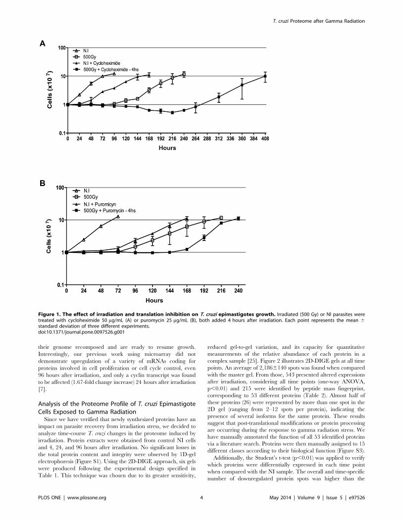
Normal growth of epimatigote cells was affected by protein
synthesis inhibition (using 50 mg/mL cycloheximide or 25 mg/mL
puromycin) and by ionizing radiation treatment (500 Gy), as
shown in Figure 1. However, irradiation promoted a more drastic
growth arrest that persisted for approximately 96 hours; after this
period, the parasites resumed normal growth, reaching the
stationary phase 216 to 240 hours after irradiation (Figure 1).
The treatment of NI cells with cycloheximide (Figure 1A) or
puromycin (Figure 1B) retarded the cell growth by at least
24 hours when compared with non-treated cells, but did not lead
to parasite death. Conversely, the combination of cycloheximide
treatment and gamma radiation was lethal to 40% of the parasites.
The remaining parasites resumed growth only 270 hours after
irradiation, reaching the stationary phase 408 hours after irradi-
ation (Figure 1A). For puromycin, a similar effect was observed,
but treated cells resumed normal growth earlier when compared
with cycloheximide-treated parasites (Figure 1B) and, in this case,
no parasite death was detected.
These observations indicate that an active translation is
important for the recovery of parasites from damage caused by
ionizing radiation. Protein synthesis blockage is potentially
impairing the translation of newly synthesized or pre-existing
mRNAs that code for proteins involved in triggering cell
proliferation. These proteins may accumulate within 24 hours
after irradiation and act later after irradiation, when parasites have
T. cruzi Proteome after Gamma Radiation
PLOS ONE | www.plosone.org 3 May 2014 | Volume 9 | Issue 5 | e97526
their genome recomposed and are ready to resume growth.
Interestingly, our previous work using microarray did not
demonstrate upregulation of a variety of mRNAs coding for
proteins involved in cell proliferation or cell cycle control, even
96 hours after irradiation, and only a cyclin transcript was found
to be affected (1.67-fold change increase) 24 hours after irradiation
[7].
Analysis of the Proteome Profile of T. cruzi EpimastigoteCells Exposed to Gamma Radiation
Since we have verified that newly synthesized proteins have an
impact on parasite recovery from irradiation stress, we decided to
analyze time-course T. cruzi changes in the proteome induced by
irradiation. Protein extracts were obtained from control NI cells
and 4, 24, and 96 hours after irradiation. No significant losses in
the total protein content and integrity were observed by 1D-gel
electrophoresis (Figure S1). Using the 2D-DIGE approach, six gels
were produced following the experimental design specified in
Table 1. This technique was chosen due to its greater sensitivity,
reduced gel-to-gel variation, and its capacity for quantitative
measurements of the relative abundance of each protein in a
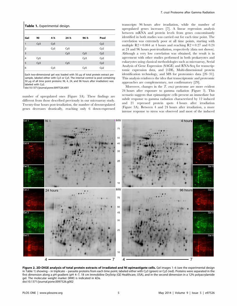
complex sample [25]. Figure 2 illustrates 2D-DIGE gels at all time
points. An average of 2,1866140 spots was found when compared
with the master gel. From those, 543 presented altered expressions
after irradiation, considering all time points (one-way ANOVA,
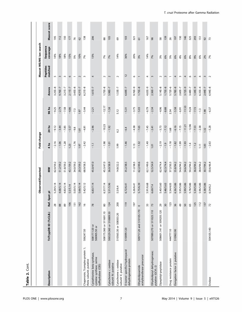
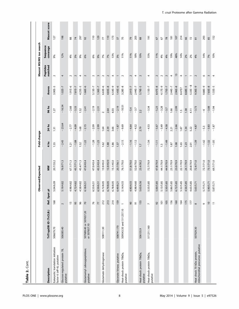
p,0.01) and 215 were identified by peptide mass fingerprint,
corresponding to 53 different proteins (Table 2). Almost half of
these proteins (26) were represented by more than one spot in the
2D gel (ranging from 2–12 spots per protein), indicating the
presence of several isoforms for the same protein. These results
suggest that post-translational modifications or protein processing
are occurring during the response to gamma radiation stress. We
have manually annotated the function of all 53 identified proteins
via a literature search. Proteins were then manually assigned to 15
different classes according to their biological function (Figure S3).
Additionally, the Student’s t-test (p,0.01) was applied to verify
which proteins were differentially expressed in each time point
when compared with the NI sample. The overall and time-specific
number of downregulated protein spots was higher than the
Figure 1. The effect of irradiation and translation inhibition on T. cruzi epimastigotes growth. Irradiated (500 Gy) or NI parasites weretreated with cycloheximide 50 mg/mL (A) or puromycin 25 mg/mL (B), both added 4 hours after irradiation. Each point represents the mean 6standard deviation of three different experiments.doi:10.1371/journal.pone.0097526.g001
T. cruzi Proteome after Gamma Radiation
PLOS ONE | www.plosone.org 4 May 2014 | Volume 9 | Issue 5 | e97526
number of upregulated ones (Figure 3A). These findings are
different from those described previously in our microarray study.
Twenty-four hours post-irradiation, the number of downregulated
genes decreases drastically, reaching only 6 down-expressed
transcripts 96 hours after irradiation, while the number of
upregulated genes increases [7]. A linear regression analysis
between mRNA and protein levels from genes concomitantly
identified in both studies was carried out for each time point. The
correlation was extremely poor at all time points, starting with
multiple R2 = 0.064 at 4 hours and reaching R2 = 0.27 and 0.24
at 24 and 96 hours post-irradiation, respectively (data not shown).
Although a very low correlation was obtained, the result is in
agreement with other studies performed in both prokaryotes and
eukaryotes using classical methodologies such as microarray, Serial
Analysis of Gene Expression (SAGE) and RNA-Seq for transcrip-
tomic expression data, and 2-DE, Multi-dimensional protein
identification technology, and MS for proteomics data [26–31].
This analysis reinforces the idea that transcriptomic and proteomic
approaches are complementary, not confirmatory [29].
Moreover, changes in the T. cruzi proteome are more evident
24 hours after exposure to gamma radiation (Figure 3). This
scenario suggests that epimastigote cells present an immediate but
subtle response to gamma radiation characterized by 12 induced
and 21 repressed protein spots 4 hours after irradiation
(Figure 3A). Between 4 and 24 hours after irradiation, a more
intense response to stress was observed and most of the induced
Figure 2. 2D-DIGE analysis of total protein extracts of irradiated and NI epimastigote cells. Gel images 1–6 (see the experimental designin Table 1) showing – in triplicate – parasite proteins from each time point, labeled either with Cy3 (green) or Cy5 (red). Proteins were separated in thefirst dimension along a pH gradient (pH 4–7, 18 cm Immobiline DryStrip (GE Healthcare, USA), and in the second dimension in a 12% polyacrylamidegel. The molecular weight marker (MW) is indicated in kDa.doi:10.1371/journal.pone.0097526.g002
Table 1. Experimental design.
Gel NI 4 h 24 h 96 h Pool
1 Cy3 Cy5 Cy2
2 Cy3 Cy5 Cy2
3 Cy3 Cy5 Cy2
4 Cy5 Cy3 Cy2
5 Cy3 Cy5 Cy2
6 Cy3 Cy5 Cy2
Each two-dimensional gel was loaded with 50 mg of total protein extract persample, labeled either with Cy3 or Cy5. The internal control (a pool containing50 mg of all time point proteins: NI, 4, 24, and 96 hours after irradiation) waslabeled with Cy2.doi:10.1371/journal.pone.0097526.t001
T. cruzi Proteome after Gamma Radiation
PLOS ONE | www.plosone.org 5 May 2014 | Volume 9 | Issue 5 | e97526

Ta
ble
2.
Pro
tein
dat
afo
rth
e5
3p
rote
ins
ide
nti
fie
din
this
stu
dy.
Ob
serv
ed
/Ex
pe
cte
dF
old
-ch
an
ge
Ma
sco
tM
S/M
Sio
nse
arc
h
De
scri
pti
on
Tri
Try
pD
BID
(TcC
LB
.)R
ef.
Sp
ot
pI
MW
4h
s2
4h
s9
6h
sA
no
va
Pe
pti
de
sm
atc
he
dS
eq
ue
nce
cov
ara
ge
Ma
sco
tsc
ore
14
-3-3
pro
tein
;p
uta
tive
51
11
67
.90
15
35
.00
/4.7
82
4.3
/29
.12
1.3
51
.08
21
.16
5.6
1E–
52
12
%7
3
40
Sri
bo
som
alp
rote
inS1
2,
pu
tati
ve5
08
55
1.2
01
95
4.8
2/4
.78
10
.9/1
5.9
1.4
53
.52
2.9
21
.32
E–5
22
4%
11
0
Act
in,
pu
tati
ve5
10
57
1.3
9o
r5
10
12
7.7
9o
r5
10
57
1.3
06
65
.75
/5.4
64
5.9
/41
.22
1.6
42
14
.92
21
2.6
58
.20
E–9
31
0%
13
6
Alp
ha
tub
ulin
,p
uta
tive
41
12
35
.96
96
.07
/4.7
44
.7/4
9.8
21
.01
21
2.6
52
10
.53
2.0
0E–
75
16
%2
48
99
5/4
.75
5.6
/49
.82
1.9
42
31
.15
24
8.8
22
.97
E–8
11
42
%8
9
10
05
.06
/4.7
55
.2/4
9.8
22
.11
24
7.2
24
5.0
81
.34
E–9
51
5%
27
5
13
65
.29
/4.7
29
.9/4
9.8
1.5
42
2.0
22
1.6
88
.44
E–9
41
4%
24
7
13
85
.47
/4.7
30
.4/4
9.8
1.0
92
2.3
42
2.2
18
.55
E–7
39
%1
55
13
95
.45
/4.7
28
.9/4
9.8
1.3
62
1.9
52
1.6
36
.52
E–8
41
3%
23
7
Am
ino
pe
pti
das
e,
pu
tati
ve,
me
tallo
-pe
pti
das
e,
Cla
nM
F,Fa
mily
M1
7,
pu
tati
ve
50
87
99
.24
03
56
.22
/6.4
45
8.9
/55
.92
1.5
52
21
.53
21
.73
5.4
0E–
51
2%
52
AT
Pas
eb
eta
sub
un
it5
09
23
3.1
80
26
5.1
5/5
.07
58
.6/5
5.7
22
.42
22
1.0
42
18
.44
7.2
6E–
75
18
%2
53
Be
tatu
bu
lin,
pu
tati
ve5
06
56
3.4
01
07
5.5
8/4
.43
45
.0/4
9.7
21
.36
24
.92
5.1
71
.02
E–7
37
%1
56
12
95
.30
/4.4
33
3.3
/49
.72
1.0
12
4.3
52
3.9
91
.53
E–7
51
3%
24
2
13
05
.15
/4.4
33
3.8
/49
.72
1.3
12
10
.73
29
.26
4.5
6E–
86
17
%4
38
17
14
.71
/4.4
32
8.6
/49
.71
.81
.69
2.0
71
.49
E–8
72
2%
73
17
34
.65
/4.4
32
5.5
/49
.72
.53
6.3
35
.29
8.4
4E–
94
11
%2
37
17
44
.58
/4.4
32
5.5
/49
.71
.16
2.5
92
.43
1.1
7E–
64
11
%2
90
17
64
.47
/4.4
32
5.0
/49
.71
.73
.58
3.5
22
.60
E–7
39
%2
16
18
34
.34
/4.4
31
9.4
/49
.71
.15
2.3
81
.93
1.2
7E–
52
6%
10
1
18
44
.25
/4.4
31
9.4
/49
.71
.56
5.9
75
.14
3.4
9E–
82
6%
11
6
18
54
.25
/4.4
32
1.2
/49
.71
.67
4.3
94
.22
3.6
1E–
62
6%
13
3
Cal
reti
culin
,p
uta
tive
51
06
85
.10
81
6.3
7/4
.49
42
.2/4
6.2
21
.07
11
.52
1.8
1E–
52
6%
89
Ch
ape
ron
inco
nta
inin
gt-
com
ple
xp
rote
in,
pu
tati
ve5
11
72
5.2
50
12
5.0
2/4
.80
69
.7/5
9.2
21
.95
21
2.8
12
16
.44
.12
E–7
39
%1
13
Ch
ape
ron
inH
SP6
0;
mit
och
on
dri
alp
recu
rso
r;G
roEL
pro
tein
;h
eat
sho
ckp
rote
in6
0(H
SP6
0)
50
76
41
.29
0o
r5
07
64
1.3
00
or
51
01
87
.55
11
75
.44
/5.1
46
6.2
/59
.22
4.9
21
9.1
92
19
.86
1.2
7E–
89
24
%4
70
18
5.5
5/5
.14
66
.0/5
9.2
24
.07
29
.24
21
0.1
81
.75
E–8
32
0%
11
1
20
5.6
5/5
.14
65
.8/5
9.2
23
.99
28
.83
29
.91
.25
E–7
71
8%
35
4
23
5.2
2/5
.14
62
.3/5
9.2
22
.03
28
.02
29
.51
6.4
2E–
87
18
%1
91
24
5.1
3/5
.14
62
.2/5
9.2
21
.72
21
1.5
22
13
.96
9.4
2E–
81
13
1%
53
3
25
5.2
1/5
.14
61
.5/5
9.2
21
.78
21
6.9
92
15
.11
.61
E–7
61
6%
20
5
T. cruzi Proteome after Gamma Radiation
PLOS ONE | www.plosone.org 6 May 2014 | Volume 9 | Issue 5 | e97526
Ta
ble
2.
Co
nt.
Ob
serv
ed
/Ex
pe
cte
dF
old
-ch
an
ge
Ma
sco
tM
S/M
Sio
nse
arc
h
De
scri
pti
on
Tri
Try
pD
BID
(TcC
LB
.)R
ef.
Sp
ot
pI
MW
4h
s2
4h
s9
6h
sA
no
va
Pe
pti
de
sm
atc
he
dS
eq
ue
nce
cov
ara
ge
Ma
sco
tsc
ore
28
5.2
9/5
.14
62
.0/5
9.2
22
.16
29
.12
29
.25
3.2
5E–
85
14
%1
95
88
4.6
8/5
.14
51
.2/5
9.2
21
.06
23
.44
22
.79
9.7
6E–
73
18
%1
12
89
4.8
3/5
.14
51
.8/5
9.2
21
.28
27
.85
27
.58
3.2
1E–
73
18
%1
50
10
65
.5/5
.14
43
.1/5
9.2
1.2
32
5.5
72
4.8
62
.11
E–7
61
5%
17
7
13
15
.03
/5.1
43
4.0
/59
.22
1.7
62
9.8
27
.33
.45
E–8
15
%7
6
16
25
.66
/5.1
42
0.3
/59
.21
.97
1.8
11
.97
4.3
1E–
72
10
%6
2
Ch
ape
ron
in;
Tco
mp
lex-
pro
tein
1;
the
tasu
bu
nit
;p
uta
tive
50
62
47
.50
16
5.4
2/5
.12
68
.9/5
8.3
22
.42
24
.81
25
.44
6.2
1E–
83
7%
13
8
Cys
tath
ion
ine
be
ta-s
ynth
ase
,cy
ste
ine
syn
thas
e,
seri
ne
sulf
hyd
ryla
se(C
BS)
50
81
77
.12
0o
r5
06
90
5.5
0o
r7
86
.83
/7.1
44
5.0
/47
.02
1.1
22
.96
22
.21
1.6
1E–
74
13
%2
06
50
81
75
.36
0o
r5
11
69
1.1
08
06
.37
/7.1
44
5.3
/47
.02
1.9
82
10
.23
21
2.1
71
.75
E–8
27
%8
8
Cyt
och
rom
ec
oxi
das
esu
bu
nit
IV;
pu
tati
ve5
06
52
9.3
60
or
51
08
89
.50
12
45
.51
/5.9
63
6.0
/38
.92
1.0
12
1.9
22
1.5
85
.98
E–7
27
%1
05
Cyt
och
rom
ec
oxi
das
esu
bu
nit
V,
pu
tati
ve5
10
56
5.3
0o
r5
08
50
3.2
02
00
5.5
/6.4
14
.8/2
2.2
1.9
96
.25
.12
1.5
3E–
72
14
%6
9
D-i
som
er
spe
cifi
c2-h
ydro
xyac
idd
eh
ydro
ge
nas
e-p
rote
in5
10
09
9.1
20
11
96
.72
/6.4
13
5.2
/38
.52
1.4
62
6.4
25
.31
6.6
9E–
71
23
6%
10
3
19
75
.26
/6.4
11
1.2
/38
.51
.13
24
.38
23
.75
9.7
6E–
96
20
%3
21
Dih
ydro
lipo
amid
eac
ety
ltra
nsf
era
sep
recu
rso
r5
09
71
7.2
0an
d5
10
10
5.1
70
05
.75
/6.3
96
2.2
/49
.62
1.3
22
7.6
52
5.0
31
.75
E–8
13
%6
1
33
5.9
1/6
.68
62
.1/4
9.6
21
.49
27
.45
26
.79
4.6
5E–
94
14
%1
67
Dih
ydro
lipo
yld
eh
ydro
ge
nas
e;
pu
tati
ve(G
CV
L-2
)5
07
08
9.2
70
or
51
10
25
.11
07
36
.69
/7.4
52
.5/5
4.9
1.0
12
2.4
12
2.3
42
.36
E–7
37
%9
8
Dip
ep
tid
yl-p
ep
tid
ase
50
86
01
.14
1o
r5
09
20
5.1
20
29
5.4
0/5
.60
62
.4/7
4.4
22
.19
25
.73
24
.96
5.1
4E–
82
3%
58
30
5.4
8/5
.63
62
.3/7
4.4
21
.92
7.1
22
8.8
81
.75
E–8
36
%1
39
Dru
gre
sist
ance
pro
tein
44
47
77
.10
12
35
.54
/4.0
53
7.2
/50
.32
1.1
81
.68
2.3
45
.72
E–6
15
%2
6
Elo
ng
atio
nfa
cto
r2
,p
uta
tive
51
09
63
.90
36
6.3
6/5
.86
55
.8/9
4.2
21
.82
26
.16
25
.58
5.7
8E–
84
6%
19
7
49
5.9
5/5
.86
54
.6/9
4.2
21
.99
27
.15
26
.91
2.6
0E–
73
4%
13
0
50
5.9
9/5
.86
54
.6/9
4.2
21
.93
21
1.2
12
11
.53
3.7
2E–
94
6%
14
6
65
5.7
8/5
.86
50
.2/9
4.2
21
.42
12
.96
29
.24
3.4
8E–
76
8%
32
5
12
55
.34
/5.8
63
6.0
/94
.22
1.2
27
.53
27
.72
1.3
4E–
95
9%
29
3
11
25
.96
/5.8
63
8.0
/94
.21
.11
22
.38
21
.36
.35
E–4
46
%1
51
13
75
.38
/5.8
63
0.7
/94
.21
.83
1.3
21
.98
2.4
4E–
73
2%
92
Eno
lase
50
41
05
.14
07
26
.54
/6.2
50
.6/4
6.4
22
.63
25
.28
26
.37
6.9
4E–
82
7%
73
T. cruzi Proteome after Gamma Radiation
PLOS ONE | www.plosone.org 7 May 2014 | Volume 9 | Issue 5 | e97526
Ta
ble
2.
Co
nt.
Ob
serv
ed
/Ex
pe
cte
dF
old
-ch
an
ge
Ma
sco
tM
S/M
Sio
nse
arc
h
De
scri
pti
on
Tri
Try
pD
BID
(TcC
LB
.)R
ef.
Sp
ot
pI
MW
4h
s2
4h
s9
6h
sA
no
va
Pe
pti
de
sm
atc
he
dS
eq
ue
nce
cov
ara
ge
Ma
sco
tsc
ore
Euka
ryo
tic
tran
slat
ion
init
iati
on
fact
or
6(e
lF-6
);p
uta
tive
50
66
79
.70
16
85
.04
/6.0
92
0.7
/33
.21
.55
1.3
11
.27
3.9
0E–
52
9%
12
7
Glu
cose
-re
gu
late
dp
rote
in7
8,
pu
tati
ve5
06
58
5.4
02
5.1
9/4
.82
76
.9/7
1.3
22
.45
22
3.6
42
16
.54
1.0
2E–
74
12
%1
98
13
4.9
8/4
.82
67
.1/7
1.3
1.2
12
2.7
72
2.6
81
.91
E–6
24
%8
8
95
4.7
2/4
.82
45
.7/7
1.3
21
.08
24
.09
23
.33
7.8
1E–
72
3%
74
96
4.5
8/4
.82
45
.4/7
1.3
1.5
21
.68
1.5
24
.25
E–5
49
%2
97
Glu
tam
amyl
cab
oxy
pe
pti
das
e;
pu
tati
ve5
07
68
9.4
0o
r5
07
65
7.2
0o
r5
07
65
7.1
07
06
.18
/6.5
14
7.6
/43
.42
1.2
22
2.1
52
2.4
11
.66
E–6
26
%9
2
76
6.5
3/6
.51
47
.0/4
3.4
21
.28
22
.09
22
.19
5.1
3E–
72
6%
11
0
77
6.5
9/6
.51
45
.5/4
3.4
21
.23
22
.99
22
.31
1.3
6E–
73
9%
12
9
Glu
tam
ate
de
hyd
rog
en
ase
50
81
11
.30
21
26
.72
/8.0
51
5.9
/45
.01
.62
1.6
42
6.6
3E–
62
7%
77
21
36
.79
/8.0
51
5.9
/45
/01
.88
2.3
92
.64
4.8
3E–
82
7%
11
0
21
46
.78
/8.0
51
5.1
/45
.01
.86
3.6
64
.03
6.5
4E–
94
13
%1
73
Gly
cera
teki
nas
e,
pu
tati
ve5
08
74
1.1
70
15
96
.49
/8.2
12
0.7
/56
.11
.37
21
.87
22
.19
1.4
3E–
61
3%
37
He
at-s
ho
ckp
rote
in7
0kD
a,p
uta
tive
50
95
43
.50
and
51
12
57
.10
15
.14
/4.5
57
6.1
/70
.02
2.1
52
9.6
92
10
.51
3.2
8E–
82
11
%7
0
90
4.9
0/4
.55
51
.8/7
0.0
21
.16
24
.64
25
.42
.91
E–7
21
1%
79
91
4.9
8/4
.60
52
.6/7
0.0
21
.12
24
.52
23
.72
.44
E–7
41
8%
20
5
He
at-s
ho
ckp
rote
in7
0kD
a,p
uta
tive
50
61
35
.91
55
5.6
3/6
.56
23
.4/3
0.2
1.7
2.7
92
.25
.79
E–5
21
0%
88
He
at-s
ho
ckp
rote
in7
0kD
a,p
uta
tive
51
12
11
.16
07
5.5
5/5
.85
72
.7/7
0.9
21
.34
24
.53
23
.34
1.1
5E–
74
15
%1
91
92
5.0
0/5
.85
47
.8/7
0.9
21
.11
25
.67
25
.23
8.4
7E–
82
11
%6
7
10
15
.13
/5.8
54
4.1
/70
.91
.32
3.4
92
3.2
84
.17
E–9
24
%6
7
10
55
.50
/5.8
54
4.9
/70
.92
1.4
92
4.5
82
3.7
1.7
5E–
72
4%
78
15
65
.86
/5.8
52
3.1
/70
.91
.87
2.2
11
.66
2.4
4E–
74
19
%1
81
16
06
.72
/5.8
52
3.0
/70
.91
.08
22
.66
22
.99
2.9
6E–
81
34
8%
10
7
16
45
.32
/5.8
52
2.1
/70
.91
.65
1.8
71
.58
.47
E74
14
%2
14
17
54
.56
/5.8
52
4.1
/70
.91
.23
1.3
41
.38
8.6
8E–
51
2%
66
17
74
.65
/5.8
52
0.8
/70
.92
.01
5.2
24
.11
5.3
3E2
81
2%
72
He
atsh
ock
70
kDa
pro
tein
,m
ito
cho
nd
rial
pre
curs
or,
pu
tati
ve5
07
02
9.3
08
5.6
5/5
.71
72
.4/7
1.0
21
.39
24
.87
23
.73
9.3
6E–
92
4%
81
95
.77
/5.7
17
2.7
/71
.02
1.4
22
5.3
24
9.8
8E–
96
14
%2
93
10
5.9
0/5
.71
73
.1/7
1.0
21
.32
23
.83
23
.08
7.8
3E–
83
7%
85
11
5.8
7/5
.71
69
.7/7
1.0
21
.05
21
.87
21
.94
1.5
5E–
54
10
%1
52
T. cruzi Proteome after Gamma Radiation
PLOS ONE | www.plosone.org 8 May 2014 | Volume 9 | Issue 5 | e97526

Ta
ble
2.
Co
nt.
Ob
serv
ed
/Ex
pe
cte
dF
old
-ch
an
ge
Ma
sco
tM
S/M
Sio
nse
arc
h
De
scri
pti
on
Tri
Try
pD
BID
(TcC
LB
.)R
ef.
Sp
ot
pI
MW
4h
s2
4h
s9
6h
sA
no
va
Pe
pti
de
sm
atc
he
dS
eq
ue
nce
cov
ara
ge
Ma
sco
tsc
ore
19
5.6
0/5
.71
67
.4/7
1.0
1.1
82
1.4
21
.53
1.1
7E–
65
12
%2
85
21
5.7
3/5
.71
67
.7/7
1.0
1.2
62
1.4
22
1.5
81
.70
E–7
41
0%
10
9
He
at-s
ho
ckp
rote
in8
5kD
a,p
uta
tive
50
96
43
.13
0o
r5
07
71
3.3
0o
r5
09
10
5.1
40
93
4.8
9/4
.79
47
.6/8
0.7
21
.44
25
.42
4.3
27
.81
E–7
23
%8
7
97
4.9
0/4
.79
43
.2/8
0.7
1.4
32
1.5
21
.15
3.2
5E–
73
5%
11
4
98
4.9
5/4
.79
43
.0/8
0.7
1.0
22
3.0
12
3.3
71
.49
E–8
23
%1
45
10
45
.30
/4.7
94
1.5
/80
.72
1.6
32
16
.15
21
8.5
52
.26
E–8
35
%1
43
12
65
.40
/4.7
93
3.5
/80
.71
.22
3.7
72
3.7
44
.34
E–8
23
%8
9
13
44
.62
/4.7
94
0.6
/80
.71
.39
2.5
82
.18
7.3
5E–
64
6%
25
8
14
84
.95
/4.7
92
8.8
/80
.71
.76
2.7
82
.47
3.4
9E–
81
1%
10
0
14
94
.86
/4.7
92
8.9
/80
.71
.67
3.5
53
.23
6.8
9E–
81
1%
62
Hyp
oth
eti
cal
pro
tein
,co
nse
rve
d5
05
98
9.1
10
18
24
.46
/4.5
01
8.4
/22
.22
1.2
53
.47
3.5
11
.92
E–5
21
1%
62
Hyp
oth
eti
cal
pro
tein
,co
nse
rve
d5
06
60
5.1
20
or
51
12
39
.11
02
02
5.7
2/4
.99
15
.1/2
8.6
2.7
57
.08
7.8
31
.34
E–9
42
2%
17
4
20
35
.67
/4.9
91
4.2
/28
.61
.87
3.9
63
.33
3.6
6E–
69
41
%9
2
20
45
.86
/4.9
91
4.1
/28
.61
.48
2.1
22
.78
1.8
8E–
76
31
%3
26
Hyp
oth
eti
cal
pro
tein
50
88
17
.20
or
50
38
01
.70
15
45
.38
/8.5
82
3.5
/66
.71
.78
4.8
04
.29
3.9
6E–
81
1%
17
Nu
cle
osi
de
ph
osp
ho
ryla
se,
pu
tati
ve5
08
98
9.9
and
50
95
69
.10
01
21
6.9
0/6
.42
34
.2/3
7.0
21
.18
25
.59
24
.38
2.9
6E–
83
16
%1
90
11
86
.38
/6.4
23
5.6
/37
.02
1.1
22
3.1
72
2.5
61
.17
E–8
14
%2
3
Olig
op
ep
tid
ase
B,
pu
tati
ve5
03
99
5.5
04
75
.86
/6.1
55
.2/8
0.8
21
.71
25
.94
27
.55
9.7
6E–
92
3%
68
63
5.8
6/6
.15
2.0
/80
.92
1.1
22
2.2
62
2.4
78
.01
E–7
23
%6
9
Par
afla
ge
llar
rod
pro
tein
35
09
61
7.2
06
06
.09
/5.9
65
6.1
/68
.61
.21
23
.51
24
.33
6.8
9E–
81
2%
24
Pe
pti
das
eM
20
/M2
5/M
40
51
02
57
.80
39
5.5
7/5
.19
55
.4/5
1.2
21
.74
25
.55
27
.07
8.4
7E–
72
6%
66
40
5.5
0/5
.19
54
.4/5
1.2
21
.64
25
.35
29
.41
2.2
6E–
81
3%
31
Pe
roxi
red
oxi
n;
tryp
are
do
xin
pe
roxi
das
e5
09
49
9.1
41
89
5.1
6/7
.92
18
.1/2
5.5
1.1
11
.42
1.7
37
.81
E–7
31
5%
17
4
Ph
osp
ho
gly
cera
teki
nas
e,
pu
tati
veo
r3
-ph
osp
ho
gly
cera
teki
nas
e,
gly
coso
mal
(PG
KA
)
51
14
19
.40
or
50
59
99
.90
or
51
14
19
.50
or
50
59
99
.10
0
74
6.7
6/7
.45
1.9
/54
.90
22
.99
25
.56
25
.35
2.4
6E–
71
3%
78
Pro
stag
lan
din
F2al
ph
asy
nth
ase
(TcP
GFS
)5
08
46
1.8
01
45
.11
/6.4
36
8.1
/42
.22
1.1
92
6.4
52
7.0
11
.59
E–6
41
4%
16
9
11
15
.91
/6.4
34
0.0
/42
.22
1.3
12
11
.76
27
.75
1.7
4E–
85
17
%2
84
11
36
.10
/6.4
34
0.0
/42
.22
1.1
12
10
.19
26
.05
1.7
5E–
88
38
%1
16
11
46
.09
/6.4
33
8.7
/42
.21
.12
26
.24
25
.06
3.1
2E–
74
13
%1
88
14
46
.15
/6.4
32
6.7
/42
.21
.49
21
.65
21
.39
5.4
9E–
84
13
%2
17
T. cruzi Proteome after Gamma Radiation
PLOS ONE | www.plosone.org 9 May 2014 | Volume 9 | Issue 5 | e97526

Ta
ble
2.
Co
nt.
Ob
serv
ed
/Ex
pe
cte
dF
old
-ch
an
ge
Ma
sco
tM
S/M
Sio
nse
arc
h
De
scri
pti
on
Tri
Try
pD
BID
(TcC
LB
.)R
ef.
Sp
ot
pI
MW
4h
s2
4h
s9
6h
sA
no
va
Pe
pti
de
sm
atc
he
dS
eq
ue
nce
cov
ara
ge
Ma
sco
tsc
ore
16
15
.52
/6.4
32
2.1
/42
.21
.74
1.8
81
.99
6.3
4E–
89
31
%9
2
Pro
tein
dis
ulf
ide
iso
me
rase
50
62
47
.10
or
50
76
11
.37
01
80
4.4
2/4
.62
0.9
/53
.51
.46
3.7
3.6
2.4
4E–
72
4%
44
Pyr
uva
ted
eh
ydro
ge
nas
eE1
be
tasu
bu
nit
;p
uta
tive
51
00
91
.80
13
25
.03
/5.0
23
0.9
/37
.81
.08
22
.21
21
.88
1.2
7E–
65
20
%1
91
13
34
.62
/5.0
24
0.7
/37
.81
.02
24
.58
23
.83
4.1
3E–
72
6%
10
8
Pyr
uva
teki
nas
e2
,p
uta
tive
50
79
93
.39
0o
r5
11
28
1.6
06
85
.97
/7.4
44
6.6
/54
.62
1.3
24
.94
24
.81
.73
E–7
12
%2
9
Pyr
uva
tep
ho
sph
ate
dik
inas
e5
10
10
1.1
40
19
45
.00
/8.2
71
3.5
/10
0.8
1.8
31
0.5
17
.42
7.8
8E–
92
3%
94
Re
cep
tor
for
acti
vate
dC
kin
ase
1,
pu
tati
ve5
11
21
1.1
20
or
51
12
11
.13
01
22
5.9
3/6
.04
35
.4/3
5.0
21
.22
26
.66
26
.06
2.3
1E–
93
11
%1
22
S-ad
en
osy
lho
mo
cyst
ein
eh
ydro
lase
51
12
29
.50
or
51
15
89
.20
01
93
5.2
5/6
.64
12
.9/4
8.4
1.4
12
1.2
32
1.1
3.7
4E–
62
7%
80
Sery
l-tR
NA
syn
the
tase
51
11
63
.1o
r5
06
77
7.8
01
40
5.3
8/5
.41
28
.9/2
5.7
1.4
21
.07
1.2
83
.36
E–6
31
9%
97
succ
inyl
-Co
Alig
ase
[GD
P-f
orm
ing
]b
eta
-ch
ain
,p
uta
tive
50
77
67
.10
15
04
.92
/5.5
82
6.2
/34
.51
.63
2.0
32
.25
1.0
8E–
62
7%
87
Th
iol2
de
pe
nd
en
tre
du
ctas
e1
;p
uta
tive
;th
iol
tran
sfe
rase
;p
uta
tive
;g
luta
thio
ne
s-tr
ansf
era
se;
pu
tati
ve
50
91
05
.70
or
50
34
19
.30
62
6.0
0/5
.83
51
.9/5
0.7
21
.18
22
.07
21
.56
4.5
6E–
62
6%
86
15
86
.21
/5.8
32
1.5
/50
.72
1.3
22
2.1
72
2.6
44
.48
E–6
13
%3
2
Tra
ns-
sial
idas
e5
09
92
7.1
01
86
4.1
8/6
.67
21
.0/5
4.7
1.6
74
.74
.71
6.1
0E–
91
4%
25
Try
par
ed
oxi
np
ero
xid
ase
48
75
07
.10
or
50
94
45
.10
or
50
48
39
.28
or
50
72
59
.10
21
06
.24
/6.7
51
1.5
/22
/42
.17
3.1
83
.42
8.0
5E–
51
5%
33
21
16
.66
/6.7
51
7.0
/22
.42
1.5
22
3.9
62
1.8
23
.25
E–8
32
2%
10
5
21
56
.75
/6.7
51
1.8
/22
.41
.44
21
.31
.25
1.8
3E–
63
19
%1
09
Tyr
osi
ne
amin
otr
ansf
era
se5
10
18
7.2
0an
d5
10
18
7.5
0o
r5
10
18
7.4
0o
r5
10
18
7.3
06
45
.83
/7.2
50
.6/4
6.1
21
.22
21
.77
21
.51
5.0
2E–
51
2%
58
79
6.2
5/6
.14
44
.9/4
6.1
22
.36
23
1.0
32
28
.73
1.3
4E–
94
16
%1
60
11
56
.21
/6.1
43
8.9
/46
.12
1.0
52
7.2
72
7.3
89
.64
E–9
41
1%
11
0
11
66
.18
/6.1
43
7.0
/46
.11
.09
25
.43
24
.73
8.4
4E–
92
14
%1
28
Vac
uo
lar
AT
Psy
nth
ase
sub
un
itB
50
60
25
.50
or
51
12
09
.10
37
5.7
1/5
.29
59
.1/5
5.5
21
.69
23
.54
24
.01
2.4
4E–
76
21
%2
07
do
i:10
.13
71
/jo
urn
al.p
on
e.0
09
75
26
.t0
02
T. cruzi Proteome after Gamma Radiation
PLOS ONE | www.plosone.org 10 May 2014 | Volume 9 | Issue 5 | e97526

and repressed protein spots were still significantly altered until
96 hours (428 spots; Figure 3B). This finding indicates a sustained
alteration in the abundance of specific T. cruzi proteins 24 hours
after gamma radiation exposure. When analyzing the T. cruzi
proteome 24 hours after irradiation, we found that, from the 59
exclusive spots, approximately 66% were repressed and 34% were
induced. However, the majority of the 23 exclusive spots found
96 hours after irradiation were induced (approximately 61%).
Exposure to Gamma Rays Increases the Levels of Shorterand/or Processed Proteins in Epimastigote Cells
When analyzing the set of upregulated proteins (especially 24
and 96 hours after irradiation), we observed a tendency for the
overexpression of shorter molecules to the detriment of longer
ones. The upregulated protein spots (red-colored dots) are mainly
at the lower part of the gel (lower molecular weight), while the
downregulated protein spots (green-colored dots) are more
sparsely distributed across the gel (Figure 4A). In addition, low
molecular weight protein spots tended to have larger fold changes
when compared with those with molecular weights close to the
expected value (Figure 4B). The Wilcoxon test was applied and
confirmed that the median values of the molecular weight of
downregulated and upregulated protein spots were different for
each time point (p,1e-09; median values of 55.45/19.39, 45.64/
19.38, and 46.51/19.42 for 4, 24, and 96 hours, respectively;
Figure 5A).
When we considered the expected molecular weight of the full-
length isoform (predicted size obtained from the TriTryDB
website) of both upregulated and downregulated proteins, we
noticed a decrease in protein size in the former case and an
increase in the latter case, thus showing the emergence of lower
molecular weight protein isoforms after irradiation (Figure 5B).
We then decided to confirm if the observed molecular weight of
these proteins in the 2D gels was in agreement with their expected
molecular weight. In the case of upregulated proteins, the
observed molecular weight was significantly lower than expected
(Figure 5B), indicating that these proteins might be processed,
yielding shorter polypeptides. It is important to note that this result
does not seem to be a consequence of protein degradation, since
clear spots can be observed in the 2D gel, indicating the presence
of a large amount of identical polypeptides in this region of the gel.
This would not be the case if proteins were degraded, considering
that in this situation peptides of variable size would be generated
and no clear spot would be observed in the gel. These results may
indicate the emergence of new protein isoforms, as the result of
protein processing, alternative splicing of mRNAs, and/or
alternative translational start/stop sites after irradiation. Alterna-
tive splicing of transcripts has the potential to expand the
repertoire of proteins. Recent studies have estimated that all
multi-exonic human genes are able to produce at least two
alternatively spliced mRNA transcripts by alternative splicing,
generating different proteins isoforms with altered structures and
biological functions [32]. In trypanosomatids, mature mRNAs are
generated after two processing events: trans-splicing to add the
spliced leader (SL) sequence to the 59 end of transcripts and
subsequent polyadenylation [17]. A genome-wide analysis com-
paring the SL addition site along the developmental cycle of the
parasite suggests that alternative trans-splicing plays an important
role in differential gene expression [33]. The occurrence of
alternative trans-splicing could be an explanation for the presence
of so many different isoforms in T. cruzi after radiation response.
A similar event has already been described in D. radiodurans,
since different isoforms of the single-strand binding (SSB) protein
were produced after ionizing radiation stress induction. SSB
proteins are vital for cell survival due to their involvement in
processes such as DNA replication, recombination, and repair.
The SSB protein spots in the gel followed a dynamic pattern of
appearance, indicating a progressive processing of the C-terminal
acidic tail, perhaps upon its interaction with ssDNA. The observed
isoelectric point (pI) and molecular weight of deinococcal SSB
isoforms were in agreement with the in silico-predicted pI and
molecular weight of the SSB proteins shortened from the C-
terminal end [16].
Intriguingly, in most of the observed processed proteins, the
identified peptide sequences were the same or nearly the same in
all sequenced protein spots and, therefore, it was impossible to
define the actual outcome of the protein processing. As a
particular case of study, the protein annotated as prostaglandin
F2 alpha synthase, which is similar to NADH-flavinoxidoreduc-
tase, is processed to a total of six different forms (Figure S2). While
the expected molecular weight for the annotated sequence is of
42 kDa, only two isoforms are nearly this size (,40 kDa) and
Figure 3. Protein spots differentially expressed at all time points. A) Number of downregulated and upregulated protein spots per timepoint. B) Venn diagram showing the overlaps of 32 protein spots differentially regulated among the three time points and of the 428 protein spotsbetween 24 and 96 hours.doi:10.1371/journal.pone.0097526.g003
T. cruzi Proteome after Gamma Radiation
PLOS ONE | www.plosone.org 11 May 2014 | Volume 9 | Issue 5 | e97526

were, in fact, the most downregulated isoforms. A third isoform
has a predicted molecular mass of 68 kDa, greatly exceeding the
expected protein size. As all of the MS/MS-identified peptides
were mapped to the C-terminal portion of the isoforms, there is no
information to characterize the N-terminal of this enlarged protein
naturally present in the NI parasites and downregulated after
exposure to gamma radiation. A smaller (29 kDa) protein is
expressed in approximately equal levels before and after radiation
exposure, while an even smaller (22 kDa) protein species is
exclusively present in irradiated cells. This is an interesting
example, representative of multiple cases, in which we have
observed the emergence of shorter isoforms of a same protein after
epimastigote irradiation. The list of processed proteins expressing
shorter isoforms after irradiation includes alpha and beta-tubulin,
D-isomer specific 2-hydroxyacid dehydrogenase-protein, elonga-
tion factor 2, glycerate kinase, pyruvate dehydrogenase E1 beta
subunit, tyrosine aminotransferase, and several heat shock proteins
(HSPs), such as HSP60, DnaK, HSP70s, and glucose-regulated
protein 78. Apart from the previously discussed SSB proteins in D.
radiodurans, very few references in the scientific literature mention
the presence of shorter protein fragments after radiation exposure
in any organism.
Interestingly, Parodi-Talice and collaborators [34] observed a
similar pattern in T. cruzi for the proteins glutamate dehydroge-
nase (GluDH), HSP70, and alpha and beta-tubulins, where lower
molecular weight isoforms were differentially expressed during
metacyclogenesis when compared with isoforms with the predicted
molecular weight. The transformation of epimastigotes into
metacyclic trypomastigotes is a complex process of differentiation,
requiring a controlled production of various proteins [34].
Similarly, a quantitative time-course proteome analysis for the
schizont-stage of Plasmodium falciparum (34 to 46 hours after
invasion) demonstrated that actin-I, enolase, HSPs, and eukaryotic
initiation factor 4A and 5A presented more than one isoform. The
isoforms also showed different expression patterns at the different
time points analyzed. P. falciparum is characterized by a complex
life cycle, undergoing extensive morphological and metabolic
changes, which reflects its capacity to survive in different host
environments [35]. According to the authors, post-translational
modifications may be a very important strategy for the parasites to
control gene expression during differentiation [34,35].
Differentially Expressed Proteins after Gamma RadiationExposure
Regarding the differentially expressed proteins, many of the
listed proteins in Table 2 and Figure S3 are related to the protein
synthesis process that seems to be upregulated, except for some
protein spots of the elongation factor 2 that show a reduction in
their levels. This may be a response to compensate for the
processing of proteins that occurs after irradiation. This response
may also enhance the synthesis of specific proteins that will
possibly play a role in the stress response. The results obtained
from the analyses of translation inhibition and proteomic profile
after irradiation place de novo protein synthesis as an important
Figure 4. Distribution of upregulated and downregulated protein spots versus molecular weight, pI, and fold change. In the scatterplots, upregulated protein spots are shown in red and downregulated protein spots are shown in green. The correlation between molecular weightand pI or fold-change ratio is shown in (A) and (B), respectively. Spots with no significant difference in expression are colored gray. The blue lineindicates the negative correlation between molecular weight and fold change.doi:10.1371/journal.pone.0097526.g004
T. cruzi Proteome after Gamma Radiation
PLOS ONE | www.plosone.org 12 May 2014 | Volume 9 | Issue 5 | e97526

cellular response to gamma radiation. The same pattern is
observed in D. radiodurans, where proteins related to translation/
folding displayed either enhanced or de novo expression in the first
hour of post-irradiation recovery. Proteins involved with DNA
repair and oxidative stress alleviation were also induced in D.
radiodurans under ionizing radiation stress [16].
Proteins involved in protein folding processes, such as chaper-
ones, are mostly downregulated post-irradiation (Figure S3). This
represents an unexpected result, since these proteins are classically
involved with stress response by stabilizing newly synthesized
protein molecules. Nevertheless, this result is in agreement with
transcriptomic data observed in microarray experiments [7]. It is
worth noting that, although HSPs are mostly downregulated,
processed forms of these molecules are upregulated and may even
be functional. Interestingly, the two chaperones localized in the
endoplasmic reticulum (calreticulin and protein disulfide isomer-
ase) are upregulated after gamma radiation exposure, which may
indicate an important role of this compartment in the ionizing
radiation stress response, suggesting the existence of an unfolded
protein response-like in this condition [36].
Another unexpected result is the downregulation of proteins
involved in the ATP metabolism (namely the beta subunit of ATP
synthase and the subunit IV of cytochrome c oxidase), although
another member of this class is upregulated (cytochrome c oxidase
subunit V). The outcome of this result is not clear and a more in-
depth study of the cell energy metabolism would be important.
Perhaps the most remarkable observation in the post-irradiation
proteome investigated here is the putative decline in the activity of
the glycolytic and amino acid metabolism pathways. Several
important enzymes of glycolysis were downregulated after gamma
radiation exposure. Accordingly, the only enzyme (pyruvate
phosphate dikinase) from gluconeogenesis listed here was upregu-
lated. Most enzymes involved in the amino acid metabolism were
also downregulated, but shorter isoforms of the GluDH were
upregulated after irradiation. They consist of three isoforms with
experimental molecular weights (15 kDa) lower than the predicted
values (45 kDa), suggesting once again the occurrence of post-
transcriptional modifications/processing of important metabolic
enzymes during the stress response. GluDH catalyzes the NAD-
and/or NADP-dependent reversible deamination of L-glutamate
to form alpha-ketoglutarate and is essential for the metabolism of
amino nitrogen in organisms ranging from bacteria to mammals
[37]. T. cruzi has a metabolism that is largely based on the
consumption of amino acids, mainly, proline, aspartate, and
glutamate, which constitute the main carbon and energy sources of
the epimastigote forms. In T. cruzi, GluDH has NADP-specific
Figure 5. Boxplots of peptide molecular weights. A) Distribution of the observed molecular weight in downregulated (green) or upregulated(red) protein spots at each time point analyzed. B) comparison between the distribution of the expected (E) and observed (O) molecular weightsamong downregulated or upregulated protein spots 24 and 96 hours after irradiation. A single asterisk corresponds to p,0.05 and a double asteriskcorresponds to p,0.001.doi:10.1371/journal.pone.0097526.g005
T. cruzi Proteome after Gamma Radiation
PLOS ONE | www.plosone.org 13 May 2014 | Volume 9 | Issue 5 | e97526

activity [38], indicating that it may serve as a pentose-phosphate
shunt-independent source of NADPH in these parasites. Taken
together, these results suggest that the parasite experiences an
overall reduction on its energy metabolism as a consequence of its
growth arrest after irradiation.
We have identified four proteins classified as redox sensors in
this study. While two of these are downregulated (both oxidore-
ductases), the other two are upregulated and these are both
tryparedoxins, which efficiently reduce hydrogen peroxide [39].
Throughout its life cycle, T. cruzi is exposed to various stresses in
different environments: the invertebrate (triatomine bugs) and the
vertebrate hosts. One of the most deleterious consequences of
oxidative stress may be the formation of DNA lesions. Guanine is
the most susceptible base to oxidation, due to its low redox
potential, and the 7,8-dihydro-8-oxoguanine (8-oxoG) is the most
common lesion. When 8-oxoG is inserted during DNA replication,
it can generate double-strand breaks, which makes this lesion
severely deleterious. Recently Aguiar et al., 2013, demonstrated
that parasites overexpressin MutT are more resistant to the
oxidative stress caused by hydrogen peroxide (H2O2) treatment.
The MutT enzyme product, 8-oxod-GMP, can generate an
oxidative stress signal, enabling the cells to overcome this stress.
MutT hydrolyses 8-oxo-dGTP in the nucleotide pool, returning it
to the monophosphate form so that it cannot be incorporated into
DNA by polymerases. Parasites overexpressing heterologous
MutT also increase the levels of cytosolic and mitochondrial
peroxidases (TcCpx and TcMPx) after H2O2 treatment. Taking
this into account and also that parasites subject to gamma
radiation experience oxidative stress and increase the levels of
some antioxidant enzymes not immediately after irradiation, but
later after irradiation, we could suggest that T. cruzi does not
respond directly to ROS production as a consequence of
irradiation, but to 8-oxo-dGMP that is generated subsequently.
The nucleotide 8-oxo-dGMP, or another secondary metabolite
generated from this process, could be acting as a second messenger
to the cell and indicating the presence of oxidative stress.
Recently, Krisko & Radman proposed a new paradigm when a
cell is subject to ionizing radiation: the proteome rather than the
genome is the primary target in radiation-induced cell death.
This paradigm has been supported by several experimental
evaluations showing that D. radiodurans has a way of protecting its
proteins from oxidative damage [40]. Indeed, a strong correlation
between intracellular Mn/Fe concentration ratios and bacterial
resistance to radiation has been shown, in which the most
resistant bacteria tolerates 300 times more Mn2+ and three times
less Fe2+ than the most radiation-sensitive bacteria [15].
Manganese ions prevent the formation of iron-dependent ROS
through the Fenton reaction, acting as chemical antioxidant
protectors. Furthermore, measurements of protein carbonyl
groups in D. radiodurans revealed that Mn2+ accumulation
prevented protein oxidation; these results were also observed in
other radioresistant bacteria [41–44].
Furthermore, the level of oxidative protein damage caused
during irradiation controls the survival of many organisms (Bdelloid
rotifers, a class of freshwater invertebrates, Caenorhabditis elegans, the
bacteria D. radiodurans, and Halobacterium salinarum), which are
extremely resistant to ionizing radiation [44–47]. The principal
factor responsible for this extraordinary radioresistance is their
great antioxidant protection of their cellular constituents, includ-
ing those required for DSB repair, allowing them to recover from
stress and continue reproduction [46].
An important finding of this study is the significant upregulation
of three hypothetical proteins after gamma radiation. This may
indicate a role for species-specific proteins in the response to stress
after ionizing radiation exposure, since these most likely represent
proteins with no homologues in other species. Similarly, in an
initial D. radiodurans proteome study, hypothetical proteins were
identified and further proved to be crucial for the response to
radiation in this bacterial species [47].
Sghaier and collaborators have recently published a study on
the amino acid composition of proteins from radiation-resistant
bacteria [48]. The authors report that such proteins bear more
small amino acids and fewer aromatic rings. We have also assessed
the amino acid composition of T. cruzi proteins in a slightly
different perspective. Amino acid counts were performed for
upregulated T. cruzi proteins after gamma radiation exposure (we
have considered as upregulated the proteins that were more
abundant than in NI cells at least in one time point) and for the
orthologues in T. brucei of T. cruzi upregulated proteins. In both
cases, amino acid counts were normalized by the count performed
in the set of all annotated proteins of the respective Trypanosoma sp.
The hypothesis was that proteins with important roles after irradia-
tion in T. cruzi would have an amino acid composition different than
what is observed in the set of all T. cruzi proteins and in the
respective orthologues in T. brucei (which is not radio-resistant).
When we compared T. cruzi proteins that were upregulated
after radiation exposure with the entire set of annotated proteins
from this parasite, we observed that the former have in general
fewer polar, hydrophobic, and small amino acids (although some
amino acids in these classes are more frequent). In addition,
upregulated proteins have fewer aromatic amino acids (except for
tyrosine, which is more frequent) and less sulfur-containing cysteine
residues.
Conclusions
Using 2D-DIGE and MS, we have identified 543 protein spots
differentially expressed after gamma radiation exposure. The
presence of multiple isoforms was observed for more than half of
the identified proteins, most of which are shorter than the
annotated protein size in the T. cruzi genome. Additionally, there
was a strong correlation for lower molecular weight peptide spots
to be overexpressed. This result could be explained by de novo
protein synthesis of different isoforms, protein processing, and/or
modification events subsequent to radiation exposure. This
observation indicates that post-translational control of gene
expression have an important role in the parasite response to
gamma radiation stress. The inhibition of protein synthesis in face
of gamma radiation was shown to have a significant effect
decreasing parasite growth and survival rates, highlighting the
importance of active translation for parasite recovery after
exposure to ionizing radiation.
We have annotated all 53 proteins identified by MS according
to their biological roles. Several proteins were represented by
multiple spots, and most of them had molecular weights lower
than predicted. As a consequence of this observation, we cannot
precisely state which biological processes are upregulated versus
downregulated, since the different protein isoforms may not
function in the same way as the full-length protein. Nevertheless,
some tendencies could be observed in this study, including
changes in the following biological processes: upregulation of the
protein synthesis process, downregulation of protein folding
(except for the upregulation of two endoplasmic reticulum
chaperones), downregulation of the ATP generation pathway,
glycolysis, and amino acid metabolism, and the upregulation of
two tryparedoxins (which reduce hydrogen peroxide in response
to oxidative stress).
T. cruzi Proteome after Gamma Radiation
PLOS ONE | www.plosone.org 14 May 2014 | Volume 9 | Issue 5 | e97526

Finally, taking into account the translation inhibition results
obtained herein, together with the observed proteomic profile after
irradiation, we can conclude that de novo protein synthesis is an
essential cellular response to gamma radiation.
Supporting Information
Figure S1 Electrophoretic analysis of total protein extracts of
irradiated and NI epimastigote cells. Total protein extracts were
obtained for each time point NI, 4, 24, and 96 hours after
irradiation. Samples were subjected to 12% SDS-PAGE and
stained with coomassie blue.
(TIF)
Figure S2 Differentially expressed isoforms of prostaglandin F2
alpha synthase. The upregulated protein spot (161) shows a lower
molecular weight when compared with the downregulated
proteins spots (14, 111, 113, 114, and 144).
(TIF)
Figure S3 Time point expression of protein spots.
(PDF)
Acknowledgments
The authors would like to thank Neuza Antunes Rodrigues for technical
assistance and Dr. Adriano Pimenta for making available the use of the
mass spectrometry facility at Departamento de Bioquımica e Imunologia,
UFMG. The authors also acknowledge the Laboratory of Gamma
Irradiation, CDTN and Dr. Marcio Tadeu Pereira for kindly providing
the access to the Co irradiator.
Author Contributions
Conceived and designed the experiments: HGSV PG SFP AMM CRM
HMA GRF. Performed the experiments: HGSV PG MB SFP HOH.
Analyzed the data: HGSV PG MB SFP AMM CRM HMA GRF.
Contributed reagents/materials/analysis tools: HGSV AMM CRM HMA
GRF. Wrote the paper: HGSV PG MB SFP GRF.
References
1. Nagajyothi F, Machado FS, Burleigh BA, Jelicks LA, Scherer PE, et al. (2012)
Mechanisms of Trypanosoma cruzi persistence in Chagas disease. CellularMicrobiology 14: 634–643.
2. Machado FS, Jelicks LA, Kirchhoff LV, Shirani J, Nagajyothi F, et al. (2012)Chagas heart disease: report on recent developments. Cardiology In Review 20:
53–65.
3. Barbosa RPA, Galvao B, Dos Santos LI, Sales PA, Marques PE, et al. (2013)
Vaccination using recombinants influenza and adenoviruses encoding amasti-
gote surface protein-2 are highly effective on protection against Trypanosoma cruzi
infection. Plos One 8: e61795.
4. Diniz LF, Urbina JA, De Andrade IM, Mazzeti AL, Martins TAF, et al. (2013)Benznidazole and posaconazole in experimental Chagas disease: positive
interaction in concomitant and sequential treatments. PLoS neglected tropical
diseases 7: e2367.
5. Clayton J (2010) Chagas disease: pushing through the pipeline. Nature 465:
S12–S15.
6. Buckner FS, Navabi N (2010) Advances in Chagas disease drug development:
2009-2010. Current opinion in infectious diseases 23: 609–616.
7. Grynberg P, Passos-Silva DG, Mourao MM, Hirata JR, Macedo AM, et al.
(2012) Trypanosoma cruzi gene expression in response to gamma radiation. PLoS
One 7: e29596.
8. Regis-Da-Silva CG, Freitas JM, Passos-Silva DG, Furtado C, Augusto-Pinto L,
et al. (2006) Characterization of the Trypanosoma cruzi Rad51 gene and its role inrecombination events associated with the parasite resistance to ionizing
radiation. Molecular and Biochemical Parasitology 149: 191–200.
9. Passos-Silva DG, Rajao MA, Nascimento de Aguiar PH, Vieira-da-Rocha JP,
Machado CR, et al. (2010) Overview of DNA repair in Trypanosoma cruzi,
trypanosoma brucei and leishmania major. Journal of Nucleic Acids 2010: 1–14.
10. Stadtman ER (1993) Oxidation of free amino acids and amino acid residues in
proteins by radiolysis and by metal-catalyzed reactions. Annu Rev Biochem 62:797–821.
11. Hutchinson F (1985) Chemical changes induced in DNA by ionizing radiation.
Proc Nucleic Acid Res Mol Biol 32: 115–154.
12. Riley PA (1994) Free radicals in biology: oxidative stress and the effects of
ionizing radiation. Int J Radiat Biol 65: 27–33.
13. Anitori RP (2012) Extremophiles Microbiology and Biotechnology. In: Gwin
KR, Battista JR, editors.Ionizing-radiation resistant microorganisms. CaisterAcademic Press.pp. 25–52.
14. Appukuttan D, Rao AS, Apte SK (2006) Engineering of Deinococcus radiodurans
R1 for bioprecipitation of uranium from dilute nuclear waste. Applied andEnvironmental Microbiology 72: 7873–7878.
15. Daly MJ, Gaidamakova EK, Matrosova VY, Vasilenko A, Zhai M, et al. (2007)Protein oxidation implicated as the primary determinant of bacterial
radioresistance. PLoS Biol 5: e92
16. Basu B, Apte SK (2012) Gamma radiation-induced proteome of Deinococcus
radiodurans primarily targets DNA repair and oxidative stress alleviation.
Molecular & cellular proteomics 11: 1.
17. Jager AV, De Gaudenzi JG, Cassola A, D’Orso I, Frasch AC (2007) mRNA
maturation by two-step trans-splicing/polyadenylation processing in trypano-somes. PNAS 7: 2035–2042.
18. Matthews KR, Tschudi C, Ullu E (1994) A common pyrimidine-rich motif
governs trans-splicing and polyadenylation of tubulin polycistronic pre-mRNAin trypanosomes. Genes Dev 8: 491–501.
19. Di Noia JM, D’Orso I, Sanchez DO, Frasch ACC (2000) AU-rich elements inthe 3-untranslated region of a new mucin-type gene family of Trypanosoma cruzi
confers mRNA instability and modulates translation efficiency. J Biol Chem 275:
10218–10227.
20. D’Orso I, Frasch AC (2001) Functionally different AU- and G-rich cis-elements
confer developmentally regulated mRNA stability in Trypanosoma cruzi by
interaction with specific RNA-binding proteins. J Biol Chem 276: 15783–15793.
21. Martınez-calvillo S, Vizuet-de-rueda JC, Florencio-martınez LE, Manning-cela
RG, Figueroa-angulo EE (2010) Gene expression in Trypanosomatid parasites.
Journal of Biomedicine and Biotechnology 2010: 1–15.
22. Marchini FK, de Godoy LMF, Rampazzo RCP, Pavoni DP, Probst CM, et al.
(2011) Profiling the Trypanosoma cruzi phosphoproteome. Plos One 6: e25381.
23. Zinoviev A, Shapira M (2012) Evolutionary conservation and diversification of
the translation initiation apparatus in Trypanosomatids. Comparative and
Functional Genomics 2012: 1–10.
24. Brener Z, Chiari E (1963) Morphological variations observed in different strains
of Trypanosoma cruzi. Revista do Instituto de Medicina Tropical de Sao Paulo 5:
220–224.
25. Marouga R, David S, Hawkins E (2005) The development of the DIGE system:
2D fluorescence difference gel analysis technology. Anal Bioanal Chem 382:
669–678.
26. Gygi SP, Rochon Y, Franza BR, Aebersold R (1999) Correlation between
protein and mRNA abundance in yeast. Molecular and cellular biology 19:
1720–1730.
27. Chen G, Gharib TG, Huang CC, Taylor JMG, Misek DE, et al. (2002)
Discordant protein and mRNA expression in lung adenocarcinomas. Molecular
& cellular proteomics 1: 304–313.
28. Washburn MP, Koller A, Oshiro G, Ulaszek RR, Plouffe D, et al. (2003) Protein
pathway and complex clustering of correlated mRNA and protein expression
analyses in Saccharomyces cerevisiae. Proceedings of the National Academy of
Sciences of the United States of America 100: 3107–3112.
29. Nie L, Wu G, Zhang W (2006) Correlation between mRNA and protein
abundance in Desulfovibrio vulgaris: a multiple regression to identify sources of
variations. Biochemical and biophysical research communications 339: 603–610.
30. Ning K, Fermin D, Nesvizhskii AI (2012) Comparative analysis of different label-
free mass spectrometry based protein abundance estimates and their correlation
with RNA-Seq gene expression data. Journal of proteome research 11: 2261–
2271.
31. Butter F, Bucerius F, Michel M, Cicova Z, Mann M, et al. (2013) Comparative
proteomics of two life cycle stages of stable isotope-labeled Trypanosoma brucei
reveals novel components of the parasite’s host adaptation machinery. Molecular
& Cellular Proteomics 12: 172–179.
32. Ezkurdia I, del Pozo A, Frankish A, Rodriguez JM, Harrow J, et al. (2012)
Comparative proteomics reveals a significant bias toward alternative protein
isoforms with conserved structure and function. Molecular biology and evolution
29: 2265–2283.
33. Michaeli S (2011) Trans-splicing in trypanosomes: machinery and its impact on
the parasite transcriptome. Future microbiology 6: 459–474.
34. Parodi-Talice A, Monteiro-Goes V, Arrambide N, Avila AR, Duran R, et al.
(2007) Proteomic analysis of metacyclic trypomastigotes undergoing Trypanosoma
cruzi metacyclogenesis. Journal of Mass Spectrometry 42: 1422–1432.
35. Foth BJ, Zhang N, Mok S, Preiser PR, Bozdech Z (2008) Quantitative protein
expression profiling reveals extensive post-transcriptional regulation and post-
translational modifications in schizont-stage malaria parasites. Genome biology
9: 177.
36. Conte L, Labriola C, Cazzulo JJ, Docampo R, Parodi AJ (2003) The interplay
between folding-facilitating mechanisms in Trypanosoma cruzi endoplasmic
reticulum. Molecular Biology of The Cell 14: 3529–3540.
37. Benachenhou-Lafha N, Forterre P, Labedan B (1993) Evolution of glutamate
dehydrogenase genes: evidence for two paralogous protein families and unusual
T. cruzi Proteome after Gamma Radiation
PLOS ONE | www.plosone.org 15 May 2014 | Volume 9 | Issue 5 | e97526

branching patterns of the archaebacteria in the universal tree of life. J Mol Evol
36: 335–346.
38. Barderi P, Campetella O, Frasch ACC, Santome JA, Hellman U, et al. (1998)
The NADP+-linked glutamate dehydrogenase from Trypanosoma cruzi: sequence,
genomic organization and expression. Biochem J 330: 951–958.
39. Krauth-Siegel RL, Comini MA (2008) Redox control in trypanosomatids,
parasitic protozoa with trypanothione-based thiol metabolism. Biochimica et
Biophysica Acta 1780: 1236–1248.
40. Krisko A, Radman M (2013) Biology of extreme radiation resistance: The way of
Deinococcus radiodurans. Cold Spring Harb Perspect Biol 5: 1–11.
41. Daly MJ, Gaidamakova EK, Matrosova VY, Vasilenko A, Zhai M, et al. (2004)
Accumulation of Mn(II) in Deinococcus radiodurans facilitates gamma-radiation
resistance. Science 306: 1025–1028.
42. Krisko A, Radman M (2010) Protein damage and death by radiation in
Escherichia coli and Deinococcus radiodurans. Proc Natl Acad Sci USA 107: 14373–
14377.
43. Daly MJ, Gaidamakova EK, Matrosova VY, Kiang JG, Fukumoto R, et al.
(2010) Small-molecule antioxidant proteome shields in Deinococcus radiodurans.PLoS ONE 5: e12570.
44. Robinson CK, Webb K, Kaur A, Jaruga P, Dizdaroglu M, et al.(2011) A major
role for nonenzymatic antioxidant processes in the radioresistance ofHalobacterium salinarum. Journal of bacteriology 193: 1653–1662.
45. Daly MJ (2012) Death by protein damage in irradiated cells. DNA repair 11: 12–21.
46. Krisko A, Leroy M, Radman M, Meselson M (2012) Extreme antioxidant
protection against ionizing radiation in Bdelloid rotifers. Proceedings of theNational Academy of Sciences of the United States of America 109: 2354–2357.
47. Zhang C, Wei J, Zheng Z, Ying N, Sheng D, et al. (2005) Proteomic analysis of
Deinococcus radiodurans recovering from gamma-irradiation. Proteomics 5: 138–
143.48. Sghaier H, Thorvaldsen S, Saied NM (2013) There are more small amino acids
and fewer aromatic rings in proteins of ionizing radiation-resistant bacteria.
Annals of Microbiology 1–9.
T. cruzi Proteome after Gamma Radiation
PLOS ONE | www.plosone.org 16 May 2014 | Volume 9 | Issue 5 | e97526


![RESEARCH ARTICLE Open Access Identification of ... · agricultural and industrial applications [1]. * Correspondence: ulhoa@icb.ufg.br 1Departamento de Bioquímica e Biologia Molecular,](https://static.fdocuments.us/doc/165x107/5f0381717e708231d409656f/research-article-open-access-identification-of-agricultural-and-industrial-applications.jpg)
![Curs 2 - Imunologia Transplantului+TR [Read-Only]](https://static.fdocuments.us/doc/165x107/577ccee91a28ab9e788e7585/curs-2-imunologia-transplantuluitr-read-only.jpg)


