Protein Purification: From industrial enzymes to cancer therapy
61
1
-
Upload
salvador-gutierrez -
Category
Documents
-
view
32 -
download
0
description
Protein Purification: From industrial enzymes to cancer therapy. 2. Jim DeKloe Solano Community College [email protected] Bio-Rad Curriculum and Training Specialists : Sherri Andrews, Ph.D. (Eastern US) [email protected] Leigh Brown, M.A. (Central US) - PowerPoint PPT Presentation
Transcript of Protein Purification: From industrial enzymes to cancer therapy

1

22
Protein Purification: From industrial enzymes to cancer therapy

3
Protein Expression and Purification Series
Instructors
Jim DeKloeSolano Community [email protected]
Bio-Rad Curriculum and Training Specialists:Sherri Andrews, Ph.D. (Eastern US)
Leigh Brown, M.A. (Central US)[email protected]
Damon Tighe (Western US)[email protected]

4
Protein Expression and Purification Series Workshop Timeline
• Introduction
• Recombinant protein expression and purification for biomanufacturing
• Dihydrofolate reductase
• Perform affinity chromatography
• Perform size exclusion (desalting) chromatography• Quantify protein concentration
• Look at SDS-PAGE results
• Look at enzyme results
• Scaling up for the BioLogic LP

5
Protein Expression and Purification Series
Option 1CentrifugationPurificationModuleOption 3
PrepackedCartridgePurificationModule Option 2
HandpackedColumnPurificationModule
Growth andExpressionModule
SDS-PAGEElectrophoresisModule
DHFREnzymaticAssayModule
PurificationModule

6
Why Teach about Protein Expression and Purification?
• Powerful teaching tool
• Real-world connections
• Link to careers and industry
• Tangible results
• Laboratory extensions
• Interdisciplinary – connects biochemistry, biomanufacturing, chemistry, biology and medical science
• Mimics a complete workflow utilized in research and industry

7
Protein Expression and Purification Series Advantages
• Follows a complete workflow including bacterial cell culture, induction, fractionation, purification, and analysis of purified protein
• Teaches affinity purification
• Work with a non-colored protein that is comparable to real world applications
• Includes ability to run at small scale using a 16k microcentrifuge or scaling up and using chromatography instrumentation
• Possibility of extensions including western blots, ELISAs, site-directed mutagenesis studies, induction experiments
8
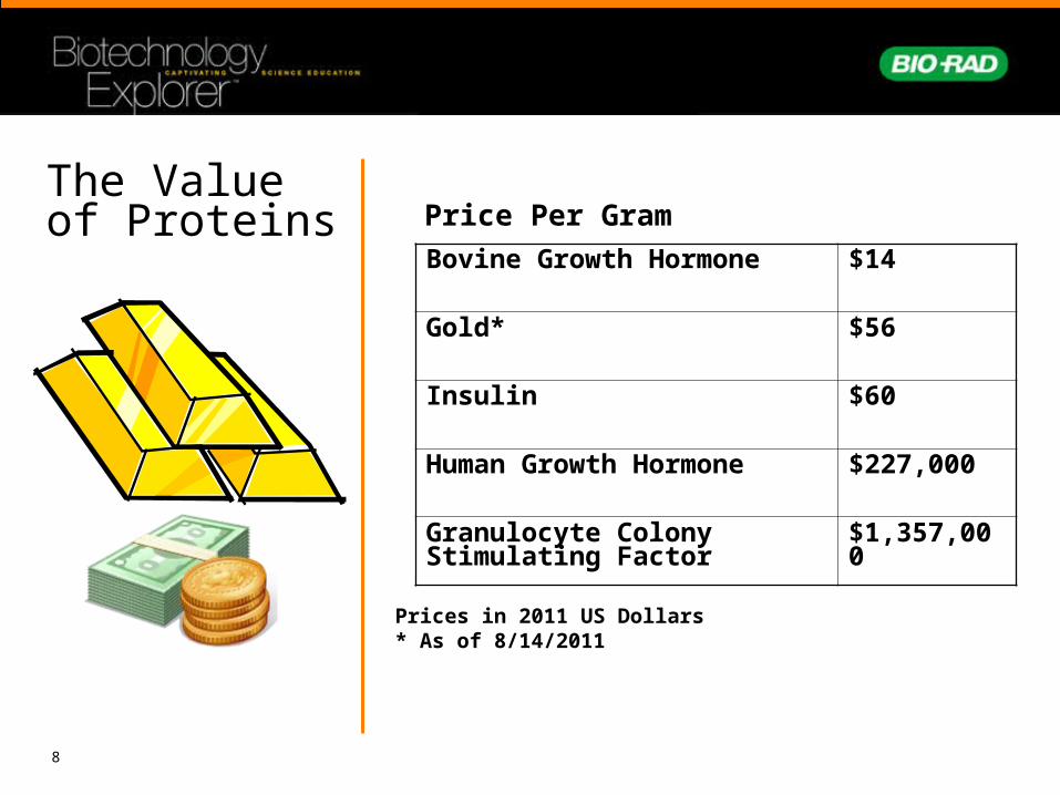
The Value of Proteins
Bovine Growth Hormone $14
Gold* $56
Insulin $60
Human Growth Hormone $227,000
Granulocyte Colony Stimulating Factor
$1,357,000
Price Per Gram
Prices in 2011 US Dollars* As of 8/14/2011

9
PROTEIN: USED IN THE TREATMENT OF:
Cell Production
Insulin Diabetes E. coli
Human growth hormone Growth disorders E. coli
Granulocyte colony stimulating factor Cancers E. ColiErythropoietin Anemia CHO cellsTissue plasminogen activator Heart attack CHO cellsHepatitis B virus vaccine Vaccination YeastHuman papillomavirus vaccine Vaccination Yeast
Protein – The product of Biotech
10
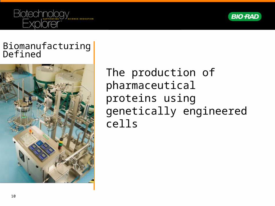
Biomanufacturing Defined
The production of pharmaceutical proteins using genetically engineered cells

11
Expression Choices Cell type:
• E. coli
• Yeast
• Mammalian–CHO

12
Expression ChoicesParameter Bacteria Yeast Mammalian
Contamination risk Low Low High
Cost of growth medium
Low Low High
Product titer (concentration)
High High Low
Folding Sometimes Probably Yes
Glycosylation No Yes, but different pattern Full
Relative ease to grow Easy Easy Difficult
Relative ease of recovery
Difficult Easy Easy
Deposition of product Intracellular Intracellular or extracellular Extracellular
Product Intracellular Often secreted into media Secreted

13
DHFR —Dihydrofolatereductase
•Converts dihydrofolate into tetrahydrofolate (THF) by the addition of a hydride from NADPH
•THF is a methyl (CH3) group shuttle required for synthesis of essential molecules
- nucleotides- amino acids

14
DHFR and Cancer
•DHFR inhibition or reduction disrupts nucleic acid synthesis affecting
-Cell growth-Proliferation
•Methotrexate – chemotherapeutic agent-Competitive inhibitor of DHFR-Methotrexate resistance - correlates with
amplification of DHFR genes

15
Induction
Biotech companies genetically engineer plasmids to place genes behind inducible promoters

16
Transcriptional Regulation in the pDHFR system
RNA Polymerase
Z Y A
Z Y ALacI
Effector (Lactose)
Z Y ALacI
lac Operon
Lactose IPTG

17
Transcriptional Regulation in the pDHFR system

18
Transcriptional Regulation in the pDHFR system
Lactose = Induced System

19
GST-DHFR-His Construct
GST – DHFR - His
Glutathione-s-transferase
•Added to increase solubility
•Can be used as a secondary purification methodology
Human dihydrofolate reductase
•Gene product of interest
•Target for chemotherapy reagents
Histidine tag
•6 Histidine tag that binds to certain metals such as nickel

20
Selection Mechanism for Mammalian cells

21
Phases of growth

22
Recovery
Separation of protein from other molecules
Purification
Separation of the protein of interest from other proteins

23
Chromatography Basics
• Mobile phase (solvent and the molecules to be separated)
• Stationary phase (through which the mobile phase travels)– paper (in paper chromatography)– glass, resin, or ceramic beads (in column
chromatography)
• Molecules travel through the stationary phase at different rates because of their chemistry.

24
Types of Column Chromatography •Ion Exchange (protein charge)
•Size Exclusion (separates on size)
•Hydrophobic Interaction (hydrophobicity)
•Affinity:•Protein A tail of Antibodies•His-tagged metal complexes (Ni)•Glutathione-s-transferase glutathione

25
Performing the chromatographic separation
•Gravity Chromatography•Spin Column Chromatography
•Chromatography Instrumentation•Small scale•Biomanufacturing scale
(bioreactors)

26
Protein Expression and Purification Series Workflow
Streak Cells
Overnight culture
Subculture, monitor, and induce
Harvest and lyse cells
Purify
Centrifugation or Instrumentation
Analyze

27
CentrifugeRCF to RPM conversion • Accurate RCF(g) is important for
chromatography resins
• RPM to RCF varies for different models of centrifuges due to variation in rotor radius
• Determine RPM for 1,000 x g. The Bio-Rad 16K microcentrifuge rotor has a radius of 7.3 cm
RCF = relative centrifugal force
RPM = rotations per minute
R = radius in cm from center of rotor to middle of spin column
1,0003,497
7.3

28
Affinity purification
A. Label column with initials. Snap off bottom tab of column, remove cap and place in 2 ml microcentrifuge tube.
B. Add 200 µl of Ni-IMAC resin slurry to empty column
C. Centrifuge for 2 minutes at 1,000 x g. After spin, discard buffer that has collected in the microcentrifuge tube.
Ni-IMAC resin slurry
200 µl
1. Pour column
2. Wash resin to remove packing buffer
3. Equilibrate resin
4. Bind GST-DHFR-His
5. Elute unbound proteins
6. Wash protein bound onto the resin
7. Elute GST-DHFR-His discard

29
Affinity purification A. Add 200 µl of
distilled H2O to column
B. Centrifuge for 2 minutes at 1,000 x g. After spin, discard water from collection tube.
Distilled H2O
200 µl
1. Pour column
2. Wash resin to remove packing buffer
3. Equilibrate resin
4. Bind GST-DHFR-His
5. Elute unbound proteins
6. Wash protein bound onto the resin
7. Elute GST-DHFR-His
discard

30
A. Add 500 µl of Equilibration buffer to column
B. Centrifuge for 2 minutes at 1,000 x g. After spin, discard Equilibration buffer and collection tube. The column is now ready to use.
Equilibrationbuffer
Affinity purification1. Pour column
2. Wash resin to remove packing buffer
3. Equilibrate resin
4. Bind GST-DHFR-His
5. Elute unbound proteins
6. Wash protein bound onto the resin
7. Elute GST-DHFR-His
500 µl
discard

31
Affinity purification
B. Gently mix for 20 min.
A. Place yellow tip closure on bottom of column. Add 600 µl Soluble Fraction to Column; Put on clear top cap.
Soluble fraction
600 µl
1. Pour column
2. Wash resin to remove packing buffer
3. Equilibrate resin
4. Bind GST-DHFR-His
5. Elute unbound proteins
6. Wash protein bound onto the resin
7. Elute GST-DHFR-His

32
His tags
N3H+-OOC
Histidine
Resin
• His tags are typically a series of 6 histidines added to the C or N terminus of a recombinant protein
Ni
Ni
Ni
Ni
N
NH
NN
H His-tagged Recombinant
Protein
• His tag and column interaction
33
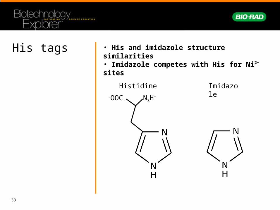
His tags
Imidazole
N3H+-OOC
Histidine
• His and imidazole structure similarities• Imidazole competes with His for Ni2+ sites

34
Affinity purification
A. Label three 2 ml tubes:
1. Pour column
2. Wash resin to remove packing buffer
3. Equilibrate resin
4. Bind GST-DHFR-His
5. Elute unbound proteins
6. Wash protein bound onto the resin
7. Elute GST-DHFR-His
“Flow through” “Wash” “ Eluate”

35 “Flow through”
“Flow through”
B. Remove yellow tip closure.
C. Place column in 2 ml collection tube labeled “Flow Through” and remove clear top cap.
D. Centrifuge for 2 min at 1,000x g. Set aside Flow Through.
1. Pour column
2. Wash resin to remove packing buffer
3. Equilibrate resin
4. Bind GST-DHFR-His
5. Elute unbound proteins
6. Wash protein bound onto the resin
7. Elute GST-DHFR-His
Affinity purification
Keep

36
A. Place column in 2 ml collection tube labeled “Wash”.
B. Add 600 µl Wash Buffer to column.
• Centrifuge for 2 min at 1,000xg. Set aside Wash fraction.
Wash Buffer
“Wash”
600 µl1. Pour column
2. Wash resin to remove packing buffer
3. Equilibrate resin
4. Bind GST-DHFR-His
5. Elute unbound proteins
6. Wash protein bound onto the resin
7. Elute GST-DHFR-His
Affinity purification
“Wash” Keep

37
Affinity purification1. Pour column
2. Wash resin to remove packing buffer
3. Equilibrate resin
4. Bind GST-DHFR-His
5. Elute unbound proteins
6. Wash protein bound onto the resin
7. Elute GST-DHFR-His
A. Place column in 2 ml collection tube labeled “Eluate”.
B. Add 400 µl Elution Buffer to column.
• Centrifuge for 2 min at 1,000xg. Set aside Eluate.
Elution Buffer
“Eluate”
400 µl
“Eluate” Keep

38
Recap so far….
Started with a complex mixture of all the soluble E. coli proteins along with the induced expressed human GST-DHFR-His
Soluble fraction
Purified the GST-DHFR-His away from the E. coli proteins by using the affinity of the 6 Histidine tag on GST-DHFR-His for Ni-IMAC beads
Flowthrough Wash Eluate
~600 µl ~600 µl ~400 µl

39
Size exclusion purification(buffer exchange)
Eluate fraction
GST-DHFR-His in 20 mM sodium phosphate, 300 mM NaCl and 250 mM imidazole
Imidazole
250 mM imidazole
solution has an A280= 0.2-0.4
W and Y contribute to A280 of proteins
NEED TO REMOVE IMIDAZOLE TO QUANTIFY PROTEIN CONCENTRATION USING A280

40
• Beads in column are made of polyacrylamide and have tiny pores
• The mixture of molecules is added to the column
• Large molecules move through the column quickly traveling around the beads
• Smaller molecules move through the pores of the beads and take longer to pass through the column
http://tainano.com/Molecular%20Biology%20Glossary.files/image047.gif
Principles of Size Exclusion Chromatography

41
Principles of Size Exclusion Chromatography
• The mass of beads in the column is called the column bed
• Beads trap or sieve and filter molecules based on size
• The separation of molecules is called fractionation
• Size of pores in beads determines the exclusion limit (what goes through the beads and what goes around the beads)
• Molecules are dissolved in a buffer

42
SizeExclusion

43
Size exclusion purification(desalting)
A. Label desalting column with your initials.
B. Invert column several times to resuspend gel.
C. Snap off bottom tip and place in a 2 ml collection tube.
1. Prepare SEC column
2. Desalt GST-DHFR-HIS with SEC column

44
1. Remove green top cap and allow excess packing buffer to drain by gravity to top of resin bed. If the column does not begin to flow, push the cap back on the column and then remove to start the flow.
D. After draining, place column in clean 2 ml tube.
E. Centrifuge for 2 min at 1,000 x g. Discard 2ml tube containing packing buffer.
discard
discard
Size exclusion purification(desalting)
1. Prepare SEC column
2. Desalt GST-DHFR-HIS with SEC column

45
A. Label new 2 ml tube “Desalted Eluate”.
B. Carefully apply 75 ul of eluate fraction directly to the center of column. Be careful not to touch resin with pipet tip.
C. Centrifuge for 4 min at 1,000 x g.
D. Repeat addition of 75 µl of Eluate fraction to column and centrifugation.
Removing the 250 mM imidazole solution by size exclusion chromatography
“Eluate”
75 µl
2 x
Size exclusion purification(desalting)
1. Prepare SEC column
2. Desalt GST-DHFR-HIS with SEC column

46
Desalted eluate ~150 µl
GST-DHFR-His
in 10 mM Tris buffer
250 mM Imidazole has been removed
Size exclusion purification(desalting)
1. Prepare SEC column
2. Desalt GST-DHFR-HIS with SEC column

47
Protein Analysis
• Determination of success of induction, lysis, and purification of GST-DHFR-His using SDS-PAGE analysis
• Measurement of concentration using the absorbance at 280 nm
• Enzymatic activity analysis

48
250
150
100 75
50
37
25 20
15
10
1 –
Pre
cisi
on
Plu
s D
ual
Co
lor
2 –
Un
ind
uce
d c
ells
3 –
Ind
uce
d c
ells
4 –
Inso
lub
le f
ract
ion
5 –
So
lub
le f
ract
ion
6 –
Co
lum
n f
low
th
rou
gh
7 –
Co
lum
n w
ash
8 –
Elu
ted
GS
T-D
HF
R-H
is
9 –
De
salt
ed G
ST
-DH
FR
-His
stan
dar
ds
Protein analysisSDS-PAGE
1. Prepare Samples
2. Prepare TGX Gel and vertical Electrophoresis apparatus
3. Load and Run Gel
4. Stain gel
5. Analyze gel

49
Protein analysis (Quantitation using A280)
Quantitation of Protein in Desalted Fraction
+100 µl Distilled H2O
Clean UV cuvette
+ 100 µl Desalted
eluateClean UV cuvette
Turn on spectrophotometer and set absorbance to 280 nm. Add 100 µl distilled H2O to clean UV compatible cuvette.
Blank spectrophotometer with distilled H2O.
Pipet 100 µl of your desalted eluate sample (GST-DHFR-His) into clean UV compatible cuvette.
Measure absorbance of sample at 280nm and record or print the value. Return sample to 2 ml tube.

50
Beer’s Law A=cl
- the molar absorptivity ((mol/L)-1 cm-1)
l - the path length of the sample (usually 1cm-cuvette)
C - the concentration of the compound in solution (mol/L)
For GST-DHFR-His
= 75,540 (mol/L)-1 cm-1
C (mol/L) = Absorbance
75,540 (mol/L)-1 cm-1 x 1 cm
Protein analysis (Quantitation using A280)
Expected results
1.3 x 10⁻⁶ – 5.3 x 10⁻⁶ M
Calculate concentration of GST-DHFR-His

51
Enzyme Assay
Absorbance at 340nm

52
Enzyme Assay
A. Set up spectrophotometer for kinetics measurements at 340 nm.
B. Blanking the instrument. Add 985 µl 1x PBS to cuvette; place in instrument, read as blank. Save cuvette with PBS
C. Running the no substrate control reaction. Add 6 µl of 10 mM NADPH to cuvette containing 985 µl 1x PBS. Add 15 µl of purified, desalted GST-DHFR-His eluate to cuvette. Cover cuvette with parafilm and invert 10 times. Immediately place cuvette in spectrophotometer and begin kinetics run.
D. As run is proceeding, record absorbance value every 15 seconds for 150 seconds. Remove and save cuvette from the spectrophotometer.
+ 985 µl 1x PBS
+ 985 µl 1x PBS
+ 6 µl NADPH
+ 15ul desalted Eluate
+ 985 µl 1x PBS

53
Enzyme Assay
E. Running the enzymatic reaction with the GST-DHFR-His, NADPH (cofactor) and DHF (substrate).Note: The enzyme reaction should be prepared while standing at the spectrophotometer. The reaction occurs extremely quickly and it is necessary to place the cuvette in the spectrophotometer and start the readings as quickly as possible once the DHF has been added.
F. Add 5 µl of 10 mM DHF to the cuvette already containing 1x PBS, your GST-DHFR-His sample and NADPH. Quickly cover the cuvette with parafilm and invert 5 times.
G. Immediately place the cuvette in the spectrophotometer and begin kinetics run. As run is proceeding, record absorbance value every 15 seconds for 150 seconds. Remove cuvette from the spectrophotometer.
+10 mM DHF

54
Instrumentation
BioLogic LPDemo
BioLogic™ LP
BioLogic DuoFlow™

55
Chromatography instrumentation
1. Pump(s)
2. Detector(s)
- UV detector
- Conductivity detector
- Pressure detector
- Fluorescence detector
3. Valves
Plus associated wiring and tubing…

56
Examining a chromatogram
BioLogic LP

57
Examining a chromatogram
BioLogic DuoFlow

58
DHFR Enzymatic Activity Calculation
Slope of Control Data x 60 = _____________ Change in Absorbance at 340 nm/minute
ΔOD, control
ΔOD, reaction
Slope of Enzyme reaction data x 60 = _____________ Change in Absorbance at 340 nm/minute
ΔOD = |ΔOD, reaction| - |ΔOD, control|
ΔC (mol/liter/min) = ΔOD ε x l
ε (extinction coefficient) = 6220 M-1 cm-1 for NADPHl (length) is the pathlength of the cuvette (usually 1 cm for most cuvettes)

59
Biomanufacturing Scaling up of the process developed during research and development

60
Bio-Rad:Curriculum Training Specialists [email protected]
http://explorer.bio-rad.com
Technical Support: 1(800)4BIORAD [email protected]
Northeast Biomanufacturing Center and Collaborative (NBC2)
http://www.biomanufacturing.org
Bio-Link (Elaine Johnson, Director)http://www.bio-link.org
Jim DeKloe:[email protected]
Resources and References

61
AVAILABLE NOW!ProteinExpressionandPurificationSeriesOrdering info
Option 1CentrifugationPurificationModuleOption 3
PrepackedCartridgePurificationModule Option 2
HandpackedColumnPurificationModule
Growth andExpressionModule
SDS-PAGEElectrophoresisModule
DHFREnzymaticAssayModule
PurificationModule
•166-5040EDU, Centrifugation Process Series
•166-5045EDU, Handpacked Column Process Series (instrumentation)
•166-5050EDU, Prepacked Cartridge Process Series (instrumentation)