Strategies in protein purification - Wolfson Centre Home...
69
Input for Purification Protocol Development Guidelines for Protein Purification Sample Preparation before Chromatography Chromatography: General Considerations 1
-
Upload
nguyenkhuong -
Category
Documents
-
view
234 -
download
3
Transcript of Strategies in protein purification - Wolfson Centre Home...

Input for Purification Protocol Development
Guidelines for Protein Purification
Sample Preparation before
Chromatography
Chromatography: General Considerations
1

PURIFICATION STRATEGY
General approach: Input for Protocol Development
Guidelines for Protein Purification. Commonly confronted decisions.
Properties of Target Protein
Sequence of Events: Cell harvesting, Cell Disruption, Extraction and
Clarification, Chromatography
Sample Preparation before Chromatography: Cell Debris Removal,
Clarification and Concentration: dialysis, filtration, ultrafiltration , others
FPPLC, columns, resolution, selectivity, efficiency and capacity
Three Phase Strategy: Linking Chromatography Techniques 2

Applications of Protein Purification In vitro Activity assays
Antibody development / production
Protein:protein interaction assays
Cell-based activity assays
Ligand-binding assays
Mass-spectrometric analysis
Structural analysis
In vivo activity assay
Don’t waste clear thinking on dirty
or not healthy proteins!!!!
Post-translational modification tests
N-terminal sequencing
Electromobility shift assay (band shift)
DNA footprinting
Protein cross-linking studies
Vaccine development/production
Probes for protein arrays/chips
Expression library screening
Other
For each application you need:
different quantities
different protein purity
start material is different, etc
different strategy
Each purification project must be
adapted to your start material and
your final needs

Protein Purification Strategy
4
FUSION PROTEIN
NON-FUSION PROTEIN
EXPRESSION
Simple Purification
ONE STEP Affinity
70 - 95% Purity
For higher purity
Capture Intermediate Purification
Polishing
Multi-Step Purification

Protein Modifications May Require Further Purification
5
COOH
NH2
Proteolytic cleavage
Glycosylation
Phosphorylation
Acylation
Misfolding, random
disulfide bridges
N or C terminal
heterogeneity
Aggregation
Oxidation of methionine Desamidation of Asp and Glu

Protein Purification - Aims
Satisfactory
expression levels
protein activity
purity
homogeneity
stability
Economical use of reagents/equipment
• Goal to Success:
Define the purification objectives
Selection or optimization of the best source or best expression conditions
A good understanding of the protein needs
Selection and optimization of the most appropriate technique for each step
7

Structural Studies: Crystallization – NMR- etc
Concentration & Storage
Fermentation
Scaling Up
Gene Cloning
Target Optimization
Target Selection
Selection of Expression Vector
Selection of Expression Host
Expression Analysis
Solubility Analysis
Purification Optimization
Characterization
Biochemical Studies Pharmaceutical Studies
Protein Production Pipeline
Purification
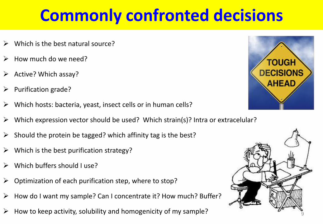
Commonly confronted decisions
Which is the best natural source?
How much do we need?
Active? Which assay?
Purification grade?
Which hosts: bacteria, yeast, insect cells or in human cells?
Which expression vector should be used? Which strain(s)? Intra or extracelular?
Should the protein be tagged? which affinity tag is the best?
Which is the best purification strategy?
Which buffers should I use?
Optimization of each purification step, where to stop?
How do I want my sample? Can I concentrate it? How much? Buffer?
How to keep activity, solubility and homogenicity of my sample? 9

Overview: separation techniques
Technique Parameter Based on for separation Gel filtration Size/Shape MW, Shape, and oligomeric state of the molecule Ion exchange/ Hydroxyapatite/ Charge interaction Asp, Glu, Lys, Arg, His
Chromatofocusing
Hydrophobic/ Hydrophobic sites Trp, Phe, Ile, Leu, Tyr, Pro Reversed phase interaction Met, Val, Ala
Affinity Biological function eg: antibody – antigen
Metal chelate Affinity for metals poly His
Covalent Covalent interaction Uses SH groups (Cys)
Multimodal Mixture Hydrophobic + Ionic Interaction 10

Input for Purification Protocol Development
General Input Sample Specific Input
11
Three phase strategy
Purification protocol
Required purity and quantity
Physical-chemical properties of target and
main contaminants
Source material information
Separation technique knowledge
Scouting runs and optimization
Economy and resources

Yields from Multistep Protein Purifications
12
Number of steps
Yield (%)
95% / step
90% / step
85% / step 80% / step 75% / step
0
20
40
60
80
100
1 2 3 4 5 6 7 8

Use relevant analytical tools
A rapid and reliable assay for the target protein: biological assay,
enzymatic, SDS-PAGE, Western, etc
Purity determination
SDS-PAGE, Native, IEF, RPC, analytical GF or EIX, etc
Total protein determination – Interference of detergents, reducing
agents, sugars, others - ultraviolet absorption, colorimetric method, etc
13

Define Properties of Target Protein (I)
Temperature Stability Need to work rapidly at low temperature
pH Stability Selection of buffers for each step
Organic Solvents Stability Selection of Conditions for RPC
Salt Stability Selection of Conditions for all steps
Co-factors for Stability or Activity Selection of Additives, pH, Salts, Buffers
Protease Sensitivity Fast removal of proteases. Protease Inhibitors
Sensitivity to Metal Ions Need of EDTA or EGTA in Buffers

Define Properties of Target Protein (II)
Redox Sensitivity Need of reducing agents to protect reduce Cys: DTT, DTE or
on the contrary, need to protect disulfide bridges
Molecular Weight/Oligomeric State Selection of Gel Filtration Media / UF
Charge Properties Selection of Ion Exchange Conditions
Biospecific affinity Selection of ligand for Affinity Medium
Post Translational Modifications Selection of Group Specific Affinity: Lectins
Hydrophobicity Solubility prediction - Selection of medium for HIC

16
Initial Bioinformatics Investigation
Using Bioinformatic Tools to Strategically Design Expression/Purification Projects
Dr. Nurit Kleinberger-Doron
http://wolfson.huji.ac.il/expression/software.html

17
Bioinformatics Tools-I
Physical and chemical parameters http://www.expasy.org/tools/protparam.html Computation of various physical and chemical parameters for a given protein: molecular
weight, theoretical pI, amino acid composition, atomic composition, extinction coefficient, estimated half-life, instability index, aliphatic index and grand average of hydropathicity (GRAVY)
http://www.scripps.edu/~cdputnam/protcalc.html Generates molecular weight information (including scanning mass spectrometry results),
estimated charges (including pI estimation), uv absorption coefficients, crystallographic solvent content percentage and Vm, and counts atoms and residues based on the protein sequence
Proteolytic Cleavage http://www.expasy.org/tools/peptidecutter/ Predicts potential cleavage sites cleaved by proteases or chemicals in a given protein
sequence http://www.cf.ac.uk/biosi/staff/ehrmann/tools/proteases.index.html Protease database of E.Coli

18
Bioinformatics Tools-II Post-translational modification prediction
http://www.expasy.org/tools/#ptm
Prediction of Ser, Thr and Tyr phosphorylation sites in eukaryotic proteins
Prediction of N-acetyltransferase A (NatA) substrates (in yeast and mammalian proteins
Prediction of O-GalNAc (mucin type) glycosylation sites in mammalian protein
Prediction of N-glycosylation sites in human proteins
Prediction of N-terminal myristoylation by neural networks
Recombinant Protein Solubility Prediction : The statistical model predicts protein solubility assuming the protein is being overexpressed in Escherichia coli.
http://www.biotech.ou.edu/
S-S bonds: Predicts cysteins that are likely to be partners in cysteine bridges
http://clavius.bc.edu/~clotelab/DiANNA/
http://gpcr.biocomp.unibo.it/cgi/predictors/cyspred/pred_cyspredcgi.cgi
FoldIndex© tries to answer to the question: Will this protein fold? http://bip.weizmann.ac.il/fldbin/findex

Is the Recombinant Protein Correctly Expressed
19
Biological activity
Analytical GF / DLS /Native PAGE / IEF
Aggregation Heterogeneity
Size Proteolytic cleavage Presence of impurities
Stability at different pH Ionic strengths Protein concentrations Detergent concentrations
SDS-PAGE and immuno blotting
MS / N-terminal sequencing Truncated forms Heterogenous N-terminus

Advantages or Disadvantages of Intra or Extracellular Expression - I
Cell wall disruption / Osmotic Shock
Extracellular expression
Recover
Clarified
sample
Cell disruption
Harvest inclusion
bodies
Intracellular expression
Insoluble in Cytoplasm
Periplasmic space
Recover Supernatant
Soluble in Cytoplasm
Cell debris removal Clarification
Purification-Chromatography
Cell removal
Clarification
Recover
Clarified
sample
Culture medium

Extracellular expression
Recover pellet
Intracellular expression
Insoluble in Cytoplasm
Periplasmic space
Recover supernatant after cell lysis
Soluble in Cytoplasm
Recover Supernatant
Purification-Chromatography
Partially pure protein Relatively low protein target in small volume
Low lipid conc. Lower degradation Correct disulphide
bond formation
Difficult to scale-up
Partially pure protein
Very large volume
Low protein concentration
Lower degradation Correct disulphide
bond formation
Recover
Clarified
sample
Recover clarified sample after cell wall lysis and osmotic shock
Culture medium
High quantities of almost pure protein
Renaturation problem
Lower sensitivity to proteases
Problems with disulphide bond
formation
Highly impure protein and lipid concentration Sensitive to proteases
Sometimes well expressed Small extraction volume
Problems with disulphide bond formation
Advantages or Disadvantages of Intra or Extracellular Expression - II

PURIFICATION STRATEGY
General approach: Input for Protocol Development
Guidelines for Protein Purification. Commonly confronted decisions.
Properties of Target Protein
Sequence of Events: Cell harvesting, Cell Disruption, Extraction and
Clarification, Chromatography
Sample Preparation before Chromatography: Cell Debris Removal,
Clarification and Concentration: dialysis, filtration, ultrafiltration , others
FPPLC, columns, resolution, selectivity, efficiency and capacity
Three Phase Strategy: Linking Chromatography Techniques 22

Extraction and Clarification Definition: Primary isolation of target protein from source material.
Removal of debris or other contaminants which are not compatible with
chromatography.
Goal: Preparation of clarified sample for further purification.
The chosen technique must be robust and suitable for all scales of
purification.
Choice of additives and buffers must be carefully
considered before scaling up
Use additives only if essential for stabilization of
product or improved extraction; select those that
are easily removed. 23

Common Substances Used in Sample Preparation
24
Minimize use of additives: they must be removed in extra purification steps or may interfere with activity assays
Tris HCl 20-50mM pH 7.5-8.0 or other buffers (HEPES, Phosphate, etc)
NaCl/KCl 0.3-0.5M (to maintain ionic strength). For soluble proteins NaCl can lower to 50mM. For some insoluble
proteins it can be increase till 1M
Glycerol 5-10% to stabilize prone to aggregate proteins (can be increase till 20%). Increase viscosity and back flow
of columns
DNaseA 25-50µg/ml (or Benzonase): degrade DNA. Reduce viscosity. Eukaryotic cells could need more Dnase
Lysozyme 0.2mg/ml for wall lysis of bacterial cells
Detergents (NP40, Triton X100, Tween 20, OG, DDM etc) for solubilization of some insoluble proteins or extraction
of membrane proteins. Use only if it does not affect protein stability!!!!
Reducing agents: 1-15mM BME, up to 2mM DTT or DTE, 1-5mM TCEP. Use only for Cys containing proteins without
disulfide bridges (maintain Cys in reduce form). Not all the IMAC columns can be use with all the reducing agents
EDTA 1-10mM Reduce oxidation damage. Chelate metal ions. Metalloprotease inhibitor. Do not use with IMAC.
Sucrose or Glucose 25mM Stabilize lysosomal membranes in eukaryotic cells. Reduce protease release.
Protease or Phosphatase inhibitors if necessary

Protease Inhibitors
25
Protease Inhibitor
Specificity of inhibition Working concentration
Antipain-dihydrochloride
Papain, Trypsin, Cathepsin A and B 1-100µM
Aprotinin Trypsin, Plasmin, Chymotrypsin, Kallikrein
2µg/ml
Benzamidine HCl Serine Proteases 0.5-4µM
Bestatin Aminopeptidases
Chymostatin Chymotrypsin and Cysteine Proteases 10-100µM
E-64 Cysteine Proteases 10µM
EDTA (or EGTA) Metalloproteases (Calcium) 2-10mM
Leupeptin Serine and Cysteine Proteases such as Plasmin, Trypsin, Papain, Cathepsin B
10-100µM
PMSF and AEBSF Serine Proteases 0.1-1mM
Pepstatin Aspartic Proteases 1µg/ml
Phosphoramidon Metalloproteinases, specifically, Thermolysin
1-10µM
Serine proteases are widely
distributed in most types of cells.
Bacterial extracts typically
contain serine and
metalloproteases.
Extracts from animal tissues
contain mainly serine-, cysteine-,
and metalloproteases. (some also
contain aspartic proteases).
Plant extracts contain large
amounts of serine and cysteine
proteases
Remove proteases early in the first purification step!!: load on capture column
immediately after lysis and clarification.

Protease Inhibitor Cocktail Set III (Cat. No. 539134) MERCK – EMD
Recommended for mammalian cells/tissue 1 ml sufficient for 20 g cells (~1 L). Dilution 1:100 to1:300
EDTA-free (good for His-Tag® protein purification)

Cell Disruption considerations
Stability of the released protein
Location of target protein within the cell (membrane, nucleus, mitochondria, etc.)
Yield and kinetics of the process. Extent of disruption: possible use of marker
substances, measure protein concentration. Balance: volume & lysis efficiency.
Suggested lysis volume for bacterial cells: 10-20% of original cell culture
Scale-up
Consider if protein purification can be performed directly from the cell lysate
without a cell debris clarification step (bed absorption chromatography) 27

28
Methods to monitor lysis
Reduction of whole cells: decrease of Abs660nm before and
after treatment.
Decrease of weight cell pellet after lysis
Monitor nucleic acid release : Increase in the Abs260nm
during lysis
(This method could be difficult because of the “haze” generated, which can alter absorbance readings. )
Microscopically : Compare cells before and after treatment

29
CELL DESINTIGRATION AND EXTRACTION: METHODS THAT DO NOT NEED SPECIAL EQUIPMENT
Freezing and thawing: Repeated cycles (can denature protein). For cells without a cell wall (animal cells). Not suitable for large scale. Not reliable method
Osmotic shock: Transfering cells from a high to a low osmotic pressure. Useful to release periplasmic proteins from Gram negative bacteria. Not reliable method
Chaotropic agents (urea, GuHCl): Extremely denaturative. Not suitable for large scale. Use for extremely insoluble proteins or inclusion bodies
Detergents (Brij, NP40, DDM, etc.): Anionic and non-ionic detergents permeabilize Gram negative cells. Can interfere in downstream process. Dissolve membrane-bound proteins. Use in combination with mechanical methods. Problematic!!! Bacterial Expression Screen - DDM (Dodecyl Maltoside) lysis - Small Affinity binding http://wolfson.huji.ac.il/purification/TagProteinPurif/DDM_Bacterial_Expr_screen.html
Organic solvents: Toluene, ether, chloroform, isoamyl alcohol, etc at different
concentration can release different materials from the cell. Extremely denaturative. Use only for solvent resistent proteins. Not reliable method
Enzymatic lysis: Lysozyme hydrolyze linkages in the peptido-glycan of bacterial cell walls. Used for pretreatment of cells in combination with mechanical methods. Yeast cell walls can be hydrolyzed with snail gut enzymes and glucanases

30
CELL DESINTIGRATION AND EXTRACTION: METHODS THAT NEED SPECIAL EQUIPMENT
Combine with chemical treatment: lysozyme, detergents, Dnase, etc.
Mixers and blenders: Useful for animal and plant tissues (Warring-blender)
Coarse grinding Grinding with a pestle and mortar of frozen mycelium. Fine grinding in a
bed mill: Useful for yeast, larger cells, algae and filamentous fungi. Use of different glass
beads (Bead-beater)
Homogenization: Animal cells. Piston/plunger device. Wheaton-Dounce homogenizer
Sonication: Bacterial cells disrupted by high frequency sound and share forces. Low scale. Very vigorous process. Heat generation. Not reliable method
High pressure lysis: Pumping cell suspension through a narrow orifice at high pressure. Mainly for bacterial cells. Very reliable and efficient method. French-press, Microfluidizer, Avestin, etc: medium scale (20-100ml). Microfluidizer, Maunton-Gaulin: For larger volumes

31 One Shot Model Microfluidizer
Avestin Emulsiflex C3
Microfluidizer
low volume benchtop machine
Cell Lysis Equipment in LSI
As French-press but for medium/larger volumes
For bacterial and yeast cells
High speed
Other applications.
HTP – Low scale: Bacterial Expression Screen - DDM (Dodecyl
Maltoside) lysis - Small Affinity binding http://wolfson.huji.ac.il/purification/TagProteinPur
if/DDM_Bacterial_Expr_screen.html

Sample Preparation before Chromatography: Cell Debris Removal, Protein Clarification and Concentration
Centrifugation For small sample volumes 15min 10000g .
For very turbid cell homogenates: 30min 50000g
Filtration before loading in chromatographic column
Pore size filter: 1 μm for particle size of chromatographic medium 90 μm and upward
Pore size filter: 0.45 μm for particle size of chromatographic medium 30 or 34 μm
Pore size filter: 0.22 μm for particle size of chromatographic medium 3, 10, 15 μm
Filtration large scale, Normal or Dead end: Hollow-fiber. Plates. Spiral Cartridge
TFF: Tangential Flow Filtration or Cross flow
Expanded Bed Adsorption
Fractional Precipitation
Ultrafiltration Membranes
32

Tangential or cross flow and Normal or dead end filtration
Membrane-Based Systems
Pressure-driven processes, such as ultrafiltration (UF), microfiltration, virus filtration, and
nanofiltration . Or electric field (electroultrafiltration, EUF)
They are mainly used for protein concentration and buffer exchange in preference to SEC on an
industrial scale.
There are charge membranes that can use as IEX, RPC,
Affinity, HIC (Pall, Mustang, etc)
Another emerging technology in membrane separation
processes is high-performance tangential flow filtration
(HPTFF).

34
Labscale™ Benchtop TFF System with
Pellicon XL module
ProFlux® M12 Benchtop TFF system with
spiral wound modules
Prep/Scale filter modules
Fully automated 80 m2 Pellicon system for concentration and diafiltration
Pellicon cassettes
Large-scale spiral wound UF/DF system
TFF: Tangential or cross flow filtration Merck

35
TFF: Tangential or cross flow filtration
Merck

36
ÄKTAcrossflow™
Fully automated filtration system for cross
flow membrane screening, process
development, and small scale processing.
Enable automation at very small scale, with
capacity ranging from liters down to 25 ml.
Kvick Cassette family
Membrane surface area
from 50 cm² to 2.5 m²
MW cutoffs (5k, 10k select,
10k, 30k, 50k, and 100k)
Hollow fiber ultrafiltration
cartridges
Available with ten
different molecular weight
Normal / Cross Flow Filtration / Ultrafiltration GE Healthcare
Syringe filters
Bottle-Top Filters
Filter capsules Membrane filtration capsules
Cross flow filtration products
Normal flow filtration products

Dialysis
A process of separating molecules in solution by the difference in their rate of diffusion
Time
Temperature
Solvent
Volume
Cut-off

Uses of dialysis
• To remove unwanted small molecules from a protein solution
- DNA
- salts
- high CMC detergents
- small proteins
• For buffer exchange
• “Desalting”

The dialysis membrane
• Molecular weight cutoff (MWCO)- the average pores size
MW>MWCO - molecule will cross membrane
MW<MWCO – molecule will not cross membrane
• MW<<MWCO cross membrane faster than MW<MWCO
8kDa
20 kDa
MWCO=10 kDa
8kDa Slow & ineffective
20 kDa
MWCO=10 kDa
Fast

• Protocol:
• Choose the membrane due to protein size.
• The “old” membranes are with cut-off of 13 kDa
• Load the sample into dialysis tubing (wash membrane and check for holes).
• Place sample into an external chamber of dialysis buffer (with gentle stirring of the buffer).
• Dialyze for 2-4 hours
• Change the dialysis buffer and dialyze for another 2-4 hours
• Change the dialysis buffer and dialyze for 2 hours - ON.
Example:
• For 10ml sample of 1M in 10L buffer – sample
will reach to 1mM at equilibrium (~4h)
• Same sample in 1L – 10mM after 4h
Then replace buffer 1L – 0.1mM after 4h.
Co
nce
ntr
atio
n.
Time
Dialysis General Considerations
Time
Buffer volume

Types of membrane
• There are more then 30 types of synthetic and natural dialysis membranes
Cellulose Polysulfone Polyethylene
Polypropylene Polyvinylidene fluoride

A process that uses semi-permeable membranes to
separate molecules on the basis of size.
It is particularly appropriate for concentration,
partial purification or for buffer exchange.
Is a gentle and non denaturing method.
The ultrafiltrate is cleared of macromolecules which are significantly larger than the cutoff of the filter
The buffer concentration in the ultrafiltrate will be exactly the same as in the concentrate
Do not replace GF, although the principles are the same: separation according to ratio of the molecule
Proteins with MW lower than the cut-off, will be retained in the concentrate if they aggregate, or are
part of a complex
Cut-off at least two or three times of the protein size
Some proteins can stick to the membranes
Ultrafiltration Concentrate
Ultrafiltrate or Retentate
Membrane with different cut-off

Protein Concentration | 2012 43
Ultra Centrifugal Devices Amicon / Millipore - Merck

Ultrafiltration devices VIVASPIN

Selecting Hollow Fiber Cartridges and Systems According to GE Healthcare

Ultrafiltration or dialysis
• Protein Desalting or Buffer Exchange
• The protein solution may be purified from low MW materials , like salts, low
MW reagents, etc
• Multiple solvent exchanges, will progressively purify macromolecules from
contaminating solutes.
• Ultrafiltration is faster than dialysis and requires less buffer
• Protein will be concentrated during ultrafiltration
• Diafiltration: Microsolutes are removed most efficiently by adding buffer to
the solution being ultrafiltered at a rate equal to the speed of filtration.

“How can I maximize recovery using Ultrafiltration?” Merck
Pick an appropriate NMWL:
Example: For a 60 kDa protein: two potential membrane choices are 10 kDa or 30 kDa NMWL
Pick devices with low non-specific binding
Check the chemical compatibility of your device
Devices can be use many times (Check before- Don’t spin to dryness)
Use an invert spin for small volumes
Use devices with vertical membrane panels
Ensure the protein is soluble at the desired final concentration
Allows simultaneous concentrating and desalting
Requires much less buffer volumes than dialysis
Allows multiple sample processing
Easy to use and relatively fast (if buffer is not viscous)

48
Expanded Bed Adsorption Chromatography Protein capture to resins without clarification (HIC, IEX and AC)

Sample Preparation: Fractional Precipitation
Ammonium Sulphate (salting-out): Stabilizes proteins. Non denaturative.
Useful before HIC or to concentrate proteins before GF
Dextran Sulphate or Polyvinylpyrrolidine: Precipitates lipoproteins
Polyethylene glycol - PEG > 4000 up to 20%w/v: Non-denaturative.
Supernatan can be used directly to IEX or AC
Acetone/Ethanol: Up to 80%. Useful for peptide or protein concentration.
Highly denaturative.
Polyethyleneimine 0.1%, Protamine Sulphate or Streptomycin Sulph. 1% :
Removal of nucleic acids. Precipitation of nucleoproteins. Can precipitate
negatively charge proteins

PURIFICATION STRATEGY
General approach: Input for Protocol Development
Guidelines for Protein Purification. Commonly confronted decisions.
Properties of Target Protein
Sequence of Events: Cell harvesting, Cell Disruption, Extraction and
Clarification, Chromatography
Sample Preparation before Chromatography: Cell Debris Removal,
Clarification and Concentration: dialysis, filtration, ultrafiltration , others
FPPLC, columns, resolution, selectivity, efficiency and capacity
Three Phase Strategy: Linking Chromatography Techniques 50

ReadyToProcess columns prepacked
Gravitation or centrifugation Disposable plastic columns
Thermo, BioRad, etc
HiTrap columns 1 & 5ml XK columns 1.6 & 2.6 cm
Prepacked Tricorn™ high-performance
columns AxiChrom column
HiScreen columns
HiScale
GE Healthcare Chromatography Columns

52
Magnetic separation
Traditional purification Magnetic bead purification
Centrifuge to pellet sample
Careful removal of supernatant required to avoid sample loss
Supernatant can be easily removed with
no sample loss

53
Resolution
Is a measure of the relative separation between two peaks
It shows if further optimization is necessary
A complete resolve peak is not equivalent to a pure substance
Resolution is proportional to: selectivity
efficiency
capacity

54
Resolution depends on efficiency and selectivity
High efficiency
Low efficiency
Efficiency is a measure of peak width (ability to elute
narrow, symmetrical peaks)
Related to the zone broadening on the column
(longitudinal diffusion of the molecules)
Expressed as the number of theoretical plates for the
column under specified experimental conditions.
Highest efficiency is achieved by:
Using small uniform bead sizes with uniform size distribution (reduce diffusion)
Good experimental technique (uniform packing, air bubbles, etc)

Resolution depends on efficiency and selectivity
High selectivity
Low selectivity
Selectivity is the ability of the
system to separate peaks
(distance between two peaks)
Selectivity depends:
1) IEX & HIC: nature and number of
ligands and experimental conditions
like pH, ionic strength, etc
2) GF: fractionation range

56
High efficiency can compensate for low selectivity…
But:
High cost
High Back Pressure
Low flow-rate
If selectivity is high, low efficiency can be tolerated
(if large peak volume is acceptable).
Good selectivity is more important than high efficiency for a good resolution
Low selectivity
High selectivity
high efficiency
low efficiency
high efficiency
low efficiency

57
SOURCE™ 30 µm
15 µm
5 µm
IEX/HIC/RPC
RPC
IEX/RPC

58
Types of capacity
• Total ionic capacity (e.g. 3.5 mM/ml)
• Available capacity (e.g. 25 mg HSA/ml)
Varies with running conditions: pH, sample, ionic strength, etc
• Dynamic capacity (e.g 25 mg HSA/ml, 300 cm/h)
flow rate dependent
also varies with pH, sample, ionic strength

59
Capacity
Available capacity is the amount of protein that can be bound under defined
experimental conditions
Dynamic capacity is available capacity at a defined flow rate.
Both capacities depend upon:
The chosen experimental conditions: pH, ionic strength of the buffer, the nature of
the counter-ion, the flow rate and the temperature.
The properties of the protein (molecular size, charge/pH relationship).
Presence of contaminants
The properties of the resin (small molecules that enter the porus matrix will have an
higher capacity).
Macroporus and highly substituted with many pores to increase surface area
Non-porus matrices have considerable lower capacity, but higher efficiency due to
shorter diffusion distances.

60
Packed bed of porous particles Two types of void volume exist!
Interparticle void volume
(preferential flow path)
Intraparticle void volume
(contains majority of
binding sites: > 90 %)
For very big molecules there is a low binding capacity if porous are not big enough, (behave
like nonporous particles) giving wider peak & lower resolution
www.biaseparations.com

61
purity
step capture
intermediate purification
polishing
isolate product, concentrate, stabilize
remove bulk impurities
achieve final purity, remove trace impurities,
structural variants, aggregates etc.
Three Phase Strategy

Selection and combination of purification techniques
Every technique offers a balance
between resolution, capacity, speed
and recovery
So, resins should be selected to meet
the objectives of the purification
step
GOAL: Fastest route to get a product
of required purity 62

Which type of chromatography resin provides the desired performance?
63
Objective: High resolution Small, uniformly sized beads
(e.g., 8-40 μm bead diameter)
Objective: High binding capacity Porous beads with high ligand
density and directed ligand coupling
Objective: High recovery Recovery is mostly dependent on buffer
conditions and on how peaks are cut
Objective: Speed Large, rigid and uniformly sized beads
provide the highest speed (e.g., 50-100 μm, highly cross-linked agarose)

For Efficient Purification Strategies
64
Capture
Intermediate Purification
Polishing
Capacity Speed
Resolution
Recovery

Capture
65
Resolution
Speed
Recovery
Capacity
GOAL:Initial purification of the target molecule from clarified source material.
Rapid isolation, and concentration (volume reduction) of the target protein
BONUS: Concentration (smaller and faster columns). Stabilization (removal of proteases)
OPTIMIZATION: Speed and Capacity: Use Macroporus and Highly Substituted matrix
Most suitable techniques: IEX / HIC / (Industry)
or Affinity /IMAC/ IEX / HIC (Academics)
Maximize binding of the target proteins and
minimize binding of contaminants during loading
Maximize protein purity during wash & elution
Higher speed that do not affect
considerably the dynamic capacity of the column

Intermediate Purification
66
Resolution
Speed
Recovery
Capacity
Goal: Removal of major impurities
Focus mainly on resolution
Continuous gradient or multi-step elution
Most suitable techniques: IEX / HIC or expensive affinity
For good resolution use around 20% of column capacity
with HIC or IEX
Use a different technique (EIX, HIC, GF, Affinity),
Or change the selectivity (same IEX at different pH,
different ligands or salts concentr for HIC, etc.):
Selectivity optimization
Increase efficiency by using non-porous smaller beads
Polishing
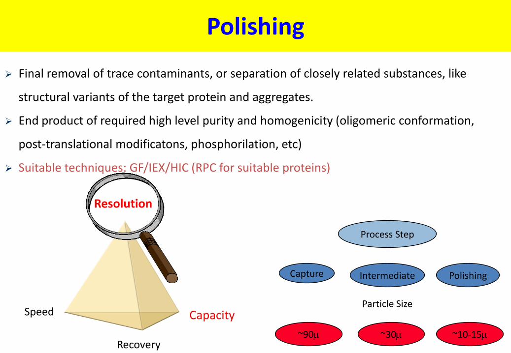
67
Resolution
Recovery
Final removal of trace contaminants, or separation of closely related substances, like
structural variants of the target protein and aggregates.
End product of required high level purity and homogenicity (oligomeric conformation,
post-translational modificatons, phosphorilation, etc)
Suitable techniques: GF/IEX/HIC (RPC for suitable proteins)
Process Step
Capture Intermediate Polishing
Particle Size
~30 ~10-15 ~90
Speed Capacity

Three Phase Strategy
68
Step
Capture
Polishing
Bead size of
chromatographic
matrix
Intermediate
purification

69
Step
Capture
Polishing
Flow rate
Intermediate
purification
Three Phase Strategy

70
Step
Capture
Polishing
RESOLUTION
Intermediate
purification
Three Phase Strategy