Prospective clinical study comparing intraligamentary anesthesia and inferior alveolar...
94
From the Department of Oral and Plastic Maxillofacial Surgery Director: Prof. Dr. med. habil. Dr. med. dent. B. Frerich Prospective clinical study comparing intraligamentary anesthesia and inferior alveolar nerve block for extraction of mandibular posterior teeth Inaugural Dissertation to Obtain the academic degree Doctor of Dentistry Faculty of Medicine University of Rostock Submitted by Ahmed Adubae / Born 09/03/1978 from Taiz/Yemen Rostock 2016 Dean: Prof. Dr. med. univ. Emil Christian Reisinger
Transcript of Prospective clinical study comparing intraligamentary anesthesia and inferior alveolar...
From the Department of Oral and Plastic Maxillofacial Surgery
Director: Prof. Dr. med. habil. Dr. med. dent. B. Frerich
Prospective clinical study comparing intraligamentary
anesthesia and inferior alveolar nerve block for extraction
of mandibular posterior teeth
Inaugural Dissertation
to
Obtain the academic degree
Doctor of Dentistry
Faculty of Medicine
University of Rostock
Submitted by
Ahmed Adubae / Born 09/03/1978
from Taiz/Yemen
Rostock 2016
Dean: Prof. Dr. med. univ. Emil Christian Reisinger
zef007
Schreibmaschinentext
urn:nbn:de:gbv:28-diss2017-0046-8
zef007
Schreibmaschinentext
zef007
Schreibmaschinentext
zef007
Schreibmaschinentext

Reviewer:
1. PD Dr. med. habil. Dr. med. Dent. Peer- Wolfgang Kämmerer, MA, Department of
Oral and Plastic Maxillofacial Surgery, University of Rostock
2. Prof. Dr. med. Dr. med. dent. Dr. med. h. c. Karsten Gundlach, Former director of
the Department of Oral and Plastic Maxillofacial Surgery, University of Rostock
3. Univ.-Prof. Dr. med. habil. Dr. med. dent. Monika Daubländer, Department of Oral
and Plastic Maxillofacial Surgery - University of Mainz
Date of submission: 26/September/2016
Date of defense: 21/March/2017

Contents/ List of tables/ List of figures
I
For the soul of my parents and for my family...
Contents/ List of tables/ List of figures
II
Table of Contents
Page
• List of tables VI
• List of figures VII
• Abbreviations
VII
1 Introduction 1
2 Review of the literature
3
2.1 Pain control in dentistry 3
2.1.1 Local anesthesia in dentistry 3
2.1.1.1 Superficial anesthesia
4
2.1.1.2 Infiltration anesthesia 5
2.1.1.3 Conduction anesthesia 5
2.1.1.4 Intraligamentary anesthesia
6
2.2 Inferior alveolar nerve block (IANB)
6
2.2.1 Techniques of IANB 7
2.2.1.1 Conventional direct technique 7
2.2.2 Anesthetic profile of IANB 8
2.2.3 Indications and contraindications of IANB 9
2.2.4 Advantages of IANB 9
2.2.5 Disadvantages of IANB 9
2.2.6 Complications of IANB 10
2.2.6.1 Failure of the anesthesia 10
2.2.6.2 Nerve injury
11
2.2.6.3 Injury to blood vessels and inadvertent intravascular injection
11
2.2.6.4 Muscle injury and trismus 12
2.2.6.5 Needle breakage 12
2.2.6.6 Self-inflicted soft tissue injury
12
2.2.6.7 Transient facial nerve paralysis 12
2.3 Intraligamentary anesthesia (ILA) 13
2.3.1 Instruments for ILA
13
2.3.1.1 Mechanical injection systems 13
2.3.1.1.1 Pistol type syringes
13
2.3.1.1.2 Penholder grip syringes 14
2.3.1.1.3 Dosing wheel systems 15
2.3.1.2 Electronically controlled injection systems 15
2.3.1.3 Needles 16
2.3.2 Technique of ILA
16
Contents/ List of tables/ List of figures
III
2.3.3 Mechanism of action of ILA 18
2.3.3.1 Vascular distribution 18
2.3.3.2 Medullary distribution 18
2.3.3.3 Combination of vascular and medullary distributions
19
2.3.4 Limitations of ILA 19
2.3.5 Anesthetic profile of ILA
20
2.3.6 Indications of ILA 20
2.3.7 Contraindications of ILA 21
2.3.8 Complications of ILA 22
2.3.8.1 Damage to periodontal tissues 22
2.3.8.2 Bone and root resorption 23
2.3.8.3 Soft tissue necrosis 23
2.3.8.4 Impaired wound healing (dry socket) 23
2.3.8.5 Bacteremia 24
2.3.8.6 Unwanted side effects 24
2.3.8.7 Hypoplasia of permanent teeth 25
3 Aims of the study 25
3.1 Problem and objectives 25
4 Materials and methods 27
4.1 Design of the study 27
4.2 Materials 27
4.2.1 Patients 27
4.2.1.1 Inclusion criteria 27
4.2.1.2 Exclusion criteria 28
4.2.2 Materials for IANB 28
4.2.3 Material for ILA 28
4.2.4 Data collection 28
4.3 Methods 29
4.3.1 Method of administrating IANB 29
4.3.2 Method of administrating ILA 30
4.3.3 Documentation of anesthetic effect 30
4.3.4 Statistical methods 32
5 Results 33
5.1 Evaluation group I 33
5.1.1 Patients and teeth 33
Contents/ List of tables/ List of figures
IV
5.1.2 General parameters 35
5.1.2.1 Age, height and body weight 35
5.1.3 Parameters of the primary objectives of this study 35
5.1.3.1 Pain of injection 35
5.1.3.2 Pain during tooth extraction 36
5.1.3.3 Unpleasantness of treatment 36
5.1.3.4 Quality of the anesthetic effect as assessed by patients 37
5.1.3.5 Need for second injection 39
5.1.4 Parameters of the secondary objectives of this study 40
5.1.4.1 Amount of local anesthetic solution used at the initial injection 40
5.1.4.2 Latency of anesthesia 40
5.1.4.3 Time between injection and start of extraction procedure 41
5.1.4.4 Time until second injection and amount of anesthetic solution at second injection 42
5.1.4.5 Duration of entire treatment 42
5.1.4.6 Quality of the anesthetic effect as assessed by the dentist 43
5.1.4.7 Duration of soft tissue anesthesia 44
5.1.4.8 Frequency of associated complications 45
5.2 Evaluation group II (patients with split-mouth technique) 45
5.2.1 Patients and teeth 45
5.2.2 Parameters of the primary objectives of this study 47
5.2.2.1 Pain of injection 47
5.2.2.2 Pain during tooth extraction 47
5.2.2.3 Unpleasantness of treatment 47
5.2.2.4 Quality of the anesthetic effect as assessed by patients 48
5.2.2.5 Need for second injection 49
5.2.3 Parameters of the secondary objectives of this study 50
5.2.3.1 Amount of local anesthetic solution used at initial injection and latency time of anesthesia 50
5.2.3.2 Time between injection and start of extraction procedure 51
5.2.3.3 Duration of entire treatment 51
5.2.3.4 Quality of the anesthetic effect assessed by the dentist 51
5.2.3.5 Duration of soft tissue anesthesia 52
5.2.3.6 Frequency of associated complications 53
6 Discussion 54
7 Conclusion 64
8 Summary 66

Contents/ List of tables/ List of figures
V
9 References 69
10 Declaration of original work 78
11 Theses 79
12 Curriculum Vitae 80
13 Acknowledgements
82
14 Appendix 83

Contents/ List of tables/ List of figures
VI
List of tables Table 1: Frequency of teeth extractions in group I 34
Table 2: Mean height and body weight related to anesthetic technique 35
Table 3: Pain of injection related to anesthetic technique 36 Table 4: Pain during tooth extraction related to anesthetic technique 36
Table 5: Unpleasantness of treatment related to anesthetic technique 37
Table 6a: Quality of anesthetic effect assessed by patients 38
Table 6b: Complete/sufficient versus insufficient/no anesthetic effect assessed by patients 38
Table 6c: Complete versus sufficient anesthetic effect assessed by patients 39
Table 7: Frequency of second injections necessary related to anesthetic technique 39
Table 8: Amount of the initially used local anesthetic solution related to anesthetic technique
40
Table 9: Latency time of anesthesia related to anesthetic technique 40
Table 10: Time between injection and start of extraction procedure 41
Table 11: Time until second injection and amount of local anesthetic for the second injections related to anesthetic technique
42
Table 12: Duration of treatment related to anesthetic technique 43
Table 13a: Quality of anesthetic effect assessed by the dentist 43
Table 13b: Complete/sufficient effect versus insufficient/no anesthetic effect assessed by the dentist
44
Table 13c: Complete versus sufficient anesthetic effect assessed by the dentist 44
Table 14: Duration of soft tissue anesthesia related to anesthetic technique 44
Table 15: Frequency of teeth extractions in group II 46
Table 16: Pain of the injection related to anesthetic technique 47
Table 17: Pain during tooth extraction related to anesthetic technique
47 Table 18: Unpleasantness of treatment related to anesthetic technique 48
Table 19a: Quality of anesthetic effect assessed by patients 49
Table 19b: Complete/sufficient effect versus insufficient/no effect assessed by patients 49
Table 20: Frequency of second injections necessary related to anesthetic technique 50
Table 21: Amount of initially used local anesthetic solution and the latency time of anesthesia 50
Table 22: Time between injection and start of extraction procedure 51
Table 23: Duration of treatment related to anesthetic technique 51
Table 24a: Quality of anesthetic effect assessed by the dentist 52
Table 24b: Complete/sufficient effect versus insufficient effect assessed by the dentist 52
Table 25: Duration of soft tissue anesthesia related anesthetic technique 52
Table 26: Frequency of occurrence of impaired wound healing (dry socket) 53

Contents/ List of tables/ List of figures
VII
List of figures Figure 1: Conventional direct technique for IANB 8
Figure 2: Ultraject® syringe (Sanofi-Aventis)
14
Figure 3: Intraligamentary injection 17
Figure 4: Mechanism of action of ILA 19
Figure 5: Bar charts showing number of extracted teeth per anesthetic technique 34
Figure 6: Box plots showing differences in pain of injection, pain during tooth extraction, and unpleasantness of treatment
37
Figure 7: Box plots showing time from injection of local anesthetic to onset of full anesthetic effect and time from injection to start of tooth extraction 41
Figure 8: Box plots comparing duration of soft tissue anesthesia in cases of ILA and IANB 45
Figure 9: Bar charts showing number of extracted teeth per anesthetic technique in group II 46
Figure 10: Box plots showing differences in pain of injection, pain during tooth extraction, and
unpleasantness of treatment in group II
48
Abbreviations C-CLAD Computer controlled local anesthetic delivery system
DPS Dynamic pressure sensor
IANB Inferior alveolar nerve block
ILA Intraligamentary anesthesia
LED Light-emitting diode
MPa MegaPascal
N Newton NSAD Non-steroid anti-inflammatory drugs
NRS Numeric rating scale
SD Standard Deviation
STA Single tooth anesthesia

Introduction
1
1. Introduction
The control of pain and discomfort has always been an important part of medicine
and dentistry and is an essential part of the successful practice in dentistry today.
Providing an efficient local anesthesia is of utmost importance in gaining patient´s
trust and cooperation in dentistry and to complete treatment successfully. Tooth
extraction is one of the most common dental procedures requiring local anesthesia.
In the upper jaw, teeth are usually extracted under local infiltration anesthesia as the
bony anatomy of the upper jaw enables the diffusion of the local anesthetic solution
in sufficient amounts into its working site. In adults, the bone anatomy in the posterior
part of the mandible allows, however, no adequate diffusion of the anesthetic
solution. In accordance, other anesthetic techniques are required for extraction of
mandibular posterior teeth. Until today, the inferior alveolar nerve block is the most
commonly used technique for providing local anesthesia in the posterior mandible
[Foster et al., 2007]. However, this technique is painful and has a relatively high
failure rate and technique immanent risks, such as transient or even persistent
damage of the lingual and/or the inferior alveolar nerve. Consequently, there is an
increasing demand for alternative local anesthetic techniques with higher success
rate and minimal risk of complications [Shabazfar et al., 2014].
Considering the legal demand of informing the patient about possible risks of an
indicated health measure and about available alternatives [Bluttner and Taubenheim,
2009], the question arises whether intraligamentary anesthesia as a primary
anesthetic technique can meet the requirement of a relatively complete and patient-
friendly local anesthetic technique and whether it can be considered as a valid
alternative to the conventional inferior alveolar nerve block for simple extraction of
mandibular posterior teeth.
So far, there are only few comparative studies of low evidence comparing
intraligamentary and inferior alveolar nerve block anesthesia in the extraction of
mandibular posterior teeth [Heizmann, 1987; Dumbrigue et al., 1997]. These studies
showed that intraligamentary anesthesia is at least not inferior to nerve block
anesthesia for this indication. Each of these two studies used a different injection
system for the administration of intraligamentary anesthesia.
The objective of the present study was to test the technique of intraligamentary
anesthesia – here using a pistol type syringe with pressure limitation (Ultraject®).
The question was whether this can be considered as a valid alternative to the inferior

Introduction
2
alveolar nerve block for extraction of mandibular posterior teeth. The primary
objectives were to evaluate the difference in pain perceived by the patients during
treatment as well as the anesthetic efficacy (complete/sufficient vs. insufficient/no
effect) based on the outcome of treatment and the degree of discomfort associated
with the extraction procedure. The pain of injection, need for second injection,
amount of anesthetic solution, and duration of the local numbness were also
assessed. A further objective of the study was to clarify whether impaired wound
healing (dry socket) was more frequent after intraligamentary anesthesia, as alluded
by some authors [Brännström et al., 1982; Meechan et al., 1987].
It was hypothesized that intraligamentary anesthesia applied using a pistol-type
syringe with a built-in pressure limiting mechanism enables a patient-friendly, pain-
and complication-less extraction of teeth in the posterior mandible. If so,
intraligamentary anesthesia could be a standard technique for this indication.

Review of the literature
3
2. Review of the literature
2.1. Pain control in dentistry
Dentistry and pain are synonymous for many patients. Therefore, many patients
avoid routine dental treatment [Thomason et al., 1999]. The patient´s perception of
dental treatment is closely related to the anesthetic experience he has had [Kohler et
al., 2008]. So, proper management of pain and attainment of adequate analgesia in
the operating field are of pivotal importance in dentistry.
In consideration of the patient´s general condition, nature and extent of the planned
therapy as well as the available facilities in the dental clinic, all of the possible pain
control methods should be considered and discussed with the patient. For effective
pain control one or more of the following methods should be kept in mind:
Administration of local anesthesia.
Psychological management of the patient and establishment of a good doctor-
patient relationship.
Medical sedation, especially in case of anxious patients.
Administration of general anesthesia for special indications.
Suggestion and possibly hypnosis.
Apart of the few indications for general anesthesia in dentistry such as in invasive
dentoalveolar surgical procedures and in treating non-cooperative patients, the
elimination of pain in dentistry has so far been conducted largely by conventional
methods of local anesthesia such as nerve block and/or infiltration anesthesia
[Csides, 2009].
2.1.1. Local anesthesia in dentistry
Local anesthesia is considered as the backbone of pain control in dentistry and is the
most widely practiced method of pain control in dentistry today. In the last decades,
however, great advances in the types of anesthetic agents, delivery devices, and
techniques have been achieved [Saxena et al., 2013]. With these advances, it
became possible to render surgical and extensive conservative dental treatment
measures bearable for the patients with minimal side effects and complications. In
dentistry, local anesthesia is mostly obtained through:

Review of the literature
4
Application of a local anesthetic agent on the mucous membrane or skin
(superficial anesthesia).
Injection of a local anesthetic solution directly into the surgical site (infiltration
anesthesia, terminal anesthesia).
Injection of a local anesthetic solution at the nerve trunk (conduction, nerve
block anesthesia).
Injection of a local anesthetic solution into the periodontal ligament space of
the tooth to be treated (intraligamentary anesthesia, periodontal ligament
injection).
Intraosseous injection of a local anesthetic solution using special injection
systems (intraosseous anesthesia).
Injection of a local anesthetic solution into the inter-dental septum (intra-septal
anesthesia).
Injection of a local anesthetic solution directly into the dental pulp (intra-pulpal
anesthesia).
Local anesthesia is considered as a safe method of pain control. Yet regardless of
how beneficial a health care procedure may be, there are always associated
disadvantages and risk of complications. Therefore, the patients should always be
informed about possible complications of the local anesthesia such as soft tissue
injury, necrosis in the injection area, nerve injury, prolonged bleeding, excessive
hematoma, needle breakage, and possible allergic reactions.
2.1.1.1. Superficial anesthesia
For superficial anesthesia, the local anesthetic, in a form of spray or gel, is applied to
the mucosa. It reaches the superficial nerve endings by local diffusion. The
anesthetic effect is limited to the mucosa and usually not sufficient to eliminate the
pain of dental treatment. It rather reduces the piercing pain of a subsequent injection.
However, superficial anesthesia is not always enough to complete elimination of this
pain of injection [Mayer, 1976; Kaufman et al., 2005]. By combining superficial
anesthesia with infiltration or conduction anesthesia, it is to be noticed that the
amount of the applied surface anesthetic must be added to the total maximum
administrable dose. Especially in children, this can easily result in exceeding the limit
of maximal dose and cause intoxication [Krüger, 1993].
Review of the literature
5
2.1.1.2. Infiltration anesthesia
For infiltration anesthesia, usually 0.5-1.5 ml of the local anesthetic solution is
injected in the immediate vicinity of the bone surface (submucosal or supraperiosteal)
most often only in the upper jaw or in the anterior part of the lower jaw. The injected
solution diffuses in the soft tissue and through the adjacent bone and reaches the
terminal nerve endings of the teeth. The anesthetic effect usually begins within 1-3
minutes after the injection and includes the region of the respective tooth, the
gingiva, and the vestibular soft tissues [Rahn, 2003]. Due to its success rate of up to
95% and its technically simple application, infiltration is the most commonly used
local anesthetic technique [Kämmerer et al., 2010]. In the posterior part of the adult
mandible, the local anesthetic solution cannot sufficiently diffuse through the thick
compact layer of bone, and hence, infiltration anesthesia is not adequate in this
region.
2.1.1.3. Conduction anesthesia
For conduction anesthesia, the local anesthetic solution is injected in the immediate
vicinity of the nerve trunk, usually at its bony exit or entry point (foramen). This results
in anesthesia of the entire area innervated by this nerve distal to the injection site.
Using only a single injection and a relatively small amount of local anesthetic
solution, a complete anesthesia of sufficiently long duration in a wide operation field
can usually be obtained with this technique [Malamed, 1997]. However, the duration
of anesthesia after a successful nerve block is significantly longer than that required
for most dental procedures, so that the patient remains limited in his disposition
ability often for hours after completion of treatment [Dirnbacher, 2003; Weber et al.,
2005; Prothmann et al., 2010]. Articulation and mastication are affected and the risk
of postoperative bite and burn injuries is increased [Chi et al., 2008]. Since the local
anesthetic has to be injected in the immediate vicinity of the nerve and because the
nerve itself cannot be exactly located during the injection, this technique of local
anesthesia is associated, on one hand, with a higher failure rate, and on the other
hand, with an increased risk of nerve injury. Nerve block anesthesia is primarily
indicated when no or only insufficient depth of anesthesia can be achieved with
infiltration anesthesia such as in the posterior part of the mandible and for extended
dental and dentoalveolar surgical procedures.

Review of the literature
6
More than 16 million nerve block anesthesias were administrated in Germany in 2013
[Bender and Taubenheim, 2014]. Commonly used nerve block anesthesia in dentistry
includes inferior alveolar, mental, and buccal nerve blocks in the lower jaw; and
superior posterior alveolar, superior anterior alveolar, nasopalatinal, and greater
palatinal nerve blocks in the upper jaw.
2.1.1.4. Intraligamentary anesthesia
The technique of intraligamentary anesthesia, i.e. the injection of the anesthetic
solution into the periodontal ligament space, was first described in the medical
literature about 100 years ago. At the beginning of the 20thcentury, in France,
attempts were made to inject the local anesthetic solution into the periodontal
ligament space to anesthetize the teeth to be extracted. The first intraligamentary
anesthesia was applied by Granjeon in 1903; this was described by Chompret and
first publicized by Bourdin in his doctoral thesis in 1925 [Bourdin, 1925].
Although intraligamentary anesthesia, already at that time, was considered to be an
interesting method of local anesthesia, it did not become established because of the
inadequacy of the injection instruments available at that time. The medico-technical
advances in the last few years have opened new possibilities for this method of local
anesthesia and enabled the dentist to apply it as a minimally invasive technique for a
variety of indications.
In the technique of intraligamentary anesthesia, the local anesthetic solution is
injected under controlled high pressure by means of special injection systems into the
periodontal ligament space of the tooth to be anesthetized. This results in a profound
anesthesia with an immediate onset of action and an anesthetic duration of
approximately 30-45 minutes using only a small amount of local anesthetic solution
(0.2 ml for each root); thereby, reducing the risk of systemic intoxication [Daubländer
and Kämmerer, 2014]. The anesthesia is limited to a single tooth and its supporting
structures while anesthesia of the lips, cheeks, and tongue is avoided.
2.2. Inferior alveolar nerve block (IANB)
IANB is the most frequently used form of conduction anesthesia when performing
restorative or surgical procedures in the posterior mandible. The local anesthetic
solution is injected in the proximity of the inferior alveolar nerve in the

Review of the literature
7
pterygomandibular space just before it enters the mandibular canal at the mandibular
foramen. It provides anesthesia of the ipsilateral mandibular teeth from the third
molar almost up to the midline and allows carrying out most dentoalveolar
procedures in the mandible.
2.2.1. Techniques of IANB
The IANB can be approached from intra- and extra-orally; in general the intraoral
approaches are most commonly used [Lipp, 1992]. Intra-orally, three techniques
have been proven to be effective (the conventional, the Gow-Gates, and the
Vazirani-Akinosi technique). In the present study only the conventional direct
technique of IANB was used because it has established itself as the dominant
technique in daily practice. Therefore, the discussion of the technique in the following
paragraphs refers only to the conventional direct technique of IANB.
2.2.1.1. Conventional direct technique
The conventional direct technique involves the insertion of the needle into the
pterygomandibular space by piercing the buccinator muscle and the deposition of the
local anesthetic solution at a level just superior to the mandibular foramen [Khoury et
al., 2011]. For the correct application, the height of the injection, the placement of the
needle, and the depth of penetration must be considered. The posterior ramus is
grasped, with the thumb placed intra-orally in the coronoid notch retracting the soft
tissue laterally. An imaginary line extends posteriorly from the fingertip in the
coronoid notch to the pterygomandibular raphe (as it turns up towards the maxilla)
and determines the height of injection. This imaginary line should be parallel to the
occlusal plane of the mandibular molars. In the majority of patients, this line will be 6-
10 mm above the occlusal plane. A long dental needle is directed from the corner of
the mouth of the contralateral side with the barrel of the syringe lying above the
mandibular premolars. The needle is inserted lateral to the pterygomandibular raphe
and advanced gently within the pterygomandibular space until bone is contacted at a
2-2.5 cm penetration depth (Figure 1). If bone is contacted too soon, the needle tip is
located too far laterally on the ramus; in case of no bone contact, the needle tip is
usually located too far medially [Malamed, 1997].

Review of the literature
8
Once the bone is contacted, the needle is withdrawn slightly to avoid subperiosteal
injection, aspiration is done in two planes and – if negative – an average of 1.5 ml of
anesthetic solution is injected slowly [Reed et al., 2012; Prama et al., 2013].
Afterwards, the needle is withdrawn for approximately 1 cm and the barrel of the
syringe is moved slightly toward the dental midline and a further 0.3-0.5 ml of local
anesthetic solution is injected to anesthetize the lingual nerve.
Figure 1: Conventional direct technique for IANB
2.2.2. Anesthetic profile of IANB
IANB provides anesthesia of the ipsilateral mandibular teeth from the third molar
almost up to the midline, the buccal soft tissue from the premolars anteriorly, the
body of the mandible, the periosteum, the periodontal ligaments, and the skin and
subcutaneous tissues of the chin and lower lip, all on the ipsilateral side [Reed et al.,
2012]. The lingual nerve and sometimes also the buccal nerve are frequently
anesthetized by the same injection technique resulting in the extension of the
anesthetized area to include the lingual gingiva, floor of the mouth, side of the tongue
and the buccal gingiva and soft tissues from third molar to the second premolar area
of the same side.

Review of the literature
9
The average latency time of the anesthesia is reported to be significantly more than 3
minutes and the duration of the soft tissue anesthesia is usually 2-4 hours [Shabazfar
et al., 2014], which is frequently significantly longer than the time required to carry out
most of the dental treatments. The simultaneous numbness of the tongue and the
lower lip is often unpleasant for many patients and the risk of a bite or burn injury of
the still anesthetized soft tissues increases [Chi et al., 2008].
2.2.3. Indications and contraindications of IANB
IANB is indicated primarily for invasive conservative, prosthetic, and surgical
treatment of multiple mandibular posterior teeth. Due to the risk of injury to the blood
vessels and hematoma formation, IANB is relatively contraindicated in patients with
bleeding diathesis or those with anticoagulant therapy. In these patients it is advised
to use another technique of local anesthesia [Stoll and Bührmann, 1983; Stoll et al.,
1986; Heizmann and Gabka, 1994].
2.2.4. Advantages of IANB
Profound anesthesia in a relatively wide area is obtained with only one
injection (useful for quadrant dentistry) [Malamed, 1997; Rahn, 2003].
Long duration of anesthesia necessary for some dentoalveolar surgical
procedures [Shabazfar et al., 2014].
2.2.5. Disadvantages of IANB
A wide area of anesthesia (not necessary for localized procedures e.g.
treatment of a single tooth) [Dirnbacher, 2002].
A concomitant anesthesia of the tongue and the lower lip is not comfortable for
many patients and could be dangerous in children and handicapped patients
[Chi et al., 2008].
The absence of reliable intraoral landmarks for the injection technique [Rahn,
2003].
A high failure rate of the anesthesia of 10% to 26% [Kaufman et al., 1984;
Heizmann, 1987; Rood, 1988].
A long latency time of anesthesia (3 to 5 minutes) [Malamed, 1997;
Dirnbacher, 2002; Shabazfar et al., 2014].

Review of the literature
10
A high risk of intravascular injection or injury of the blood vessels with a
positive aspiration (11%-30%, highest of all intraoral injection techniques)
[Weber, 1981; Lipp and Daubländer, 1998; Evers and Haegerstam, 2000].
A high risk of nerve Injury [Pogrel, 2007].
2.2.6. Complications of IANB
Even if the IANB is considered as a gold standard technique of local anesthesia in
the lower jaw, a significant number of cases with unwanted side effects and
complications related to this technique have been reported in the literature [Rood,
1988; Pogrel and Thamby, 2000; Pogrel, 2007; Choi et al., 2009; Gaffen and Hass,
2009; Chevalier et al., 2010; and others]. Here, some of these are listed:
2.2.6.1. Failure of the anesthesia
The reported failure rate of the IANB ranges between 10% and 26% [Kaufman et al.,
1984; Heizmann, 1987; Rood, 1988]. This relatively high rate of failure is, in
particular, due to the fact, that the mandibular foramen is neither clinically palpable
nor can be exactly localized [Rahn, 2003]. The needle is guided by variable
unreliable intraoral anatomical structures, which occasionally result in the application
of the anesthetic solution far away from the nerve trunk. In addition, the position of
the mandibular foramen changes with skeletal growth both in craniocaudal and
anteroposterior directions [Nicholson, 1985].
Technical errors such as inaccurate injection technique, lack of knowledge of the
anatomy, lack of experience and needle deviation are reported as the most frequent
causes of failure [Madan et al., 2002]. In addition to the technical errors, anatomical
variations, inflammation or infection in the site of injection, inactive anesthetic solution
and extremely anxious patients are other possible causes of failure of the IANB
[Boronat López and Peñarrocha Diago, 2006].
Concerning the anatomical variations, four of these were described, namely the
presence of a retromandibular foramen, an accessory mylohyoid nerve, a bifid
mandibular nerve, and a contralateral innervation of the anterior teeth [Desantis and
Liebow, 1996; Boronat López and Peñarrocha Diago, 2006]. The presence of a
retromolar foramen is reported with a prevalence of 7.7% [Sawyer and Kiely, 1991].
In 0.4% of cases, the inferior alveolar nerve presents two or even three branches

Review of the literature
11
passing through accessory foramina [Boronat López and Peñarrocha Diago, 2006].
Langlais et al. [1985] recorded in a series of 6000 panoramic X-ray, 57 bifid canals
(0.95%). These findings indicate that bifid canals are not so unusual.
2.2.6.2. Nerve injury
Prolonged and possibly permanent alterations in sensation due to nerve injury could
occur after dental injections. The nonsurgical cases of nerve injury in dentistry are
almost exclusively related to IANB injection and appear to affect the lingual nerve
60%-70% more frequently than the inferior alveolar nerve [Malamed, 1997; Pogrel
and Thamby, 2000; Gaffen and Hass, 2009]. A combination of injury of both nerves is
also possible [Pogrel and Thamby, 2000].
According to Pogrel [2007], permanent nerve involvement after IANB may occur in
about 1 of 20.000-850.000 patients. The lingual nerve is more prone to injury as it
lays only 3 to 5 mm away from the mucosa and the intraoral landmark for IANB (the
pterygomandibular raphe). When the mouth is opened, the lingual nerve is held taut
within the interpterygoid fascia, and because of its fixation, it is prone to be injured by
the needle [Harn and Durham, 1990; Hillerup and Jensen, 2006; Smith and Lung,
2006].
The exact biological mechanism of nerve injury remains the subject of debate in the
literature. The most common and most tenable theories are direct trauma to the
nerve with the needle, intraneuronal hematoma, and local anesthetic neurotoxicity
[Smith and Lung, 2006]. An intraneuronal injection of the local anesthetic is more
probable with a second injection since the piercing of the already anesthetized nerve
is not noticed by the patient.
2.2.6.3. Injury to blood vessels and inadvertent intravascular injection
By injection in the highly vascularized area, the risk of injury to the blood vessels
and/or intravascular injection is relatively high [Rood, 1972]. The rate of the
inadvertent vascular contact during IANB injection ranges between 11% and 20%
[Weber, 1981; Lipp and Daubländer, 1998; Taghavi Zenouz et al., 2008]. Evers and
Haegerstam [2000] claimed a positive aspiration rate of more than 30%.
An intravascular injection can result in severe systemic intoxication. In patients with
anticoagulation therapy and those with bleeding disorders, the mere infringement of a

Review of the literature
12
blood vessel could result in serious bleeding into the medial pterygoid muscle or the
pterygomandibular space and the surrounding soft tissue [Carter et al., 2003],
potentially even leading to a fatal result.
2.2.6.4. Muscle injury and trismus
Injury of the medial pterygoid muscle, particularly by repeated injections, could result
in the development of hematoma and trismus. The injured muscle becomes tender,
producing discomfort when opening the mouth. During a period of sleep, when the
muscles are not in use, the muscles go into spasm leaving the patient with
significantly reduced mouth opening in the morning [Malamed, 1997; Wright, 2011].
2.2.6.5. Needle breakage
Material errors, incorrect injection technique in a hurry and a sudden or unexpected
movement of the patient during the injection could result in breakage of the needle
[Daubländer and Kämmerer, 2014]. This complication became rare with the
significant improvement of material properties of the needles and the use of
disposable needles that are not to be resterilized. However, all reported cases of
needle breakages occurred mostly in cases of IANB [Ethunandan et al., 2007;
Pogrel, 2009].
2.2.6.6. Self-inflicted soft tissue injury
The duration of the anesthesia after a successful nerve block generally exceeds by
far the time required for a routine dental treatment. During this time, the
responsiveness of the patient is decreased. The area anesthetized is relatively wide
and this is associated with an increased risk of an accidental bite injury to the lip
and/or tongue, especially by children und handicapped patients [Davidson and Craig,
1987; Bendqude et al., 2001; Chi et al., 2008].
2.2.6.7. Transient facial nerve paralysis
Transient facial nerve palsy as a complication of the IANB is a rarely reported
incident. The most common cause of this complication is the deposition of the local

Review of the literature
13
anesthetic too far posteriorly in the body of the parotid gland i.e. close to the facial
nerve [Chevalier et al., 2010; Tzermpos et al., 2012].
2.3. Intraligamentary anesthesia (ILA)
2.3.1. Instruments for ILA
Even if ILA can principally be administrated by using the conventional aspiration
syringes [Walton and Abbott, 1981], special syringe systems for intraligamentary
injection have been developed and are preferable. Injection systems for ILA should
provide a mechanism for amplifying the injection pressure applied by the dentist in
order to overcome the encountered resistance when injecting the anesthetic solution
into the dense periodontal ligament space. This should also give the dentist the
opportunity to feel the individual anatomic variation and to adjust the applied injection
pressure accordingly.
2.3.1.1. Mechanical injection systems
In order to create a pressure required to overcome the interstitial resistance of the
periodontal ligament tissues for injection of the anesthetic solution, different syringe
systems have been developed by different manufacturers and are available on the
market since the late 1970s.
2.3.1.1.1. Pistol type syringes
Via an integrated lever mechanism, these syringe systems amplify mechanically the
injection pressure applied by the dentist. Examples for these syringe types are Peri-
press® (Resista, Omegna, Italy) and Ligmaject® (Henke-Sass Wolf, Tuttlingen,
Germany). However, with these syringe systems, the dentist cannot feel the
individual anatomical variations of the periodontium and adjust the injection pressure
accordingly. A mechanical amplification of the injection pressure without pressure
limiting mechanism allows the local anesthetic to be injected rapidly into the
periodontal ligament space – and hence with higher pressure than required. In
accordance, the recorded injection pressure applied by these systems varies from 10
to 344 Newton [Leilich et al., 1985].
Review of the literature
14
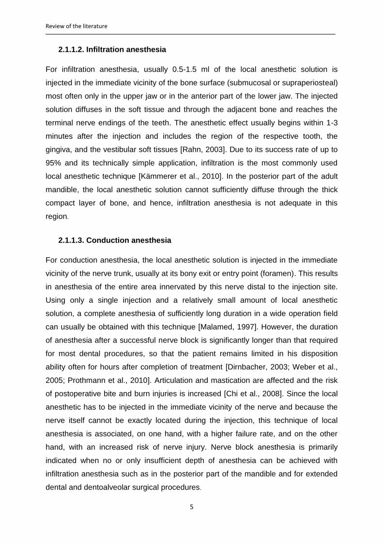
In order to get a better control on amplifying the injection pressure, a pistol type
syringe with an integrated pressure limiting mechanism was developed in the mid-
1980s [Rahn et al., 1987]. This syringe system has been brought onto the market by
Hoechst (Sanofi-Aventis, Frankfurt am Main, Germany) under the name of Ultraject®
(Figure 2). The Ultraject® syringe consists of a screw-able holder for the local
anesthetic cartridge with a plastic protection tube and a fixture for attachment of the
screw-able needle, a body of the syringe consisting of a toothed piston rod and a
pawl for locking the piston rod, a trigger lever and a handle with the mechanism of
pressure limitation. The automatic pressure limiting mechanism ensures that the
applied pressure does not exceed 120 N. When pulling the trigger lever too quickly,
the pressure transmission will stop automatically.
Figure 2: Ultraject® syringe (Sanofi-Aventis)
2.3.1.1.2. Penholder grip syringes
In 1984, Bayer and Ronvig (Daugaard, Denmark) introduced the penholder grip
syringe systems [Zugal, 2001]. In these syringe systems, a wing mounted laterally on
the handle of the syringe adopts the function of the trigger lever. This wing transfers
the applied force via an integrated multistage lever system to the piston rod and then
to the cartridge. Examples of these systems are Citoject® (Heraeus-Kulzer, Hanau,
Germany) and Paroject® (Ronvig, Daugaard, Denmark) syringes.
Instead of the usual 0.2 ml of local anesthetic released by each pull on the trigger
lever of the pistol type syringe systems, only 0.06 ml of anesthetic solution is
released by each stroke on the dosing lever (wing) of these syringe systems.

Review of the literature
15
Therefore, in order to apply the required amount of about 0.2 ml local anesthetic per
injection site, the wing must be squeezed three times per each injection site. The
duration of the injection and the amount of the injected local anesthetic can
consequently be better controlled and adjusted by the dentist.
2.3.1.1.3. Dosing wheel syringes
With this syringe system, the injection pressure is amplified by means of a dosing
wheel. The force exerted on the dosing wheel is amplified by a ratio of 5.5:1, due to
the different sizes of the wheels, and transmitted directly by turning the dosing wheel
from back to front on the toothed piston rod. With each rotational movement of the
dosing wheel, 0.1 ml of local anesthetic is released. To apply the desired 0.2 ml per
injection point, two rotational movements of the dosing wheel are necessary
[Glockmann and Taubenheim, 2010]. This syringe system enables to feel the
individual tissue resistance directly with thumb or index finger [Tobien and Schulz,
2000]. An example of this syringe system is the SoftJect® (Henke-Sass Wolf,
Tuttlingen, Germany).
2.3.1.2. Electronically controlled injection systems
In addition to the mechanical injection systems for the manual administration of ILA,
electronically controlled injection systems were developed in the last two decades.
The first computer-controlled local anesthetic delivery system (C-CLAD™) for
periodontal ligament injection “The Wand®” (Milestone Scientific Inc., New jersey,
USA) was introduced in 1997, and has been followed by other systems from different
manufacturers, such as CompuMed® (Utah Medical Products, Inc., Utah, USA),
CompuDent®, and STA®- System (Milestone Scientific Inc., New Jersey, USA) [Von
Haussen, 2011].
With the new technology of computer-controlled local anesthetic delivery systems,
the injection time was adjusted to the measured resistance of the periodontal tissues
at the site of the injection. The local anesthetic is administered into the periodontal
ligament space very slowly via a foot pedal or manually controlled by means of the
hand piece [Shepherd et al., 2001; Hochman et al., 2006, 2007]. With a similar
application mode, the various systems differ mainly in the number of selectable

Review of the literature
16
application programs, the control by hand or foot, as well as the display options
regarding the application process by LED or digital visualization.
In the newer devices such as the STA™ system with the STA-Wand® hand piece
(Milestone Scientific Inc., New Jersey, USA), in addition to extended speed modes,
an additional mode for computer-controlled information about the pressure prevailing
in the periodontal tissue during injection (dynamic pressure sensor technology (DPS))
is offered. The pressure is tested repeatedly in a second and can be read on the
display. The information provided by the DPS is to give the clinician indications of the
tissue properties, the interstitial pressure and thus liquid absorption capability of the
tissue [Hochman et al., 2007].
2.3.1.3. Needles
The selection of adequate needles for the periodontal ligament injection is of
paramount importance. The sulcus between tooth and alveolar bone is extremely
narrow; for this reason, the external diameter of the injection needle used should not
be larger than 0.3 mm (= 30 G). In order to keep the injection pressure as low as
possible, the inner diameter of the needle should be as large as possible in relation
to the outer diameter. The tip of the needle should have an extra-short bevel to
prevent it from bending while in contact with the tooth, which would increase the
resistance. Current recommendation is to use system-adapted injection needles, 0.3
mm (= 30G) with a length between 12 and 16 mm with the necessary stiffness to
prevent bending during insertion [Endo et al., 2008].
2.3.2. Technique of ILA
The local anesthetic solution is injected under controlled high pressure into the
periodontal ligament space between root and alveolar bone. At least one injection
point is generally to be defined for each root of the tooth to be anesthetized.
Principally it can be injected at any point along the gingival sulcus; however, the
mesiofacial and distofacial points are more favorable, as they are easily accessible
[Heizmann and Gabka, 1994]. If repeated injections are required, another point of
injection has to be selected; repeated injections at the same point could cause
tearing and detachment of the periodontal ligament fibers [Plagmann and Jagenow,
1984]. The needle is introduced through the gingival sulcus, at an angle of 30-40

Review of the literature
17
degrees to the long axis of the tooth, and is advanced 2-3 mm into the periodontal
ligament space between root and alveolar bone [Dreyer et al., 1983; Daubländer and
Kämmerer, 2014] (Figure 3). Further advance of the needle into the periodontal
ligament space beyond this depth would usually not be possible. The bevel of the
needle tip should be directed towards the alveolar bone and away from the root
surface. This needle position permits the anesthetic solution to spread into the bone
instead of squirting out of the ligamental space. To reduce the injection pain, a few
drops of local anesthetic solution can be deposited simultaneously with or right
before insertion of the needle. The handle of the syringe is then squeezed firmly until
backpressure is achieved. A resistance to the injection must always be present, if no
resistance is felt, the position of the needle might be incorrect; this has to be
controlled and changed. The injection pressure is sustained to deposit the local
anesthetic solution slowly into the dense periodontal ligament space.
According to the actual state of art, 0.2 ml of local anesthetic is to be injected for
each root over at least 20 seconds. At two rooted teeth, one injection is required for
each root, wherein the injection time for the second injection should be a little longer
(more than 20 seconds). At three-rooted teeth, three injections are required (one for
each root) and the injection time for the third injection should be obviously longer
(more than 25 seconds) [Endo et al., 2008; Bender and Taubenheim, 2014].
Figure 3: Intraligamentary injection

Review of the literature
18
The injection time has a significant influence on the pressure required to overcome
the tissue resistance during injection of a defined amount of local anesthetic.
Obviously, the injected volume is slowly absorbed by the tissue, so that the injection
resistance decreases gradually [Tobien and Schulz, 2000]. Due to the
incompressibility of liquids and the narrow of the available periodontal ligament space
into which the anesthetic solution is to be injected, a too rapid injection can lead to
deflection of the tooth in the alveolus and to the clinical symptoms of feeling of
elongation, pressure pain, and pre-contact [Huber and Wilhelm-Höft, 1988; Zugal,
2001].
2.3.3. Mechanism of action of ILA
Formerly, it was assumed, that the intraligamental injection forces the local
anesthetic solution primarily along the periodontal ligament space until it reaches the
apical area of the root producing local anesthesia of only one tooth. This assumption
has been disproved on the basis of animal experiments conducted by several
authors [Walton and Garnick, 1982; Garfunkel et al., 1983; Plagmann and Jagenow,
1984; Tagger et al., 1994; Kämmerer et al., 2012; and others]. Histological and
radiological studies show a little or no solution in the periodontal ligament space after
injection of ink containing local anesthetic solution and local anesthetic containing
radiologic detectable substances in the periodontal ligament space of the
experimental animals. Today, there are three theories about the working mechanism
of the ILA:
2.3.3.1. Vascular distribution
Under the influence of the relatively high injection pressure, the local anesthetic
solution injected into the periodontal ligament space is forced through the lamina
cribriformis into the alveolar bone and its blood vessels. It finally reaches the dental
pulp via the arterial blood supply and exerts its action on the nerve endings in the
dental pulp [Castagnola et al., 1982; Erlemeier, 1991; Müller and Henne, 1991].
2.3.3.2. Medullary distribution
The injected local anesthetic solution is primarily forced into the bone marrow space
of the alveolar bone through perforations formed by the blood vessels (nutrient
Review of the literature
19
canals); from this point, it spreads further through the bony canals and reaches the
periapical area of the root and exerts its action extra-pulpally [Smith and Pashley,
1982; Smith and Walton, 1983; Müller and Henne, 1991; Tagger et al., 1994].
2.3.3.3. Combination of vascular and medullary distribution
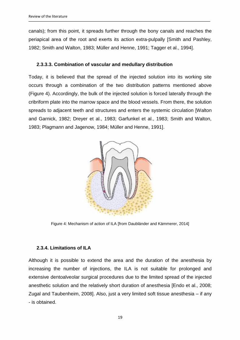
Today, it is believed that the spread of the injected solution into its working site
occurs through a combination of the two distribution patterns mentioned above
(Figure 4). Accordingly, the bulk of the injected solution is forced laterally through the
cribriform plate into the marrow space and the blood vessels. From there, the solution
spreads to adjacent teeth and structures and enters the systemic circulation [Walton
and Garnick, 1982; Dreyer et al., 1983; Garfunkel et al., 1983; Smith and Walton,
1983; Plagmann and Jagenow, 1984; Müller and Henne, 1991].
Figure 4: Mechanism of action of ILA [from Daubländer and Kämmerer, 2014]
2.3.4. Limitations of ILA
Although it is possible to extend the area and the duration of the anesthesia by
increasing the number of injections, the ILA is not suitable for prolonged and
extensive dentoalveolar surgical procedures due to the limited spread of the injected
anesthetic solution and the relatively short duration of anesthesia [Endo et al., 2008;
Zugal and Taubenheim, 2008]. Also, just a very limited soft tissue anesthesia – if any
- is obtained.

Review of the literature
20
2.3.5. Anesthetic profile of ILA
The local anesthetic effect is limited to the target tooth, its supporting tissues, and in
a limited extent to the neighboring mesial and distal teeth without concomitant
anesthesia of the tongue, lips, and checks. The amount of local anesthetic solution
required to produce the intended anesthesia is very small in comparison to the other
conventional techniques of local anesthesia. This makes it possible to undertake
dental treatment at many teeth in the same appointment without substantially
increasing the risk of overdosing and systemic intoxication [Heizmann and Gabka,
1994; Endo et al., 2008].
The anesthetic effect starts immediately after the injection and the maximum
anesthetic effect is reached within 30 seconds after the injection [Gray et al., 1987;
Plagmann, 1987; Zugal, 2001]. This very rapid onset of action enables the dentist to
evaluate the success of the anesthesia and to begin the treatment immediately. The
duration of the anesthesia after the periodontal ligament injection is approximately
30-45 minutes; this time is usually sufficient to perform the majority of dental
treatments. At the same time, the patient can resume his normal activities shortly
after completion of the treatment. The speech and chewing functions are minimally
affected and the risk of postoperative bite and burn injuries decreases drastically.
Due to injection into the periodontal ligament space, there is practically no risk of
damage to the blood vessels and/or the nerve; therefore, it is not mandatory to inform
the patient about these complications and to obtain an informed consent as it is in the
case of nerve block anesthesia [Kaltenbach et al., 2006].
2.3.6. Indications of ILA
Generally, ILA can be used as a primary or as a supplementary method for treating
the individual teeth in the upper and lower jaws. Indications of the ILA include:
Minimal invasive dental surgical procedures [Malamed, 1982; Faulkner, 1983;
Kaufman et al., 1983; Edwards and Head, 1989; Heizmann and Gabka, 1994;
Dumbrigue et al., 1997, Kämmerer et al., 2010].
All dental restorative therapeutic measures including removal of caries,
preparation for crown and bridge [Zugal, 2001; Kämmerer et al, 2015].
Endodontic therapy especially when subsequent injections are required while
using rubber dam [Weber et al., 2006; Glockmann et al., 2007].
Review of the literature
21
Completion of anesthesia in case of incomplete anesthesia or failure of other
methods of local anesthesia [Heizmann and Gabka, 1994].
Periodontal therapy (closed periodontal therapy measures) [Prothmann et al.,
2009].
ILA offers particular advantages in:
Differential diagnosis of unclear pulpal pain. Since with ILA every tooth can be
anesthetized individually, sensitivity can be tested tooth by tooth [Simon et al.,
1982; Littner et al., 1983; Garfunkel et al., 1985; Zugal et al., 2005].
Dental treatment of patients with cardiovascular diseases, as only a small
amount of local anesthetic solution is required [Garfunkel et al., 1985; Endo et
al., 2008].
Dental treatment of patients with hemorrhagic diathesis and those under
anticoagulant therapy [Stoll and Bührmann, 1983; Stoll et al., 1986].
Treatment of children and patients with mental disabilities [Anand et al., 2005];
the pain of injection is virtually very slight due of the fine needles used
[Einwag, 1982]. Additionally, the risk of postoperative bite trauma is reduced
due to the short duration of the anesthesia and the absence of the
concomitant anesthesia of lip and tongue [Davidson and Craig, 1987].
Treatment of patients with dental fear and anxiety. A minimal injection pain,
immediate onset of profound anesthesia with a limited regional extension
plays an important role in the reduction of fear and anxiety of the patient
[Adubae et al.,2016].
Using instruments and anesthetics that comply with the state of art and science, ILA -
in the hand of an experienced dentist - is a method of local anesthesia, which is
applicable for the above-mentioned indications [Bender and Taubenheim, 2014].
2.3.7. Contraindications of ILA
In addition to the conditions that make local anesthesia contraindicated in general,
ILA is relatively contraindicated in:
Patients with risk of endocarditis du to the risk of bacteremia [Rahn et al.,
1986].
Immunocompromised patients.

Review of the literature
22
Ankylosed teeth.
2.3.8. Complications of ILA
Certain complications and unwanted side effects in connection with the ILA had been
supposed by some authors [Phillips, 1943] and summarized by Giovannitti and Nique
[1983]. The supposed complications were then studied systematically in the following
years by many researchers. The results of some of these studies will be discussed in
the following paragraphs.
2.3.8.1. Damage to periodontal tissues
It was claimed that insertion of the needle through the junctional epithelium and
injection of the local anesthetic solution under high pressure into the periodontal
ligament space, causes inflammation and permanent damage to the periodontal
tissues [Phillips, 1943]. However, the results of the many histological studies
conducted by many investigators showed that only minimal histological effects in the
periodontium were induced by intraligamental injections; these changes regenerated
within a short time. The authors concluded that the ILA is a safe and reliable method
with only minimal transient and reversible inflammatory changes in the periodontal
tissue [Walton and Garnick, 1982; Fuhs et al., 1983; Galili et al., 1984; Anneroth et
al., 1985; Müller and Henne, 1991; Tagger et al., 1994; and others].
Only repeated injections at the same site can cause tear and detachment of the
periodontal ligament fibers from the bone [Plagmann and Jagenow, 1984]. For this
reason, it is advised that the needle is inserted at another point when a second
injection is required.
The effects of the intraligamental injection on the pulpal tissue were studied by Lin et
al. [1985]; neither pathological changes such as degeneration and/or ischemic
necrosis, nor inflammation in the pulps of the examined teeth were observed in that
study. The harmlessness of intraligamentary injection on pulpal tissues was
confirmed by studies conducted by many other investigators [Gray et al., 1987;
Grund et al., 1992; Torabinejad et al., 1993].
Review of the literature
23
2.3.8.2. Bone and root resorption
Reversible bone and root resorptions, confined to the area of the crestal bone, with a
complete healing within 25 days after intraligamental injection were reported in
animal models [Walton and Garnick, 1982]. Pertot and Déjou studied the histological
effect of the mere needle insertion and of the injection of a saline solution on bone
and root resorption at 105 sites in the periodontium of five dogs, with observation
periods of 7, 25 and 45 days. The injections were made with a specially designed
syringe equipped with a miniaturized force transducer. They observed that, at 7 days,
both needle penetration and the injection of saline solution resulted in increased
osteoclastic, odontoclastic activity and in bone and root resorption. The extent of
bone resorption was greater after injection of saline solution than that after the mere
needle penetration without injection. At 25 and 45 days, healing had occurred in all
cases. These findings suggest that the injection pressure is sufficient to initiate
osteoclastic but reversible bone resorption [Pertot and Déjou, 1992].
2.3.8.3. Soft tissue necrosis
In a clinical study conducted by Kaufman et al. [1983], only one case of mucosal
inflammation with marginal necrosis was observed after administration of ILA to 258
teeth in a total of 187 patients. Five other patients reported a post-operative pain that
lasted for 2 days after treatment. In another clinical study conducted by Zugal et al.
[2005], no case of necrosis or any other tissue damage was observed in 205
documented cases of ILA. Glockmann et al. [1997] could not find a significant
increase in the probing depth, within 3 months after periodontal ligament injection.
2.3.8.4. Impaired wound healing (dry socket)
Tsirlis et al. studied the frequency of occurrence of dry socket after extraction of
mandibular molars under ILA and IANB. In that study, 305 extractions of mandibular
molars in two groups of patients were documented. The first group of patients
received IANB, whereas the second group received ILA as local anesthetic
technique. Eleven cases of dry socket were observed, five of which were in the first
group and six in the second group. The authors concluded that the use of ILA did not
result in a higher incidence of dry socket than did IANB [Tsirlis et al., 1992]. In
another study, Heizmann and Gabka compared ILA, IANB and infiltration anesthesia

Review of the literature
24
in the extraction of mandibular and maxillary teeth. The results of that study showed
no significant differences in the frequency of occurrence of dry socket after the three
local anesthetic methods [Heizmann and Gabka, 1994].
2.3.8.5. Bacteremia
Numerous dental therapeutic measures, including the periodontal ligament injection,
can trigger a bacteremia. A sepsis due to promoting bacteria through the injection
needle into the tissue and into the blood stream can possibly occur. Walton and
Abbott [1981] clarified that this is probably the case in intraligamental injections, but
in no greater extent than with other dental treatments. The resulted bacteremia is
usually transient. In 1986, Rahn et al. published the results of a study on the
incidence of bacteremia after ILA. They found that the incidence of bacteremia is
significantly higher after ILA when a high injection pressure is used. The risk of
endocarditis represents, according to Rahn et al. a clear restriction to the use of ILA.
Though, studies proving the clinical relevance are lacking.
2.3.8.6. Unwanted side effects
Unwanted clinical side effects such as postoperative discomfort, pressure pain, a
sensation of tooth elongation and pre-contact after abatement of the ILA have been
described in various publications [Malamed, 1982; Faulkner, 1983; Kaufman et al.,
1983; Plagmann, 1987]. The causes of these unwanted effects often lay in the fact,
that the injection of the anesthetic solution did not take place under adequate
consideration of the individual anatomical conditions of the patient.
In their study, Huber and Wilhelm-Höft showed that the tooth could move in its socket
under the influence of the high injection pressure. During intraligamentary injection, a
volume of liquid is pumped under pressure into a chamber, which is already filled
completely. Since liquids are incompressible, an extension of the alveolus or a shift of
periodontal fluid cushion under the influence of the high injection pressure comes
primarily into consideration [Huber and Wilhelm-Höft, 1988]. In order to avoid these
unwanted effects, the anesthetic solution should be injected slowly, giving time for
the anesthetic solution to be absorbed by the tissue [Zugal et al., 2005]. By
increasing the injection time, the injection pressure required to overcome the tissue
resistance decreases gradually.
Review of the literature
25
2.3.8.7. Hypoplasia of permanent teeth
The pressure used during intraligamental injection to primary teeth can force the
anesthetic solution into the underlying tooth germs of the permanent teeth
[Brännström et al., 1982]. In an experimental animal study, Brännström et al. [1984]
noticed cases of enamel hypoplasia and/or hypomineralization in a total of 15
permanent teeth after application of ILA to 16 primary teeth of two monkeys using the
Peripress® injection syringe. Based on this result, Brännström suggested that great
care should be taken, when using ILA on primary teeth close to developing
permanent teeth. However, the diffusion of the injected solution into germs of
permanent teeth could not be observed after periodontal ligament injection of ink
containing local anesthetic solutions to six primary teeth of two young sheep
[Kämmerer et al., 2012], and at 58 sites to primary teeth in five dogs [Tagger et al.,
1994]. To our knowledge, such effects on permanent teeth have never been reported
in humans yet.
3. Aims of the study
3.1. Problem and objectives
Considering the relatively high failure rate and the risk immanent in the technique of
IANB, as well as the legal demand of informing the patient about possible risks of an
indicated health measure and about available alternatives [Bluttner and Taubenheim,
2009], the question arises whether the ILA as a primary anesthetic technique can
meet the requirement of a complete and patient-friendly local anesthetic technique
and whether it can be considered as a valid alternative to the conventional IANB for
extraction of mandibular posterior teeth.
In this study it was to be tested whether the technique of ILA administered with pistol
type syringes with pressure limiting mechanism can be considered as a valid
alternative to the IANB for extraction of mandibular posterior teeth. The primary
objectives were particularly to evaluate the difference in pain experienced by the
patients during treatment as well as the efficacy of the local anesthesia
(complete/sufficient vs. insufficient/no effect) and the need for second injection based
on the outcome of treatment and the degree of discomfort associated with the
extraction procedure. Of secondary interest were the differences in the amount of

Review of the literature
26
anesthetic solution, latency time of the anesthesia, duration of treatment and duration
of local numbness. A further objective was to clarify whether impaired wound healing
after tooth extraction (dry socket) was more frequent after ILA than following IANB.
The precise, very complex influence between systemic diseases of the patients and
the wound healing processes or the frequency of wound healing disturbance were
not to be examined here, but it was rather to ascertain whether more wound healing
disturbances occur after ILA.
Matrials and methods
27
4. Materials and methods
4.1. Design of the study
This present study was a prospective, randomized comparative clinical trial
conducted in the Department for Oral and Plastic Maxillofacial Surgery of the
University Medical Centre Rostock, Germany after approval of the local ethics
committee of the Faculty of Medicine- University of Rostock (No A 2014-0129) in a
period of 12 months from April 2014 to April 2015. Two different techniques of dental
local anesthesia (ILA and IANB) were compared with the help of several parameters.
4.2. Materials
4.2.1. Patients
Adult patients of both sexes with one or more mandibular posterior teeth indicated for
extraction were selected for this study. These patients were referred to the
Department for Oral and Plastic Maxillofacial Surgery of the University Medical
Centre Rostock for dental extractions. The patients were assigned randomly into
those who received ILA and those who received IANB as local anesthetic technique
for the indicated dental extraction. In the cases of bilateral dental extraction, ILA was
administered first on one side and tooth was extracted on this side. After completing
the treatment and documentation on this side, IANB was then administered on the
other side and another tooth was extracted. Although this could potentially affect the
patients´ perception of pain during subsequent procedures, it was reasoned that this
would be the more logical approach since the claimed onset of ILA is immediate.
4.2.1.1. Inclusion criteria
Included in the study were patients of both sexes at least 18 years old and with
clinical indication for dental local anesthesia because of indicated extraction of one or
more mandibular posterior teeth. Only teeth requiring simple extractions were
included in the study.
When more than one tooth on one side of the mandible was to be extracted under
ILA, each tooth was considered as independent sample, as each tooth requires its
own ILA anesthesia. When, however, many teeth on one side to be extracted under
IANB, only one tooth that best fulfils the inclusion criteria was considered. After

Matrials and methods
28
injection and extraction of this tooth was accomplished and documented, the other
tooth/teeth was/were then extracted but not included in the study.
4.2.1.2. Exclusion criteria
Exclusion criteria were the following: patients under 18 years of age, incapacitated
patients, patients with contra-indications for any of the components of the anesthetic
solution (allergy to articaine, epinephrine, and sulfite), patients with American Society
of Anesthesiologist classification > 2, chronic or simultaneous taking of psychotropic
or anti-inflammatory (NSAD) drugs in temporal context with the dental treatment, lack
of compliance, pregnancy, as well as infection in the area of injection. Teeth with
acute apical infections or drainage of pus from the gingival sulcus or surrounding
tissues, and teeth with more than 0.5 mm mobility in any direction were not included
in the study.
4.2.2. Materials for IANB
Inferior alveolar nerve blocks were administered using disposable syringes (BD
Discardit II™, 5 ml; Becton, Dickinson and Company, New Jersey, USA) and 25
gauge/42 mm needles (Sterican®; B. Braun Melsungen AG, Melsungen, Germany).
The local anesthetic agent used for IANB was Ultracain D-S 2 ml ampoules (articaine
40 mg/ml plus suprarenin 0.006 mg/ml; Sanofi-Aventis GmbH, Frankfurt am Main,
Germany).
4.2.3. Materials for ILA
For the administration of ILA, pistol-type syringes with built-in safety mechanism for
limiting the mechanical pressure (Ultraject®; Sanofi-Aventis GmbH, Frankfurt am
Main, Germany) and 30 gauge short bevel/16 mm needles (Heraeus Kulzer GmbH,
Hanau, Germany) were used. The local anesthetic agent used for ILA was Ultracain
D-S 1.7 ml cartridges (articaine 40 mg/ml plus suprarenin 0.006 mg/ml; Sanofi-
Aventis GmbH, Frankfurt am Main, Germany).
4.2.4. Data collection
Prior to the actual treatment, the purposes, nature of the study as well as the
intended procedure and its possible complications were explained to the patients and

Matrials and methods
29
a written consent was obtained from each patient. A copy of the study information
and the consent form was given to each patient.
For data collection, an evaluation sheet was created, which was divided into
questions to be answered both by the dentist and the patients. The evaluation sheet
was divided into four parts. The first part was to be filled out in the first appointment
and involved the patient´s personal data such as name, birth date, sex, weight,
height and telephone number as well as the name of the dentist and the date of
treatment. The medical and dental histories, as well as the history of drug ingestion
were collected and noticed in this part. It was particularly asked if the patient took
analgesic or sedative drugs and whether allergic reaction to local anesthetics or to
the stabilizers contained in the solution was occurred in the past. At the second
appointment and before the beginning of the actual treatment, the data collected at
the first appointment were always controlled and updated. At this appointment, each
patient was asked, whether he/she had questions in relation to the study and the
agreement of the patients to participate in the study was reassured.
The second part of the evaluation sheet was to be filled out by the dentist and
involved the statement of the tooth to be extracted, local anesthetic technique used,
time of injection, and the amount of injected solution. The onset time of anesthesia,
quality of the anesthesia obtained, need for second injection, observed signs of
contact with the nerve or the blood vessels, as well as the exact time of the start of
extraction procedure were also noticed by the dentist.
In the third part were questions to be answered by the patients and dealt with the
quality of the anesthesia obtained, need for second injection and the intensity of the
pain perceived during injection and extraction procedure. The fourth part was to be
filled out one day after tooth extraction (some time via telephone) and involved the
duration of anesthesia. The other points of this part were to be filled out at the third
appointment 3-5 days later and involved signs of possible complication such as dry
socket. This evaluation sheet is shown in the appendix (Page: 83).
4.3. Methods
4.3.1. Method of administering IANB
The technique used for IANB was the intraoral conventional direct method described
previously in paragraph 2.2.1.1 page no. 7-8.

Matrials and methods
30
4.3.2. Method of administering ILA
First, the local anesthetic cartridge with the protective tube was inserted into the
cartridge holder. The pawl was depressed and the piston rod was withdrawn from the
syringe body until it stopped. Then, the cartridge holder was screwed onto the
syringe. Next, a thin needle was placed onto the cartridge holder and screwed tightly.
The trigger lever of the syringe was pressed slightly until few drops of anesthetic
solution appeared at the needle tip. The syringe was now ready for injection.
The injections were carried out using the injection technique described previously in
paragraph 2.3.2 page no. 16-18.
4.3.3. Documentation of anesthetic effect
The exact time of injection, the technique of local anesthesia used and the injected
amount of local anesthetic solution were noticed by the dentist in the evaluation
sheet. Immediately after injection, each patient had to determine how painful the
injection was using 11- points segmented numeric rating scale (NRS). Each patient
had to select a number from a horizontal bar (0-10; 0 = no pain, 10 = extreme pain)
that best reflected the intensity of his pain. In order not to influence the perception of
the injection pain by the patient, no topical anesthesia was used.
Signs of complications during and immediately after the injection and the incidence of
a positive aspiration were also noticed in the evaluation sheet. The appearance of
blood in the syringe or the appearance of reddish discoloration of the anesthetic
solution after aspiration was categorized as a positive aspiration. When positive, the
anesthetic solution was discarded and the injection was repeated with a new
anesthetic solution and a new needle. As criteria of direct nerve contact, subjective
experience of a sudden strike of pain by the patient during the advance of the needle
in the soft tissue and/or the experience of prolonged hypoesthesia or dysesthesia
after treatment was defined.
Numbness was tested with a dental probe on the gingiva immediately after the
injection and further each 10 seconds in case of ILA and each 30 seconds in case of
IANB till full numbness was declared and the time of onset of the anesthetic effect
was noticed.
The extraction procedures started with elevators and forceps. The time of beginning
the treatment was also noticed in the evaluation sheet. The subjective quality of the
Matrials and methods
31
anesthesia was documented both by the dentist and the patient in separated fields
using a Likert- scale (complete, sufficient, insufficient and no effect). The anesthesia
was assessed as complete when it was possible to remove the tooth without pain
and discomfort. The ability to remove the tooth with mild but tolerable pain and
discomfort was assessed as sufficient anesthesia. Anesthesia was assessed as
insufficient when anesthetic effect was reported by the patient subjectively but the
tooth could not be extracted successfully with tolerable pain and discomfort. A severe
pain during the extraction with an absence of subjective anesthetic effect was
assessed as no anesthetic effect. Cases of insufficient and no anesthetic effect after
the first injection were considered as primary anesthetic failure. The patient had to
decide on the basis of the remaining pain, whether a second injection was required.
The need for a second injection was documented on the evaluation sheet.
When needed, the second injection was done using the same anesthetic technique
as for the first injection. The time of the second injection and the injected amount of
local anesthetic solution were also documented by the dentist. The subjective quality
of anesthesia (complete, sufficient, insufficient and no effect) was documented again.
If the anesthesia, after the second injection was still incomplete and the completion of
the treatment without tolerable pain was not possible, a combination of the both
anesthesia techniques was undertaken. These cases were considered as cases of
secondary anesthesia failure.
After complete removal of the tooth, the socket was cleaned carefully, the sharp bony
edges were removed and the time of the end of extraction procedure was
documented. The treatment was finished with a placement of a piece of sterile gauze
on the extraction wound. Patients were asked to remain seated for several minutes
after completion of treatment and to evaluate the overall pain and unpleasantness of
the entire treatment again using the 11- points segmented numeric rating scale
(NRS). Postoperative instructions were given to the patient orally and in written form.
Prescription of postoperative antibiotics was done in only few cases with a height risk
of wound healing disturbances.
One day later, the patients were asked (sometimes via telephone) about the duration
of soft tissue anesthesia and possible adverse effects.
The wounds were examined for signs of retarded healing (dry socket) at a second
appointment within one week after tooth extraction. The criteria for the diagnosis of a
dry socket were: empty alveolus, denuded bone surface being very sensitive to

Matrials and methods
32
probing, extreme pain that lasted more than 3 days after extraction and unpleasant
taste and/or odor.
4.3.4. Statistical methods
For descriptive analysis of categorical data, absolute and relative frequencies were
calculated. For continuous data, minimum, maximum, median and mean values,
skewedness, quartiles and standard deviations were calculated. Categorical data
were visualized via bar charts, consistent data via boxplots. For further explorative
data analysis, Kolmogorov-Smirnov test was employed to test for a difference among
ILA and IANB. In cases of p-values <0.05, Mann-Whitney-U test and in cases of p-
values >0.05, Students’ t-test for independent samples were employed. The influence
of categorical variables was shown with chi-square tests and cross tables. A global
significance level was chosen to 0.05. All analyses were carried out using SPSS
Statistics version 22 (IBM, Armonk, NY, USA).

Results
33
5. Results
Two hundred sixty-six adult patients of both sexes (176 males, 90 females) with one
or more mandibular posterior teeth (total number of teeth= 301) indicated for
extraction were included in this study. 98 patients had co-morbidities (cardiovascular
diseases n= 42; diabetes mellitus n= 16; malignant tumors n= 26; osteoporosis n= 7;
dysfunction of thyroid gland n= 7). Beta-blockers were taken by 38 patients,
anticoagulants by 14 patients, bisphosphonates by five patients, insulin by nine
patients and seven took medications for dysfunction of the thyroid gland. Three of
266 patients reported acute dental pain before the treatment.
For data evaluation, patients were categorized into two evaluation groups (evaluation
group I and II) based on whether only one or both anesthetic techniques were used
in individual patients (unilateral or bilateral tooth extraction). Group I (patients n= 238,
teeth n= 245) involved the patients who received randomly either ILA or IANB for
indicated unilateral dental extraction while group II (patients n= 28, teeth n= 56)
involved the patients who received both ILA and IANB because of indicated bilateral
dental extraction (split-mouth).
ILA was compared with IANB in each evaluation group separately and the results in
group I were then compared with that in group II (split-mouth).
5.1. Evaluation group I
5.1.1. Patients and teeth
A total of 238 patients (153 males, 85 females) in the age group of 19-87 years
(mean: 54.3; standard deviation (SD): 16.78) with height between 150-195 cm
(mean: 173.4; SD: 9.23) and body weight between 45-156 kg (mean: 83.5; SD: 17.7)
were included in this group. In these patients, a total of 245 teeth (Table 1) were
extracted by one and the same dentist using the same local anesthetic agent
(articaine with epinephrine 1:200.000) and the two different local anesthetic
techniques. 105 teeth (in 98 patients) were extracted under ILA, whereas 140 (in 140
patients) were extracted under IANB following randomization (Figure 5).

Results
34
Tooth number Number of teeth extracted Percent
34 15 6.12
35 21 8.57
36 31 12.65
37 52 21.23
38 4 1.63
44 13 5.31
45 21 8.57
46 39 15.92
47 46 18.78
48 3 1.22
Total 245 100
Table 1: Frequency of tooth extractions in group I
Figure 5: Bar charts showing number of extracted teeth per anesthetic technique

Results
35
5.1.2. General parameters
5.1.2.1. Age, height and body weight
The mean age of the patients who underwent tooth extraction under ILA was 56
years (SD: 15.6), whereas the mean age of the patients in whom IANB was used was
52.6 years (SD: 17.95). Comparing the means of age, no statistically significant
difference could be found (p= 0.219). The mean height of the patients was 172.58
cm (SD: 9.11) in case of ILA and 174.26 cm (SD: 9.34) in case of IANB. Concerning
the mean body weight of the patients, it was 82.35 kg (SD: 16.37) and 84.73 kg (SD:
19.09) respectively (Table 2). When comparing the means of height (p= 0.259) and of
body weight (p= 0.406) there was no significant statistical difference. Similarly, no
statistically significant differences were found between ILA and IANB in relation to the
existence of preoperative pain (p= 1), and the reported general diseases (p= 0.285).
Anesthesia Patient
(n)
Mean Standard
deviation
Standard error of the
mean value
Height
(cm)
ILA
IANB
ILA
IANB
98
140
98
140
172.58
174.26
82.35
84.73
9.11
9.34
16.37
19.09
1.07
1.01
1.93
2.06 Body
weight
(kg)
Table 2: Mean height and body weight related to anesthetic technique
In accordance, the general parameters of patients treated with ILA and those treated
with IANB were relatively homogenous and there were no significant statistical
differences between ILA and IANB.
5.1.3. Parameters of the primary objectives of this study
5.1.3.1. Pain of injection
The pain of injection was assessed by the patients with a mean of 2.19 points on the
numeric rating scale (NRS) in case of ILA and a mean of 3.65 points in case of IANB

Results
36
(Table 3; Figure 6). Comparison of the pain of the two anesthetic techniques showed,
that the injections were significantly less painful in case of ILA (p< 0.001).
Anesthesia Injection
(n)
Mean Standard
deviation
Standard error of
the mean value
Pain of injection
(NRS)
ILA 105 2.19 1.79 0.174
IANB 140 3.65 1.90 0.161
Table 3: Pain of injection related to anesthetic technique
5.1.3.2. Pain during tooth extraction
Immediately after tooth extraction, the pain experienced during the extraction
procedure was assessed by the patients with a mean of 1.95 points in case of ILA
and a mean of 1.66 points in case of IANB (Table 4 and Figure 6). In accordance, no
significant statistical difference was seen (p= 0.211).
Anesthesia Teeth
(n)
Mean Standard
deviation
Standard error of
the mean value
Pain during tooth
extraction (NRS)
ILA 105 1.95 1.71 0.167
IANB 140 1.66 1.90 0.161
Table 4: Pain during tooth extraction related to anesthetic technique
5.1.3.3. Unpleasantness of treatment
An assessment with a mean of 2.30 points was given by the patients for tooth
extraction under ILA in contrast to a mean of 2.54 points for extraction under IANB
(Table 5 and Figure 6). Again, no significant statistical difference was seen (p= 0.31).
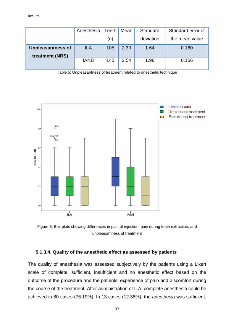
Results
37
Anesthesia Teeth
(n)
Mean Standard
deviation
Standard error of
the mean value
Unpleasantness of
treatment (NRS)
ILA 105 2.30 1.64 0.160
IANB 140 2.54 1.96 0.166
Table 5: Unpleasantness of treatment related to anesthetic technique
Figure 6: Box plots showing differences in pain of injection, pain during tooth extraction, and
unpleasantness of treatment
5.1.3.4. Quality of the anesthetic effect as assessed by patients
The quality of anesthesia was assessed subjectively by the patients using a Likert
scale of complete, sufficient, insufficient and no anesthetic effect based on the
outcome of the procedure and the patients’ experience of pain and discomfort during
the course of the treatment. After administration of ILA, complete anesthesia could be
achieved in 80 cases (76.19%). In 13 cases (12.38%), the anesthesia was sufficient.

Results
38
In 11 cases (10.48%), the anesthesia was insufficient and in one case (0.95%), there
was no anesthetic effect (Table: 6a).
After IANB, complete anesthesia could be achieved in 109 cases (77.86%). In six
cases (4.29%) the anesthesia was assessed as sufficient. In 23 cases (16.42%),
anesthesia was insufficient and in two cases (1.43%) there was no anesthetic effect
(Table 6a).
Table 6a: Quality of anesthetic effect assessed by patients
Inability to extract the tooth after the initial injection was considered as primary (initial)
anesthesia failure; accordingly, the initial failure rate of anesthesia was 11.43% in
case of ILA and 17.85% in case of IANB. When comparing the complete/sufficient
effect versus the insufficient/no anesthetic effect of the two techniques of local
anesthesia (the proportion of teeth extracted successfully versus the proportion of
teeth which could not be successfully extracted after the initial injection), no
significant statistical difference could be seen (p= 0.082; Table 6b).
Anesthesia
ILA IANB
(n) (%) (n) (%)
Quality of the
anesthetic effect
Total number of teeth
Complete/sufficient
Insufficient/no effect
93
12
105
88.57
11.43
100
115
25
140
82.14
17.86
100
Table 6b: Complete/sufficient versus insufficient/no anesthetic effect assessed by patients
Anesthesia
ILA IANB
(n) (%) (n) (%)
Quality of the anesthetic effect
Total number of teeth
Complete
Sufficient
Insufficient
No effect
80
13
11
1
105
76.19
12.38
10.48
0.95
100
109
6
23
2
140
77.86
4.29
16.42
1.43
100

Results
39
Comparing the “complete” versus “sufficient”-ratings assessed by the patients, a
statistically significant difference was found in favor for ILA (p = 0.03; Table: 6c).
Anesthesia
ILA IANB
(n) (%) (n) (%)
Quality of the
anesthetic effect
Total number of teeth
Complete
Sufficient
80
13
93
76.19
12.38
88.57
109
6
115
77.86
4.28
82.14
Table 6c: Complete versus sufficient anesthetic effect assessed by patients
5.1.3.5. Need for second injection
Second injection of local anesthetic was required in 12 cases (11.43%) of ILA and in
25 cases (17.86%) of IANB (Table 7). Statistically, no significant difference could be
found (p= 0.197).
Anesthesia
ILA IANB
(n) (%) (n) (%)
Second injection No 93 88.57 115 82.14
Yes 12 11.43 25 17.86
Total number of teeth 105 100 140 100
Table 7: Frequency of second injections necessary related to anesthetic technique
The anesthetic technique used for the second injection was always the same
technique as for the initial injection. After second injections, complete anesthesia
could be attained in 11 cases out of the 12 cases of ILA and in 23 cases of the 25
cases of IANB. In one case of ILA (0.95%) and in two cases of IANB (1.42%), the
extraction procedure could not be completed even after second injections. These
cases were in the same patients who assessed the anesthesia as “no effect” after
initial injection and were categorized as secondary anesthesia failure. In these three
cases, a combination of the both anesthetic techniques was used to successfully
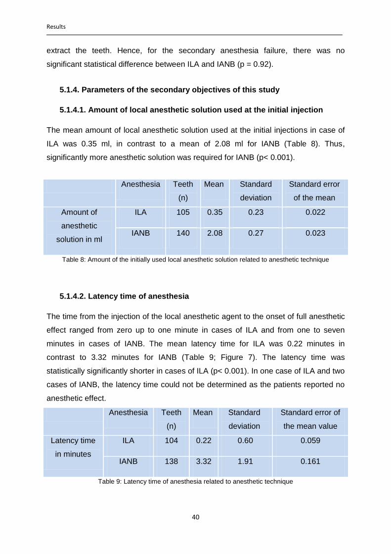
Results
40
extract the teeth. Hence, for the secondary anesthesia failure, there was no
significant statistical difference between ILA and IANB (p = 0.92).
5.1.4. Parameters of the secondary objectives of this study
5.1.4.1. Amount of local anesthetic solution used at the initial injection
The mean amount of local anesthetic solution used at the initial injections in case of
ILA was 0.35 ml, in contrast to a mean of 2.08 ml for IANB (Table 8). Thus,
significantly more anesthetic solution was required for IANB (p< 0.001).
Anesthesia Teeth
(n)
Mean Standard
deviation
Standard error
of the mean
value Amount of
anesthetic
solution in ml
ILA 105 0.35 0.23 0.022
IANB 140 2.08 0.27 0.023
Table 8: Amount of the initially used local anesthetic solution related to anesthetic technique
5.1.4.2. Latency time of anesthesia
The time from the injection of the local anesthetic agent to the onset of full anesthetic
effect ranged from zero up to one minute in cases of ILA and from one to seven
minutes in cases of IANB. The mean latency time for ILA was 0.22 minutes in
contrast to 3.32 minutes for IANB (Table 9; Figure 7). The latency time was
statistically significantly shorter in cases of ILA (p< 0.001). In one case of ILA and two
cases of IANB, the latency time could not be determined as the patients reported no
anesthetic effect.
Anesthesia Teeth
(n)
Mean Standard
deviation
Standard error of
the mean value
Latency time
in minutes
ILA 104 0.22 0.60 0.059
IANB 138 3.32 1.91 0.161
Table 9: Latency time of anesthesia related to anesthetic technique
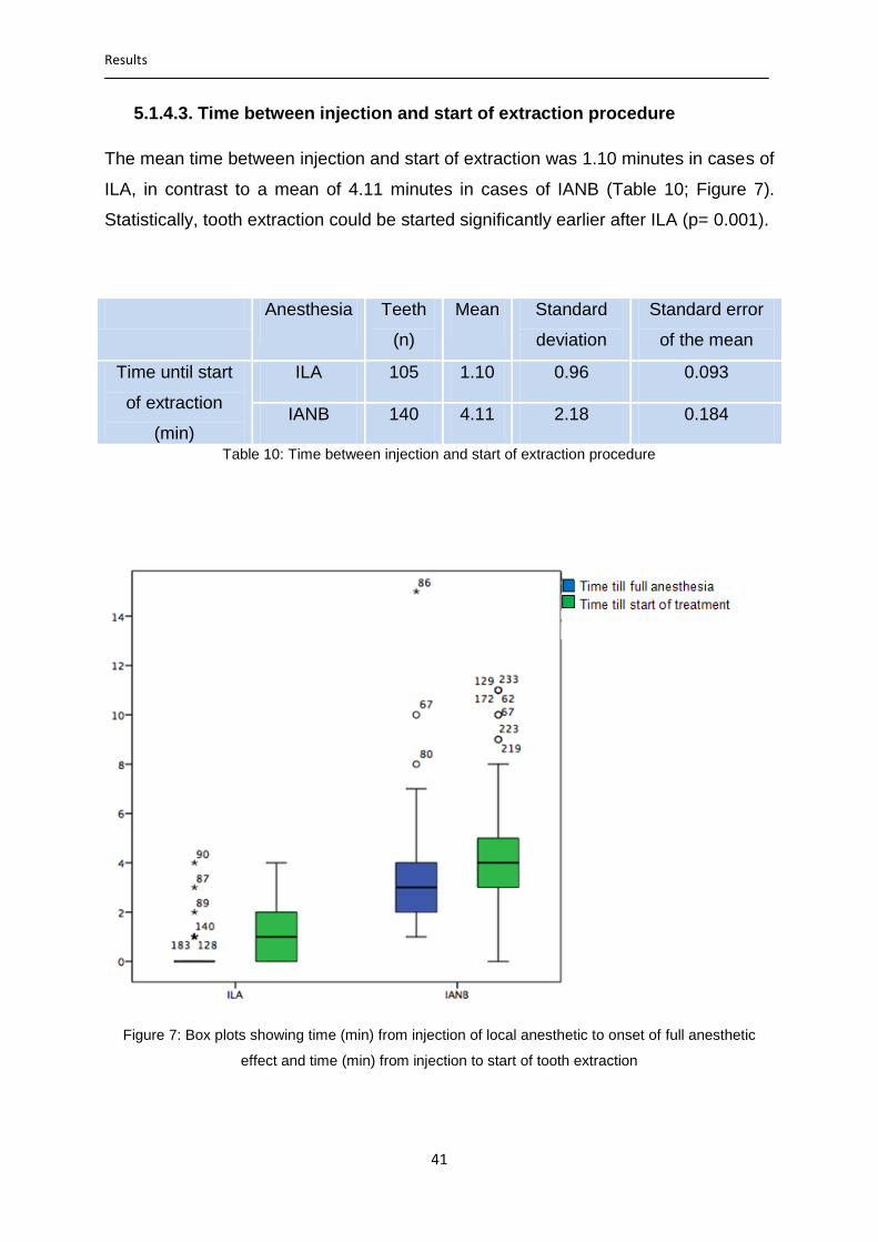
Results
41
5.1.4.3. Time between injection and start of extraction procedure
The mean time between injection and start of extraction was 1.10 minutes in cases of
ILA, in contrast to a mean of 4.11 minutes in cases of IANB (Table 10; Figure 7).
Statistically, tooth extraction could be started significantly earlier after ILA (p= 0.001).
Anesthesia Teeth
(n)
Mean Standard
deviation
Standard error
of the mean
value Time until start
of extraction
(min)
ILA 105 1.10 0.96 0.093
IANB 140 4.11 2.18 0.184
Table 10: Time between injection and start of extraction procedure
Figure 7: Box plots showing time (min) from injection of local anesthetic to onset of full anesthetic
effect and time (min) from injection to start of tooth extraction

Results
42
5.1.4.4. Time until second injection and amount of anesthetic solution at
second injection
The time between the initial injection and the required second injection was
statistically significantly longer in case of IANB (p< 0.001). As far as the amount of
local anesthetic solution for the second injection is concerned, significantly higher
amount of local anesthetic was required for the second injection in the case of IANB
(p< 0.001; Table 11).
Anesthesia Teeth
(n)
Mean Standard
deviation
Standard error
of the mean
value
Time until second
injection (min)
ILA 12 2.23 1.35 0.407
IANB 25 7.35 2.44 0.509
Amount of local
anesthetic for the
second injection
(ml)
ILA 12 0.28 0.71 0.213
IANB 25 1.59 0.48 0.100
Table 11: Time until second injection and amount of local anesthetic for the second injections related
to anesthetic technique
5.1.4.5. Duration of entire treatment
In the cases of ILA, the total duration of treatment (time from injection to the end of
treatment) ranged from one minute to 27 minutes. On average, the total duration of
the treatment was 5.6 minutes. In the cases of IANB, it ranged from four minutes to
36 minutes. On average, the total duration of the treatment in the cases of IANB was
10.7 minutes (Table 12). So, treatment under ILA lasted statistically significantly
shorter when compared to IANB (p< 0.001). Unfortunately, in two cases of ILA, the
duration of treatment was not documented.
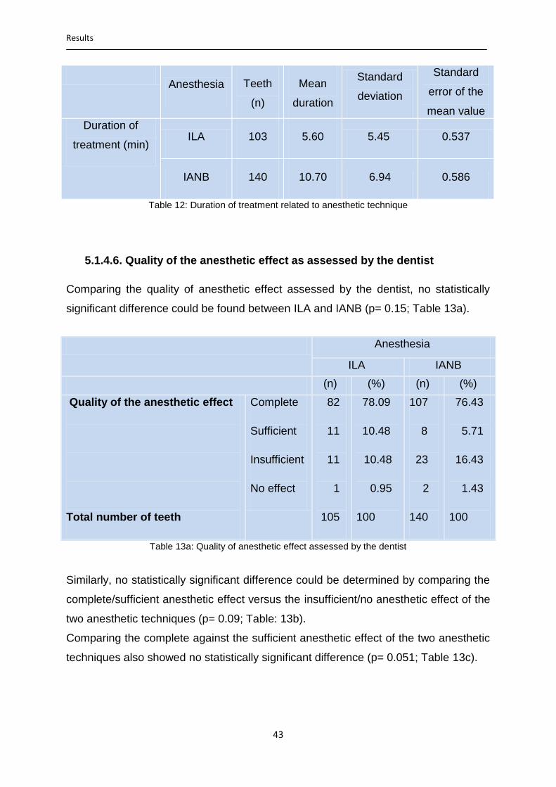
Results
43
Anesthesia Teeth
(n)
Mean
duration
Standard
deviation
Standard
error of the
mean value
Duration of
treatment (min) ILA 103 5.60 5.45 0.537
IANB 140 10.70 6.94 0.586
Table 12: Duration of treatment related to anesthetic technique
5.1.4.6. Quality of the anesthetic effect as assessed by the dentist
Comparing the quality of anesthetic effect assessed by the dentist, no statistically
significant difference could be found between ILA and IANB (p= 0.15; Table 13a).
Table 13a: Quality of anesthetic effect assessed by the dentist
Similarly, no statistically significant difference could be determined by comparing the
complete/sufficient anesthetic effect versus the insufficient/no anesthetic effect of the
two anesthetic techniques (p= 0.09; Table: 13b).
Comparing the complete against the sufficient anesthetic effect of the two anesthetic
techniques also showed no statistically significant difference (p= 0.051; Table 13c).
Anesthesia
ILA IANB
(n) (%) (n) (%)
Quality of the anesthetic effect
Total number of teeth
Complete
Sufficient
Insufficient
No effect
82
11
11
1
105
78.09
10.48
10.48
0.95
100
107
8
23
2
140
76.43
5.71
16.43
1.43
100

Results
44
Anesthesia
ILA IANB
(n) (%) (n) (%)
Quality of the
anesthetic effect
Total number of teeth
Complete/sufficient
Insufficient/no effect
93
12
105
88.57
11.43
100
115
25
140
82.14
17.86
100
Table 13b: Complete/sufficient versus insufficient/no anesthetic effect assessed by the dentist
Anesthesia
ILA IANB
(n) (%) (n) (%)
Quality of the
anesthetic effect
Total number of teeth
Complete
Sufficient
82
11
93
78.09
10.48
88.57
107
8
115
76.43
5.71
82.14
Table 13c: Complete versus sufficient anesthetic effect assessed by the dentist
5.1.4.7. Duration of soft tissue anesthesia
The duration of soft tissue anesthesia in cases of ILA ranged from 20 minutes up to
80 minutes (average 47.66 minutes). In comparison, for IANB, duration of soft tissue
anesthesia ranged from 130 minutes up to 360 minutes (average 228.56 minutes)
and was statistically significantly longer (p< 0.001; Table 14 and Figure 8).
Anesthesia Teeth
(n)
Mean Standard
deviation
Standard error
of the mean
value value Duration of soft
tissue
anesthesia (min)
ILA 104 47.66 33.461 3.265
IANB 138 228.56 53.382 4.512
Table 14: Duration of soft tissue anesthesia related to anesthetic technique

Results
45
Figure 8: Box plots comparing duration of soft tissue anesthesia (min) in cases of ILA and IANB
5.1.4.8. Frequency of associated complications
No signs of nerve contact were observed in any patient. A positive aspiration was
observed in six cases (4.3%) of IANB. Impaired wound healing (dry socket) was
observed in six cases (5.7%) after ILA and in three cases (2.1%) after IANB. A
comparison of the frequency of occurrence of impaired wound healing after ILA and
IANB showed no statistically significant difference (p= 0.178). No case of soft tissue
necrosis or clinically relevant bacteremia was observed in this study.
5.2. Evaluation group II (patients with split- mouth technique)
5.2.1. Patients and teeth
In 28 of the total 266 patients (23 males, 5 females) ILA was administrated on one
side of the mandible while IANB was administrated on the other side, as they had
teeth to be extracted on both sides of the mandible. Thus, 56 teeth (Table 15) were
extracted in these patients (ILA n= 28, IANB n= 28; Figure 9). These 28 patients were
a little older than those in the main group (22-87 years, mean 59.2; SD 14.7), were

Results
46
grown higher (160-186 cm, mean 176.57; SD 6.59) and a little less heavy (45-118 kg,
mean 80.8; SD 15.1).
Tooth number Number of teeth extracted Percent
34 3 5.36
35 3 5.36
36 9 16.07
37 12 21.23
38 0 0
44 2 3.57
45 4 7.14
46 8 14.29
47 13 23.21
48 2 3.57
Total 56 100
Table 15: Frequency of tooth extractions in group II
Figure 9: Bar charts showing number of extracted teeth per anesthetic technique in group II
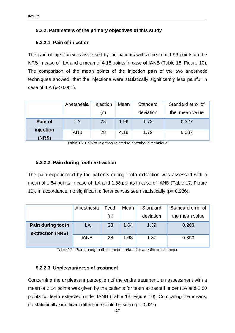
Results
47
5.2.2. Parameters of the primary objectives of this study
5.2.2.1. Pain of injection
The pain of injection was assessed by the patients with a mean of 1.96 points on the
NRS in case of ILA and a mean of 4.18 points in case of IANB (Table 16; Figure 10).
The comparison of the mean points of the injection pain of the two anesthetic
techniques showed, that the injections were statistically significantly less painful in
case of ILA (p< 0.001).
Anesthesia Injection
(n)
Mean Standard
deviation
Standard error of
the mean value
Pain of
injection
(NRS)
ILA 28 1.96 1.73 0.327
IANB 28 4.18 1.79 0.337
Table 16: Pain of injection related to anesthetic technique
5.2.2.2. Pain during tooth extraction
The pain experienced by the patients during tooth extraction was assessed with a
mean of 1.64 points in case of ILA and 1.68 points in case of IANB (Table 17; Figure
10). In accordance, no significant difference was seen statistically (p= 0.936).
Anesthesia Teeth
(n)
Mean Standard
deviation
Standard error of
the mean value
Pain during tooth
extraction (NRS)
ILA 28 1.64 1.39 0.263
IANB 28 1.68 1.87 0.353
Table 17: Pain during tooth extraction related to anesthetic technique
5.2.2.3. Unpleasantness of treatment
Concerning the unpleasant perception of the entire treatment, an assessment with a
mean of 2.14 points was given by the patients for teeth extracted under ILA and 2.50
points for teeth extracted under IANB (Table 18; Figure 10). Comparing the means,
no statistically significant difference could be seen (p= 0.427).
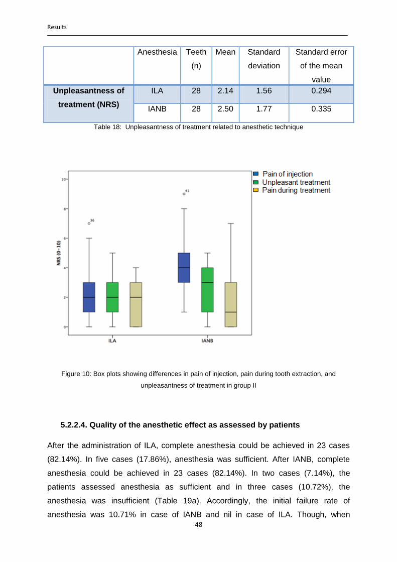
Results
48
Anesthesia Teeth
(n)
Mean Standard
deviation
Standard error
of the mean
value
Unpleasantness of
treatment (NRS)
ILA 28 2.14 1.56 0.294
IANB 28 2.50 1.77 0.335
Table 18: Unpleasantness of treatment related to anesthetic technique
Figure 10: Box plots showing differences in pain of injection, pain during tooth extraction, and
unpleasantness of treatment in group II
5.2.2.4. Quality of the anesthetic effect as assessed by patients
After the administration of ILA, complete anesthesia could be achieved in 23 cases
(82.14%). In five cases (17.86%), anesthesia was sufficient. After IANB, complete
anesthesia could be achieved in 23 cases (82.14%). In two cases (7.14%), the
patients assessed anesthesia as sufficient and in three cases (10.72%), the
anesthesia was insufficient (Table 19a). Accordingly, the initial failure rate of
anesthesia was 10.71% in case of IANB and nil in case of ILA. Though, when

Results
49
comparing the quality of the anesthetic effect as assessed by patients, no statistically
significant difference was seen (p= 0.177).
Table 19a: Quality of anesthetic effect assessed by patients
Comparing the complete/sufficient anesthetic versus the insufficient/no effect of the
two techniques of local anesthesia (Table 19b), no statistically significant difference
could be seen (p= 0.236).
Anesthesia
ILA IANB
(n) (%) (n) (%)
Quality of the
anesthetic effect
Total number of teeth
Complete/sufficient
Insufficient/no effect
28
0
28
100
0
100
25
3
28
89.29
10.71
100
Table 19b: Complete/sufficient versus insufficient/no effect assessed by patients
5.2.2.5. Need for second injection
Second injection of local anesthetic was required in three cases of IANB (10.7%). In
contrast, no second injection was required in case of ILA (Table 20). Even so, no
statistically significant difference could be found (p= 0.236).
Anesthesia
ILA IANB
(n) (%) (n) (%)
Quality of the anesthetic effect
Total number of teeth
Complete
Sufficient
Insufficient
23
5
0
28
82.14
17.86
0
100
23
2
3
28
82.14
7.14
10.72
100
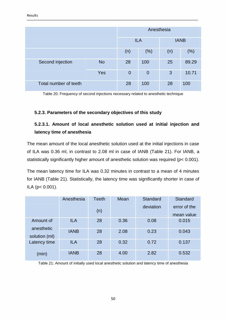
Results
50
Anesthesia
ILA IANB
(n) (%) (n) (%)
Second injection No 28 100 25 89.29
Yes 0 0 3 10.71
Total number of teeth 28 100 28 100
Table 20: Frequency of second injections necessary related to anesthetic technique
5.2.3. Parameters of the secondary objectives of this study
5.2.3.1. Amount of local anesthetic solution used at initial injection and
latency time of anesthesia
The mean amount of the local anesthetic solution used at the initial injections in case
of ILA was 0.36 ml, in contrast to 2.08 ml in case of IANB (Table 21). For IANB, a
statistically significantly higher amount of anesthetic solution was required (p< 0.001).
The mean latency time for ILA was 0.32 minutes in contrast to a mean of 4 minutes
for IANB (Table 21). Statistically, the latency time was significantly shorter in case of
ILA (p< 0.001).
Anesthesia Teeth
(n)
Mean Standard
deviation
Standard
error of the
mean value
Amount of
anesthetic
solution (ml)
ILA 28 0.36 0.08 0.015
IANB 28 2.08 0.23 0.043
Latency time
(min)
ILA 28 0.32 0.72 0.137
IANB 28 4.00 2.82 0.532
Table 21: Amount of initially used local anesthetic solution and latency time of anesthesia

Results
51
5.2.3.2. Time between injection and start of extraction procedure
The mean time between injection of local anesthetic and start of the extraction was
one minute in cases of ILA, in contrast to a mean time of 4.11 minutes in cases of
IANB (Table 22). Statistically, tooth extraction could be started significantly earlier
after ILA (p< 0.001).
Anesthesia Teeth
(n)
Mean Standard
deviation
Standard
error of the
mean value
Time until start
with the
extraction (min)
ILA 28 1.00 0.77 0.145
IANB 28 4.11 2.39 0.452
Table 22: Time between injection and start of extraction procedure
5.2.3.3. Duration of entire treatment
On average, the total duration of the treatment from injection to the end of the
extraction procedure was 4.81 minutes in case of ILA and 8.64 minutes in case of
IANB (Table 23). A comparison of the total duration of treatment under ILA and IANB
indicated, that the treatment under ILA lasted statistically significantly shorter than
under IANB (p= 0.006).
Anesthesia Teeth
(n)
Mean Standard
deviation
Standard error
of the mean
value Duration of
treatment
(min)
ILA 28 4.81 4.25 0.818
IANB 28 8.64 5.53 1.046
Table 23: Duration of treatment related to anesthetic technique
5.2.3.4. Quality of the anesthetic effect as assessed by the dentist
When comparing the quality of anesthetic effect as assessed by the dentist, no
statistically significant difference could be found between ILA and IANB (p= 0.611).
Similarly, no statistically significant difference could be determined when comparing
the complete/sufficient versus the insufficient anesthetic effects (p= 0.491; Table 24a
& b).

Results
52
Table 24a: Quality of anesthetic effect assessed by the dentist
Anesthesia
ILA IANB
(n) (%) (n) (%)
Quality of the
anesthetic effect
Total number of teeth
Complete/sufficient
Insufficient
28
0
28
100
0
100
25
3
28
89.29
10.71
100
Table 24b: Complete/sufficient versus insufficient effect assessed by the dentist
5.2.3.5. Duration of soft tissue anesthesia
On average, the soft tissue anesthesia lasted for 46.07 minutes after ILA in contrast
to an average duration of 244.29 minutes after IANB (Table 25). The soft tissue
anesthesia lasted statistically significantly longer after IANB (p< 0.001).
Anesthesia Teeth
(n)
Mean Standard
deviation
Standard
error of the
mean value
Duration of
soft tissue
anesthesia
(min)
ILA 28 46.07 16.39 3.097
IANB 28 244.29 59.60 11.264
Table 25: Duration of soft tissue anesthesia related to anesthetic technique
Anesthesia
ILA IANB
(n) (%) (n) (%)
Quality of the anesthetic effect
Total number of teeth
Complete
Sufficient
Insufficient
25
3
0
28
89.28
10.72
0
100
24
1
3
28
82.14
7.14
10.72
100
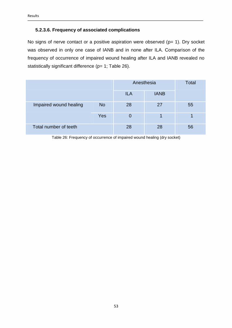
Results
53
5.2.3.6. Frequency of associated complications
No signs of nerve contact or a positive aspiration were observed (p= 1). Dry socket
was observed in only one case of IANB and in none after ILA. Comparison of the
frequency of occurrence of impaired wound healing after ILA and IANB revealed no
statistically significant difference (p= 1; Table 26).
Anesthesia Total
ILA IANB
Impaired wound healing No 28 27 55
Yes 0 1 1
Total number of teeth 28 28 56
Table 26: Frequency of occurrence of impaired wound healing (dry socket)

Discussion
54
6. Discussion
In this study, the efficacy of ILA for extraction of mandibular posterior teeth was
compared to IANB in a clinical routine setting. The results obtained in this study
indicate, that ILA – here using Ultraject® syringe - is at least as efficient as IANB for
this indication. In accordance, data showed no statistically significant differences
between the success rates and the profoundness of local anesthesia obtained with
the two anesthetic techniques. In certain aspects, ILA is found to be even superior to
IANB. Hence, ILA was significantly less painful to the patients, had a significantly
shorter latency time, enabled a shorter duration of treatment and required a
significantly smaller amount of local anesthetic solution in comparison to IANB. The
results show furthermore, that ILA - properly applied – can be used as a primary
anesthetic technique unrestricted for almost all nonsurgical extractions of mandibular
posterior teeth.
This study was a prospective, randomized, comparative clinical trial with a sample
size comparable to - if not more than - that of similar studies [Heizmann and Gabka,
1994; Dumbrigue et al., 1997]. Heizmann and Gabka [1994] compared ILA
administered with Citoject® syringe (teeth= 110/ maxillary= 56; mandibular= 54)
against IANB (teeth= 110/ maxillary= 0; mandibular= 110) and infiltration anesthesia
(teeth= 110/ maxillary= 76; mandibular= 34) in extraction of maxillary and mandibular
teeth on a total of 255 patients (ILA= 83; IANB= 85; infiltration= 87). Dumbrigue et al.
[1997] compared ILA (teeth= 24) administered using a pressure syringe (Ligmaject®)
against IANB (teeth= 21) administered using conventional aspirating syringes on 16
patients requiring bilateral mandibular dental extraction (split-mouth).
In this present study, ILA administered with Ultraject® syringe (teeth n=133) was
compared to IANB administered with conventional disposable injection syringes
(teeth n= 168) for extraction of mandibular posterior teeth on a total of 266 patients
(ILA= 126; IANB= 168). These 266 patients were categorized into two evaluation
groups (evaluation group I and II) based on whether only one or both anesthetic
techniques were used in individual patients (unilateral or bilateral tooth extraction).
238 patients with unilateral mandibular dental extractions who received randomized
either ILA or IANB as anesthetic technique were included in evaluation group I.
Evaluation group II (split-mouth) consisted of 28 patients with bilateral mandibular
dental extractions who received both ILA and IANB, each on one side of the
mandible. ILA was compared with IANB in each evaluation group and the results in

Discussion
55
the two groups were compared to each other. In group I, a total of 245 teeth were
extracted (ILA= 105; IANB= 140) from a total of 238 patients (ILA= 98; IANB= 140)
whereas in group II, a total of 56 teeth (ILA= 28; IANB= 28) was extracted from 28
patients.
The general patients´ parameters (number, age, height, and body weight, medical
and dental histories) of the patients anesthetized with ILA and those anesthetized
with IANB were relatively similar and showed no statistically significant differences.
The patients in evaluation group II were a little older, were grown higher and a little
less heavy than those in evaluation group I.
All of the injections, teeth extractions, and data collection in this study were
performed by only one dentist allowing the influence of the dentists´ variables to be
minimized. As mentioned above, in evaluation group II (28 patients, 56 teeth), the
study was designed as a split-mouth technique. Here, instead of having different
patients for each anesthetic technique, the same patients received first ILA and then
IANB. Hence, the influence of the patients’ variables on the results could be
minimized as well. The study data of this group of patients were evaluated separately
and the results were found to be in a high coincidence with those of the evaluation
group I.
The Ultraject® syringes used in this study for the administration of ILA are equipped
with a built-in safety pressure control mechanism; so that, independently of the
individual injection behavior, the injection of local anesthetics always takes place
patient-friendly, thereby reducing the unwanted effects. Ultraject® syringes have a
pressure limitation of 120 N. Converted to the surface area on which the pressure is
applied (the inner diameter of 0.3 mm needle), this results in a final injection pressure
of about 3.2 MPa [Zugal et al. 2005].
Concerning the general medical conditions of the patients, only a small proportion of
the entire study sample showed co-morbidities. Therefore, it may be assumed that
there was no statistically significant influence of the general medical history on the
results or on the effects of anesthesia. The same applies to the history of regular
intake of medication. In the entire study sample, only three patients reported acute
dental pain before treatment. Therefore, a statement about the correlation between
the pain prior to treatment and the failure of the anesthesia can – unfortunately -

Discussion
56
neither be excluded nor confirmed. Von Haussen [2011] claimed a positive
correlation between preoperative pain and failure of ILA. He reported about failure of
ILA - administered with STA™-system - in five from a total of eight patients who had
reported about pain preoperatively. The influence of pain medication on the results
can be ruled out since patients who have taken a pain medication before treatment
were excluded from this study.
The pain perceived by patients during the different steps of treatment is an important
aspect in the evaluation of the efficacy of anesthetic techniques. In accordance, an
objective method of pain measurement should be employed. Though, the basic
problem is that pain is a personal subjective experience and cannot be visualized or
felt by another person in the same way. So, only indirect methods – such as
physiological, behavioral and report by the patient - can be used for this purpose. In
this study, a numeric rating scale (NRS) consisting of 11 points (0= no pain; 10
extreme pain) was used. The patient selected a number from 0 to 10 that best
reflected the intensity of pain he/she felt. Although this method can be affected by
fear and expectation of pain by the patient, it is the most reliable and commonly used
method [Ardakani et al., 2010].
Regarding the pain of injection, a statistically significant difference between ILA and
IANB was found in this study. The injections in cases of ILA were significantly less
painful to the patients than that in cases of IANB (p< 0.001). In both evaluation
groups, the injection of ILA was rated with significantly fewer points on the NRS (2.2
points in group I; two in group II) than that of IANB (3.7 points in group I; 4.2 in group
II). However, the injection pain in evaluation group II was rated to be relatively less in
cases of ILA compared to IANB. In other words, the injections of IANB seemed to be
more painful than ILA in group II when compared to group I. In group II, ILA was
administered first on one side, and then IANB was administered in the same
appointment on the other side of the mandible. This could potentially affect the
patients´ perception of pain during subsequent injections and may explain why the
injection pain of IANB was rated worse. This result is in accordance with that in many
other previous studies. Einwag [1982] reported an absence of pain response for ILA
injection. Heizmann and Gabka [1994] reported about injection pain in 7.2% of 110
cases of tooth extraction under ILA and in 10% of 110 cases under IANB. Zugal
[2001] noticed injection pain in 27 (13.2%) from 205 cases of ILA, while Weber [2005]

Discussion
57
reported injection pain in two (1.5%) of 137 cases of ILA and in 35 (26.9%) of 130
cases of IANB. In these four studies mentioned above, the evaluation was based on
the patients´ report about injection pain with yes/no answers. Kaufman et al. [2005]
reported injection pain with a mean of 2.74 points in 45 cases of ILA and a mean of
3.54 points in 61 cases of IANB. Prama et al. [2013] reported injection pain with a
mean of 1.7 points in 20 cases of ILA and a mean of 3.4 points in 20 cases of IANB,
while Kämmerer et al. [2015] reported on injection pain with a mean of 1.55 points in
22 cases of ILA and a mean of 3.05 points in 20 cases of IANB. In the last three
studies mentioned above, a numeric rating scale from 0-10 was used for evaluation
of injection pain.
However, not all authors came to the same conclusions. Dumbrigue et al. [1997]
found that the injections of ILA were associated with more pain and discomfort to the
patient than that of IANB. Moreover, the authors concluded, that the profoundness of
local anesthesia was less after ILA. However, it must be noticed that Dumbrigue – in
the statistical analyses of the injection pain - has not taken each extracted tooth as
an independent sample, but considered the total number of patients. As in some
patients, more than one tooth was extracted under ILA, multiple intraligamental
injections were required in the same quadrant. This may account for the observed
greater patients´ discomfort during injections associated with ILA. Furthermore,
Dumbrigue has used Ligmaject® syringe for the administration of ILA. When such
pistol type syringes without safety pressure limiting mechanism are used, the
injection takes place rapidly under higher pressure than required and the injection
pain increases obviously [Walton and Abbott, 1981; Giovannitti and Nique, 1983]. In
addition, the sample size in the study of Dumbrigue et al. was small (16 patients, 45
teeth). Hence, the results of such studies, with small study sample should not be
taken without caution and reservation.
It must also be recognized, that there several other procedural factors possibly
associated with pain experience during the injection of local anesthetic such as type
of anesthetic solution, amount of injected solution, temperature of the anesthetic
solution, injection rate, location of injection and experience of the dentist [Arjen et al.,
2009].
Regarding the amount of local anesthetic solution required, a statistically significant
difference was found between ILA and IANB both in group I and II (p< 0.001). The

Discussion
58
mean amount of local anesthetic solution used at initial injections in both evaluation
groups was similar in case of ILA (0.4 ml) and similar in case of IANB as well (2.1
ml). In each group, a significantly higher amount of local anesthetic solution was
required for IANB, both at initial and second injections. This fact is technique
immanent and, of course, coincides with that used by many other Authors. Stoll and
Bührmann [1983] used a mean amount of 0.3-0.4 ml of local anesthetic in 57 cases
of ILA. Heizmann and Gabka [1994] used a mean of 0.3 ml for ILA and 2.4 ml for
IANB, while Dirnbacher [2003] injected a mean amount of 0.4 ml for ILA and 1.8 ml
for IANB. By Weber [2005], a mean of 0.47 ml for ILA and 1.9 ml for IANB were
required. Dombrigue et al. [1997] injected a mean of 0.2-0.4 ml anesthetic for ILA and
1.9 ml for IANB. However, Rahn et al. [1987] used a mean of 0.8 ml of local
anesthetic for ILA and was able to reach only 60% success rate in 47 cases.
The latency time of anesthesia was determined through testing the anesthetic effect
on the gingiva at regular time intervals using a sharp dental probe. ILA – technique
immanent – has virtually no latency. However, according to the observation by the
dentist in this study, a latency of 30 seconds up to one minute must be expected in
inflamed tissues. In this study, ILA was found to have a statistically significant shorter
latency time than IANB (p< 0.001). The mean latency time after ILA was 0.22 minute
in group I and 0.32 minute in group II. In case of IANB, mean latency times of 3.32
minutes and four minutes were determined in group I and II, respectively. This result
corresponds to that in many previous studies. A practically no latency after ILA was
reported by many authors [Gray et al., 1987; Cohen et al., 1993; Heizmann and
Gabka, 1994; Zugal et al., 2005; Weber et al., 2006]. After IANB, mean latency times
of three minutes and 3.80 minutes were reported by Heizmann & Gabka [1994] and
by Dirnbacher et. al. [2003], respectively. For Weber [2005], this value was 4.6
minutes.
Of course, electric pulp testing would have given more objective fundamental
information in regard of the pulp anesthesia. Therefore, the used methods for
subjective evaluation of gingival anesthesia should be considered to be a possible
bias of this study.
Being able to begin with the extraction procedure immediately after the injection of
the local anesthetic, ILA does offer the advantages of saving time, reducing the
treatment time and the duration of patients´ stay on the dental chair, which in turn
Discussion
59
reduces the psychological distress of the patients. Furthermore, by eliminating the
need of bridging the long latency time by changing the room and working at other
patients (“room-hopping”), the patient stays under continuous observation by the
dentist after the injection and actions can be taken immediately in case of eventual
complications.
In relation to the total duration of treatment, measured from the time of injection until
the end of the extraction procedure, a significant difference was found between ILA
and IANB . The mean duration of treatment in group I was 5.6 minutes after ILA and
10.7 minutes after IANB. In group II, these values were 4.8 and 8.6 minutes after ILA
and IANB, respectively. In both evaluation groups, the duration of treatment was
significantly longer in the cases of IANB (p< 0.001; 0.006 in group II). This was
especially due to the longer latency time of anesthesia after IANB.
With an average treatment time of 10.70 minutes and an average latency time of 3.32
minutes, 31% of the total treatment time was required for waiting for the anesthetic
effect of IANB. Only in the remaining 69% of the time, extraction procedures took
place. Measured from the beginning to the end of the extraction procedure,
Heizmann and Gabka [1994] reported a mean treatment time of 3.5 minutes after ILA
and 6.4 minutes after IANB. The mean times measured from the beginning to the end
of extraction procedures obtained in this present study – after subtracting the mean
latency times – were 5.4 and 7.4 minutes for ILA and IANB, respectively (4.5 and 4.6
minutes in group II). These mean times of treatment are a little bit longer but
comparable to that obtained by Heizmann and Gabka [1994].
Since the treatments in all cases of ILA and IANB had always been completed while
the maximal effect of anesthesia was still present, it is unlikely that the differences in
the duration of treatment could have had effects on the results, in particular, the
quality of anesthesia.
In regard to the pain experienced by the patients during the extraction procedures, no
statistically significant difference between ILA and the IANB could be found in this
study (p= 0.211; 0.936 in group II). Similarly, no significant difference could be found
regarding the unpleasantness of the entire treatment under ILA and IANB (p= 0.31;
0.427 in group II).

Discussion
60
In this present study, a success of local anesthesia was judged when teeth can be
extracted without or with mild but tolerable pain and discomfort. In group I, an initial
success rate of 88.6% in case of ILA (99% after second injections) and 82.14% in
case of IANB (98.6% after second injections) could be achieved. The initial success
rate in group II was 100% for ILA and 89.3 (100% after second injections) for IANB.
The difference in success rate of ILA and IANB after the first injections was
statistically not significant (p= 0.082; 0.236 in group II). Similarly, no statistically
significant difference between the success rates of ILA and IANB could be found
after second injections both in group I and II (p= 0.92, one respectively).
The initial success rates of ILA and IANB achieved in this study are comparable to
that reported by others. Heizmann [1989] could achieve an initial success rate of
86.4% with ILA (99.1% after second injections) and 84.5% with IANB (100% after
second injections) in extraction of 110 maxillary and mandibular teeth. Considering
the only extraction of mandibular teeth, the success rate achieved by Heizmann was
81.5% in extraction of 54 teeth with ILA and 84.5 in extraction of 110 teeth with IANB.
However, not all authors came to the same result. Some authors could achieve a
higher success rate with ILA in dental extraction [Faulkner, 1983; Kaufman et al.,
1983]; and others achieved a lower success rate than that in this present study
[Edward and Head, 1989; Dombrigue et al., 1997]. Faulkner [1983] could achieve
97% success rate in extraction of 100 teeth. Kaufman et al. [1983] reported a 95%
success rate in a total of 22 cases. In the preceding studies [Faulkner, 1983;
Kaufman et al., 1983], success was defined as completion of the extraction
procedures without any discomfort. All cases in which additional anesthesia was
required or in which the procedure could not be completed at that visit were
considered as failures.
Edwards and Head [1989] reported a 79% success rate of ILA in extraction of 14
teeth. Dombrigue et al. [1997] reported about success rate of 75% in extraction of 24
mandibular posterior teeth with ILA and 95% in extraction of 21 mandibular posterior
teeth with IANB. Second injections were required in 12 cases (50%) of ILA and in one
case (4.8%) of IANB.
In this present study, in addition to the evaluation by the patients, the success of the
anesthesia was also evaluated by the dentist based on his 12 years long experience
in dental extraction under local anesthesia and served as a control of the patients’
evaluation of success to minimize the bias that might be caused by overestimation by

Discussion
61
the patients. The evaluations of anesthetic success made by the patients are found
to coincide in the greatest part with that indicated by the patients.
Another point of discussion is related to the total duration of soft tissue anesthesia.
The duration of soft tissue anesthesia after IANB is – technique immanent –
significantly longer than that after ILA. In accordance, the results of this study showed
that the total duration of marginal soft tissue anesthesia was statistically significantly
longer after IANB than after ILA (p< 0.001). The average duration of ILA was 47.7
minutes in group I and 46 minutes in group II whereas the average duration of IANB
was 228.6 minutes; 244 minutes in group II.
The average duration of ILA obtained in this study is higher than that obtained by
some authors such as 30 minutes [Heizmann, 1989; Dirnbacher, 2002; 2003; and
Endo et al., 2008] and 31.8 minutes [Weber, 2005]; but lower than that obtained by
some others such as 56 minutes [Cisides, 2009], 74.1 minutes [Langbein, 2011] and
61 minutes [Kämmerer et al., 2014]. The duration of IANB obtained in this study is
comparable to that reported in the literature [Heizmann and Gabka, 1994;
Dirnbacher, 2002, 2003; Weber, 2005; Endo et al., 2008; Cisides, 2009; Kämmerer et
al., 2014]. These authors reported that the soft tissue anesthesia lasted for more than
200 minutes after IANB.
This duration of soft tissue anesthesia after IANB may be considered to be
unnecessarily long and without reasonable ratio to the treatment time. In accordance,
for IANB, the average duration of treatment was 10.7 minutes whereas the average
duration of soft tissue anesthesia was 228.6 minutes. This means, that only 4.7% of
the total duration of anesthesia was used for the actual treatment. In consequence,
the patients remained limited in their dispositions for hours after the end of treatment.
In those cases, functions of mastication and speech are usually disrupted and the
risk of bite or thermal injury, especially in children and in patients with mental
disabilities, is increased.
No signs of nerve injury were noticed in this study after IANB. According to the
literature, a persistent or even permanent nerve involvement after IANB may occur in
one from 20,000-850,000 cases [Pogrel, 2007]. Consequently, a direct comparison of
this result with that obtained in this study is difficult due to the small sample size.

Discussion
62
A positive aspiration was observed in six cases (8.4%) of IANB. This frequency of
positive aspiration is comparable to that of 8.2% reported by Persson et al. [1974] but
differs from that reported by other authors such as 11.7% [Bartlett, 1972], 11%
[Rood, 1972], 30% [Evers and Haegerstam, 2000] and 4.3% [Vasconcelos et.al.,
2008]. With ILA, positive aspiration or a contact with a large blood vessel can virtually
be excluded.
Expected was an increase in the frequency of occurrence of dry socket after ILA as
the local anesthetic solution with its vasoconstrictor content is injected in the direct
vicinity of the extraction site, which could impair the building of blood coagulum
necessary for the wound healing. In addition, adrenaline and other vasoactive agents
possess fibrinolytic activity [Mannucci et al., 1985]. In the entire study sample of this
study, however, impaired wound healing (dry socket) was observed in six cases
(4.5%) after ILA and in four cases (2.4%) after IANB. In accordance, there was no
significant difference between ILA and IANB in regard to the frequency of occurrence
of dry socket (p= 0.178; one in group II). This result coincides with others [Tsirlis et
al., 1992; Heizmann and Gabka, 1994]. Tsirlis et al. [1992] observed dry socket in six
cases (4.3%) after 139 dental extractions with ILA and in five cases (3%) after 166
extractions with IANB using 2% lidocaine with 1:80.000 epinephrine as a local
anesthetic in all cases. Heizmann and Gabka [1994] reported on six cases of dry
socket (5.5%) after extraction of 110 teeth with ILA and on five cases (4.5%) after
extraction of other 110 teeth with IANB using 4% articaine with 1:200,000 adrenaline
as a local anesthetic for both anesthetic techniques. In this present study, 4%
articaine with 1:200.000 adrenaline was the local anesthetic used for both anesthetic
techniques as well. Whether the choice of local anesthetic agents and the different
concentrations of vasoconstrictor in local anesthetic agents might influence the
incidence of dry socket after ILA, remains a subject of debate in the literature.
Meechan et al. [1987] claimed that the choice of local anesthetic agents could have
an influence on the frequency of occurrence of dry socket. He observed a significant
increase in the frequency of occurrence of dry socket after using Xylocaine (2%
lignocaine with 1:80.000 adrenaline) as compared to Citanest (3% Prilocaine with
0.03 IU/ml felypressin). He concluded that this difference in the incidence of dry
socket was due to the difference in the haemostatic ability of the two
vasoconstrictors. Meyer [1971] considered the influence of the vasoconstrictor to be

Discussion
63
important. However, Nitzan [1983] could not find a significant different in the
incidence of dry socket after teeth extractions under local and general anesthesia; he
concluded that vasoconstrictor can have no effect on the production of dry socket.
Birn [1973] stated that any vasoconstrictor effect on the incidence of dry socket is
unproven.
Testing the possible influences of different adrenaline concentrations in local
anesthetic agents on the efficacy of ILA for dental extraction and on the possibly
associated complications is the subject of our research in the future. We assumed
that the amounts of adrenaline administered during ILA are very small and do not
contribute considerably to the production of dry socket.
Even if it was no objective of this study, no signs or symptoms of manifested
bacteremia were observed in this study. Rahn et al. [1986] noticed a significant
increase in the incidence bacteremia after ILA and advised the use of conventional
anesthesia by patients with increased risk of endocarditis. In our opinion, the risk of
bacteremia during intraligamental injection is not considerably higher than that during
other dental treatment measures. In patients prone to endocarditis, in accordance to
the German guidelines, antibiotic prophylaxis is carried out anyway.
Since a large proportion of the data analyzed in this study was obtained from the
patients´ own answers of questions concerning the study parameters, the data are
not completely objective and may reflect individual, personal assessments. Pain
threshold and its tolerance may vary in individual patients; and the perception of pain
can be modified by psychological, social and situational factors. For this reason, it
must be recognized that there are inherent limitations in conclusions that may be
drawn from a study thus designed.

Conclusion
64
7. Conclusion
Based on the results of this study, the following conclusions can be derived:
ILA represents a safe and reliable method of local anesthesia with a success rate
comparable to that of the conventional inferior alveolar nerve block for mandibular
dental extraction. So, it can be considered as a reliable alternative to IANB.
Moreover, it does offer several advantages for the patient and dentist over the
conventional IANB.
The injection of ILA is significantly less painful to the patients than the conventional
nerve block anesthesia making it a preferable method of anesthesia especially for
children and patients with fear and anxiety.
ILA has virtually no latency, so that it is possible to evaluate the anesthetic effect and
to start with treatment immediately after the injection of the local anesthetic solution.
Being able to begin with treatment immediately after the injection, ILA does offer the
advantages of saving time, reducing the total treatment time and the duration of
patients´ stay on the dental chair, which in turn reduces the psychological distress of
the patients.
For ILA, only a small amount of local anesthetic solution is required. In consequence,
the risk of overdosing and systemic reactions due to local anesthetics’ toxicity is
lower than that after IANB. This risk should be considered, especially in children and
in patients with cardiovascular diseases as well as when treating many teeth in
different quadrants in the same appointment.
ILA enables a shorter duration of anesthesia in comparison to the nerve block
anesthesia. This duration is usually enough to complete the majority of dental
treatments. At the same time, the patient can resume his normal activity shortly after
the completion of treatment. Additionally, the local anesthetic effect is limited to the
region of the tooth to be treated without concomitant anesthesia of the lip and
tongue. The speech and chewing functions are minimally affected and the risk of
postoperative bite and burn injuries decreases.
The risk of injury to the blood vessels and hematoma formation with ILA can
practically be excluded. With the increasing proportion of elderly patients and patients
with cardiovascular diseases taking anticoagulants, the number of risk patients in the
daily dental practice increases. ILA should be considered as the anesthetic method
of choice for these patients.

Conclusion
65
With ILA, there is practically no risk of nerve injury. Considering the actual
jurisprudences, the patients must be informed about the risks and complications of an
indicated health measure and about the alternative therapy measure. So, ILA must
be offered as an alternative to the nerve block anesthesia. In recent judicial
procedures about injuries to the inferior alveolar nerve, the attending dentists were
condemned because they had not offered ILA as a possible alternative and had not
given the patient the possibility of choosing another method of anesthesia.
Taking into account the potential complications of the inferior alveolar nerve block,
ILA should be considered as a preferred anesthetic technique especially for single
tooth treatment while IANB should be limited only to cases in which ILA failed to
provide the sufficient anesthesia and in cases of dentoalveolar surgical procedures
requiring a longer time or for treating many teeth in the same quadrant.

Summary
66
8. Summary
Usually, the nature of the bone of the adult posterior mandible requires the use of
regional nerve block to obtain anesthesia for most dental procedures. Inferior alveolar
nerve block (IANB) is considered as a reliable technique of local anesthesia and is
still the most commonly used technique in the lower jaw, especially when treating
many teeth in the same quadrant. However, IANB has a relatively high failure rate
and technique immanent risk of complications, such as transient or even persistent
damage of the lingual and/or the inferior alveolar nerve. In consequence, the need for
an alternative anesthetic technique with higher success rate and lower risk of
complications is increased. Intraligamentary anesthesia (ILA) is considered to be an
efficient and clinically safe technique of local anesthesia and appears to be an
interesting alternative. For ILA, the local anesthetic solution is injected directly into
the periodontal ligament space of the tooth to be treated and shows its effect only in
this region. So far, there is a lack of respective studies on this topic.
The purpose of this prospective, randomized clinical trial was to test whether ILA
applied using a pistol-type syringe with a built-in pressure limiting mechanism
(Ultraject®) can be considered as a valid alternative to – or offers advantages for the
patients over - the conventional IANB in extraction of mandibular posterior teeth.
266 adult patients of both sexes (176 males, 90 females) with one or more
mandibular posterior teeth indicated for extraction were included in this study. 301
extractions of mandibular posterior teeth (ILA= 133 teeth, IANB= 168 teeth) were
evaluated. Patients were categorized randomly into those who received ILA and
those who received IANB as a local anesthetic technique for the indicated dental
extraction. When bilateral mandibular dental extractions were indicated, ILA was
randomly used first on one side and then IANB on the other. All injections, teeth
extractions, and data collection were carried out by one and the same dentist. The
anamnestic patient data, as well as the descriptive study data were collected using
especially designed evaluation sheet. The study parameters were assessed using
Likert scale or numeric rating scale.
After treatment and before statistical data evaluation, the patients were further
categorized into two evaluation groups based on whether only one anesthetic
technique (unilateral) or both techniques (bilateral) were used in the individual
patients. In evaluation group I were patients (patient n= 238, teeth n= 245) with

Summary
67
unilateral dental extraction who received either ILA (teeth n= 105) or IANB (teeth n=
140), while in evaluation group II (split-mouth) were patients with indicated bilateral
dental extraction (patient n= 28, teeth n= 56) and who received both ILA (teeth n= 28)
and IANB (teeth n = 28), each in one side of the mandible. ILA was compared with
IANB within each evaluation group and the results of the two groups were then
compared with each other.
The two techniques of dental local anesthesia were compared with the help of
several parameters (pain of injection, pain during tooth extraction, anesthetic quality,
need for second injection, amount of anesthetic solution required, duration of
anesthesia, and frequency of complications). The primary objectives were to evaluate
the difference in pain perceived by the patient during treatment as well as the
anesthetic efficacy (complete/sufficient vs. insufficient/no effect) based on the
outcome of treatment and the degree of discomfort associated with the extraction
procedure. Of secondary interest were the amount of local anesthetic solution
required, the duration of treatment, the duration of soft tissue anesthesia, and the
frequency of complications. Each extracted tooth was considered as an independent
sample.
The collected data were tabulated and analysed statistically. A global significance
level was chosen to 0.05. For explorative data analysis, Kolmogorov-Smirnov test
was employed to test for a difference between the two anesthetic techniques. In
cases of p-values <0.05, Mann-Whitney-U test and in cases of p-values >0.05,
Students’ t-test for independent samples were employed.
The results in evaluation groups I and II were found to coincide in the greatest part
with each other. The injection of ILA was statistically significantly less painful for the
patients in both evaluation groups (p< 0.001). In evaluation group II, IANB was
statistically much more painful for the patients than ILA in comparison to that in group
I. In group II, ILA was administered first on one side, and then IANB was
administered in the same appointment on the other side of the mandible. This could
potentially affect the patients´ perception of pain during subsequent injections and
may explain why the injection pain of IANB was rated worse. The difference in pain
experienced by the patients during tooth extraction under ILA and IANB was
statistically not significant (p= 0.211; 0.936 in group II). Similarly, no statistically
significant difference could be found when comparing the unpleasantness of
treatment (p= 0.31; 0.427 in group II).

Summary
68
The success rate of ILA in extraction of mandibular posterior teeth was 88.6% after
just one injection (100% in group II); this rate increased to 99% after second injection.
IANB, however, had a success rate of 82.2% after first injection (89.3% in group II),
and 98.6% after second injection (100% in group II). The difference between the
success rates of ILA and IANB was statistically not significant (p= 0.082; 0.236 in
group II).
The latency between the injection of the local anesthetic and the onset of the full
anesthetic effect was significant shorter after ILA (p< 0.001); thus, it was possible to
start with the extraction immediately after the injection, shortening the total time
required for the treatment and saving time. In addition, the amount of local anesthetic
solution used was substantially less in cases of ILA when compared with IANB (p<
0.001).
The duration of soft tissue anesthesia after IANB was significantly longer than that
after the ILA (p< 0.001) and exceeded by far the time required for the extraction.
Regarding the frequency of associated complications, no case of nerve injury was
observed. Positive aspiration was observed in six cases (4.3%) of IANB in group I
and in nil case in group II. In the entire study sample (group I and II), dry socket was
observed in six cases (4.5%) after ILA and in four cases (2.4%) after IANB. In
accordance, there was no significant difference between ILA and IANB in regard to
the frequency of occurrence of dry socket (p= 0.178; one in group II).
Based on the results of this present study, it can be concluded that ILA represents a
safe and reliable alternative to the conventional IANB in extraction of mandibular
posterior teeth. Taking into account the potential complications of the inferior alveolar
nerve block, ILA should especially be considered as a preferred anesthetic technique
for single tooth treatment while IANB should be limited only to cases in which ILA
failed to provide the sufficient anesthesia and in cases of dentoalveolar surgical
procedures requiring a longer time or for treating many teeth in the same quadrant.
References
69
9. References
Adubae A, Buttchereit I, Kämmerer PW (2016). Extraktion unter ILA bei einer Phobie-Patientin. Zahnärztl Mitt 106(5):42-45 Anand P, Wilson R, Sheehy EC (2005). Intraligamental analgesia for post-operative pain control in children having dental extractions under general anaesthesia. Eur J Paediatr Dent 6:10-15. Anneroth G, Danielsson KH, Evers H, Hedström KG, Nordenram A (1985). Periodontal ligament injection. An experimental study in the monkey. Int Oral Surg 14:538-543. Ardakani FE, Bahrololoumi Z, Booshehri MZ, Azam AN, Ayatollahi F (2010). The position of lingual as an indix for inferior alveolar nerve block injection in 7-11 year-old children. JODDD 4:47-51. Bartlett SZ (1972). Clinical observations on the effects of injections of local anesthetic preceded by aspiration. Oral Surg Oral Med Oral Pathol 33:520-526.
Bender W, Taubenheim L (2014). Minimalinvasive Lokalanästhesie- keine Aufklärungspflicht. ZWP online- Das Nachrichenportal für die Dentalbranche. Bendqude V, Akkareddy B, Jawale BA, Chaudhary S (2011). An unusual pattern of self-inflicted injury after dental local anesthesia: a report of 2 cases. J Contemp Dent Pract 12:404-407. Birn H (1973). Etiology and pathogenesis of fibrinolytic alveolitis (dry socket). Int. J. Oral Surg 2:211-263. Bluttner A,Taubenheim L (2009). Aufklärungspflicht des Zahnarztes: Risiken der Leitungsanästhesie. Zahnärztl Mitt 5A:104-106. Boronat López A , Peñarrocha Diago M (2006). Failure of locoregional anesthesia in dental practice. Review of the literature. Med Oral Patol Oral Cir Bucal 11:10-13. Bourdain, C.L (1925). L’Anesthésie par l’injection intra-ligamentaire pour l’extraction des dents. These de Doctorat, Editions de la Semaine Dentaire, Paris. Brännström M, Lindskog S, Nordenvall KJ (1984). Enamel hypoplasia in permanent teeth induced by periodontal ligament anesthesia of primary teeth. J Am Dent Assoc 109:735-736.
Brännström M, Nordenvall KJ, Hedström KG (1982). Periodontal tissue changes after intraligamentary anesthesia. ASDC J Dent Child 49:417-423.

References
70
Carter G, Gross AN, Lloyd J, Tocchetti R (2003). Current concepts of the management of dental extractions for patients taking warfarin. Aust Dent J 48:89-96. Castagnola L, Chenaux G, Colombo A (1982). Intraligamentary anesthesia with the peripress syringe. Quintessenz Int 13:927-930. Chevalier V, Arbab-Chirani R, Tea SH, Roux M (2010). Facial palsy after inferior alveolar nerve block: case report and review of the literature. Int J Oral Maxillofac Surg 39:1139-1142. Chi D, Kanellis M, Himadi E, Asselin ME (2008). Lip biting in a pediatric dental patient after dental local anesthesia: a case report. J Pediatr Nurs 23:490-493. Choi EH, Seo JY, Jung BY, Park W (2009). Diplopia after inferior alveolar nerve block anesthesia: report of 2 cases and literature review. Oral Surg Oral Med Oral Pathol Oral Radiol Endod. 107:21-24. Cohen HP, Cha BY, Spangberg LS (1993). Endodontic anesthesia in mandibular molars: A clinical study. J. Endod 19:370-373. Csides M (2009). Die intraligamentäre Anästhesie als primäre Methode der zahnärztlichen Lokalanästehsie unter besonderer Betrachtung der angewandten Injektionssysteme und der damit generierten Effekte beim Patienten Diss med. univ. Jena. Daubländer M, Kämmerer PW (2014). Lokalanästhesie in der Zahnmedizin. Berlin: Forum-med-dent Aventis. Davidson L, Craig S (1987). The use of periodontal ligament injection in children. J. Dent 15:204-208.
DeSantis JL, Liebow C (1996). Four common mandibular nerve anomalies that lead to local anesthesia failures. J Am Dent Assoc 127:1081-1086. Dirnbacher T (2002). Intraligamentäre Anästhesie vs. Leitungs- und Infiltrationsanästhesie in der Praxis, Diss.med. univ. jena. Dirnbacher T (2003). Reduzierung unerwünschter Effekte bei der zahnärztlichen Lokalanästhesie, Diss. med. univ.Jena. Dirnbacher T, Glockmann E, Taubenheim L (2003). Die intraligamentäre Anästhesie. Welche Anästhesie ist die richtige?. Zahnärztl Mitt 23:44-51. Dreyer WP, van Heerden JD, de V. Joubert JJ (1983). The route of periodontal ligament injection of local anesthetic solution. J Endod 9:471-474. Dumbrigue HB, Lim MV, Rudman RA, Serraon A (1997). A comparative study of anesthetic techniques for mandibular dental extraction. Am J Dent 10:275-278.

References
71
Edwards RW, Head TW (1989). A clinical trial of intraligamentary anesthesia. J Dent Res 68:1210-1213. Einwag J (1982). Die intraligamentäre Anästhesie im Kindes- und Jugendalter. Dtsch Zahnärztl Z 37:874-876. Endo T, Gabka J, Taubenheim L (2008). Intraligamentary anesthesia: benefits and limitations. Quintessence Int 39: 15-25. Erlemeier EM (1991). Aktuelle Aspekte der intraligamentären Anästhesie. Die Quintessenz 42:471-481. Ethunandan M, Tran A.L, Anand R, Bowden J, Seal M.T, Brennan P.A (2007). Needle breakage following inferior alveolar nerve block: Implications and management. BDJ 202:395-397. Evers H, Haegerstam G (2000). Zahnärztliche Lokalanästhesie. Deutscher Ärzte-Verlag, Köln.
Faulkner RK (1983). The high-pressure periodontal ligament injection. Br Dent J 154:103-105. Foster W, Drum M, Reader A, Beck M (2007). Anesthetic efficacy of buccal and lingual infiltrations of lidocaine following an inferior alveolar nerve block in mandibular posterior teeth. Anesth Prog 54:163-169. Fuhs QM, Walker WA, Gough RW, Schindler WG, Hartman KS (1983). The periodontal ligament injection: Histological effects on the periodontium in dogs. J Endod 9:411-415. Gaffen AS, Hass DA (2009). Retrospective review of voluntary reports of nonsurgical paresthesia in dentistry. J Can Dent Assoc 75:579-579f. Galili D, Kaufman E, Garfunkel AA, Michaeli Y (1984). Intraligamentary anesthesia-- a histological study. Int J Oral Surg 13: 511-516. Garfunkel AA, Kaufman E, Galili D (1985). Intraligamentary anesthesia (transligamentary anesthesia) for health compromized patients. Gerodontics 1:63-64. Garfunkel AA, Kaufman E, Marmary Y, Galili D (1983). Intraligamentary- intraosseous anesthesia. A radiographic demonstration. Int J Oral Surg 12:334-339. Giovannitti JA, Nique TA (1983). Status report: the periodontal ligament injection. J Am Dent Assoc 106:222-224. Glockmann E, Glockmann I, Kulick R (1997). Intraligamentäre Anästhesie. Aspekte der regionalen Schmerzausschaltung in der Zahnheilkunde. Aktuelles Wissen Hoechst. 55-64.

References
72
Glockmann E, Taubenheim L (2010). Minimalinvasive Schmerzausschaltung - Intraligamentäre Anästhesie. Zahnärztlicher Fach-Verlag, Herne. Glockmann E, Weber M, Taubenheim L (2007). Schmerzausschaltung vor Vitalextirpationen. Endodontie (16) 3:197-204. Gray RJM, Lomax AM, Rood JP (1987). Periodontal ligament injection: with or without a vasoconstrictor? Br Dent J 162:263-265. Grund P, Raab WM, Martin S (1992). Die Infiltrationsanästhesie – Veränderungen von Pulpadurchblutung und Schmerzschwelle. Dtsch Zahnärztl Z 47: 262-265. Harn SD, Durham TM (1990). Incidence of lingual nerve trauma and postinjection complications in conventional mandibular block anesthesia. J Am Dent Assoc121:519-523. Heizmann R (1987). Die intraligamentale zahnärztliche Lokalanästhesie im Vergleich zu den üblichen Anästhesieformen bei der Zahnextraktion. Diss.med., Freie Universität, Berlin. Heizmann R, Gabka J (1994). Nutzen und Grenzen der intraligamentären Anästhesie. Zahnärztl Mitt 84: 46-50. Hillerup S, Jensen R (2006). Nerve injury caused by mandibular block analgesia. Int J Oral Maxillofac Surg 35:437-443. Hochman MN (2007). Eine neue Technologie zur Einzelzahnanästhesie und Weiterentwicklung der intraligamentären Anästhesie. Quintessenz (58) 3: 301-307. Hochman MN, Friedman MJ, Williams W, Hochman CB (2006). Interstitieller Gewebedruck bei dentalen Injektionen- Eine klinische Studie. Quintessenz (57) 9: 931-938. Huber HP, Wilhelm-Höft C (1988). Auswirkungen der intraligamentären Anästhesie auf die Zahnbeweglichkeit. Dtsch Zahnärztl Z 43: 313-316. Kaltenbach M, Glockmann E, Taubenheim L (2006). Zahnarzthaftung: Thematisierung der Risiken und Alternativen der Leitungsanästhesie. Zahnärzteblatt RLP 9:68-70. Kämmerer PW, Palarie V, Schiegnitz E, Ziebart T, Al-Nawas B, Daubländer M (2012). Clinical and histological comparison of pulp anaesthesia and local diffusion after periodontal ligament injection and intrapapillary infiltration Anaesthesia. J Pain Relief 1:1-5. Kämmerer PW, Schiegnitz E, von Haussen T, Shabazfar N, Kämmerer P, Willershausen B, Al-Nawas B, Daubländer M (2015). Clinical efficacy of a computerised device (STA™) and a pressure syringe (VarioJect INTRA™) for intraligamentary anaesthesia. Eur J Dent Educ 19:16-22.

References
73
Kämmerer PW, Shabazfar N, Al-Nawas B (2010). Zahnextraktionen unter intraligamentärer Anästhesie: Limitationen und Nebenwirkungen gängiger Methoden der zahnärztlichen Lokalanästhesie. Dent Implantol 14:306-313. Kaufman E, Galili D, Garfunkel AA (1983). Intraligamentary anesthesia: A clinical study. J Prosthet Dent 49:337-339. Kaufman E, Epstein JB, Naveh E, Gorsky M, Gross A, Cohen G (2005). A survey of pain, pressure, and discomfort induced by commonly used oral local anesthesia injections. Anesth Prog 52:122-127.
Kaufman E, Weinstein P, Milogram P (1984). Difficulties in achieving local anesthesia. J Am Dent Assoc 108:205-208.
Khoury JN, Mihailidis S, Ghabriel M, Townsend G (2011). Applied anatomy of the pterygomandibular space: improving the success of inferior alveolar nerve blocks. Aust Dent J 56:112-121. Kohler BR, Casellon L, Laissle G (2008). Gow-Gate technique: A pilot study for extraction procedures with clinical evaluation and review. Anesth Prog 55:2-8. Krüger E (1993). Operationslehre für Zahnärzte. 8. A., Quintessenz-Verlag Berlin. Langbein A (2011). Patientenschonende Lokalanästhesie bei zahnärztlichen therapeutischen Maßnahmen unter besonderer Betrachtung der intraligamnetären Anästhesie als primäre Methode der Schmerzausschaltung. Diss. Me., Ludwig-Maximilians-Universität, München. Langlais RP, Broadus R, Glass BJ (1985). Bifid mandibular canals in panoramic radiographs. J Am Dent Assoc110:923-926. Leilich G, Arndt R, Frenkel G (1985). In-vivo-Messungen zur Ermittlung der bei der intraligamentären Injektion angewandten Kräfte. Zahnärztl Welt (94)1:51-53. Lin L, Lapeyrolerie M, Skribner J, Shovlin F (1985). Periodontal ligament injection: effects on pulp tissue. J Endod 11:529-534. Lipp MDW (1992). Die Lokalanästhesie in der Zahn-, Mund- und Kieferheilkunde. Quintessenz-Verlag, Berlin. Lipp MDW, Daubländer M (1998). Exogene und endogene Adrenalin-Plasma-Spiegel während zahnärztlicher Behandlung unter Lokalanästhesie. Symposium 20 Jahre Ultracain, in: Aktuelles Wissen Hoechst 167-177. Littner MM, Tamse A, Kaffe I (1983). A new technique of selective anesthesia for diagnosing acute pulpitis in the mandible. J Endod 9:116-119. Madan GA, Madan SG, Madan AD (2002). Failure of inferior alveolar nerve block: exploring the alternatives. J Am Dent Assoc 133:843-836.

References
74
Malamed SF (1982). The periodontal ligament (PDL) injection: An alternative to inferior alveolar nerve block. Oral Surg Oral Med Oral Pathol 53:117-121. Malamed SF (1997). Hand book of local anesthesia. 4th ed., St. Louis: Mosby- year book. 193- 212. Mannucci PM, Aberg M, Nilsson IM, Robertson B (1985). Mechanism of plasminogen activator and factor VIII increase after vasoactive drugs. Br. J. Haematol 30: 81-93.
Mayer R (1976). Die Angst vor der Spritze. Zahnärztl Welt 85:1000-1006 Meyer RA (1971). Effect of anaesthesia on the incidence of alveolar osteitis. J. Oral Surg 29: 724-726. Meechan JG, Venchard GR, Rogers SN, Hobson RS, Prior I, Tavares C, Melnicenko S (1987). Local anesthesia and dry socket. A clinical investigation of single extractions in male patients. Int J Oral Maxillofac Surg 16:279-284. Müller W, Henne J (1991). Experimentelle Untersuchungen zur Effektivität der intraligamentären Anästhesie. Dtsch Zahnärztl Z 46:815-816. Nicholson ML (1985). A study of the position of the mandibular foramen in the adult human mandible. Anat Rec 212:110-112. Nitzan DW (1983). On the genesis of dry socket. J. Oral Maxillofac. Surg 41: 706-710. Persson G, Keskitalo E, Evers H (1974). Clinical experiences in oral surgery using a new self-aspirating injection system. Int J Oral Surg 3:428-434.
Pertot WJ, Déjou J (1992). Bone and root resorption. Effects of the force developed during periodontal ligament injections in dogs. Oral Surg Oral Med Oral Pathol 74:357- 365.
Phillips WH (1943). Anatomic considerations in local anesthesia. J Oral Surg. 1: 112-121.
Plagmann H-Chr (1987). Möglichkeiten und Grenzen der intraligamentären Anästhesie (ILA). Quintessenz 38:1711-1721. Plagmann H-Chr, Jagenow U (1984). Tierexperimentelle Studie zur Reaktion der desmodontalen Gewebe auf intraligamentäre Injektion. Dtsch Zahnärztl Z 39:677-682. Pogrel MA (2007). Permanent nerve damage from inferior alveolar nerve block- an update to include articaine. J Calif Dent Assoc 35:271-273.

References
75
Pogrel MA (2009). Brocken local anesthetic needles: a case series of 16 patients, with recommendations. J Am Dent Assoc 140: 1517-1522.
Pogrel MA, Thamby S (2000). Permanent nerve involvement resulting from inferior alveolar nerve blocks. J Am Dent Assoc 131:901-907.
Prama R, Padhye L, Pawar H, Rajput N (2013). Efficacy of intraligamentary injections as a primary anesthetic technique for mandibular molars & A comparison with inferior alveolar nerve block. Indian J. of Multidisc. Dent. 3:785-791.
Prothmann M, Claußnetzer N, Taubenheim L, Rossaint R (2009). Systematische Behandlung von Parodontopathien unter intraligamentärer Anästhesie. Parodontologie (20)2:139-148. Prothmann M, Taubenheim L, Rossaint R (2010). Alternativen zu leitungs- und Infiltrationsanästhesie. Zahnärztl. Welt (119) 9:398-405. Rahn R (2003). Zahnärztliche Lokalanästhesie. Forum-med-dent Aventis. Frankfurt am Main. Rahn R, Frenkel G, Atamni F, Shah PM, Schäfer V (1986). Bakteriämie bei intraligamentärer Anästhesia. Zahnärztl Mitt (75)18: 2121-2129. Rahn R, Shah PM, Haindl U, Frenkel G (1987). Intraligamentäre Anästhesie mit druckbegrenzender Spritze. Die Quintessenz 8: 1329-1336. Reed KL, Malamed SF, Fonner AM (2012). Local anesthesia part 2: Technical consideration. Anesth Prog 59:127-136. Roahen JO, Marshall FJ (1990). The effects of periodontal ligament injection on pulpal and periodontal tissues. J Endod 16: 28-33. Rood JP (1972). Inferior dental nerve block. Routine aspiration and a modified technique. Br Dent J 132:103-105. Rood JP (1988). Inadequate Analgesia. Dent Update 15: Supplement I, 8-11. Sawyer DR, Kiely ML (1991). Retromolar foramen: a mandibular variant important to dentistry. Ann Dent 50:16-18. Saxena P, Gupta SK, Newaskar V, Chandra A (2013). Advances in dental local anesthesia techniques and devices: An update. Natl J Maxillofac Surg 4:19-24.
Shabazfar N, Daubländer M, Al-Nawas B, Kämmerer P.W (2014). Periodontal intraligament injection as alternative to inferior alveolar nerve block – meta- analysis of the literature from 1979 to 2012. Clin Oral Invest 18:351-358. Shepherd PA, Eleazer PD, Clark SJ, Scheetz JP (2001). Measurement of intraosseous pressures generated by the Wand, hight-pressure periodontal ligament syringe, and the Stabident system. J Endod 27: 381-384.

References
76
Simon DE, Jacobs TL, Senia ES, Walker WA (1982). Intraligamentary anesthesia: Aid in endodontic diagnosis. Oral Surg Oral Med Oral Pathol 54:77-78. Smith MH, Lung KE (2006). Nerve injuries after dental injection: A review of the literature. J Can Dent Assoc 72:559-564.
Smith GN, Pashley DH (1983). Periodontal ligament injection: Evaluation of systemic effects. Oral Surg Oral Med Oral Pathol 56:571-574 Smith GN, Walton RE (1983). Periodontal ligament injection: distribution of injected solutions. Oral Surg Oral Med Oral Pathol 55:232-238.
Stoll P, Bührmann K (1983). Die intraligamentäre Anästhesie bei der Zahnextraktion von Patienten mit hämorrhagischer Diathese. Zahnärztl Welt 92:54-55.
Stoll P, Krekeler G, Bührmann K (1986). Lokale Schmerzausschaltung bei hämorrhagischer Diathese. Dtsch Zahnärztl Z 41:405.
Tagger M, Tagger E , Sarnat H (1994). Periodontal ligament injection - spread of the solution in the dog. J Endod 20:283-287. Tagger E, Tagger M, Sarnat H, Mass E (1994). Periodontal ligament injection in the dog primary dentition: spread of local anaesthetic solution. Int J Paediatr Dent 4:159-166. Taghavi Zenouz A, Ebrahimi H, Mahdipour M, Pourshahidi S, Amini P, Vatankhan M (2008). The incidence of intravascular needle entrance during inferior alveolar nerve block injection. J Dent Res Dent Clin Dent Prospects 2:38-41. Thomason WM, Dixon GS, Kruger E (1999). The west coast study. II: dental anxiety and satisfaction with dental services. N Z Dent J 95:44-48. Tobien V, Schulz D (2000). Veränderte intradesmodontale Injektion. ZMK. (16) 5: 332-333. Torabinejad M, Peters DL, Peckham N, Rentschler LR, Richardson J (1993). Electron microscopic changes in human pulps after intraligamental injection. Oral Surg Oral Med Oral Pathol 76: 219-224. Tsirlis AT, Iakovidis DP, Parissis NA (1992). Dry socket: frequency of occurrence after intraligamentary anesthesia. Quintessence Int 23:575-577. Tzermpos FH, Cocos A, Kleftogiannis M, Zarakas M, Iatroul I (2012). Transient delayed facial nerve palsy after inferior alveolar nerve block anesthesia. Anesth Prog 59:22-27. Van Wijk AJ, Hoogstraten J (2009). Anxiety and pain during dental injections. J Dent 37: 700-704.

References
77
Vasconcelos BC, Freitas KC, Canuto MR (2008). Frequency of positive aspirations in anesthesia of the inferior alveolar nerve by the direct technique. Med Oral Patol Oral Cir Bucal 13:371-374.
Von Haussen T (2011). Evaluation einer Kohortenstudie in Hinsicht auf die klinische Praktikabilität zweier unterschiedlicher intraligamentärer Spritzensysteme. Diss. med., Universität. Mainz. Walton RE, Abbott BJ (1981). Periodontal ligament injection: a clinical evaluation. J Am Dent Assoc 103: 571-575. Walton RE, Garnick JJ (1982). The periodontal ligament injection: histologic effects on the periodontium in monkeys. J Endod 8:22-26. Weber E (1981). Arzneimittelkomplikationen durch Unverträglichkeit und Allergie. Zahnärztl Mitt 71:1004-1010 Weber M (2005). Reduzierung der unerwünschten Nebeneffekte bei der zahnärztlichen Lokalanästhesie unter besonderer Berücksichtigung der Erfordernisse für endodontische Maßnahmen. Diss. med., Universität, Jena.
Weber M, Taubenheim L, Glockmann E (2006). Schmerzausschaltung vor indizierten endodontischen Behandlungen. ZWR Dtsch Zahnärzteblatt. 115(10): 421-433. Wright EF (2011). Medial pterygoid trismus (myospasm) following inferior alveolar nerve block: case report and literature review. Gen Dent 59:64-67. Zugal W (2001). Die intraligamentäre Anästhesie in der zahnärztlichen Praxis. Zahnärztl Mitt (91) 6:46-52. Zugal W, Taubenheim L (2008). Komplikationen und Grenzen der intraligamentären Anästhesie. Stomatologie (105) 8:187-195. Zugal W, Taubenheim L, Schulz D (2005). Triade des Anästhesie - Erfolgs: Instrumente – Anästhetika – Methoden-Beherrschung. Z Stomatol 102(1):9-14.

Declaration of orginal work
78
10. Declaration of original work
I hereby declare that the work presented here is, to the best of my knowledge and
belief original and the results of my own investigations, except as acknowledged, and
has not been submitted, either in part or whole, for a degree at this or any other
University. Formulations and ideas taken from other sources are cited as such. This
work has not been published yet.
Rostock, den 22.07.2016 Ahmed Adubae

Theses
79
11. Theses
From the results of this present study, the following theses are derived:
1) Intraligamentary anesthesia is significantly less painful to the patient than the
conventional inferior alveolar nerve block.
2) Intraligamentary anesthesia has significantly shorter latency time compared to
the inferior alveolar nerve block.
3) Intraligamentary anesthesia enables a shorter duration of treatment.
4) Intraligamentary anesthesia has a shorter duration of soft tissue anesthesia
that is usually enough to complete the majority of dental treatment measures.
5) The profoundness of intraligamentary anesthesia is comparable to that of
conventional inferior alveolar nerve block.
6) The success rate of intraligamentary anesthesia in extraction of mandibular
posterior teeth is comparable to that of inferior alveolar nerve block.
7) The periodontal ligament injection of local anesthetic solution is not associated
with a significant increase in the frequency of occurrence of dry socket.

Curriculum
80
12. Curriculum Vitae
Ahmed Adubae
Dethardingstraße 86 – 18057 Rostock
- Personal data:
Name: Adubae, Ahmed
Date of birth: 09.03.1978
Place of birth: Taiz – Yemen
Marital status: Married, three children.
Nationality: Previously Yemeny, actually German
Occupation: Oral surgeon
- Education and study
Post-Graduate
2009-2012 Master Degree in Oral Surgery (Fachzahnarzt)
Zahnärztekammer Mecklenburg-Vorpommern.
Under-Graduate
1998-2003 Bachelor Degree in dentistry,
Faculty of Medicine and Health Sciences, Aden University,
Yemen.
Primary and Secondary School
1994-1997 Secondary education, Lutfi Aman Secondary School, Aden.
1985-1994 Primary education, Al-Ghazaly School, Taiz.

Curriculum
81
- Professional Experience
2012-2016 Oral surgeon – Department of Oral and Plastic Maxillofacial
Surgery, University of Rostock, Germany
2009-2012 Postgraduate student, Department of Oral and Plastic
Maxillofacial Surgery, University of Rostock. 2008-2009 One year course for German language at University of Rostock.
2004-2008 Instructor and Dentist – Department of Oro-maxillofacial Surgery,
Faculty of Dentistry, University of Aden, Yemen.
2003-2004 Dentist – Department of Oro-maxillofacial Surgery, Al-Gamhoria
Teaching Hospital, Aden, Yemen.
- Articles and Publications
Adubae A, Buttchereit I, Kämmerer W-P (2016). Extraktion unter ILA bei einer
Phobie-Patientin. ZAHNÄRZTLICHE MITTEILUNGEN 106(5):42-45·
Ahmed Adubae
Acknowledgements
82
13. Acknowledgements
This thesis would not have been possible without the guidance and the help of
several individuals who in one way or another contributed and extended their
valuable assistance and available support in the preparation and completion of this
study. I would like to owe my deepest gratitude to:
Priv. Doz. Dr. med. habil. Dr. med. dent. Peer Wolfgang Kämmerer, supervisor of this
thesis, for the opportunity of doing this research and for all the support given during
the development of this work.
Prof. Dr. med. Dr. med. dent. Bernhard Frerich, Director of the Department of Oral
and Plastic Maxillofacial Surgery, for all the effort and support during my time at his
Department and the development of this work.
Dr.med. dent. Ingo, Buttchereit, special thanks for the encouragement, for all the fun
moments spent together and for the help in tacking of photos.
All the colleagues in the Department of Oral and Plastic Maxillofacial Surgery, for
receiving me so well and providing me a friendly work environment.
My family, for everything they taught me and for being present at all time.
My wife Nagwa for the love, friendship, affection and patience. None of this would
have been possible without you.
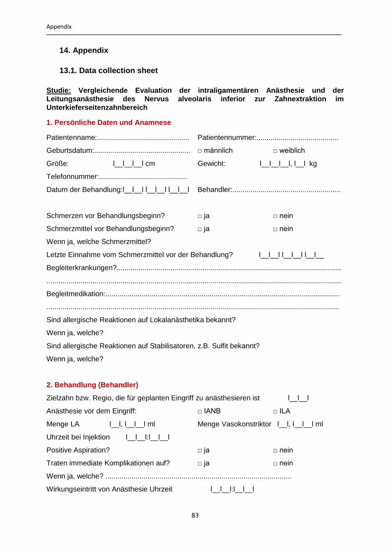
Appendix
83
14. Appendix
13.1. Data collection sheet
Studie: Vergleichende Evaluation der intraligamentären Anästhesie und der
Leitungsanästhesie des Nervus alveolaris inferior zur Zahnextraktion im
Unterkieferseitenzahnbereich
1. Persönliche Daten und Anamnese
Patientenname:.............................................. Patientennummer:.........................................
Geburtsdatum:................................................ □ männlich
□ weiblich
Größe: l__l__l__l cm Gewicht: l__l__l__l, l__l kg
Telefonnummer:............................................
Datum der Behandlung:l__l__l l__l__l l__l__l Behandler:......................................................
Schmerzen vor Behandlungsbeginn? □ ja
□ nein
Schmerzmittel vor Behandlungsbeginn? □ ja
□ nein
Wenn ja, welche Schmerzmittel?
Letzte Einnahme vom Schmerzmittel vor der Behandlung? l__l__l l__l__l l__l__
Begleiterkrankungen?................................................................................................................
...................................................................................................................................................
Begleitmedikation:.....................................................................................................................
..................................................................................................................................................
Sind allergische Reaktionen auf Lokalanästhetika bekannt?
Wenn ja, welche?
Sind allergische Reaktionen auf Stabilisatoren, z.B. Sulfit bekannt?
Wenn ja, welche?
2. Behandlung (Behandler)
Zielzahn bzw. Regio, die für geplanten Eingriff zu anästhesieren ist l__l__l
Anästhesie vor dem Eingriff: □ IANB □ ILA
Menge LA l__l, l__l__l ml Menge Vasokonstriktor l__l, l__l__l ml
Uhrzeit bei Injektion l__l__l:l__l__l
Positive Aspiration? □ ja
□ nein
Traten immediate Komplikationen auf? □ ja
□ nein
Wenn ja, welche? .............................................................................................
Wirkungseintritt von Anästhesie Uhrzeit l__l__l:l__l__l

Appendix
84
Prüfmethode □ Schleimhaut □ Zahn
Behandlungsbeginn Uhrzeit l__l__l:l__l__l
Anästhesiewirkung:
□ Vollständig □ Ausreichend □ Ungenügend □ Keine
Nachinjektion erfoderlich? □ Ja □ Nein
Wenn ja, welche Anästhesiemethode □ IANB □ ILA
Uhrzeit l__l__l:l__l__l Menge LA l__l, l__l__l ml
3. Behandlung (Patient)
Behandlungsuhrzeit l__l__l:l__l__l
Anästhesiewirkung:
□ Vollständig □ Ausreichend □ Ungenügend □ Keine
Nachinjektion erfoderlich? □ Ja □ Nein
Wenn ja, Anästhesiewirkung nach der Nachinjektion
□ Vollständig □ Ausreichend □ Ungenügend □ Keine
Ende des schmerzhaften Eingriffs Uhrzeit l__l__l:l__l__l
Ende der Gesamtbehandlung Uhrzeit l__l__l:l__l__l
Geplante Behandlung wurde in vollem
Umfang abgeschlossen
□ Ja □ Nein
Wenn nein, Grund:
□ Ungenügende Anästhesie □ anderer Grund
Schmerz bei der Injektion ( Patient) ( Skala: 0=gar nicht; 10= sehr stark
0--------1---------2--------3---------4---------5---------6---------7---------8---------9--------10
Unangenehme Behandlung (Patient)
0--------1---------2--------3---------4---------5---------6---------7---------8---------9--------10
Schmerzhafte Behandlung (Patient)
0--------1---------2--------3---------4---------5---------6---------7---------8---------9--------10
Wirkungseintritt:
Misserfolg:
4. Nachbehandlung
Datum der Nachbehandlung ggf. Patienten Kontakt? : l__l__l l__l__l l__l__l
Wie lange dauerte es bis nach Beendigung der zahnärztlichen Behandlung das
Taubheitsgefühl an Wange, Zunge und Lippe vollständig abgeklungen war?

Appendix
85
Uhrzeit l__l__l:l__l__l
Traten Beschwerden auf?
Welcher Art?
Ist Wundheilungsstörung vorhanden? □ Ja □ Nein
Zweiter Nachbehandlungstermin: Datum: l__l__l l__l__l l__l__l
Ist Wundheilungsstörung vorhanden? □ Ja □ Nein
Bemerkungen?