Proprioceptors involved in stinging response of the ... · Proprioceptors involved in stinging...
46
Instructions for use Title Proprioceptors involved in stinging response of the honeybee, Apis mellifera Author(s) Ogawa, Hiroto; Kawakami, Zenji; Yamaguchi, Tsuneo Citation Journal of Insect Physiology, 57(10): 1358-1367 Issue Date 2011-10 Doc URL http://hdl.handle.net/2115/47379 Type article (author version) File Information JIP57-10_1358-1367.pdf Hokkaido University Collection of Scholarly and Academic Papers : HUSCAP
Transcript of Proprioceptors involved in stinging response of the ... · Proprioceptors involved in stinging...

Instructions for use
Title Proprioceptors involved in stinging response of the honeybee, Apis mellifera
Author(s) Ogawa, Hiroto; Kawakami, Zenji; Yamaguchi, Tsuneo
Citation Journal of Insect Physiology, 57(10): 1358-1367
Issue Date 2011-10
Doc URL http://hdl.handle.net/2115/47379
Type article (author version)
File Information JIP57-10_1358-1367.pdf
Hokkaido University Collection of Scholarly and Academic Papers : HUSCAP

1
MS Ref. No.: IP-D-11-00093
Proprioceptors involved in stinging response of the honeybee, Apis
mellifera.
Hiroto OGAWA1,2*, Zenji KAWAKAMI1 AND Tsuneo YAMAGUCHI1
1 Department of Biology, Faculty of Science, Okayama University, Okayama 700 Japan
2 Department of Biological Sciences, Faculty of Science, Hokkaido University, Sapporo
060-0810, Japan
Key words: proprioceptor, stinging response, campaniform sensilla, mechanosensory
hair, sensory feedback, honeybee
*Correspondence to: Hiroto Ogawa
Department of Biological Sciences, Faculty of Science, Hokkaido University
Kita 10-jyo, Nishi 8, Kita-ku, Sapporo 060-0810, Japan
Phone/Fax: +81-11-706-3525
E-mail: [email protected]

2
Abstract
Two types of mechanosensitive proprioceptor organ are present on the stinging
apparatus of the honeybee: campaniform sensilla and mechanosensory hairplates. The
campaniform sensilla are located on the surface of the tapering sting-shaft, which
comprises an unpaired stylet and paired lancets. Each sensillum on the lancet differs
from that on the stylet in terms of their topography and external morphology. The
sensory afferents of the campaniform sensilla display slow-adapted firing responses to
deformation of the cuticle that would be caused by the action of inserting the sting into
a substrate, and their afferent signals induce and/or prolong the stinging response. By
contrast, the mechanosensory hairplates are located at basal cuticular plates and on the
posterior surface of the lancet valves. Two fields of hairplates on the second ramus at
the ventral edge of the groove and on the antero-lateral edge of the oblong plate respond
synchronously to protraction of the lancet. During the stinging response, these hairplates
are likely to detect any sliding movement of the lancet and its position relative to the
stylet. Afferent signals produced by them are likely to provide important information to
the neuronal circuit for the generation and modulation of the stinging motor pattern.

3
1. Introduction
The exocuticle of insects and crustaceans contains a large number of
mechanosensitive proprioceptors of various morphological types. These provide sensory
information about the state and performance of exopodites and have a part in controlling
the movement and posture of effectors during locomotion (reviewed by Bässler and
Büschges, 1998; Pearson, 1993; Zill et al., 2004). In the honeybee, Apis mellifera, two
kinds of external proprioceptor have been described on the stinging apparatus:
campaniform sensilla and hairplates with trichoid sensilla (Hermann and Douglas,
1976a, b; Shing and Erickson, 1982). The campaniform sensilla located on the sting
probably detect the depth of sting insertion by assaying the increasing cuticular
deformation that occurs with successively deeper penetrations, whereas the hairplates
located on the cuticular plates of the stinging apparatus are likely to be proprioceptors
detecting the relative position of movable parts of the sting during the stinging response
(Shing and Erickson, 1982). However, there are no published physiological studies
available on the response characteristics and functional roles of these proprioceptive
organs in the stinging response.
The stinging response is the culminating stage of the defensive behavior of the
honeybee, involving penetration of a substrate by the shaft of the sting and the release
of venom from the venom sac (Breed et al., 2004; Collins et al., 1980). Recently, the
stinging response has been used for a new conditioning protocol in the honeybee, and
has received much attention in the framework of learning and memory (Carcaud et al.,
2009; Giurfa et al., 2009; Roussel et al., 2010; Vergoz et al., 2007). During the stinging
response, in addition to the protraction of the entire shaft out of the abdomen tip, the
paired ventral parts of the sting (i.e. the ‘lancet’ versus the unpaired dorsal component,
the ‘stylet’) exhibit alternating rhythmic sliding (Snodgrass, 1956; Dade, 1962). This

4
movement results from the coordinated action of four pairs of stinging muscle (M196s,
M197s, M198s, and M199s) and enables the sting to be inserted deeper into the target.
A previous study described that afferent inputs to the terminal abdominal ganglion
(TAG), in which the central pattern generator for the stinging movements is located,
modulate the frequency of the rhythmic sliding and maintain the relationship between
cycle period and burst duration of the stinging muscle activity at various frequencies
(Ogawa et al., 1995). The sensory signals from the mechanosensitive proprioceptors in
the stinging apparatus also appear to have an effect on the stinging motor patterns, but it
is currently unknown how and which proprioceptors are involved in the modulation of
the stinging response.
In the present study, we first describe the topography, detailed morphology and
central projection of the proprioceptors, from which the effective stimulus to each
receptor is inferred. We also analyze the electrophysiological responses of these
receptors to the inferred effective stimuli, and examine the effects of the afferents on the
stinging motor activity.

5
2. Materials and methods
2.1. Animals
Specimens of honeybees (Apis mellifera L.) were obtained from outdoor colonies at
Okayama University. All the experiments presented were performed with foraging bees.
2.2. Preparation
Bees were anesthetized at 4C for 20 min. The abdomen was severed from the thorax
and pinned on a paraffin platform. Following longitudinal lateral-line incision, the terga
of the 3–6th segments and the gut were removed to expose the stinging apparatus. To
avoid any venom leaking from the preparation, the acid gland was coated with Vaseline.
2.3. Morphology
The topography and location of proprioceptors mediated by the stinging response
were examined in whole sting shafts and basal cuticular plates that were removed from
abdomens, under a transmission light microscope (BH-RFL, Olympus, Tokyo, Japan).
The exact distribution and the external structure of proprioceptors were examined with a
scanning electron microscope (SEM; T-300, JOEL, Tokyo, Japan) in isolated stinging
apparatus that had been fixed with acetone (Wako Pure Chemical, Osaka, Japan),
dehydrated, CO2-critical-point dried and then coated with gold.
Sectional planes of the campaniform sensilla were examined using a transmission
electron microscopy (TEM). The lancets and stylet dissected from the stinging
apparatus were prefixed for 2 h at 4°C with 4% glutaraldehyde (in 0.1-M sodium
phosphate buffer, pH 7.4, Sigma-Aldrich, St Louis, MO, USA). They were then
post-fixed for 2 h at 4C in a 2% OsO4 solution in the same buffer, and finally
dehydrated and imbedded in Epon 812 araldite (CY-230, Ciba Geigy, Tokyo, Japan).

6
Ultra-thin serial sections were double-stained with uranyl acetate and lead citrate, and
observed under the TEM (H-300, Hitachi, Tokyo, Japan).
To stain the central projection of the sensory neurons innervating the proprioceptors,
10% agar gel consisting of 10% NiCl2 was located either at the cut end of the sting
(stylet or lancet) or at the end of hair sensilla on the valve for 8–24 h at 4°C. After
filling, the 6th and terminal abdominal ganglia were isolated into honeybee saline (NaCl
270 mM, KCl 3.2 mM, MgCl2 10 mM, CaCl2 1.8 mM, NaHCO3 7.1 mM, Dextrose 50
mM, Tris-Buffer 10 mM, pH 7.4, Wako). The nickel ions were then precipitated within
the neurons by addition of rubeanic acid (Wako) to the honeybee saline. After fixation
with 70% ethanol, the ganglia were dehydrated and cleared with methyl salicylate for
whole-mount viewing. The ganglion stained with the nickel ion was intensified,
according to the method of Bacon and Altman (1977). The stains were drawn as a
whole mount using a camera lucida attached to the microscope (Olympus).
2.4. Electrophysiology
Electrophysiological experiments were performed with the exposed stinging
apparatus in the above-mentioned manner. For extracellular recordings of neuronal
responses of the proprioceptors, a glass suction electrode filled with honeybee saline
was placed on a cut proximal stump of the lateral nerve, A8 or A9, innervating the
proprioceptors. A reference electrode was placed in the abdomen. Campaniform sensilla
were stimulated by a stainless steel probe attached to a micromanipulator (Narishige,
Tokyo, Japan). The tip of the probe was placed on the surface of the sting, and pressed
against the cuticle. The bend of the sting shaft or the barbs of the lancet led to excitation
of the campaniform sensilla. To stimulate the hair sensilla on the second ramus or on

7
the oblong plate, a lancet was moved forward and backward alternately via a small
stainless wire connected to the arm of a vibrator.
For recordings of electromyograms (EMGs), electropolished tungsten wires (ø = 70
µm) were inserted into the stinging muscles (a protractor M198 and a retractor M199;
see Ogawa et al., 1995). A reference electrode was placed in the abdomen. Recordings
of the neuronal activity of sensory afferents and the EMGs were viewed on an
oscilloscope and stored on magnetic tape.

8
3. Results
Two types of proprioceptive sense organ were observed on the honeybee stinging
apparatus (Fig. 1). One is the campaniform sensilla, which are strain-sensitive
mechanoreceptors distributed on the long shaft of the sting. The second type is
mechanosensory hairplates located on the basal cuticular plates.
3.1. Campaniform sensilla
3.1.1. Topography and external morphologies
The campaniform sensilla on the sting were classified into two groups according to
their distributions. The first group was observed on distal barbs of the lancet (Fig.
2A,C). One sensillum was associated with each of the 2nd to 7th barbs from the tip of
the lancet, which has ten barbs in the honeybee (Fig. 3). The total number of the first
group of sensilla was 8.11 ± 1.17 (mean ±SD, number of samples = 45) for both sides of
the lancet. The second group of campaniform sensilla was observed on wide area of the
dorsolateral surface of the stylet (Fig. 2B, C). The total number of sensilla on one stylet
was 63.66 ± 10.46 (mean ± SD, number of samples =12), more than that of the sensilla
on the lancet. Most of sensilla in the second group were not distributed near the
mid-line of the shaft but near the lateral side instead. The topography suggests that the
campaniform sensilla on the lancet detect distortions of the barb when the sting is
inserted into an object, and that those on the stylet detect deformation of the entire sting
shaft during the insertion. In addition to the difference in their distributions, the sensilla
on the lancet and those on the stylet differed from each other in their external
morphology. Electron microscopic observation revealed that surface of the
campaniform sensillum on the lancet rises up quaquaversally, whereas that on the stylet
hollows in the cuticle (Fig. 4A, B). The diameter of these sensilla was 2.29 ± 0.26 µm

9
(mean ± SD, number of measured sensilla = 9, number of animals = 3) across the major
axis. The major axis of each sensillum on the lancet corresponded to the orientation of
the sting shaft (Fig. 4C).
3.1.2 Electrophysiological responses
Extracellular recording of the proximal cut-end of the lateral nerve 8 (A8; Fig. 10)
including sensory afferents from the campaniform sensilla on the lancet, showed that
deformation in the barb of lancet evoked a discharge of action potentials in A8 (Fig.
5A1). The sensory afferents fired rapidly in response to the deformation stimulus.
Although the spike discharge was sustained in the presence of an ongoing stimulus,
spike frequency of the discharge gradually declined over the stimulus time (Fig. 5B1). In
these recordings, the discharge contained some sensory units with different spike
amplitude. Bending of the sting shaft elicited bursting responses in the lateral nerve 9
(A9; Fig. 10) including the afferents from the sensilla on the stylet (Fig. 5A2, A3). The
discharge recorded from A9 also contained several spike units. Both dorsal- and
ventral-bending stimuli elicited tonic discharge of the sensory afferents in the temporal
profile (Fig. 5B2). The response of the sensilla on the stylet to the bending stimulus was
not adapted as obvious in the spike frequency compared with the response to the
distortion of the lancet barb. This result suggests that the afferent signals from the
sensilla on the lancet respond to dynamic deformation of the barb derived from the
movement of the sting into a substrate, whereas the sensilla distributed on the stylet
detect the sustained distortion of the whole shaft of the sting during the insertion.
3.1.3. Stinging motor pattern evoked by stimulus to campaniform sensilla

10
In a preparation of the isolated abdomen as described in Materials and Methods,
pinching of a tip of the lancet where the campaniform sensilla are located induced the
stinging response, during which the right and left protractor muscles, M198s,
rhythmically contracted in antiphase (Fig. 6A). This indicates that afferents of the
campaniform sensilla on the lancet provide sensory signals triggering the stinging
response. The pinching of the stylet bulb also elicited the stinging response (Fig. 6B).
However, the induction of the stinging response required such a strong pinching
stimulus that the stylet shaft was distorted. It thus appears that the threshold of the
campaniform sensilla on the stylet for triggering the stinging response is higher than
that on the lancet.
In the stinging motor pattern induced by pinching of the stylet, the duration of each
discharge (burst duration) and the interval between successive discharges (cycle period)
of M198s, which are standard parameters for describing the stinging motor activity
(Ogawa et al., 1995), were longer than those measured in the response to the lancet
pinching (Fig. 6B1). Furthermore, under a sustained pinching stimulus to the stylet, the
burst duration and the cycle period became increasingly long (Fig. 6B2). Statistic
analysis showed that both of these parameters for the motor pattern induced by stimulus
to the stylet were significantly longer than for those of the pattern induced by stimulus
to the lancet (p <0.05, independent two-sample t-test for two-tailed hypothesis,
Fig.7A1,2). This elongation of the burst duration and the cycle period has also been
observed when the sting was inserted into a soft object, such as a rubber block (Ogawa
et al., 1995). It was hypothesized that the pinching of the stylet augmented the friction
of lancet movement and prolonged the cycle period and burst duration.
Next, to clarify the function of campaniform sensilla in the modulation of the
stinging motor rhythm, we analyzed the motor patterns evoked by tactile stimulus to the
11
abdominal sternite of the honeybee, in which the whole sting was coated and fixed with
wax (Fig. 6C). In this condition, the rhythmic movement of the lancets was disturbed
without exciting the campaniform sensilla on the stylet. As was seen when the sting was
inserted into a rubber block, both the burst duration and the cycle period measured in
the wax-coated condition were significantly longer than those in intact preparations with
a non-coated, uninserted sting (p <0.05, independent two-sample t-test for two-tailed
hypothesis, Fig. 7 A1,2). By contrast, the tactile stimulus to the abdomen induced fewer
(only three) cycles of rhythmic contraction in the wax-coated condition (Fig. 6C). There
were also fewer burst cycles in the whole response in the wax-coated condition
compared with that in the rubber-inserted condition (p <0.05, Mann-Whitney's U test,
Fig. 7 A3). This result suggests that the afferent signals of campaniform sensilla on the
sting contribute to the triggering and continuance of the stinging movement, but take no
part in the elongation of the stinging motor rhythm.
Furthermore, we analyzed the relationship between the burst duration and the cycle
period measured from EMGs of M198, which is a useful index characterizing the
stinging motor program mediated by sensory feedback (Ogawa et al., 1995). In Fig. 7B,
the burst duration is plotted against the cycle period under various conditions. The
motor patterns induced by stimulus of the stylet showed a linear correlation between the
burst duration and the cycle period as well as the motor pattern evoked by tactile
stimulus to the sternite, which is one of the characteristic stinging motor patterns
(Ogawa et al., 1995, Fig. 7B1). The coefficients of correlation are 0.9383 for the data of
the stylet-stimulated pattern and 0.9607 for the data of the sternite-stimulated pattern in
the rubber-inserted condition. The first-order regression lines of the correlation are
y = 0.5409x – 11.812 for the plots the stylet-stimulated pattern and y = 0.4394x + 3.493
for the plots of the sternite-stimulated pattern in the rubber-inserted condition. Statistic

12
analysis also showed that there was no difference in the proportion of burst in the cycle
period between the stylet-stimulated and sternite-stimulated patterns in the
rubber-inserted condition (p >0.05, independent two-sample t-test for two-tailed
hypothesis, Fig. 7A4). These results demonstrated that the stinging responses triggered
by the campaniform sensilla on the stylet are the same in terms of their motor program
as the response to stimulus of the abdomen (Ogawa et al., 1995). The analysis of the
data of the motor patterns evoked by lancet-pinching revealed little correlation between
the burst duration and the cycle period (the coefficients of correlation is 0.3338),
because the plots for the lancet-stimulated patterns were more narrowly distributed than
were those for the patterns evoked by stylet or sternite stimulus. However, the burst
proportion of the lancet-stimulated pattern was close to that of the sternite-stimulated
pattern under the uninserted condition. The input from campaniform sensilla on the
lancets is also likely to evoke the stinging motor pattern. The sensory feedback for
stabilizing and modulating the motor patters probably functions successfully in the
stinging response evoked by mechanical stimulus of the campaniform sensilla.
Using this analysis, we compared the stinging motor patterns recorded under the
wax-coated condition with those under the rubber-inserted condition. The motor
patterns under the wax-coated condition showed a correlation between the burst
duration and cycle period (Fig. 7B2). The coefficient of correlation for the motor
patterns in the wax-coated condition was 0.9170. However, the first-order regression
line of the correlation for the data of the wax-coated condition (y = 0.686x–18.928) was
steeper in its slope compared with the line of the motor pattern in the rubber-inserted
condition; in addition, the proportion of burst in the cycle period recorded in the
wax-coated condition was significantly larger than that recorded in the rubber-inserted
condition (p <0.05, independent two-sample t-test for two-tailed hypothesis, Fig. 7A4).

13
It was therefore suggested that sensory feedback from the campaniform sensilla on the
sting is not essential for the basic structuring of the stinging motor program but that it
could make a minor contribution to the modulation of the duration of activity of the
stinging muscle.
3.2. Hairplate sensilla
3.2.1. External morphologies
The mechanosensory hairplates are found on three fields of movable parts of the
stinging apparatus: the first is located on the second ramus at the ventral edge of the
groove, the second is located on anterolateral edge of the oblong plate, directly beneath
the triangular plate, and the third is located on the posterior side of the lancet valve (Fig.
8). The plate on the second ramus comprised 20–30 hairs, which were arranged in one
or two lines along the first ramus (Fig. 8A). Each hair sensillum is cone shaped,
approximately 2.19±0.28 m in root diameter and 13.65±3.21 m in length (mean ± SD,
number of measured sensilla = 12, number of animals = 2). Their arrangement suggests
that the hair sensilla on the second ramus would be able to detect the rhythmic thrusting
movement of the lancet during the stinging response, because the first ramus connecting
to the lancet slides on the second ramus by contraction of the protractor M198. The
hairplate on the oblong plate is approximately 80 50 m in size and contains
approximately 20 hairs (Fig. 8B). Each hair sensillum is also cone shaped, 3.05 ± 0.32
µm in root diameter and 24.01 ± 5.20 m in length (mean ± SD, number of measured
sensilla = 18, number of animals = 2), which is slightly longer than the hairs on the
second ramus. Given that this hairplate is engaged with the triangular plate, it is likely
to be able to detect the relative position of the triangular plate connecting to the lancet
via the first ramus, in addition to thrusting of the lancet. Mechanosensory hairs on the

14
valve of the lancet, which were observed using a light microscope but not with SEM,
were thinner and longer than the cone-shaped hair sensilla on either the second ramus or
the oblong plate (Fig. 8C). It is suggested that the hairplate on the valve is likely to
detect the fluid pressure or flow velocity of the venom within the bulb of the stylet
during the stinging response.
3.2.2. Electrophysiological responses
Artificial thrusting of the lancet on the stylet with a wire probe evoked spike
discharges in the 9th lateral nerve (A9; Fig. 10), which includes afferent axons of the
hairplates on the second ramus and the oblong plate. Some spike units with a different
amplitude were contained by the discharges recorded throughout the movement. Their
response to sliding of the lancet was transient and directionally sensitive: protraction of
the lancet excited the sensory afferents, whereas retraction evoked few responses (Fig.
9A1). When the lancet was moved in alternating sinusoidal or triangular waves, the
spike frequency of the afferent units was altered in sync with both types of movement.
There was no difference between the two types of stimulus in terms of the responses of
the afferents (Fig. 9A2, A3): The afferents responded maximally during the protraction
phase to each movement (Fig. 9B). Furthermore, it was confirmed that the firing
responses of the afferents were synchronized with protraction of the lancet at different
frequencies (0.5–4.5 Hz; Fig. 9C). These results showed that the afferent of the
mechanosensory hairplates on the second ramus and the oblong plate could provide
sensory signals synchronized with the rhythmic movement of the lancet during the
stinging response. It is suggested that these afferent signals convey sensory information
about the relative position of the first ramus to the second ramus or about the stroke of
the lancet. The hairplates might have important roles in the modulation of the stinging
15
motor rhythm, such as the elongation of the burst duration and cycle period of stinging
muscle activity, when the sting is inserted into a substrate.
3.3. Central projections of afferents from proprioceptors
Afferent axons from the mechanoreceptors on the lancet and the triangular and
quadrate plates enter TAG via the 8th lateral nerve, A8, because these parts are
developed from the 8th sternum. By contrast, axons from the mechanoreceptors on the
stylet, oblong plate and sheath of the sting all of which are developed from the 9th
sternum, merge into the 9th lateral nerve, A9 with motor nerve branches innervating
stinging muscles, including M198, and then enter the TAG.
We stained the afferent fibers of the campaniform sensilla at the tip of the lancet and
the bulb of the stylet (Fig. 10A, B). Most of projections of the campaniform sensilla of
both the lancet and stylet were confined to the ipsilateral hemisphere in the TAG,
although part of the axon arborizes to the contralateral side across a mid-line.
Furthermore, other branches extend to the ipsilateral side of the 6th ganglion through a
connective nerve cord. The branches in the projection from the stylet were fewer than
those from the lancet, which might result from shortage in the uptake of the nickel into
the axons. Unfortunately, we were unable to stain the afferents of the hairplates on the
second ramus and the oblong plate, because it was technically too difficult to sort only
the nerve roots to these sensilla. The projection patterns of afferent axons from the
hairplate on the lancet valve within the TAG were similar to those of the campaniform
sensilla. However, there was no process extending to the 6th ganglion (Fig. 10C).

16
4. Discussion
4.1. Two groups of campaniform sensilla
The campaniform sensilla are the proprioceptive organs of arthropods. They are
embedded in the cuticle and are excited by compression perpendicular to their axis
(Pringle, 1938). In surface view, individual receptors appear as small-diameter, domed
caps. In the sting of the honeybee, campaniform sensilla are found on each barb of the
lancet and the stylet (Shing and Erickson, 1982). In the current study, we showed that
the two groups of campaniform sensillum on the sting differ from each other in terms of
their distribution and external morphology: the first type of sensilla is located on each
barb of the lancet and have a large domed cap at the surface of cuticle, whereas the
second type is distributed over the whole stylet and have a small cap within a cuticular
sinkhole. Their external structures appear to reflect the difference in their sensitivity and
response to deformation of cuticle. The major axis of both types of campaniform
sensillum, however, corresponds to the orientation of the sing shaft. This observation
suggests that both types of campaniform sensilla are sensitive to bending of the sting or
deformation of the barbs.
4.2. Physiological roles of campaniform sensilla
The campaniform sensilla are mechanosensitive organs that monitor forces via
cuticle strain in the exoskeleton of insects (Pringle, 1938). These sensilla are arranged
on various limbs of insects in groups with distinctive distributions, where cuticular
strain is induced both by passive loading of the leg and during muscle-driven
locomotion (Hofmann and Bässler, 1982; Hustert et al., 1981; Pringle, 1938; Spinola
and Chapman, 1975). Sensory signals from the femoral and trochanteral campaniform
sensilla affect the timing of the central pattern generator for insect walking, and have a
17
primary role in coordinating movements of the distal leg joints (Akay et al., 2001, 2004).
In the cockroach, for example, sensilla responding to an increase or decrease in the
force of the legs contribute to the onset of swing and reinforce leg coordination during
walking (Keller et al., 2007; Ridgel et al., 1999; Noah et al., 2001, 2004; Zill et al.,
2009). In our experiments, the pinching stimulus to the campaniform sensilla situated
on the sting evoked the stinging response, and the stinging movement was sustained for
as long as the sting was pinched. These results demonstrate that the sensory signals
from campaniform sensilla on the sting have key roles in trigger and continuance of the
stinging response.
The sting shaft of the honeybee is usually enfolded within the sting chamber located
at posterior end of the abdomen. Therefore, distortion in the sting activating the
campaniform sensilla is caused only if the sting is inserted into the target object. As
soon as the sting is inserted, the stinging apparatus is isolated from the main body and
left on the surface of the inserted object. Even after isolation from the body, the stinging
apparatus continues its rhythmic stinging action so that the sting shaft can be inserted
successively deeper, and the venom injected into the target. Sustained inputs from the
campaniform sensilla excited by deformation in the inserted sting are likely to be
effective enough to prolong the stinging action after isolation of the sting from the body
of the honeybee.
We also identified campaniform sensilla on the stings of various species of Aculeata,
including the Asiatic honeybee Apis cerana and four species of wasps: Polistes
chinensis A, Polistes rothneyi I, Vespa simillima X, Vespa mandarinia J (Supplemental
Fig. 1). The number and distribution of sensilla in A. cerana are identical on the lancets
and the stylet with those in A. mellifera (Supplemental Fig. 1A1). By contrast,
differences occur between honeybees and wasps in the number and distribution of the

18
campaniform sensilla on the sting, although each sensillum is similar in size and
morphology to those seen in honeybees (Supplemental Fig. B1). There are more
campaniform sensilla on the stylet of wasps compared with the stylet of honeybees; the
total number is approximately 50, 130, 160 and over 200 in P. chinensis, P. rothneyi, V.
simillima and V. mandarinia, respectively. Most of the sensilla are distributed around
the bulb of the stylet in the honeybee, whereas the campaniform sensilla are located as a
cluster at root of the stylet in the wasps (Supplemental Fig. 1B2). The stylet of the wasp
sting is slender without the ‘bulb’ in which the lancet valves pump and deposit the
venom, because wasps eject the venom by contraction of the muscle covering a venom
sac, rather than by pumping of the lancet valves (Supplemental Fig. 2). In wasps, the
muscles connecting the stinging apparatus with the abdomen are well developed, and
the sting is oriented to the target object. The cluster of the campaniform sensilla at the
base of the stylet would, therefore, provide effective sensory signals for control of the
sting toward the target. By contrast, the similar arrangement of the campaniform
sensilla on the barb of the lancet between the honeybees and the wasps (Supplemental
Fig. 1A2–5) suggests that these sensilla provide essential signals to trigger and sustain the
stinging response in various species of Aculeata.
4.3. Two types of hairplate on the stinging apparatus
The hairplate is an assembly of mechanosensory hairs in a characteristic grouping
that is excited by the deflection of each hair as it comes into contact with the exopodites
of the arthropod; thus, they monitor the movement of the effectors directly. In insects,
the hairplates are implicated in various types of proprioceptive reflex to maintain the
posture of the body and exopodites. Electrophysiological studies have shown that the
hairplates at the leg joints detect the position and movement of the legs (stick insects:

19
Wendler, 1971; Büschges and Schmitz, 1991; cockroaches: Wong and Pearson, 1976;
locusts: Bräunig and Hustert, 1985a,b). These receptors also provide sensory-feedback
signals synchronized with leg movement during walking and to effect the generation of
walking motor activity (Wong and Pearson, 1976; Wendler, 1966; Bässler, 1986).
In the stinging apparatus of the honeybee, the location and external morphology of
the hairplates give some indication of the kind of stimulus that activates them. The
hairplates with short, thick hair sensilla were identified on two fields of the basal
cuticular plates, the second rami and the oblong plates, which are attached to the
moving parts of the lancet as it slides along the stylet. These hairplates probably detect
the skeletal movements of the first ramus or the triangular plate, and provide sensory
information about the relative position and movement of the lancet on the stylet during
the stinging response. By contrast, the long, thin hair sensilla located on the distal
surface of the lancet valve appear to detect the flow of the venom as it fills the stylet
bulb. It is suggested that the afferent signal from this hairplate has a role in monitoring
the amount of venom that is drained from venom sac.
4.4. Proprioceptive feedback to the stinging pattern generator
In the stinging response of the honeybee, the rhythmically sliding movements of the
lancet are produced by alternating contractions of a set of stinging muscles (a protractor,
M198 and a retractor, M199). A study using EMG illustrated the relationships among
three temporal components of the stinging motor pattern: the interval between
successive discharges of a muscle; the duration of discharge; and the time lag between
the right and left discharges of homologous units (Ogawa et al, 1995). All three
components elongate linearly as the sting is inserted deeper into a soft substrate and the
tension on the lancets is increased. The maintenance of these relationships and

20
modulation of the stinging motor pattern require sensory feedback from the
proprioceptors on the stinging apparatus.
To effect the rhythmic motor pattern generation in the nervous system, the afferents
from the mechanoreceptors need to provide sensory signals synchronized with the
movement of the effectors. The hairplates and campaniform sensilla at the leg joint of
insects provide motor neurons and interneurons in the central pattern generator (CPG)
with periodic sensory signals synchronized with the stepping movement during walking,
and can effect the generation of walking motor activity (reviews in Burrows, 1996;
Bässler and Büschges, 1998; Zill et al., 2004). The afferents from the hairplates on the
second ramus and the oblong plate discharge spikes in phase over the protraction of the
lancet, and maximally respond to the peak of the lancet protraction. This result shows
that these proprioceptors are capable of monitoring the correct position of the lancet
during the stinging response. It is suggested that the hairplate organs on the base plates
of the stinging apparatus provide the stinging CPG with proprioceptive feedback, which
has an important role in the patterning of the stinging motor activity.
In the stinging response without a mechanical load to the sting shaft, the stinging
muscles contract in quick rhythm and the right and left stroke of the sliding of the lancet
are in synch with each other. As the sting is inserted into an object and friction of the
sting increases, the burst duration of protractor, M198 becomes longer (Ogawa et al.,
1995). This elongation, which was observed in the motor pattern evoked by mechanical
stimulus to the campaniform sensilla on the stylet, enables a constant depth of insertion
to be maintained by one contraction of M198 on each side of the stylet. This system
ensures quick and certain insertion of the sting. The information about protraction of the
lancet, which is provided by the hairplates, is vital to the control of lancet sliding for the
motor pattern generating system.
21
Deafferentation, by cutting of the sensory nerves, causes the stinging motor pattern
to become more variable and destabilized (Ogawa et al., 1995). The present study
demonstrated that the afferents from the campaniform sensilla have little effect on
modulation of the stinging motor pattern, because the burst duration is significantly
longer even if sliding of the lancets is suppressed by wax coating without distortion of
the sting shaft, as shown by the statistical analysis in Fig. 7. Furthermore, this
deafferentation of the campaniform sensilla did not alter the temporal characteristics of
the stinging motor program, including the correlation between burst duration and the
cycle period of the stinging muscle activity. Therefore, it is thought that the afferent
signals of the campaniform sensilla are not essential for stinging pattern generation.
However, owing to technical difficulties, it was impossible to record separately the
responses of the afferents from the hairplates on the second ramus and the oblong plate.
Although details of the distinct response-property of each hairplate are unclear, these
proprioceptors could provide important sensory feedback signals for stinging motor
pattern generation. Neuronal components receiving the sensory feedback from the
hairplates have been unknown in the stinging CPG. Further analysis of neuronal circuit
of the CPG will provide additional details of the sensory feedback mechanism in the
stinging response of the honeybee.

22
References
Akay, T., Bässler, U., Gerharz, P., Büschges, A., 2001. The role of sensory signals from
the insect coxa-trochanteral joint in controlling motor activity of the femur-tibia
joint. Journal of Neurophysiology, 85, 594–604.
Akay, T., Haehn, S., Schmitz, J., Büschges, A., 2004. Signals from load sensors
underlie interjoint coordination during stepping movements of the stick insect leg.
Journal of Neurophysiology, 92, 42–51.
Bässler, U., 1986. Afferent control of walking movements in the stick insect Cuniculina
impigra. II. Reflex reversal and the release of the swing phase in the restrained
foreleg. Journal of Comparative Physiology A, 158, 351-362.
Bässler, U., Büschges, A., 1998. Pattern generation for stick insect walking
movements–multisensory control of a locomotor program. Brain Research
Reviews, 27, 65–88.
Breed, M.D., Guzmán-Novoa E., Hunt, G.J., 2004. Defensive behavior of honeybees:
organization, genetics, and comparisons with other bees. Annual Review of
Entomology, 49, 271-298.
Bräunig, P., Hustert, R., 1985a. Actions and interactions of proprioceptors of the locust
hindleg coxo-trochanteral joint. I. Afferent responses in relation to joint position
and movement. Journal of Comparative Physiology A, 157, 73-82.
Bräunig, P. Hustert, R.,, 1985b. Actions and interactions of proprioceptors of the locust
hindleg coxo-trochanteral joint. II. Influence on the motor system. Journal of
Comparative Physiology A, 157, 83-89.
Büschges, A., Schmitz, J., 1991. Nonspiking pathways antagonize the resistance reflex
in the thoraco-coxal joint of stick insects. Journal of Neurobiology, 22, 224-237.
23
Burrows, M., 1996. The Neurobiology of an Insect Brain, Oxford University Press,
Oxford, pp. 393-401.
Carcaud. J., Roussel, E., Giurfa, M., Sandoz, J.C., 2009. Odour aversion after olfactory
conditioning of the sting extension reflex in honeybees. Journal of Experimental
Biology, 212, 620-626.
Collins, A.M., Rinderer, T.E., Tucker, K.W., Sylvester, H.A., Lackett, J.L., 1980. A
model of the honeybee defensive behaviour. Journal of Apicultural Research, 19,
224-231.
Dade, H.A., 1962. Anatomy and dissection of the honeybee. International Bee Research
Association, London, pp. 51-56.
Giurfa, M., Fabre, E., Flaven-Pouchon, J., Groll, H., Oberwallner, B., Vergoz, V.,
Roussel, E., Sandoz, J.C., 2009. Olfactory conditioning of the sting extension
reflex in honeybees: Memory dependence on trial number, interstimulus interval,
intertrial interval, and protein synthesis. Learning and Memory, 16, 761-765.
Hermann, H.R., Douglas, M.E., 1976. Comparative survey of the sensory structures on
the sting and ovipositor of hymenopterous insects. Journal of the Georgia
Entomological Society, 11, 223-239.
Hermann, H.R., Douglas, M.E., 1976. Sensory structures on the venom apparatus of a
primitive ant species. Annals of the Entomological Society of America, 69,
681-686.
Hofmann, T., Bässler, U., 1982. Anatomy and physiology of trochanteral campaniform
sensilla in the stick insect, Cuniculina impigra. Physiological Entomology, 7,
413–426.
Hustert, R., Pflüger, H.J., Bräunig, P., 1981. Distribution and specific central
projections of mechanoreceptors in the thorax and proximal leg joints of locusts.

24
III. External mechanoreceptors: the campaniform sensilla. Cell Tissue Research,
216, 97–111.
Keller, B.R., Duke, E.R., Amer, A.S., Zill, S.N., 2007. Tuning posture to body load:
decreases in load produce discrete sensory signals in the legs of freely standing
cockroaches. Journal of Comparative Physiology A, 193, 881-891.
Noah, J.A., Quimby, L., Frazier, S.F., Zill, S.N., 2001. Force detection in cockroach
walking reconsidered: discharges of proximal tibial campaniform sensilla when
body load is altered. Journal of Comparative Physiology A, 187, 769-784.
Noah, J.A., Quimby, L., Frazier, S.F., Zill, S.N., 2004. Sensing the effect of body load
in legs: responses of tibial campaniform sensilla to forces applied to the thorax in
freely standing cockroaches. Journal of Comparative Physiology A, 190, 201-215.
Ogawa, H., Kawakami, Z., Yamaguchi, T., 1995. Motor pattern of the stinging response
in the honeybee, Apis mellifera. Journal of Experimental Biology, 189, 39-47.
Pearson, K.G., 1993. Common principles of motor control in vertebrates and
invertebrates. Annual Review of Neuroscience 16, 265-297.
Roussel, E., Sandoz, J.C., Giurfa, M., 2010. Searching for learning-dependent changes
in the antennal lobe: simultaneous recording of neural activity and aversive
olfactory learning in honeybees. Frontiers in Behaviorsal Neuroscience, 4, 155.
Pringle, J.W.S.,1938. Proprioception in insects. II. The action of the campaniform
sensilla on the legs. Journal of Experimental Biology, 15, 114-131.
Ridgel, A.L., Frazier, S.F., Dicaprio, R.A., Zill, S.N., 1999. Active signaling of leg
loading and unloading in the cockroach. Journal of Neurophysiology, 81,
1432-1437.
Shing, H., Erickson, E.H., 1982. Some ultrastructure of the honeybee (Apis mellifera L.)
sting. Apidologie, 13, 203-213.

25
Snodgrass, R.E., 1956. Anatomy of the honeybee. Ithaca, Comstock, pp. 154-167.
Spinola, S. M., Chapman, K.M., 1975. Proprioceptive indentation of the campaniform
sensilla of cockroach legs. Journal of Comparative Physiology A, 96, 257-272.
Vergoz, V., Roussel, E., Sandoz, J.C., Giurfa, M., 2007. Aversive learning in honeybees
revealed by the olfactory conditioning of the sting extension reflex. PLoS One. 2,
e288.
Wendler, G., 1966. The co-ordination of walking movements in arthropods. Symposia
of Society for Experimental Biology, 20, 229-249.
Wendler, G., 1971. Korperhaltung bei der tabheuschrecke Carausius morosus: Ihre
Beziehung zur Schwereorientierung und Mechanismen ihrer Regelung.
Verhandlungen der Deutschen Zoologischen Gesellschaft, 65, 214-219.
Wong, R.K.S., Pearson, K.G., 1976. Properties of the trochanteral hairplate and its
function in the control of walking in the cockroach. Journal of Experimental
Biology, 64, 233-249.
Zill, S, Schmitz, J, Büschges, A., 2004. Load sensing and control of posture and
locomotion. Arthropod Structure and Development, 33, 273-286.
Zill, S.N., Keller, B.R., Duke, E.R., 2009. Sensory signals of unloading in one leg
follow stance onset in another leg: transfer of load and emergent coordination in
cockroach walking. Journal of Neurophysiology, 101, 2297-2304.

26
Figure legend
Fig. 1. Overview of the proprioceptors on the stinging apparatus. (A) Drawing
indicating location of the stinging apparatus in the honeybee (square enclosed
area). (B) Schematic illustration of the distribution of proprioceptors on the
stinging apparatus (lateral view). Dark-gray areas indicate a field on which the
mechanosensory hairplates are located, and light-gray areas indicate a field on
which the campaniform sensilla are located (see also Fig. 8). 1r, first ramus; 2r,
second ramus; AGld, acid gland; Lct, lancet; Ob, oblong plate; Qd, quadrate plate;
Sty, stylet; Tri, triangular plate; Vlv, valve on lancet; Vmsc, venom sac.
Fig. 2. Topography of campaniform sensilla on the sting. (A, B) Transmission light
micrograph of the lancet tip viewed ventrally (A) and the stylet bulb viewed
laterally (B). The preparations are whole mounted. Arrows indicate the
campaniform sensilla. (C) Drawings of the lancet and stylet in a given sample,
indicating distribution of campaniform sensilla on the sting. Left drawing in C
shows ventral side of the lancet. Middle and right drawings show dorsal and
lateral sides of the stylet, respectively. Each sensillum was marked as a black dot.
The sensilla on the lancet are located at base of the each barb by ones, while most
of sensilla on the stylet are distributed on the lateral side of the sting shaft from
bulb to tip.
Fig. 3. Distribution of the campaniform sensilla on the lancet. Total numbers of
appearance of the sensilla in 45 samples were counted along the sting: in most

27
of samples, the campaniform sensilla are observed at base of the 3rd, 4th and 5th
barbs from the tip of sting.
Fig. 4. Electron micrographs of campaniform sensilla on the sting. (A) Scanning
electron micrographs (SEM) showing external morphology of the campaniform
sensillum on the lancet (A1) and on the stylet (A2). (B) Cross section images of the
sensillum on the lancet (B1) and on the stylet (B2). (C) SEM images of ventral (C1)
and lateral (C2) views of the lancet, showing alignment of the campaniform
sensilla at base of the lancet barbs. Scale bars: 1 µm in A, B, 10 µm in C1, 5 µm in
C2.
Fig. 5. Responses of the campaniform sensilla to sting deformation. (A) Typical
recordings from afferents in the lateral nerve A8 or A9. Lower traces monitor the
movement of a stainless steel probe for deformation stimulus. In A1, the sensilla
on the lancet responded to distortion of the lancet barbs. In A2 and A3, the sensilla
on the stylet responded to ventralward or dorsalward bending of the stylet shaft,
respectively. Schematic diagrams on right insets indicate the stimulus procedures.
All responses in A were recorded from different preparations. (B) Histogram of
the spike frequency in responses of large afferent units to deformation of the barbs
(B1) and ventralward bending of the shaft (B2). Each bar shows mean firing rate in
100-ms bin, which was calculated from recordings of 4 (for B1) or 5 (for B2) trials
in different two preparations.
Fig. 6. Electromyograms of left and right protractor muscles, M198s during the stinging
responses. (A, B) Stinging motor patterns elicited by pinching stimuli to the tip of

28
lancets in A, or to the bulb of stylet in B1 and B2. Each upward arrowhead under
the lower traces in A and B1 shows a time point of transient pinching stimulus
EMG shown in B2 were stinging motor pattern when the pinching of the stylet
was retained. A line under the lower trace in B2 indicates the duration of the
pinching stimulus. Both the duration of each discharge (the burst duration) and the
interval between successive discharges (the cycle period) of M198s became
longer gradually. (C) Motor pattern evoked by tactile stimulus to sternite when the
whole sting was fixed with wax. In this condition, the rhythmic movement of the
lancets was disturbed while the campaniform sensilla were not excited. The burst
duration and the cycle period were also much longer in the stinging motor pattern
in C. The traces in A, B and C were obtained from different preparations.
Fig. 7. Statistic analysis of the stinging motor patterns. (A) Summary of four kinds of
parameters measured from EMG of M198, including the burst duration (A1), the
cycle period (A2), the number of cycles per whole response (A3) and the
proportion of the burst duration to the cycle period (A4). The data measured from
the stinging responses to pinching stimulus to the lancet (Lct) or stylet (Sty) were
showed by black bars. Gray bars indicate the data measured from the motor
patterns evoked by tactile stimulus to the abdominal sternite in different
conditions, in which the sting was not inserted (uninserted), inserted into the
rubber block (rubber) or fixed with wax (wax-coated). (B) Relationships between
the cycle period and the burst duration in the stinging motor patterns evoked by
different stimuli in various conditions. Open squares indicate the data in the
lancet-stimulated pattern. Open circles indicate the data in the style-stimulated
pattern. Cross marks, filled lozenges and gray triangle indicate the data in the

29
stenite-stimulated patterns in uninserted, rubber-inserted and wax-coated
conditions, respectively. Lines are first-order regression lines for each data set:
solid line for the patterns in rubber-inserted condition, broken lines for the
style-stimulated patterns in B1 and for the patterns in wax-coated condition in B2.
Numbers of measured-cycles were 25 for the lancet-stimulation, 51 for the
stylet-stimulation, 140 for the uninserted condition, 66 for the rubber-inserted
condition and 25 for the wax-coated condition. Numbers of responses are 5 for the
lancet stimulation, 4 for the stylet-stimulation, 45 for the uninserted condition, 8
for the rubber-inserted condition and 8 for the wax-coated condition. Numbers of
animals are 3 for the lancet-stimulation, 4 for the stylet-stimulation and 6 for the
sternite–stimulations in each type of conditions, respectively.
Fig. 8. External morphology of the hairplates on the stinging apparatus. (A, B) SEM
images of the hairplates on the 2nd ramus (A) and on the oblong plate (B). (C)
Transmission light micrographs of the hairplate on the lancet valve. Left diagram
shows location of the hairplates (dark-gray areas). Abbreviations as in Fig. 1.
Fig. 9. Responses of the hairplates to lancet movements. (A) Typical responses to three
types of the movement. A1, A2 and A3 show responses to a ramp-hold-release
protraction, periodic sliding in sinusoidal or triangular waves at 0.5 Hz,
respectively. Upper traces show extracellular recordings of the sensory afferents
within the lateral nerve A9, which innervates the hairplate on the 2nd ramus or the
oblong plate. Lower traces monitor relative position of sliding lancet, in which the
upward trace means protraction of the lancet. Peak-to-peak amplitude of the
stimulation was adjusted to maximum stroke of the lancet sliding. All traces were

30
recorded from different preparation. (B) Histograms of spike frequency obtained
from large afferent units. B1 and B2 show the responses to sinusoidal or triangular
sliding, respectively. Each bar with error bar shows mean ± SEM of the firing rate
in 100-ms bin, which was calculated from 8 cycles in the recordings shown in A2
and A3. The spike frequency was maximum at most protracted position. (C)
Responses to sinusoidal sliding of the lancet in different frequency. In C1-C4, the
lancet was periodically moved in 0.5, 1.5, 2.5 and 3.5 Hz, respectively. All
responses in C were recorded from a single preparation different from that for A
and B.
Fig. 10. Innervation and central projection of the afferents from proprioceptors on the
stinging apparatus. (A, B) Drawings of the afferent fibers from the campaniform
sensilla on the lancet in A and on the stylet in B within the 6th and terminal
abdominal ganglia. (C) Drawing of the afferent fibers from the hairplate sensilla
on the lancet valve within the TAG. 6th AG, sixth abdominal ganglion; TAG,
terminal abdominal ganglion.
Supplemental Fig. 1. Arrangement of the campaniform sensilla on the sting in five
species of bees and wasps. (A) Typical patterns in distribution of the campaniform
sensilla on the lancet of Apis cerana (A1), Polistes chinensis (A2), Polistes
rothneyi (A3), Vespa simillima (A4) and Vespa mandarinia (A5). Each sensillum is
marked as a black dot. On the sting of wasps, the campaniform sensilla are located
at not only base of the barb but also lancet shaft. (B) SEM images showing
external morphology of the campaniform sensilla on the lancet (B1) and cluster of
the campaniform sensilla located at base of the stylet (B2) in Polistes rothneyi.

31
Supplemental Fig. 2. Schematic illustration of the stinging apparatus (dorsal view) in
Polistes chinensis. Six pairs of stinging muscles (M188, M196, M197, M198 and
M199) lie on three pairs of plates (the quadrate plate, oblong plate and triangular
plate, which are visible after the removal of stinging muscles as shown on the
right side. AGld, acid gland; BGld, alkali gland; Ob, oblong plate; Qd, quadrate
plate; Sh, sheath; Tri, triangular plate; Vmsc, venom sac.

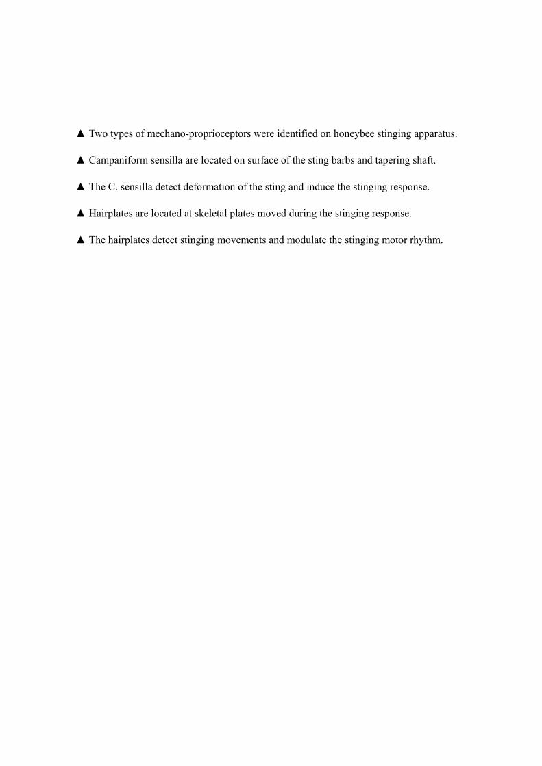
▲ Two types of mechano-proprioceptors were identified on honeybee stinging apparatus.
▲ Campaniform sensilla are located on surface of the sting barbs and tapering shaft.
▲ The C. sensilla detect deformation of the sting and induce the stinging response.
▲ Hairplates are located at skeletal plates moved during the stinging response.
▲ The hairplates detect stinging movements and modulate the stinging motor rhythm.