Program and abstractsinrcworld.org/2014/Final Program.pdfChaim Pick (Israël) Tom Prisinzano (USA)...
122
45 th meeting of the International Narcotics Research Conference Montreal, Quebec, Canada July 13‐18, 2014 Program and abstracts
Transcript of Program and abstractsinrcworld.org/2014/Final Program.pdfChaim Pick (Israël) Tom Prisinzano (USA)...

45thmeetingoftheInternationalNarcoticsResearchConferenceMontreal,Quebec,CanadaJuly13‐18,2014
Programandabstracts

Committees/Acknowledgments
Program Committee
Louis Gendron (Canada, Chair) Catherine Marie Cahill (USA, Co‐chair) Michael Bruchas (USA) Ivy Carroll (USA) Charles Chavkin (USA) Lakshmi Devi (USA) Susan Ingram (USA) Jose Moron Concepcion (USA) Minoru Narita (Japan) Amynah Pradhan (USA) Lawrence Toll (USA, Treasurer) Tuan Trang (Canada) John Traynor (USA, INRC President)
Executive Committee
John Traynor (USA, President/Secretary) Lakshmi Devi (USA, Past President) Lawrence Toll (USA, Treasurer/Vice President) Elena Bagley (Australia) Stephen Husbands (UK) Thomas Koch (Germany) Dominique Massotte (France) Jay McLaughlin (USA) Jose Moron‐Concepcion (USA) Chaim Pick (Israël) Tom Prisinzano (USA) Ichiro Sora (Japan)
Acknowledgments
This conference would have not been possible without the strong support of public institutions and sponsors.
‐ Alkermes, Inc. (USA) ‐ ASPET (The Neuropharmacology division) ‐ Centre de Recherche du CHUS (Canada) ‐ Charles River (USA) ‐ Denator AB (Sweden) ‐ Faculté de médecine et des sciences de la santé
Université de Sherbrooke (Canada) ‐ Hotchkiss Brain Institute
University of Calgary (Canada) ‐ Institut de pharmacologie de Sherbrooke
Université de Sherbrooke (Canada) ‐ Institute of Neuroscience, Mental Health and Addiction (INMHA)
Canadian Institute for Health Research (Canada) ‐ National Institute on Drug Addiction (NIDA) ‐ Quebec Pain Research Network
Fonds de la Recherche du Québec – Santé (Canada) ‐ Reckitt Benckiser Pharmaceuticals (Canada) ‐ Stoelting Co. (USA) ‐ Tourisme Montréal (Canada) ‐ Trevena Inc. (USA)
Cover Images from: Tourisme Montréal, Robert Burch, Michel Caty, Victor Lamich Diaz,, Benoit Cécile

Thank you to our Sponsors Quebec Level Sponsors
Montreal Level Sponsors
Other Sponsors Granting Agencies
The Neuropharmacology Division of ASPET has contributed towards
the ongoing training of future generations of neuropharmacologists


The Hotchkiss Brain Institute (HBI) is a neuroscience research, training
and education centre at the University of Calgary
Currently celebrating its 10th Anniversary, the HBI
connects more than 700 scientists, clinician-scientists,
trainees and staff, working to train tomorrow’s leaders in
brain and mental health.
With a seamless integration of research and education,
and a strong partnership between the science and
clinical communities, we are doing world-class
research that is of considerable benefit to the
community at large.
The HBI’s foundational research themes:
Axon Biology and Regeneration
Cerebral Circulation
Neural Systems & Behaviour
Connect to our
translational research programs:
Spinal Cord & Nerve Regeneration
Multiple Sclerosis
Stroke & Vascular Dementia
Depression & Psychosis
find out more at hbi.ucalgary.ca

CANCER: BIOLOGY, PROGNOSIS, AND DIAGNOSIS
• Understand the mechanisms underlying cancer biology. • Integrate fundamental knowledge into clinical practice. • Develop new diagnostic strategies and therapeutic approaches.
INFLAMMATION AND PAIN
• Elucidate the mechanisms underlying immune response, inflammatory diseases, and pain. • Prevent pain and inflammation. • Treat patients and provide relief.
DIABETES, OBESITY, AND CARDIOVASCULAR COMPLICATIONS
• Understand the fundamental mechanisms of diabetes and obesity. • Identify the health and social determinants underlying the development of diabetes and obesity. • Prevent cardiovascular and other consequences.
HEALTH: POPULATIONS, ORGANIZATION, AND PRACTICES
• Understand the health–disease continuum and its determinants. • Improve the health of individuals, as well as their care and services pathway. • Increase the efficacy and effectiveness of recourse to resources as well
as to preventive, diagnostic, therapeutic, and rehabilitation interven-tions.
• Develop, implement, and assess best practices.
MEDICAL IMAGERY
• Develop novel imaging approaches based on positron emission tomography (PET) and magnetic resonance imaging (MRI). • Create new radiotracers for diagnosis and therapeutic management. • Improve Canada's isotope supply chain.
MOTHER & CHILD
• Improve mother and child health, from conception to adolescence. • Understand maternofetal health, perinatal inflammation, and rare hereditary diseases. • Identify and prevent hazards related to the environment of newborns.
MULTIDISCIPLINARY RESEARCH THRUSTS AT THE CUTTING EDGE OF HEALTH ISSUES
FOR MORE INFORMATION, CLICK cr.chus.qc.ca OR CALL (819) 820-6480
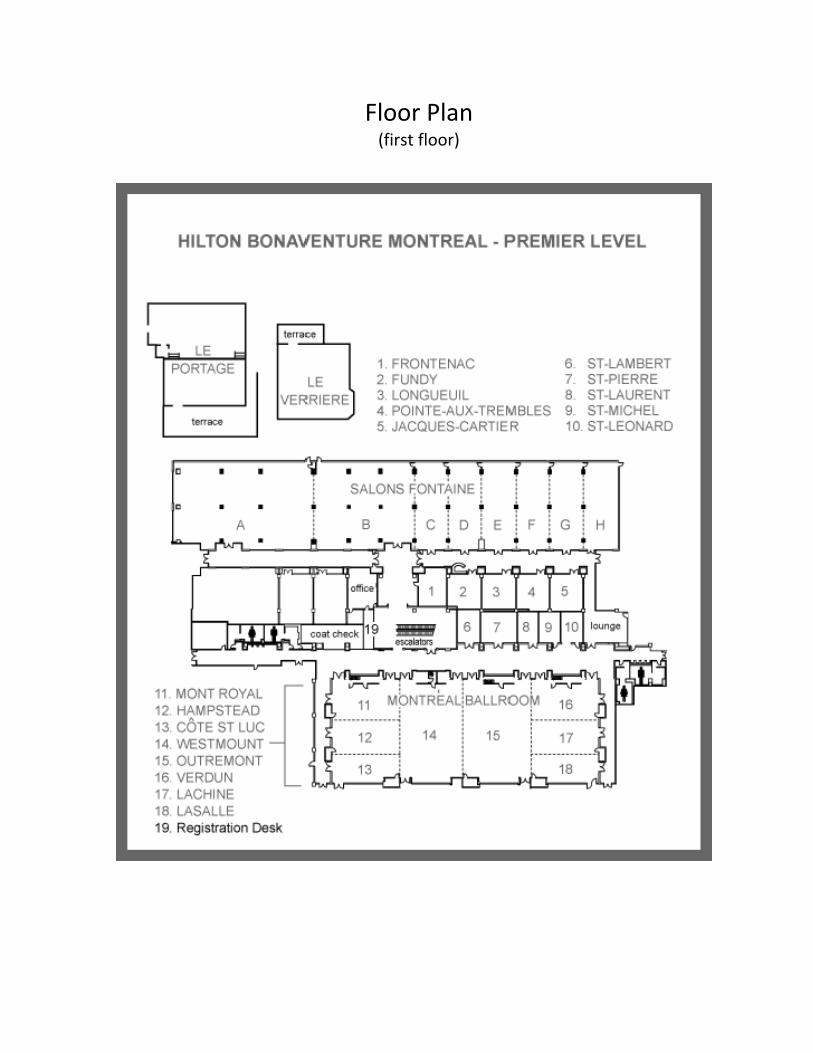
Floor Plan (first floor)

Program
Sunday July 13th 15:00‐20:00 Registration 18:00‐20:00 Welcome reception (Le Portage)
Monday July 14th 8:00‐15:00 Registration 6:30‐8:00 Breakfast provided (Fontaine A) 7:50‐8:00 Welcoming Remarks (Louis Gendron and John Traynor) Plenary lecture #1 (Westmount) 8:00‐8:45 Roger Sunahara (University of Michigan)
“Structural and functional characterization of the receptor‐G protein cooperativity” Symposium #1 (Westmount)
RECEPTOR CRYSTAL STRUCTURE AND NOVEL OPIOID CHEMISTRY Chair: Peter Schiller (Institut de Recherche Clinique de Montréal) Discussion leader: Ivy Carroll (Research Triangle Institute)
8:45‐9:10 Gustavo Fenalti (Scripps Research Institute) “Molecular Control of Opioid Receptor Signaling”
9:10‐9:35 Nurulain Zaveri (Astraea Therapeutics) “Which way is up? Rethinking Nociceptin Ligand Design and SAR From Nociceptin Opioid Receptor Crystal Structure and Active‐State Homology Model comparisons”
9:35‐10:00 Philip Portoghese (University of Minnesota) “MOR heteromers as targets for treatment of inflammatory and neuropathic pain”
10:00‐10:20 Nutritional break 10:20‐10:45 Stephen Husbands (University of Bath)
“Relapse prevention: targeting multiple receptors”
Hot Topics
10:45‐10:55 ‐D. Tourwé “Synthesis and biological evaluation of a compact, conformationally constrained bifunctional opioid agonist‐neurokinin‐1 antagonist”
10:55‐11:05 ‐K.E. Livingston “Probing Allosteric Modulation of the Mu Opioid Receptor”
11:05‐11:15 ‐P.D. Mosier “Analogs of salvinorin A bearing sulfur‐containing substituents at position C2 are high‐affinity kappa‐opioid receptor partial agonists with a potentially novel binding mode”

11:15‐11:25 ‐A.A. Harland “Synthesis, Optimization, and Evaluation of Mixed‐Efficacy ‐Opioid Receptor Agonists/ ‐Opioid Receptor Antagonists: A Balancing Act”
11:25‐11:35 ‐R.D. Howells “Novel small molecule triazoles modeled from naltrindole potently inhibit the proliferation of human multiple myeloma cells”
11:35‐12:00 Discussion period 12:00‐13:00 Lunch provided (Fontaine A) Symposium #2 (Westmount)
REVISITING THE THERAPEUTIC POTENTIAL OF DELTA OPIOID RECEPTOR LIGANDS Chair: Junzo Kamei (Hoshi University)
Discussion leader: Emily Jutkiewicz (University of Michigan) 13:00‐13:25 Gregory Scherrer (Stanford University School of Medicine)
“Delta Opioid Receptor Function in Skin: Controlling Pain Where it Starts” 13:25‐13:50 Jon Violin (Trevena Inc.)
“Biased ligands at mu and delta opioid receptors: targeting selective signalling to develop improved therapeutics”
13:50‐14:15 Manojkumar Puthenveedu (Carnegie Mellon University) “Increasing delta opioid receptor bioavailability”
14:15‐14:40 Amynah Pradhan (University of Illinois at Chicago) “The Potential of Delta Opioid Receptor Agonists for the Treatment of Migraine”
14:40‐15:00 Nutritional break
Hot Topics 15:00‐15:10 ‐M. Bigliardi‐Qi
“Delta Opioid receptors: Important Role In wound healing”
15:10‐15:20 ‐A. Saitoh “The DOR2 antagonist naltriben abolishes KNT‐127‐induced anxiolytic‐like effects in rats”
15:20‐15:30 ‐A.B. Tudashki “Ligand ability to evoke Gi activation and to promote internalization of DOR can predict response duration over a short period”
15:30‐16:00 Discussion period 16:00‐18:00 Poster session #1 (odd numbers; Fontaine B‐C)

Tuesday July 15th 8:00‐15:00 Registration 6:30‐8:00 Breakfast provided (Fontaine A) Founder’s lecture (Westmount) 8:00‐9:00 Brigitte Kieffer (McGill University) and
Christopher Evans (University of California Los Angeles) “Cloning: twice in one day”
Symposium #3 (Westmount)
CHANGES IN REWARD CIRCUITRY IN CHRONIC PAIN. Chair: Howard Fields (University of California San Francisco)
Discussion leader: Catherine M. Cahill (University of California Irvine) 9:00‐9:25 Marco L. Loggia (Harvard Medical School)
“Disrupted brain circuitry for pain‐related reward/punishment in fibromyalgia”
9:25‐9:50 Lucia Hipolito (Columbia University) “Chronic pain increases opioid self‐administration through an accumbal dopaminergic mechanism”
9:50‐10:10 Nutritional break
10:10‐10:35 Anna Taylor (University of California Irvine)
“Neuropathic Pain Modulates Dopaminergic Circuitry: Role for Microglial Activation” 10:35‐11:00 Zhizhong Pan (University of Texas)
“Pain facilitates response to opioid reward via a shared epigenetic pathway” Hot Topics
11:00‐11:10 ‐S.D. Comer “Abuse liability of oxycodone and morphine in buprenorphine‐maintained participants with or without chronic pain”
11:10‐11:20 ‐A. Bendiksen “The Use of Opioids in the Treatment of Young People with Chronic Non‐Malignant Pain at a Multidisciplinary Pain Clinic in Denmark”
11:20‐11:30 ‐I. Gomes “Analysis of Morphine Regulated Striatal Synaptic Networks using Proteomics and Network Analysis”
11:30‐12:00 Discussion period 12:00‐13:00 Lunch provided (Fontaine A) 12:00‐13:00 Lunch (Executive committee) (Salon Fundy) Symposium #4 (Westmount)
DISSECTING THE FUNCTIONS OF NOCICEPTIN/ORPHANIN FQ RECEPTORS Chair: Michael Bruchas (Washington University)
Discussion leader: Larry Toll (Torrey Pines Institute)
13:00‐13:25 Raül Andero Galí (Emory University) “Amygdala‐dependent fear is regulated by opioid receptor‐like 1 in mice and humans with PTSD”
13:25‐13:50 Kelly Standifer (University of Oklahoma College of Pharmacy) “Neuroinflammatory actions of N/OFQ following traumatic stress”
13:50‐14:15 Thomas Jhou (Medical University of South Carolina) “Nociceptin/Orphanin‐FQ in the rostromedial tegmental nucleus”
14:15‐14:40 Robert Innis (National Institutes of Mental Health) “Imaging of nociceptin/orphanin FQ peptide (NOP) receptors in human brain and whole‐body using a novel positron emission tomography radioligand, [11C]NOP‐1A”
14:40‐15:00 Nutritional break
Hot Topics 15:00‐15:10 ‐A. Ozawa
“EGFP‐NOP Mice: Location in Spinal Cord and DRG”
15:10‐15:20 ‐S.M. Spangler “Dissecting Nociceptin Receptor Modulation of Reward”
15:20‐15:30 ‐Y. Zhang “Effect of the nociception/orphanin FQ peptide receptor antagonist JTC‐801 on cytokine expression following exposure to the single prolonged stress model of PTSD”
15:30‐16:00 Discussion period 16:00‐18:00 Poster session #2 (even numbers; Fontaine B‐C)
Wednesday July 16th 8:00‐12:00 Registration 6:30‐8:00 Breakfast provided (Fontaine A) Plenary lecture #2 (Westmount) 8:00‐8:45 Antonello Bonci (NIDA intramural program director) “Optogenetic approaches to study synaptic plasticity and substance abuse” Symposium #5 (Westmount)
DYSPHORIA AND AVERSION IN ADDICTION AND CHRONIC PAIN Chair: Chris Evans (University of California Los Angeles) Discussion leader: Charles Chavkin (University of Washington) 8:45‐9:10 Julie Kauer (Brown University)
“Acute stress activates kappa opioid receptors and triggers persistent synaptic changes in the VTA”
9:10‐9:35 Joel Schlosburg (Scripps Research Institute, San Diego) “Dynorphin‐kappaopioidreceptoractivationregulatesthedynamicsofdrug‐takingintheprogressiontowardsaddiction”
9:35‐10:00 Brendan Walker (Washington State University) “Quiescent kappa‐opioid receptors in the medial prefrontal cortex: the rollercoaster of cognitive control”

10:00‐10:20 Nutritional break
Hot Topics 10:20‐10:30 ‐A. Riegel
“The Functional Rewiring of Cortical Synapses in a Translational Model of Neuropathic Pain” 10:30‐10:40 ‐L.G. Rosen
“Opiate exposure state controls a molecular switch in opiate reward memory formation in the basolateral amygdala‐prefrontal cortical pathway”
10:40‐10:50 ‐A. Lesniak “Opioid receptor expression in mice with persistent, long‐term neuropathic pain”
10:50‐11:00 ‐S.Schattauer“Ligand‐directedsignalingatkappaopioidreceptors:differentialmechanismsofJNKMAPKactivationbyU50,488andnorBNI”
11:10‐11:20 ‐G.R.Matyas“Development of a Vaccine against Heroin”
11:20‐11:45 Discussion period 12:00‐13:30 Data Blitz (lunch boxes provided)
Thursday July 17th
8:00‐15:00 Registration 6:30‐8:00 Breakfast provided (Fontaine A) Plenary lecture #3 (Westmount) 8 :00‐8:45 Yves De Koninck (Université Laval)
“Expanding your toolbox; new approaches to probe the brain with light”
Symposium #6 (Westmount) (Sponsored by the CIHR’s Institute of Neuroscience, Mental Health and Addiction)
WORKSHOP ON NEW TECHNOLOGIES IN NEUROSCIENCE AND DRUG ADDICTION Chair:Fred Nyberg (Uppsala University) Discussion leader: Jose Moron‐Concepcion (Columbia University)
8:45‐9:10 Maxime Descoteaux (Université de Sherbrooke) “Diffusion MRI: imaging the wiring of the brain”
9:10‐9:35 Philippe Seguela (McGill University) "Optogenetic control of pain pathways in vivo"
9:35‐10:00 Sotiris Masmanidis (University of California Los Angeles) “Large‐scale electrophysiology and virtual reality for reward circuit research”
10:00‐10:20 Nutritional break
Hot Topics 10:20‐10:30 ‐K. Skold
“Post‐sampling changes in opioid peptide levels and the effect of tissue stabilization”

10:30‐10:40 ‐A.K. Fakira “NR2B‐mediated changes in hippocampal spine morphology following morphine conditioned place preference”
10:40‐10:50 ‐M.J. Schmidt “Spatiotemporal Control of Mu‐Opioid Signaling and Behavior”
10:50‐11:15 Discussion period Symposium #7 (Westmount)
DISSECTING OPIOID NEURAL CIRCUITS 11:15‐12:00 YOUNG INVESTIGATOR AWARD SYMPOSIUM
Chair: John Traynor (University of Michigan)
Discussion leader: Elyssa Margolis (University of California San Francisco) Michael Bruchas (Washington University) “Spatiotemporal Control of Opioid Systems in Reward and Aversion”
12:00‐13:00 Lunch provided (Fontaine A) 13:00‐13:25 Thomas Kash (University of North Carolina)
“Dynorphin controls the gain of an amygdalar anxiety circuit“ 13:25‐13:50 Matthew Banghart (Harvard University)
“A photochemical approach to peptidergic neuromodulation” Hot Topics
13:50‐14:00 ‐E.N. Bobeck “Endosomal Endothelin Converting Enzyme‐2: A regulator of opioid receptor trafficking”
14:00‐14:10 ‐K.J. Tonsfeldt “Extrasynaptic GABAA receptors in the periaqueductal gray are involved in descending pain modulation and chronic inflammatory pain‐induced plasticity”
14:10‐14:20 ‐L. Kotecki “GIRK channel activation in midbrain GABA neurons is not required for opioid‐induced locomotor stimulation”
14:20‐14:45 Discussion period 14:45‐15:00 Nutritional break Symposium #8 (Westmount)
GENETIC AND EPIGENETIC REGULATION IN ADDICTION AND CHRONIC PAIN Chair: Laura Stone (McGill University) Discussion leader: Mary Jeanne Kreek (The Rockefeller University)
15:00‐15:25 Luda Diatchenko (McGill University) “Expansion of mu‐opioid receptor gene locus: new functional variants”
15:25‐15:50 Pamela Kennedy (University of California Los Angeles) “Chromatin Cross‐Talk in the Nucleus Accumbens: Implications for Cocaine‐Induced Behavioral and Molecular Adaptations”
15:50‐16:15 Marcello Wood (University of California Irvine) “The role of HDAC3 in acquisition and extinction of drug‐seeking behavior”

Hot Topics
16:15‐16:25 ‐O. Levran “Dopaminergic system gene polymorphisms and heroin addition: further support for a protective effect of casein kinase 1e (CSNK1E) variants”
16:25‐16:35 ‐J.S. Wieskopf “Broad spectrum analgesic effcacy of IBNtxA is mediated by exon 11‐associated splice variants of the mu‐opioid receptor gene”
16:35‐16:55 Discussion period 16:55‐17:45 Business meeting (Westmount) Shuttles to the Banquet dinner leave between 18:15 and 18:30 from the Hilton 19:00‐late Banquet Dinner
Friday July 18th
End of the Conference and Departure

INRC 2014 Awardees
Dr Brigitte Kieffer
B. L. Kieffer is Professor at McGill Univerity and at the Université de Strasbourg France. Her team uses mouse genetic approaches to tackle the role of opioid receptors in brain physiology and disorders, and to search for novel genes involved in psychiatric disorders. She has developed and shared exquisite genetic tools worldwide, and has developed innovative research lines with strong impact in neuroscience and biomedical research. Her work has important implications for the development of treatment of pain, drug abuse and emotional disorders. She is part of the Center for Opioid Receptors and Drugs of Abuse or CSORDA.
Pr. Kieffer is recipient of the Jules Martin (French Academy of Science, 2001) and the Lounsbery (French and US Academies of Science, 2004) Awards, and has become an EMBO Member in 2009. In 2012 she received the Lamonica Award of Neurology (French Academy of Science) and was nominated Chevalier de la Légion d’honneur. In December 2013 she was elected as a member of the French Academy of Sciences. She started as the Scientific Director of the Douglas Hospital Research Centre, affiliated to McGill University in January 2014.
Dr Christopher Evans Christopher Evans received his Ph.D. from Imperial College London, conducting his thesis research on endorphins and enkephalins, at the Medical Research Council Institute in Mill Hill. After a postdoctoral fellowship at Stanford University, he joined the UCLA faculty in the Department of Psychiatry and Biobehavioral Science. His research accomplishments have included identification of a number of novel endogenous opioid peptides and the cloning of the first opioid receptor.
Dr. Evans is currently Director of the UCLA Brain Research Institute and the Stefan Hatos Professor directing the Shirley and Stefan Hatos Center for Neurophamacology in the UCLA Semel Institute. He is also director of a NIH‐funded center – The Center for Opioid Receptors and Drugs of Abuse or CSORDA with the broad aim of understanding the action of opioid drugs such as morphine and heroin at the molecular, cellular and behavioral levels.
Dr Michael Bruchas Michael Bruchas’s research training is in GPCR pharmacology and neuroscience. His graduate work focused on adrenergic receptors while his post‐doctoral work was in the department of pharmacology, at the University of Washington, Seattle, in Charles Chavkin’s laboratory. Here he studied opioid receptor biased signaling in behavior using mouse genetics and behavioral approaches. His laboratory at Washington University (St. Louis) investigates interactions between stress, GPCRs, neuropeptides, and neural circuits in affective behavior.
Recent efforts by his group have focused on developing tools for wireless optogenetics and optical GPCRs for examining signaling pathways and behavior in vivo. Using a variety of genetic, pharmacological, engineering, and optogenetic approaches, he will discuss recent efforts by his team to dissect the role of opioid peptides and receptor signalling in models of affective behavior.
Founder’s lecture
Young Investigator Award

Dr Brigitte Kieffer, Founder’s Award, INRC 2014
When a scientist receives an honour it is usually for a single major contribution to advancing the field, but for Brigitte Kieffer I would identify three major contributions to opioid science which have been world‐leading and have had a very significant impact on both INRC and on the wider scientific community. Firstly, her cloning of the delta opioid receptor (published in PNAS), secondly the development and characterisation of the mu opioid receptor knockout mouse (published in Nature) and thirdly the development and characterisation of the first conditional opioid receptor knockout mouse, with a fascinating behavioural phenotype that will be published shortly. In between there have been many other significant publications, including really elegant work on the role of opioids in emotional responses (in Nature Genetics) and in maternal attachment (in Science).
Brigitte first came to an INRC meeting in 1990 at Noordwijkerhout, and at that point several laboratories had spent nearly a decade trying to clone the first opioid receptor. In the 1980’s I listened to a lot of talks on the cloning of the opioid receptor that really had nothing to say! I felt sorry for a string of PhD students who had to write a thesis with precious little to show for their heroic efforts. So when Brigitte Kieffer and Chris Evans independently succeeded in cloning the first opioid receptor in 1992 these were landmark publications. Chris published in Science and Brigitte published in PNAS, though I know she originally submitted the manuscript to Nature. The editors foolishly said “it was just the cloning of another G‐protein coupled receptor” and rejected the manuscript. This paper is now cited over 850 times and I am sure the editors of Nature are kicking themselves. Nature did not make the same mistake twice and when Brigitte submitted her phenotypic characterisation of the first opioid receptor knockout mouse in December 1996 the referee and editors reports were glowing. The citation count for this paper is now approaching four figures. She presented this seminal work at the INRC meeting in Long Beach earlier that year; a clearcut deletion of the mu opioid receptor and a remarkable loss of key behaviours that this receptor is involved in. I recall there was stunned silence in the room, as INRCers listened to this major field advance. Avram Goldstein who was standing next to me remarked “this is almost too good to be true”. But it was one of those set of experiments that we would all love to be involved with; I was privileged to be one of the collaborators on this work. Sometimes, experiments don’t work out as you had planned. Brigitte had set out to make a delta opioid receptor knockout mouse, and succeeded first in making a mu knockout – the kappa gene knockout came next (again a first) and the delta knockout last. Sometimes the direction of science is rather serendipitous.
There is a third landmark discovery to come and I suspect in her Founders lecture she might talk about her work in developing a conditional deletion of an opioid receptor to enable us to identify the role of these receptors in specific regions in the brain. This work and the previous development of knockout mice, have taken many years and stoic perseverance to come to fruition. It reflects Brigitte’s critical attention to detail, a characteristic I have seen when writing both grant applications and research publications with her. She has very many successful collaborations in both Europe and worldwide, and in looking at the author lists of her papers you might think she collaborates with everyone. This is not true – she chooses her collaborators very carefully. I remember her telling me when she cloned the delta opioid receptor that she gave the clones to everyone who asked; but far too many of those that asked did not do anything useful with them! So when the opioid knockouts were developed she took a much more cautious approach, and it has delivered over 200 high quality publications.
Brigitte is a person who is in love with her science and her success has been recognised by four national prizes from both French and US academies of science. She was appointed as Director of the world‐leading IGBMC in Strasbourg in 2012 where most of her opioid research has been carried out, but she always said that she must not allow leadership and management to compromise the important science she still had to do. So this year she moved to be scientific director of the Douglas Institute at McGill University in Canada – there is more to come, investigating opioid involvement in emotion.
For me, it has been an honour to collaborate with Brigitte for 20 years. There has been some great science and great friendship. In the early 1980’s you would have found Hans Kosterlitz (one of the founders of INRC) in the bar late at night at INRC meetings, working hard and playing hard. From 1992, you would find Chris Evans and Brigitte Kieffer (and me!) there too. As I said, great science and great friendship; it is what INRC is all about.
Professor Ian Kitchen University of Surrey, UK April 2014
Dr Christopher Evans, Founder’s Lecture Award, INRC 2014
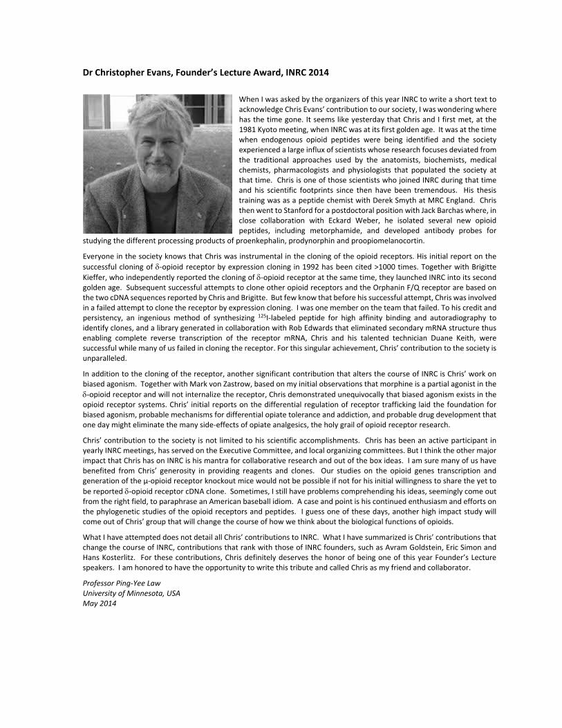
When I was asked by the organizers of this year INRC to write a short text to acknowledge Chris Evans’ contribution to our society, I was wondering where has the time gone. It seems like yesterday that Chris and I first met, at the 1981 Kyoto meeting, when INRC was at its first golden age. It was at the time when endogenous opioid peptides were being identified and the society experienced a large influx of scientists whose research focuses deviated from the traditional approaches used by the anatomists, biochemists, medical chemists, pharmacologists and physiologists that populated the society at that time. Chris is one of those scientists who joined INRC during that time and his scientific footprints since then have been tremendous. His thesis training was as a peptide chemist with Derek Smyth at MRC England. Chris then went to Stanford for a postdoctoral position with Jack Barchas where, in close collaboration with Eckard Weber, he isolated several new opioid peptides, including metorphamide, and developed antibody probes for
studying the different processing products of proenkephalin, prodynorphin and proopiomelanocortin.
Everyone in the society knows that Chris was instrumental in the cloning of the opioid receptors. His initial report on the
successful cloning of ‐opioid receptor by expression cloning in 1992 has been cited >1000 times. Together with Brigitte
Kieffer, who independently reported the cloning of ‐opioid receptor at the same time, they launched INRC into its second golden age. Subsequent successful attempts to clone other opioid receptors and the Orphanin F/Q receptor are based on the two cDNA sequences reported by Chris and Brigitte. But few know that before his successful attempt, Chris was involved in a failed attempt to clone the receptor by expression cloning. I was one member on the team that failed. To his credit and persistency, an ingenious method of synthesizing 125I‐labeled peptide for high affinity binding and autoradiography to identify clones, and a library generated in collaboration with Rob Edwards that eliminated secondary mRNA structure thus enabling complete reverse transcription of the receptor mRNA, Chris and his talented technician Duane Keith, were successful while many of us failed in cloning the receptor. For this singular achievement, Chris’ contribution to the society is unparalleled.
In addition to the cloning of the receptor, another significant contribution that alters the course of INRC is Chris’ work on biased agonism. Together with Mark von Zastrow, based on my initial observations that morphine is a partial agonist in the
‐opioid receptor and will not internalize the receptor, Chris demonstrated unequivocally that biased agonism exists in the opioid receptor systems. Chris’ initial reports on the differential regulation of receptor trafficking laid the foundation for biased agonism, probable mechanisms for differential opiate tolerance and addiction, and probable drug development that one day might eliminate the many side‐effects of opiate analgesics, the holy grail of opioid receptor research.
Chris’ contribution to the society is not limited to his scientific accomplishments. Chris has been an active participant in yearly INRC meetings, has served on the Executive Committee, and local organizing committees. But I think the other major impact that Chris has on INRC is his mantra for collaborative research and out of the box ideas. I am sure many of us have benefited from Chris’ generosity in providing reagents and clones. Our studies on the opioid genes transcription and generation of the µ‐opioid receptor knockout mice would not be possible if not for his initial willingness to share the yet to
be reported ‐opioid receptor cDNA clone. Sometimes, I still have problems comprehending his ideas, seemingly come out from the right field, to paraphrase an American baseball idiom. A case and point is his continued enthusiasm and efforts on the phylogenetic studies of the opioid receptors and peptides. I guess one of these days, another high impact study will come out of Chris’ group that will change the course of how we think about the biological functions of opioids.
What I have attempted does not detail all Chris’ contributions to INRC. What I have summarized is Chris’ contributions that change the course of INRC, contributions that rank with those of INRC founders, such as Avram Goldstein, Eric Simon and Hans Kosterlitz. For these contributions, Chris definitely deserves the honor of being one of this year Founder’s Lecture speakers. I am honored to have the opportunity to write this tribute and called Chris as my friend and collaborator.
Professor Ping‐Yee Law University of Minnesota, USA May 2014

TABLE OF CONTENTS
Plenary lectures
Roger K. Sunahara 12Allosteric modulation of the orthosteric binding site by G proteins: mechanism of high affinity agonistbinding
Antonello Bonci, MD 13Optogenetic approaches to understanding synaptic plasticity and substance use disorders.
Yves De Koninck 14Expanding your toolbox; new approaches to probe the brain with light
Founder’s lecture
Brigitte Kieffer and Christorpher Evans 15Cloning: Twice in One Day
Young Investigator Award
Michael R. Bruchas 16Spatiotemporal Control of Opioid Systems in Reward and Aversion
Symposium #1
Gustavo Fenalti 17Molecular Control of δ-Opioid Receptor Signaling
NURULAIN T. ZAVERI, Ph.D. 18Which way is up? Rethinking Nociceptin Ligand Design and SAR from Nociceptin Receptor CrystalStructure and Active-State Homology Model comparisons
Philip S. Portoghese 19MMG22 selectively targets a putative MOR-mGluR5 heteromer for treatment of chronic inflammatorypain
Stephen Husbands 20Potential Relapse Prevention Agents Targetting Multiple Opioid Receptors
Symposium #2
Gregory Scherrer 21Delta Opioid Receptor Function in Skin: Controlling Pain Where it Starts
Jonathan Violin 22Biased ligands at mu and delta opioid receptors: targeting selective signalling to develop improvedtherapeutics
Manojkumar A. Puthenveedu 23Increasing delta opioid receptor bioavailability
Amynah A Pradhan 24Delta Opioid Receptor Agonists for the Treatment of Migraine
Symposium #3
Marco L. Loggia 25Disrupted Brain Circuitry for Pain-Related Reward/Punishment in Fibromyalgia
1
Lucia Hipolito 26Chronic pain increases opioid self-administration through an accumbal dopaminergic mechanism
Anna Taylor 27Neuropathic Pain Modulates Dopaminergic Circuitry: Role for Microglial Activation
Zhizhong Pan 28Pain facilitates response to opioid reward via a shared epigenetic pathway
Symposium #4
Raul Andero Galı 29Amygdala-dependent fear is regulated by Oprl1 in mice and humans with PTSD
Kelly Standifer 30NEUROINFLAMMATORY ACTIONS OF N/OFQ FOLLOWING TRAUMATIC STRESS
Thomas Jhou 31Nociceptin/Orphanin-FQ and opioid mechanisms involving the rostromedial tegmental nucleus
Robert Innis 32Imaging of nociceptin/orphanin FQ peptide (NOP) receptors in human brain and whole-body using anovel positron emission tomography radioligand, [11C]NOP-1A
Symposium #5
Julie A. Kauer 33Blocking kappa opioid receptors, even after stress, rescues LTPGABA and prevents reinstatement ofcocaine seeking
Joel E Schlosburg 34Dynorphin-kappa opioid receptor activation regulates the dynamics of drug-taking in the progressiontowards addiction
Brendan M. Walker 35Quiescent kappa-opioid receptors in the medial prefrontal cortex: the rollercoaster of cognitive control
Symposium #6
Maxime Descoteaux 36Diffusion MRI : imaging the wiring of the brain
Philippe Seguela 37Optogenetic control of pain pathways in vivo
Sotiris Masmanidis 38Large-scale electrophysiology and virtual reality for reward circuit research
Symposium #7
Thomas Kash 39Kappa Opioid Receptor signaling controls the gain of an amygdalar anxiety circuit
Matthew Banghart 40A photochemical approach to peptidergic neuromodulation
Symposium #8
2

Luda Diatchenko 41Mu-opioid receptor gene locus: new functional variants
Pamela Kennedy 42Chromatin Cross-Talk in the Nucleus Accumbens: Implications for Cocaine-Induced Behavioral andMolecular Adaptations
Marcelo A. Wood 43The role of HDAC3 in acquisition and extinction of drug-seeking behavior
POSTERS
1. A. Samoshkin 45Structural and functional interaction between 6TM MOR isoform and β2-adrenoreceptors
2. Aaron M. Bender 45Synthesis and evaluation of mixed efficacy mu opioid receptor (MOR), delta opioid receptor (DOR)ligands
3. Achla Gupta 46The fungal collybolides as a new class of sesquiterpenes that selectively interact with the kappa opioidreceptor
4. Adie Wilson-Poe 46Chronic Morphine Treatment Alters Cannabinoid Modulation of GABAergic Synaptic Transmission inthe Periaqueductal Grey
5. Akihiko Ozawa 47EGFP-NOP Mice: Location in Spinal Cord and DRG
6. Akiyoshi Saitoh 47The DOR2 antagonist naltriben abolishes KNT-127-induced anxiolytic-like effects in rats.
7. Amanda K. Fakira 48NR2B-mediated changes in hippocampal spine morphology following morphine conditioned place pref-erence
8. Andrea Bedini 48Prolonged activation of Toll-like receptor 4 (TLR4) down-regulates NOPr expression in human glioblas-toma cells, hampering nociceptin ability to counteract TLR4-mediated elevation of [Ca2+]i and induc-tion of NF-kB
9. Andrzej W. Lipkowski 49Neuroprotective effects of biphalin in a mouse model of mild traumatic brain injury (mTBI)
10. Anette Bendiksen 49The Use of 0pioids in the Treatment of Young People with Chronic Non-Malignant Pain at a Multidis-ciplinary Pain Clinic in Denmark.
11. Anna Lesniak 50Opioid receptor expression in mice with persistent, long-term neuropathic pain
12. Arthur Riegel 50The Functional Rewiring of Cortical Synapses in a Translational Model of Neuropathic Pain
13. Aubrie A. Harland 51Synthesis, Optimization, and Evaluation of Mixed-Efficacy µ-Opioid Receptor Agonists/ δ-Opioid Re-ceptor Antagonists: A Balancing Act
3

14. Bernard P. Roques 51DENKIs (Dual Enkephalins Inhibitors), a pharmacological and therapeutic breakthrough
15. Bigliardi, PL 52Delta Opioid receptors: Important Role In wound healing
16. Brady Atwood 52Effects of alcohol on mu opioid receptor-mediated plasticity at dorsal striatal inputs
17. Brenda M. Ray 53Naltrexone, like Nalmefene, is a Kappa opioid receptor partial agonist
18. Brian Cox 53Effects of morphine on GABAergic plasticity on dopamine neurons of the VTA: roles of PKA, AKAP,calcineurin, and GABA-A receptor trafficking
19. Brian Reed 54Trypanosoma brucei as Novel Vaccine Platform for Oxycodone
20. C. Mollereau 54Mass spectrometry analysis of agonist-induced MOP receptor phosphorylation in the mouse brain.
21. CJ Schier 55The activity of HIV-1 Tat and morphine on Toll-like receptor 4 signaling Implications for mechanism ofactions contributing to HIV-related neuroinflammation
22. Caroline C. Churchill 55Adeno-associated virus mediated gene transfer of arginine decarboxylase reduces morphine analgesictolerance.
23. Christine Neumann 56The Delta-Opioid Receptor Affects Epidermal Homeostasis via ERK-Dependent Inhibition of Transcrip-tion Factor POU2F3
24. Christoph Stein 56Effect of Peripheral Opioid Receptor Blockade on Opioid Analgesic Demand: A Randomized ClinicalTrial
25. Christopher R. McCurdy 57SYNTHESIS AND EVALUATION OF DUAL-ACTIVITY OPIOID-NPFF LIGANDS FOR RECEPTORAFFINITY, ANTINOCICEPTION AND TOLERANCE LIABILITIES.
26. Courtney Donica 57Platelet-Derived Growth Factor Receptor-beta Antagonism Restores Morphine Analgesic EfficacyAgainst Neuropathic Pain
27. Cristina D Peterson 58Intrathecal delivery of a bivalent ligand targeting a mu opioid Receptor mGLuR5 Receptor heteromerreduces nerve-injury hyperalgesia
28. Cullen L. Schmid 58Novel ligands at the mu opioid receptor that display bias toward G protein coupling over βarrestin2recruitment
29. Dana Selley 59Cannabinoid CB1 Receptor Induction of ΛFosB Mediates Mu Opioid Receptor Cross-Sensitization inthe Nucleus Accumbens
4

30. Derek Simon 59Calmodulin-dependent Kinase II Protein is Increased in the Hippocampus of Oxycodone Self-administered Adult C57Bl/6 Mice.
31. Devon Collins 60Oxycodone causes greater locomotor activation in female compared to male C57BL/6J mice
32. Dirk Tourwe 60SYNTHESIS AND BIOLOGICAL EVALUATION OF A COMPACT, CONFORMATIONALLY CON-STRAINED BIFUNCTIONAL OPIOID AGONIST - NEUROKININ-1 ANTAGONIST
33. Dominique Massotte 61CO-LOCALIZATION OF MU AND DELTA OPIOID RECEPTORS IN THE NERVOUS SYSTEM USINGDOUBLE FLUORESCENT KNOCK-IN MICE
34. Dr. Andrea Bedini 61Ligand-directed signaling at mu opioid receptors: differential mechanisms of MAPK activation by mor-phine and fentanyl
35. Dr. Selena Schattauer 62Nalfurafine is a KOR agonist that produces analgesia, pruritus, inhibition of luteinizing hormone release,and ERK1/2 MAPK activation, but has low potency for p38 MAPK activation
36. Dr. Selena Schattauer 62Ligand-directed signaling at kappa opioid receptors: differential mechanisms of JNK MAPK activationby U50,488 and norBNI
37. Eriko Nakata 63OPIOID DELTA RECEPTOR-MEDIATED β-ARRESTIN SIGNALING MODULATES CONVULSIVEEFFECTS
38. Erin Bobeck 63Endosomal Endothelin Converting Enzyme-2: A regulator of opioid receptor trafficking
39. Francia Fang 64Increased Emergency Department Visits Caused by Tramadol Abuse are Associated with Opioid-RelatedOver-Dose Death in the U.S.
40. Fraser McIntosh 64Tramadol is an effective analgesic in an acute and chronic rat pain model
41. Gary R. Matyas 65Development of a Vaccine against Heroin
42. Georgina Thompson 65Comprehensive analysis of factors influencing quantification of biased signalling at the mu-opioid re-ceptor
43. Gina F. Marrone 66Supraspinal endomorphin analgesia requires exon 11 variants of the mu opioid receptor (MOR-1)
44. H. Machelska 66Pain inhibition by leukocytic opioid receptors
45. Hamid I Akbarali 67The role of beta-arrestin2 in the development of morphine tolerance in the enteric nervous system
5

46. Hanieh Bagheri Tudashki 67Ligand ability to evoke Gαi activation and to promote internalization of DOR can predict responseduration over a short period.
47. Heather Leduc-Pessah 68Involvement of Microglial P2X7 Receptors in the Development of Morphine Analgesic Tolerance
48. Heather Leduc-Pessah 68Toll-like receptor 4 mutant and null mice retain morphine-induced tolerance, hyperalgesia, and physicaldependence
49. Helen Sanderson 69Agonist-induced phosphorylation of Thr370 and Ser375 in the COOH-terminus of the mu-opioid receptor
50. Helmut Schmidhammer 69Synthesis and Biological Characterization of Tritium-Labelled HS665, a Highly Potent and SelectiveAgonist at the κ Opioid Receptor
51. Hiroshi Ueda 70Anti-opioid NMDA receptor theory underlying the lack of morphine analgesia in fibromyalgia-like modelmice
52. Helene Beaudry 70Cyclin-depedent kinase 5 regulates Mu and Delta opioid receptor activities
53. Iness Charfi 71Molecular determinants of delta-opioid receptor recycling
54. Ivone Gomes 71Analysis of Morphine Regulated Striatal Synaptic Networks using Proteomics and Network Analysis
55. JP McLaughlin 72Evaluating the optimal ratio of mixed MOR/KOR agonism to prevent liabilities of use and cocaine-conditioned place preference.
56. Jacob Mahoney 72Mechanism for the allosteric effects of G proteins on orthosteric ligand binding to GPCRs
57. Jane Aldrich 73Characterization of the Antinociceptive and Kappa Opioid Receptor Antagonist Activity of Analogs ofthe Macrocyclic Tetrapeptide CJ-15,208
58. Jeffrey Wieskopf 73Broad spectrum analgesic efficacy of IBNtxA is mediated by exon 11-associated splice variants of themu-opioid receptor gene
59. Jenny Morgenweck 74Functionally selective kappa opioid receptor agonist efficacy in pruritis
60. Jessica Anand 74DEVELOPMENT OF PERIPHERALLY AVAILABLE MIXED EFFICACY MOR/DOR LIGANDS THATDISPLAY REDUCED DEVELOPMENT OF TOLERANCE
61. Jin Xu 75Exploring pharmacological functions of mu opioid receptor carboxyl termini in gene targeted mousemodels
6

62. Joel Schlosburg 75Optimization of an active vaccine against heroin: immunotherapeutic approaches towards long-termblockade of heroin activity
63. John M. Streicher 76Identification of Novel Signaling Regulators of the Mu Opioid Receptor
64. Jordan G. McCall 76Wireless devices for optogenetic and pharmacological dissection of neural circuits in behavior
65. K. A. Hymel 77Stress-induced increases in depression-like and cocaine-seeking behaviors are reversed by disruption ofmemories during reconsolidation
66. Karen J Tonsfeldt 77Extrasynaptic GABAA receptors in the periaqueductal gray are involved in descending pain modulationand chronic inflammatory pain–induced plasticity.
67. Karim Nagi 78STRATIFICATION OF OPIOID ANALGESICS ACCORDING TO BIAS FOOTPRINTS AT MU- (MOR)AND DELTA-OPIOID RECEPTORS (DORs).
68. Karl Skold 78Post-sampling changes in opioid peptide levels and the effect of tissue stabilization
69. Kathryn Livingston 79Probing Allosteric Modulation of the Mu Opioid Receptor
70. Kazuo Nakamoto 79The involvement of brain GPR40 signaling in regulation of the descending pain inhibitory system
71. Khaled Abdallah 80Mapping delta opioid receptors in the rat central nervous system using an irreversible biotinylated ligand
72. Laura G. Rosen 80Opiate exposure state controls a molecular switch in opiate reward memory formation in the basolateralamygdala-prefrontal cortical pathway
73. Lee-Yuan Liu-Chen 81Functional selectivity at the kappa opioid receptor (KOPR): from in vitro to in vivo
74. Lydia Kotecki 81GIRK channel activation in midbrain GABA neurons is not required for opioid-induced locomotor stim-ulation
75. Magdalena I. Bujalska-Zadrozny 82Influence of disulfiram on the development of tolerance on analgesic action of morphine
76. Magdalena I. Bujalska-Zadrozny 82Effect of new form of magnesium salt on analgesic activity of tramadol
77. Mariana Spetea 83Anti-inflammatory and Antinociceptive Action of 6β-Tryptophan Substituted 14-O-methyloxymorphonein Mouse Models of Inflammatory Bowel Diseases
7

78. Mark R. Nilges 83Endomorphin analog analgesics lack reinforcement qualities and are promising candidate medicationsfor the treatment of opioid addiction
79. Martin J. Schmidt and Madison A. Baird 84Spatiotemporal Control of Mu-Opioid Signaling and Behavior
80. Matthew Hearing 84Repeated Morphine Promotes Bidirectional and Cell-Type Specific Adaptations in AMPAR plasticity inMedium Spiny Neurons of the Accumbens Core and Shell
81. Matthew Randesi 85Variants of stress-related genes and their role in heroin self-exposure, addiction, and response to treat-ment
82. Meritxell Canals 85Delineating the dynamics of µ Opioid Receptor signalling and regulation
83. Minghua-Hua Li 86Postnatal changes in excitability and GABAergic transmission in rat rostral ventromedial medulla (RVM)neurons
84. Nicole Burma 86Spinal P2X7R receptors are critically involved in the development of morphine physical dependence
85. Orna Levran 87Dopaminergic system genes polymorphisms and heroin addiction: further support for a protective effectof casein kinase 1ε (CSNK1E) variants
86. Osamu Nakagawasai 87Angiotensin II type 1 receptor-mediated increase in spinal p38 MAPK phosphorylation leads to theinduction of nociceptive behavior in mice
87. P. D. Mosier 88Analogs of salvinorin A bearing sulfur-containing substituents at position C2 are high-affinity kappa-opioid receptor partial agonists with a potentially novel binding mode
88. Philippe Bourassa 88Surface plasmon resonance as a label-free approach to monitor the mu opioid receptor-mediated sig-naling.
89. Pierre-Eric Lutz 89Opioid signaling in early-life adversity: a translational study
90. Reagan L. Pennock 89Differential effector coupling does not underlie resistance or susceptibility to desensitization of presy-naptic mu opioid and GABAB receptors
91. Ream Al-Hasani 90Bimodal role for nucleus accumbens dynorphinergic neurons in aversion and reward
92. Richard D. Howells 90Novel small molecule triazoles modeled from naltrindole potently inhibit the proliferation of humanmultiple myeloma cells.
93. Rob Hill 91Tolerance to morphine respiratory depression: reversal by ethanol
8

94. Ruihua Chen 91Differential Modulation of the Signaling of µ–Opioid Receptor Agonists by the Positive Allosteric Mod-ulator, BMS-986121
95. Sandra D. Comer 92Abuse liability of oxycodone and morphine in buprenorphine-maintained participants with or withoutchronic pain
96. Sarah L Withey 92Reversal by Tamoxifen of tolerance to morphine-induced respiratory depression.
97. Seikwan Oh 93SYNTHESIS OF 3,4,5-TRIMETHOXYCINNAMIC ACID DERIVATIVES AND THEIR ANTI-NARCOTIC EFFECT
98. Shay Q. Neufeld 93Opioid modulation of inhibitory synapses in striatal patch compartments
99. Shogo Tokuyama 94Involvement of ERM proteins in the Development of Morphine Analgesic Tolerance through P-glycoprotein at the Blood–Brain Barrier
100. Shuanglin Hao 94Interleukin 4 mediated by HSV vector attenuates morphine tolerance and physical withdrawal in rats
101. Sima Ajami 95Substance Dependence Treatment Data Set; According to the Comparative Study of Substance De-pendence Treatment Information System
102. Sima Ajami 95The Advantages and Barriers to Implement Substance Dependence Treatment Information System(SDTIS)
103. Skylar Spangler 96Dissecting Nociceptin Receptor Modulation of Reward
104. St-Louis E., Parent J.-L., Gendron L. 96Analysis of the second intracellular loop of DOP: A role in the membrane targeting of DOP
105. Stephanie Puig 97Platelet-derived growth factor circuitry underlying opioid tolerance
106. Steven D. Chang 97Quantitative Analysis of Functional Selectivity at the Nociceptin Opioid Receptor
107. Susruta Majumdar 98Utilizing the Ugi multi-component reaction to synthesize opioids
108. Tomoe Kanbara 98THE CONTRIBUTION OF Gi/o PROTEIN TO OPIOID ANTINOCICEPTION IN AN OXALIPLATIN-INDUCED NEUROPATHY RAT MODEL
109. Vadim Yuferov 99Association of the OPRK1 intronic SNP rs6473797 with prolactin response to dynorphin administrationin normal male volunteers
9

110. Vinod Tiwari 99Subcutaneous injection of DALDA inhibits ongoing neuropathic pain and persistent inflammatory painin rats
111. Wataru Nemoto 100Antagonistic effect of angiotensin (1-7) on angiotensin II-induced nociceptive behavior in mice
112. Yan Zhou 100Involvement of kappa opioid receptors in chronic escalation alcohol drinking in mice
113. Yanan Zhang 101Tetrahydroisoquinoline-based orexin 1 receptor antagonists: structure-activity relationships at the 1-benzyl position
114. Yi-Ting Chiu 101Deciphering agonist- and species-dependent KOPR phosphorylation using LC-MS/MS and phosphos-pecific KOPR antibodies
115. Yingxian Pan 102A heroin addiction severity-associated intronic single nucleotide polymorphism modulates alternativepre-mRNA splicing of mu opioid receptor gene, OPRM1, as a splicing modifier via hnRNPH interactions
116. Yong Zhang 102EFFECT OF THE NOCICEPTIN/ORPHANIN FQ PEPTIDE RECEPTOR ANTAGONIST JTC-801ON CYTOKINE EXPRESSION FOLLOWING EXPOSURE TO THE SINGLE PROLONGED STRESSMODEL OF PTSD
117. ZG. Lu 103Rescue of IBNtxA analgesia, but not morphine, in a double E1/E11 MOR-1 knockout mouse modelusing lentiviral-mediated gene delivery of the 6 transmembrane (6TM) domain variant mMOR-1G
118. Zafiroula Georgoussi 103RGS4 and RGS2 proteins, new modulators of the κ-opioid receptor signaling
10

ORAL PRESENTATIONS
11

Allosteric modulation of the orthosteric binding site by G proteins:mechanism of high affinity agonist binding
Roger K. Sunahara
Department of Pharmacology, University of Michigan Medical School
Recent advances in the structural biology of G protein-coupled receptors (GPCRs) have unraveled manyintricacies of ligand binding. Our recent efforts to elucidate the crystal structure of the β2-adrenergicreceptor (β2AR), in a complex with the stimulatory G protein, Gs, have provided insight into how GPCRactivation leads to GDP release, the principle step that precedes GTP binding and G protein activation.The structural data also reveal a possible mechanism through which G proteins in turn allostericallymodulate hormone binding. These and more recent high resolution structural data using β2AR-selectivecamelid antibodies (nanobodies) suggest that occupation of the binding site for the G protein C-terminusstabilizes high affinity agonist binding. The conformation changes results in the formation of a cap overthe orthosteric site, located at the extracellular face of the receptor, slowing ligand dissociation. Here weprovide functional evidence to suggest that this ‘closed’ conformation garners high affinity agonist-bindingproperties through slowing the dissociation rates of ligands bound at the orthosteric site. Moreover, cou-pling of a nucleotide-free G protein or nanobody to a ligand-free, or empty receptor, can also stabilizethe ‘closed’ conformation and physically prevent a ligand’s access to the hormone binding site. Takentogether these structural changes and the slower observed ligand dissociation rates account for the Gprotein-dependent high affinity binding properties of agonists.
12
Optogenetic approaches to understanding synaptic plasticity and substanceuse disorders.
Antonello Bonci, MD
National Institute on Drug Abuse
The ventral tegmental area (VTA), nucleus accumbens (NAC) and prefrontal cortex (PFC) are all part ofthe limbic system and play a fundamental role in motivation, reward- and drug-dependent behaviors. Afew years ago, my laboratory has shown that drugs of abuse such as cocaine can produce long-term synap-tic plasticity and that the duration of such plasticity is dependent upon the modality of drug or rewardadministration. By applying a multidisciplinary approach that includes electrophysiology, optogeneticsand behavioral procedures, my laboratory has produced a series of studies aimed at defining the pathwaysthat control and modulate reward and drug-dependent behaviors. During my presentation, I will presentthe latest data on the cellular mechanisms and pathways that underlie reward substance use disorders.
13

Expanding your toolbox; new approaches to probe the brain with light
Yves De Koninck
Universite Laval and Institut universitaire en sante mentale de Quebec, Canada
The future of neuroscience lies in our ability to assess, in a context sensitive manner, how each componentof the enormously complex CNS integrates, processes and transfers neurochemical information. Althoughcolossal advances in understanding cell signalling events have been made using ex vivo preparations, thehighly reactive and plastic CNS imposes in vivo studies to assess their relevance to normal function andpathology. Thus, the true enabling discovery technologies will be those that bridge single cell molecularsignalling studies with whole animal physiological and behavioural assessments.
The recent advent of photoactivatable proteins to generate novel sensors and actuators open new arraysof possibilities on this front. Yet, harnessing their full potential remains limited by properties of light suchas diffraction, absorption and scattering which restrict resolution and depth of observation/intervention.Thus, our ability to probe and control cellular and molecular events across the length and time scales(from subcellular compartments to neuronal networks; from milliseconds to days) in vivo hinges on thedevelopment of novel techniques to deliver light and measure events with extreme sensitivity and precision.
I will describe recent techniques developed to undertake these challenges. At one end of the spectrum,fluorescence fluctuation approaches to conduct quantitative analysis of molecular interactions in situ atpreviously unachievable resolutions. At the other end of the spectrum, optical approaches to enable singlecell signalling and electrophysiology studies in deep brain structures. I will also describe means to detect-ing direct molecular constituents, such as lipids, without any label or contrast agent, which is critical fortranslation to human studies. I will finally discuss challenges that remain to develop methods to probeand control neuronal activity non-invasively during behavior.
This work was funded by the Canadian Institutes of Health Research, the Pfizer-Fonds Recherche Quebec– Sante Innovation Fund and the Natural Science and Engineering Research Council of Canada.
14
Cloning: Twice in One Day
Brigitte Kieffer and Christorpher Evans
Douglas Research Centre, McGill University, Canada; UCLA, USA
Cloning of opioid receptors was a significant and frustrating blockade for the progress of INRC researchfrom the mid 80’s till the final cloning of the delta opioid receptor in 1992. There were many approachesattempted; some based on receptor purification, others on subtractive cloning or G-protein coupled re-ceptor homology cloning. However, expression cloning won the day with two groups, one in Strasbourg,led by Brigitte Kieffer, and one in Los Angeles, led by Chris Evans, successfully cloning the delta opioidreceptor from NG108-15 cells using very similar strategies and over the same time period. Papers weresubmitted in the summer of 1992 and the delta opioid receptor sequence known by all by the winter of 1992.
As a result of cloning the delta opioid receptor, sequences of the kappa and mu opioid receptors werequickly cloned or discovered by homology. A fourth receptor, the Opioid Receptor-Like (ORL) Receptorwas also discovered by several homology cloners and a new GPCR system revealed. The cloning of theopioid receptor family opened many new exciting possibilities for opioid research. These included; theidentification of the selectivity and structure activities of the opioid receptor family, the ability to tagand follow receptor trafficking within live cells both in vitro and in vivo, and the ability to knockout andknock-in opioid receptors to determine receptor participation in innate and drug-modified behaviors. Thecloning clearly pushed the opioid field to another era of discovery but was just one piece of the puzzle.
The Holy Grail remains as to whether separating analgesic from addictive mu opiates can be achieved, andligand-biased signaling at these receptors may hold promise. Current questions in the field also includeevaluation of the true therapeutic potential for delta agonists or kappa antagonists in the treatment ofmood disorders. Finally, the complexity of endogenous opioid physiology, including receptor signaling andwithin-system interactions throughout the broad opioidergic neural networks, will surely maintain thisfascinating system at the center of neuroscience research.
15

Spatiotemporal Control of Opioid Systems in Reward and Aversion
Ream Al-Hasani, Jordan G. McCall, Gunchul Shin, Sung-Il Park, Jenny M. Wong, Omar S. Mabrouk,Nicole A. Capik, Gavin Schmitz, Blessan Sebastian, Daniel Y. Hong, Michael J. Krashes, Bradford B.Lowell, Thomas L. Kash, Robert T. Kennedy, John Rogers, Michael R. Bruchas
Washington University (RAH, JGM, GS, BS, DYH, MRB), University of Michigan (JMW, OSM), Uni-versity of North Carolina (NAC, TK), University of Illinois (GS,SP, JR), Harvard University (BL, MK)
Stress responses are largely controlled by specific neurotransmitters and their receptors in the centralnervous system. Many of these signals are conveyed through activation of both neuropeptide (i.e. CRFand Opioid) and monoamine (norepinephrine, dopamine, serotonin) receptor systems. These receptors areseven transmembrane spanning G-protein coupled receptors (GPCR) and they can stimulate a variety ofsignaling cascades following neurotransmitter/neuropeptide release. Neuropeptide and monoamine circuitsare engaged by stress, and elicit a complex array of behavioral responses relevant to anxiety, addiction,and depression. Two neuropeptide systems of major interest in stress responsivity include dynorphinopioid peptides and CRF. These systems and circuits have classically been studied using pharmacologicalapproaches, in vivo and in vitro electrophysiology and biochemical methods. Here we will describe recentadvances in optogenetic technology including development and implementation of cellular scale wirelessoptogenetic devices for in vivo behavioral measures. In addition, we report divergence of behavioral re-sponses using optical control of discrete brain region subnucleai containing dynorphin expressing neurons.We find that optical control of this neuropeptide system in select regions results in differences in rewardand aversion behavior. Finally, we will also discuss recent advances in controlling various monoamine andpeptide GPCR signaling pathways with optogenetic strategies and how these technologies reveal novelinsights into neuromodulator function in stress-induced affective behavior.
16

Molecular Control of δ-Opioid Receptor Signaling
Gustavo Fenalti, Patrick M. Giguere, Vsevolod Katritch, Xi-Ping Huang, Aaron A. Thompson, VadimCherezov, Bryan L. Roth, Raymond C. Stevens
Department of Integrative Structural and Computational Biology, The Scripps Research Institute, 10550North Torrey Pines Road, La Jolla, CA 92037, USA; National Institute of Mental Health PsychoactiveDrug Screening Program / Department of Pharmacology and Division of Chemical Biology and MedicinalChemistry, University of North Carolina Chapel Hill Medical School, Chapel Hill, NC 27599, USA
Opioids represent widely prescribed and abused medications, although their signal transduction mecha-nisms are not well understood. Here we present the 1.8A high-resolution crystal structure of the humanδ-opioid receptor (δ-OR), revealing the presence and fundamental role of a sodium ion mediating allostericcontrol of receptor functional selectivity and constitutive activity. The distinctive δ-OR sodium ion sitearchitecture is centrally located in a polar interaction network in the 7-transmembrane bundle core, withthe sodium ion stabilizing a reduced agonist affinity state, and thereby modulating signal transduction.Site-directed mutagenesis and functional studies reveal that changing the allosteric sodium site residueAsn131 to Ala or Val augments constitutive arrestin-ergic signaling. Mutation of Asp95Ala, Asn310Ala,and Asn314Ala transform classical δ-opioid antagonists like naltrindole into potent β-arrestin-biased ago-nists. The data establish the molecular basis for allosteric sodium ion control in opioid signaling, revealingthat sodium-coordinating residues act as “efficacy-switches” at a prototypic G protein-coupled receptor.
17

Which way is up? Rethinking Nociceptin Ligand Design and SAR fromNociceptin Receptor Crystal Structure and Active-State Homology Model
comparisons
NURULAIN T. ZAVERI, Ph.D.
Astraea Therapeutics
The antagonist-bound crystal structure of the nociceptin opioid receptor (NOP) was recently reportedalong with those of the other opioid receptors bound to opioid antagonists. Crystal structures of theactive-state opioid receptor GPCRs bound to opioid agonists still remain to be determined. We recentlyreported the first homology model of the ‘active-state’ of the nociceptin receptor (Daga and Zaveri, 2012).Using this NOP ‘active-state’ homology model for docking NOP agonists and for molecular dynamics sim-ulations was used to understand NOP ligand structure-activity relationships and agonist-induced receptoractivation. However, comparing binding poses of NOP antagonists in the NOP crystal structure to thebinding pose of NOP agonists revealed some interesting differences, which may be important to considerduring lead optimization of NOP receptor ligands. These insights have assisted our structure-based drugdesign efforts for novel nociceptin ligand discovery. Grant Support: R01DA014026 and R01DA027811.
18
MMG22 selectively targets a putative MOR-mGluR5 heteromer fortreatment of chronic inflammatory pain
P.S. Portoghese, E. Akgun, M.M. Lunzer, M.I. Javed, A.J. Beitz, B.A. Smeester
Medicinal Chemistry, College of Pharmacy; Veterinary and Biomedical Sciences, University of Minnesota
The management of chronic pain with clinically employed analgesics is problematic due to developmentof tolerance and other adverse effects. As an approach to this problem, a bivalent ligand (MMG22) con-taining both mu agonist and mGluR5 antagonist pharmacophores recently was reported (PNAS USA,2013:110, 11595) to produce potent antinociception via a putative MOR-mGluR5 heteromer in mice withLPS-induced acute inflammatory pain . Here we present studies of MMG22 in a chronic inflammatory painmodel in mice implanted with fibrosarcoma cells. As intrathecal MMG22 exhibited 250,000-times greaterpotency than a mixture of mu opioid agonist (M19) and mGluR5 antagonist (MG20) monovalent ligands,the data are consistent with interaction of MMG22 with the protomers of a putative MOR-mGluR5 het-eromer. Since a 572-fold potency increase of MMG22 observed over a period of 21 days after implantationof fibrosarcoma cells in mice mirrored the time-course increase of hyperalgesia, we investigated the possi-bility that activated NMDAR might be inhibited via a MMG22-occupied heteromer. After inhibition ofNMDAR-mediated hyperalgesia with MK801, the potency of MMG22 was reduced over a thousand-foldand was similar to that of control mice and TLR4 KO mice. These results suggest that the NMDAR isinhibited through physical association with a MMG22-occupied MOR-mGluR5 heteromer via allostericinteraction with the mGluR5 protomer. Presumably, the potent antinociception of MMG22 is due to acombination of NMDAR inhibition and activation of the MOR protomer. There was no significant dif-ference in the ED50 of morphine between control and LPS-pretreated mice in the presence or absence ofMK801, supporting the crucial role of inhibited mGluR5 protomer in blocking activated NMDAR. Giventhe high potency, long duration (<24 hrs) of action, and absence of tolerance, MMG22 has potential asan analgesic for chronic inflammatory pain.
This project was supported by NIH grant DA030316.
19

Potential Relapse Prevention Agents Targetting Multiple Opioid Receptors
Stephen Husbands
University of Bath
Current treatments for drug addiction are only marginally effective; 70% of treated addicts relapse withinthe first year following treatment, while the majority of drug addicts take more than one addictive drugat any one time (poly-drug abuse).Buprenorphine and mu opioid (MOP) receptor antagonists have been combined and shown, in preclinicaland early clinical studies, to be effective as a treatment of cocaine, alcohol and opiate abuse (particularlyrelapse prevention) and also against major depressive disorder. The objective of this project has been tomimic these combinations by developing single chemical entities that have potent kappa and delta opioid(KOP, DOP) receptor antagonist and measurable nociceptin/ORL-1 (NOP) receptor partial agonist ac-tivity coupled with substantially lower efficacy at MOP receptors than displayed by buprenorphine. Leadcompounds have been identified within the C7-methyl orvinol series. One such compound, BU10119, hasbeen confirmed as an opioid antagonist in vivo, exhibiting no antinociceptive activity in the acetic acidwrithing assay, while in the warm water tail withdrawal (50oC) assay BU10119 (10mg/kg sc) completelysuppressed the antinociceptive activity of the KOP receptor agonist EKC, and was not surmountable upto 10mg/kg EKC (approx 100-times the ED50 dose of EKC) indicating very potent KOP antagonism.Antagonism lasted for 24h, indicating an extended, but not overly long, duration of action. In the aceticacid writhing assay BU10119 (1 mg/kg, sc) completely prevented the effect of 1 mg/kg morphine (sc),confirming MOP antagonism. In an assay for hyperalgesia, which responds to very low opioid agonist ac-tivity and NOP agonism there was a significant effect of BU10119 that was blocked by the NOP antagonistJ113397 but not blocked by the MOP antagonist naltrexone, confirming NOP receptor agonism in vivo.Importantly, this compound which has MOP, KOP and DOP antagonist activity and NOP partial agonistactivity is effective at blocking reinstatement of cocaine seeking in rats induced by the pharmacologicalstressor yohimbine; a model of stress-induced relapse to cocaine.Supported by NIDA Grant DA07315
20

Delta Opioid Receptor Function in Skin: Controlling Pain Where it Starts
Gregory Scherrer
Stanford University
The delta opioid receptor (DOR) has long been considered a promising target for the treatment of chronicpain. Specifically, numerous studies indicate that DOR agonists can reduce tactile hypersensitivity inrodent models of neuropathic or inflammatory pain. However, how and where DOR operates along neuralcircuits to control pain has remained unclear. Here I will provide evidence that the skin is a major locus ofDOR-mediated modulation of nociception. Early during embryonic development discrete populations oflarge-diameter somatosensory neurons of the dorsal root ganglia (DRG) start to express DOR. These neu-rons, in which DOR expression is maintained during adulthood, will give rise to myelinated mechanosensoryfibers that innervate the glabrous and hairy skin. Additionally DOR is expressed by cutaneous unmyeli-nated mechanonociceptors that co-express the G protein-coupled receptor MrgprD. Recordings using anex vivo somatosensory system preparation demonstrated that DOR-expressing DRG neurons respond tomechanical stimulation of the skin. In these mechanosensory neurons, DOR is present at the plasma mem-brane and trafficked along both central and peripheral axons. DOR-positive peripheral axons terminateeither as free nerve endings that meander through the epidermis to innervate the stratum granulosum, asseen for MrgprD-positive fibers, or innervate differentiated structures in the skin, including Merkel celltouch domes, hair follicles or Meissner corpuscles, to form touch-detecting organs. I will also discuss thefunction of DOR in these peripheral terminals and how DOR regulates neuronal excitability in skin.Financial support: National Institute on Drug Abuse grant DA031777.
21
Biased ligands at mu and delta opioid receptors: targeting selectivesignalling to develop improved therapeutics
Jonathan Violin
Trevena Inc.
On-target adverse effects have limited the clinical utility of drugs targeting opioid receptors. ”Biasedligands” stabilize subsets of receptor conformations compared to classical agonists and antagonists, andmay offer the possibility of dissecting beneficial and adverse pharmacology at the same receptor.
TRV130 is a G protein-biased ligand at the mu opioid receptor: it stimulates G protein coupling withpotency and efficacy similar to strong opioid analgesics, but stimulates markedly reduced coupling tobeta-arrestins. This profile translates to an improved therapeutic index in preclinical models of pain andopioid-related adverse events. Early clinical trials of TRV130 suggest that this profile may deliver en-hanced analgesia with improved safety and tolerability compared to morphine. These results support theconcept that biased ligands can deliver differentiated pharmacology at opioid receptors.
Similarly, emerging data suggests that biased ligands targeting the delta opioid receptor may be ableto reduce the on-target seizure liability that has hindered drug discovery targeting selective delta opioidagonists. Preclinical studies of beta-arrestin knockout mice and G protein-biased ligands suggests thatanalgesic, anti-depressant, and anti-Parkinsonian benefits of delta opioid receptor activation can be en-gaged without engaging seizure liability.
Further discovery and characterization of biased ligands may uncover new opportunities to deliver dif-ferentiated therapeutics with improved efficacy, safety and tolerability.
Conflict of interest: Dr. Violin is an employee of Trevena Inc, a pharmaceutical company developingsome of the compounds that will be described in this presentation.
22

Increasing delta opioid receptor bioavailability
Manojkumar A. Puthenveedu
Dept of Biological Sciences, Carnegie Mellon University
The delta opioid receptor (DOR) is an attractive candidate as an alternate target for pain management.However, DOR agonists are not very effective in vivo, although they are efficient at initiating signaling invitro. One potential explanation for this lower efficacy is that DOR is predominantly retained in intra-cellular compartments in neurons, therefore not being available at the cell surface to bind ligands. Thisleads to the simple hypothesis that release of this retention will increase surface delivery of DOR, and,therefore, higher efficacy of DOR agonists. We have developed a tractable model using PC12 cells andprimary sensory neuronal cell cultures to test this hypothesis at a molecular level. I will present our workon the molecular mechanisms and functional relevance of the retention and regulated surface delivery ofDOR. This work is supported by internal funds from CMU.
23
Delta Opioid Receptor Agonists for the Treatment of Migraine
Amynah A Pradhan
Department of Psychiatry, University of Illinois at Chicago
Migraine is an extraordinarily common brain disorder for which therapeutic options continue to be lim-ited. The aim of this study was to use new and established preclinical mouse models to examine theeffects of delta opioid receptor agonists on basic mechanisms of migraine. The nitric oxide donor, ni-troglycerin, is a consistent migraine trigger in humans, and was shown to evoke both acute mechanicaland thermal hyperalgesia in mice. Chronic intermittent administration of nitroglycerin resulted in a pro-gressive and sustained hyperalgesia, as well as conditioned place aversion. Several delta opioid receptoragonists, including SNC80, significantly reduced nitroglycerin-evoked hyperalgesia. SNC80 also abolishednitroglycerin-induced conditioned place aversion, suggesting that delta opioid receptor activation may alsoalleviate the negative emotional state associated with migraine. In addition, we found that SNC80 sig-nificantly attenuated cortical spreading depression, a model of migraine aura that is considered useful inpredicting the efficacy of migraine preventive therapies. We have also used conditional knockout mice todetermine the different roles of central vs. peripheral delta opioid receptors in mediating the migraine-relieving effects of delta agonists. Together, our data demonstrates that delta opioid receptor agonistsmodulate multiple basic mechanisms of migraine, and indicate that delta opioid receptors are a promisingtherapeutic target for this disorder.
This research was supported by NIH-NIDA Grants DA05010 and DA031243, the Shirley and Stefan HatosResearch Foundation, and the Department of Psychiatry UIC.
A.Pradhan has a contract with Trevena.
24

Disrupted Brain Circuitry for Pain-Related Reward/Punishment inFibromyalgia
Marco L. Loggia
Harvard Medical School, Charlestown, MA
While patients suffering from fibromyalgia (FM) are known to exhibit hyperalgesia, the central mecha-nisms contributing to this altered pain processing are not fully understood. In this study we investigatepotential dysregulation of the neural circuitry underlying cognitive and hedonic aspects of the subjectiveexperience of pain such as anticipation of pain and of pain relief.FMRI was performed on 31 FM patients and 14 controls while they received cuff pressure pain stimuli ontheir leg, calibrated to elicit a pain rating of 50/100. During the scan, subjects also received visual cuesinforming them of impending pain onset (pain anticipation) and pain offset (relief anticipation).
Patients exhibited less robust activations during both anticipation of pain and anticipation of relief withinregions commonly thought to be involved in sensory, affective, cognitive and pain-modulatory processes.In healthy controls, direct searches and region-of-interest analyses in the ventral tegmental area (VTA)revealed a pattern of activity compatible with the encoding of punishment: activation during pain antic-ipation and pain stimulation, but deactivation during relief anticipation. In FM patients, however, VTAactivity during pain and anticipation (of both pain and relief) periods was dramatically reduced or abol-ished.FM patients exhibit disrupted brain responses to reward/punishment. The VTA is a source for reward-linked dopaminergic/GABAergic neurotransmission in the brain and our observations are compatible withreports of altered dopaminergic/GABAergic neurotransmission in FM. Reduced reward/punishment sig-naling in FM may relate to the augmented central processing of pain and reduced efficacy of opioidtreatments in these patients.
25

Chronic pain increases opioid self-administration through an accumbaldopaminergic mechanism
Lucia Hipolito1, Elaine Zhong1, Jose Gonzalez-Romero1, Laszlo Virag1, Robert Whittington1, Sandra D.Comer2, Susan M. Carlton3, Michael R. Bruchas4, Jose A. Moron 1
(1) Department of Anesthesiology, Columbia University, 630 West 168th St., P&S Box 46, New York,NY 10032 USA. (2) Department of Psychiatry, Division on Substance Abuse, New York State Psychi-atric Institute, College of Physicians and Surgeons of Columbia University, New York, NY 10032 USA.(3) Department of Neuroscience & Cell Biology. University of Texas Medical Branch Galveston, Texas77555 USA. (4) Departments of Anesthesiology, Department of Anatomy and Neurobiology, WashingtonUniversity Pain Center, Washington University School of Medicine, Box 8054 St. Louis, MO 63110 USA.
Prescription opioid use is at an all-time high, and abuse of this class of medications has skyrocketedin the past two decades. Here we investigated the relationship between chronic pain and opioid self-administration. We found a striking rightward shift in the dose response curve for heroin self-administrationin the presence of chronic pain with a concomitant alteration of dopamine release in the nucleus accum-bens. Together, these results identify a previously unknown connection between chronic pain and loss ofdopaminergic signaling in opioid self-administration that induces opioid intake at very high doses, whichmay act to promote opioid dose escalation and perhaps abuse.
This work was supported by NIH, NIDA grants DA027460 (JAM), DA035144 (MRB), and by the NationalCenter for Research Resources and the National Center for Advancing Translational Sciences, NationalInstitutes of Health, through Grant Number UL1 RR024156.
26

Neuropathic Pain Modulates Dopaminergic Circuitry: Role for MicroglialActivation
Anna M.W.Taylor, Annie Castonguay, Atefeh Ghogha, Niall P. Murphy, Yves Dekoninck, Christopher J.Evans, Catherine M. Cahill
UC Irvine, UC Los Angeles, Laval University
The use of opioids for the treatment of non-cancer chronic pain is controversial because of suboptimalanalgesia and risk of iatrogenic addiction. The mesolimbic dopamine system, consisting of dopaminergiccell bodies in the ventral tegmental area (VTA) projecting to the ventral striatum, is of particular interestin pain research because of its overlapping function in the negative affective state of pain, opioid reward,and analgesia. In this study, we examine how chronic pain alters signaling in the mesolimbic dopaminesystem and the effect this has on the reinforcing and analgesic properties of opioids.A neuropathic pain condition was modeled in C57bl/6 male mice via a loose ligation of the left sciaticnerve with a polyethylene cuff (chronic constriction injury; CCI). Ventral striatal dopamine release wasmeasured via microdialysis. After behavioral testing, brains were isolated and prepared for molecular andchemical analysis.Systemic morphine two weeks after nerve injury failed to stimulate dopamine release in the ventral stria-tum. Nerve-injury also led to a disruption in the chloride (Cl-) gradient in GABAergic neurons in theVTA, caused by a decrease in K+/Cl- co-transporter function. This disrupted Cl- gradient in GABAneurons led to an increased inhibition on dopaminergic neurons as measured by lowered cFOS activationin dopamine cell bodies stimulated by systemic morphine in nerve-injured animals. GABAergic KCC2function could be restored by interfering with BDNF signaling or by chronic treatment with the microglialinhibitor, minocycline. This treatment also recovered morphine-stimulated dopamine release in the ventralstriatum in nerve-injured animals.These results point to a global dysregulation in mesolimbic reward processing in chronic pain states thatmay undermine the analgesic and hedonic attributes of opioids. Interfering with BDNF signaling or block-ing microglial activation may represent a novel therapeutic target to ameliorate the analgesic effectivenessof opioids in chronic pain states.This work is supported by NIH, CIHR and The UCLA Hatos Center. The authors declare no conflict ofinterest.
27
Pain facilitates response to opioid reward via a shared epigenetic pathway
Zhi Zhang, Wenjuan Tao, Yuan-Yuan Hou, Wei Wang, Paul J. Kenny, Zhizhong Z. Pan
University of Texas MD Anderson Cancer Center
In an animal model of chronic pain, we show that pain facilitates the acquisition of behaviors of opioid re-ward. The pain effect is mediated by pain- and opioid-mediated upregulation of the transcription regulatorMeCP2 in the central nucleus of the amygdala (CeA) that integrates emotional responses. The downstreameffects of MeCP2 upregulation include repression of the histone dimethyltransferase G9a and activation ofBDNF function. These findings suggest a shared epigenetic regulation pathway of MeCP2–G9a–BDNF inCeA regulation of emotional responses to pain and opioid reward.
Supported by NIH National Institute on Drug Abuse grants DA023069 and DA027541. The authorsdeclare no financial conflict of interest.
28

Amygdala-dependent fear is regulated by Oprl1 in mice and humans withPTSD
Raul Andero Galı
Emory University
The amygdala-dependent molecular mechanisms driving the onset and persistence of posttraumatic stressdisorder (PTSD) are poorly understood. Recent observational studies have suggested that opioid analgesiain the aftermath of trauma may decrease the development of PTSD. Using a mouse model of dysregulatedfear, we found altered expression within the amygdala of the Oprl1 gene (opioid receptor-like 1), whichencodes the amygdala nociceptin (NOP)/orphanin FQ receptor (NOP-R). Systemic and central amygdalainfusion of SR-8993, a new highly selective NOP-R agonist, impaired fear memory consolidation. In hu-mans, a single-nucleotide polymorphism (SNP) within OPRL1 is associated with a self-reported historyof childhood trauma and PTSD symptoms (n = 1847) after a traumatic event. This SNP is also associ-ated with physiological startle measures of fear discrimination and magnetic resonance imaging analysisof amygdala-insula functional connectivity. Together, these data suggest that Oprl1 is associated withamygdala function, fear processing, and PTSD symptoms. Further, our data suggest that activation ofthe Oprl1/NOP receptor may interfere with fear memory consolidation, with implications for preventionof PTSD after a traumatic event.
29

NEUROINFLAMMATORY ACTIONS OF N/OFQ FOLLOWINGTRAUMATIC STRESS
Kelly M. Standifer, Courtney L. Donica, Cindy D. Simpson-Durand and Yong Zhang
Department Pharmaceutical Sciences, College of Pharmacy; Oklahoma Center for Neuroscience and Deptof Cell Biology, College of Medicine; University of Oklahoma HSC, OKC, OK, USA
Post-traumatic stress disorder (PTSD) frequently shares co-morbidity with chronic pain and negativelyimpacts the course of treatment for both disorders. We recently reported that persistent mechanical al-lodynia and thermal hyperalgesia were produced in an animal model of PTSD, single prolonged stress(SPS), in the absence of additional physical injury. The appearance of allodynia was preceded by elevatedTNFα in serum, and followed by elevated nociceptin/orphanin FQ (N/OFQ) in the CSF at day 9 and invarious brain regions associated with pain and emotional processing between days 9-28. In addition, levelsof the pro-inflammatory cytokine, IL-6, or its mRNA were elevated in spinal cord and amygdala during thesame time period. The neuropeptide, N/OFQ, has immunomodulatory actions in addition to modulatingnociceptive sensitivity, anxiety and many other responses. When rats received daily injections of the NOPreceptor antagonist JTC-801 on Day 7 of SPS, JTC-801 treatment reversed allodynia and hyperalgesia,and returned N/OFQ and IL-6 levels back to baseline by day 21.
To better understand how N/OFQ modulates cytokine expression, cytokine mRNA levels in U937 hu-man monocyte cells were assessed by quantitative PCR (qPCR). N/OFQ-induced up-regulation of IL-6mRNA was blocked by NOP receptor antagonists and inhibitors of NFκB, p38 and Akt signaling. It wasnot blocked by inhibitors of PKC or ERK MAP kinase. This suggests potential mechanisms by whichN/OFQ increases IL-6. Our findings suggest that N/OFQ may maintain chronic pain states by stimulat-ing IL-6 up-regulation during chronic stress and that this up-regulation may contribute to allodynia andhyperalgesia in the PTSD model.
Animal protocol was approved by the OUHSC IACUC committee & U.S. Army Research and MaterielCommand ACURO office, and complied with the Animal Welfare Act and adhered to the principlesdescribed in the Guide for Care and Use of Laboratory Animals. This study was supported by the De-partment of the Army DMRDP W81XWH-11-2-0077 and the Oklahoma Center for the Advancement ofScience and Technology. No conflict exists.
30

Nociceptin/Orphanin-FQ and opioid mechanisms involving the rostromedialtegmental nucleus
Thomas Jhou
Medical University of South Carolina
The rostromedial tegmental nucleus (RMTg) was identified in 2009 as a midbrain GABAergic region thatencodes negative reward prediction errors, projects intensely to midbrain dopamine neurons, and receivesmajor inputs from the lateral habenula, frontal cortex, and other regions involved in motivated behav-ior. Subsequent studies showed the RMTg to be a critical modulator of aversive behavior, and of somephysiological and motivational effects of mu opioids. We now report that RMTg neurons in both ratsand transgenic mice express mRNA for prepronociceptin (PNOC), the precursor to the peptide nociceptin(also called orphanin FQ), as well as nocicstatin. The predicted function of PNOC in these neurons issomewhat different from its apparent roles in other brain regions, adding to an already diverse view ofPNOC roles in motivated behavior.
This work is supported by 1R03DA034431 and 1R01DA037327. No financial conflicts of interest aredeclared.
31

Imaging of nociceptin/orphanin FQ peptide (NOP) receptors in humanbrain and whole-body using a novel positron emission tomography
radioligand, [11C]NOP-1A
T. G. Lohith1, S. S. Zoghbi1, C. L. Morse1, N. A. Goebl2, J.T. Tauscher2, V.W. Pike1, R. B. Innis1, M.Fujita1
1 Molecular Imaging Branch, NIMH, Bethesda, MD, USA, 2 Eli Lilly & Co., Lilly Research Laboratories,Indianapolis, IN, USA
Introduction: Nociceptin/orphanin FQ peptide (NOP) receptor is a new member of opioid receptor thatmay be linked to pathophysiology of pain and other neuropsychiatric disorders. To image NOP receptorin vivo, we developed a new positron emission tomography (PET) ligand, [11C]NOP-1A, that has highaffinity (Ki = 0.15 nM) at NOP and high binding (50 - 70% specific) in monkey brain. Next, we assessedthe ability of [11C]NOP-1A to image and quantify NOP receptors for the first time in human brain andestimate its radiation safety profile in humans.
Methods: After i.v. injection of [11C]NOP-1A, two hour dynamic PET scans were performed in healthysubjects for brain (n = 7), brain test and retest in same subject (n = 11), and whole-body imaging (n= 9). During brain scans, arterial blood was sampled serially to measure [11C]NOP-1A concentrations inplasma. From brain scans, total distribution volume (VT; a measure of receptor density) was determinedby kinetic modeling. Whole-body scans provided radioactivity biodistribution and radiation exposure es-timates (effective dose).
Results: [11C]NOP-1A showed high radioactivity uptake ( 5–7 peak SUV) in brain. VT values (mL/cm3)were high, ranged from 10.1 in temporal cortex to 5.6 in cerebellum, well identified ( 1.1% SE), and stableafter 70 min scan time indicating minimal entry of radiometabolites in brain. The retest variability andreliability of VT were moderately good averaging 12% and 0.46 for large brain regions. In whole-body,the radioactivity distributed in NOP receptor expressing peripheral organs - such as heart, pancreas andspleen. The average effective dose was 4.3 µSv/MBq, which is safe and similar to other 11C-labeled radi-oligands.Conclusion: [11C]NOP-1A is an excellent PET ligand to study changes in NOP receptor density or recep-tor occupancy by novel drugs in living subjects.
Disclosures: Study was supported by NIMH IRP and by CRADA with Eli Lilly & Co. Authors de-clare no conflicts of interest.
32

Blocking kappa opioid receptors, even after stress, rescues LTPGABA andprevents reinstatement of cocaine seeking
Abigail M. Polter, Rachel A. Bishop, Lisa A. Briand, R. Christopher Pierce, Julie A. Kauer
1Department of Molecular Pharmacology, Physiology, and Biotechnology, Brown University, ProvidenceRI, 02908 2Department of Psychiatry Perelman School of Medicine at the University of PennsylvaniaPhiladelphia, PA 19104
Stress can be an important precipitating factor that drives escalation or relapse of drug-seeking behavior,and dopaminergic neurons in the ventral tegmental area (VTA) are an important site of convergence ofdrugs and stress. We previously identified a long-term potentiation of GABAergic synapses onto theseneurons (LTPGABA). Multiple drugs of abuse and stress block or inhibit LTPGABA (Nugent et al, Na-ture, 2007; Niehaus et al, Eur J Neurosci 2010), suggesting that this common mechanism may play a rolein addiction and stress-related diseases. Our recent work shows that the block of LTPGABA by stressrequires glucocorticoid (GR, Niehaus et al 2010) and kappa opioid receptors (KOR, Graziane et al, Neu-ron, 2013), however, it remains unknown how long this lasts. As previously shown, LTP of GABAergicsynapses onto dopamine neurons in VTA slices can be triggered by application of a nitric oxide donor(part of the LTP signaling cascade) (IPSC amplitude, 155±28% of baseline values). After a five-minutecold water swim stress, LTPGABA is blocked for at least five days and returns to normal levels by tendays. Thus, stress causes long-lasting, but not permanent, alterations in plasticity of VTA GABAergicsynapses. Remarkably, administration of the KOR antagonist nor-BNI 2 hours (131±7% of baseline) or 24hours (138±15% of baseline) after stress also rescues LTPGABA and preliminary data suggest rescue evenat 4 days after stress. In parallel, post-stress administration of nor-BNI (2 hours after stress) also preventsreinstatement of cocaine self-administration. Our results show that a single exposure to acute stress cancause days-long changes in the reward circuitry mediated by KORs rescued by a KOR antagonist. Noconflicts of interest. This research was supported by NIH grants DA011289 (JAK), MH019118 (NMGand AMP),AA007459 (AMP), DA033372 (LAB), DA15214 (RCP), DA22339 (RCP), DA33641 (RCP),DA18678 (RCP) and a NARSAD Young Investigator Award (AMP).
33
Dynorphin-kappa opioid receptor activation regulates the dynamics ofdrug-taking in the progression towards addiction
Schlosburg JE, Vendruscolo LF, Park PE, Cohen A, Schmeichel BE, Whitfield TW, Crawford EF, & KoobGF
1) Committee on the Neurobiology of Addictive Disorders, The Scripps Research Institute, La Jolla, CA;2) Skaggs School of Pharmacy & Pharmaceutical Sciences, UCSD, La Jolla, CA
In an effort to understand the neurobiology, and potential therapeutic targets, involved in drug addiction,identifying underlying differences between non-dependent use and escalating drug dependence is critical.Increasing efforts are focusing on the negative reinforcing properties of drug withdrawal and activation ofbrain stress/dysphoric systems, which oppose the positive reinforcing/euphoric effects of drugs of abuse.Key amongst these systems is the dynorphin-kappa opioid receptor system, which has established roles indecreasing dopamine response to drugs of abuse, as well as stress-induced drug relapse.Using an extended-access model of drug self-administration (6-23 hr/session), we have demonstrated thata single systemic injection of the long-acting kappa opioid antagonist, nor-BNI, has powerful effectivenessin suppressing the progressive escalation and increased motivation for drug infusions typical in the tran-sition to dependent drug-taking. Though to varying degrees, nor-BNI significantly blunts the increaseddrug-seeking in self-administration of heroin, methamphetamine, cocaine, and nicotine. This inhibitionof drug escalation is paralleled by concurrent decreases in progressive ratio breakpoints for drug infusion,indicating that nor-BNI prevents enhancement of drug reinforcement efficacy. Evidence also suggests thatincreasing drug intake produces increased prodynorphin content in key brain regions involved in reward(e.g. nucleus accumbens) and stress (e.g. central nucleus of the amygdala), which may guide points ofintervention for preventing continued progression of the addiction cycle. However, the specific patternof prodynorphin dysregulation appears to be drug-specific. Using intracranial infusions of nor-BNI, orvirally-mediated prodynorphin shRNA knockdown, we can replicate systemic effects into these same iden-tified regions. Together, the evidence suggests dynorphin-kappa receptor overactivation is a key mediatorin excessive drug-seeking and taking.
Research supported by DA004043. Training for JES & TWT provided by AA007456 and DA033726,respectively. The authors declare no conflicts of interest with this research.
34

Quiescent kappa-opioid receptors in the medial prefrontal cortex: therollercoaster of cognitive control
Brendan M. Walker
Washington State University
Understanding the neurobiological basis of impulsive-like behavior is critically important for the treatmentof numerous neuropsychiatric disorders. Impulsivity declines with age, but is a hallmark symptom of nu-merous neuropsychiatric disorders, including addictive disorders such as alcohol dependence. Utilizing acombination of neurobehavioral techniques and agonist-stimulated GTPγS assays, we demonstrate thatcentral kappa-opioid receptor (KOR) stimulation induces an impulsive phenotype in rodents with greatereffects in younger animals than those that are older. Additional analyses identified that within the me-dial prefrontal cortex (mPFC), KORs show increased function in younger compared to older Wistar rats.Furthermore, mPFC KOR function declined with the maturity to the extent of becoming ‘silent’ in olderrats. However, following chronic alcohol exposure, KOR function in the mPFC increased significantlyand may account for the cognitive deficits commonly observed in alcoholics. In support of this hypothe-sis, site-specific administration of a KOR agonist into the mPFC was shown to induce working memorydeficits. The present data highlight the role of the mPFC KORs as an endogenous impulse/cognitive con-trol mechanism and are could be highly relevant in the clinical management of impulse/cognitive controldeficits commonly observed in many neuropsychiatric disorders. Disclosures: Support for this researchwas provided in part by R01AA020394-01 from the National Institute on Alcohol Abuse and Alcoholismand the WSU Alcohol and Drug Abuse Research Program according to the State of Washington InitiativeNo. 171. BMW is a consultant for H. Lundbeck A/S, Denmark.
35

Diffusion MRI : imaging the wiring of the brain
Maxime Descoteaux
Sherbrooke Connectivity Imaging Lab (SCIL), Computer Science, Universite de Sherbrooke
In this talk, diffusion MRI (dMRI) will be introduced, from its basics to its many applications for humanbrain mapping. We will see how one can go from raw diffusion data to full brain white matter structuralconnectivity. This computerized process is called tractography. In particular, we will see how diffusiontractography is currently at the heart of connectomics studies and how it can be used for research andclinical applications. Connectomics is at the heart of modern neurosciences, neuroimaging and neuroin-formatics.
36

Optogenetic control of pain pathways in vivo
Philippe Seguela
Montreal Neurological Institute- McGill University
Nociceptors are specialized primary sensory neurons activated by noxious stimuli. Their selective activationand/or silencing can control pain perception. Using a conditional genetic strategy, we generated transgenicrodent models in which pain is either optically evoked by activation of excitatory channelrhodopsin-2 innociceptors or inhibited by silencing nociceptor activity using hyperpolarizing opsins. Our approach con-sists of expressing the proton pump archeorhodopsin (ArchT) fused to EGFP in the Nav1.8+ nociceptors,using the Nav1.8-Cre driver line. Cellular distribution of the ArchT-EGFP actuator was assessed in fluo-rescence in DRG, trigeminal ganglia, sciatic nerve, glabrous skin and dorsal horn of the spinal cord, andshowed a selective expression of ArchT in nociceptive somata and distal processes. Strong labeling both inspinal laminae I and II and in glabrous skin demonstrated an efficient trafficking of ArchT from cell somato central terminals and peripheral targets. Electrophysiological recordings confirmed significant outwardphotocurrents and hyperpolarizations in ArchT-expressing cultured DRG neurons in response to yellowlight (589 nm) stimulation. These light-evoked inhibitions were sufficiently large to block electrically-as well as chemically-induced action potentials. Analgesic effects of transdermal illumination with yel-low light were observed in freely moving transgenic mice. A complete behavioral analysis is currentlyin progress as sensory phenotyping is conducted under several acute and chronic pain conditions. Ourresults validate the development of constitutive transgenic models in which the function of genetically-defined neuronal populations in somatosensory pathways can be optically and non-invasively controlledwith high spatiotemporal resolution. This technology allows the selective interrogation of candidate cellu-lar targets in search of genetic or pharmacological treatment of chronic pain conditions resistant to opioids.
37

Large-scale electrophysiology and virtual reality for reward circuit research
Sotiris Masmanidis
University of California, Los Angeles
I will introduce a recently developed technology, nanofabricated silicon-based probes, for recording large-scale neural activity across multiple brain areas. This technology routinely allows simultaneous recordingof over one hundred neurons from the basal ganglia and related brain areas. Additionally, I will discuss thedevelopment of a virtual conditioned place preference assay relying on olfactory cues, and some prelimi-nary results in combining these techniques to understand the network-level mechanisms of reward-basedlearning and addiction.
38

Kappa Opioid Receptor signaling controls the gain of an amygdalar anxietycircuit
Thomas Kash, Nicole Crowley
University of North Carolina Chapel Hill
A large body of behavioral evidence indicates that activation of kappa opioid receptors (KOR) increasesanxiety-like behavior. However, to date the circuit that mediates KOR induced anxiety has yet to beidentified. Using a combination of electrophysiological and optogenetic techniques we found that activa-tion of KOR inhibits glutamate release in the bed nucleus of the stria terminalis (BNST) and preventsthe anxiolytic phenotype seen with activation of basolateral amygdala (BLA) to BNST projections. Inaddition, optically-evoked dynorphin-induced heterosynaptic plasticity of glutamate inputs in the BNSTis demonstrated. These results demonstrate a local dynorphin-dependent, neuropeptidergic mechanism ofinhibiting an anxiolytic pathway, providing a discrete therapeutic target for anxiety treatment
39

A photochemical approach to peptidergic neuromodulation
Matthew R. Banghart1, Shay Q. Neufeld1, John T. Williams2, Bernardo L. Sabatini1
1Howard Hughes Medical Institute, Department of Neurobiology, Harvard Medical School, Boston, MA,USA 2Vollum Institute, Oregon Health-Sciences University, Portland, OR, USA
The mammalian striatum is rich in the opioid neuropeptides enkephalin and dynorphin, as well as thetachykinin Substance P. Despite their abundance, little is known about their role in modulating striatalactivity. The inability to deliver these relatively large and hydrophobic molecules with precision in braintissue has limited quantitation of their actions and hindered studies into the kinetics of peptidergic trans-mission. The use of photoactivatable or ‘caged’ signaling molecules is widespread in neuroscience due tothe spatiotemporal precision afforded by light. To facilitate our studies into peptidergic transmission inthe striatum, we have developed caged opioid, tachykinin and cholecystokinin neuropeptides and a cagedanalog of the opioid antagonist naloxone. Photochemical release of opioid peptides during electrophysio-logical experiments from striatal brain slices revealed two temporal domains of opioid signaling that arespecific to distinct targets within the striatal microcircuit. Furthermore, we have successfully integratedoptogenetic tools with our photochemical reagents using dual-color one-photon excitation to study pep-tidergic modulation of genetically identified synapses in the striatum. These tools should be generallyuseful to the opioid research community.
This work was supported by the Helen Hay Whitney Foundation (MRB), the Charles A. King Trust/Bankof America Co-Trustee (MRB), the National Institute on Drug Abuse (K99DA034648 to MRB; RO1DA08163to JTW), the Canadian Institute of Health Research (SQN), the National Institute of Mental Health(RO1MH085418 & R01MH100568 to BLS), and the Howard Hughes Medical Institute (BLS).
40
Mu-opioid receptor gene locus: new functional variants
L. Diatchenko1, S.A. Shabalina5, A.A. Samoshkin1, C.T. Viet2, M. Convertino3, W. Maixner4, N.V.Dokholyan3, B. Schmidt2.
(1) Alan Edwards Centre for Research on Pain, McGill University, Montreal, Quebec, Canada; (2) Oraland Maxillofacial Surgery, NYU, New York, NY, USA; (3) Computational and Experimental Biophysics,UNC, Chapel Hill, NC, USA; (4) Center for Neurosensory Disorders, UNC, Chapel Hill, NC, USA; (5)NCBI, NIH, Bethesda, MD, USA
Opioids are the most widely used analgesics to treat moderate to severe pain. However, the analgesicefficacy of opioids is compromised by wide range of optimal dose response and side-effects such as toler-ance, dependence and hyperalgesia (OIH). The primary target for clinically used opioids is the µ-opioidreceptor (MOR). Genetic variability within human OPRM1 gene locus, coding for MOR receptor, hasbeen proposed to explain variation in responses to opioids, with the primary focus on the non-synonymouschange A118G. Through the association study design we recently identified new functional genetic vari-ant within OPRM1 gene locus, SNP rs563649, that led to the cloning and characterization of new humanMOR isoform MOR-1K. MOR-1K isoform codes for 6 transmembrane (TM) MOR variant, instead of 7TMstructure that is characteristic for GPCR, and shows substantial functional differences in comparison withmajor isoform.Supported by NIH/NIDA grant # 5R41DA032293-02;Conflict of interest: Shareholder and Consultant for Algynomics Inc
41

Chromatin Cross-Talk in the Nucleus Accumbens: Implications forCocaine-Induced Behavioral and Molecular Adaptations
Pamela J Kennedy (1), Jian Feng (2), AJ Robison (3), I Maze (4), Ana Badimon (2), Ezekiel Mouzon (2),Dipesh Chaudhury (5), Diane Damez-Werno (2), Stephen J Haggarty (6), Ming-Hu Han (5), Rhonda Bassel-Duby (7), Eric Olson (7) & Eric Nestler (2).
(1) Department of Psychology, University of California, Los Angeles, Los Angeles, California, USA. (2)Fishberg Department of Neuroscience and Friedman Brain Institute, Icahn School of Medicine at MountSinai, New York, New York, USA. (3) Department of Physiology, Michigan State University, East Lans-ing, Michigan, USA. (4) Laboratory of Chromatin Biology and Epigenetics, The Rockefeller University,New York, New York, USA. (5) Department of Pharmacology and Systems Therapeutics, Friedman BrainInstitute, Mount Sinai School of Medicine, New York, New York, USA. (6) Center for Human GeneticResearch, Department of Neurology, Massachusetts General Hospital, Harvard Medical School, Boston,Massachusetts, USA. (7) Department of Molecular Biology, University of Texas Southwestern MedicalCenter, Dallas, Texas, USA.
Repeated exposure to cocaine results in increased histone acetylation and decreased histone methylationin the nucleus accumbens (NAc), both of which promote drug-induced alterations in gene expression.Histone deacetylases (HDACs) are a family of enzymes that regulate the acetylation of histone tails. Weshow that local knockdown of HDAC1 from the NAc of floxed HDAC1 mice blocks cocaine-induced behav-ioral plasticity. We further show that pharmacological inhibition of class I HDACs in the NAc increasesglobal levels of histone acetylation (H3K9ac and H3K14ac), but also induces repressive histone methyla-tion (H3K9me2), and blocks cocaine-induced molecular and behavioral adaptations. Repeated exposureto cocaine increased expression of many GABAA receptor subunit genes in the NAc and increased thefrequency of inhibitory post-synaptic currents on medium spiny neurons. These effects of cocaine wereblocked by HDAC inhibition which was correlated with a prominent increase in repressive histone methy-lation at the promoters of GABAA receptor subunit genes. Our findings suggest a novel mechanism bywhich prolonged and selective HDAC inhibition can alter behavioral and molecular adaptations to cocaine.
Kennedy: Canadian Institutes for Health Research (ref 90086)Eric Nestler: NIDA and NIH
42
The role of HDAC3 in acquisition and extinction of drug-seeking behavior
Melissa Malvaez, George Rogge, and Marcelo A. Wood
University of California Irvine
Repeated use of drugs of abuse causes persistent alterations in gene expression responsible for the long-term behavioral and structural changes in central reward pathways. Recently, it has been suggested thatepigenetic mechanisms are responsible, in part, for these drug-induced changes in gene expression. Epige-netic regulation of gene expression may provide transient and potentially stable conditions, which in turnmay ultimately participate in the molecular mechanisms required for neuronal changes subserving long-lasting changes in drug-seeking behavior. Our research is focused on understanding the role of chromatinmodifying enzymes in the acquisition and extinction of context-drug associated memory formation. Inparticular, we examine how the histone acetyltransferase CREB-binding protein (CBP) and the histonedeacetylase 3 (HDAC3) are pivotally involved in regulating histone acetylation required for transcriptionunderlying context-cocaine associated memory formation using the conditioned place preference (CPP)paradigm. One exciting result of this research is that HDAC inhibition after establishing a CPP sig-nificantly facilitates extinction of drug-seeking behavior in a manner that is refractive to reinstatement.Thus, understanding chromatin modifying mechanisms that establish and maintain drug-dependent plas-ticity changes may lead to a better understanding of substance abuse disorders as well as novel approachesfor treatment.
This research is currently supported by NIDA.
43

POSTER PRESENTATIONS
44

No. 1 - Structural and functional interactionbetween 6TM MOR isoform andβ2-adrenoreceptors
A. Samoshkin1, C.T. Viet2, M. Convertino3, W. Maixner4,N.V. Dokholyan3, B. Schmidt2, L. Diatchenko1
1Alan Edwards Centre for Research on Pain, McGill University,Montreal, Quebec, Canada; 2Bluestone Center for Clinic Research,New York University, New York, New York, USA; 3Departmentof Biochemistry and Biophysics, University of North Carolina atChapel, Chapel Hill, NC, USA; 4Center for Pain Research andInnovation, School of Dentistry, University of North Carolina atChapel Hill, Chapel Hill, NC, USA
Opioids are the most widely used analgesics to treat mod-erate to severe pain. However, the analgesic efficacy ofopioids is compromised by side-effects such as tolerance,dependence and hyperalgesia (OIH). The primary targetfor opioids is the 7 transmembrane (7TM) µ-opioid re-ceptor (MOR). 6TM, an alternatively spliced isoform ofMOR, lacks the first TM and extracellular domains andhas been associated with high pain sensitivity and poorresponse to morphine (Shabalina et al., 2009). Two inde-pendent groups recently showed that, 6TM is not localizedat the cell membrane, instead is retained in the intracellularcompartments (Gris et al., 2010; Majumdar et al., 2011).Current literature hypothesizes that 6TM localization andfunctional activities could be strongly linked to a chaper-one or a specific GPCR partner. It was also suggested theβ2-adrenoreceptor (AR) could be such co-factor (Liang etal., 2007; Hudson et al., 2010). To investigate this impor-tant possible link between 6TM MOR and AR we screenedthree AR subtypes (β1-, β2- and β3-ARs) for their abilityto traffic 6TM to the cell surface. We expressed 6TM aloneor in combination with three AR subtypes and we screenedfor their ability to alter 6TM localization in human cells.We observed that, when expressed alone, 6TM was mainlypresent in the intracellular compartments and colocalizedwith marker for ER but not with the marker for plasmamembrane. When coexpressed with β2-ARs, 6TM wastranslocated from ER compartments to the plasma mem-brane. However, upon coexpression with β1- or β3- AR,6TM retained its intracellular localization, suggesting thatβ2 could be the only subtype of AR responsible for 6TMalterations. To further investigate the mechanism of action,we demonstrate that coexpression of 6TM with β2-ARs inneuronal Be(2)C cells increases the percentage of cells withpositive Ca2+ response upon treatment by a selective lig-and for 6TM - 10µM IBNtxA (Majumdar et al., 2011),compared to the cells transfected with 6TM alone.Our datastrongly suggest that β2-AR could be a structural and func-tional partner of 6TM MOR.
No. 2 - Synthesis and evaluation of mixedefficacy mu opioid receptor (MOR), delta
opioid receptor (DOR) ligands
Aaron M. Bender1,2, Aubrie A. Harland1,2, Mary J.Clark3, Emily M. Jutkiewicz3, John R. Traynor3, Henry I.Mosberg1,2
1. Department of Medicinal Chemistry, College of Pharmacy,University of Michigan, Ann Arbor, Michigan 48109, United States2.Interdepartmental Program in Medicinal Chemistry, University ofMichigan, Ann Arbor, Michigan 48109, United States 3. Departmentof Pharmacology, University of Michigan, Ann Arbor, Michigan 48109
Although opioid analgesics represent the gold standard forthe treatment of acute and chronic pain, their usage isoften accompanied by undesirable side effects such as thedevelopment of dependence and tolerance. A considerableamount of research has thus been done to find a potentanalgesic that does not display these negative attributes.In general, clinically used opioids such as morphineevoke both the desired and undesired effects throughactivation of the mu opioid receptor (MOR). Numerousreports have indicated that the undesired MOR-relatedside effects may be ameliorated by concomitant ligandinteraction with the delta opioid receptor (DOR). Ourcurrent research is focused on the development of smallmolecule opioid ligands that display a mixed MOR/DORefficacy profile. Here, we describe a diverse series ofstructural modifications to opioid peptidomimetic 1, whichemploys a tetrahydroquinoline (THQ) scaffold and ismodeled on a series of cyclic tetrapeptide opioid agonists.Compound 1 is currently the only small molecule MORagonist/DOR antagonist reported to cross the blood brainbarrier, and is approximately equipotent to morphine inthe mouse warm water tail withdrawal assay followingintraperitoneal administration. Structural modificationsfocused on increasing the metabolic half life of compound1 will be presented, based on the results of metaboliteidentification studies.We also describe a series of 4-substituted piperidine andpiperazine compounds based on THQ 2, a compound thatshows balanced, low nanomolar binding affinity for MORand DOR. We have shown that by changing the lengthand flexibility profile of the side chain in this position,binding affinity is improved at both receptors by asignificant degree. Furthermore, several of the compoundsin this series also display good efficacy at MOR, whilesimultaneously displaying DOR antagonism.
45

No. 3 - The fungal collybolides as a new classof sesquiterpenes that selectively interact with
the kappa opioid receptor
Achla Gupta1, Ivone Gomes1, Adrien Cave3, Heidi E.
Hamm3, Joseph Parello3, and Lakshmi A. Devi1,2
1Department of Pharmacology and Systems Therapeutics, 2Depart-ment of Neuroscience and Friedman Brain Institute, Icahn Schoolof Medicine at Mount Sinai, New York, NY, USA; 3Departmentof Pharmacology, Vanderbilt University School of Medicine,Nashville,TN ,USA
Selective kappa opioid receptor agonists and antagonistshave potential as therapeutics for the treatment ofpsychiatric and neurological disorders, as well as obesity.Most kappa receptor ligands are nitrogenous compoundswith tertiary amine function. An exception to this issalvinorin A, a non-nitrogenous diterpene extracted fromthe hallucinogenic Mexican mint Salvia divinorum thatexhibits potent and highly selective agonistic activity atkappa opioid receptors. The structure of salvinorin A hasa furyl-δ-lactone motif; this motif is also present in colly-bolides, sesquiterpenes extracted from the fungus Collybiamaculata. We therefore examined whether collybolideand its sesquiterpene diastereoisomer, 9-epi-collybolidefunction as opioid receptor ligands. Competition bindingassays as well as functional GTPγS binding and cAMPassays carried out with human embryonic kidney cellsexpressing either mu, delta or kappa opioid receptors showthat both compounds are highly selective kappa opioidreceptor agonists. Interestingly, collybolides display abiphasic binding and signaling profile with a transitionat very low ligand concentration (EC50 >10-12 M) thatis reported for the first time and likely originates from areceptor population functionally different from the majorpopulation associated with the transition at higher ligandconcentration. Interestingly, 7-epi-collybolide, a hemisyn-thetic epimer at the level of the furan group, also exhibitsa partial agonistic activity at higher concentrations. Takentogether, these studies show that collybolides represent anovel class of selective kappa opioid receptor agonists thatcould be developed as therapeutics in the treatment ofdrug addiction, mood disorders or pain.
This work was supported by NIH grants DA008863,DA019521, and NS026880 to L.A.D.
No. 4 - Chronic Morphine Treatment AltersCannabinoid Modulation of GABAergic
Synaptic Transmission in the PeriaqueductalGrey
Wilson-Poe, A.R., & Vaughan. C.W.
University of Sydney, Faculty of Medicine, Kolling Institute ofMedical Research
Cannabinoids and opioids produce antinociception bymodulating GABAergic synaptic transmission in adescending analgesic pathway from the midbrain peri-aqueductal grey (PAG). While chronic opioid treatmentproduces opioid tolerance, it has recently been shown toenhance cannabinoid-induced antinociception within thePAG. This study examined the effect of chronic opioidtreatment on opioid and cannabinoid modulation ofGABAergic synaptic transmission in the PAG. MidbrainPAG slices were prepared from untreated animals, andanimals that had undergone chronic morphine (4x 5mg/kg), or saline pre-treatment over 2 days. Whole cellvoltage-clamp recordings were made from neurons withinthe ventrolateral PAG. In slices from untreated animals,the pan-cannabinoid receptor agonist WIN55212 andthe opioid agonist DAMGO reduced the amplitude ofelectrically evoked GABAa receptor-mediated inhibitorypostsynaptic currents (IPSCs) in PAG neurons, withEC50s of 30 and 100 nM, respectively. The inhibitionof evoked IPSCs produced by WIN55212 (30 nM) andDAMGO (100 nM) was similar in PAG neurons frommorphine and saline treated animals. However, wheneIPSCs were recorded in the presence of the CB1 antago-nist AM251, DAMGO-mediated inhibition of the currentwas significantly greater in the morphine-treated neurons.Finally, AM251 increased the frequency of spontaneousminiature IPSCs in PAG neurons from chronic morphine,but not saline treated animals. These results indicate thatwhile the potency of cannabinoid agonist-induced inhi-bition of GABAergic synaptic transmission is unaffectedby chronic morphine treatment, there is an enhancementof endocannabinoid-mediated tonic inhibition. Thus,endocannabinoid modulation of synaptic transmissionshould be an important consideration in the developmentof novel opioid analgesic therapies.
Research was supported by the National Institute onDrug Abuse of the National Institutes of Health underAward Number F32DA034464.
46

No. 5 - EGFP-NOP Mice: Location in SpinalCord and DRG
Akihiko Ozawa1, Jinhua Wu1, Daniela Mercatelli1, GloriaBrunori1, Sarah Low2, Gregory Scherrer2, , BrigitteKieffer3; Lawrence Toll1
1 Torrey Pines Institute for Molecular Studies, Port St. Lucie, FL,USA, 2 Stanford University School of Medicine, Stanford, CA USA,3 Douglas Institute, McGill University, Montreal, Canada
NOP receptors and N/OFQ are found throughout thebrain, in the spinal cord, dorsal root ganglia, and a va-riety of peripheral tissues. To better characterize the local-ization of the receptor, immunohistochemical experimentswere conducted on mice in which the NOP receptor is re-placed by an active NOP-eGFP receptor C-terminal fusionprotein. After perfusion and fixation, sections from dorsalroot ganglia and spinal cord were stained with anti-GFPantibodies, as well as with antibodies to NF200, CGRP,PKCγ, and with the isolectin IB4. In the DRG, NOP-eGFP receptors are expressed on large, medium, and rel-atively small NF200+ (myelinated) cells that are mostlyCGRP-, suggesting they are not the peptidergic Aδ no-ciceptors. This suggests that the NOP-eGFP receptorsmight be on myelinated low-threshold mechanosensory neu-rons, including down-hair afferents. Down-hair afferents arethe most sensitive mechano-sensitive somatosensory neu-rons and contribute to touch perception. Down-hair affer-ents have myelinated axons and relatively small diametercell bodies, and are NF200+. These cells contain neithermu nor delta receptors. They project to the ventral bor-der of inner lamina II of the spinal cord where PKCγ+spinal neurons are located. There are also a number ofsmall cells that are NOP-eGFP+, NF200-, CGRP+: theseare likely the typical peptidergic nociceptive C-fibers, all ofwhich co-express mu receptors. In the spinal cord, NOP-eGFP receptors are highly expressed in laminae I-III, co-localizing with PKCγ (ventral border of lamina II inner) aswell as with IB4 (dorsal border of lamina II inner), but ap-pear to be expressed ventrally throughout the spinal cord.In most of the cells visualized, whether they be from thespinal cord, DRG, or brain, NOP receptors appear to be lo-cated throughout the cell, rather than being localize to theplasma membrane. In summary, our immunohistochemi-cal characterization reveals information regarding the cellstypes containing NOP receptors in DRG and the laminarlocation in the spinal cord, information that will be usefulin understanding the mechanism by which NOP receptorsalter a nociceptive response.This work was funded by NIH
No. 6 - The DOR2 antagonist naltribenabolishes KNT-127-induced anxiolytic-like
effects in rats.
Akiyoshi Saitoh (1), Azusa Sugiyama (1,2), Jun-Ichiro Oka(2), Hiroshi Nagase (3), Mitsuhiko Yamada (1)
(1) Dept. of Neuropsychopharmacology, NIMH, NCNP, Tokyo,Japan, (2) Lab. Pharmacol, Fac. Pharm. Sci., Tokyo Univ. Sci.,Chiba, Japan, (3), Dept. Med. Chem, IIIS, WPI, Tsukuba Univ.,Ibaraki, 305-8577, Japan
The DOR2 antagonist naltriben abolishes KNT-127-induced anxiolytic-like effects in rats.Akiyoshi Saitoh (1), Azusa Sugiyama (1,2), Jun-Ichiro Oka(2),Hiroshi Nagase (3), Mitsuhiko Yamada (1)(1) Dept. of Neuropsychopharmacology, NIMH, NCNP,Tokyo, Japan, (2) Lab. Pharmacol, Fac. Pharm. Sci.,Tokyo Univ. Sci., Chiba, Japan, (3) Dept. Med. Chem,IIIS, WPI, Tsukuba Univ., Ibaraki, JapanObjective: We previously reported that the delta opi-oid receptor (DOP) agonists KNT-127 produce potentanxiolytic-like effects in animal models. Here, we reporton the roles of DOR subtypes (DOR1 and DOR2) play inmediating KNT-127-induced anxiolytic-like effects.Methods: Male Wistar rats were used for behavioralexperiments. The anxiolytic-like effect was evaluated usingthe elevated-plus maze test. As reference, the antinoci-ceptive effects were evaluated using the formalin test.KNT-127 was administered 30 min before the test session.The DOR1-selective antagonist 7-benzylidenenaltrexone(BNTX) and the DOR2-selective antagonist naltriben(NTB) were administered 30 min before KNT-127 treat-ment.Results: Pretreatment with NTB completely abolishedKNT-127-induced anxiolytic-like effects in rats. By con-trast, BNTX produced no effect at a dose that completelyblocked the antinociceptive effects of KNT-127.Conclusion: We clearly demonstrated that the DOR2-selective antagonist, but not the DOR1-selective antag-onist, abolishes the anxiolytic-like effects of the DORagonist KNT-127, suggesting different roles of these DORsubtypes in anxiety. We propose that DOR2-selectiveagonists would be good candidates for future developmentof anxiolytic drugs.Financial Disclosures: The authors report no biomedicalfinancial interests or potential conflicts of interest. Thisresearch was supported by research grants from anIntramural Research Grant (24-2) for Neurological andPsychiatric Disorders of NCNP.
47

No. 7 - NR2B-mediated changes inhippocampal spine morphology followingmorphine conditioned place preference
A.K. Fakira1, G.S. Portugal1, R. Al-Hasani2,3, S.A.Golden5, S.J. Russo5, M.R. Bruchas2,3,4, D. Sulzer6 andJ.A. Moron Concepcion1.
1Department of Anesthesiology, College of Physicians and Surgeons,Columbia University Medical Center, New York 2Departmentof Anesthesiology, Division of Basic Research, 3Department ofAnatomy–Neurobiology, and 4Washington University Pain Center,Washington University School of Medicine, St. Louis, Missouri5Fishberg Department of Neuroscience and Friedman Brain Institute,Graduate School of Biomedical Sciences at the Ichan School ofMedicine at Mount Sinai, New York, New York 6Departments ofNeurology, Psychiatry, and Pharmacology, Columbia UniversityMedical Center, New York, New York,
The hippocampus plays a critical role in the developmentof opiate preference and cravings triggered by environmen-tal cues. The non-medical use of prescription drugs occursin 13% of young adults and 1.7% of the overall population.Moreover, relapse to opiate addiction occurs at alarmingrates which signifies the need for improved therapeutics.To understand the mechanisms that control the formationand retrieval of drug associated memories we investigatehow morphine exposure in a novel context modifies hip-pocampal function. Following morphine conditioned placepreference (CPP), levels of the NR2B NMDA receptor sub-unit are increased in post-synaptic density fractions of thehippocampus which persists after extinction of morphineCPP. This may be important for cue dependent relapse ashippocampal infusion of the NR2B antagonist ifenprodilblocks morphine induced reinstatement. Furthermore, hip-pocampal LTP is impaired after morphine CPP and ex-tinction, indicating that long lasting changes in synapticfunction occur after repeated morphine exposure. To in-vestigate this, we are examining dendritic spines on CA1pyramidal neurons of which there are three main varieties;thin, stubby and mushroom. The formation and retrac-tion of these spines is controlled by Rho family GTPasesby regulation of actin polymerization. Preliminary datasuggests that the density of thin spines is decreased follow-ing morphine CPP. Interestingly, it is known that increasedRhoA activity can decrease the number of thin spines andthat RhoA regulators p250GAP and kalirin-7 both formdirect interactions with the NR2B subunit. Future stud-ies using a combination of 2-photon and confocal imagingwill determine how NR2B-RhoA signaling is involved inalterations of spine morphology after morphine CPP. Al-together, our data highlight new mechanisms underlyingcontext-dependent drug seeking behavior and may lead todevelopment new therapeutic approaches to preventing re-lapse.NIH grant R01 DA025036
No. 8 - Prolonged activation of Toll-likereceptor 4 (TLR4) down-regulates NOPrexpression in human glioblastoma cells,
hampering nociceptin ability to counteractTLR4-mediated elevation of [Ca2+]i and
induction of NF-kB
Andrea Bedini, Monica Baiula, Marco Caprini, FrancescoFormaggio, Jacopo Baldi and Santi Spampinato
Department of Pharmacy and Biotechnology (FaBiT) – University ofBologna – Italy
INTRODUCTION: The bidirectional interaction betweenneurons and neighboring glia contributes to the onset andmaintenance of chronic pain. Toll-like receptor 4 (TLR4) isa key glial activator inducing the release of cytokines andchemokines. The endogenous peptide nociceptin/orphaninFQ (N/OFQ) and its receptor (NOPr) are involved in paintransmission and inflammatory response. Interestingly,NOPr is expressed in glia and may be activated by N/OFQreleased by other neuronal or immune cells, possiblyplaying a role in the glial adaptive responses to thealtered activity of nociceptive neurons in chronic pain.We aimed to investigate any cross-talk between TLR4-and NOPr-mediated intracellular signaling in a model ofhuman glia exposed to E. coli lipopolysaccharide (LPS), aTLR4 agonist.MEHTODS: Experiments were carried out in U87-MGglioblastoma cells after acute (0-24h) or prolonged (72h)exposure to LPS (0.05–10ng/ml) and/or N/OFQ (1µM).Interleukin-1β (IL-1β) mRNA levels were detected byqPCR; LPS-induced elevation of [Ca2+]i and transcrip-tional activation of NF-kB reporter gene were detectedby calcium imaging and dual reporter gene assays,respectively. LPS-induced changes of NOPr transcriptsand density of NOPr binding sites were investigated byqPCR and receptor binding assays.RESULTS: An acute activation of TLR4 by LPS inducesa significant increase of IL1-β mRNA that is blocked bythe simultaneous exposure to N/OFQ, which is also ableto counteract LPS-induced increase of [Ca2+]i and NF-kBactivation. The selective NOPr antagonist [Nphe(1)]-Nociceptin (1-13)-NH2 prevents the inhibitory effect ofN/OFQ. Interestingly, a prolonged activation of TLR4by LPS significantly down-regulates NOPr expressionthrough transcriptional processes requiring the activationof p38MAPK. In these conditions, N/OFQ-mediatedinhibition of LPS-induced increase of [Ca2+]i and NF-kBactivation is absent. These findings suggest a relevant roleplayed by N/OFQ/NOPr system, balancing glial activationafter an acute exposure to LPS; this action is lost aftera prolonged exposure to LPS that mimics activation ofTLR4 observed in chronic pain.
Funded by FARB2012, RFO2012, RFO2013
48

No. 9 - Neuroprotective effects of biphalin ina mouse model of mild traumatic brain injury
(mTBI)
Anna Lesniak1, Mariusz Sacharczuk2, Chaim G Pick3,Andrzej W. Lipkowski1
1Mossakowski Medical Research Centre, Polish Academy of Sciences,Warsaw, PL 02106, 2Institute of Genetics and Animal Breeding,Polish Academy of Sciences, Jastrzebiec, PL 05552, 3Tel AvivUniversity, Tel Aviv, Israel,
Traumatic brain injury (TBIs) concussion is often a resultof traffic accidents, contact sports as well as battlefield orterrorist explosions. TBI is classified based on severity. Amild form of traumatic brain injury (mTBI), sometimescalled mild head injury – MHI, often results in the post-concussion syndrome (PCS). Unfortunately, PCS is usuallyunderestimated, because the immediate physical symptomsdecrease rapidly and conventional neuroimaging studies ofthe brains of most mTBI victims often do not express anyradiological evidence of brain lesions. However, cognitiveimpairments persist for weeks, months or even yearsafter the incident. A mouse weight drop model mirrorswell the mTBI-induced long-lasting learning and memoryimpairments observed in humans [1]. Recent resultsindicate that opioids, especially biphalin show promisinganti-neurodegenerative properties [2,3]. Therefore, wedecided to assess if an immediate post-injury injectionof biphalin provided any benefits in mTBI behavioralimpairments. After a systemic administration of biphalinwe observed an improvement in spatial and recognitionmemory in the Morris Water Maze and Novel ObjectRecognition tests 7 and 30 days post-trauma. Our newdata suggest that opioid receptor activation may provideneuroprotection of post-traumatic neurodegenerationprocesses. Further investigations will be carried out inthe development of optimal post-accidental therapeutictime-window for efficacious treatment of mTBI.Acknowledgements: This work was supported by thePolish National Science Center (NCN), grant no.2011/03/N/NZ4/02021 for Anna Lesniak. The technicalsupport by Zdzislawa Kowalska is highly acknowledged.References:1. Zohar O, Rubovitch V, Milman A, Schreiber S, PickCG (2011), Acta Neurobiol Exp 71: 36-452. Kawalec M, Kowalczyk JE, Beresewicz M, LipkowskiAW, Zablocka B (2011), Neurochem Res 36: 2091-2095,3. Yang L, Wang H, Shah K, Karamyan VP, AbbruscatoTJ (2011). Brain Res. 1383:307–316
No. 10 - The Use of 0pioids in the Treatmentof Young People with Chronic Non-Malignant
Pain at a Multidisciplinary Pain Clinic inDenmark.
Anette Bendiksen, M.D.
The Multidisciplinary Pain Clinic, Friklinikken, Give, Denmark.
Background: In Denmark the general opinion among doc-tors is that as far as possible opioids should be avoided inthe treatment of young people with chronic non-malignantpain.Aim: To study the use of opioids in young patients from17-28 years of age with chronic non-malignant pain treatedby trained pain specialists in a multidisciplinary pain clinicin Denmark with a total of 3000 referrals a year.Method: A retrospective systematic review of the recordsof all 60 young patients (45 female and 15 male) treatedmedically in the clinic during 12 months from 2013-2014.Results: 52% were in opioid therapy when referred to ourclinic. At the end of their therapy 65% of all patients wereon opioid treatment, 64% of the women and 66% of themen. In the clinic they were all placed on antidepressantsand anticonvulsants before opioids were started. Theopioids used when well treated were methadone in 51% ofthe patients with a mean dose of 18 mg a day (4,5-40mg),buprenorphine in 10% with a mean dose of 0,36 mg a day(0,12-0,6 mg), slow release morphine in 8% with a meandose of 18 mg a day (15-20mg), slow release tramadolein 26% with a mean dose of 225 mg a day (50-400 mg)and tapentadol in 5% with a mean dose of 350 mg a day(300-400mg).Conclusion: Despite the view on opioid usage in youngpeople with chronic non-malignant pain and the attemptto avoid addictive drugs, this study shows that opioidsare necessary analgesics in young patients too in orderto achieve a satisfactory pain relief. Relatively low dosesand only long-acting opioids were used. There was nodifference between female and male patients with regardto opioid use. Methadone was the preferred opioid becauseit was well tolerated by most, and because of low level oftolerance development.
49

No. 11 - Opioid receptor expression in micewith persistent, long-term neuropathic pain
Jonsson A., Gronbladh A., Lesniak A., Hallberg M.,Nyberg F.
Division of Biological Research on Drug Dependence, Department ofPharmaceutical Biosciences, Uppsala University, Uppsala, Sweden
IntroductionChronic pain arising from peripheral nerve lesions serves achallenge due to its multifactorial character and consequentresistance to treatment. Despite extensive research there isstill not enough knowledge about this phenomenon in theaspect of developing effective pain-relief. There is muchcontroversy around the use of opioids in neuropathic pain.Numerous studies bring forward hypotheses explainingreduced effectiveness of opioid analgesia under neuropathicpain conditions. Among them are the alterations in opioidreceptor expression, but studies employing different modelsof neuropathy demonstrate variable results. A decrease orno change in opioid receptor expression was reported, whileothers claim that this is a dynamic process changing overtime. Therefore, our aim was to study the expression of 3types of opioid receptors in a mouse model of long-lastingneuropathic pain.MethodsTo study opioid receptor expression we utilized a sparednerve injury (SNI) mouse model. Male, NMRI mice weresacrificed after 40 days after nerve ligation or sham surgeryand the dorsal and ventral portions of the lumbar andsacral parts of the spinal cord were removed. The relativeabundances of opioid receptor mRNAs was quantified inqPCR analysis using specific primer sets.ResultsTranscripts of all 3 opioid receptors were detected in thedorsal horn of the spinal cord ipsilateral to the ligation.But their amount did not differ as compared to the shamor control mice. There was also no alteration in theexpression level of either of the receptors.ConclusionLack of alterations in opioid receptor expression in thespinal cord dorsal horn in long-term neuropathy is thefirst step towards a notion that opioids may be adequatefor clinically meaningful analgesia in patients with a longhistory of neuropathic pain. However, opioid receptorfunctionality and interaction with other neurotransmittersystems demands further investigation to support thisclaim.AcknowledgementsThis work was supported by the Berzelii Technology Cen-tre for Neurodiagnostics and Swedish Medical ResearchCouncil (Grant 9459).
No. 12 - The Functional Rewiring of CorticalSynapses in a Translational Model of
Neuropathic Pain
B. Harlan (1), H. Hughes (1), W. Buchta (1), R. Wang (2),T. Shippenberg (2), and A.C. Riegel (1)
(1) Department of Neurosciences, Medical University of SouthCarolina (MUSC), Charleston SC 29425; (2) Integrative NeuroscienceBranch, National Institute on Drug Abuse (NIDA), Baltimore, MD21224
Neuropathic pain is a progressive neurological diseaseand often refractory to therapeutic intervention. Thenegative affect and emotion associated with chronicpain suggests a rewiring of higher cognitive functionsand a dysfunctional cortex. To investigate the cellularmechanisms underlying this behavioral state, we used thespared nerve injury (SNI) model of neuropathic pain inwild-type and transgenic rats to assess structural andneuronal plasticity in the prelimbic cortex. This strategyenabled us to link a persistent (<40d) SNI-inducedregional increase in C-Fos activation and increased densityof long/thin dendritic spine filaments ballistically loadedwith fluorescent DiI, with an overactive release of calciumfrom the endoplasmic reticulum (ER) and neuronalhyperexcitability. The increased firing measured in brainslices from SNI animals was not attenuated by blockadeof NMDA/AMPA receptors or activation of GABA-A/Breceptors, indicating an adaptation beyond synapticinputs. Instead, the hyperexcitability was linked to afailure of intrinsic-inhibition mediated by KCNQ (Kv7)ion channels, a loss of the slow after-hyperpolarization(AHP) and an elimination of inhibitory accommodation(spike-frequency adaptation)—all of which could be re-stored by blockade of PKA, manipulation of calcium storesor stabilization of KCNQ channels with retigabine. TheseSNI-induced functional adaptations demonstrated anunexpected specificity to CaMKII-positive, deep cortical(5/6) layer neurons in the prelimbic cortex contralateral tothe SNI.Likewise, the SNI-induced structural changes in spinedensity and filament length suggestive of new synapticconnections in prelimbic layer 5/6 neurons were notobserved in pyramidal neurons in the visual cortex. Takentogether these findings demonstrate (1) the complexity ofthe neuroadaptations during neuropathic pain, and (2)that the enduring hyperexcitability in the cortex duringthis state often attributed to NMDA-dependent LTP mayactually result from an ER calcium-dependent loss ofintrinsic inhibition.
This work was supported by NIDA/NIH (DA033342& DA028811). No conflicts of interest.
50

No. 13 - Synthesis, Optimization, andEvaluation of Mixed-Efficacy µ-OpioidReceptor Agonists/ δ-Opioid Receptor
Antagonists: A Balancing Act
Aubrie A. Harland2, Larisa Yeomans1, Aaron M. Bender2,Mary J. Clark3, Jessica Anand1,3, Qin Wang3, IraPogozheva1, John R. Traynor3, Henry I. Mosberg1,2
1 Department of Medicinal Chemistry, College of Pharmacy,University of Michigan, Ann Arbor, Michigan 48109, United States2 Interdepartmental Program in Medicinal Chemistry, University ofMichigan, Ann Arbor, Michigan 48109, United States 3 Departmentof Pharmacology, Medical School, University of Michigan, AnnArbor, Michigan 48109, United States
Several studies have shown that selective δ-opioid recep-tor (DOR) antagonism in the presence of a µ-opioid re-ceptor (MOR) agonist or administration of a MOR agonistin DOR knockout mice greatly reduces the developmentof MOR-mediated morphine tolerance and dependence.1-3 Unfortunately, multi-drug regimens and administrationof “drug cocktails” are often impractical and difficult toimplement in clinical practice due to undesired side ef-fects and decreased patient compliance. With these lim-itations in mind, our lab is interested in creating and syn-thesizing a bi-functional ligand, or series of bi-functionalligands, with a mixed-efficacy profile that display MORagonism/DOR antagonism. Although our original opioidpeptidomimetic series displayed the desired profile and val-idated our tetrahydroquinoline (THQ) scaffold as being asuitable and bioavailable template, the compounds in thisoriginal series displayed 10-130-fold preference for MORover DOR, and many of the compounds still exhibitedconsiderable affinity and efficacy at the κ-opioid receptor(KOR).4 In a subsequent generation of compounds, we haveexplored two different modifications to our original scaf-fold in attempt to balance the affinity at MOR and DORwhile reducing KOR affinity and efficacy. One modificationincluded replacement of a THQ scaffold with a tetrahy-dronaphthalene (THN) scaffold. However, this change notonly unbalanced MOR and DOR affinities by preferentiallyincreasing affinity at MOR, it also disrupted the efficacyprofiles at all three receptors. Additionally, we modifiedthe original scaffold through N-acetylation of the nitrogenin the THQ core. This modification not only improvedDOR affinity, but also reduced KOR affinity without sig-nificantly altering efficacy profiles, thereby creating a bi-functional ligand with a cleaner and more balanced MORagonist/DOR antagonist profile.
No. 14 - DENKIs (Dual EnkephalinsInhibitors), a pharmacological andtherapeutic breakthrough
Bernard P. Roques
Universite Paris Descartes, INSERM U1022, 4 avenue del’Observatoire, 75006 Paris ; Pharmaleads, 11 rue Watt, 75013 Paris,France
Enkephalins are endogenous peptide-ligands of opioidreceptors with affinities for the mu (MOR) and delta(DOR) receptors in the range of morphine [1]. They arereleased by intense stimuli such as pain but their effectsare transient (≤ min) due to their rapid degradationby two metallopeptidases, NEP and APN. As early as1984, we have developed inhibitors named DENKIs (DualENKephalinase Inhibitors) increasing concentration andhalf-life of ENKs, specifically in the painful area, henceinducing a “physiological” analgesia. The most advancedDENKIs are PL37 and PL265 both in clinical trials.DENKI-induced analgesia is first new concept emerging inthe clinical treatment of pain since decades [1]. DENKIshave two main indications : i) via i.v., severe acute orchronic pain due to surgery, traumatisms or cancer; ii)p.o., severe neuropathic pain, such as direct nerve sufferingdue to chemical (antitumor drugs), metabolic (diabetes),infectious (shingles) or mechanical (accidental sectionduring surgery) lesions.Both types of pain are unsatisfactorily treated. Morphine(MO) remains the current treatment, but its side-effectsrestrain its use. As DENKIs are devoid of MO side effectsP37 can be substituted, combined or alternated withMO. Furthermore, the analgesia induced by protectedENKs is amplified by their stronger effects than MO onthe emotional status of the suffering patient by DORsrecruitment in the whole limbic system of reward. This isfar from negligible when treating severe chronic pain.Current medications for neuropathic pain have beendeveloped for other indications: depression for duloxetine,seizures for pregabalin but are effective in less than half ofpatients, with total relief in only 30% of responders.Opiates are sometimes prescribed in neuropathic pain,but two issues limit their use: i) an inhibitory effect onthe spino-bulbo-spinal descending pathway, reducing theiranalgesic effects [1]; ii) need of high doses generatingnumerous and inacceptable side-effects (tolerance andconstipation). As shown, none of these inconvenients wereobserved with ENKs. Finally, a six-fold synergy has beendemonstrated when PL37 and pregabalin are combined.
[1] B.P. ROQUES, M.C. FOURNIE-ZALUSKI andM. WURM, Inhibiting the breakdown of endogenousopioids and cannabinoids to alleviate pain. Nature Rev.Drug Discovery, 2012, 11, 292-311.
51

No. 15 - Delta Opioid receptors: ImportantRole In wound healing
Bigliardi-Qi M., 1 Neumann C., 1, Bigliardi PL. 1,2
1Institute of Medical Biology, Agency for Science, Technology &Research (A*STAR) 8A Biomedical Grove, 06-06 (Immunos), Singa-pore 138648 2Department of Internal Medicine, School of Medicine,National University Hospital, 5 Lower Kent Ridge Road,Singapore
In addition to its analgesic functions, the peripheral opioidreceptor system affects skin homeostasis through effectson cell differentiation, proliferation, apoptosis, migrationand adhesion. Our group has shown that delta-opioidreceptor (DOR) knockout mice reveal as phenotype anatrophic epidermis and over-expression of keratin 10. Inaddition, the DOR knockout mice have delayed woundhealing in a burn wound model and have on the woundmargin epidermal hypertrophy and over-expression ofdermal MMP-2 and Collagen IV. This suggests that DOReffects keratinocyte migration and/or proliferation duringskin homeostasis.
Recent results from our group reveal that not onlykeratinocytes, but also human fibroblasts are under controlof delta opioid receptors and its ligands. We used anin-vitro wound scratch model using human immortalizedkeratinocytes (N/TERTS) and primary human fibroblastsand we could observe that the DOR ligands were muchmore stimulating migration than MOR ligands and thatthere exists a complicated agonist/antagonist relationship.New results on mice by using topically applied opioidligands show a significant improvement of wound healingin a full thickness, punch biopsy model. In addition,for the past a few years, it has been our major task toask what is the molecular consequence of delta opioidreceptor activation in keratinocytes and how could thatinfluence skin differentiation, homeostasis and finallywound healing. Our discovery of a transcription factor ex-clusively expressed in epidermis and acts as a bi-functionaltranscription factor activating and repression of keratinexpression is involved in this important signaling pathway.
no conflict of interests to declare
No. 16 - Effects of alcohol on mu opioidreceptor-mediated plasticity at dorsal striatal
inputs
Brady K. Atwood & David M. Lovinger
National Institute on Alcohol Abuse and Alcoholism, NIH, Bethesda,MD, USA
The dorsal striatum is one brain area in which the opioidsystem is strongly impacted by alcohol use. Alcohol useproduces changes in mu opioid receptor (MOR) levelsand functionality in dorsal striatum. Alcohol also altersthe levels of enkephalins, the endogenous MOR ligandsin the striatum. It is likely that these changes will alterMOR-mediated signaling. It is unknown however, whateffect alcohol has on MOR-mediated synaptic plasticityin this brain region. We previously showed that MORmediates long-term depression (mOP-LTD) of excitatorytransmission in dorsal striatum and that this plasticitywas sensitive to in vivo oxycodone exposure. mOP-LTDis mutually occlusive with endocannabinoid-LTD andendocannabinoid-LTD is impaired by both oxycodone andethanol exposure. We therefore hypothesized that alcoholwould impair mOP-LTD at dorsal striatal inputs similarto oxycodone.
To determine the effects of in vivo oxycodone andethanol exposure, we used single I.P. injections of oxy-codone and a chronic intermittent ethanol exposure(CIE) paradigm using vapor chambers in C57BL/6J mice.Whole-cell patch clamp electrophysiology was used toprobe striatal inputs in mouse brain slices for changes toMOR-mediated synaptic plasticity induced by exposure tooxycodone or alcohol.
DAMGO induces mOP-LTD of excitatory transmis-sion in dorsal striatal brain slices. A single in vivoexposure to oxycodone disrupted mOP-LTD for multipledays following exposure. We also found that, similarto oxycodone, 2 weeks of CIE disrupted the ability ofDAMGO to induce mOP-LTD in the dorsal striatum ofmouse brain slices. Therefore two distinct drugs of abusecause the same disruption to MOR-mediated plasticity inthe dorsal striatum. We are currently using an optogeneticapproach to determine whether these effects are broad orrestricted to specific striatal inputs.
No conflict of interest. Work is supported by theDivision of Intramural Clinical & Biological Research ofNIAAA.
52

No. 17 - Naltrexone, like Nalmefene, is aKappa opioid receptor partial agonist
Ray, B.M., Ducat, E., Altschuler, J., Rahm, S., Reed, B.,Kreek, MJ
The Laboratory of the Biology of Addictive Diseases, The RockefellerUniversity, New York, USA
In prior published work from this Laboratory, nalmefene(10 mg) and naloxone (10 mg), both selective mu opioidreceptor antagonists, were administered intravenously (IV)to healthy human subjects on separate days. In males,nalmefene caused a modest rise in prolactin levels, whereasnaloxone did not, demonstrating the kappa opioid receptorpartial agonist properties of nalmefene, as documentedin appropriate binding studies. We have now comparednalmefene to naltrexone administered IV to evaluatekappa opioid receptor agonist activity of naltrexone. Intotal, 13 healthy subjects participated: 5 males and 8females. Males and females were analyzed separately, sincefemales have higher and more variable basal prolactinlevels than males. In males, naltrexone or nalmefene (10mg, IV) were given on separate study days. The serumprolactin levels area under the curve (AUC; from 0-120minutes post-antagonist administration) was similar forboth drugs. The AUC following naltrexone administrationwas elevated compared to the AUC for the baselineday (Student’s T-test; p>0.005). The AUC followingnalmefene administration was similarly elevated comparedto baseline (Student’s T-test, p>0.01). In females, therewas an apparent elevation in serum prolactin levels afternaltrexone, as well as after nalmefene administrationcompared to baseline. However, due to the wide variabilityboth at baseline and after antagonist administration,the apparent changes were not significant. These datademonstrate for the first time that naltrexone is a kappaopioid receptor partial agonist in humans.
Support: Dr. Miriam and Sheldon G. Adelson Med-ical Research Foundation.The authors declare no conflict of interest.
No. 18 - Effects of morphine on GABAergicplasticity on dopamine neurons of the VTA:
roles of PKA, AKAP, calcineurin, andGABA-A receptor trafficking
Brian M. Cox, Matthieu Dacher, Shawn Gouty, JayarajKodangattil & Fereshteh S. Nugent
Department of Pharmacology, Uniformed Services University,Bethesda MD 20814 USA
Morphine treatment modulates the synaptic strength ofGABAergic synapses onto dopamine (DA) neurons of theVTA by modulating both long-term potentiation (LTP)and long-term depression (LTD). Morphine inhibits a formof LTP mediated by NO and cGMP at these GABAsynapses (Nugent et al, Nature 446:1086, 2007). A singleexposure to morphine also blocks a recently discovered formof long-term depression (LTD-GABA) in DA neurons ofVTA (Dacher & Nugent, Neuropharmacol. 61:1166, 2011).In LTD-GABA, the postsynaptic A-kinase-anchoring scaf-folding protein 79/150 (AKAP79/150) signaling complexselectively regulates inhibitory synaptic transmission bytethering protein kinase A (PKA) and calcineurin (CaN) toGABAergic synapses (Dacher et al, J. Neurosci. 33:2650,2013). Using immunofluorescence and whole-cell patchclamp recording in rat midbrain slices, we report that post-synaptic DA D2 receptor activation in the VTA inducesLTD of GABAergic synapses on DA neurons through CaNactivation accompanied by PKA inhibition in a process re-quiring AKAP79/150 as a bridging signaling molecule. Ac-tivation of CaN through AKAP induces a clathrin-mediatedloss of GABA-A receptors in the synapse and a reductionin the magnitude of the IPSP, a process that is inhibited byprior morphine exposure. In contrast, disruption of AKAP-mediated anchoring of PKA does not affect glutamater-gic synapses onto DA neurons, suggesting that the PKA-AKAP-CaN complex is uniquely situated at GABAA re-ceptor synapses where it regulate plasticity associated withGABAA receptor trafficking. Opiate–induced disruptionof GABAergic LTP and LTD mediated through these re-cently elucidated signaling pathways in the VTA has thepotential to persistently alter the output of individual DAneurons and of the VTA as a whole. These actions maycontribute to the reinforcing and/or addictive properties ofopiates.[Supported by the Whitehall Fndn and NARSAD(to FN) and USU].
53

No. 19 - Trypanosoma brucei as NovelVaccine Platform for Oxycodone
Brian Reed, Jason Pinger, Pete Stavrapoulos, EduardoButelman, Mary Jeanne Kreek, Nina Papavasiliou
Laboratory of the Biology of Addictive Diseases; Laboratory ofLymphocyte Biology - The Rockefeller University
Vaccination against abused drugs holds promise as ameans to prevent drug addiction, both as an aide toprevent recidivism in drug addicted persons in recovery, aswell as to prevent progression from those in early stagesof the drug addiction trajectory to prevent developmentof addiction. Prior attempts to develop such vaccinesdemonstrated that immune system production of specificantibodies to drugs of abuse can suppress the effectsof these drugs, but indicate a need for better vaccineplatforms with more sustained production of higher levelsof antibodies, and with reduced individual variability.Trypanasoma brucei brucei, a nonvirulent (in humans)strain related to virulent T. brucei strains responsiblefor African sleeping sickness, has properties of immunestimulation suggesting it may serve as an improvedvaccine platform compared to conventional approaches,e.g. non-specific hapten conjugation to proteins such askeyhole limpet hemocyanin, co-administered with adju-vant. Oxycodone was conjugated via the 6-ketone-positionusing amino-oxy-propanethiol linkage to a peptide whichwas then enzymatically coupled to the N-terminus of thesingle uniform coat protein of T.brucei, with up to 100million uniformly presented oxycodone molecules presenton each T.brucei organism. In our initial studies in mice,we detected oxycodone specific antibodies following 2successive injections of 1.0x10⁶ T. Brucei with conjugatedoxycodone. In parallel studies, we also detected antibodiesspecific for a different small molecule, nitrophenol, fol-lowing inoculation with T. brucei coupled to nitrophenol.This seminal proof-of-concept study will serve as the basisof further studies of the immune response to T.brucei as asmall molecule vaccine platform.
Supported by: NIH-NIDA-CEBRA grant R21-DA036365(P.S., E.B.), NIH-NCATS-CTSA UL1-TR000043 PilotAward (B.R., J.P.), Dr. Miriam and Sheldon G. AdelsonMedical Research Foundation (M.J.K.) The authors haveno conflicts of interest.
No. 20 - Mass spectrometry analysis ofagonist-induced MOP receptor
phosphorylation in the mouse brain.
C. Mollereau1, C. Froment1, O. Schilz1, L. Mouledous1, S.Schulz2.
1 Institut de Pharmacologie et Biologie Structurale CNRS/Universitede Toulouse, 205 route de Narbone, 31077 Toulouse, France 2Institute of Pharmacology and Toxicology, Jena University Hospital,Friedrich Schiller University Jena, 1 Drackendorfer strasse, D607747Jena, Germany
Phosphorylation of the mu-opioid (MOP) receptor isthought to be one of the initial steps leading to thedevelopment of analgesic tolerance. Many in vitro datahave shown that variation in the efficacy of several opioiddrugs in terms of desensitization, arrestin recruitement andinternalization is correlated to differential MOP receptorphosphorylation. High-efficacy agonists such as DAMGOinduce higher levels of MOP receptor phosphorylation andinternalization than low-efficacy agonists such as morphine.The relative importance of particular residues has beenapproached by site-directed mutagenesis and developmentof phosphosite-specific antibodies and, more recently, bymass spectrometry analyses which is the only methodable to demonstrate the presence of multiphosphorylationon a single receptor. Phosphorylation of MOP receptoroccurs primarily at S363 and within a cluster of serine andthreonine residues (T370, and 375STANT379) within thecytoplasmic tail of the receptor. Depending on the drug,multiple phosphorylation sites have been identified andshown to be sequentially and differentially phosphorylatedby diverse GRK. Most of these data have been obtainedin cellular models and it is now essential to study thepost-translational modifications of MOP receptors in vivoin order to understand the molecular basis for the diversityof acute and long-term opioid effects.Here, we describe for the first time by using NanoflowLiquid Chromatography-tandem Mass Spectrometry(NanoLC-MS/MS), the analysis of endogenous MOPreceptor phosphorylation from the brain of mice ad-ministered with different opioids. The agonist-inducedphosphorylated residues, including multi-phosphorylatedclusters on a single receptor, were identified and theirrelative abundance quantified by a label-free method.The data contribute to demonstrate drug-selective andhierarchical multiphosphorylation of MOP receptor invivo. A part of this work is reported in Gluck et al. (BiolPsy, in press).Financial support : Region Midi-Pyrenees and Europeanfunds (FEDER)
54

No. 21 - The activity of HIV-1 Tat andmorphine on Toll-like receptor 4 signaling
Implications for mechanism of actionscontributing to HIV-relatedneuroinflammation
CJ Schier1, N El-Hage1, EM Podhaizer1, M Rodriguez2,PE Knapp1, KF Hauser1
1Virginia Commonwealth University, Richmond, VA 23298, 2Univer-sidad Central del Caribe School of Medicine, Bayamon, PR 00960
Opiate drug use by HIV-1 positive individuals is reportedto exacerbate HIV-associated neuroinflammation. In addi-tion to µ-opioid receptors, opiate drugs reportedly interactwith the Toll-like receptor 4 complex (TLR4) in models ofchronic pain and neuroinflammation. Likewise, TLR4 is aplausible target for HIV-1 virotoxins such as Tat; there-fore, we aimed to clarify the role of morphine and Tat inTLR4 activation using acute HIV–morphine in vitro mod-els. We initially employed a human, HEK cell line engi-neered to stably express CD14 and MD2 either with TLR4[TLR4(+) cells] or without TLR4 [control, TLR4(-) cells],and containing a NF-κB-inducible reporter that quantifiesTLR4 activation. We found that an acute, 16-h exposureto HIV-1 Tat1-86 (100 nM) increased TLR4 reporter ac-tivity; however, at the time and conditions assessed, ex-posure to morphine (500 nM–5 µM) did not activate theTLR4 reporter. Using an ELISA, we noted that a 24-h ex-posure to Tat, but not morphine, increased CCL2 releasein TLR4(+), but not TLR4(-) cells. LPS-EK and LPS-RS were used as positive and negative controls to assureTLR4 specificity. We sought to confirm these results us-ing acutely treated, primary, mixed-glial cultures isolatedfrom wild-type and TLR4-null mice. Wild-type, but notTLR4-null mixed glia, showed increased release of CCL2and CCL5 after 12 h of stimulation with TLR4 agonist LPS-EK, Tat, or combined Tat and morphine, but not morphinealone. Model validation using TLR2 (peptidoglycan) andTLR3 (Poly IC) agonists assessing the capacity of TLR4-null glia and macrophages to activate NF-κB independentlyof TLR4 are planned. Overall, our results suggest thatacute exposure to Tat, but not morphine, activates TLR4and chemokine release. Thus, TLR4 expression plays alarge role in the acute, Tat-induced chemokine response.Further experiments are required to explore more chronicHIV and opiate exposures, in which prolonged elevationsin TLR4 levels may lead to fundamentally different inter-actions with morphine compared to acute models. Support:NIH T32 DA007027, K02 DA027374, and R01 DA034231.The authors declare no conflicts of interest.
No. 22 - Adeno-associated virus mediatedgene transfer of arginine decarboxylase
reduces morphine analgesic tolerance.
Caroline C. Churchill1, Daniel Schuster2, Lalitha Belur3,Stephen Schnell2, Maureen Riedl2, Cristina Peterson1,,Kelley F. Kitto1, R. Scott McIvor3, Lucy Vulchanova4,Carolyn A. Fairbanks1, 2,5,6
Departments of 1Experimental and Clinical Pharmacology, 2Neu-roscience, 3Genetics Cell Biology and Development, 4MolecularVeterinary Biosciences 5Pharmaceutics, 6Pharmacology, Universityof Minnesota, Minneapolis, MN 55455 (USA).
Background: The enzyme arginine decarboxylase (ADC),decarboxylates arginine to produce agmatine. Agmatinehas been shown to prevent the development of opioidanalgesic tolerance when given by many routes of ad-ministration and in multiple models of tolerance. Sinceagmatine is produced endogenously, the opportunity arisesto develop a method for enhancing expression of ADC andpotentially the therapeutic effects of agmatine, locally andconstiutively. We have recently characterized a method fordelivery gene therapeutics to the central nervous systemusing simple intrathecal injection of adeno-associated virus(AAV) vectors by direct lumbar puncture in consciousmice. We report here the effects of gene transfer of ADCin a model of opioid induced tolerance.
Methods: ICR-CD1 male mice were pre-treated witheither AAV5-ADC vector or saline eight weeks prior totesting. Thermal sensory responses were determined usingthe warm water tail flick immersion test (52.5°C). Micewere give seven subcutaneous (s.c.) injections of eithermorphine (3 mg/kg (4 times) and 5 mg/kg (3 times)) orsaline over the course of 3 days. On day four, tail flick testwas performed and cumulative dose-response curves to s.c.morphine (1, 3, 10, 20 mg/kg) were constructed in eachof the four pre-treatment groups (Saline-Saline, Saline-Morphine, AAV5-ADC-saline, AAV5-ADC-morphine.)Relative potency (Tallarida 1987) was calculated for eachdose-response curve and the development of morphinetolerance assessed in each of the two primary treatmentgroups.
Results: A significant rightward shift in the mor-phine dose-response curve was observed in controlsubjects, as expected. However, in subjects pre-treatedwith AAV5-ADC, morphine tolerance did not develop.
Conclusion: We conclude that the observed AAV5-ADC prevention in morphine tolerance is likely dueto production of the decarboxylase product of ADC,agmatine, which has been shown repeatedly to preventopioid tolerance.
55

No. 23 - The Delta-Opioid Receptor AffectsEpidermal Homeostasis via ERK-DependentInhibition of Transcription Factor POU2F3
Neumann, C. 1, Bigliardi-Qi, M. 1 , Widmann, C. 2,Bigliardi, P.L. 1, 3
1 Experimental Dermatology Laboratory, Institute of MedicalBiology, A*STAR, 138648 Singapore; 2 Department of Physiology,University of Lausanne, 1005 Lausanne, Switzerland; 3 NUH/NUS,National University/Hospital of Singapore, 119228 Singapore
The expression of several neurohormones, neurotransmit-ters, and neuropeptides, including pro-opiomelanocortinderivatives, such as β-endorphin and [Met5]-Enkephalin,as well as expression of the corresponding receptors hasbeen shown in skin. In view of the constant environmentalassaults that the skin must endure, the presence of thisneuroendocrine system has been suggested to be importantfor cutaneous homeostasis and during wound healing.However, there is currently a lack of understanding of themolecular mechanisms by which neuropeptide signallingcan modulate keratinocyte function.Here, we show by Western blot that DOPr signallingspecifically activates the ERK 1/2 MAPK pathway inhuman keratinocyte cell lines. Quantitative imagingsuggested a reduced proliferation rate of DOPr activatedkeratinocytes in culture. Furthermore, DOPr activationmarkedly delays induction of keratin intermediate filamentKeratin 10 (KRT 10) and KRT 1 during in vitro differen-tiation, observed both by quantitative real-time PCR andWestern blot, and abolished the induction of KRT 10 inan organotypic skin model. This is accompanied by dereg-ulation of involucrin, loricrin, and filaggrin, illustrated bya markedly reduced induction of their expression uponinitiation of differentiation in vitro. Additionally, POU2F3was identified as a transcription factor mediating theDOPr-induced regulation of keratinocyte differentiationrelated genes. It was revealed that DOPr-mediatedERK-dependent downregulation of this factor affects keyaspects of keratinocyte function.Complementing previous studies in DOPr-deficient mice,these data suggest that DOPr activation in human ker-atinocytes significantly influences epidermal morphogenesisand homeostasis, and might have a major impact duringthe wound healing process.
The authors have declared that no competing inter-ests exist.These studies were supported by funding from SwissNational Science Foundation (SNSF) grant 31003A-116811to M. Bigliardi-Qi and by the Singapore BiomedicalResearch Council of the Singapore Agency for Science,Technology and Research (A*STAR).
No. 24 - Effect of Peripheral Opioid ReceptorBlockade on Opioid Analgesic Demand: A
Randomized Clinical Trial
Christina A. Jagla, Peter Martus, Christoph Stein
Freie Universitaet Berlin - Charite
Peripherally mediated opioid analgesia is a promisingapproach to pain treatment that avoids the deleterious sideeffects of centrally acting opioids and nonsteroidal anal-gesics. To gauge the therapeutic potential of this strategy,we assessed the proportion of morphine analgesia mediatedoutside the central nervous system. We hypothesizedthat the selective blockade of peripheral opioid receptorsby methylnaltrexone (MNX) will increase the patients’demand for morphine to achieve satisfactory postoper-ative pain relief. In a double-blind, placebo-controlled,sequential two-center trial, 50 patients undergoing kneereplacement surgery were randomized to receive MNX (0.9mg/kg s.c.) (hospital I: n = 14; hospital II: n = 11) orsaline (hospital I: n = 13; hospital II: n = 12) at the end ofsurgery. Primary outcome was the cumulative amount ofi.v. morphine administered over 8 hours postoperatively.Secondary outcomes were pain scores at rest and duringmovement, vital signs and adverse side effects. AfterMNX, demands for morphine (hospital I: 35.31 ± 12.99 mgvs. 25.51 ± 7.92 mg, P=0.03; hospital II: 35.42 ± 11.73mg vs. 24.8 ± 7.84 mg, P = 0.02; pooled data: P > 0.001;means ± SD) were strongly (by about 40 %) increased.Secondary outcomes were similar in all groups (P <0.05). Thus, a significant proportion of analgesia producedby systemically administered morphine is mediated byperipheral opioid receptors. Drugs that selectively activatesuch receptors should have the potential to producepowerful clinical pain relief.
The study was funded by International AnesthesiaResearch Society, Helmholtz Gemeinschaft, and EuropeanCommission (FP7-602891-2).
56

No. 25 - SYNTHESIS AND EVALUATIONOF DUAL-ACTIVITY OPIOID-NPFF
LIGANDS FOR RECEPTOR AFFINITY,ANTINOCICEPTION AND TOLERANCE
LIABILITIES.
CR McCurdy1, VB Journigan1, C Mesangeau1, CN
Kapanda1, SJ Cutler1, SO Eans2, ML Ganno2, JPMcLaughlin2
1 Dept of Biomolecular Sciences, The University of Mississippi,University, MS 38677, USA 2 Torrey Pines Institute for MolecularStudies, Port St. Lucie, FL 34987, USA
Neuropeptide FF (NPFF) receptor antagonists preventmorphine-mediated antinociceptive tolerance. Dual-actingcompounds with opioid agonist and NPFF antagonist mayproduce antinociception without tolerance.Methods: In an effort to produce selective, non-peptideNPFF probes, we identified compounds with dual affinityat NPFF and opioid receptors. The affinity and activityof these compounds for opioid and NPFF receptors wasdetermined with radioligand competition binding assaysas well as functional assays. Three compounds wereselected for in vivo investigation in the mouse warm-watertail-withdrawal test, as well as an initial screen for acuteantinociceptive tolerance. Antinociceptive activity andopioid selectivity of these compounds was determined afterintracerebroventricular (icv) administration.Results: A series of compounds were generated andexamined in receptor binding and functional assays.Structure-activity relationships were identified in the seriesof compounds, conveying increased affinity in the nMrange for opioid receptors and the uM range for NPFFreceptors with opioid agonism and NPFF antagonismin functional assays. Compounds 1, 2 and 3 producedequipotent antinociception lasting at least 50 min, withED50 (and 95%CI) values of 6.9(4.7-9.5), 5.0(4.2-5.8) and2.7(2.1-3.7) nmol, icv, respectively that was antagonized bypretreatment with MOR or KOR antagonists. Moreover,although repeat treatment with morphine shifted theED50 value 9.6-fold rightward, the 1.6-fold rightward shiftcaused by compound 1 did not produce significant acuteantinociceptive tolerance.Conclusions: Our investigations of structure activityrelationships yielded ligands that retained the receptoraffinity and activity of the parent compounds. Compoundsselected from the in vitro screening demonstrated fullantinociception, with one compound showing an absence ofacute antinociceptive tolerance. These results suggest thedevelopment of dual-action opioid-NPFF ligands may offertherapeutic promise as analgesics with fewer liabilities ofuse.
Funding: Supported by funds from the University ofMississippi and the State of Florida
COI: none to declare
No. 26 - Platelet-Derived Growth FactorReceptor-beta Antagonism RestoresMorphine Analgesic Efficacy Against
Neuropathic Pain
Donica, CL, Cui, Y, Shi, S, and Gutstein, HB
Department of Anesthesiology, Department of Biochemistry andMolecular Biology, The University of Texas – MD Anderson CancerCenter, Houston, Texas
Background: Chronic, intractable pain is a problem ofpandemic proportions. Pain caused by nerve injuries(neuropathic pain) is extremely difficult to treat. Forcenturies, opiates such as morphine have been the first-linetreatment for severe chronic pain. However, opiates areoften ineffective against neuropathic pain, leaving fewoptions for suffering patients. In these studies, we deter-mined whether PDGFR-beta inhibition could improve theeffectiveness of morphine for neuropathic pain treatment.
Results and Findings: Spinal nerve ligation was per-formed in male Sprague-Dawley rats. The clinically usedPDGFR antagonist imatinib did not relieve mechanicalpain in a nerve injury model as determined by Von Freyassay. Surprisingly, combining imatinib with a previouslyineffective dose of morphine led to complete pain relief.Scavenging released platelet-derived growth factor B(PDGF-B) also markedly augmented the analgesic effectof morphine. Tolerance to the analgesic effect of morphinedid not develop when the PDGFR was inhibited orPDGF-B was scavenged.
Conclusions: These findings suggest the novel hy-pothesis that PDGF-B released by injured nerves rendersanimals resistant to morphine, implying that PDGFR-betainhibition could potentially eliminate the tremendoussuffering caused by neuropathic pain.
These studies were funded by CPRIT grant awardedto HG and PhRMA Foundation Fellowship to CD.
57

No. 27 - Intrathecal delivery of a bivalentligand targeting a mu opioid ReceptormGLuR5 Receptor heteromer reduces
nerve-injury hyperalgesia
Cristina D. Peterson, Kelley F. Kitto, Eyup Akgun, MaryLunzer, Phil S. Portoghese, Carolyn A. Fairbanks
University of Minnesota
Background: Reports on the presence of MOR andmGluR5 in the spinal cord, and the existence of MOR-mGluR5 heteromer in HEK293 cells coexpressed withMOR and mGluR5 has raised the likelihood that thisputative heteromer may be present in the CNS. Such aheteromer could be involved in the inflammatory process,given the known functional interaction of mGluR5 withthe NMDA receptor. Thus, a bivalent ligand (MMG22)that contains both mGluR5 antagonist and mu agonistpharmacophores should antagonize the mGluR5 pro-tomer and activate the MOR protomer of the putativeMOR-mGluR5 heteromer. This in fact proved to be thecase, in that intrathecal MMG22 has been reported toproduce exceptional antinociception without tolerance inmouse models of inflammatory pain (Akgun, E et al. 2013).
Methods: ICR-CD1 males mice were used. Sensorythresholds were determined using an electronic vonFrey algesiometer. Mice were subjected to spared nerveinjury (Decostered and Woolf, 2001). At multiple dayspost-injury, tactile hypersensitivity levels were measured todetermine magnitude of hyperalgesia. MMG22, morphine,MPEP, or the combination of morphine + MPEP wereadministered intrathecally according to the method ofHylden and Wilcox (1980). Sensory thresholds wereevaluated post-injection at 5, 12, 20, 60, 90, and 120minutes post-injection. Data were analyzed by ANOVAand dose-response curves constructed by the method ofTallarida (1997).
Results: MMG22 (1, 100, 1000 pmol) was effectivein reversing nerve-injury induced tactile hypersensitivityat 5 and 12 minutes post injection. Higher doses (1-10nmol) were effective for a duration equivalent to that ofeffective doses of morphine, MPEP or the combination.
Conclusion: We conclude that MMG22 demonstratesanti-hyperalgesic efficacy in a mouse model of neuropathicpain. The potency of MMG22 appears to be greater thanthat of either morphine or MPEP given individually or incombination.
No. 28 - Novel ligands at the mu opioidreceptor that display bias toward G protein
coupling over βarrestin2 recruitment
Cullen L. Schmid (1), Nicolette C. Ross (2), Zhizhou Yue(2), Jenny Morgenweck (1), Michael D. Cameron (1,3),Thomas D. Bannister (2) and Laura M. Bohn(1)
(1) The Departments of Molecular Therapeutics and Neuroscience;(2) Translational Research Institute—Medicinal Chemistry; (3)Translational Research Institute—Drug Metabolism and Pharma-cokinetics, The Scripps Research Institute, Jupiter, Florida 33458
Introduction:Advances in the understanding of functionally selectiveligands, or drugs that differentially and preferentiallyactivate or inhibit signaling pathways downstream ofreceptor binding, have led to drug development effortsspecifically targeting certain pathways in the hope ofenhancing the beneficial aspects of receptor activationwhile avoiding negative effects in vivo. While opioids,which mediate their effects through activation of the muopioid receptor (MOR), are the gold standard for the treat-ment of moderate to severe pain, their therapeutic use islimited by their significant adverse side effects. Previouslywe have demonstrated in mice that the absence of thescaffolding and regulatory protein βarrestin2 (βarr2-KOmice) results in enhanced and prolonged antinociceptionto morphine without the development of antinociceptivetolerance, respiratory suppression and constipation.Based on these findings, we hypothesize that functionallyselective compounds which activate MOR without induc-ing interactions with βarrestin2 would recapitulate theeffects of morphine in the βarr2-KO mice and would serveas more effective analgesics with reduced side effect profiles.
Methods:Herein, we utilize cell lines to evaluate novel ligandsfor their ability to activate MOR-mediated signalingcascades and induce βarrestin2 interactions with thereceptor. Pharmacokinetic and antinociceptive profilesof lead compounds were then determined in C57Bl/6J mice.
Results:Through this screening process, we have identifiedcompounds that display bias towards the activationof MOR-mediated signaling cascades and away fromβarrestin2 recruitment. These compounds are also brainpenetrant and induce potent antinociception.
Conclusions:These functionally selective MOR agonists may serveas useful tools in vivo for evaluating the contribution ofβarrestin2 interactions with the receptor to MOR-mediatedphysiologies and may aid drug discovery efforts for thedevelopment of more effective opioid analgesics.
Funding from NIDA: DA033073
58
No. 29 - Cannabinoid CB1 ReceptorInduction of ΛFosB Mediates Mu Opioid
Receptor Cross-Sensitization in the NucleusAccumbens
Dana E. Selley1, Matthew F. Lazenka1, Aaron Tomarchio1,
Eric J. Nestler2 and Laura J. Sim-Selley1
1Department of Pharmacology & Toxicology, Virginia Common-wealth University, Richmond, VA; 2Department of Neuroscience,Friedman Brain Institute, Mt. Sinai School of Medicine, New York,NY
Introduction. Repeated administration of abused drugs,such as opiates and Λ9-tetrahydrocannabinol (THC), themajor psychoactive constituent in marijuana, induces theAP1 transcription factor ΛFosB in the striatum. Repeatedcannabinoid agonist treatment also cross-sensitizes rodentsto the reinforcing and motor stimulant effects of mu opioidreceptor (MOR) agonists. We reported that inducibletransgenic expression of ΛFosB in D1/dynorphin positivemedium spiny striatal neurons enhanced MOR signaling inthe nucleus accumbens (NAc) and enhanced the rewardingeffects of morphine without affecting MOR expression.Moreover, repeated THC administration induced ΛFosB inthe NAc, but inducible transgenic expression of ΛFosB didnot enhance cannabinoid CB1 receptor (CB1R) signalingin this region.Methods. Mice were injected twice daily for one weekwith 10 mg/kg THC or a ramping dose paradigm of10, 30 and 60 mg/kg THC, with the dose increasingevery other day. ΛFosB induction was determined byimmunoblot, and ΛFosB co-localization was determinedby dual immunohistochemistry. G-protein activation wasdetermined by agonist-stimulated [35S]GTPγS binding,either in membranes isolated from dissected brain regionsor by [35S]GTPγS autoradiography.Results. Repeated THC administration induced ΛFosBin the NAc and dorsal striatum. ΛFosB was co-localizedwith dynorphin, suggesting induction in D1/dynorphinpositive medium spiny neurons. Similarly to transgenicover-expression of ΛFosB, repeated THC administrationenhanced MOR-mediated G-protein activation in theNAc, whereas CB1R-mediated G-protein activity was notincreased. This effect was limited to NAc because neitherTHC nor inducible transgenic over-expression of ΛFosBaltered MOR-mediated G-protein activation in the dorsalstriatum. Importantly, inducible transgenic expression ofΛcJun, a dominant negative inhibitor of AP1-mediatedtranscription, blocked THC-induced enhancement of MORactivity in NAc.Conclusion. These results suggest that cross-sensitizationof MOR-mediated G-protein activation by repeated THCis mediated by induction of ΛFosB.Supported by DA014277, DA008227, DA030404 andDA030227 from NIDA.
No. 30 - Calmodulin-dependent Kinase IIProtein is Increased in the Hippocampus ofOxycodone Self-administered Adult C57Bl/6
Mice.
Derek P. Simon, PhD, Yong Zhang, PhD, and MaryJeanne Kreek, MD
The Laboratory of the Biology of Addictive Diseases, the RockefellerUniversity, New York, NY 10065
Prescription opioid (PO) abuse is reaching epidemic lev-els amongst all socio-economic groups in the United States.PO such as oxycodone are powerfully addictive, incur ahigh risk of fatal overdose, and lead to use of even moredangerous opioids. Progression to addiction involves learn-ing to associate drug use with drug reward, thus the forma-tion of drug-associated memories is an essential componentof addictive disease. The hippocampus is acknowledgedas the brain region critical for the formation/storage ofmemories. Memory consolidation in hippocampal neuronsinvolves augmentation of glutamatergic synaptic strengththrough repeated activation of Calmodulin-dependent ki-nase II (CamKII). However, the mechanisms underly-ing drug-associated reward learning are still unclear foroxycodone addiction. To examine molecular correlatesin the hippocampus with oxycodone-reward learning, weconducted an oxycodone self-administration experiment.Adult C57Bl/6 male mice were implanted with an intra-venous catheter and allowed to self-administer oxycodone2 hours/day for 14 days (0.25mg/kg/nose poke). Saline-yoked animals were used as control. Mice were then sac-rificed and the entire hippocampus was dissected and pro-tein lysate analyzed by Western Blot. We found a statis-tically significant increase in the amount of CamKII pro-tein in the hippocampus of oxycodone-self administeredmice compared to controls, though no difference in phos-phorylated CamKII, or the memory-related factors NM-DAR1 and Egr1, was observed. These data suggest thatan increase of CamKII-dependent drug-associated memoryformation may have occurred in the oxycodone-self admin-istered animals and that CamKII may represent a poten-tial therapeutic target for the disruption of drug-associatedreward learning. This work was supported by the KopfFamily Postdoctoral Fellowship (DPS), NIH R01DA029147(YZ) and the Dr. Miriam and Sheldon G. Adelson Medi-cal Research Foundation (MJK). No conflicts of interest todeclare.
59

No. 31 - Oxycodone causes greater locomotoractivation in female compared to male
C57BL/6J mice
D Collins, Y Zhang, S Rahm, B Reed, MJ Kreek
The Laboratory of the Biology of Addictive Disease, The RockefellerUniversity, New York, NY
Nonmedical use of the oxycodone is a major publichealth problem. Males and females may present differentbehavioral consequences related to its use. Unfortunately,many rodent studies of oxycodone’s behavioral effects areconducted in mixed groups or males only and subsequentlygeneralized to females. Here, we compare locomotoractivity data in drug-naıve vs. oxycodone-exposed maleand female mice.
Adult male and female C57BL/6J mice (10 weeksold) were randomly assigned to one of two groups (5mice/sex/group), drug-naıve and oxycodone-treated, wereinjected (IP) 3x/day (every 6hr; 1st injection at 0800) for14 days. Naıve mice received only saline at each injection.Oxycodone-treated mice received oxycodone (10 mg/kg)at each injection. Each day, immediately following the1st injection, locomotor activity, defined as total distancetraveled, was measured in a standard photobeam enclosureduring a 2-hr session.
Three-way ANOVA, sex x drug x session, with re-peated measures on the last factor, revealed a maineffect of sex on locomotor activity in both oxycodone-and saline-treated mice [F (1,16) = 29.06, p > 0.0001]as well as a main effect of drug [F(1,16) =189.09, p> 0.00001] with no significant interaction of sex anddrug. Student-Neuman-Keuls post-hoc tests revealed asignificant increase in locomotor activity in oxycodone-versus saline-injected animals in both sexes [p > 0.0005].
Taken together, these results suggest that sex-baseddifferences produce unique oxycodone-induced locomotor-stimulating effects in male and female C57BL/6J mice.
This work was supported by NIH 1R01DA029147(YZ), the Adelson Medical Research Foundation (BR& MJK), and the David Rockefeller Graduate Program(DTC). The authors declare no conflict of interest.
No. 32 - SYNTHESIS AND BIOLOGICALEVALUATION OF A COMPACT,
CONFORMATIONALLY CONSTRAINEDBIFUNCTIONAL OPIOID AGONIST -NEUROKININ-1 ANTAGONIST
Dirk Tourwe1, Karel Guillemyn,1 Patrycia Kleczkowska,4
Olivier Van der Poorten,1 Isabelle Van den Eynde,1 AttilaKeresztes,2 Eva Varga,2 Josephine Lai,2 Frank Porreca,2
Nga N. Chung,3 Carole Lemieux,3 Joanna Mika,5 WiolettaMakuch,5 Barbara Przewlocka,5 Jozef Vanden Broeck,6
Andrzej W. Lipkowski,4 Peter W. Schiller,3 and StevenBallet1*
1 Laboratory of Organic Chemistry, Departments of Chemistryand Bioengineering Sciences, Vrije Universiteit Brussel, Brussels,Belgium, 2 Department of Pharmacology, University of Arizona,Tucson AZ, U.S.A., 3 Department of Chemical Biology andPeptide Research, Clinical Research Institute, Montreal, Canada, 4Neuropeptide Laboratory, Medical Research Centre, Polish Academyof Sciences, Warsaw, Poland, 5 Department of Pain Pharmacology,Institute of Pharmacology, Polish Academy of Sciences, Krakow,Poland, 6 Animal Physiology and Neurobiology Department,Katholieke Universiteit Leuven, Leuven, Belgium.
The co-administration of an opioid agonist combined withNK1 receptor (NK1R) antagonists, as separate compounds,or as one hybrid compound, led to an enhanced antinoci-ceptive potency, with suppressed tolerance effects upon pro-longed administration.[1][2]We reported the compact peptidomimetic Dmt-D-Arg-Aba-Gly-(3,5-(CF3)2)NMe-benzyl, (SBCHM01), possessing amixed opioid agonist-NK1R antagonist profile (MOR Ki:0.4 nM, DOR Ki: 10.4 nM, NK1R pA2: 7.8), with po-tent antinociceptive properties in the mouse tail flick as-say when given iv, indicating that it was able to cross theBBB.[3] The compound however induced tolerance. Con-trol over the relative potency of a dual ligand with over-lapping pharmacophores represents a major challenge. Wehave made modifications in SBCHM01 in order to influ-ence opioid potency and NK1R antagonism, which iden-tified Dmt-D-Arg-Aba-beAla-NMe-benzyl (KGCHM02), aligand with improved opioid potency (MOR Ki: 0.08 nM,DOR Ki: 0.3 nM), and slightly reduced NK1R antagonism(pA2: 6.44). In the mouse tail flick assay, after iv ad-ministration, KGCHM02 was less potent than SBCHM01,indicating that its BBB permeability is reduced. The hy-brids were injected it and compared to their opioid partslacking the NK1R part in a neuropathic pain model(CCI).The hybrids, but not the parent opioid parts decreased allo-dynia in the von Frey test, KGCHM02 being more potentthan SBCHM01. Also in the cold plate test, the hybridsdecreased hyperalgesia in contrast to the opioid parts, witha potency stronger than that of morphine.[1] Misterek et al; Life Sci.54,393(1994)[2] Largent-Milnes etal.;Br. J. Pharmacol.161,986(2010)[3]Ballet J.Med. Chem.54,2467(2011)
60

No. 33 - CO-LOCALIZATION OF MU ANDDELTA OPIOID RECEPTORS IN THENERVOUS SYSTEM USING DOUBLEFLUORESCENT KNOCK-IN MICE
Eric Erbs*, Lauren Faget*, Gregory Scherrer°, AudreyMatifas*, Laurent Vasseur*, Pierre Veinante#, Brigitte L.Kieffer*, Dominique Massotte#
INCI UPR 3212, CNRS, Strasbourg, France,* IGBMC UMR7104,Illkirch, France, °Stanford University, CA USA
We have generated double knock-in mice expressing deltaopioid receptors in fusion with the green fluorescent proteinand mu receptors in fusion with a red fluorescent protein.We have used these animals to map the two receptors in thebrain, spinal cord and dorsal root ganglia. Special atten-tion was given to the profile of mu-delta co-expression at thecellular level. Our data indicate that neurons co-expressingthe two receptors are particularly abundant within regionsinvolved in nociception. This suggests potential physiolog-ical relevance and prompts to investigate in vivo the func-tional role of mu-delta heteromers to assess their potentialas a new therapeutic target
No. 34 - Ligand-directed signaling at muopioid receptors: differential mechanisms of
MAPK activation by morphine and fentanyl
Andrea Bedini, Jamie Rose Kuhar, Erica Melief, CharlesChavkin
Department of Pharmacology, University of Washington, Seattle(USA) Department of Pharmacy and Biotechnology, University ofBologna (Italy)
INTRODUCTION: The ability of opioid ligands toselectively activate particular signaling pathways overothers represents an intriguing opportunity in opioidpharmacology to develop more effective drugs. However,in order to exploit ligand-directed signaling at mu opioidreceptors (MOR), a deeper investigation of the differentsignaling modalities elicited by opioid ligands is necessary.The aim of this research, therefore, has been to characterizethe differential processes of JNK and ERK1/2 MAPKactivation by two prototypical MOR agonists, morphineand fentanyl.METHODS: Wild type, JNK1-/-, JNK2-/-, JNK3-/-, orGRK3-/- male C57/Bl6 mice were administered saline,morphine (10mg/kg, s.c), or fentanyl (0.3mg/kg, s.c.)30-60 minutes prior to harvesting spinal cords. Spinal cordcell lysates were analyzed for phospho-JNK immunore-activity. To further characterize mechanisms of JNKactivation, additional experiments were carried out inMOR expressing HEK293 cells. Cells were pretreated withsiRNA against arrestin-2 or arrestin-3, or inhibitors of Srcfamily kinases or PKC, and then treated with morphine orfentanyl (10µM, 30 min). Cell lysates were analyzed forphospho-JNK and phospho-ERK1/2 immunoreactivity.RESULTS: Both morphine and fentanyl selectivelyincrease pJNK2-ir in vivo. Morphine activation of JNKwas GRK3-independent, whereas fentanyl activation ofJNK was GRK3-dependent. In vitro, morphine andfentanyl both activated ERK1/2 in a Src-dependent butarrestin-independent manner. Morphine activation of JNKwas PKC- and Src-dependent, but arrestin-independent,whereas fentanyl-mediated activation of JNK was arrestin2- and Src-dependent, but PKC-independent. Thesedistinct modalities in JNK activation may representthe molecular basis of the differences observed betweenmorphine vs. fentanyl-induced tolerance, which areJNK-dependent and JNK-independent, respectively.
Work supported by DA030074, DA07278, DA020570.
61

No. 35 - Nalfurafine is a KOR agonist thatproduces analgesia, pruritus, inhibition ofluteinizing hormone release, and ERK1/2
MAPK activation, but has low potency forp38 MAPK activation
Selena Schattauer, Jamie Rose Kuhar, Allisa Song, AshleyAngell, Robert Steiner, Charles Chavkin
Department of Pharmacology and Department of Physiology andBiophysics, University of Washington, Seattle (USA)
Previous studies have shown the aversive propertiesof KOR activation to be GRK3/p38 MAPK-mediated,indicating that KOR agonists that selectively activateG-protein signaling without activating p38 may havetherapeutic potential as non-dysphoric analgesics. Clinicaltrials investigating the efficacy of the KOR agonistnalfurafine in treating pruritus in dialysis patients did notreport dysphoric side effects, indicating nalfurafine mayhave an attractive pharmacology for clinical use. Thisresearch aimed to characterize nalfurafine activation ofERK1/2 and p38 MAPK, as well as the in vivo effects ofnalfurafine in analgesia, pruritus, and luteinizing hormone(LH) release.HEK293 cells expressing human or rat KOR were treatedwith 10pm-10µM nalfurafine or 1µM U50,488 prior toanalysis of phospho-ERK1/2 (5 min) or phospho-p38(30 min) immunoreactivity to measure the efficacy andpotency of nalfurafine for activation of these MAPKs.Analgesia was measured in male C57/BL6 mice usingthe warm water tail withdrawal assay before and 30 minafter U50,488 (1-30mg/kg, i.p.) or nalfurafine (1-150µg/kgi.p.) To measure pruritus, male wild type, MOR-/-, orKOR-/- C57/Bl6 mice were administered saline or 50µg/kgnalfurafine 20 minutes prior to 5’GNTI (50µg/kg, s.c.)and scratches over 30 minutes were counted. To measureLH, blood was collected prior to or 20 minutes followingadministration of saline or 1-50µg/kg nalfurafine in femaleC57/Bl6 mice 13 days post-ovariectomy.Nalfurafine was 250 fold more potent for ERK1/2activation as compared to p38 activation in hKORexpressing HEK293 cells, and 20 fold more potent forERK1/2 activation as compared to p38 activation in rKORexpressing HEK293 cells. U50,488 (15 and 30mg/kg) andnalfurafine (50 and 150µg/kg) produced a norBNI-sensitiveanalgesic response in the warm water tail withdrawalassay. 5’GNTI increased scratching in a KOR- andMOR-independent manner and nalfurafine pretreatmentresulted in a KOR-dependent and MOR-independentreduction in scratching. U50,488 and nalfurafine bothreduced LH levels in ovariectomized mice.
Work supported by DA030074, DA07278, DA020570.
No. 36 - Ligand-directed signaling at kappaopioid receptors: differential mechanisms of
JNK MAPK activation by U50,488 andnorBNI
Selena Schattauer, Andrea Bedini, Charles Chavkin
Department of Pharmacology, University of Washington, Seattle(USA) Department of Pharmacy and Biotechnology, University ofBologna (Italy)
Exploiting ligand-directed signaling at kappa opioid recep-tors (KOR) represents an opportunity for the developmentof safer and more effective analgesic drugs, but furtherinvestigation of signaling modalities is needed. It haspreviously been shown that both U50,488 and norBNI canboth stimulate KOR-dependent phosphorylation of JNKMAPK in vivo and in vitro; however norBNI activation ofJNK results in receptor desensitization, whereas U50,488activation of JNK does not. Therefore, the aim of thisresearch is to characterize the process of JNK activationby these ligands to better understand the differentialeffects on desensitization. HEK293 or wild type mouseembryonic fibroblast (MEF) cells expressing KOR-GFPwere treated with norBNI or U50,488 (10µM), and celllysates were analyzed for phospho-JNK immunoreactivity.U50,488 resulted in biphasic JNK activation in HEK293cells, with peaks at 15 and 60 min, whereas norBNIresulted in monophasic JNK activation peaking at 60min. In contrast, U50,488 caused JNK activation in MEFcells peaking at 15min whereas norBNI was ineffective.To further characterize the signaling mechanisms, KORexpressing HEK293 cells were treated with norBNI orU50,488 following pretreatment with either Src familykinase inhibitors, Rho family GTPase inhibitors, or siRNAagainst arrestin-2 or arrestin-3. JNK activation by U50,488had an early, arrestin-independent component (15min) anda slower arrestin-3 dependent component (60min). Thisis similar to the previously reported biphasic activationof ERK by U50,488. The monophasic norBNI activationof JNK was significantly reduced by both arrestin-2 andarrestin-3 siRNA. Inhibition of Rac1, RhoA, or Src pre-vented JNK activation by norBNI at 60min and U50,488at 15 and60 min. U50,488 and norBNI were both able toincrease KOR/arrestin interactions, as assessed by BRETanalysis using venus tagged arrestins and luciferase taggedKOR. These differences in signaling cascades mediatingJNK activation by norBNI and U50,488 may account forthe differences in JNK-mediation desensitization.
Work supported by DA030074, DA07278, DA020570.
62
No. 37 - OPIOID DELTARECEPTOR-MEDIATED β-ARRESTIN
SIGNALING MODULATES CONVULSIVEEFFECTS
Eriko Nakata (1), Junichi Sakai (1), Takashi Kanda (1),Yoshikazu Watanabe (1, 2), Daisuke Saito (1), ToshihiroTakahashi (1), Takashi Iwai (2), Shigeto Hirayama (2),Hideaki Fujii (2), Tomio Yamakawa (1) and Hiroshi Nagase(2, 3)
(1) Discovery Research Laboratories, Nippon Chemiphar Co. Ltd.,Saitama, Japan, (2) School of Pharmacy, Kitasato University, Tokyo,Japan and (3) International Institute for Integrative Sleep Medicine,University of Tsukuba, Ibaraki, Japan
Opioid δ receptor agonists are potential therapeutic agentsfor pain and various indications. However, convulsiveeffects through the δ receptor activation were reported inpreclinical studies. Although successful separation betweenconvulsions and the desired pharmacological effects hasbeen reported for some δ agonists, its mechanism has notyet been explored.We designed and synthesized the characteristic δ receptoragonists with a novel fundamental oxazatricyclodecanestructure, and identified their unique pharmacologicalprofile. With regard to four δ receptor agonists, whichhad a slightly different structure and showed similar Gprotein-mediated agonistic activities in cAMP assay usingthe δ receptor-expressing CHO cells, we analyzed their δreceptor mediated β-arrestin2 signaling activities in vitroand convulsive effects in mice. All compounds showedanalgesic activities in acetic acid-induced writhing testand two compounds induced the convulsion. Interestingly,two δ agonists without the convulsion appeared to be aconsequence reduced β-arrestin2 recruitment and followedinternalization activities, suggested that the δ receptor-induced convulsive effects are mediated by β-arrestin2signaling.These results indicated that δ agonists preferred G protein-mediated signaling to β-arrestin2-mediated signaling couldbe useful therapeutic agents.
This study was supported by the Japan Science andTechnology Agency (JST) grants.
No. 38 - Endosomal Endothelin ConvertingEnzyme-2: A regulator of opioid receptor
trafficking
Erin N. Bobeck1, Achla Gupta1, Wakako Fujita1, IvoneGomes1, and Lakshmi A. Devi1,2
1Department of Pharmacology and Therapeutics; 2The FriedmanBrain institute Icahn Medical School at Mount Sinai; New York, NY
Trafficking of opioid receptors following activation hasbeen studied extensively, however very few studies haveevaluated the fate of the internalized neuropeptide.It is generally assumed that the internalized peptideis hydrolyzed by endopeptidases; virtually nothing isknown about specific enzyme(s) involved in this process.Previously we have shown endothelin-converting enzyme-2(ECE-2), a metallopeptidase, to be involved in opioidpeptide processing in vitro and opioid actions in vivo.In this study, we examined the involvement of ECE-2 inmodulating µ and δ opioid receptor trafficking and itsimpact on receptor function. In cells stably expressingECE-2 and µ or δ receptors, opioid peptide treatmentleads to an increase in colocalization of µ or δ receptorswith ECE-2 within the endosomal compartment. Followingremoval of the peptide, there is a decrease in colocalizationwith ECE-2, and this is prevented by ECE-2 inhibition.We also find that ECE-2 inhibition leads to substantialdecrease in receptor recycling and signaling without signif-icantly affecting receptor internalization. Interestingly wefind that ECE-2 inhibition affects recycling of receptorsinternalized in response to only those agonists that aresubstrates of ECE-2. Similar results were obtained withdorsal root ganglion cells and primary cortical neurons thatendogenously express the receptors and ECE-2. To assessthe role of ECE-2 in vivo, we examined the effect of ECE-2inhibition on intrathecal DAMGO or fentanyl-mediatedantinociception. We find that the co-administrationof the ECE-2 inhibitor prolongs DAMGO-mediatedantinociception (as compared to controls) but not thefentanyl-mediated antinociception. Finally, we find thatadministration of the ECE-2 inhibitor significantly affectsstress-induced analgesia in wild-type mice, but not inmice lacking δ receptors suggesting opioid receptors andECE-2 in this process. Taken together these data reveal anovel role of ECE-2 in modulating the opioid system bypostendocytic processing of opioid peptides.Supported by NIH grants, DA019521 and NS026880 (toLAD); ENB was supported by the NIDA PostdoctoralTraining Program DA007135.
63

No. 39 - Increased Emergency DepartmentVisits Caused by Tramadol Abuse are
Associated with Opioid-Related Over-DoseDeath in the U.S.
Francia Fang (1), Jing Wu (2)
(1) Chapel Hill High School, Chapel Hill, NC 27516. USA (2) Uni-versity of North Carolina at Chapel Hill, Chapel Hill, NC 27599. USA
Introduction: Tramadol is an uncontrolled narcotic opioidin the U.S. and its increased abuse or misuse poses serioushealth risks and adverse outcomes. To better understanda geographic pattern associated to its adverse healtheffects, a comprehensive analysis of numbers of emergencydepartment (ED) visits caused by Tramadol abuse and itscorrelation with opioid over-dose death were conducted.
Methods: The cohort data (2004 - 2010) was col-lected from three major public drug abuse surveillancesystems including (1) drug abuse-related ED visits fromDrug Abuse Warning Network (DAWN); numbers ofindividual deaths due to tramadol abuse from (2) TheCenters for Disease Control and Prevention (CDC)Morbidity and Mortality Weekly Report and (3) TheAnnual Medical Examiners Report of Florida Departmentof Law Enforcement (FDLE).
Results: The results from DAWN ED visits demon-strated a substantial increase in the number of Tramadolabuse among the middle-aged group and that womencomprise 58% of the ED visits. A correlation study ofthe numbers of ED visit and prescribed opioid over-dosedeath from CDC and FDLE showed that the increased EDvisits involved in Tramadol are positively correlated withthe increase in overdose death rates in the US (r2= 0.741;r = 0.861) nationally as well as in the state of Florida(r2= 0.978; r = 0.989). The analytic result is statisticallysignificant (p > 0.01).
Conclusions: The study results showed that increasedabuse of Tramadol positively correlated with increaseddeaths due to opioid over-dose and a further legal andregulatory implementation may be required to reduce thenon-control access of Tramadol and subsequent public riskdue to Tramadol abuse.
No. 40 - Tramadol is an effective analgesic inan acute and chronic rat pain model
Fraser McIntosh, Harunor Rashid, Kari L Riggs, Kimber-ley H Chappell
Charles River, Elanco
BACKGROUND:The analgesic tramadol is a widely used drug for a vari-ety of chronic pain conditions including osteoarthritis (OA)patients who have failed to respond to non-steroidal anti-inflammatory drugs (NSAIDS).OBJECTIVE:The aim of these studies was to assess whether tramadolwould be efficacious in a rat model of OA pain which ex-hibits both NSAID sensitive and insensitive phases of jointpain.METHODS:Rat Formalin: Animals(n=8 per group) were administeredeither vehicle, tramadol (20-80mg/kg PO)or duloxetine be-fore injection of formalin (30uL) into the left footpad. No-cifensive behaviours were captured by video camera andanalysed later offline. Phase 1 (0-5mins) and Phase 2(20-35mins) responses were quantified for all groups.Rat Mono-iodoacetate(MIA): Joint pain was induced byinjection of MIA (25uL) into the left knee of each ani-mal (n=8 per group). Dynamic weight bearing (DWB) re-sponses were quantified using the BioSebTM system. Base-line responses were analysed either on Day 1 or Day 21 andanimals were randomized to treatment groups. Tramadolwas administered (60mg/kg PO) and DWB responses weremeasured at 0.5,2,4 and 48hours. For each animal, datawas quantified as weight borne on the left limb (injured) -right limb (uninjured).RESULTS:Treatment with tramadol resulted in a significant dose de-pendent reduction in both Phase 1 and Phase 2 responsesin the rat formalin model. Based on these data, 60mg/kg(oral, BID) was evaluated in the rat MIA model. In theearly NSAID sensitive phase, tramadol demonstrated sig-nificant efficacy (40-60%) at all timepoints following bothsingle and BID dosing. In the late NSAID insensitive phaseof the model, tramadol was similarly effective (35-55%).CONCLUSIONS:Tramadol is an effective analgesic demonstrating efficacy inboth NSAID sensitive and insensitive phases of a chronicjoint pain model. The rat MIA model employing a quan-titative, non-subjective DWB endpoint, is a useful systemto study OA joint pain.
64

No. 41 - Development of a Vaccine againstHeroin
Gary R. Matyas1, Oscar Torres1,4, Rashmi Jalah1,4,
Kenner C. Rice2,3, Fuying Li2,3, Joshua F.G. Antoline2,3,Kejun Cheng2,3, Malliga R. Iyer3, Arthur E. Jacobson2,3,Zoltan Beck1,4, Carl R. Alving1
1Laboratory of Adjuvant and Antigen Research, US Military HIVResearch Program, Walter Reed Army Institute of Research, 503Robert Grant Avenue, Silver Spring, MD 20910, USA
One novel approach to the treatment of drug abuse isto develop vaccines that prevent the pharmacologicaleffects of the drug. Although the vaccines do not reducethe physical dependence of the drug in addicts, vaccineswill prevent the drug-induced euphoria and may also beimportant in preventing relapse and drug overdose. Themechanism of protection of vaccines is to induce antibodiesthat bind to the drug, sequester it in the blood and preventit from the crossing the blood-brain barrier. However,the development of a vaccine to heroin has a numberof challenges. Heroin is too small to be immunogenicwhen injected and therefore, surrogate heroin haptensmust be coupled to a carrier to induce antibody responsesto heroin. Both high titer and high affinity antibodiesthat have a long duration are required. Heroin vaccinedevelopment is particularly difficult because heroin israpidly metabolized to 6-acetylmorphine and morphineafter injection. Consequently, a vaccine for heroin mustinduce antibodies that bind not only to heroin, but also itsmetabolites. Two heroin haptens, DiAmHap and MorHap,were tested as antigens for a candidate heroin vaccine.MorHap is a morphine analog containing the functionalgroup used for coupling at the 6 hydroxyl position.DiAmHap is a heroin analog in which amide groups aresubstituted for the acetyl groups at the 3 and 6 hydroxylpositions and the coupling group is at the bridge nitrogenat position 17. MorHap and DiAmHap were coupled totetanus toxoid as the carrier and mixed with liposomescontaining monophosphoryl lipid A as an adjuvant. Micewere immunized and boosted twice at 3 week intervals.Nine weeks after the primary immunization, sera wereassayed for antibodies to the hapten and the animals weretested by a hot-plate nociception assay. Antibody titerswere very high with endpoint titers of 3 and 2.5 millionfor MorHap and DiAmHap immunized mice, respectively.Both groups of immunized animals exhibited reducedantinociceptive effects of heroin after the injection ofheroin. MorHap immunization induced antibodies thatreacted with 6-acetylmorphine and morphine, whereasimmunization with DiAmHap induced antibodies thatreacted with heroin, 6-acetylmorphine and morphine. Thisstudy indicates that stable heroin haptens can be used invaccine formulations and that resulting antibodies blockthe pharmacological effects of heroin.
No. 42 - Comprehensive analysis of factorsinfluencing quantification of biased signalling
at the mu-opioid receptor
Georgina Thompson, Arthur Christopoulos and MeritxellCanals
Monash Institute of Pharmacological Sciences, Drug DiscoveryBiology Laboratory. Parkville, Victoria, Australia
The dynamic structure of GPCRs allows different ligandsto stabilise the receptor into unique active conformations,resulting in differential activation cell signalling pathwaysand, eventually, in distinct physiological outcomes. Thisphenomenon known as “signalling bias” or “functional se-lectivity” can be exploited to design new opioids that se-lectively activate signalling pathways that lead to analgesiawhilst minimising side effects that are elicited by activa-tion of other signalling pathways. However, bias is not anabsolute quality, it is a dynamic and multidimensional phe-nomenon; it is always relative to a reference ligand, and itis dependent on both cellular protein complement as wellas the spatiotemporal properties of the different signallingendpoints.By quantifying signalling bias at the µ opioid receptor(MOR) across multiple different signalling pathways in twodifferent cell lines, we show how the cellular backgroundinfluences the direction of biased agonism. In addition, wehave examined the impact of signalling kinetics on the de-tection of bias. This work has shown that the time pointsselected for measuring activation of different signalling ef-fectors can significantly alter the bias observed across dif-ferent pathways.Altogether this demonstrates the changeable nature of lig-and bias, which presents a significant challenge when at-tempting to predict biased signalling in vivo. However thesestudies have also identified ligands that maintain uniquesignalling bias profiles in different cell lines which may bepredictive of distinct physiological outcomes. A greater un-derstanding about biased signalling in native tissue and invivo, will enable links between distinct signalling bias pro-files and specific physiological responses to be established,providing essential new information for the design opioidswith effective analgesia and less side effects.
65

No. 43 - Supraspinal endomorphin analgesiarequires exon 11 variants of the mu opioid
receptor (MOR-1)
Marrone G.F. 1, 2, Grinnell S.G. 1, 2, Pan Y.X. 1, PintarJ. 4, Majumdar, S. 1, Pasternak G.W. 1, 2,3
1Mol. Pharmacol. and Chem., Mem. Sloan-Kettering Cancer Ctr.,New York, NY; 2Dept. of Neurosci., 3Dept. of Pharmacol., WeillCornell Med. Col., New York, NY, 4Dept. of Neurosci. and CellBio., Robert Wood Johnson Medical School, Piscataway, NJ
Multiple mu opioid receptors (MOR-1) have been confirmedwith the cloning of several mu splice variants. These vari-ants are generated by distinct promoters associated withexon 1 and exon 11. Previously, our lab observed thatthe potency of morphine-6β-glucuronide, heroin, and fen-tanyl analgesia was reduced in mice lacking truncated 6transmembrane (6TM) exon 11 variants of MOR-1. Incontrast, morphine analgesia was intact. We now presentevidence that the mu selective peptides endomorphin-1and endomorphin-2 also require exon 11 variants for theiranalgesic actions supraspinally. An endomorphin-2 ana-log, DAPP (dmt-DAla-Phe-Phe-NH2), produces analgesiavia exon 11 variants of MOR-1 as well. In vitro, approxi-mately 50% of [125I]DAPP binding was lost in the brainsof exon 11 knockout mice, whereas no specific binding wasdetected in mice lacking both MOR-1 exon 1 and exon 11variants. Consistent with binding in mouse brain, we ob-served increased binding in CHO cells coexpressing MOR-1and exon 11 6TM variant MOR-1G compared to CHO cellsexpressing MOR-1 alone. No binding was observed in cellsexpressing MOR1G alone. Together, our results indicatethat 6TM exon 11 variants are involved in endomorphinanalgesia and binding.
No. 44 - Pain inhibition by leukocytic opioidreceptors
M.O. Celik, D. Labuz, H. Machelska
Department of Anesthesiology, Charite – Universitatsmedizin Berlin,Campus Benjamin Franklin, Berlin, Germany
Introduction: Traditionally, opioids produce analgesia byacting at opioid receptors on sensory neurons and blockingthe release of excitatory neurotransmitters. Althoughopioid receptors are also expressed by leukocytes, theirsignificance to pain transmission has not been addressed.Here we tested the hypothesis that activation of opioidreceptors in leukocytes releases opioid peptide Met-enkephalin (ENK), which activates peripheral neuronalreceptors, to ameliorate neuropathic pain.Methods: As a model of neuropathy we used a chronicconstriction injury (CCI) of the sciatic nerve in mice.Mechanical sensitivity was evaluated with von Freyfilaments. Immune cells were isolated from damagednerves, and ENK extracellular levels were measured byradioimmunoassay.Results: In vivo, exogenous µ-, δ- and κ-opioid receptoragonists injected at the CCI site infiltrated by opioidpeptide-containing leukocytes produced analgesia, whichwas attenuated by ENK inactivation or leukocyte deple-tion. The decreased analgesia was restored by re-injectionof wild type, but not of µ-, δ- or κ-receptor lackingleukocytes. In vitro, the exogenous opioids secretedENK from leukocytes. This release was abolished bythe respective opioid receptor antagonists, by blockingGαi or Gβγ subunits, by removing intracellular, but notextracellular calcium, and was attenuated by inhibitors ofphospholipase C or inositol 1,4,5-trisphosphate receptors.Conclusion: Our findings indicate that activation ofGαi-coupled opioid receptors in immune cells leads tothe intracellular calcium-regulated secretion of ENK, andconsequently decreases neuropathy-induced mechanicalsensitivity. The leukocytic opioid receptor-induced opioidsecretion represents a previously unknown mechanisticbasis for peripheral pain relief.
There is no conflict of interest. Supported by theDeutsche Forschungsgemeinschaft grant.
66

No. 45 - The role of beta-arrestin2 in thedevelopment of morphine tolerance in the
enteric nervous system
Hamid I Akbarali, Tricia H. Smith, Dipanjana D. De,Hercules Maguma and William L. Dewey
Department of Pharmacology and Toxicology, Virginia Common-wealth University
Clinical and anecdotal data suggest that tolerance to opi-oids does not develop to constipation upon long-term treat-ment. This is supported by studies demonstrating lack oftolerance development in the isolated mouse colon upon re-peated morphine exposure. However, tolerance develops toupper gastrointestinal motility and in the isolated ileum. Inthe ileum, but not the colon, tolerance is associated witha decrease in β-arrestin2 (BARR2). We have previouslyshown that tolerance to morphine occurs in the colon fromBARR2 knock-out (KO) mice. We have now examined a)localization of BARR2 in the myenteric plexus, b) devel-opment of tolerance in a single adult mouse enteric neuronfrom the BARR2 KO mice, and c) changes in the expres-sion of the µ-OR in the ileum and colon following expressionof morphine antinociceptive tolerance. Immunohistochem-istry was performed on whole-mount longitudinal musclemyenteric plexus from mouse ileum. Enteric neurons wereisolated, cultured and cell excitability examined by whole-cell patch clamp recordings. The mRNA expression forµ-OR was determined by qPCR. BARR2 was specificallylocalized in the cell bodies of neurons in the myenteric gan-glia co-expressing ChAT, but not Substance P or nNOS.Isolated neurons from the ileum of wild-type mice exposedto morphine (10 µM) overnight or from morphine-pelleted(5 days) mice, responded to naloxone (1 µM) with an in-creased number of evoked action potentials and a decreasedrheobase, indicative of enhanced excitability upon precipi-tated withdrawal. Neurons from the colon of wild-type miceremained unchanged. However, neurons from the colon ofBARR2 KO mice when treated with morphine overnight, responded with enhanced excitability to naloxone treat-ment. In morphine-pelleted mice, the expression of OPRM-1 was significantly reduced compared to controls in theileum but not the colon. The expression of OPRM-1 wasreduced in the colon of BARR2 KO mice. The decrease inileum of wild-type and colon of BARR2 KO mice was dueto reduced mRNA stability. These studies suggest that therole of BARR2 in the enteric neurons is to maintain theexpression of µ-OR. Supported by NIH DA024009.
No. 46 - Ligand ability to evoke Gαi activationand to promote internalization of DOR can
predict response duration over a short period.
Hanieh Bagheri Tudashki, Derek N. Robertson, Peter W.Schiller and Graciela Pineyro
Centre de recherche, Sainte-Justine Hospital, Montreal; Departmentof Pharmacology, Faculty of Medicine, University of Montreal;Laboratory of Chemical Biology and Peptide Research, ClinicalResearch Institute of Montreal.
Opioids are the most powerful analgesics but their pro-longed use can cause the development of analgesic toler-ance. The tolerance may be associated with the durationof response to cAMP One approach to assess the produc-tion of cAMP in response to activation of adenylate cyclaseis through the use of radiolabeled precursors. This methodrequires long periods of activation of the receptor to havea detectable level of cAMP production. During this pe-riod, we cannot distinguish the actual response regulatedby the delta opioid receptor (DOR). For this study, we useda biosensor based on BRET technology that provides mea-surements of cAMP levels as a function of real time. Theaim of our study was to determine whether there is a cor-relation between the internalization profile of DOR whenstimulated by different ligands, with respect to the dura-tion of signaling in the short-term (≤15 min) and long term(120 min). During the 15 min of stimulation, inhibition ofcAMP production by DOR activation is characterized byan increase in the inhibition of cAMP, and after 6-7 min-utes, a reduction in inhibition of production cAMP. Thisevolution of the duration of cAMP inhibition is biphasicand is explained in part by the efficiency of ligands to pro-mote Gαi activation and by the profile of internalizationfor each of the different ligands used in this study. Forexample, deltorphin II which displayed high efficiency topromote Gαi activation and internalization shows a morepronounced decline in cAMP response. Unlike morphinewhich displayed low efficiency to promote Gαi activation,and a poor sequestration, displaying a minimal responsedecay of inhibition of cAMP. Blocking internalization ofDOR by dynasore reduced response decay for internaliz-ing agonists (e.g.: deltorphin II). However, ligand abilityto promote internalization of DOR does not explain the ki-netic profile of a persistent inhibition of cAMP over a longerperiod of 120 min. In conclusion this data indicates thata ligands ability to evoke G protein activation or promoteendocytosis were predictive of response duration over shortbut not sustained periods of cAMP inhibition.
67

No. 47 - Involvement of Microglial P2X7Receptors in the Development of Morphine
Analgesic Tolerance
Heather Leduc-Pessah 1, Nicholas Weilinger 2, ChurmyFan 1, Roger Thompson 2, Tuan Trang 1
Departments of Comparative Biology & Experimental Medicine,and Physiology & Pharmacology 1, Hotchkiss Brain Institute 2,University of Calgary, Calgary, Alberta, Canada
Introduction: Morphine is indispensable in the treatmentof acute and chronic pain. However, its use is limited bythe development of analgesic tolerance, such that higherand more frequent doses are required to achieve the samelevel of pain control. Growing evidence suggests that mi-croglia are causally involved in the development of opioidtolerance. In the present study we examined the impor-tance of ATP gated P2X7 receptors (P2X7R) expressed onspinal microglia in the development of morphine tolerance.Methods: Sprague Dawley rats were administered mor-phine (15mg/kg; IP) once daily over 7 days. Morphineanti-nociception was assessed using thermal tail flick andmechanical paw pressure tests. Cultured BV2 microglialcells were treated with 5 day morphine(1µM). Changes inP2X7R protein expression were assessed by Western blot,whereas modulation of P2X7R function was determined bywhole cell patch clamp recordings and calcium imaging.Results: We found that repeated morphine administrationcaused a progressive decline in morphine anti-nociceptionand a loss in morphine analgesic potency, the two key fea-tures of morphine analgesic tolerance. The developmentof morphine tolerance correlated with an increase in spinaldorsal horn microglial P2X7R protein expression. Phar-macologically blocking P2X7Rs with the selective antag-onist A740003 attenuated the development of tolerancebut did not reverse established tolerance. In BV2 mi-croglial cells, repeated morphine treatment increased to-tal P2X7R protein expression, an effect recapitulated bythe mu-opioid receptor agonist DAMGO, and suppressedby the mu-receptor antagonist, CTAP. In addition to in-creased P2X7R expression, we found that repeated mor-phine markedly potentiated P2X7R-mediated calcium re-sponses and inward current in BV2 microglia.Conclusion: Taken together, our findings suggest that mi-croglial P2X7Rs are causally involved in the development,but not expression, of morphine analgesic tolerance. Wealso discovered that microglial P2X7R expression and func-tion are critically modulated by activity of mu-opioid recep-tors.Acknowledgements: Supported by CIHR, NSERC and CFIAuthors report no conflict of interest
No. 48 - Toll-like receptor 4 mutant and nullmice retain morphine-induced tolerance,hyperalgesia, and physical dependence
Heather Leduc-Pessah, Theresa Alexandra Mattioli,Graham Skelhorne-Gross, Chris J.B. Nicol, Brian Milne,Tuan Trang, Catherine M. Cahill
University of Calgary, Queen’s University and University of CaliforniaIrvine
Introduction: The innate immune system modulatesopioid-induced effects within the central nervous systemand one target that has received considerable attention isthe toll-like receptor 4 (TLR4). Here, we examined thecontribution of TLR4 in the development of morphinetolerance, hyperalgesia, and physical dependence intwo inbred mouse strains: C3H/HeJ mice which havea dominant negative point mutation in the TLR4 generendering the receptor non-functional, and B10ScNJ micewhich are TLR4 null mutants.
Methods: Opioid physical dependence was estab-lished using a 5-day escalating morphine dosing paradigmwhere mice received intraperitoneal injections of systemicmorphine at 8 h intervals. On day 5 mice were challengedwith a single injection of the opioid receptor antagonist,naloxone (2 mg/kg), to precipitate morphine withdrawaland signs of withdrawal were scored. Morphine analgesictolerance and hyperalgesia were assessed in mice admin-istered morphine (10mg/kg; i.p) once daily for 5 days orescalating morphine twice daily for 7 days, respectively.Anti-nociception was assessed using thermal tail flickand mechanical paw pressure tests. Gene expression wasanalyzed using quantitative real-time PCR and microglialactivation and cfos immunoreactivity were assessed usingimmunohistochemical labelling of fixed spinal cord sections.
Results: We found that neither acute antinociceptiveresponse to a single dose of morphine, nor the developmentof analgesic tolerance to repeated morphine treatment,was affected by TLR4 genotype. Likewise, opioid inducedhyperalgesia and opioid physical dependence were notaltered in TLR4 mutant or null mice. We also examinedthe behavioural consequence of two stereoisomers ofnaloxone: (-) naloxone, an opioid receptor antagonist,and (+) naloxone, a purported antagonist of TLR4.Both stereoisomers of naloxone suppressed opioid inducedhyperalgesia in wild-type control, TLR4 mutant, andTLR4 null mice.
Conclusion: Collectively, our data suggest that TLR4is not required for opioid-induced analgesic tolerance,hyperalgesia, or physical dependence.
Funding: CIHR (TT, CMC) and NSERC (TT, TAM)
68

No. 49 - Agonist-induced phosphorylation ofThr370 and Ser375 in the COOH-terminus of
the mu-opioid receptor
H.S.Sanderson, G.Henderson and E.Kelly
University of Bristol
Agonist-induced activation of the mu-opioid receptor(MOPr) leads to phosphorylation of multiple residues inthe COOH-terminus of the receptor (1,2,3). The role ofthese phosphorylation events in MOPr function is stilllargely unknown, however phosphorylation of Ser375 isthought to be mediated by GRKs (1,4) and leads toarrestin association and MOPr internalization (2). Inthe present study we used phospho-specific antibodiesto study the agonist-induced phosphorylation of Thr370and Ser375. HA-tagged MOPr was stably expressed inHEK293 cells and following agonist addition, the receptorwas immuno-precipitated from cells (1,3) and subject toWestern blotting.
Under receptor saturating concentrations, DAMGO,Endomorphin-2 and methadone all induced robust phos-phorylation of Thr370 and Ser375 within 10 min. Incontrast, morphine induced lower levels of phosphorylationat both theseresidues. The role of PKC in phosphorylation ofThr370 and Ser375 was studied using the PKC inhibitorGF109203X (10 µM, added 30 min before agonist). Pre-treatment with GF109203X abolished morphine-inducedphosphorylation of Thr370 but did not affect morphine-induced phosphorylation of Ser375. Pre-treatment withGF109203X also reducedDAMGO- and Endomorphin-2-induced phosphorylation ofThr370, but did not affect the ability of these agonists toinduce phosphorylation of Ser375.
In summary, agonist-induced activation of MOPrleads to phosphorylation of Thr370 and Ser375 in theCOOH-terminus of the receptor, with morphine inducingless phosphorylation than other agonists such as DAMGOand Endomorphin-2. Furthermore, whereas inhibitionof PKC blocked morphine-induced phosphorylation ofThr370, it did not affect morphine-induced phosphoryla-tion of Ser375. This indicates that PKC activity plays akey role (either directly or indirectly) in morphine-inducedphosphorylation of Thr370.
(1) Chen Y-J et al, J Neurochem 124:189, 2013(2) Lau EK et al, Sci Signal 4:ra52, 2011(3) Just S et al, Mol Pharmacol 83:633, 2013(4) Doll C et al, Br J Pharmacol 167:1259, 2012
No. 50 - Synthesis and BiologicalCharacterization of Tritium-Labelled HS665,a Highly Potent and Selective Agonist at the
κ Opioid Receptor
Helmut Schmidhammer1, Elena Guerrieri1, Jayapal ReddyMallareddy2, Geza Toth2, Mariana Spetea1
1Department of Pharmaceutical Chemistry, Institute of Pharmacy,University of Innsbruck, Innsbruck, Austria; 2Institute of Biochem-istry, Biological Research Centre, Hungarian Academy of Sciences,Szeged, Hungary
Introduction: Activation on the κ opioid (KOP) receptorleads to significant analgesia, while it is not involved in theunwanted side effects of the µ opioid (MOP) receptor. Be-sides the analgesic activity, KOP agonists have also shownother beneficial effects such as antipruritic, anti-arthritic,anti-inflammatory, and neuroprotective effects. On theother hand, inhibition of the KOP receptor may be usefulfor the treatment of depression, anxiety, stress and drugaddiction. Several N-substituted diphenethylamine ana-logues have been synthesized and biologically characterizedfor opioid receptor activities. The N-cyclobutylmethylsubstituted analogue (HS665) was identified as a novelhighly selective KOP agonist with potent antinociceptiveaction mediated through KOP receptor mechanisms [1].Methods: Bromination of HS665 with N-bromosuccinimidein dichloromethane afforded the dibrominated ana-logue, which was tritium labelled with tritium gas overPdO/BaSO4 catalyst to yield [3H]HS665 with a specificactivity of 30.65 Ci/mmol. Kinetic studies and opioidbinding characteristics of [3H]HS665 were determinedusing radioligand binding assays.Results: In Chinese hamaster ovary (CHO) cells expressingthe human KOP receptors (CHO-hKOP), binding of thenew tritiated opioid ligand was demonstrated to be rapid,specific and saturable. [3H]HS665 specifically labelleda single class of opioid binding sites with an affinity inthe subnanomolar range, together with an extremely lownonspecific binding (>10%). Furthermore, the abilityof a number of type-selective opioid ligands to displaceits binding from CHO-hKOP cells was assessed. Thecompetition binding experiments proved the KOP receptorspecificity of this novel radioligand.Conclusion: Altogether, these properties clearly define theprospective of the tritium-labelled HS665 to become animportant and useful research tool for investigating andunderstanding KOP receptor pharmacology.[1] M. Spetea, I. P. Berzetei-Gurske, E. Guerrieri and H.Schmidhammer (2012). J. Med. Chem. 55, 10302-10306.
69

No. 51 - Anti-opioid NMDA receptor theoryunderlying the lack of morphine analgesia in
fibromyalgia-like model mice
Hiroshi Ueda, Yosuke Matsushita, Takehiro Mukae
Department of Pharmacology and Therapeutic Innovation, NagasakiUniversity Graduate School of Biomedical Sciences
We have developed an experimental fibromyalgia model inmice, which had been given intermittent cold stress (MolPain, 2008). As seen in fibromyalgia patients, these miceshow long-lasting hyperalgesia and allodynia for manyweeks. Female-dominant gender difference is also observed.From the point of therapeutic view, such abnormal painbehaviors could be effectively inhibited by gabapentinoidsand many anti-depressants (Mol Pain 2011). Among all,no morphine analgesia is observed (Neurosci Lett, 2010).Based on the assumption that excess stress may driveendogenous opioid systems, we speculated that opioidtolerance might be caused by the actions of endogenousopioids. In addition, we have previously demonstratedthat NMDA receptor subunit NR2A plays a crucial rolein the development of morphine analgesic tolerance (Jof Neurosci, 2003). In the present study we will discussthe possible roles of NMDA receptor system in such“endogenous opioid tolerance” by use of specific TG miceand therapeutic drugs.
No. 52 - Cyclin-depedent kinase 5 regulatesMu and Delta opioid receptor activities
Helene Beaudry, Andree-Ann Mercier-Blais, CarolineDelaygue, Christine Lavoie, Jean-Luc Parent, WitoldNeugebauer and Louis Gendron
Institut de pharmacologie de Sherbrooke, Faculte de medecine et dessciences de la sante, Universite de Sherbrooke, Quebec, Canada
Introduction: We have shown that chronic morphine andCFA-induced inflammation increase the density of cell sur-face delta opioid receptors (DOPs) in rodent spinal cordand dorsal root ganglia neurons. Among other mechanisms,receptor phosphorylation represents an important step inthe regulation of receptor trafficking and functions. Inter-estingly, the phosphorylation of DOP on Thr161 by Cyclin-dependent kinase 5 (Cdk5) was shown to be involved inreceptor trafficking. The present study therefore aimed todetermine the role of Cdk5 in regulating the functions ofDOP in inflamed- and morphine-treated rats. Since mu(MOP) and DOP have been shown to be coregulated, wealso sought to determine if Cdk5-mediated regulation ofDOP also affects MOP.Methods: The role of Cdk5 on the regulation of opioid re-ceptors in inflamed- and morphine-treated rats was stud-ied using roscovitine as a selective kinase inhibitor andC11-DOPi2, a cell-penetrant peptide mimicking endoge-nous DOP’s second intracellular loop.Results: We found that roscovitine dose-dependently (1-30µg, i.t.) reduced the antinoniceptive effect of deltorphine(Dlt II) in morphine-treated rats. Similarly roscovitine in-duced a robust decrease in Dlt II-induced antihyperalgesiain the CFA inflammation model. Administration of theC11-DOPi2 mimicking peptide also produced a robust de-crease in Dlt II-mediated antinociceptive and antihyperal-gesic effects. Interestingly, although MOP does not con-tain the putative phosphorylation motif for Cdk5, we ob-served that roscovitine significantly increased the DAMGO-induced antinociception and antihyperalgesia. Moreover,blockade of DOP phosphorylation with DOPi2 significantlyincreased MOP antinociceptive and antihyperalgesic ef-fects.Conclusion: Together, our results demonstrate that Cdk5 isa key player of DOP regulation in inflamed- and morphine-treated rats. Moreover, our results indicate that the reg-ulation of DOP by Cdk5 is sufficient to modulate MOPfunctions by an indirect process. Further studies are re-quired to understand the precise mechanisms of action ofCdk5 on MOP and DOP. Supported by CIHR, FRQS andNSERC. Authors declare no financial disclosure.
70

No. 53 - Molecular determinants ofdelta-opioid receptor recycling
Iness Charfi, Graciela Pineyro
Sainte-Justine research center, University of Montreal
Opioids are the most potent analgesics for the treatmentof severe pain. Delta opioid receptor (DOR) agonists in-duce their analgesic actions with fewer side effects thanthose of mu receptor, making them a target of interest fordevelopment of novel analgesics. However, they induce tol-erance to analgesia. Recent hypotheses suggest that dis-tinct profile of tolerance of different opioid analgesics couldresult from the stabilization of different ligand-specific con-formations of the receptor, each having different traffick-ing profiles. Here, we compared different DOR ligands toassess whether they display different post-endocytic traf-ficking (recycling) properties. The experiments were donein HEK293 cells and in cortical neurons transfected withDORs. Our results indicate that in HEK293 cells, DORsinternalized by DPDPE colocalize with the mannose-6-phosphate receptor (M6PR),a marker for late endosomes(LE) and trans-golgi network (TGN). Inhibition of Rab9and TIP47 (two proteins involved in the transport betweenLE and TGN) affects DOR recycling. Furthermore, the ki-nase PKD (which is known to intervene in trafficking fromTGN to the membrane), the Rab7 (which enables retromer-mediated transport from sorting endosome (SE) to TGN)and Rab11 (which mobilizes recycling cargo from perinu-clear compartment to the membrane) were also involved inthis process. We can conclude that in HEK293 cells, theDOR may recycle from the SE directly or via the LE and/orthe TGN. Then we studied DOR recycling in neurons. Heretoo, DORs colocalized with M6PR and the recycling wasdependant on Rab9 and TIP47. We can conclude to datethat in neurons, translocation from LE to TGN constitutesa route for internalized DORs to reach the membrane. Ouroutlook is to study these mechanisms for different DORagonists and to correlate these properties with the poten-tial of drugs to induce tolerance. These results will enablethe development of ligands with a longer analgesic activity.(CIHR, NSERC)
No. 54 - Analysis of Morphine RegulatedStriatal Synaptic Networks using Proteomics
and Network Analysis
Ivone Gomes1, Steven D. Stockton Jr. 1,2, Tong Liu3,Chandrakala Moraje1, Lucia Hipolito-Cubedo4, AviMa’ayan1,, Jose Moron-Concepcion4, Hong Li3 andLakshmi A. Devi1,2
1Department of Pharmacology and Systems Therapeutics, 2Depart-ment of Neuroscience and Friedman Brain Institute, Mount SinaiSchool of Medicine, New York, New York, 3Center for AdvancedProteomic Research and Department of Biochemistry and MolecularBiology, New Jersey Medical School Cancer Center, UMDNJ,Newark, NJ, 4 Department of Anesthesiology, Columbia UniversityMedical Center, New York, NY
The clinical use of morphine in the treatment of chronicpain is limited by the development of tolerance, depen-dence and addiction. These undesired effects are thoughtto be due to alterations in synaptic transmission andneuroplasticity within the reward circuitry, particularlyin the striatum. In this study, using a combination ofsubcellular fractionation, quantitative proteomics andsystems biology approaches, we examined changes instriatal postsynaptic density fractions following chronicmorphine administration. Differential isotopic labeling andmass spectrometry analysis of fractions from saline andmorphine treated animals identified over 2,600 proteins ofwhich 38 were differentially altered (25 upregulated and 13downregulated) in response to morphine. Several of thesedifferentially regulated proteins were validated by Westernblot analysis. Next, using graph theory-inspired networkanalysis we identified a morphine-regulated network whereupregulated and downregulated proteins were connectedby 40 significant intermediates. Follow up studies witha few of the interesting intermediates in the network(such as caspase-3, receptor-interacting serine/threonineprotein kinase 3 and E3-ubiquitin ligase neural precursorcell expressed developmentally down-regulated protein 4(NEDD4)) revealed that they were localized to the post-synpase and upregulated in response to chronic morphineadministration. Finally, since many of the proteins inthe morphine-regulated network predicted alterations inproteasomal degradation, we hypothesized that the globalubiquitination state of the postsynaptic density proteinswere substantially altered by chronic morphine treatment.Direct examination revealed a significant increase in thelevel of ubiquitination of postsynpatic density proteins inmorphine treated animals as compared to saline treatedanimals. Taken together, these findings suggest anemerging role for posttranslational modification of proteinssuch as ubiquitination in opiate tolerance, dependence andaddiction.This work was supported by NIH grants DA008863,DA019521, and NS026880 to L.A.D.
71

No. 55 - Evaluating the optimal ratio of mixedMOR/KOR agonism to prevent liabilities of
use and cocaine-conditioned place preference.
JP McLaughlin1, SO Eans1, ML Ganno1, NC Ross1, SN
Senadheera2, JV Aldrich2
1:Torrey pines Institute for Molecular Studies, Port St Lucie, FL34987, USA; 2:Dept of Medicinal Chemistry, The University ofKansas, Lawrence, KS 66045, USA
Mixed activity mu opioid receptor (MOR) and kappaopioid receptor (KOR) agonists may prevent the acute re-warding effects of abused drugs. We used two macrocyclictetrapeptide mixed opioid agonists with differing ratiosof MOR and KOR agonism to evaluate the balanced ofmixed MOR/KOR agonism that would minimize liabilitieswhile preserving the therapeutic ability to suppress therewarding effects of cocaine.
Methods: The ratio of MOR to KOR agonist activi-ties possessed by the two peptides CJ-15,208 (Ross et al.,2012) and [D-Ala1,D-Trp4]CJ-15,208 (Aldrich et al., 2014)were determined in the 55oC warm-water tail-withdrawaltest, using wild-type C57BL/6J, MOR knockout (MORKO) and KOR knockout (KOR KO) mice. Sedation andrespiratory depression were evaluated in the rotorod assayand CLAM system. Conditioned place preference (CPP)testing directly evaluated the rewarding effect of thepeptides and their ability to prevent cocaine CPP.
Results: CJ-15,208 and [D-Ala1,D-Trp4]CJ-15,208produced equipotent antinociception in wild-type mice,with ED50 (and 95%CI) values of 1.74(0.62-4.82) and3.03(2.16-4.59) nmol, icv, respectively. CJ-15,208 showedan equal mix of MOR and KOR agonism, whereas [D-Ala1,D-Trp4]CJ-15,208 antinociception was predominantlyKOR mediated. Neither compound produced sedationat doses up to 100 nmol icv. CJ-15,208 produced initialconditioned place aversion (CPA) at a 3 nmol icv dose andprevented cocaine-CPP, but at higher doses demonstratedMOR-mediated CPP. In contrast, [D-Ala1,D-Trp4]CJ-15,208 (3 nmol icv) was without effect in the conditionedplace preference assay, but demonstrated CPA at higherdoses that also prevented cocaine-CPP. Both of theseeffects were absent in KOR KO mice.
Conclusions: The addition of MOR agonism to KORagonism alleviates some liabilities attributed to KOR-selective agonists, yet retains the ability to counteract therewarding effects of cocaine. However, an even ratio ofMOR/KOR agonist activity proved detrimental at higherdoses, suggesting an upper limit to the desired amount ofMOR agonism.
Funding: NIDA grant R01 DA032928 and fundsfrom the State of Florida.
Conflicts of interest: none.
No. 56 - Mechanism for the allosteric effectsof G proteins on orthosteric ligand binding to
GPCRs
Brian DeVree1, Giselle Velez-Ruiz1, Jacob Mahoney1,
Soren Rasmussen2, Adam Kuszak1, Jan Steyaert3, ElsPardon3, Brian Kobilka4, and Roger Sunahara1
1University of Michigan, 2University of Copenhagen, 3Vrije Univer-siteit Brussel, 4Stanford University
G protein-coupled receptors (GPCRs) are allostericmachines that translate extracellular ligand binding intointracellular signaling via transition between distinctconformational subsets. Agonist-mediated stabilizationof an active GPCR conformation, and thus promotionof G protein interaction, is a fundamental tenet ofGPCR biology. In addition, G protein-mediated allostericenhancement of agonist affinity has been observed in manyGPCRs. However, the intricacies of this high-affinity stateremain poorly understood, and the mechanism by which Gproteins allosterically regulate orthosteric ligand bindingis unclear. Based on structural data, we hypothesizedthat transition to an active receptor conformation leadsto constriction of the orthosteric binding site around itsligand, thus slowing dissociation and enhancing affinity.We used equilibrium and kinetic radioligand bindingassays to characterize the effects of G protein and Gprotein-mimetic nanobodies on the ligand-binding proper-ties of multiple GPCRs, and also investigated the roles ofindividual residues within β2-adrenergic receptor (β2AR).When bound to nucleotide-free Gs heterotrimer, β2ARdisplayed a Bmax decrease which was restored by GDP orGTPγS. [3H]DHAP association was slowed when β2ARwas bound to nucleotide-free Gs heterotrimer, and GDPreversed this effect in a concentration-dependent manner.The Gs mimetic nanobody Nb80 slowed both [3H]DHAPassociation and dissociation. However, this effect wasnot antagonist-specific, as Nb80 also inhibited associationof the agonist [3H]Formoterol. Together, these datasuggest that transit into or out of the β2AR orthostericsite is restricted due to a “closed” orthosteric site inthe active β2AR conformation (bound either to Gs orNb80). Furthermore, it appears that high-affinity agonistbinding is only supported when receptor is bound tonucleotide-free G protein. Interestingly, the M2 muscarinicacetylcholine receptor and mu opioid receptor displayedligand-binding phenomena similar to those we observedfor β2AR, suggesting that this behavior may extend toGPCRs beyond β2AR and the amine receptor family.
This work was supported by AHA 13PRE17110027.
72

No. 57 - Characterization of theAntinociceptive and Kappa Opioid Receptor
Antagonist Activity of Analogs of theMacrocyclic Tetrapeptide CJ-15,208
J.V. Aldrich (1), S.N. Senadheera (1), T.F. Murray (2),M.L. Ganno (3), S.O. Eans (3), E. Mizrachi (3), and J.P.McLaughlin (3)
1) Department of Medicinal Chemistry, The University of Kansas,Lawrence, KS 66045, USA; 2) Creighton University School ofMedicine, Omaha, NE 68178, USA; 3) Torrey Pines Institute forMolecular Studies, Port St. Lucie, FL 34987, USA
The macrocyclic peptide CJ-15,208 (cyclo[Phe-D-Pro-Phe-Trp], Saito et al., J. Antibiot. 2002, 55, 847) exhibits mixedopioid agonist/kappa opioid receptor (KOR) antagonistactivity in vivo (Ross et al., Br. J. Pharmacol. 2012, 165,1097) and is a promising lead compound for developingnovel therapeutics for the treatment of pain and drugabuse. We synthesized analogs of this lead peptide toexplore the influence of the aromatic residues on theanalogs’ opioid activity profiles.
Methods. The analogs were synthesized by a combi-nation of solid phase synthesis with cyclization in solutionunder optimized reaction conditions. The peptides wereevaluated in radioligand binding assays for mu, kappaand delta opioid receptor affinities in vitro, and in themouse 55 oC warm water tail withdrawal assay in vivo forantinociception and opioid antagonist activity.
Results. The analogs generally exhibited potent antinoci-ception in vivo comparable to CJ-15,208 following bothintracerebroventricular and oral administration in spite ofreductions in opioid receptor affinities in vitro; multipleopioid receptors contributed to the antinociception ofeach analog. In contrast to the parent peptides, onlyone of the analogs exhibited KOR antagonism in vivo.Interestingly, when screened in an acute tolerance model,the antinociceptive tolerance observed varied markedlyamong the peptides, from no significant display of toleranceto a rightward shift in the dose-response curve equal to orgreater than that exhibited by morphine.
Conclusions. Changes in the macrocyclic tetrapep-tide structure were well tolerated with retention ofantinociceptive activity, while the KOR antagonism wasvery sensitive to changes in the aromatic residues. Theidentification of compounds with potent antinociceptionthat exhibit minimal tolerance in the initial screeningis a promising development in the search for potentialanalgesics with improved liability profiles.
Research supported by NIDA grant R01 DA018832.
No. 58 - Broad spectrum analgesic efficacy ofIBNtxA is mediated by exon 11-associated
splice variants of the mu-opioid receptor gene
Jeffrey S. Wieskopf1, Ying-Xian Pan2, Jaclyn Marcovitz1,
Alexander H. Tuttle1, Susruta Majumdar2, Gavril W.Pasternak2, and Jeffrey S. Mogil1
1Dept. of Psychology and Alan Edwards Centre for Research on Pain,McGill University, Montreal, QC H3A 1B1 CANADA; 2Departmentof Neurology and Molecular Pharmacology and Chemistry Pro-gram, Memorial Sloan-Kettering Cancer Center, New York, NY 10065
Mu-opioids remain vastly important for the treatment ofpain, and would represent ideal analgesics if their analgesiceffects could be separated from their many side effects. Arecently synthesized compound, iodobenzoylnaltrexamide(IBNtxA), acting at 6-transmembrane (6-TM) splicevariants of the mu-opioid receptor gene, was shown to havepotent analgesic actions against acute, thermal pain accom-panied by a vastly improved side-effect profile comparedto 7 TM-acting drugs such as morphine. Whether suchanalgesia can be seen in longer lasting and non-thermalalgesiometric assays is not known. The current studydemonstrates potent and efficacious IBNtxA inhibitionof a wide variety of assays, including inflammatory andneuropathic hypersensitivity and spontaneous pain. Wefurther demonstrate the dependence of such analgesia on6-TM mu-opioid receptor variants using isobolographicanalysis and the testing of Oprm1 (the mu-opioid receptorgene) exon 11 null mutant mice. Finally, the effect of nervedamage (spared nerve injury) and inflammatory injury(complete Freund’s adjuvant) on expression of mu-opioidreceptor variant genes in pain-relevant central nervoussystem loci was examined, revealing a complex pattern ofchanges. These findings are supportive of the potentialvalue of 6-TM-acting drugs as novel analgesics.
The Louise and Alan Edwards Foundation (J.S.M.)and the National Institute on Drug Abuse of the NIH(DA02615, DA06241, DA07242, G.W.P.). JSW wassupported by a Vanier Canada Graduate Scholarship fromthe Canadian Institutes for Health Research. None ofthe authors has a conflict of interest related to this research.
73

No. 59 - Functionally selective kappa opioidreceptor agonist efficacy in pruritis
J. Morgenweck (1), K. Frankowski (2), J. Aube (2),L.M.Bohn (1)
(1) Department of Molecular Therapeutics and Neurosciences, TheScripps Research Institute, Jupiter, FL 33458, (2) Department ofMedicinal Chemistry, The University of Kansas, Lawrence, KS 66045
Introduction: The only clinically utilized KOR agoniston the market is the partial agonist Nalfurafine which isused in the treatment of pruitis or itch. Conversely, KORantagonists induce acute pruritis emphasizing that KORmay be a very good target for the treatment of pruitis.The mechanism through which KOR modulates thisresponse remains unclear. In this study we behaviorallyinvestigate the role of KOR and the GPCR regulatoryprotein, βarrestin2, in the itch response in mice.
Methods: Itch is evaluated by using an establishedmouse model of acute pruritis wherein a single subcuta-neous (s.c.) injection of an itch-inducing irritant (eitherKOR antagonist or chloroquine phosphate) is used toinduce robust scratching bouts that are quantified over a60 minute observation period.
Results: Two KOR antgonists, 5’GNTI and norBNI,induce itch in C57BL/6J mice in a dose-dependent mannerwhich is decreased (but not eliminated) in KOR KOmice supporting the involvement of the KOR system inpruritis. This response can be dose-dependently reducedwith pretreatment of the conventional KOR agonist,U50,488H, as previously shown. A functionally selectiveKOR agonist that preferentially promotes G proteinsignaling over βarrestin2 recruitment is also efficacious insuppressing the itch response. Like U50,488H, the biasedKOR agonist is also efficacious in preventing chloroquinephosphate-induced itch. To further test the βarrestin2involvement in this response, itch was also evaluated βarr2WT and KO mice.
Conclusions:These observations suggest that agonist efficacy at KORfor treating pruitis may be due to its utilization of Gprotein signaling pathways. Further studies are needed todetermine the mechanistic underpinnings of KOR functionin the development and treatment of pruitis.
Support: NIH/NIDA: R01DA031927 (LMB and JA)Disclosure:
No. 60 - DEVELOPMENT OFPERIPHERALLY AVAILABLE MIXED
EFFICACY MOR/DOR LIGANDS THATDISPLAY REDUCED DEVELOPMENT OF
TOLERANCE
Jessica P. Anand (1), Vanessa R. Porter-Barrus (1), EmilyM. Jutkiewicz (2), John R. Traynor (2)*, and Henry I.Mosberg (1)
(1)(1) Department of Medicinal Chemistry, and (2) Departmentof Pharmacology, University of Michigan, Ann Arbor, MI, 48109, USA
It has been shown that the co-administration of a mu opi-oid receptor (MOR) agonist with a delta opioid receptor(DOR) antagonist displays MOR mediated analgesia, butalso produces reduced negative side effects notably the de-velopment of tolerance and dependence, features that limitthe clinical use of opioid analgesics. Administering drugcocktails has considerable disadvantages related to poten-tial diverse pharmacokinetic properties. Therefore we haveexplored the development of multifunctional ligands thatdisplay both MOR agonism and DOR antagonism in a sin-gle molecule. We have generated a series of constrainedmixed efficacy peptides using our previously published ho-mology models of the opioid receptors to guide ligand de-sign. These peptides displayed low nM affinity for MORand DOR in vitro with selectivity over the kappa opioidreceptor. This series of ligands also displayed MOR ef-ficacy similar to morphine and reduced DOR activation,with some compounds showing a complete lack of stimula-tion at DOR. As low bioavailability is typical of peptides,we installed a C-terminal sugar moiety, in which the sidechain hydroxyl of Ser is covalently O-linked to a glucose orlactose as a means of improving blood-brain barrier pene-tration. Both glycosylated and unglycosylated derivativeswere assessed in vivo for antinociceptive properties. Thoseligands that produced antinociception were further evalu-ated for the development of tolerance and dependence. Lig-ands in this series displayed a reduced development of acutetolerance, as desired. This demonstrates that although pep-tides are traditionally considered to be poor drug scaffolds,they can be viable tools for exploring therapeutic peptideanalgesics as well as potentially developing molecules withreduced dependence liability and extended therapeutic use.This work was funded by NIH grants DA003910 (H.I.M.)MH083754 (J.R.T), DA007267 (V. P.-B.), DA007281 andGM007767 (J.P.A.)
74

No. 61 - Exploring pharmacological functionsof mu opioid receptor carboxyl termini in
gene targeted mouse models
J. Xu1, V. Le Rouzic1, ZG. Lu1, M. Xu1, T. Brown1, W.Hoefer2, G.C. Rossi2, G.W. Pasternak1, Y.-X. Pan1
1 Department of Neurology the Molecular Pharmacology andChemistry Program, Memorial Sloan-Kettering Cancer Center, NewYork, NY 10065, U.S.A. ;2Department of Psychology, CW PostCollege, Long Island University, Brookville, NY 11568, U.S.A.
Mu opioid receptor (OPRM1) gene undergoes extensive al-ternative pre-mRNA splicing, creating an array of splicevariants or isoforms that are conserved from rodent to hu-man. These splice variants have been categorized into threemajor types based upon receptor structure, full-length car-boxyl (C-) terminal variants with 7-transmembrane (TM)domains, truncated variants containing 6-TM domains andtruncated variants with single TM. Structurally, all the C-terminal variants shared the entire receptor structure pre-dicted from exons 1/2/3 with the original MOR-1, exceptthat they have different intracellular C-terminal tails. Thefunctional significance of these C-terminal variants has beensuggested by differences in region-specific expression, muagonist-induced G protein coupling, phosphorylation, inter-nalization and post-endocytic sorting. However, little hasbeen known for their in vivo function. In the present study,we generate three gene targeted mouse models: 1) mE3Min which all C-terminal tails are eliminated by introducinga stop codon in the end of exon 3; 2) mE4M and 3) mE7M,in which C-terminal tails encoded by exon 4 or exon 7 aredisrupted by inserting a stop codon in the beginning of exon4 or exon 7, respectively. All three mouse models displayednormal analgesic responses toward various mu opioids, buteach mouse model exhibited unique profiles in terms ofmu opioid-induced tolerance, physical dependence and re-warding. For example, mE7M homozygous mice showedresistance to morphine tolerance, contrasting to mE4M ho-mozygous mice that were vulnerable to morphine tolerance.Eliminating C-terminal tails encoded by exon 7 had no ef-fect on morphine-6β-glucuronide (M6G) dependence. How-ever, disrupting C-terminal tails encoded by exon 4 signif-icantly reduced M6G dependence. Our data highlight thefunctional importance of the C-terminal tails in mediatingthe actions of various mu opioids, and suggest distinct rolesof individual C-terminal tails in the complex mu opioid ac-tions. (Supported by DA13997 & DA029244 (Y.-X.P) andDA02615 & DA07242 (G.W.P) from the National Instituteon Drug Abuse; CA08748 (Core grant, MSKCC) from theNational Cancer Institute)
No. 62 - Optimization of an active vaccineagainst heroin: immunotherapeutic
approaches towards long-term blockade ofheroin activity
Schlosburg JE1,2, Bremer PT2, Stowe GN2, Vendruscolo
LF1, Lockner JW2,Lively JM2, Koob GF1, & Janda KD2
1Committee on the Neurobiology of Addictive Disorders; 2Depart-ments of Chemistry, Immunology, and Microbial Science, The ScrippsResearch Institute, La Jolla, CA 92037
Heroin continues to be the most statistically likely drugto result in addiction among users, with few effectivetherapeutic options that result in long-term abstinence.While opioid antagonists can be administered in slow-release formulations, these treatments often preclude othercommon treatment options, have potential for side-effects,and require moderate medical monitoring. We havepreviously demonstrated a novel heroin vaccine haptendesign that is capable of preventing heroin antinocicep-tion, conditioned place preference, reinstatement, andre-escalation of self-administration with high specificitywithout targeting opioid receptor activity. This vaccineattached heroin to keyhole limpet hemocycanin (KLH),via a thiol linker attached at the nitrogen bridgehead,allowing the metabolically labile portion of heroin toremain exposed. When delivered in alum adjuvant, theresult was generally high titer levels of antibodies that hadbinding specificity for heroin (4±1 µM) and especially highaffinity its primary metabolite, 6-acetylmorphine (35±1nM).Using serum antibody titer ELISA measurements andquantitative screening methods of heroin-induced hotplate and tail immersion responses (54°C) in mice, wehave engaged in a multi-step optimization of the originalvaccine. The process systematically examined all thecomponents of the heroin vaccine formulation: route ofadministration, protein immunogen, chemical linkers,and adjuvants. We have carried the top performingvariations forward through each successive test, withthe final result being an optimized vaccine formulationthat appears to have roughly 5-10 times the capacity inpreventing heroin psychoactivity of the original vaccine,shifting heroin dose-response curves over 20-fold versusunvaccinated animals. The blockade of heroin activity iseffective for at least several months, and may representan effective adjunct therapeutic option for heroin addiction.
Research Supported by DA026625 and the PearsonCenter for Alcoholism and Addiction Research. Theauthors declare no conflicts of interest for this research.
75
No. 63 - Identification of Novel SignalingRegulators of the Mu Opioid Receptor
Justin LaVigne, Katie Edwards, Kathlene Pangilinan,Kyle Hong, Nate Mullen and John M. Streicher
University of New England, Department of Biomedical Sciences,Biddeford, ME USA
Chronic pain is a serious health concern with high costsand a significant impact on patient quality of life. Amulti-decade drug discovery effort targeted at findingnew pain therapeutics has met with limited success.Recent work has demonstrated that specific signalingcascades (i.e. βarrestin2) downstream of the mu opioidreceptor (MOR) mediate specific behavioral effects, andthat functionally selective drugs that specifically activatethe desired cascades can have an enhanced therapeuticindex. However, very few regulatory proteins which canbe targeted in this manner have been described. In orderto facilitate the identification of such targets, we haveapplied two methods of unbiased screening to find novelsignaling regulators of the MOR. For one, we have createda MOR-CHO cell line, and used co-immunoprecipitation ofdrug activated receptor complexes followed by proteomicanalysis in an attempt to find novel associated proteins.For the other, we will transfect a MOR-HEK cell line witha pooled shRNA library, covering 15,000 human genes.Populations of cells with increased or decreased signalingwill be isolated by flow cytometry, and the candidate geneswhich regulate MOR induced signaling will be identifiedby next-gen sequencing. From these data, we will identifya list of potential signaling regulators of the MOR, andwill further test the effect of candidate knockdown onMOR induced signaling. Using this approach, we haveinitial confirmation that a novel scaffold protein previouslyunassociated with the MOR regulates MOR inducedERK activation without altering receptor expression oractivation. We have also begun to test other candidateregulators with a similar approach. Overall, we hopeto use this data to further define the MOR signalingcomplex, and to identify new targets and strategies fordrug discovery to treat chronic pain.Conflicts: NoneFunding: COBRE Pilot Project Grant (underP20GM103643)
No. 64 - Wireless devices for optogenetic andpharmacological dissection of neural circuits
in behavior
Jordan G. McCall, Gunchul Shin, Sung il Park, Jae-WoongJeong, Daniel V. Harburg, Ream Al-Hasani, Edward R.Siuda, Martin J. Schmidt, Daniel S. Brenner, Robert W.Gereau, IV, John A. Rogers, and Michael R. Bruchas
(1) Division of Biological and Biomedical Sciences (DBBS), Neuro-sciences Program, (2) Dept. of Anesthesiology, (3) Anatomy andNeurobiology, (4) Pain Center, Washington University, St. Louis,MO, USA, (5) Dept. of Materials Science and Engineering, (6)Chemistry, (7) Mechanical Science and Engineering, (8) Electricaland Computer Engineering, (9) Beckman Institute for AdvancedScience and Technology (10) Frederick Seitz Materials Research Lab-oratory, University of Illinois at Urbana-Champaign, Urbana, IL, USA
The rise of optogenetics provides a unique opportunity toadvance both materials engineering and neuroscience. Wereport a series of novel devices for studying and perturbingintact neural systems through optogenetics, microfluidicpharmacology, and electrophysiology. Unlike optogeneticapproaches that rely on rigid, glass fiber optics coupled toexternal light sources, these novel devices utilize flexiblesubstrates to carry microscale, inorganic light emittingdiodes (µ-ILEDs) and multimodal sensors into the brain.As with previous versions of these devices, wireless controlis possible using radiofrequency (RF) power scavenging.Increasing the carrier frequency of this RF signal allowsfor miniaturization of these harvesters. We demonstratethat these devices can be accurately implanted into deepbrain structures while mounted completely subcuta-neously under the scalp. Because of their architecture,light from these devices can be delivered at differentangles allowing complete dorsal-ventral targeting of deepbrain structures. We demonstrate the utility of theseindependently-controlled µ-ILEDs in isolating anatomicalsubregions of dynorphinergic neurons. In addition tooptogenetic control, we also present devices for wirelessdelivery of conventional and UV-caged opioid agonists andantagonists. These wireless microfluidic devices introducea second wireless scheme based on battery-powered micro-processors for combined optogenetic and pharmacologicalmanipulation of neural circuits. Here we demonstrate theability to wirelessly deliver the µ-opioid receptor agonist,DAMGO, in a homecage environment. Finally, acute invivo electrophysiological study of opioid circuits is possiblewith completely dissolvable recording and stimulatingelectrodes. These devices allow for chronic implantation ofa transient electrode that is fully resorbed by brain tissueover time. In sum, we present a diverse toolset of devicescapable of achieving previously untenable control ofneuropeptide and monoamine neural circuits. Supportedby NIDA R01DA037152 (MRB), NIH Common FundNINDS R01NS081707 (MRB, RWG, JAR), and NIMHF31MH101956 (JGM).
76
No. 65 - Stress-induced increases indepression-like and cocaine-seeking behaviorsare reversed by disruption of memories during
reconsolidation
K. A. Hymela, S. O. Eansa, K. Sitchenkob, S. M. Gomesa,b, A. L. Lukowskya, J. M. Medinaa, E. I. Sypekb, A. N.Careyb,c and J. P. McLaughlina,b
aTorrey Pines Institute of Molecular Studies, Port St. Lucie, Florida34987, USA; bDepartment of Psychology, Northeastern University,Boston, Massachusetts 02115, USA; cDepartment of Psychology,Simmons College, Boston, Massachusetts 02115, USA
Maladaptive behavioral responses characteristic of post-traumatic stress disorder (PTSD) are notably resistantto treatment after establishment. We hypothesized thatdisrupting memory reconsolidation during stress re-exposure may reverse previously established stress-inducedincreases in depression-like and cocaine-seeking behaviors.C57BL/6Jmice were subjected to repeated social defeatstress (SDS), and examined for time spent immobilein a subsequent forced swim test (FST). AdditionalSDS-exposed mice were place conditioned with cocaine,and tested for conditioned place preference (CPP). Todetermine if memory disruption could reverse PTSD-likesequelae, mice were administered vehicle or drugs provento disrupt memory formation during reconsolidation(propranolol (1 or 10 mg/kg, i.p.) or cycloheximide (2.2 or4.4 mg/kg, i.p.)) both 1 h prior and 2 h after re-exposureto a single additional trial of SDS. Mice were then againtested for FST or CPP the next day. SDS-exposed micedisplayed increases in socially defeated postures across SDStrials and subsequently demonstrated significant increasesin time spent immobile in the FST or in the cocaine-pairedchamber compared to controls. Vehicle-treatment followedby additional SDS-exposure did not alter these behaviors,but propranolol- or cycloheximide-treatment reversedeach of the potentiated responses in a dose-dependentmanner. Overall, these results demonstrate that whilerepeated exposure to a social defeat stressor subsequentlyincreased depression-like and cocaine-seeking behaviors,pharmaceutical disruption of traumatic memories madelabile by re-exposure to SDS during reconsolidation mayhave therapeutic value in the treatment of establishedPTSD-related behaviors.
Source of Funding: This research was supported bya Northeastern University Provost’s RSDF award to JPMand funds from the State of Florida, Executive Office ofthe Governor’s Department of Economic Opportunity.
Conflict of Interest: none declared
skdfhagvbujagvk;
No. 66 - Extrasynaptic GABAA receptors inthe periaqueductal gray are involved indescending pain modulation and chronicinflammatory pain–induced plasticity.
Karen J. Tonsfeldt, Katherine L. Suchland, Minghua Li &Susan L. Ingram.
Dept. of Neurological Surgery, Oregon Health & Science University,Portland, OR
Plasticity changes induced by chronic inflammatory painhave been described in many areas of the descending mod-ulatory pathway, but remain relatively unstudied in theperiaqueductal gray (PAG). Opioid inhibition of GABAer-gic synaptic activity in the PAG activates a descendingantinociceptive pathway. The extrasynaptic, tonic chlo-ride current produced by delta-subunit containing GABAAreceptors has been identified in the PAG, but its role inchronic inflammatory pain has not been explored. Extrasy-naptic and synaptic GABAA currents were measured inmale and female rats treated with Complete Freud’s Ad-juvant (CFA) or untreated (naıve). Electrophysiologicalrecordings were made from slices containing the PAG sixdays after CFA injection. Basal GABA mIPSC frequencywas increased in CFA (2.2 ± 0.5 Hz, n=12 cells) comparedto saline pretreated rats (1.1 ± 0.2 Hz, n=8 cells). Theamplitude of the GABA mIPSC was significantly increasedin CFA treated rats (45 ± 4 pA, n=10), compared to naıverats (32 ± 4 pA, n=11). Superfusion with the GABAA an-tagonist bicuculline (BIC) reduced the holding current by18 ± 4 pA (n = 10) in naive male rats. The GABAA antag-onist picrotoxin (300 µM) changed the holding current by 7± 2 pA (n=6) and this effect was enhanced by CFA treat-ment in female rats (16 ± 6 pA, n=3). When measuredusing qRT-PCR, CFA-treated rats had significantly moreGABAA-delta subunit mRNA (2.8 ± 0.4 (n=4) comparedto naıve rats (1.1 ± 0.4 relative units, n=3).These studiesindicate that the extrasynaptic GABAA current mediatedby receptors containing the delta subunit may be an impor-tant site of plasticity during chronic inflammatory pain.We used the delta subunit agonist THIP to examine howactivation of the tonic current alters pain behavior. On itsown, THIP (100 ng) microinjected into the vlPAG had noeffect on hot plate latency but significantly attenuated mor-phine (5 µg)-induced antinociception. In addition, DS2, anew allosteric agonist of the delta subunit, shifts the mor-phine dose-response curve of morphine to the right. Thesedata support a role of the extrasynaptic GABAA receptorin pain modulation in vivo.Support: AG023477 (KJT) and DA027625 (SLI)
77

No. 67 - STRATIFICATION OF OPIOIDANALGESICS ACCORDING TO BIASFOOTPRINTS AT MU- (MOR) AND
DELTA-OPIOID RECEPTORS (DORs).
Karim Nagi (1,2), Johanie Charbonneau (1,2), Victoria
Lukashova (1), Christian Le Gouill (1), Michel Bouvier (1)
and Graciela Pineyro (1,2)
(1) University of Montreal and (2) Ste-Justine Hospital ResearchCenter, Montreal, Quebec, Canada.
Opioid analgesics are elective in the treatment of severepain but their use is restricted by severe side effects.Signaling bias has been proposed as a viable means forimproving this situation but we still ignore the whethersignaling preferences are indeed predictive of desired andunwanted actions of opioid analgesics. To start addressingthese issues HEK293 cells were transfected with differentBRET-based biosensors to compare clinically availableopioids and selective DOR agonists in their efficiencyto induce: activation of different G proteins (Gαi/o/z),stimulation of analgesic effectors (Kir3.1/3.2 channels),inhibition of cAMP production and recruitment of βarr2in presence or absence of GRK2, GRK5 and GRK6. Biaswas calculated from operational transduction coefficients(log(Tau/KA)) using Met-enkephalin as the standard(Met-ENK). Responses elicited by rat DORs indicatedthat deltorphin II defined a group on its own being the onlyligand with no bias between Kir3 and cyclase responses.For the rest of ligands Kir3 log(Tau/KA) ratios were atleast one order of magnitude greater than those observedfor cyclase inhibition. Among them βarr2 responsesallowed to define three additional groups: a) drugs thatfailed to elicit any βarr2 response (buprenorphine andTIPP); b) drugs whose log(Tau/KA) ratios for differentβarr2 readouts were not different from those correspondingto cyclase responses (morphine, oxycodone and DPDPE)and c) drugs whose efficiency to inhibit cAMP productionwas at least one order of magnitude lower than thatdisplayed for βarr2 recruitment (loperamide, AR-M100390and SNC-80). Although with different efficiencies, allligands activated Gαi1/αo/αz proteins, displaying no biasamong these responses. Comparing results obtained withrat DORs to human DORs and to rat MORs indicatedthat bias profiles were reasonably maintained among DORsof different species but varied among receptor subtypes. Ifthese signaling profiles are predictive of in vivo outcomes,they should allow to segregate ligands into groups withdistinct potential for analgesia and/or side effects.
Funding: This work was supported by Pfizer (M.Band G.P).
No. 68 - Post-sampling changes in opioidpeptide levels and the effect of tissue
stabilization
Jenny Gustafsson1, Lova Segerstrom1, Karl Skold2, MatsBoren2, Ingrid Nylander1
1Department of Pharmaceutical Biosciences, Uppsala University,Uppsala, Sweden 2Research and Development, Denator AB, Uppsala,Sweden
Aim of Investigation: Immediately after sacrifice ofexperimental animals or tissue excision during sampling,enzymes present in tissues may start to degrade proteinsand peptides, and thus give rise to analytical results basedon post-mortem conditions that are not representative tothe in vivo conditions. The present project evaluates theuse of conductive heat transfer for sample stabilization(enzymatic inactivation) in a routine radioimmunoassayprotocol for measurement of opioid peptides in rat brain.
Methods: The Stabilizor T1 instrument (DenatorAB, Gothenburg, Sweden) was used for stabilization.Different post-mortem treatments were tested on stabilizedand non-stabilized tissue and the effect was evaluated onthe levels of opioid peptides in rat hypothalamus (HT),striatum (Str), and cingulate cortex (Cg). Dynorphin A(Dyn A), Dynorphin B (Dyn B), Leu-enkephalin-Arg6(Leu-Arg), and Met-enkephalin-Arg6Phe7 (MEAP) wasanalysed using radioimmuneassay (RIA).
Results: Levels of Dyn A, Dyn B, and MEAP inHT were higher in stabilized tissue than in non-stabilized.Leu-Arg, a degradation product from the dynorphins,showed an opposite pattern. Stabilization before freezingresulted in higher Dyn A, Dyn B, and MEAP compared tosamples which were stabilized in a frozen state.
Conclusions: This work proves that samples enzy-matically stabilized using the Stabilizor T1 instrumentcan be analyzed using standard RIA protocols and keepthe sample integrity. All of the peptide levels were stabletwo hours post-stabilization in room temperature, provingthe instrument’s application in the clinic. However,to fully evaluate the results obtained in this project,complimentary mass spectroscopy analyzes of the differentpost-mortem treatment groups are necessary to determinefrom which peptide, or peptides, the measured immunore-activity is derived from.
Acknowledgments: Funding was provided from theSwedish Medical Research Council and ERAB
78

No. 69 - Probing Allosteric Modulation of theMu Opioid Receptor
Livingston KE, Traynor JR
University of Michigan; Department of Pharmacology
The mu opioid receptor (MOPr), particularly the orthos-teric site, is the major target for all clinically used opi-oid drugs. We have begun to investigate positive allostericmodulation of MOPr as a novel avenue for managing painand have recently characterized the first positive allostericmodulator (PAM) of MOPr, BMS-986122 (BMS). The ob-jective of the current study was to investigate the probeselectivity, and possible mechanism of action of BMS fora variety of orthosteric MOPr ligands by radioligand bind-ing assays and by activation of G protein using the [35S]-GTPγS assay. In membranes from C6 glioma cells express-ing MOPr, BMS alone did not displace orthosteric ligandsfrom MOPr, nor did it alter basal [35S]-GTPγS binding.As a MOPr PAM, BMS increased the binding affinity ofseveral clinically used opioid drugs, including methadoneand loperamide, as well as endogenous peptides includingmethionine-enkephalin and β-endorphin. In addition, BMSenhanced the potency of these ligands to cause activationof G protein with allosteric cooperatively (α) values rang-ing from 6 to 18. In contrast, binding affinity and potencyto stimulate [35S]-GTPγS binding of the opioids morphineand congeners as well as fentanyl was unaffected by BMS.For these compounds, there was an enhancement of maxi-mal G protein stimulation. Sodium ions are known to stabi-lize inactive states of G protein-coupled receptors, includ-ing MOPr, thereby reducing agonist affinity. To test thehypothesis that BMS promotes active-like states of MOPr,we compared the sensitivity of MOPr agonists to BMS withtheir sensitivity to Na+ ions and found an excellent cor-relation. Moreover, the potency of Na+ ions to reduceDAMGO binding was markedly attenuated in the presenceof BMS. Overall, these findings show that BMS exhibitsdistinct probe dependence for MOPr agonists and that thismay be related to an ability of BMS to interfere with thebinding of Na+ ions to the receptor. Supported by T32DA007267.
No. 70 - The involvement of brain GPR40signaling in regulation of the descending pain
inhibitory system
Kazuo Nakamoto (1), Naoya Sato (1), Takashi Nishinaka(1), Aizawa Fuka (1), Mitsumasa Mankura (2), YutakaKoyama (3), Fumiyo Kasuya (4) and Shogo Tokuyama (1)
(1) Department of Clinical Pharmacy, School of PharmaceuticalSciences, Kobe Gakuin University, (2) Bizen Kasei Chemical Co.Ltd., (3) Laboratory of Pharmacology, Faculty of Pharmacy, OsakaOhtani University, (4) Biochemical Toxicology Laboratory, Facultyof Pharmaceutical Sciences, Kobe Gakuin University
Omega-3 polyunsaturated fatty acids (PUFAs) such asdocosahexaenoic acid (DHA) have an antinociceptiveeffect against pain caused by inflammation and neu-ropathy; however, the underlying mechanisms remainpoorly understood. Previously, we demonstrated thatDHA-induced antinociception, effectuated by increasedβ-endorphin release, may be mediated by GPR40, along-chain fatty acid receptor. Here, we examine theinvolvement of GPR40 signaling in the supraspinal areain the regulation of the descending pain inhibitory systemcomprising serotonergic and noradrenergic neurons.Immunohistochemistry revealed that GPR40 protein wascolocalized with NeuN (a neuron marker) in the medullaoblongata, but not with GFAP (an astrocyte marker).In addition, GPR40 protein colocalized with tryptophanhydroxylase (TPH, a serotonergic neuron marker) anddopamine beta hydroxylase (DBH, a noradrenergic neuronmarker). A single intracerebroventricular (i.c.v.) injectionof GW9508, a GPR40 agonist, significantly increased thenumber of c-Fos-positive cells in the locus coeruleus andraphe magnus of the medulla oblongata compared withthe 0.2%DMSO-treated group. In addition, the number ofneurons double-labeled for c-Fos and TPH and DBH wereincreased in the mice treated with GW9508, comparedwith the 0.2%DMSO-treated group. The pre treatmentof 6-hydroxy dopamine (6-OHDA), a neuron toxic, orp-chrolophenylalanine (PCPA) significantly inhibitedGW9508-induced aninociception. Interestingly, the i.c.v.injection of GW9508 significanly increased noradrenalineand serotonine level in the spinal cord. Furthermore, thei.c.v. injection of GW1100, a GPR40 antagonist, signifi-cantly increased formalin-induced pain-related behavior inthe late phase, but not in the early phase, compared withthe 0.2%DMSO-treated group. In conclusion, our findingssuggest that the GPR40 signaling pathway may play animportant role in the regulation of the descending paincontrol system.This study was supported by a Grant-in-Aid for ScientificResearch (C) (24592364) from the Ministry of Education,Culture, Sports, Science and Technology, Japan.
79

No. 71 - Mapping delta opioid receptors inthe rat central nervous system using an
irreversible biotinylated ligand
Khaled Abdallah (1), Jane V. Aldrich (5), Xin Wang (5),Jean-Luc Parent (2,3,4) & Louis Gendron (1,3,4)
(1) Departement de Physiologie et Biophysique, (2) Service deRhumatologie, Departement de Medecine, Faculte de Medecine etdes sciences de la sante, (3) Institut de Pharmacologie de Sherbrooke,Universite de Sherbrooke and (4) The Centre de recherche duCHUS, Sherbrooke, Quebec J1H5N4, Canada, and (5) Departmentof Medicinal Chemistry, The University of Kansas, Lawrence, Kansas66045, USA.
Over the last decades, growing evidence revealed thatdelta opioid receptor (DOP) agonists may be of interestfor the treatment of chronic pain, emotional disorders,and neurodegenerative diseases. The expression patternof DOP in the central nervous system (CNS), however,remains a matter of controversy. In fact, the selectivityof most commercially available DOP antibodies has beenchallenged. To address this issue, we used an antibody-freeapproach to map DOP in the brain and the spinal cordof rats. We combined an irreversible affinity label (AL)derivative of the DOP antagonist TIPP with a biotinderivative (DSB) to take advantage of the high affinity ofbiotin derivatives to bind avidin. Brain and spinal cordslices were incubated with 100 nM of TIPP-AL-DSB andthen the labeled receptors detected using an avidin-biotin-peroxidase reaction followed by 3,3’diaminobenzidine(DAB) staining. This pharmacological labeling of DOPrevealed a wide distribution of the receptor throughoutthe CNS with high staining in the olfactory bulb, cortex,striatum, hypothalamus, thalamus, some pontine nuclei,and brainstem as well as in the spinal cord. At thecellular level DOP labeling appeared to be relativelyhomogenous within the neuronal cell bodies and dendritessharing different morphological features in size and shape.Topographical analysis indicated a selective laminarorganization of DOP mainly in cerebral cortex and spinalcord. While DOP staining in the somatosensory cortexwas mostly observed in pyramidal cells of layer V, it waslargely distributed throughout the gray matter of thespinal cord (mostly in laminae I-V of the dorsal horn andin some motoneurons of the ventral horn). Notably, thepattern of DOP expression observed with TIPP-AL-DSBappears to be very similar to previous anatomical studies(binding and in situ hybridization studies). In conclusion,TIPP-AL-DSB is a useful tool to study DOP and couldpotentially be used as an alternative to antibodies.Financial support: Supported by CIHR grant MOP273137No conflict of interest to declare
No. 72 - Opiate exposure state controls amolecular switch in opiate reward memory
formation in the basolateralamygdala-prefrontal cortical pathway
Laura G. Rosen, Danika Lyons, Walter J. Rushlow, StevenR. Laviolette
Department of Anatomy and Cell Biology, Department of Psychiatry,Schulich School of Medicine and Dentistry, The University of WesternOntario
The potent rewarding effects of opiate class drugs facilitatethe formation of strong associative memories linked tothe drug experience that play a key role in triggeringrelapse. These opiate reward memories are encoded andstored in the basolateral amygdala (BLA) and medialprefrontal cortex (mPFC) along a temporal gradient.Interestingly, intra-BLA processing of opiate-relatedreward memories is mediated by dopamine D1R and D2Rsignaling as a function of opiate exposure state, where D1Rreceptors are required for acute opiate memory formationin the previously drug-naıve state, but D2R signaling isnecessary for memory formation during opiate dependenceand withdrawal. Linkages between D1R and ERK 1/2and between D2R and CaMKIIα suggest these signalingmolecules may underlie the state-dependent opiate memoryformation “switch”. Using an unbiased place conditioningprocedure paired with targeted microinfusions, we showthat associative memories are processed in the BLAvia an ERK-dependent mechanism in the naıve state,but via a CaMKIIα-dependent mechanism following thetransition to dependence and withdrawal. Interestingly,intra-mPFC memory acquisition requires CaMKIIαsignaling in the drug-naıve state, but does not appear torequire ERK1/2 in either opiate exposure state. Westernblotting analyses demonstrated a further dissociation inthese signaling molecules across the BLA-mPFC pathway.Protein analysis revealed a reduction in both ERK1/2and CaMKIIα expression in BLA tissue, but an increasein both CaMKIIα and ERK1/2 in the mPFC. Together,these results demonstrate a functional interaction betweenthe BLA and mPFC during the processing and storageof opiate-related associative memories, during the switchfrom the non-dependent, to dependent/withdrawn opiateexposure states.
Funding sources: CIHR grant to SRL
80

No. 73 - Functional selectivity at the kappaopioid receptor (KOPR): from in vitro to in
vivo
DiMattio KM, Cowan A, Tallarida RJ, Liu-Chen L-Y
Temple University School of Medicine, Center for Substance AbuseResearch and Department of Pharmacology, Philadelphia, PA, USA
It has been previously shown that analgesia at theKOPR is G protein mediated and dysphoria is β-arrestinmediated. There have been several recent reports ofligand bias at the KOPR. However, there have been nocomprehensive reports of the degree of bias for multipleavailable KOPR agonists at the human and mouse KOPRs.Here we examined the ability of over 20 KOPR agonists toactivate G proteins and to internalize the receptor in vitro.We used clonal mouse neuroblastoma (N2a) cells stablytransfected with the human or mouse KOPR (hKOPR andmKOPR, respectively) for our in vitro studies. We usedagonist-induced [35S]GTPγS binding to membranes as ameasure of G protein activation and KOPR internalizationas a readout of β-arrestin recruitment employing on-cellwestern technique on live cells. The method of Ehlertand colleagues was used to quantify the degree of ligandbias. Several ligands were identified to be biased foreither the G protein or β-arrestin pathway, with severalspecies differences. We chose a G biased compound,MOM-Salvinorin B, an arrestin biased ligand, U50,488H,and a balanced ligand, nalfurafine (TRK820) identified invitro at the mKOPR to perform in vivo behavioral studiesin CD1 mice. Three different behavioral tests were used:(1) inhibition of compound 48/80-induced scratching, (2)the formalin test, and (3) conditioned place aversion. Wehypothesized that there would be separation between thedose-response relationship for analgesia and dysphoria inthe arrestin biased and G protein biased ligands. At alldoses of the three compounds that produced analgesiaand anti-scratching effects, significant aversion was seenwith U50,488 and MOM-Salvinorin B, whereas nalfurafineonly produced significant aversion at the highest dose of20 µg/kg. Nalfurafine was the only compound studied tohave a separation between the ED50 values for antinoci-ception and anti-scratching vs. aversion. Thus, there aredifferences in ligand bias between in vitro and in vivo, thereasons for which will be discussed.This work was supported by NIH grants T32 DA007237(K.M.D.) and R01 DA017302 (L.Y.L-C). The authorsdeclare no conflicts of interest.
No. 74 - GIRK channel activation in midbrainGABA neurons is not required for
opioid-induced locomotor stimulation
Lydia Kotecki1, Matthew C. Hearing2, Ezequiel Marron
Fernandez de Velasco1, Nicole Victoria1, Zhilian Xia1,Marco Pravetoni1, C. David Weaver3, Kevin Wickman1
1Department of Pharmacology, University of Minnesota, Minneapo-lis, MN 2Department of Neuroscience, University of Minnesota,Minneapolis, MN 3Department of Pharmacology, Vanderbilt Instituteof Chemical Biology, Vanderbilt University Nashville, TN
The ventral tegmental area (VTA) and interconnectedbrain regions, including the nucleus accumbens and medialprefrontal cortex comprise, in part, the mesocorticolimbicdopamine (DA) system. The mesocorticolimbic DA systemmediates the reward-related behavioral effects of drugs ofabuse, including opioids. Opioids are believed to indirectlystimulate dopamine (DA) release from direct inhibition ofGABAergic neurons located in the VTA and/or rostrome-dial tegmental nucleus (RMTg). Innervation of VTA DAneurons by VTA GABA neurons is evident, however, thespecific mechanism whereby these local GABA neurons dis-inhibit VTA DA neurons remains unclear. Reward-relatedeffects of opioids, including locomotor stimulation, requireactivation of mu opioid receptors (MOR). MOR activationmodulates downstream effectors that decrease neuronal ex-citability, including G protein-gated inwardly-rectifying K+(GIRK/Kir3) channels. GIRK channels are localized tomidbrain GABA neurons and have long been thought to un-derlie the direct inhibitory effect of opioids on VTA GABAneurons, and consequently, the disinhibition of VTA DAneurons and opioid-induced reward-related behavior. Here,we show that selective genetic and pharmacologic manip-ulations of GIRK channel activity in the VTA or RMTgdo not disrupt the locomotor stimulatory effects of opi-oids. The prototypical GIRK channel is a heterotetramerformed by GIRK1 and GIRK2 subunits, however, VTA DAneurons do not express Girk1. Leveraging this knowledge,we microinfused a novel GIRK channel agonist, ML297,which selectively activates GIRK1-containing heteromers.This approach permitted selective inhibition of midbrainGABAergic, but not DAergic neurons. Neither intra-VTAor -RMTg infusion of ML297 was sufficient to promote lo-comotor stimulation. In contrast to our hypothesis, thesedata suggest that GIRK channel activation is not requiredfor reward-related behavior induced by opioids.Supported by DA034696, DA011806, DA007097
81

No. 75 - Influence of disulfiram on thedevelopment of tolerance on analgesic action
of morphine
Anna E. de Corde, Magdalena I. Bujalska-Zadrozny
Medical University of Warsaw, Poland
INTRODUCTIONThe use of opioids in the treatment of chronic pain islimited by a rapid development of opioid tolerance.It is know that dopamine participates in the developmentof tolerance on analgesic activity of opioids. Sincedisulfiram and its metabolites inhibit the activity ofdopamine-B-hydrolase leading to increase of dopaminelevel, we hypothesize that disulfiram may delay opioidtolerance.METHODSThe studies were performed on male Wistar rats. Twotypes of nociceptive stimuli, mechanical (Randall-Selittotest) and thermal (Tail-flick test and Plantar test) wereused.RESULTSDisulfiram administered alone on 21 Connecticut daysslightly increased the threshold for mechanical (Randall-Selitto test) and thermal stimuli (tail-flick and plantartests) in dose-dependent manner.Morphine administered alone on 21 consecutive daysgradually increased the threshold for mechanical andthermal stimuli. The antinociception reached its maximumon day 6 in Randall-Selitto test and tail-flick test or onday 4 in plantar test, then following repeated treatmentslowly decreased and achieved baseline values on day 14(Randall-Selitto test), on day 13 (plantar test) and on day21 (tail-flick test).Premedication with disulfiram (12.5 mg/kg p.o.) preventedthe development of tolerance on analgesic activity of mor-phine (25 mg/kg p.o.) for consecutive 21 days. Similarbut stronger effect was observed after premedication withdisulfiram at doses 25 or 50 mg/kg p.o. before morphine.CONCLUSIONSDisulfiram delays the development of tolerance on analgesicaction of morphine in acute pain model after mechanicaland thermal stimuli.
Research subject implemented with CePT infrastruc-ture financed by the EU – the European RegionalDevelopment Fund within the Operational Program“Innovative economy” for 2007-2013.
No. 76 - Effect of new form of magnesium salton analgesic activity of tramadol
Magdalena I. Bujalska-Zadrozny, Anna E. de Corde
Medical University of Warsaw, Poland
INTRODUCTIONAs previously reported, magnesium sulphate administeredparenterally significantly increased an opioid analgesiain different kinds of pain. It seems that the combinedadministration of opioid and magnesium may offer in thefuture very valuable therapeutic possibilities, moreoverthe concomitant administration of those compounds orallywould hypothetically allow for the use of this combinationin outpatient conditions.Since the typical form of magnesium salts are weaklyand slowly absorbed from the gastrointestinal tract, wedecided to investigate how magnesium salt in new andbetter absorbed form administered orally modify theopioid analgesia, in comparison with conventional form ofmagnesium salt.METHODSThe studies were performed on male Wistar rats. Twotypes of nociceptive stimuli, mechanical (Randall-Selittotest) and thermal (Tail-flick test), were used.RESULTSConventional and new forms of magnesium salts at adose of 15 mg of magnesium ions/kg did not modify thenociceptive threshold after mechanical and thermal stimuli.Tramadol administered at a low dose of 125 mg/kg hadonly a little antinociceptive effect.A pre-treatment with a new form of magnesium saltenhanced the analgesic activity of orally administeredtramadol significantly faster and more effectively incomparison to the conventional form of magnesium salt.CONCLUSIONSA new form of magnesium salt enhances analgesic effect oftramadol. This observation may be relevant clinically.
Research subject implemented with CePT infrastruc-ture financed by the EU – the European RegionalDevelopment Fund within the Operational Program“Innovative economy” for 2007-2013.This work was supported by the National Centre forResearch and Development of Poland.
82

No. 77 - Anti-inflammatory andAntinociceptive Action of 6β-TryptophanSubstituted 14-O-methyloxymorphone in
Mouse Models of Inflammatory BowelDiseases
Mariana Spetea1, Tanila Ben Haddou1, Marta Sobczak2,
Jakub Fichna2, Helmut Schmidhammer1
1Department of Pharmaceutical Chemistry, Institute of Phar-macy, Center for Molecular Biosciences Innsbruck, Universityof Innsbruck, Innsbruck, Austria; 2Department of BiomolecularChemistry, Faculty of Medicine, Medical University of Lodz, Lodz, Po
Introduction: Inflammatory bowel diseases (IBD) consti-tute a large group of severe and chronic gastrointestinal(GI) disorders characterized by intestinal inflammation,mucosal damage, and severe pain that is still inadequatelymanaged. µ Opioid (MOP) receptors are distributedthroughout the central and peripheral nervous systems andvarious non-neuronal tissues. Several lines of evidence showthat MOP receptors are involved in the pathophysiologyof inflammatory GI disorders, directing toward the clinicalrelevance of targeting MOP receptors for the treatmentof IBD. Herein, we describe the anti-inflammatory andantinociceptive actions of 6β-tryptophan substituted14-O-methyloxymorphone (HS1333), a novel peripherallyacting MOP agonist, in mouse models of IBD.Methods: Opioid receptor activities were determinedusing radioligand binding and [35S]GTPγS functionalassays. A fluorescence cell-based assay was used tomonitor the nuclear factor-κB (NF-κB) activity uponstimulation with tumor necrosis factor-α (TNF-α) andlipopolysaccharide (LPS). Anti-inflammatory effects wereassessed in trinitrobenzene sulfonic acid (TNBS)- anddextran sodium sulphate (DSS)-induced colitis in mice.Antinociceptive action of HS1333 was evaluated in mousemodels of abdominal pain after.Results: HS1333 displays high MOP binding affinity and isa potent and full agonist toward the MOP receptor. It sig-nificantly inhibits, in a concentration-dependent manner,NF-κB activation in TNF- and LPS-stimulated humanmonocytic THP-1 Blue cells. In mice, the MOP receptoragonist (0.1 and 1 mg/kg, s.c.) twice daily attenuatesTNBS- and DSS-induced colitis. Dose-dependent andsignificant antinociceptive effects were produced by s.c.HS1333 in acetic acid-induced abdominal stretching andmustard oil-induced acute colitis in mice.Conclusion: Novel MOP agonists acting in the peripherywith combined immunosuppressive and antinociceptiveproperties may provide a new approach for the treatmentof IBD. (Supported by the Austrian Science Fund (FWF):P21350, Tyrolean Research Fund (TWF): UNI-0404/949and S&T Cooperation Fund Austria-Poland 2013-2014:PL13/2013.
No. 78 - Endomorphin analog analgesics lackreinforcement qualities and are promisingcandidate medications for the treatment of
opioid addiction
Mark R. Nilges, Chloe Cable, James E. Zadina
Neuroscience Program, Departments of Medicine and Pharmacology,Tulane University
Endomorphins (EM) have the highest selectivity of any en-dogenous peptide for the pain modulating mu-opioid re-ceptor (MOR). We have synthesized several metabolicallystable, blood-brain barrier permeable EM analogs that haveequal or greater analgesic potency than morphine, the cur-rent gold standard MOR agonist. Morphine and otherexogenously derived opioid analgesics produce detrimen-tal side effects such as respiratory depression, tolerance,glial cell activation, and have a high potential for abuse.We have shown that EM analogs do not induce respira-tory depression, produced substantially less tolerance thanmorphine, and do not induce markers of microglia or as-trocyte activation. In abuse liability assays, intravenous(i.v.) morphine produced a robust and dose-dependent con-ditioned place preference effect, but the analogs did not.In self-administration models, lever pressings for morphineprogressively escalated as lever pressing requirements in-creased or the available mg/kg dose decreased, suggestinghigh abuse liability. By contrast, rats with access to i.v.EM analog infusions lever pressed no more than controlswith access to i.v. vehicle, suggesting the analogs lack rein-forcing qualities. In this study we trained rats to discrimi-nate i.v. infusions of morphine from vehicle in a 2-lever foodchoice drug-discrimination (DD) task to assess the substitu-tion potential of the analogs for morphine. Methadone fullysubstitutes for morphine, but suppresses motor/appetitiveresponses for food in the DD model. A compound whichlacks self-administration but substitutes for morphine with-out motor/appetitive impairment has potential as a phar-macotherapy for opioid addiction. We found that analogs 2and 4 fully substituted for morphine, but did not impairmotor/appetitive responding. This suggests the analogsmay provide a better therapeutic window than methadonesince motor impairment is a major limitation of methadonemaintenance therapy. These data indicate EM analogs havea better safety profile than morphine, lack reinforcementqualities, provide potent analgesia, and are novel candi-dates for opioid addiction pharmacotherapy.Support: VA, DOD, ONR
83

No. 79 - Spatiotemporal Control ofMu-Opioid Signaling and Behavior
Martin J. Schmidt1*, Madison A. Baird1*, Edward Siuda1,Will Planer1, Vijay Samineni1,3, Bryan Copits1,3, SkylarSpangler1,4, Robert W. Gereau IV1-4, and Michael R.Bruchas1-4
1.Department of Anesthesiology, Washington University in St.Louis 2.Department of Neurobiology, Washington University inSt. Louis 3.Washington University Pain Center 4.Program inNeuroscience/DBBS, *Co-presenters
Tools for isolating microcircuitry and kinetics of opioid ac-tion with discrete spatiotemporal control in freely movinganimals are not yet available. We created optically sensi-tive mu opioid receptors (oMORs) for using optogeneticsto drive mu opioid-like signaling in in vivo at the level ofmicrocircuitry with cell-type specificity. Here, we presentthe initial characterization of these receptors in vitro andin vivo. oMOR chimeric receptor constructs were designedto incorporate the extracellular loops of rat rhodopsin withthe intracellular components of rat mu opioid receptors.First, we compared the pharmacology of oMOR vs. wild-type MOR in inhibiting cAMP, activating MAPKs, andinternalizing in response to photo-stimulation. Sustainedphoto-stimulation of oMOR caused suppression of forskolin-induced cAMP, activated pERK MAPK and caused recep-tor internalization with kinetics that are consistent with en-dogenous MOR activation. For in vivo cell-type specificity,oMORs were cloned and packaged into a cre-dependentadenovirus (AAV5-EF1a-oMOR-WPRE) and injected intoVGAT-IRES-Cre (GABA-selective) mice in brain struc-tures relevant for mu opioid-driven behaviors such as con-ditioned place preference and analgesia. As expected, thevirus was expressed in the VTA and RMTg of VGAT-IRES-Cre mice. Whole cell slice physiology recordings fromthese mice showed that photo-stimulation of oMOR causeda robust outward GIRK current, in a similar manner toDAMGO at wild-type MOR. Furthermore, 10mW 473nmlaser light delivered through fiber optic ferrules caused sig-nificant real time place preference. Here we show thatoMORs are light sensitive Gi-coupled GPCRs that signaland traffic similar manner as compared to MOR and are suf-ficient to promote reward-like behavior in mice. This toolwill enable spatiotemporal dissection of neural pathways,behaviors associated with mu opioid receptor activation,and could generally be used to examine effects of inhibitoryG-protein activation in neural circuits. Authors have nocompeting financial interests. Supported by R01DA037152to MRB, and HHMI to MAB.
No. 80 - Repeated Morphine PromotesBidirectional and Cell-Type Specific
Adaptations in AMPAR plasticity in MediumSpiny Neurons of the Accumbens Core and
Shell
Matthew Hearing, Jakub Jedynak, Rachel Fischer, andMark Thomas
Department of Neuroscience, University of Minnesota
Introduction: In animal models of addiction, plasticity ofglutamatergic synapses in the nucleus accumbens (NAc)exerts powerful control over drug-seeking behavior. How-ever, little is known about whether, how or when experi-ence with drugs may trigger synaptic plasticity in this keynucleus. Here we identify adaptations in glutamatergic sig-naling within the NAc following withdrawal from repeatedmorphine exposure.Methods: Whole-cell voltage-clamp recordings were per-formed in medium spiny neurons (MSNs) in sagittal brainslices containing the NAc core or shell regions from adultC57BL/J6 and Drd1a-tdTomato mice. Mice received 5daily injections of morphine (10 mg/kg, i.p.) or saline,followed by 10-14 d of withdrawal, at which point animalswere left undisturbed (no challenge) or injected with salineor morphine 24 h before electrophysiological study (chal-lenge).Results: Initial studies in NAc shell MSNs of wild-typemice showed a significant increase in the ratio of AM-PAR:NMDAR currents, a reduction in paired-pulse ratios,and an increase in miniature excitatory postsynaptic cur-rent (mEPSC) amplitude, but not frequency in morphine-treated mice following withdrawal (no challenge). In con-trast, preliminary data from the core indicate that with-drawal from morphine does not significantly alter any ofthese parameters. Studies using Drd1a-tdTomato miceshow that within the shell, AMPAR/NMDAR ratios, aswell as mEPSC amplitude and frequency are increased inD1- but not in non-D1-MSNs (i.e., D2-MSNs), while a sig-nificant reduction in frequency was found in D2-MSNs ofthe core. Whether AMPAR:NMDAR ratios are altered inthe core is currently being explored. In drug-challengedmice, data indicate that a single re-exposure to morphineduring withdrawal becomes a potent stimulus for synapticdepression and reverses the initially observed potentiationwithin the shell. These effects, and the role of depotenti-ation in the expression of sensitization are currently beingexplored further.Conclusion: Our results indicate that in vivo morphine ex-erts dynamic bidirectional control over excitatory synapticstrength in NAc that displays neuronal and anatomical se-lectivity.
84

No. 81 - Variants of stress-related genes andtheir role in heroin self-exposure, addiction,
and response to treatment
M. Randesi(1), Orna Levran(1), V. Yuferov(1), Joel Correada Rosa(2), P. Blanken(3), W. van den Brink(3), J.M. vanRee(3), M.J. Kreek(1)
(1) The Laboratory of the Biology of Addictive Diseases, TheRockefeller University, New York, NY, USA; (2) Center for Clinicaland Translational Science, The Rockefeller University, New York,NY, USA; (3) Central Committee on the Treatment of HeroinAddicts (CCBH), Utrecht, the Netherlands
Here we report part of a larger study designed to identifygenetic variants that are associated with susceptibilityto develop heroin addiction and response to treatmentthereof. The goal was to determine if specific variants (120)of stress-related genes (26) contribute to heroin addictionin a unique cohort of 597 Dutch Caucasian subjects, con-sisting of four groups: 1) subjects with no history of druguse (n=153); 2) subjects self-exposed to illicit opiates atleast 5 times but not addicted (n=163); 3) former heroin-dependent subjects successfully methadone-maintained(n=143); and 4) treatment-resistant heroin-dependentsubjects, offered heroin-assisted methadone treatment(n=138). Genotyping was performed using a customGoldenGate array (Illumina). Initially, a one-way ANOVAwas performed to test for differences in genotype fre-quency among the four groups of subjects for each singlenucleotide polymorphism (SNP), separately. Four SNPsfrom three genes were identified as nominally significant(p>0.05): arginine vasopressin receptor 1B (AVPR1B)SNP rs33933482; galanin (GAL) SNP rs3136541; and in theglucocorticoid receptor gene (NR3C1) SNPs rs10482672and rs17339455. Analysis comparing the control group tothose subjects self-exposed without addiction, found twoSNPs in the neuropeptide Y gene (NPY), rs2234759 andrs6536721, as nominally significant. When the methadonemaintained subjects were compared to the group thatrequired augmentation with heroin, four SNPs in fourgenes were nominally significant: corticotropin releasinghormone receptor 1 (CRHR1) SNP rs4792887; glycinereceptor, alpha 1 (GLRA1) SNP rs2964608; NPY SNPrs16148; and NR3C1 SNP rs10482672. Further analysesare in progress to determine if there are differences be-tween the combined groups 1+2 and combined groups 3+4.
Supported by pilot funds from NIDA [MJK], CCBHZonMw 310000465, and The Dr. Miriam & Sheldon G.Adelson Medical Research Foundation.
The authors report no conflicts of interest.
No. 82 - Delineating the dynamics of µ OpioidReceptor signalling and regulation
Meritxell Canals and Michelle Halls
Drug Discovery Biology Theme, Monash Institute of PharmaceuticalSciences, Monash University
Receptor signal compartmentalisation (the restriction ofsecond messengers in space and time) provides a mechanismwhereby receptors can direct the assembly of focused “plat-forms” that facilitate second messenger production, the or-ganisation and scaffolding of effectors, and co-ordinationof regulatory events. Decades of research have focused,without success, on the differential regulation and inter-nalisation of µOR by morphine compared to DAMGO, inan effort to design more effective analgesics. To date, thereare no studies examining compartmentalised signalling ofµOR in live cells, no information on µOR spatiotempo-ral signalling profiles, and no knowledge of whether thisis regulated by receptor trafficking. Whether µOR resideswithin biochemically-defined lipid-rich plasma membrane(PM) regions is controversial, and the lack of a unifyingview is likely due to the invasive nature of methods usedto isolate these domains. We now show that the regulationof µOR trafficking and signal compartmentalisation are in-terdependent. We have used BRET to measure µOR en-docytic trafficking and have correlated this with signallingmeasured from bulk compartments in single cells usingFRET biosensors. µOR initially resides within a uniquePM microdomain. Morphine stimulation of µOR initiatesa PM-localised Gαi/o-Gβγ-PKC activation. PKC phos-phorylation of the receptor causes sequestration of µORand a sustained signalling profile: sustained membrane-PKC phosphorylation, and sustained cytosolic ERK phos-phorylation. In contrast, DAMGO does not activate PKCallowing µOR translocation within the PM and a tran-sient signalling profile: transient Gαi/o-mediated cytosolicERK phosphorylation, βArr recruitment, receptor internal-isation, and transient βArr-mediated nuclear ERK phos-phorylation. Our results suggest that µOR-mediated sig-nalling is highly compartmentalised and ligand-dependentand that distinct µOR membrane localisations lead to di-verse spatiotemporal signalling.
85

No. 83 - Postnatal changes in excitability andGABAergic transmission in rat rostral
ventromedial medulla (RVM) neurons
Ming-Hua Li, Katherine L. Suchland, and Susan L. Ingram
Oregon Health & Science University
Neurons in the rostral ventromedial medulla (RVM) playcritical and complex roles in pain modulation. Recentstudies have shown that electrical stimulation of the RVMproduces pain facilitation in young animals (postnatal day(PN) > 21) but biphasic pain facilitation and inhibitionin adults, and that opioids applied in this region havedifferent effects at different ages. The cellular mechanismsunderlying these changes in the influence of the RVM onpain behaviors are not known. This is in part becausewhole-cell patch-clamp studies in RVM to date havebeen in young (PN > 16) animals and the fact thatthe organization and abundance of myelinated fibersin this region make it a challenging area for whole-cellpatch-clamp recording in adults. Neurotransmission inseveral systems, including GABA and glutamate neuro-transmission, undergo developmental changes that matureby PN 21-30. Thus, we focused on optimizing whole-cellpatch-clamp recordings for RVM neurons in animals olderthan PN day 30 and compared the results to animals 10- 21 days PN. Our results demonstrate that there aresubstantial differences in neuronal excitability, GABAergicneurotransmission and opioid action in RVM neuronsbetween these time periods. Differences in these propertiesof RVM neurons may underlie the developmental changesin descending control of pain from the RVM to the spinalcord.
These studies were supported by the grants fromNIH (DA027625, S.L.I.), and America Heart Association(13SDG14590005, MH.L.).
No. 84 - Spinal P2X7R receptors are criticallyinvolved in the development of morphine
physical dependence
Nicole Burma & Tuan Trang
Departments of Comparative Biology & Experimental Medicine, andPhysiology & Pharmacology, Hotchkiss Brain Institute, University ofCalgary, Calgary, Alberta, Canada
Introduction: Opioids, such as morphine, are among themost powerful and widely prescribed analgesics for manag-ing pain. However, repeated opioid use can lead to phys-ical dependence, a crippling condition that manifests as awithdrawal syndrome upon drug cessation. The spinal dor-sal horn is a primary site of action for opioids, and cellu-lar changes in this region are causally implicated in opioidphysical dependence. In the present study, we examinedthe importance of spinal ATP-gated P2X7 purinoceptors(P2X7R) in the development of morphine physical depen-dence.Methods: Opioid physical dependence was established us-ing a 5-day escalating morphine dosing paradigm. Adultmale Sprague Dawley rats were given twice daily intraperi-toneal injections of morphine at 8 hour intervals (day 1:10 mg/kg; day 2: 20 mg/kg; day 3: 30 mg/kg; day 4: 40mg/kg). To examine the contribution of spinal P2X7Rsin the development of morphine physical dependence, in-trathecal injections of the P2X7R antagonist brilliant blueG (BBG) or A740003 were co-administered with morphine.On day 5, rats received a morning injection of 45 mg/kgand 2 hours later challenged with a single injection of theopioid receptor antagonist, naloxone (2 mg/kg), to precip-itate morphine withdrawal.Results: We found that naloxone challenge precipitated arobust withdrawal syndrome in morphine treated animals.The severity of morphine withdrawal correlated with in-creased spinal expression of ionized calcium-binding adap-tor molecule (iba-1), a cellular marker of microglial activa-tion, and the upregulation of c-Fos, a marker of neuronalactivation. Intrathecal administration of the P2X7R an-tagonist, BBG or A740003, significantly attenuated the be-havioural signs of withdrawal. Blocking spinal P2X7R alsoprevented the naloxone-induced increase in iba-1 and c-Fosexpression.Conclusions: Blocking P2X7Rs during repeated morphinetreatment decreases withdrawal symptoms and reduces ac-tivation of spinal microglia. Collectively, our findings reveala novel and critical role of spinal P2X7Rs in the develop-ment of morphine dependence.[Supported by grants from CIHR, NSERC, and CFI]Authors declare no conflicts of interest.
86

No. 85 - Dopaminergic system genespolymorphisms and heroin addiction: furthersupport for a protective effect of casein kinase
1ε (CSNK1E) variants
*Levran, O.1, Peles, E.2,3, Randesi, M.1, Correa da Rosa,J.4, Rotrosen, J.5, Adelson, M.1,2,6, Kreek, M.J.1
1The Laboratory of the Biology of Addictive Diseases, The Rocke-feller University, New York, NY, USA, 2Dr. Miriam and Sheldon G.Adelson Clinic for Drug Abuse Treatment and Research, Tel AvivElias Sourasky Medical Center, Tel Aviv, Israel, 3Sackler Facultyof Medicine, Tel Aviv University, Tel Aviv, 4Center for Clinicaland Translational Science, The Rockefeller University, 5VA NewYork Harbor Healthcare System and NYU School of Medicine, NewYork, 6Dr. Miriam and Sheldon G. Adelson Clinic for Drug AbuseTreatment and Research, Las Vegas, NV, USA
The dopaminergic reward pathways have been implicatedin the etiology of drug addictions. We have previouslyreported a tentative association of CSNK1E (casein kinase1ε) SNPs and haplotype with heroin addiction in subjectsof European ancestry.
The aims of this association study were to deter-mine if genetic variants in genes encoding proteins of thedopaminergic pathway account for the heritable factors insusceptibility to heroin addiction, as well as to replicateour previous findings in a larger cohort. The study wasperformed on 828 unrelated former severe heroin addictstreated at methadone maintenance treatment programs inNYC, Las Vegas and Israel, as well as 233 healthy controls.The study was limited to subjects of predominantlyEuropean ancestry, verified by STRUCTURE analysis of155 AIMs. A total of 118 SNPs in 13 genes were genotypedusing the Illumina GoldenGate platform. After filteringfor quality, HWE, MAF and complete LD, 82 SNPs wereanalyzed for association by logistic regression using thePLINK program.
Nominally significant associations were observed at12 SNPs located in the genes CSNK1E, DRD2/ANKK1,DRD3,and DBH (P>0.05). The association of the intronicCSNK1E SNP rs1534891 (a protective effect of the minorallele) remained significant after correction for multipletesting. This study supports our previous report of associ-ation of this SNP and a related haplotype with protectionfrom heroin addiction, in subjects of European descent.CSNK1E encodes a serine/threonine-selective enzymeisoform that interacts with several signaling pathwaysincluding circadian rhythms and DARPP-32 regulation ofthe dopaminergic pathway. Csnk1e has been implicated innegative regulation of sensitivity to stimulants and opioids,in rodents. CSNK1E may be a target in the treatment ofdrug addiction.
Supported by the Dr. Miriam and Sheldon G. AdelsonMedical Research Foundation and 2UL1 RR024143-06(JC).The authors have no conflict of interest.
No. 86 - Angiotensin II type 1receptor-mediated increase in spinal p38
MAPK phosphorylation leads to theinduction of nociceptive behavior in mice
Osamu Nakagawasai1, Wataru Nemoto1, Yoshiki Ogata1,
Fukie Yaoita1, Takeshi Tadano2, Koichi Tan-No1
1 Department of Pharmacology, Tohoku Pharmaceutical University,Japan; 2 Laboratory of Environmental and Health Sciences,College of Medical Pharmaceutical and Health Sciences, KanazawaUniversity, Japan.
It has been demonstrated that angiotensin II (Ang II) par-ticipates in either the inhibition or the facilitation of no-ciceptive transmission depending on the brain area. How-ever, the role of spinal Ang II in nociceptive transmissionremains unclear. Therefore, in order to elucidate the roleof Ang II in nociceptive transmission in the spinal cord,we examined the effect of intrathecal (i.t.) administrationof Ang II into mice. I.t. administration of Ang II pro-duced a behavioral response in mice mainly consisting ofbiting and/or licking of the hindpaw and the tail along withslight hindlimb scratching directed toward the flank. Thebehavior induced by Ang II was dose-dependently inhib-ited by intraperitoneal injection of morphine, suggestingthat the behavioral response is related to nociception. Thenociceptive behavior was also inhibited dose-dependentlyby i.t. co-administration of losartan, an Ang II type 1(AT1) receptor antagonist, and SB203580, a p38 MAPKinhibitor. However, the Ang II type 2 (AT2) receptor antag-onist PD123319, the upstream inhibitor of ERK1/2 phos-phorylation U0126, and the JNK inhibitor SP600125 hadno effect on Ang II-induced nociceptive behavior. West-ern blot analysis showed that the i.t. injection of Ang IIinduced phosphorylation of p38 MAPK in the lumbar dor-sal spinal cord, which was inhibited by losartan, withoutaffecting ERK1/2 and JNK. Furthermore, we found thatAT1 receptor expression was relatively high expressed inthe lumbar superficial dorsal horn. Our data show thati.t. administration of Ang II induces nociceptive behav-ior accompanied by the activation of p38 MAPK signalingmediated through AT1 receptors. This observation indi-cates that Ang II may act as a neurotransmitter and/orneuromodulator in the spinal transmission of nociceptiveinformation.
87

No. 87 - Analogs of salvinorin A bearingsulfur-containing substituents at position C2are high-affinity kappa-opioid receptor partialagonists with a potentially novel binding mode
P. D. Mosier (1), P. R. Polepally (2), E. Vardy (3), J. K.Zjawiony (2), B. L. Roth (3)
(1) Dept. of Med. Chem., Virginia Commonwealth Univ., Richmond,Virginia, 23298, USA, (2) Dept. of Pharmacognosy, Univ. ofMississippi, University, Mississippi, 38677, USA, (3) Dept. ofPharmacol., Univ. of North Carolina, Chapel Hill, North Carolina,27599, USA
The mechanism by which the highly affine and non-basicagonist salvinorin A and its analogs interact with andmodulate the function of the kappa-opioid receptor (KOP)is not only intriguing from a receptor–ligand interactionstandpoint but also highly relevant to the potentialtreatment of several disease states, including but notlimited to those associated with visceral pain (e.g. irritablebowel disease; IBS). Here we report the affinity, potencyand putative interaction modes of 19 salvinorin A analogswith either an S-alkyl or S-aryl thiocarbonate or dualester/S-aryl- or ester/S-alkyl-thioether substituent at theC2-position at the human KOP receptor. Affinity datafor the compounds at the wild-type KOP receptor weredetermined using a competitive binding assay with thearylacetamide agonist [3H]U69,593 as the radioligand.Nine of the compounds were shown to have substantialbinding affinity (Ki 4–177 nM). Notably, positional isomerswere found to have greatly varying Ki values. Functionaldata for these compounds at the wild-type KOP receptorwas determined using a cellular cAMP response assayand showed that each of the tested compounds havepartial agonist activity (EC50 = 35–1850 nM) relative tosalvinorin A. Experimentally-guided automated dockingroutines were used to derive putative binding modes forthe partial agonists using the crystal structure of thehuman KOP receptor bound to the antagonist JDTic(PDB ID = 4DJH). Interestingly, a “reverse” bindingmode not previously reported was identified in which the2-substituent interacts with transmembrane (TM) helix 2,TM3 and extracellular loop 2 (EL2), while the diterpenoidsalvinorin scaffold interacts with TM3, TM6 and TM7.These results suggest that the uncharged nature of theligands combined with the spacious and largely symmetricorthosteric binding pocket allow salvinorin A analogs toeffectively bind and stabilize active conformations of theKOP receptor through alternative binding modes.
Support: NIH-NIMH MH-R01DA017204 (BLR).
No. 88 - Surface plasmon resonance as alabel-free approach to monitor the mu opioid
receptor-mediated signaling.
P. Bourassa1, H. Bagheri Tudashki4, G. Pineyro5, M.Grandbois2,3 and L. Gendron1,3
1. Departement de physiologie et biophysique, 2. Departementde pharmacologie, 3. Institut de pharmacologie, Universite deSherbrooke, Quebec, Canada. 4. Departement de pharmacologie, 5.Departement de psychiatrie, Universite de Montreal, Quebec, Canada
Nowadays, most analgesics used for the treatment ofmoderate to severe pain conditions target the mu opioidreceptor (MOP). Interestingly, all MOP agonists donot activate the numerous receptor-mediated signalingcascades to the same level. In this study, we used SurfacePlasmon Resonance (SPR) spectroscopy as a label-freeapproach to investigate the contribution of G proteincoupling, ERK1/2 activation and receptor internalizationto the SPR signals generated by various ligands in livingHEK cells expressing the human Flag-MOP.
In the present study, we found that SPR signals triggeredby DAMGO and morphine were similar. However, weobserved that they were differently affected by variouspharmacological inhibitors. Indeed, U0126, dynasore andcholera toxin (CTX) all affected the amplitude of thesignal induced by DAMGO and morphine. CTX alsoaffected the maximal slope of morphine SPR response.However, it had no effect on the agonist-induced ERKphosphorylation. The SPR signals induced by DAMGOand morphine were completely abolished by pertussis toxin(PTX) pretreatment, indicating an important role for Gαi.We also evaluated four MOP antagonists (CTAP, CTOP,naloxone and naltrexone). We found that CTOP, naloxonenaltrexone and CTAP, which intriguingly is the lone ligandof our study to induce a rapid decrease in reflectance, allgenerated PTX-sensitive SPR signals. Our BRET analysisfurther revealed that MOP antagonists can trigger confor-mational changes within the Gαiβγ heteromer. Together,our results suggest that CTOP, naloxone and naltrexoneare partial agonists while CTAP is likely an inverse agonist.
We demonstrated that SPR spectroscopy is a sensi-tive approach allowing for the monitoring of aggregatecellular responses occurring as a result of receptor acti-vation. With this cell-based, label-free approach we showthat it is possible to detect responses for ligands for whichno responses are normally detected in formal biochemicalassays.
Conflict of interest: authors have no conflict of in-terest to declare.Funding: Supported by NSERC-CIHR CollaborativeHealth Research Project grant #RPCS-385882-2010 (M.Gand L. G) and by NSERC grant #311997 (G. P)
88

No. 89 - Opioid signaling in early-lifeadversity: a translational study
Pierre-Eric Lutz, Dominique Filliol, Sabine Dhir, Em-manuel Darcq, Michael Meaney, Brigitte Kieffer, GustavoTurecki
Douglas Hospital Research Centre, McGill University, Montreal,Canada
Introduction. Childhood maltreatment (CM) represents anextreme form of emotional pain that affects children of allages, race, economic, and cultural backgrounds. There isa strong relationship between CM and mental health out-comes. Accordingly, sexual and physical abuse, as well asprofound emotional neglect, are among the strongest pre-dictors of psychiatric pathology, such as depression and sui-cide.Hypothesis. We hypothesize that CM disrupts opioidsignaling in brain structures processing emotional pain,thereby increasing the risk of depression and suicidethroughout life. We also propose that epigenetic adapta-tions in the opioid system genes may account for such long-term effects.Methods. Our project combines the study of human post-mortem tissues and a robust and validated rat model. Wefocus our analyses on three brain regions critically regulat-ing emotional pain: the anterior insula, the anterior cingu-late cortex and the dorso-medial thalamus.In human, we measure the expression (quantitative PCR)and G-protein coupling ([35S]-GTPγS) of opioid receptors,as a function of past histories of severe CM, depression andsuicide. In rat, we quantify the expression of the opioidsystem as a function of naturally occurring variations inmaternal care. Our group of investigators has provided im-portant evidence that low levels of maternal care in thismodel are relevant to the study of neurobiological adapta-tions to early-life adversity, which may be conserved acrossspecies.Results. Our results indicate that a history of severe CMassociates with a significant decrease in kappa opioid recep-tor mRNA levels in the human anterior insula. Surprisingly,CM did not modify the maximal coupling efficiency of thekappa opioid receptor (Emax) in this brain region.Conclusion. This is the first study reporting how CM mayimpact the opioid system in the human brain. As a nextstep, we are currently investigating potential epigeneticmechanisms accounting for maladaptive opioid signaling as-sociated with CM, focusing on DNA methylation.Funding sources:Canadian Institute for Health ResearchFondation Fyssen (PEL) and Fondation Bettencourt-Shueller (PEL)
No. 90 - Differential effector coupling doesnot underlie resistance or susceptibility to
desensitization of presynaptic mu opioid andGABAB receptors
Reagan L. Pennock and Shane T. Hentges
Colorado State University, Department of Biomedical Sciences
Mu opioid receptor (MOR)-mediated inhibition of GABArelease onto proopiomelanocortin (POMC) neurons in thehypothalamus resists acute desensitization. In contrast,acute desensitization of GABAB receptor (GABABR)-mediated inhibition of GABA release is observed at roughlyone quarter of POMC neurons. It is currently unclearwhether this differential susceptibility to desensitization isdue to differing physical properties of presynaptic MORsand GABABRs, or due to differential effector coupling.In the present study, whole-cell voltage clamp recordingswere made in POMC neurons. The ability of MOR andGABABR agonists to suppress inhibitory postsynaptic po-tentials (IPSCs) was measured in the presence of pharma-cological blockers that target known effectors of MORs andGABABRs. Neither DAMGO- nor baclofen-induced inhi-bition of IPSCs was occluded when voltage-dependent K+channels were blocked or when a Ca2+-free external record-ing solution was used. DAMGO and baclofen also inducedrobust inhibition of IPSCs induced by ionomycin-mediatedCa2+ influx into terminals presynaptic to POMC neurons.Together, these data suggest that MOR- and GABABR-mediated inhibition of GABA release onto POMC neu-rons does not require the activation of voltage-dependentK+ channels or the inhibition of calcium influx. However,when recordings were made in an external solution con-taining Sr2+ instead of Ca2+, Sr2+-evoked IPSCs were ro-bustly inhibited by both DAMGO and baclofen. This resultsuggests that MOR- and GABABR-mediated inhibition ofGABA release can occur through the inhibition of voltage-dependent Ca2+ channels (VDCC). Together with the ob-servation that presynaptic inhibition of GABA release byMORs and GABABRs is not occluded by unregulated cal-cium influx due to ionomycin, it appears that inhibition ofrelease from both types of receptors can be mediated by ei-ther the inhibition of Ca2+ channels or by direct inhibitionof vesicular release. Altogether, the results demonstratethat presynaptic MORs and GABABRs are coupled simi-larly, and that differential receptor-effector coupling is un-likely to explain differential desensitization of presynapticreceptors. NIH grant R01DA032562
89

No. 91 - Bimodal role for nucleus accumbensdynorphinergic neurons in aversion and
reward
Ream Al-Hasani*, Jordan G. McCall*, Jenny M. Wong,Omar S. Mabrouk, Nicole A. Capik, Gavin Schmitz,Blessan Sebastian, Daniel Y. Hong, Michael J. Krashes,Bradford B. Lowell, Thomas L. Kash, Robert T. Kennedy,Michael R. Bruchas
Washington University in St. Louis
The dynorphin/kappa opioid system is implicated in stressand vulnerability to drug abuse. It is thought that stresscauses dynorphin release activating kappa-opioid receptors(KOR) within dopaminergic and serotonergic nuclei andtheir ventral striatal targets. Consequently, much attentionhas focused on these systems in the modulation of KOR-mediated responses. Despite our current knowledge of cen-tral dynorphinergic cell body populations, a clear descrip-tion of the axonal projections of these neurons is unknown.To address this we crossed the Cre-dependent tdTomato(Ai9) reporter mouse to a mouse expressing Cre recom-binase under the same promoter as dynorphin (Dyn-Cre)so only dynorphinergic cells express tdTomato, allowingcomplete visualization of dynorphinergic circuitry through-out the brain. We show robust dynorphin expression incell bodies throughout the brainstem and forebrain. Wewere also able to use these mice in conjunction with viralretrograde approaches to isolate and identify NAc dynor-phinergic projections throughout the brain. Dynorphin-ergic neurons within the accumbens are particularly im-plicated in stress and drug abuse. Therefore we investi-gated whether specific modulation of dynorphinergic neu-ronal firing in the NAc is sufficient to induce aversive be-haviors. We virally targeted channelrhodopsin-2 to stri-atal dynorphinergic neurons and optogenetically activatedneuronal populations in both the dorsal and ventral NAcshell. Activation of dorsal NAc shell induces a place pref-erence and is positively reinforcing in an FR1 operant taskparadigm while activation of ventral NAc shell drives con-ditioned and real-time aversive behavior. This photoacti-vation of dynorphinergic neurons in the ventral NAc alsoincreased dynorphin release, as measured using microdial-ysis and mass spectroscopy. Understanding the mecha-nisms by which the dynorphin/kappa opioid system reg-ulates negative affective behaviors will provide valuable in-sight into potential treatments for drug abuse and depres-sion. Work supported by NIDA R01DA033396 (M.R.B,R.A), NIH Common Fund, NINDS R01NS081707 (M.R.B),NIMH F31MH101956 (J.G.M). Authors declare no conflictof interest.
No. 92 - Novel small molecule triazolesmodeled from naltrindole potently inhibit theproliferation of human multiple myeloma cells.
R.D. Howells1, T.C. Yoon1, A.M. Meillier1, Y. Peng2, andW.J. Welsh2
Department of Biochemistry and Molecular Biology, Rutgers-NJMedical School1, Newark, NJ; Department of Pharmacology,Rutgers-RWJ Medical School, Piscataway, NJ2.
Multiple myeloma (MM) is an incurable malignancy de-rived from plasma cells that proliferates in the bone mar-row. In 2014, the American Cancer Society estimated that24,050 people will be diagnosed with MM, and 11,090 willdie of the disease. Current therapies have improved the5-year survival estimate to 40%, however, there is an obvi-ous need for new, effective treatment options. We reportedthat naltrindole (Nti) inhibited human MM cell prolifera-tion in vitro and in a murine xenograft model in vivo, byinteraction with a non-opioid receptor target (Mundra etal., 2012). Molecular modeling of the Nti pharmacophoreled to the synthesis of a series of trisubstituted triazoles,and in the present study, we have discovered a lead com-pound, MM-900, that is 50-fold more potent than Nti ininhibiting the proliferation of MM cells. For cell prolifera-tion assays, human U266 cells are plated in 12-well dishes ata density of 100,000 cells/ml in RPMI 1640 medium con-taining 10% FBS. U266 cells were incubated for 72 h at37oC in a humidified atmosphere containing 5% CO2 inthe presence and absence of Nti or MM-triazoles at concen-trations ranging from 5-50 µM. Following incubation, cellswere gently dispersed and aliquots were analyzed in du-plicate in a Vi-Cell instrument to quantify the number ofviable cells/ml and the overall percent viability. In these as-says, Nti inhibits MM cell proliferation with an EC50 of 20µM. Approximately 20 MM-triazoles have been screened,and the majority displayed EC50‘s in the 10-80 µM range,however, 3 compounds inhibited MM cell growth at signif-icantly lower concentrations. Thus, MM-900, MM-902 andMM-902S reduced U266 cell proliferation with EC50‘s of400, 490 and 520 nM, respectively, reflecting an increasein potency relative to Nti of 40-50-fold. We have shownthat Nti significantly inhibits human MM tumor growth inSCID mice with daily dosing of 15 mg/kg, ip, therefore,the most active MM-triazoles that are obtained will also betested in vivo in the mouse/human xenograft model. It isanticipated that this approach will yield novel therapeuticcompounds to be used alone or in combination with currentdrugs used to treat MM.
90

No. 93 - Tolerance to morphine respiratorydepression: reversal by ethanol
R Hill, E Kelly, G Henderson
University of Bristol
Ethanol has been reported to reverse both morphinecellular tolerance in rat locus coeruleus neurones (1) andmorphine antinociceptive tolerance in vivo in mice (2).The present investigation examined the effect of a low doseof ethanol on tolerance to morphine respiratory depressionin mice.
Respiration (minute volume) was measured by wholebody plethysmography in male mice (CD-1) breathing 95%air/5% CO2. To measure changes in respiration, the datafor each mouse was normalised to pre-drug baseline andthe area under the curve (AUC) calculated in arbitraryunits for a 30 min post-drug period. All data are given asmean ± standard error.
Morphine (10 mg/kg ip; n = 8) rapidly depressedrespiration. In comparison to saline injected animals theAUC for morphine was -11.0 ± 1.3. Ethanol (0.3 g/kgip; n = 6) caused no significant depression of respirationcompared to saline (n=8) with AUC = -1.4 ± 0.6 Vs -0.7± 0.6 respectively.
Tolerance to morphine was induced by subcutaneousimplantation of a 75mg morphine pellet. After 6 days ofpellet implantation respiration had returned to pre pelletimplantation levels (minute volume pre pellet 154 ± 5ml/min, n = 12; 6 days after pellet implantation 149 ± 4ml/min, n = 12).
In morphine pelleted mice, challenge with morphine(10 mg/kg ip; n = 6) on day 6 caused significantlyless depression of respiration than in placebo pelletimplanted mice (AUC = -3.3 ± 0.6 for morphine treatedVs -10.4 ± 0.6 for placebo, P>0.001). When morphinepelleted mice (n = 6) were injected simultaneously withmorphine (10 mg/kg) and ethanol (0.3 g/kg) therewas a significant depression of respiration compared tomorphine alone (AUC = -8.3 ± 0.8 Vs -3.3 ± 0.6, P>0.001).
These data provide evidence that ethanol reversestolerance to morphine respiratory depression as previouslyobserved with both cellular and antinociception tolerance(1, 2).
1.Llorente et al, Mol Pharmacol 84:252, 20132.Hull LC et al, J Pharmacol Exp Ther 345:512,2013
No. 94 - Differential Modulation of theSignaling of µ–Opioid Receptor Agonists by
the Positive Allosteric Modulator,BMS-986121
Shweta Bhargava, Ruihua Chen, Gregg Crumley, JianxiLiu, Yeung-Wai Lock, Wendy S. Miller, Aniket Patel,Thomas M. Stormann, Marla L. Watt, and Donald J. Kyle
Discovery Research, Purdue Pharma L.P., Cranbury, New Jersey,United States, 08512
The search for novel opioids with strong analgesic prop-erties and reduced side effect profiles has been ongoingfor decades. Efforts have focused on compounds actingat the orthosteric, “opiate” site on the µ opioid receptor(MOR). Recently, BMS-986121 was described as a positiveallosteric modulator (PAM) acting on the MOR. Under-standing how such a PAM molecule modulates signaling ofµ agonists via G protein and β-arrestin2 (β-Arr2) pathwaysmay aid the discovery of novel, analgesic µ PAMs with re-duced side effect profiles. We examined the effects of BMS-986121 on MOR activation of G protein and β-Arr2 signal-ing by a panel of opioid agonists. BMS-986121 effects onagonist-stimulated MOR internalization and MOR/β-Arr2co-localization were also examined. Methods: Agonist-stimulated GTPγS binding was used to measure G proteinsignaling. Signaling events in the β-Arr2 pathway werestudied with: a β-Arr2 recruitment assay (DiscoveRx); flu-orescent SnapTag-MOR and β-Arr2-ZsGreen proteins withhigh content imaging analysis on a GE INCell 6000 de-vice were used to measure MOR internalization and co-localization with β-Arr2. Results: DAMGO was a full ag-onist for GTPγS binding and β-Arr2 recruitment with nobiased signaling observed; it also exhibited full efficacy forMOR internalization, with internalized MOR and β-Arr2highly co-localized. BMS-986121 increased DAMGO po-tency for all parameters with no effect on efficacy, leav-ing the unbiased signaling profile intact. Morphine wasa G protein-biased, partial agonist, with modest internal-ization of MOR and minimal subsequent co-localizationwith β-Arr2. BMS-986121 increased the potency and ef-ficacy of morphine for both G protein and β-Arr2 signal-ing. The PAM enhanced morphine potency and efficacyfor MOR internalization as well as co-localization with β-Arr2. Buprenorphine was a weak, partial agonist for Gprotein signaling with negligible recruitment of β-Arr2 giv-ing a G protein-biased profile. Buprenorphine producedno MOR internalization or MOR/β-Arr2 co-localization.BMS-986121 enhanced buprenorphine efficacy for variousparameters, with little impact on potency.
91
No. 95 - Abuse liability of oxycodone andmorphine in buprenorphine-maintained
participants with or without chronic pain
Comer S.D., Metz V., Vosburg S.K., Mogali S., ManubayJ.M., Jones J.D., Vogelman J., Sullivan M.A.
New York State Psychiatric Institute and Columbia University, NewYork, NY 10032
Abuse of prescribed opioids is currently a serious publichealth concern in the U.S. The problem is complicated bythe fact that it is difficult to differentiate between opi-oid abusers who do not need these medications for painand patients who may be misusing opioids (e.g., takinglarger doses than prescribed) because they are experienc-ing inadequate pain relief. The purpose of our study isto examine the subjective and reinforcing effects of mor-phine (MOR) and oxycodone (OXY) in participants meet-ing DSM-V criteria for opioid use disorders who either do ordo not have chronic pain. All participants live in the hospi-tal and are maintained on sublingual buprenorphine (BUP;4 mg BID) throughout the 4-week study. Following a 1-week BUP stabilization period, participants receive MOR(360 mg), OXY (120 mg), or placebo (PBO) orally duringseparate laboratory sessions. The drugs are administeredunder single-blind conditions and labeled Drug A, B, or C.Participants then have the opportunity to choose to self-administer Drug A, B, or C during 5 separate choice ses-sions (1 choice opportunity per day). Preliminary analysesof the data were conducted for 7 participants with chronicpain (6M,1F; average pain score (0-10): 4.3±1.2) and 9without pain (8M,1F; average pain score: 1.3±0.6). Rat-ings of “High” and “Good effects” significantly increasedafter administration of MOR in those with pain and afterOXY in those without pain compared to PBO, but drug“Liking” only increased in those without pain and only af-ter OXY. Furthermore, the percentage of drug choices didnot differ in patients with pain (37±11%, 37±15%, 26±13%for PBO, MOR, and OXY, respectively), but choice ofOXY (58±12%) was greater than PBO (24±9%) and MOR(16±11%) in those without pain. The reasons cited fordrug choice also differed between the groups. The majorityof participants without pain chose OXY and MOR in orderto “Get high,” whereas those with pain chose MOR most of-ten to “Enhance the study medication (BUP)” and OXY to“Get high.” These data suggest that opioid-seeking behav-iors and the reasons for them differ between opioid userswith and without chronic pain. Supported by DA16759(SDC).
No. 96 - Reversal by Tamoxifen of toleranceto morphine-induced respiratory depression.
Sarah L Withey, Eamonn P Kelly, Graeme Henderson
University of Bristol
Opioid analgesics are the leading class of prescriptiondrugs that cause unintentional overdose deaths throughrespiratory depression. In single neurones and in studies ofantinociception, tolerance to morphine is reversed by bothinhibitors of protein kinase C (PKC) and ethanol (Hullet al., 2013; Llorente et al., 2013). The investigation ofPKC activity in respiration experiments in freely movinganimals is limited by the lack of PKC inhibitors that crossthe blood-brain barrier. Here we investigate the effectsof Tamoxifen, a drug known to inhibit PKC (O’Brian etal., 1985), on tolerance to morphine-induced respiratorydepression.
To induce tolerance mice were implanted with a 75mg morphine pellet (sc) for 6 days. On day 6, respirationwas recorded using individual plethysmograph chambers.To assess the level of tolerance mice were given a challengedose of morphine (10 mg/kg ip). Changes in minutevolume were recorded for 30 minutes post challenge andanalysed as a percentage of pre-challenge baseline.
Morphine treated animals demonstrated significanttolerance to the respiratory depressant effects of morphine,when compared to naıve controls. On morphine challenge,respiration was 92.8 ± 3.5 % and 60.7 ± 4.3 % of pre-druglevels respectively, p>0.001. Tamoxifen (0.6 mg/kg ip),injected 30 minutes before morphine, significantly reversedtolerance to respiratory depression in morphine treatedanimals (on morphine challenge respiration was 59.3 ± 5.3% of pre-drug levels p>0.001). Tamoxifen alone had noeffect on respiration in morphine pelleted or naıve animals.
We have demonstrated that tolerance to respiratorydepression by morphine can be reversed by the PKCinhibitor, Tamoxifen. Previously we have reported thatethanol reverses tolerance to both the antinociceptive andrespiratory depressant effects of morphine. This couldsuggest that ethanol exerts its effects by inhibition of PKC.
ReferencesHull LC et al., (2013), J Pharmacol Exp Ther. 345(3):512-9Llorente J et al., (2013), Mol Pharmacol. 84(2):252-60O’Brian CA, et al., (1985), Cancer Res. 45; 2462
Research funded by the BBSRC.
92

No. 97 - SYNTHESIS OF3,4,5-TRIMETHOXYCINNAMIC ACID
DERIVATIVES AND THEIRANTI-NARCOTIC EFFECT
K. Yun, J. Kim, Y. Lee, J. Kim, S. Oh
Dept. of Neuroscience and TIDRC, School of Medicine, EwhaWomans Univ., Seoul, Korea
The purpose of this study is to characterize the puta-tive anti-narcotic effects of 3,4,5-trimethoxycinnamic acid(TMCA) constituent of Polygala tenuifolia. A simple syn-thesis and biological evaluation of 3,4,5-trimethoxyphenylacrylamides 1a-f as novel antinarcotic agents is described.TMCA derivatives were administered 30 min prior to theinjection of morphine in C57BL/6 mouse for 7 days. It wasfound that compounds 1d-f exhibited good inhibitory ef-fects on the morphine withdrawal syndrome in mice. Inter-estingly, TMCA derivatives showed specific binding affinityon serotonin receptors, especially on the 5-HT1A receptor.The cells were treated with 3,4,5-trimethoxyphenyl acry-lamides 1a-f (10 uM) for 30 min, pERK expression was el-evated by tested compounds in cortical neuronal cells. Theelevation of pERK expression by 3,4,5-trimethoxyphenylacrylamides treatment were suppressed by WAY 100635 (1uM), 5-HT1A receptor-specific antagonist. Compounds 1aand 1c demonstrated significant action to pERK expres-sion and good response to WAY 100635 in cultured corti-cal neurons among those analogues. These results suggestsimple synthesis of 3,4,5-trimethoxyphenyl acrylamides andkey fragments are useful for the development of antinarcoticagents.
No. 98 - Opioid modulation of inhibitorysynapses in striatal patch compartments
Matthew R. Banghart, Shay Q. Neufeld, Bernardo L.Sabatini
Howard Hughes Medical Institute, Department of Neurobiology,Harvard Medical School, Boston, MA, USA
Opioid peptides and their receptors are prominent in thedorsal striatum, a brain region critical for the generationof purposeful movements and goal-directed behavior.Mu-opioid receptors (MORs) are highly enriched instriatal patch compartments, distinct limbic microcircuitsembedded within the predominately sensorimotor dorsalstriatum. Previous work has revealed patch specificsuppression of inhibition by the MOR agonist DAMGO,however it is not known which pre- and post-synaptic neu-rons within patches are targeted by DAMGO. In addition,the action of opioid peptides, which can activate multipleopioid receptors, is not understood. Using a combinationof transgenic mice, optogenetics, and photoactivatablepeptides, we identified the inhibitory synapses suppressedby opioid peptides, investigated the underlying receptors,and defined the kinetics of modulation.We validated a transgenic mouse line that allows simul-taneous observation of patches and post-synaptic cellidentity. We confirmed that enkephalin (enk) – the onlyendogenous ligand for MORs in striatum – suppresses localinhibition onto both direct and indirect pathway striatalprojection neurons selectively in patches. To determinethe synaptic targets of enk we conditionally expressedChR2 in specific cell types and found that particularsubsets of inhibitory synapses were suppressed by enk.Investigation of the underlying receptors revealed thatMOR is not the only relevant opioid receptor. Finally, weprobed the time course of opioid peptide modulation withcaged enk, allowing instantaneous delivery of saturatingpeptide concentrations. Surprisingly, maximal synapticsuppression required the presence of peptide for severalminutes. These results delineate how opioid release instriatum could shift the balance of limbic-associatedstriatal output by suppressing a subset of inhibitorysynapses in patches.
This work was supported by the Charles A. King Trust(MRB), National Institute on Drug Abuse (K99DA034648to MRB), Canadian Institute of Health Research (SQN),the National Institute of Mental Health (RO1MH085418& R01MH100568 to BLS), and Howard Hughes MedicalInstitute (BLS).
93

No. 99 - Involvement of ERM proteins in theDevelopment of Morphine Analgesic
Tolerance through P-glycoprotein at theBlood–Brain Barrier
Shogo Tokuyama (1), Takuro Kobori (1,2), Shinichi Harada(1), Hideo Takahashi (2) and Kazuo Nakamoto (1)
(1) Department of Clinical Pharmacy, School of PharmaceuticalSciences, Kobe Gakuin University, (2) Department of Pharmacology,Kinki University, Faculty of Medicine.
Introduction: Many of opioid analgesics such as morphineare recognized as substrate for P-glycoprotein (P-gp), adrug efflux transporter. Therefore, P-gp has considerableimpact on pharmacokinetics and pharmacological effectsof opioid analgesics. Cumulative evidence proposes thatchronic treatment with morphine increases P-gp expressionin the brain capillary endothelial cells (BCECs), an integralcomponent of blood brain barrier (BBB), leading to thedevelopment of morphine analgesic tolerance. However,the detailed mechanism remains unexplained.Ezrin/radixin/moesin (ERM) are scaffold proteins reg-ulating the plasma membrane localization of some drugtransporters such as P-gp in peripheral tissues, althougha few reports focus its role in the central nervous systemas well. In this study, we investigated the involvement ofERM in the development of morphine analgesic tolerancethrough altered P-gp expression in BCECs.Methods: Male ddY mice aged 4 weeks were given mor-phine (50 mg/kg/day) subcutaneously for 5 days and itsanalgesic effect against thermal stimuli was evaluated withtail-flick test every day during experimental period. Eachprotein expression in the membrane fraction of BCECs wasdetermined using western blotting. The protein-proteininteractions between P-gp and each ERM proteins, ortheir immunohistological localization were analyzed bymeans of immunoprecipitation or immunofluorescenceassay, respectively.Results & Conclusion: Repeated treatment with morphinedecreased its analgesic effect and increased P-gp proteinexpression in BCECs. Under the same condition, thelevels of total ERM proteins were similar between salineand morphine group. The protein expression of moesinwas significantly increased whereas that of ezrin wassignificantly decreased and no change was observed inthat of radixin. Furthermore, immunoprecipitation orimmunofluorescence studies revealed the interactionbetween moesin and P-gp along with co-localization.In conclusion, an increase in moesin accounts, at leastin part, for the increased expression of P-gp in BCECs,leading to the development of morphine analgesic tolerance.
No. 100 - Interleukin 4 mediated by HSVvector attenuates morphine tolerance and
physical withdrawal in rats
Xuexing Zheng, Shue Liu, Wenwen Zheng, Roy C. Levitt,Keith A. Candiotti, David A. Lubarsky & Shuanglin Hao
Department of Anesthesiology, University of Miami Miller School ofMedicine, Miami, FL33136
Introduction:Morphine is one of the most effective and potent analgesicscurrently available. Their utility in the management ofpain is well recognized, however, extending the applicationof opioids to the management of chronic pain has met withconsiderable resistance. This resistance is due in part toconcerns related to the development of tolerance, incorrectdrug usage, and addiction. Recent evidence suggestschronic morphine may induce the release of proinflam-matory cytokines which are involved in morphine tolerance.
Methods:In the morphine tolerance model, chronic morphine wasadministrated intraperitoneally for 7 days. Thermallatency was measured using hotplate test. HSV vectorexpressing IL-4 or control vector was injected into thehindpaws. Neurochemical factors in the spinal cordand/or DRG were examined using western blots and/orimmunohistochemistry. In the physical withdrawal model,rats were treated with escalated morphine for 5 daysfollowed by naloxone precipitation. HSV vector expressingIL-4 or control vector was injected into the periaqueductalgray (PAG) 1 week before morphine. Physical withdrawalsymptoms were observed.
Results:Subcutaneous inoculation of these vectors into hindpawsdelayed the development of chronic morphine tolerance inrats. IL-4 overexpression mediated by the HSV vectorsreduced TNFα and the phosphorylation of p38 in morphinetolerance in the DRG and the spinal dorsal horn in chronicmorphineFurthermore, we found that HSV-mediated IL-4 overex-pression in the PAG reduced the morphine withdrawal.
Conclusion:These results support the concept that proinflammatoryfactors may play an important role in the pathogenesis ofmorphine tolerance induced by chronic morphine. Thesestudies indicate that the nonreplicative HSV-derivedvectors expressing immunomodulatory molecules mightprove a novel approach (gene therapy) to morphinetolerance/physical withdrawal.
The study was supported by the NIH DA20078 (S.H.),DA026734 (S.H.), and DA034749 (S.H.). The authorsdeclare that there are no competing interests in the work.
94

No. 101 - Substance Dependence TreatmentData Set; According to the Comparative
Study of Substance Dependence TreatmentInformation System
Sima Ajami1, Zahra Mellat-Karkevandi2
Isfahan University of Medical Sciences
Addiction is a phenomenon that causes structural changesin different systems of society. Studies show for planningof addiction prevention and treatment, it is necessary tocreate an information management system. In addictiontreatment, without access to identification and causal in-formation, distribution of personal trusteeships and treat-ment equipment won’t be possible. In order to plan foraddicts’ treatment, information has to be collected by in-terviewing and examining patient and regarding substanceusing history and social activity. This information helpsclinicians to specify treatment plan for detoxification andcontinuing supportive treatment. The aim of this study wasto establish substance dependence treatment data sets ac-cording to a comparative study on America, Australia, andEngland Minimum Data Set. In this study, which was anapplied one and from comparative-exploratory study, datasets of substance dependence treatment information sys-tems in America, Australia, and England were compared.Sample countries were chosen based on related available in-formation on the internet and also development of thesecountries in health information management. Informationsource included library sources, electronic sources and coun-seling with specialists inside and outside of country. Find-ings analysis was done in theory-comparative method andby using comparative table. In America admission data setand discharge data set, in Australia established-level itemsand episode-level items, and in England adults and youngpeople, were titles of main groups in data set of substancedependence treatment. Presence of a standard data set insubstance dependence treatment field is base of establishinga treatment information system and so base of this diseaseprevention and treatment. Thus, according to lack of thisdata set in Iran, we hope that data sets studied in thisresearch can be a good guidance to establish a MinimumData Set (MDS) for substance dependence treatment.Key words: Data Set; Minimum Data Set; Information Sys-tem; Substance Dependence; Substance Dependence Treat-mentConflict of interest: Nil
No. 102 - The Advantages and Barriers toImplement Substance Dependence Treatment
Information System (SDTIS)
Sima Ajami, Zahra Mellat-Karkevandi
Isfahan University of Medical Sciences
Addiction is a phenomenon that causes structural changesin different systems of society. Studies show for planningof addiction prevention and treatment, it is necessary tocreate an information management system. In addictiontreatment, without access to identification and causal in-formation, distribution of personal trusteeships and treat-ment equipment won’t be possible. The aim of informa-tion system is informing others, thus information systemgives proper information to users based on their informa-tion need. The information system is a set of organizedcomponents which is responsible for collecting, storing anddistributing of information. Substance dependence infor-mation systems refer to systems which collect, analyse andreport data related to substance dependence information.The aim of this study was to identify advantages and barri-ers to implement Substance Dependence Treatment Infor-mation System (SDTIS). We employed the following key-words and their combinations in different areas of arti-cles. In this study, 22 of collected articles and reportswere selected based on their relevancy. The most impor-tant ones are recording sufficient, complete and accurateinformation and easy and timely access to them; supportingclinical decision making; supporting audits of clinical prac-tice; helping prevention, control and treatment planning;monitoring and enhancing the quality of care; evaluatingrisk factors; being effectively used for research purposes;helping creation a network among treatment centers andadministrative centers and allowing longitudinal trackingof client health-related outcomes by having linkages withother healthcare information systems. Also, it may be nec-essary to modify the assessment to transfer informationwhen patients move between levels of care. In general, itis a useful and credible tool in measuring improvements inhealthcare and one of vital prerequisites for creating andusing an Electronic Health Record (EHR) in every country.Developing formal protocols to examine data quality andconsistency shortly after admission can help us to solve thelast problem. Key words: Registry, Management Informa-tion System, Substance Dependence
95
No. 103 - Dissecting Nociceptin ReceptorModulation of Reward
Skylar M. Spangler 1,2,3,4; Elisha E. Peterson 2,4; Ream
Al-Hasani 2,3,4; William J Planer 2,3,4; Garret D. Stuber5,6,8; Thomas Kash 7,8; Thomas Jhou 9,10; Michael R.Bruchas 1,2,3,4
1) Division of Biology and Biomedical Sciences (Neuroscience) 2)Dept of Anesthesiology 3) Anatomy and Neurobiology 4) WashingtonUniversity Pain Center Washington University School of Medicine,Saint Louis, MO, USA 5) Dept of Psychiatry 6) Dept o
Nociceptin/Orphanin FQ Opioid Receptor (NOPR) and itsendogenous ligand, nociceptin (NOFQ) have been shown toaffect the rewarding properties of drugs of abuse. BothNOPR and N/OFQ are highly expressed in the Ven-tral Tegmental Area (VTA), widely regarded as a criticalanatomical region for both drug and natural reward. Thecircuitry of the NOPR/NOFQ receptor system in the con-text of reward, however, remains unknown. The aim ofthis study was to dissect the role of NOPR and NOFQ inreward modulation. First, we pharmacologically targetedNOPR receptors with potent and selective NOPR agonist,SCH 221510. Consistent with prior studies on NOPR ago-nism, we demonstrated that SCH221510 significantly atten-uates drug-seeking behavior in a cocaine conditioned placepreference (CPP) model. Then, to determine whether theeffect of SCH221510 was specific to NOPR expressed indopaminergic VTA (DA-VTA) neurons, we targeted a cre-dependent NOPR virus into the VTA of NOPR knockoutcrossed with Tyrosine hydroxylase-Cre mice. Our prelimi-nary data suggests activation of NOPR specifically withinthe dopaminergic neurons of the VTA is sufficient to atten-uate drug seeking behavior in a CPP model. To determinethe source of endogenous nociceptin to the VTA that mod-ulates reward behavior, we generated a novel Cre mouseline, Noci-IRES-Cre, in which the promoter for the NOFQgene drives Cre recombinase expression. We then created areporter mouse by crossing a Noci-Cre mouse with a Cre-dependent tdTomato (Ai9) reporter mouse. In this reportermouse, tdTomato is expressed in NOFQ positive neuronsand allows us to examine both cell bodies and projectionsites. We show NOFQ is expressed in the expected cellbodies of brain loci involved in reward and affective behav-ior, including the Rostromedial Tegmental nucleus (RMTg)and the VTA, which inhibit DA-VTA neurons, and alsothe Central Amygdala (CeA). We optogenetically targetedthese nociceptin containing cells and evaluated their effectson reward and aversion behavior. These data provide thegroundwork for NOPR containing neural circuits that reg-ulate reward and aversive-like behavior.
No. 104 - Analysis of the second intracellularloop of DOP: A role in the membrane
targeting of DOP
Etienne St-Louis, Jean-Luc Parent, Louis Gendron
Universite de Sherbrooke
We have previously shown that activation of the mu opioidreceptor and inflammation increase the plasma membranedensity of delta opioid receptors (DOPs) in neurons.Interestingly, this effect is accompanied by an increasedanalgesic efficacy of DOP agonists. In a recent study, wefound that inhibiting cyclin-dependent kinase 5 (cdk5)impairs the regulation of DOP triggered by morphineand inflammation. The regulation of DOP by cdk5 islikely mediated by the phosphorylation of T161 localizedwithin the second intracellular loop (ICL2) of DOP. Theexact mechanisms involved in the cdk5-mediated DOPregulation are however unknown.We have identified a putative COPB1 binding site (KxK)near to the cdk5 consensus phosphorylation motif in theICL2 of DOP. Since COPB1 is involved in the retrogradetransport of proteins from the Golgi to the endoplasmicreticulum (ER), we hypothesized that the cdk5-mediatedphosphorylation of T161 attenuates the ability of COPB1to bind the 164KAK putative ER retention motif, thuspromoting the export of DOP to the cell surface. To studythe role of this putative retention motif in the regulation ofDOP, we generated a mutation in the ICL2. Using ELISA,we found that the K166A mutant has a higher level of cellsurface expression compared to the wildtype Flag-DOP.The K166A mutation also induced a mobility shift ofFlag-DOP on SDS-PAGE compatible with a variation inthe glycosylation levels. Finally, our preliminary resultssuggest that COPB1 can be co-immunoprecipitated withwildtype Flag-DOP. A reduction in the association ofCOPB1 with the K166A mutant remains to be determined.Our results suggest that the KxK motif localized in theICL2 of DOP is part of the cellular mechanisms involvedin the retention of DOP. Mutation of this motif possiblydecreases the ability of COPB1 to associate and retainDOP in the ER. Because this motif is adjacent to theputative cdk5 phosphorylation site, we now propose thatcdk5 regulates DOP by promoting the phosphorylationof T161, which interferes with the association of COPB1with DOP.
Financial support: Supported by CIHR grant MOP273137No conflict of interest to declare
96

No. 105 - Platelet-derived growth factorcircuitry underlying opioid tolerance
Stephanie Puig, Howard B. Gutstein
University of Texas MD Anderson Cancer Center. Department ofAnesthesiology and PeriOp. Medicine.
For centuries, opioids have been the gold standard fortreating severe pain. However, with repeated dosing, theanalgesic effect of opioids decreases. The mechanismsunderlying opioid tolerance remain poorly understood. Werecently made the groundbreaking discoveries that inhi-bition of the platelet-derived growth factor receptor beta(PDGFR-β) signaling prevented morphine tolerance fromoccurring and completely reversed established tolerance.Based on these findings, we hypothesize that 1) PDGFR-βsignaling is a general mediator of opioid tolerance; 2)PDGFR-β expressing cells in the pain processing pathwaysform neural circuits underlying tolerance; and 3) Thesecircuits are modified by opioid exposure. Using immuno-histofluorescence and confocal microscopic imaging, wehave defined the specific types of cells expressing thePDGFR-β and the platelet-derived growth factor-BB(PDGF-BB) in the dorsal root ganglia (DRG) and thesubstantia gelatinosa (SG) under basal conditions. Theseproteins were characterized in various cell subtypes of theSG and the DRG. Experiments are underway investigatingthe co-localization of PDGFR-β and the MOR underbasal conditions. We are also determining whether thedistribution and co-localization of these markers is alteredby chronic administration of various opioids. These studieswill provide unique insights into the neural substratesunderlying opioid tolerance.
The authors declare no conflict of interest.Tis research is funded by a CPRIT grant to H.B. Gutstein.
No. 106 - Quantitative Analysis of FunctionalSelectivity at the Nociceptin Opioid Receptor
Steven D. Chang, Skylar Spangler, Wayne Mascarella,Vsevolod Gurevich, F. Ivy Carroll, Michael R. Bruchas
Washington University in St. Louis, RTI International, VanderbiltUniversity
The Nociceptin/Orphanin FQ Opioid Receptor (NOPR)is the most recently discovered and least characterized G-protein coupled receptor in the opioid receptor family. Theactions of NOPR and its endogenous ligand, nociceptin,have been thought to play a role in neuromodulation in thecontexts of pain, opioid tolerance and addiction, as wellas the modulation of stress and anxiety behaviors. It isthought that the NOP receptor, like other opioid recep-tors and GPCRs, may bind functionally selective ligandsthat can bias signal transduction, resulting in diverse be-havioral effects. The potential for functional selectivity atthe NOPR has yet to be explored. To quantitatively studythe signal transduction profiles of NOPR ligands in-depth,we used real-time live-cell cAMP and Bioluminescence Res-onance Energy Transfer (BRET) assays to study the ligand-induced Gα-protein activation and arrestin 2/3 recruitmentat NOPR. Additionally, we employed high-resolution confo-cal microscopy to identify differences ligand-induced recep-tor internalization. We screened multiple NOPR-selectiveligands, as well as novel, custom-designed small moleculesaimed at elucidating structural significance in signal trans-duction. We have found that the NOPR, like other opioidreceptors exhibits different signaling profiles, depending onthe ligand type. Furthermore, we show that minute changesin ligand structure can illicit opposing signaling profilesranging from agonism to inverse agonism. Additionally,we propose plausible ligand-receptor docking configurationsthat may help to elucidate the relationship between con-formation and signal transduction at the NOP receptor.Finally, we show that ligands are capable of eliciting dif-ferent degrees of receptor internalization. Together, thesedata provide new and quantitative insight into the struc-tural relationship between ligands and signal transductionat the NOP receptor, which will ultimately facilitate ad-ditional studies examining NOPR ligand bias in behavior.Supported by: NIH R00 DA025182 and T32 DA007261.
97

No. 107 - Utilizing the Ugi multi-componentreaction to synthesize opioids
Andras Varadi, Joan J Subrath, Valerie LeRouzic, GavrilW. Pasternak, Gina Marrone, and Susruta Majumdar
Department of Neurology, Memorial Sloan-Kettering Cancer Center,NY, NY 10065
In multicomponent reactions (MCRs), two or morestarting materials react simultaneously to yield a singleproduct. High structural diversity can be achieved in justone synthetic step, reaction yields are generally high andthe purification of products is simple. Thus, MCRs aresuitable for the quick and convenient synthesis of drug-likelibraries.Our approach utilizes the isocyanide-based four-componentUgi reaction to obtain synthetic opioid scaffolds ina one-pot, single step fashion. Reaction between N-phenethylpiperidone (ketone), aniline (amine), propionicacid (carboxylic acid) and isocyanides yielded a smalllibrary of bis-amide analogs of carfentanyl. Our leadcompound showed high affinity for mu (MOR) and delta(DOR) opioid receptors with poor affinity for kappa(KOR) receptors in radioligand binding assays. It hadmoderate analgesic potency in vivo but showed a reason-able side-effect profile over morphine.This MCR approach has the promise to deliver morepotent and efficacious small-molecule opioids with anadvantageous side-effect profile and potential clinicalapplicability.This work was supported by research grant from theNational Institute on Drug Abuse (DA034106) to SM and(DA06241) to GWP.
No. 108 - THE CONTRIBUTION OF Gi/oPROTEIN TO OPIOID
ANTINOCICEPTION IN ANOXALIPLATIN-INDUCED NEUROPATHY
RAT MODEL
Tomoe Kanbara (1, 2), Atsushi Nakamura (1, 2) MasahiroShibasaki (2), Tomohisa Mori (2), Tsutomu Suzuki (2),Minoru Hasegawa (1), Gaku Sakaguchi (1), ToshiyukiKanemasa (1)
(1) Pain & Neurology, Medicinal Research Laboratories, SHIONOGICo., Ltd., Osaka, Japan; (2) Department of Toxicology, HoshiUniversity School of Pharmacy and Pharmaceutical Sciences, Tokyo,Japan
Purpose: Oxaliplatin is a chemotherapeutic agent thatinduces chronic refractory neuropathy. To determinewhether opioids are effective for this type of neuropathy,we investigated the efficacies of morphine, oxycodone,and fentanyl, and the mechanisms underlying opioidantinociception, in oxaliplatin-induced neuropathy in rats.Method: We assessed the antinociceptive effects of opioidsin rats with oxaliplatin-induced neuropathy using the vonFrey test. To investigate the µ-opioid receptor functionof each opioid in the oxaliplatin model, a [35S]-GTPγSbinding assay was performed.Result: Rats exhibited significant mechanical allodyniafollowing 2 weeks of chronic oxaliplatin administration.Within the range of doses that did not induce seda-tion, morphine (3 mg/kg, subcutaneously [s.c.]) andoxycodone (0.3–0.56 mg/kg, s.c.) completely reversedoxaliplatin-induced mechanical allodynia, whereas fentanyl(0.0056–0.03 mg/kg, s.c.) showed partial antinociception.In the oxaliplatin model, the antinociceptive effects of theoptimal doses of morphine and oxycodone were completelyinhibited by pertussis toxin (PTX; 0.5 µg/rat, i.c.v.), aGi/o protein inhibitor, while the partial effect of fentanylwas not affected by PTX. In the [35S]-GTPγS bindingassay, activation of µ-opioid receptor by fentanyl, but notby morphine or oxycodone, in the mediodorsal thalamusof oxaliplatin-treated rats was significantly reduced.Conclusion: Morphine and oxycodone showed robustantinociceptive effects via PTX-sensitive Gi/o proteinactivation in oxaliplatin-induced neuropathy. The partialantinociception of fentanyl in the oxaliplatin model mightresult in part from the loss of that protein activation.The degree of Gi/o protein activation might be relatedto the potency of antinociceptive effect of opioids onoxaliplatin-induced neuropathy.
Conflict of interestNone of the authors have any conflicts of interest todisclose relating to this submission.
98

No. 109 - Association of the OPRK1 intronicSNP rs6473797 with prolactin response todynorphin administration in normal male
volunteers
Yuferov V, Levran O, Ducat E, Randesi M, Ho A, KreekMJ
Laboratory of the Biology of Addictive Diseases, The RockefellerUniversity, New York, USA
Pituitary prolactin (PRL) secretion is under inhibitory hy-pothalamic tuberoinfundibular dopaminergic control in hu-mans. Extra pituitary brain prolactin has anxiolytic andanti-stress properties through activation of PRL receptors.The kappa opioid receptor (OPRK1) and its ligands dynor-phin peptides (Dyn) modulate the activity of the mesolim-bic, nigrostriatal and hypothalamic dopaminergic neurons.This study evaluated the effect of an OPRK1 variant, whichwe have previously shown to be associated with heroinaddiction, on prolactin response to dynorphin adminis-tration in healthy males. Method. Twenty four healthymale volunteers participated in the study conducted in theRockefeller University Hospital. On each testing day, sub-jects received Dyn1-13 i.v.. at doses of 0, 120 and 500µg/kg. Serum prolactin levels were determined at sequen-tial time points from 0 to 120 min by immunoradiometricassay. The change in prolactin response of each dynorphindose from the saline (delta area under the curve, AUC)was calculated for each subject. OPRK1 intronic SNPrs6473797 was genotyped using Illumina GoldenGate ar-ray. A two-way ANOVA, Genotype by Condition (LowDose, High Dose), with repeated measures on the sec-ond factor, was used to examine delta AUCs. All sub-jects gave consent for genetic studies. Results. Therewas no significant main effect of Genotype, F(2,21)=2.41,P=0.11. There was a significant main effect of Condition,F(1,21)=36.50, p>0.00001, and a significant Genotype XCondition interaction, F(2,21)=3.94, p>0.05. While therewas no difference among genotypes at the low dose, indi-viduals with the GG genotype had significantly lower re-sponse to the higher dynorphin dose than individuals withAA and AG (p>0.05). Conclusion. The results suggestthat the OPRK1 intron 2 variant alters prolactin levels inresponse to Dyn1-13 administration. This alteration maybe due to lower expression of the receptor and therefore lessinhibition of dopaminergic tone in the hypothalamus. Sup-port: NIH NIDA-P60-05130 (MJK), NIH-NCRR CTSA,UL1-TR000043, Dr. Miriam and G. Sheldon Adelson Med-ical Research Foundation. The authors declare no conflictof interest.
No. 110 - Subcutaneous injection of DALDAinhibits ongoing neuropathic pain and
persistent inflammatory pain in rats
Vinod Tiwari, Shao-Qiu He, Fei Yang, Qian Xu, RonenShechter, Alene Carteret, Yun Guan, Srinivasa N. Raja
Department of Anesthesiology and Critical Care Medicine, JohnsHopkins University, School of Medicine, Baltimore, MD, USA
Introduction & Methodology: Patients with tissue ornerve injury commonly suffer from ongoing and/orparoxysmal spontaneous pain. Although ongoing painis the most common and bothersome clinical complaint,most preclinical animal studies rely on reflex response toevoked sensory hypersensitivities which does not alwayscorrelate with analgesic efficacy in humans. Here, we usedthe conditioned place preference (CPP) to examine ifsubcutaneous injection of dermorphin [D-Arg2,Lys4](1-4)amide (DALDA), a mu-opioid receptor (MOR) agonist,can reduce ongoing pain associated with nerve injury andtissue inflammationResults: Compared to the preconditioning, rats with L5spinal nerve ligation (SNL) surgery spent significantlymore time in the DALDA (5, 10 mg/kg s.c., n=8-12/dose)paired chamber during post-conditioning. Importantly,neither dose of DALDA (5 and 10 mg/kg, s.c.,) inducedCPP in naive rats, suggesting that DALDA-induced CPPin SNL rats is likely due to the reward from pain relief,but not due to drug penetration into central nervoussystem (CNS) which will directly activate CNS rewardcircuitry, an action known to CNS-penetrating mu-opioids(e.g., morphine). Intraperitoneal (i.p.) pretreatment withmethylnaltrexone (5 mg/kg), a peripherally restrictedMOR-preferring antagonist, at 10 min before DALDA in-jection blocked DALDA-induced CPP in SNL rats, furthersuggesting a peripheral mechanism for DALDA-inducedrelief of ongoing pain. Gabapentin (60 mg/kg, i.p), whichwe used as a positive control, also induced CPP in SNLrats, but not in naive rats. In a rat model of persistentinflammatory pain induced by intraplantar injection offormalin (50 µL, 1%), DALDA pretreatment (10 mg/kgs.c,) significantly inhibited spontaneous pain behavior at15-60 min (phase II) after formalin injection, as comparedto saline pretreatment.Conclusions: Current findings suggest that subcutaneousinjection of DALDA may alleviate ongoing pain associatedwith neuropathic and inflammatory insults in rats, andthe drug action may be preferentially by targeting MORsin the peripheral nervous system. Further studies arerequired to examine the underlying neurophysiologicalmechanisms.
Grant/Other Support: NIH Grant NS70814, NS26363.
99

No. 111 - Antagonistic effect of angiotensin(1-7) on angiotensin II-induced nociceptive
behavior in mice
Wataru Nemoto1, Yoshiki Ogata1, Osamu Nakagawasai1,Fukie Yaoita1, Takeshi Tadano2, Koichi Tan-No1
1Department of Pharmacology, Tohoku Pharmaceutical University,4-4-1 Komatsushima, Aoba-ku, Sendai 981-8558, Japan; 2Labo-ratory of Environmental and Health Sciences, College of MedicalPharmaceutical and Health Sciences, Kanazawa University, Kanazawa
We have recently demonstrated that intrathecal (i.t.) ad-ministration of angiotensin II (Ang II) induces nociceptivebehavior in mice accompanied by a phosphorylation of p38MAPK mediated through Ang II type 1 (AT1) receptors(Nemoto et al., Mol. Pain 2013;9:38). The N-terminal frag-ment of Ang II, Ang (1-7), plays a pivotal role in counter-balancing many of the well-established actions induced byAng II. However, the role of Ang (1-7) in spinal nocicep-tive transmission remains unclear. Therefore, we examinedwhether i.t. administration of Ang (1-7) can inhibit theAng II-induced nociceptive behavior in mice. The noci-ceptive behavior induced by Ang II was dose-dependentlyinhibited by the co-administration of Ang (1-7) (0.03-0.3pmol), whereas Ang (1-7) (0.3-30 pmol) alone did not elicitnociceptive behavior. The inhibitory effect of Ang (1-7)was reversed by the co-administration of A779, a Mas re-ceptor antagonist, at a dose of 0.1 pmol, suggesting that theinhibitory effect of Ang (1-7) was mediated through Mas re-ceptors. Western blot analysis showed that the increase inspinal p38 MAPK phosphorylation following the i.t. ad-ministration of Ang II was also inhibited by Ang (1-7), andthe Ang (1-7) induced-inhibition was prevented by A779.Furthermore, we found that spinal Mas receptors are co-localized with AT1 receptors in the superficial dorsal horn.Our data show that the i.t. administration of Ang (1-7)attenuates an Ang II-induced nociceptive behavior and isaccompanied by the inhibition of p38 MAPK phosphory-lation mediated through Mas receptors. This observationindicates that Ang (1-7) may modulate nociceptive trans-mission in the spinal cord.
No. 112 - Involvement of kappa opioidreceptors in chronic escalation alcohol
drinking in mice
Yan Zhou, F. Ivy Carroll, Mary Jeanne Kreek
Rockefeller University, NY; Research Triangle Institute, NC
Alcohol consumption affects multiple neurobiological sys-tems including the dynorphin/kappa opioid receptor (KOP-r) system. Specifically, activation of the KOP-r system hasbeen implicated in the negative reinforcement aspects of al-cohol addiction. Though KOP-r antagonists are found toreduce alcohol consumption in alcohol-dependent rats, it isunclear whether KOP-r activation is involved in high alco-hol drinking in mice. Therefore, the present study was un-dertaken to determine if pharmacological blockade of KOP-r could alter alcohol drinking (consumption & preference at7.5, 15 and 30% concentrations) in male adult C57BL/6Jmice, using two protocols: 1) 5-day drinking-in-the-dark(DID) with 1 recording/day (4-h); and 2) 3-week chronicescalation drinking (CED) (2-bottle choice & each otherday) with 3 recordings/day (4, 8 & 24-h). Selective KOP-r antagonist JDTic (3, 10 or 20 mg/kg, ip) administrationwas given 1 day before the test after mice established drink-ing behavior. Saccharin DID drinking (0.2%) with JDTicwere tested as control. The alcohol (2g/kg) induced con-ditioned place preference (CPP) was tested with JDTic at20mg/kg. Mice exposed to the DID for 5 days at each of3 alcohol concentrations established stable drinking behav-ior, with 4-6 g/kg/4-h alcohol consumption. JDTic had noeffect on alcohol or saccharin DID drinking. With the CEDprotocol, mice exposed to 15% alcohol vs. water gradu-ally developed high alcohol consumption from 12g/kg/dayat the start to 20g/kg/day after 3 weeks, with increasedpreference ratio [PR] from 0.4-0.6 to 0.7-0.8. In contrastto the DID, after 3-week CED drinking, JDTic at 10 &20mg/kg (but not 3mg/kg) decreased alcohol drinking atall 3 time points. The total daily consumption was reducedto 15g/kg after 20mg/kg JDTic, with no effects on thePR. The alcohol CPP expression was unaltered by JDTicat 20mg/kg. The results show that pharmacological block-ade of KOP-r reduced the CED, but not the DID binge,drinking or CPP rewarding expression, providing new evi-dence for the involvement of the KOP-r system in alcoholaddiction. Support: Dr. Miriam & Sheldon G. AdelsonMed Res Foundation, DA09045. No conflict of interest.
100

No. 113 - Tetrahydroisoquinoline-based orexin1 receptor antagonists: structure-activityrelationships at the 1-benzyl position
Yanan Zhang, David Perrey, Nadezhda German, AnnDecker, Brian P. Gilmour, and Brian F. Thomas
Research Triangle Institute
Blockade of the OX1 receptor has been suggested to be apromising strategy for the treatment of drug addiction. Un-fortunately, currently available orexin antagonists are eithermixed OX1/OX2 antagonists designed to treat sleep disor-ders or OX1 selective antagonists with undesirable phar-macological properties. To address the pressing need forpotent and selective OX1 antagonist, we have developedorexin antagonists based on a tetrahydroisoquinoline scaf-fold, the core structure of both the dual OX1/OX2 recep-tor antagonist ACT-078573 and the OX2 selective antago-nist TCS-OX2-29. Recently, we reported the synthesis andin vitro and in vivo evaluation of a series of substitutedtetrahydroisoquinolines, one of which showed excellent invitro OX1 potency and selectivity and attenuated cocaine-induced conditioned place preference in rats (J. Med Chem.,2013, p6901). We herein report our efforts in further ex-ploring the structure-activity relationships focusing on the1-benzyl position of the tetrahydroisoquinoline. Structuralfeatures important for OX1 potency and selectivity havebeen identified including the requirement for aromatic func-tionality at the 1-position. Combined structural modifi-cations from several positions have been conducted whichresulted in compounds having low nanomolar potency atthe OX1 receptor and excellent selectivity over the OX2receptor. These finding will help expedite the develop-ment of potent and selective OX1 antagonists as potentialmedications for the treatment of OX1-mediated disorderssuch as drug addiction. This work was supported by re-search grants from the National Institute on Drug Abuse(DA032837 and DA026582).
No. 114 - Deciphering agonist- andspecies-dependent KOPR phosphorylation
using LC-MS/MS and phosphospecific KOPRantibodies
Yi-Ting Chiu, Chongguang Chen, Anika Mann*, StefanSchulz*, Lee-Yuan Liu-Chen
Center for Substance Abuse Research and Department of Pharma-cology, Temple University School of Medicine, Philadelphia, PA19140 *Institute of Pharmacology and Toxicology, Jena UniversityHospital, Friedrich-Schiller-University, Jena, Germany 07747
We found that etorphine caused lower phosphorylationand internalization of the KOPR than U50,488H. Herewe examined the sites of phosphorylation by the twoagonists. The human and mouse KOPRs were tagged atN-terminus with FLAG and at C-terminus with 6xHis(FhK6H and FmK6H, respectively) and stably expressedin Neuro2A mouse neuroblastoma cells. Cells weretreated with vehicle or 10 µM U50,488H for 30 min.FhK6H and FmK6H were affinity-purified with Ni-NTAagarose followed by anti-FLAG M2 antibody-agarose,resolved with SDS–PAGE and stained. The FhK6H orFmK6H protein, a diffused band of 52kDa, was excisedand in-gel micro-digestion was performed with Glu-C.LC-MS/MS analysis revealed that U50,488H promotedhKOPR phosphorylation at S356, T357, S358 and T363with T363 having the most hits. U50,488H enhancedmKOPR phosphorylation at S356, T357, T363 and S369with S369 having the most hits. Peptides containing 1, 2and 3 phosphates were detected. Rabbit antiserum wasgenerated against KLH conjugates of four phosphopeptidescontaining pS356/pT357, pS358, pT363, and pS369.Antiserum was tested for phosphopeptide specificity bydot-blot and those with high specificity were purified.Cells were treated with saline, U50,488H (10 µM) oretorphine (1 µM) for 30 min, solubilized and receptor waspartially purified with a Ni-NTA agarose. For FhK6H andFmK6H, purified phosphopeptide antibodies all detecteddiffuse bands of 52kDa. Staining intensity of hKORPwith anti-pS358 and anti-pS363 was in the order of U50 <etorphine < control, whereas that with anti-pS356/pT357was U50 < etorphine control. The U50,488H effect wasblocked by naloxone. Staining intensity of mKOPR withanti-pS356/pT357 was U50< etorphine control; with anti-T363 was U50<< etorphine < control; with anti-S369 wasU50<etorphine << control. These results indicate thatKOPR phosphorylation is agonist- and species-dependent.We will further investigate the dose-response relationshipand time courses of the two agonists in phosphorylatingdifferent residues to disclose possible differences in site andhierarchy of phosphorylation in both KOPRs.This work was supported by NIH grant DA17302. Theauthors have no conflicts of interest.
101

No. 115 - A heroin addictionseverity-associated intronic single nucleotide
polymorphism modulates alternativepre-mRNA splicing of mu opioid receptorgene, OPRM1, as a splicing modifier via
hnRNPH interactions
J. Xu1, Z.G. Lu1, M. Xu1, L. Pan1, Y. Deng1, X. Xie2, H.Liu2, S. Ding3, Y.L. Hurd4, G.W. Pasternak1, R.J. Klein5,L. Cartegni6, W. Zhou2 and Y.-X. Pan1
1Dept. of Neurology and the Molecular Pharmacology andChemistry Program, Memorial Sloan-Kettering Cancer Center, NewYork, NY 10065, USA 2Lab. of Behavioral Neuroscience, NingboAddiction Research and Treatment Center, School of Medicine,Ningbo University, Ningbo, Zhejiang, P.R. China 3Lab. of ClinicalBiochemistry, Ningbo No. 2 Hospital, Zhejiang, P.R. China 4Depts.of Psychiatry and Neuroscience, Icahn School of Medicine at MountSinai, New York, NY 10029, USA 5Dept. of Medicine, MemorialSloan-Kettering Cancer Center, New York, NY 10065, USA 6SusanLehman Cullman Lab for Cancer Research, Ernest Mario School ofPharmacy Rutgers, The State University of New Jersey, Piscataway,NJ 08854, USA
Single nucleotide polymorphisms (SNP) in the OPRM1gene have been implicated to associate with vulnerabilityof dependence on opioids such as heroin. The functionalsignificance of several SNPs has been demonstrated in in-fluencing opioid binding, G protein coupling, and receptorexpression, as well as promoter activity. Yet, little has beenknown for the role of SNPs on OPRM1 alternative splic-ing. The present studies identify association of an intronicSNP (rs9479757) with the heroin addiction severity amongChinese male heroin abusers, and describe its roles as anOPRM1 alternative splicing modifier. Although no signifi-cant difference between heroin abuser (n=332) and normalcontrol (n=190) groups, this SNP were significantly asso-ciated with the heroin addiction severity (Mild. Moderateand Severe groups) defined by criteria including route, du-ration and daily dose of heroin intake among 332 heroinabusers. Haplotype-based analysis together with additional22 SNPs further supported strong association of rs9479757with the heroin addiction severity, suggesting that this SNPis a candidate marker for predicting degree of heroin addic-tion in Chinese male heroin abusers. In vitro EMSA andUV-crosslinking studies indicated that hnRNPH is the ma-jor binding partner for the G-containing SNP site, whereasthe A to G transition significantly lowers affinity toward hn-RNPH. Minigene studies indicate that the A to G transitionfacilitates exon 2 skipping, leading to altered expressionof the OPRM1 splice variants, which was consistent withthe data from human postmortem brains, suggesting thatthe G-containing SNP site functions as an intronic splicingenhancer. The roles of this SNP in modulating OPRM1alternative splicing through hnRNPH was further demon-strated by using siRNA, antisense morpholino oligos, andRNA affinity purification-coupled LC-MS/MS approachesin human neuroblastoma Be(2) cells. Our studies suggest afunctional link between a SNP-containing splicing modifierand the heroin addiction severity through the underlyingmechanisms involving hnRNPH.
No. 116 - EFFECT OF THENOCICEPTIN/ORPHANIN FQ PEPTIDERECEPTOR ANTAGONIST JTC-801 ONCYTOKINE EXPRESSION FOLLOWING
EXPOSURE TO THE SINGLEPROLONGED STRESS MODEL OF PTSD
Yong Zhang (1), Cindy D. Simpson-Durand (1) and Kelly
M. Standifer (1, 2)
(1) Department Pharmaceutical Sciences, College of Pharmacy, (2)Oklahoma Center for Neuroscience and Dept of Cell Biology, Collegeof Medicine, University of Oklahoma HSC, OKC, OK, USA
Post-traumatic stress disorder (PTSD) is frequently asso-ciated with increased inflammatory cell activity and higherlevels of circulating cytokines in plasma. Cytokine profilesin animal models of PTSD have not been reported to date.We previously reported that single-prolonged stress (SPS),an established animal model for PTSD, induces long-lastingmechanical and thermal allodynia. Further, JTC-801, anN/OFQ peptide (NOP) receptor antagonist, reversed SPS-induced pain- and anxiety-like behavior, as well as ele-vated N/OFQ levels and NOP receptor expression in brain.To test our hypothesis that neuroinflammation is a piv-otal component of the mechanism underlying pain and/orPTSD, we examined the effect of JTC-801 on cytokine pro-tein and gene expression in brain after SPS. Male Sprague-Dawley rats were subjected to sham or SPS, followed byonce daily injection of JTC-801 (6 mg/kg, i.p.) or vehiclefrom day 7 until 21 days after initiation of SPS. CytokinemRNA levels were measured using real-time quantitativePCR. JTC-801 alleviated SPS-induced mechanical allody-nia and reversed thermal threshold to baseline levels. JTC-801 reversed increased IL-2 and IL-6 mRNA in amygdalaafter SPS. In hippocampus, there was a significant interac-tion effect between SPS and JTC-801 on IL-6 mRNA lev-els. SPS increased IL-1α and IL-2 mRNA in periaqueductalgrey (PAG), and TNF-α mRNA in hippocampus, but thiswas not reversed by JTC-801. When cytokine proteins wereexamined using Rat Cytokine 10-Plex Panel, there was asignificant interaction effect between SPS and JTC-801 onIL-6 level in spinal cord. Higher IL-4, IL-10, IL-12, GM-CSF and IFN-γ levels were noted in hippocampus afterSPS, which were not reversed by JTC-801. These resultssuggest that inflammatory cytokine expression in brain wasdifferentially affected by SPS, and only partially mediatedthrough the NOP receptor; expression of neuroinflamma-tory mediators may contribute to the maintenance of PTSDsymptoms. Animal protocol was approved by the OUHSCIACUC committee and U.S. Army Research and MaterielCommand ACURO office. This study was supported by theDepartment of the Army DMRDP W81XWH-11-2-0077.No conflict exists.
102
No. 117 - Rescue of IBNtxA analgesia, butnot morphine, in a double E1/E11 MOR-1
knockout mouse model usinglentiviral-mediated gene delivery of the 6transmembrane (6TM) domain variant
mMOR-1G
ZG. Lu1, J. Xu1, G.C. Rossi2, S. Majumdar1, G.W.Pasternak1, Y.-X. Pan1
1 Department of Neurology and the Molecular Pharmacology andChemistry Program, Memorial Sloan-Kettering Cancer Center, NewYork, NY 10065, U.S.A. 2Department of Psychology, Long IslandUniversity, Post Campus, Brookville, NY 11568, U.S.A.
Extensive alternative pre-mRNA splicing of the muopioid receptor (OPRM1) gene creates multiple variantsor isoforms that are conserved from rodent to human.Expression of these variants is controlled by two distinctpromoters associated with exon 1 (E1) and exon 11(E11). Almost all the full-length (7TM) splice variantsand the single TM variants are under the control ofthe E1 promoter whereas the truncated 6TM variantsare transcribed through the E11 promoter. The E11knockout (KO) mouse has provided important functionalinformation on the 6TM variants. Loss of the E11 variantseliminates IBNtxA (3-iodobenzoyl-6β-naltrexamide)analgesia while morphine analgesia was intact. IBNtxAis a new potent opiate analgesic lacking the traditionalside effects associated with classical opiates, includingrespiratory depression, constipation, physical dependenceand reinforcing behavior. In contrast, morphine analgesiais lost in an E1 knockout mouse while IBNtxA analgesiaremained intact. Recently, we developed a new KO mousemodel in which both E1-associated and E11-associatedvariants are disrupted (E1/11 double KO), leading theloss of all MOR-1 splice variants. No mu opioids showanalgesic activity In this E1/11 double KO mouse,including morphine, M6G and IBNtxA. In the currentstudy we used a lentiviral-mediated gene delivery of the6TM variant mMOR-1G to determine if we could rescue(i.e. restore) IBNtxA analgesia in the E1/11 double KOmouse. Intrathecal (i.t.) or intracerebroventricular (i.c.v.)administration of lentiviral particles expressing mMOR-1Gin E1/11 double KO mice restored IBNtxA analgesia, butnot morphine analgesia. These studies confirm a role forE11 variants in IBNtxA analgesia and illustrates the utilityof Lentiviral-mediated gene delivery in studying in vivo thefunction of the OPRM1 splice variants and as a potentialtherapeutic strategy to pain management. (Supported byDA13997 & DA029244 (Y.-X.P) and DA02615 & DA07242(G.W.P) from the National Institute on Drug Abuse; anda core grant CA08748 from the National Cancer Instituteto MSKCC.
No. 118 - RGS4 and RGS2 proteins, newmodulators of the κ-opioid receptor signaling
Maria-Pagona Papakonstantinou and Zafiroula Georgoussi
Laboratory of Cellular Signalling and Molecular Pharmacology,Institute of Biosciences and Applications, National Centre forScientific Research �Demokritos�, Athens, Greece
Previous studies have shown that RGS4 associates with theC-termini of µ- and δ-opioid receptors in living cells andplays a key role in Gi/Go protein coupling selectivity andsignaling of these receptors (Georgoussi et al., 2006; Leon-tiadis et al., 2009). To deduce whether similar partnersare involved in κ-opioid receptor (κ-OR)signaling and de-fine the ability of members of B/R4-RGS family to inter-act with this receptor we performed pulldown experimentsusing GST fusion peptides encompassing specific regionsof the κ-OR. These experiments indicated that RGS2 andRGS4 interact within a specific amino acid stretch of thecarboxyl terminus of κ-OR. Co-immunoprecipitation stud-ies indicated that RGS2 and RGS4 associate with κ-ORconstitutively and upon receptor activation and confer se-lectivity for coupling with a specific subset of G proteins.Functional assays have shown that both members of B/R4-RGS family attenuate κ-OR mediated inhibition of adenylylcyclase and ERK1,2 phosphorylation displaying a differen-tial amplitude in their functional effect. Collectively, ourresults demonstrate that RGS2 and RGS4 are new inter-acting partners and negative modulators of κ-OR signal-ing.This work was supported by the EU grant “Normolife”(LSHC-037733) and the European COST Action CM1207(GLISTEN).
103

Join us next year in Pheonix, Arizona
at Arizona Biltmore
June 15th – 19th 2015