Preterm Brain Injury, Antenatal Triggers, and Therapeutics ...
42
cells Review Preterm Brain Injury, Antenatal Triggers, and Therapeutics: Timing Is Key Daan R.M.G. Ophelders 1,2, † , Ruth Gussenhoven 1, † , Luise Klein 1,3, † , Reint K. Jellema 1 , Rob J.J. Westerlaken 1,2 , Matthias C. Hütten 1,2 , Jeroen Vermeulen 4 , Guido Wassink 5 , Alistair J. Gunn 5 and Tim G.A.M. Wolfs 1,2, * 1 Department of Pediatrics, Maastricht University Medical Center, 6202 AZ Maastricht, The Netherlands; [email protected] (D.R.M.G.O.); [email protected] (R.G.); [email protected] (L.K.); [email protected] (R.K.J.); [email protected] (R.J.J.W.); [email protected] (M.C.H.) 2 School for Oncology and Developmental Biology (GROW), Maastricht University, 6229 ER Maastricht, The Netherlands 3 School for Mental Health and Neuroscience (MHeNS), Maastricht University, 6229 ER Maastricht, The Netherlands 4 Department of Pediatric Neurology, Maastricht University Medical Center, 6202 AZ Maastricht, The Netherlands; [email protected] 5 Department of Physiology, Faculty of Medical and Health Sciences, University of Auckland, Private bag 92019, Auckland 1023, New Zealand; [email protected] (G.W.); [email protected] (A.J.G.) * Correspondence: [email protected] † These authors contributed equally to this work. Received: 8 June 2020; Accepted: 5 August 2020; Published: 10 August 2020 Abstract: With a worldwide incidence of 15 million cases, preterm birth is a major contributor to neonatal mortality and morbidity, and concomitant social and economic burden Preterm infants are predisposed to life-long neurological disorders due to the immaturity of the brain. The risks are inversely proportional to maturity at birth. In the majority of extremely preterm infants (<28 weeks’ gestation), perinatal brain injury is associated with exposure to multiple inflammatory perinatal triggers that include antenatal infection (i.e., chorioamnionitis), hypoxia-ischemia, and various postnatal injurious triggers (i.e., oxidative stress, sepsis, mechanical ventilation, hemodynamic instability). These perinatal insults cause a self-perpetuating cascade of peripheral and cerebral inflammation that plays a critical role in the etiology of diffuse white and grey matter injuries that underlies a spectrum of connectivity deficits in survivors from extremely preterm birth. This review focuses on chorioamnionitis and hypoxia-ischemia, which are two important antenatal risk factors for preterm brain injury, and highlights the latest insights on its pathophysiology, potential treatment, and future perspectives to narrow the translational gap between preclinical research and clinical applications. Keywords: preterm brain injury; hypoxia-ischemia; chorioamnionitis; timing; therapeutic hypothermia; stem cells; annexin A1; erythropoietin; biomarkers 1. Patterns of Preterm Brain Injury 1.1. Diffuse White Matter Injury At present, the most common type of brain injury in preterm infants is diffuse white matter injury (dWMI) [1]. Historically, cystic periventricular WMI or leukomalacia (cPVL), characterized by Cells 2020, 9, 1871; doi:10.3390/cells9081871 www.mdpi.com/journal/cells
Transcript of Preterm Brain Injury, Antenatal Triggers, and Therapeutics ...
cells
Review
Preterm Brain Injury, Antenatal Triggers, andTherapeutics: Timing Is Key
Daan R.M.G. Ophelders 1,2,† , Ruth Gussenhoven 1,†, Luise Klein 1,3,† , Reint K. Jellema 1,Rob J.J. Westerlaken 1,2, Matthias C. Hütten 1,2, Jeroen Vermeulen 4, Guido Wassink 5,Alistair J. Gunn 5 and Tim G.A.M. Wolfs 1,2,*
1 Department of Pediatrics, Maastricht University Medical Center, 6202 AZ Maastricht, The Netherlands;[email protected] (D.R.M.G.O.); [email protected] (R.G.);[email protected] (L.K.); [email protected] (R.K.J.);[email protected] (R.J.J.W.); [email protected] (M.C.H.)
2 School for Oncology and Developmental Biology (GROW), Maastricht University, 6229 ER Maastricht,The Netherlands
3 School for Mental Health and Neuroscience (MHeNS), Maastricht University, 6229 ER Maastricht,The Netherlands
4 Department of Pediatric Neurology, Maastricht University Medical Center, 6202 AZ Maastricht,The Netherlands; [email protected]
5 Department of Physiology, Faculty of Medical and Health Sciences, University of Auckland,Private bag 92019, Auckland 1023, New Zealand; [email protected] (G.W.);[email protected] (A.J.G.)
* Correspondence: [email protected]† These authors contributed equally to this work.
Received: 8 June 2020; Accepted: 5 August 2020; Published: 10 August 2020�����������������
Abstract: With a worldwide incidence of 15 million cases, preterm birth is a major contributor toneonatal mortality and morbidity, and concomitant social and economic burden Preterm infants arepredisposed to life-long neurological disorders due to the immaturity of the brain. The risks areinversely proportional to maturity at birth. In the majority of extremely preterm infants (<28 weeks’gestation), perinatal brain injury is associated with exposure to multiple inflammatory perinataltriggers that include antenatal infection (i.e., chorioamnionitis), hypoxia-ischemia, and variouspostnatal injurious triggers (i.e., oxidative stress, sepsis, mechanical ventilation, hemodynamicinstability). These perinatal insults cause a self-perpetuating cascade of peripheral and cerebralinflammation that plays a critical role in the etiology of diffuse white and grey matter injuriesthat underlies a spectrum of connectivity deficits in survivors from extremely preterm birth.This review focuses on chorioamnionitis and hypoxia-ischemia, which are two important antenatalrisk factors for preterm brain injury, and highlights the latest insights on its pathophysiology,potential treatment, and future perspectives to narrow the translational gap between preclinicalresearch and clinical applications.
Keywords: preterm brain injury; hypoxia-ischemia; chorioamnionitis; timing; therapeutic hypothermia;stem cells; annexin A1; erythropoietin; biomarkers
1. Patterns of Preterm Brain Injury
1.1. Diffuse White Matter Injury
At present, the most common type of brain injury in preterm infants is diffuse white matterinjury (dWMI) [1]. Historically, cystic periventricular WMI or leukomalacia (cPVL), characterized by
Cells 2020, 9, 1871; doi:10.3390/cells9081871 www.mdpi.com/journal/cells
Cells 2020, 9, 1871 2 of 42
large cystic lesions in the deep periventricular white matter, was the most common type of whitematter injury in preterm babies. Fortunately, this type of white matter injury has become muchless common [2], presumptively due to improved obstetric and neonatal healthcare [3]. However,more diffuse types of white matter injuries are increasingly recognized [1,4]. This diffuse WMI isclinically characterized by either diffuse microscopic, punctate lesions, decreased white matter volume,and the thinning of the white matter tracts [5,6].
In humans, cortical myelination begins at approximately 34 weeks of gestation and progressesin a caudal-to-rostral fashion [7,8]. However, cortical myelination is still incomplete at full term,and axonal myelination progressively increases over the first few years of life [9]. Intra-cortical myelincontinues to increase until approximately 30 years of age [10,11]. These myelinated axons supportbrain connectivity by enabling rapid action potential transmission and providing axonal protection.
The predominant cells responsible for myelination are the oligodendrocytes (OLs). OLs undergocritical developmental maturation during the third trimester of pregnancy that renders themparticularly vulnerable for perinatal insults [12]. OLs originate from neuronal stem cell (NSC)-derivedoligodendrocyte precursor cells (OPCs) that differentiate into immature pre-myelinating OLs (pre-OLs),and finally into mature OLs that, upon contact with axons, produce myelin that encapsulatesthese axons [12]. In particular, the pre-OLs, that are the most abundant cell type from 24–30weeks of gestation, are considered the key cellular target in preterm brain injury [13]. Pre-OLsexhibit maturation-dependent characteristics, including overexpression of excitatory amino acidreceptors and an immature anti-oxidant system that render them vulnerable to injurious events [14,15].Under physiological conditions, a consistent pool of OPCs is maintained through the continuousproliferation of OPCs. During development, these OPCs then migrate from the subventricular zoneinto specific white matter regions, where they differentiate into pre-OLs and subsequent maturemyelinating OLs. Due to inflammatory/hypoxic insults, pre-OL decrease in number and local OPCsare triggered to proliferate and replenish the OL pool. In chronic diffuse WMI, the maturation oflate pre-OLs is believed to arrest at this pre-myelinating stage, contributing to myelination failureafter WMI [16]. The precise mechanisms that lead to a maturational arrest are unclear but likelyinclude excessive accumulation of hyaluronic acid produced by reactive astrocytes, chronic microglialactivation, and changes in the cell cycle (e.g., cell cycle exit) of oligodendrocyte progenitors [17].
1.2. Grey Matter Injury
Increasing evidence shows that diffuse white matter injury leads to disturbances in grey matterstructures, including the cerebral and cerebellar cortex, thalamus, hippocampus, and basal ganglia [18].As in the white matter, grey matter abnormalities are associated with aberrations in neuronal processessuch as impaired neuronal dendritic arborization, rather than cell death alone [19,20]. Although datafrom multiple animal models have now demonstrated that diffuse WMI is not directly correlated withaxonal damage [21,22], it has been shown that oligodendrocytes and neuronal function are inherentlyintertwined. Oligodendrocytes play an integral role in axonal development and function. Mild diffuseloss of white matter reduces the functional integrity of neuronal axons, thus impairing neuronal growth,development and function after preterm birth [20]. Two specific types of neurons, the GABAergicinterneurons, and subplate neurons, are of particular interest in the pathophysiology of preterm braininjury. Subplate neurons comprise a transient neuronal cell population, located just below corticallayer 6, and are essential for the development of thalamocortical connections and accurate formationof distinct cortical layers [19]. The GABAergic interneurons within the subcortical white matterestablish connections between adjacent neurons and modulate the neural circuitry. These processes arepredominant when vulnerability to preterm WMI is the highest, and indispensable in the developmentand function of cortical networks [23]. After transient hypoxemia in a fetal ovine model, although nosignificant cell death was seen in the basal ganglia or subplate [23,24], there was a loss of interneuronsand disruption of perineuronal nets in the parasagittal cortex [25]. Furthermore, in a similar study,transient hypoxemia was associated with marked impairment of dendritic arborization and functional
Cells 2020, 9, 1871 3 of 42
dysmaturation [23,24], underlining their potential clinical relevance to the pathophysiology of pretermbrain injury.
1.3. Cerebellar Injury
There is increased recognition of cerebellar involvement in the adverse neurodevelopmentaloutcomes after preterm birth [26]. Historically, the cerebellum was considered to be solely responsiblefor motor function, and altered cerebellar function was linked to ataxia. However, there is accumulatingevidence that the cerebellum also plays an important role in the high prevalence of non-motor deficits(i.e., cognition, learning, and behavior) in survivors of prematurity [27].
Clinical data show that a disturbed cerebellar structure in prematurely born children can still bedetected at school age/adolescence with diffusion tensor imaging (DTI), and is associated with adverseneurodevelopmental outcome [28]. For example, cerebellar cognitive affective syndrome has beenlinked to prematurity-related cerebellar injury [27,29].
In particular, during the third trimester of pregnancy, the cerebellum exhibits a rapid increasein growth and development [30]. This accelerated growth is characterized by the proliferation andmigration of granule precursor cells from the external granular layer (EGL) to the internal granularlayer (IGL) of the cerebellar cortex. This process is essential for the structural and functional integrityof the cerebellum [30]. At this stage of development, the cerebellum is particularly vulnerable to insultsthat can disrupt normal development (e.g., hypoxia-ischemia (HI), inflammation) and the consequencesof preterm delivery, including chronic lung injury [31]. Experimental studies in fetal sheep [32] haveshown that perinatal asphyxia increased neuronal death and oxidative stress, and reduced cerebellarstrata and astrocytes in the preterm cerebellum. These recent findings suggest that cerebellar injurylikely contributes to the morbidities associated with ‘encephalopathy of prematurity’ and highlightthe importance of protecting the cerebellum against injurious perinatal insults to prevent long-termneurodevelopmental impairment.
2. Pathophysiology of Preterm Brain Injury
2.1. Intrauterine Infection and/or Inflammation
Chorioamnionitis (i.e., antenatal infection/inflammation) is defined as inflammation of the fetalmembranes (chorion and amnion), and histologically characterized by diffuse infiltration of neutrophilsinto these membranes. Chorioamnionitis is the most common cause of preterm birth (11–40%), and itsincidence increases with decreasing gestation age [33,34]. Chorioamnionitis is present in 3–5% ofdeliveries at term; however, in deliveries between 21–24 weeks of gestation, chorioamnionitis isconfirmed in 94% of placentas [35]. Chorioamnionitis is considered to be a polymicrobial syndromethat includes Mycoplasmas species (Ureaplasma species in particular), Gardnerella vaginalis, and Fusobacteriaspecies as the most common isolated bacteria, but viral and fungal species are also reported [36].The routes by which microorganisms invade the amniotic cavity are ascending from the lower genitaltract (most common), hematogenous spread through the placenta, accidental introduction via invasiveprocedures (i.e., amniocentesis/percutaneous umbilical cord blood sampling), contamination viaintrauterine contraceptive devices, and retrograde spread via the fallopian tubes [37]. Since theascending route is the most common infectious route, it is not surprising that microbial invasionof the amniotic cavity is more frequently found in pregnancies with preterm premature ruptureof the membranes (PPROM) and with longer duration of labor. However, intact membranes canalso be invaded by microbes and cervical insufficiency, twin gestations, meconium-stained amnioticfluid, presence of genital tract pathogens (e.g., sexually transmitted infections, group B Streptococcus,bacterial vaginosis) and idiopathic vaginal bleeding are associated with increased risk of intra-amnioticinfection. Chorioamnionitis is an independent risk factor for adverse fetal organ development,including the brain [38]. Specifically, chorioamnionitis is linked to increased risk of intraventricularhemorrhage [39] and neurologic impairment/injury [40,41], including cerebral palsy (CP) [42].
Cells 2020, 9, 1871 4 of 42
2.2. Fetal Immune Response Syndrome
When the fetus is directly exposed to intrauterine inflammation/infection via direct skin contact orswallowing and breathing movements in utero, the fetal immune system responds with the release ofpro-inflammatory cytokines that induce a fetal inflammatory response syndrome (FIRS) [33]. FIRS ischaracterized by an elevation of fetal plasma interleukin-6 (IL-6), a pro-inflammatory cytokine involvedin hematopoiesis, maturation of antibody-producing plasma cells, T-cell activation, and differentiationand secretion of acute-phase proteins by the liver. The elevation of fetal plasma IL-6 concentrations isan independent risk factor for severe neonatal morbidity, including intraventricular hemorrhage (IVH)and PVL [43,44].
A study including 2390 extremely preterm infants (<27 weeks’ gestation) showed that fetalexposure to histological and clinical chorioamnionitis was associated with an increased risk of cognitiveimpairment at 18–22 months of corrected age, compared to infants not exposed to chorioamnionitis orhistological chorioamnionitis alone [45]. These combined data suggest that the release of inflammatorycytokines (i.e., FIRS) in the course of intrauterine infection may play a crucial role in the developmentof preterm brain injury [44]. Fetal systemic inflammation is a known inducer of cerebral inflammatoryresponses and is associated with adverse neurodevelopmental outcomes [46].
2.3. Hypoxia-Ischemia
Perinatal brain injury after severe hypoxic-ischemia (HI), also known as hypoxic-ischemicencephalopathy (HIE), is associated with high morbidity and mortality rates and occurs in 1–8 per1000 live births in term infants [47]. Interestingly, the incidence of HIE in preterm infants is higher(4–48 per 1000 preterm newborns), indicating that HI plays an important role in the pathophysiologyof preterm brain injury [48,49]. It has been shown that in term-born infants, the damage is directly atthe level of the neurons (grey matter), whereas in the premature infants, the damage is occurring moreat the level of the white matter with secondary damage to the grey matter [50].
Conceptually, brain injury caused by HI can be described in three phases [51]. The initialphase (primary energy failure) during which there is a decreased supply of oxygen andbrain cells switch to anaerobic metabolism, which generates lactate and much less adenosinetriphosphate (ATP) per molecule of glucose. As ATP becomes depleted and membrane pumpsstart to fail, there is accumulation of intracellular sodium and calcium and cytotoxic edema,with the release of excitatory neurotransmitters (especially glutamate) and free fatty acids fromdegrading cellular structures. In turn, glutamate activates N-methyl-d-aspartate (NMDA) andα-amino-3-hydroxy-5-methyl-4-isoxazolepropionic acid (AMPA) receptors on pre-oligodendrocytesand neurons, resulting in reactive oxygen and nitrogen species (ROS and RNS) production.After reperfusion, excitatory amino acid levels resolve and there is apparent recovery of cerebraloxidative metabolism. Although this latent phase is relatively short (typically lasting for approximately6 h), the evolving neurotoxic cascade is still reversible. Consequently, we consider this critical periodas the key “therapeutic window of opportunity” for many neuroprotective therapies [52].
Although the restoration of blood and oxygen supply to the brain limits HI brain damage,reperfusion exacerbates cell injury in the secondary phase. During this phase, that typically extendsfrom 6 to 72 h after the insult, restored reperfusion leads to induction of an inflammatory reactionin the brain and subsequent synthesis of ROS, mitochondrial dysfunction, and persistent glutamateexcitotoxicity that eventually results in cell death. Cell death mechanisms include apoptosis, autophagy,and necrosis. Whereas apoptosis and autophagy are controlled paths to cell death through intracellularprogramming, early-onset necrosis represents premature cell death via unregulated degradation of cellcomponents. In turn, these cell components function as danger-associated molecular patterns (DAMPs)that trigger a sterile (non-infectious) cerebral inflammatory response, characterized by aberrantmicroglia activation that ultimately can lead to altered brain development and injury. Targeting theseprocesses in this secondary phase of HI-driven preterm brain injury may offer an additional windowof pharmaceutical opportunity. In addition, combining interventions in the primary and secondary
Cells 2020, 9, 1871 5 of 42
phases might allow synergistic pharmacological effects. The potential neuroprotective therapies withinthis context are further outlined below.
Following the secondary phase, there are persistent mechanisms that prevent endogenous repairremodeling and re-organization, which potentially lasts weeks to years. During this tertiary phase ofbrain injury, epigenetic changes and chronic inflammation sensitize the brain to further injury [53].
3. Preterm Brain Injury—Timing Is Key
3.1. Antenatal Inflammation
Previous studies of short-term antenatal exposure to different microbial stimuli (inclusing gramnegative lipopolysaccharide (LPS), Ureaplasma Parvum (UP) and Candida Albicans) in preterm ovinelambs indicate that timing rather than the specific inflammatory trigger has a greater impact onabnormal histological outcome in the fetal brain [54,55]. This critical role of timing was recentlyconfirmed and explored in an LPS kinetics study. In this study, we found that intra-amniotic exposureto LPS induced a rapid systemic and neuroinflammatory response within 12 h after LPS exposure,whereas white matter injury was not detected until 15 days post-LPS exposure [56].
The mechanisms potentially responsible for this impaired white matter development,amongst others, include excessive microgliosis, loss of metabolic support, and disturbed glutamatehomeostasis [57,58], resulting in subsequent cell death, and then the maturational arrest of newly formedpreOLs that impair the developing immature brain. Moreover, inflammation-induced epigeneticchanges during early development can lead to substantial lasting neurodevelopmental impairmentslater in life [31,32]. These findings highlight that the pathophysiological and treatment window afterendotoxin-induced preterm brain damage is both protracted and dynamic.
As we discuss later, multiple experimental studies have identified recombinant erythropoietin(EPO) as a potential neurotherapeutic after inflammation and HI-induced perinatal brain damage,with inconsistent outcomes reported in clinical studies. Within this context, we investigated the temporalexpression of the EPO receptor (EPO-R) in the white matter, cortex, and hippocampus of LPS-exposedpreterm fetal sheep. We found that the total EPO-R expression remained unchanged followingintra-amniotic LPS exposure. However, the expression of the phosphorylated EPO receptor (pEPO-R),representing its activated form, displayed a distinct time-dependent switch from over-expressionto under-expression. Specifically, after 5 h of LPS exposure, the pEPO-R expression was increasedin the white matter and cortex. In contrast, at two days post-LPS exposure, a decrease in pEPORexpression was observed that persisted until eight days post-LPS [56]. Such a suboptimal window forEPO treatment, as identified following antenatal infection and potential other perinatal inflammatoryhits, might form an explanation for the existing inconsistencies between different clinical EPO studies.
These findings suggest that the timing and onset of inflammation is a key determinant for thetype and trigger of pharmacological interventions, which will be discussed below.
3.2. Hypoxia-Ischemia
In preterm fetal sheep, global HI caused a potent neuroinflammatory response, characterized byexcessive microglial activation, from 3 h [59] until 21 days post-HI [60–73]. This was associated withacute loss of pre-OLs at 72 h after the insult, and restoration of pre-OL numbers at seven days and14 days post-reperfusion, in association with the increased restorative proliferation of oligodendrocyteprogenitors [63,74,75]. Importantly, myelinating OLs remained significantly reduced at these timepoints, suggesting maturational arrest contributing to hypomyelination [16,63,75,76]. These resultswere recently extended by van den Heuij et al., who demonstrated persistent hypomyelination at21 days after HI, with a significantly increased ratio of immature to mature OLs, despite no change inthe total number of (Olig-2 positive) oligodendrocytes [73].
Importantly, although hypothermia is also protective in preterm fetal sheep after HI, early initiationof therapeutic hypothermia as early as possible within the first 6 h appears to be essential for optimal
Cells 2020, 9, 1871 6 of 42
neuroprotective effects, again underscoring the concept that timing is crucial for pathophysiologicalchanges and concomitant treatment [77].
3.3. Blood-Brain Barrier
In both acute HI and chronic neuroinflammation, the disruption of the blood-brain barrier (BBB)is increasingly recognized as an important contributor to brain injury [78]. Critically, BBB disruptionallows inflammatory mediators, such as cytokines and inflammatory cells to enter the brain parenchyma,thereby contributing to further disease progression.
In a preterm ovine model of cerebral ischemia, Malaeb et al. showed increased expressionof claudin-5, decreased expression of ZO-1 and -2, but stable levels of occludin and claudin-1 at72 h after ischemia [79]. These results were complemented by Chen et al., who demonstratedimpaired BBB function at 48 h post-ischemia [80]. However, the peak of BBB permeability occurredat 4 h post-reperfusion, suggesting that BBB function improves relatively soon after injury [80].In an ovine model of global HI, we recently extended these findings by showing albumin leakageand a temporal depletion of Annexin A1 (ANXA1) expression in the cerebrovasculature at 24 hpost-reperfusion [81]. ANXA1 is a downstream effector molecule of glucocorticoids with pro-resolvingproperties, and ANXA1 in the endothelial cells of the BBB promotes cytoskeletal stability and enhancestight junction formation [81]. In addition, we observed upregulation of endogenous ANXA1 inmicroglia after the HI indicating an additional function of ANXA1 in the microglial response toinjury [81]. This finding fits the concept that ANXA1 induces non-inflammatory phagocytosis of celldebris and promotes an anti-inflammatory microglial phenotype that contributes to tissue repair [82–84].
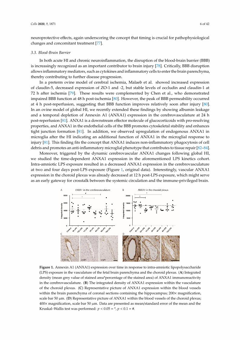
Moreover, triggered by the dynamic cerebrovascular ANXA1 changes following global HI,we studied the time-dependent ANXA1 expression in the aforementioned LPS kinetics cohort.Intra-amniotic LPS exposure resulted in a decreased ANXA1 expression in the cerebrovasculatureat two and four days post-LPS exposure (Figure 1, original data). Interestingly, vascular ANXA1expression in the choroid plexus was already decreased at 12 h post-LPS exposure, which might serveas an early gateway for crosstalk between the systemic circulation and the immune-privileged brain.
Figure 1. Annexin A1 (ANXA1) expression over time in response to intra-amniotic lipopolysaccharide(LPS) exposure in the vasculature of the fetal brain parenchyma and the choroid plexus. (A) Integrateddensity (mean grey value of stained area*percentage of the stained area) of ANXA1 immunoreactivityin the cerebrovasculature. (B) The integrated density of ANXA1 expression within the vasculatureof the choroid plexus. (C) Representative picture of ANXA1 expression within the blood vesselswithin the brain parenchyma of coronal sections containing the hippocampus; 200× magnification,scale bar 50 µm. (D) Representative picture of ANXA1 within the blood vessels of the choroid plexus;400×magnification, scale bar 50 µm. Data are presented as mean/standard error of the mean and theKruskal–Wallis test was performed: p < 0.05 = *, p < 0.1 = #.
Cells 2020, 9, 1871 7 of 42
3.4. Immune System Activation
3.4.1. Innate Immune System
Despite differences in etiology (infectious versus sterile inflammation), cross-talk betweenthe peripheral immune system and the immune-privileged brain parenchyma is central in thepathophysiology of preterm brain injury. The biochemical cascade initiated after HI and antenatalinfection results in the release of danger-associated molecular patterns (DAMPs) and cytokines (IL-6 inparticular) from the circulation contribute to the activation of cerebral endothelium and disruptionof the blood-brain barrier, as elegantly reviewed by Mallard et al. [85], thereby undermining centralnervous system (CNS) immune privilege and allowing active recruitment of leukocytes that contributeto the neuroinflammatory response, which is primarily mediated by microglia [63,86,87].
Microglia, the resident innate immune cells of the brain, play an important role in brain homeostasisand immune surveillance. They can recognize a wide range of signals (i.e., pathogen-associatedmolecular patterns and damage-associated molecular patterns) that indicate a threat to thestructural and functional integrity of the CNS through various pattern recognition receptors (PRRs),glutamate receptors, and purinergic receptors, resulting in activation [88–90]. Upon activation,they produce pro-inflammatory cytokines, chemokines, reactive oxygen species, and excitotoxicmolecules [91,92]. Strikingly, the complete depletion of microglia in models of HIE increases braininjury, indicating that microglia are crucial for tissue repair [93–95]. Accordingly, the prevention ofexcessive microglia activation while simultaneously promoting their phagocytic and neurotrophicfunction can have protective effects. These findings fit well with the concept of type 1 (M1) and type 2(M2) microglia dichotomy, depending on microenvironmental cues, microglia can rapidly change theirphenotype to pro-inflammatory, cytotoxic (type 1) cells, or anti-inflammatory regenerative (type 2) cells,respectively, and intermediate phenotypes [96,97]. Efforts have been made to characterize the dynamicsof the microglia response after HI (6 h–7 days), showing that 6 h after HI, there is a mixed responseof M1 and M2 markers that normalizes 24 h to seven days after HI [98]. Others report increased(iNOS), specifically in microglia, and a general increase in pro-inflammatory and anti-inflammatorygenes within the brain tissue 24 h after an HI event, that were increased by previous sensitization withLPS [99].
Consistent with an acute inflammatory response of resident microglia, clinical studiesdemonstrated that cytokine levels reach a peak at 12 h after HI [100]. This systemic accumulation ofDAMPs immediately after the injurious event has been associated with a massive activation of theperipheral immune system and with a rapid mobilization of immune effector cells (i.e., neutrophils,monocytes, T-cells) from the spleen [91,101]. These mobilized effector cells can invade the neonatalbrain through a disrupted blood-brain barrier and aggravate the existing injury [91,101]. The reportsaiming to characterize microglia phenotype after HI mostly disregard the contribution of infiltratingmonocytes, recruited from the periphery and differentiating into macrophages, to neuroinflammationas macrophages and microglia share a similar expression profile. Similarly, to microglia, macrophagesare plastic in phenotype and can either exacerbate brain injury or promote regeneration. There isevidence that monocytes are recruited into the brain after HI as levels of monocyte chemoattractantprotein-1 (MCP-1) increase acutely within the brain and in mouse models in which only mature myeloidcells from the periphery express an enhanced green fluorescent protein, fluorescence is detected 24 hand seven days after HI within the CNS [102,103]. In the same model, antibody-mediated depletionof monocytes was neuroprotective but only in male mice. This suggests monocytes contribute tobrain damage in male mice [102]. Contrarily, it has been shown that mice pre-conditioned with LPSgenerated splenic monocytes that protect the brain in a stroke model [104]. It seems that dependingon the context, monocytes are a double-edged sword, and influencing their phenotype would be aninteresting therapeutic approach.
In a mouse model that expressedgreen fluorescent protein only in myeloid cells, granulocytesinfiltrated the brain at 24 h and seven days after HI as well, though they contributed less compared to
Cells 2020, 9, 1871 8 of 42
monocyte infiltration to the total number of infiltrating CD11b+ cells (a marker of macrophages andmicroglia) [102]. In line with this finding, neutrophils were detected in the cerebrovasculature hoursafter HI and the depletion of neutrophils before HI reduced brain swelling [63,105,106]. However,when neutrophils were depleted later than 4–8 h after reperfusion, the protective effect of neutropeniawas abolished, suggesting an important timing effect of neutrophils in neonatal HI [106].
3.4.2. Astrocytes
In the healthy neonatal brain, astrocytes, along with pericytes, are an important component of theneurovascular unit (NVU) that regulate blood flow, ion balance, support neurons, and exert anti-oxidantactions [107–109]. At 25–40 weeks of gestation, astrocytes, predominating the NVU, cover 60% of thecerebrovasculature with their end-feet [108,110]. As previously described, during neuroinflammation,endothelial cells of the BBB become activated, releasing pro-inflammatory cytokines. Astrocytes barereceptors of the innate immune system, secrete matrix metalloproteinases (MMPs), and cytokines ofpro- and anti-inflammatory nature, potentiate excitotoxicity through iNOS, increase their expression ofthe glial fibrillary acidic protein (GFAP), and proliferate and form glial scars after injury [107,111–113].Their immune cell function and close coupling to the cerebrovasculature strongly suggest that theyactively contribute to the neuroinflammatory cascade.
Depending on the timing and phase of neuroinflammation, astrocytes could be detrimental orneuroprotective in adult pathologies [113]. Further studies enforced the dual role of astrocytes andtermed them A1 and A2 astrocytes by analogy with the M1 and M2 forms of microglia, the firstone being most abundantly present in adult neuropathologies [113,114]. In response to neonatalhypoxia-ischemia and interleukin 1-beta (IL-1β)-induced brain injury, A2 astrocytes highly expressingCyclooxygenase-2 (COX-2) predominatly leading to oligodendrocyte progenitor maturation arrest [115].The inhibition of the COX-2/prostaglandin E2 pathway improved white matter outcome [115].
Recently, Disdier et al. exposed fetal sheep in utero to prolonged intravenous LPS andassessed changes in the NVU. They found astrogliosis with reactive astrocyte morphology (astrocyticfeet-swelling) in the cerebral cortex and white matter, and a decrease in astrocytic vessel coverage inthe white matter together with microglia activation which might impair brain maturation and lead toinjury [116]. Similarly, we showed in an ovine model of infection-induced fetal neuroinflammation,in which intra-amniotically administered Candida Albicans actively invaded the systemic circulation,that astrocyte numbers and GFAP expression increased two and five days after the infection along withwhite matter injury and microglial activation [55]. By contrast, chronic intra-amniotic exposure of UPdecreased astrocyte numbers and resulted in white matter loss in fetal sheep [54]. This decrease in thenumber of astrocytes after 42 days of UP exposure is important since these cells possess several essentialfunctions in brain development, including the regulation of the extracellular glutamate homeostasis,which provided structural and metabolic support to surrounding cells (e.g., oligodendrocytes) andmodulate neuronal connections [57,117]. The changes in astrocyte function or density also result inaltered neurological outcomes. In particular, the altered astrocyte (GFAP) protein expression anddisrupted astrocyte maturation have been implicated in the pathogenesis of neurodevelopmentaldisorders such as autism and CP [118,119].
In the context of neonatal HI induced brain injury, astrogliosis is observed with reactive astrocytesexhibiting swollen end-feet [120–122]. Moreover, marked astrogliosis was observed at three and sevendays after global HI in ovine fetal models [62,123,124]. Importantly, inflammation-induced opening ofastrocytic gap junction (connexin) hemichannels, as recently reviewed by Galinsky et al. [125], is akey regulating event in the evolution of oligodendrocyte and neuronal injury. Likely mechanismsinclude modulation of intracellular calcium handling, blood-brain barrier integrity, purinergic receptorsignaling, and inflammasome pathway activation. These cellular processes are likely to initiate acycle of excessive ATP release, which propagates activation of purinergic receptors on microglia andastrocytes to trigger inflammation-induced injury of neurons and oligodendroglia [125].
Cells 2020, 9, 1871 9 of 42
3.4.3. Adaptive Immunity
Pathological inflammation can also be mediated by cells of the adaptive immune system.Lymphocytes have been found to contribute to pathophysiology of neonatal brain injury. In particular,T- and B-cells were observed in post-mortem brains of premature infants with PVL and in brainsof rodent models for HI three to seven days after HI [63,65,66,126]. Further, lymphocyte deficientmice showed decreased white matter injury after HI [126], and splenectomy prior to HI in neonatalrats resulted in significant neuroprotection [87]. Perinatal chronic hypoxia in mice also led to theinfiltration of autoreactive, myelin-specific CD4+ T-cells contributing to white matter injury [127].The contribution of peripheral immune effector cells to brain injury has been confirmed and extendedby our data in a preclinical model of neonatal HIE, showing that neuroinflammation mediated bymicroglia was associated with marked mobilization of the peripheral immune system and splenicinvolution [63,65]. In addition, we showed that splenic T cell tolerance, induced by mesenchymalstem cell treatment, resulted in decreased cerebral infiltration of T lymphocytes and correlated withwhite matter protection [65]. However, Herz et al. showed, in a rodent HI model, that peripheral T-celldepletion exacerbates brain injury, suggesting that T-lymphocytes also have neuroprotective roles inthe context of neonatal brain injury [128].
4. Treatment
Following birth, all preterm infants receive supportive care, including adequate respiratorysupport, blood pressure support, normalization of fluid-electrolyte balance, and blood glucose levels,and, if necessary, antibiotic treatment, and treatment of seizures. Preterm brain injury evolves over timein a period of hours to weeks, creating a window of opportunity for treatment. However, therapeuticoptions to improve the outcomes of preterm neonates suffering from brain injury are very limited.
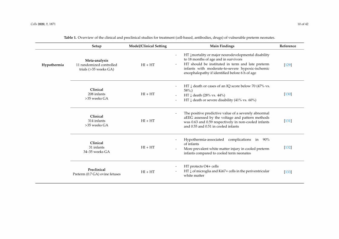
Given the dominant role of inflammation in the induction of preterm brain injury, we considerthat immune-modulatory interventions have a high potential to support recovery from brain injuryin vulnerable neonates. Immune modulation can be directly effective or indirectly by improvingthe efficacy of other therapies. A summary of studies on neuroprotective therapies for preterm andnear-term neonates is provided in Table 1.
Cells 2020, 9, 1871 10 of 42
Table 1. Overview of the clinical and preclinical studies for treatment (cell-based, antibodies, drugs) of vulnerable preterm neonates.
Setup Model/Clinical Setting Main Findings Reference
HypothermiaMeta-analysis
11 randomized controlledtrials (>35 weeks GA)
HI + HT
- HT ↓mortality or major neurodevelopmental disabilityto 18 months of age and in survivors
- HT should be instituted in term and late preterminfants with moderate-to-severe hypoxic-ischemicencephalopathy if identified before 6 h of age
[129]
Clinical208 infants
>35 weeks GAHI + HT
- HT ↓ death or cases of an IQ score below 70 (47% vs.58%;)
- HT ↓ death (28% vs. 44%)- HT ↓ death or severe disability (41% vs. 60%)
[130]
Clinical314 infants
>35 weeks GAHI + HT
- The positive predictive value of a severely abnormalaEEG assessed by the voltage and pattern methodswas 0.63 and 0.59 respectively in non-cooled infantsand 0.55 and 0.51 in cooled infants
[131]
Clinical31 infants
34–35 weeks GAHI + HT
- Hypothermia-associated complications in 90%of infants
- More prevalent white matter injury in cooled preterminfants compared to cooled term neonates
[132]
PreclinicalPreterm (0.7 GA) ovine fetuses HI + HT
- HT protects O4+ cells- HT ↓ of microglia and Ki67+ cells in the periventricular
white matter[133]
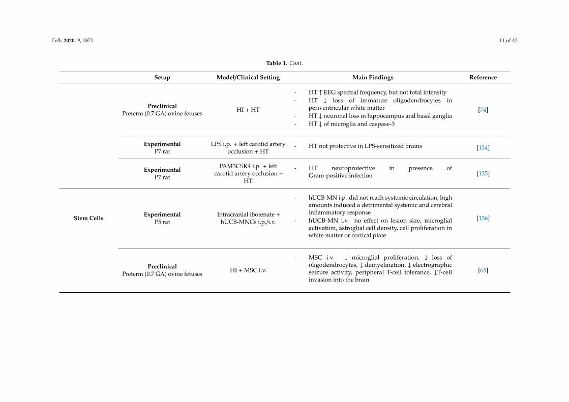
Cells 2020, 9, 1871 11 of 42
Table 1. Cont.
Setup Model/Clinical Setting Main Findings Reference
PreclinicalPreterm (0.7 GA) ovine fetuses HI + HT
- HT ↑ EEG spectral frequency, but not total intensity- HT ↓ loss of immature oligodendrocytes in
periventricular white matter- HT ↓ neuronal loss in hippocampus and basal ganglia- HT ↓ of microglia and caspase-3
[74]
ExperimentalP7 rat
LPS i.p. + left carotid arteryocclusion + HT
- HT not protective in LPS-sensitized brains [134]
ExperimentalP7 rat
PAM3CSK4 i.p. + leftcarotid artery occlusion +
HT
- HT neuroprotective in presence ofGram-positive infection [135]
Stem Cells ExperimentalP5 rat
Intracranial ibotenate +hUCB-MNCs i.p./i.v.
- hUCB-MN i.p. did not reach systemic circulation; highamounts induced a detrimental systemic and cerebralinflammatory response
- hUCB-MN i.v. no effect on lesion size, microglialactivation, astroglial cell density, cell proliferation inwhite matter or cortical plate
[136]
PreclinicalPreterm (0.7 GA) ovine fetuses HI + MSC i.v.
- MSC i.v. ↓ microglial proliferation, ↓ loss ofoligodendrocytes, ↓ demyelination, ↓ electrographicseizure activity, peripheral T-cell tolerance, ↓T-cellinvasion into the brain
[65]

Cells 2020, 9, 1871 12 of 42
Table 1. Cont.
Setup Model/Clinical Setting Main Findings Reference
PreclinicalPreterm (0.7 GA) ovine fetuses HI + MAPC i.v.
- MAPC i.v. ↓number of seizures, preventeddecrease in baroreceptor reflex sensitivity afterglobal HI, ↓ microglial proliferation, prevention ofhypomyelination, modulation of the peripheral splenicinflammatory response
[67]
PreclinicalPreterm (0.7 GA) ovine fetuses HI + hAECs i.n.
- hAECs↑brain weight, ↑restoration of immature/matureOLs and ↑ myelin basic protein, ↓ microgliaand astrogliosis, partially improved cortical EEGfrequency distribution, ↓ loss of cortical area,↓cleaved-caspase-3 expression, ↑neuronal survival indeep grey matter nuclei
[73]
ExperimentalP4 Rat IVH +UCBC i.c.v.
- UCBCs ↓ post-hemorrhagic hydrocephalusdevelopment, ↓astrogliosis, ↓cell death, ↓expression ofpro-inflammatory cytokines in CSF, ↑corpus-callosalthickness, ↑myelin basic protein expression,↑behavioral tests vs. IVH group
[137]
Clinical trial(NCT02274428, phase I),IVH; mean GA 26.1 ± 0.7
weeks
IVH + UCBC i.c.v.- No infant died or showed serious adverse effects related
with stem cell transplantation [138]
ExperimentalP7 rat
Unilateral HI + MAPCi.v./i.c.v.
- i.v. or intracerebral MAPC ↑ motor and neurologicscore, hippocampal cell preservation vs. veh group [139]
Cells 2020, 9, 1871 13 of 42
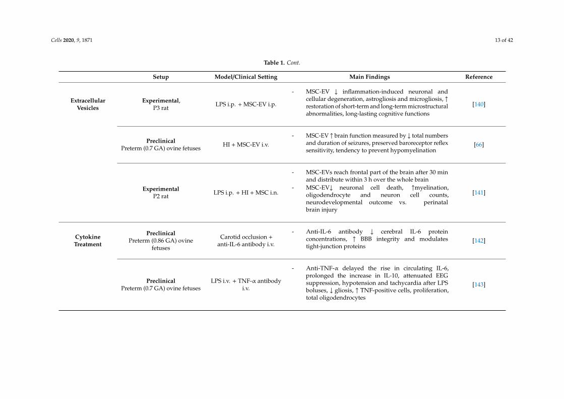
Table 1. Cont.
Setup Model/Clinical Setting Main Findings Reference
ExtracellularVesicles
Experimental,P3 rat LPS i.p. + MSC-EV i.p.
- MSC-EV ↓ inflammation-induced neuronal andcellular degeneration, astrogliosis and microgliosis, ↑restoration of short-term and long-term microstructuralabnormalities, long-lasting cognitive functions
[140]
PreclinicalPreterm (0.7 GA) ovine fetuses HI + MSC-EV i.v.
- MSC-EV ↑ brain function measured by ↓ total numbersand duration of seizures, preserved baroreceptor reflexsensitivity, tendency to prevent hypomyelination
[66]
ExperimentalP2 rat LPS i.p. + HI + MSC i.n.
- MSC-EVs reach frontal part of the brain after 30 minand distribute within 3 h over the whole brain
- MSC-EV↓ neuronal cell death, ↑myelination,oligodendrocyte and neuron cell counts,neurodevelopmental outcome vs. perinatalbrain injury
[141]
CytokineTreatment
PreclinicalPreterm (0.86 GA) ovine
fetuses
Carotid occlusion +anti-IL-6 antibody i.v.
- Anti-IL-6 antibody ↓ cerebral IL-6 proteinconcentrations, ↑ BBB integrity and modulatestight-junction proteins
[142]
PreclinicalPreterm (0.7 GA) ovine fetuses
LPS i.v. + TNF-α antibodyi.v.
- Anti-TNF-α delayed the rise in circulating IL-6,prolonged the increase in IL-10, attenuated EEGsuppression, hypotension and tachycardia after LPSboluses, ↓ gliosis, ↑ TNF-positive cells, proliferation,total oligodendrocytes
[143]
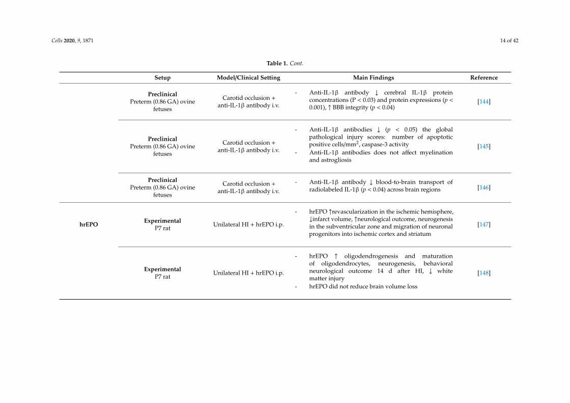
Cells 2020, 9, 1871 14 of 42
Table 1. Cont.
Setup Model/Clinical Setting Main Findings Reference
PreclinicalPreterm (0.86 GA) ovine
fetuses
Carotid occlusion +anti-IL-1β antibody i.v.
- Anti-IL-1β antibody ↓ cerebral IL-1β proteinconcentrations (P < 0.03) and protein expressions (p <0.001), ↑ BBB integrity (p < 0.04)
[144]
PreclinicalPreterm (0.86 GA) ovine
fetuses
Carotid occlusion +anti-IL-1β antibody i.v.
- Anti-IL-1β antibodies ↓ (p < 0.05) the globalpathological injury scores: number of apoptoticpositive cells/mm2, caspase-3 activity
- Anti-IL-1β antibodies does not affect myelinationand astrogliosis
[145]
PreclinicalPreterm (0.86 GA) ovine
fetuses
Carotid occlusion +anti-IL-1β antibody i.v.
- Anti-IL-1β antibody ↓ blood-to-brain transport ofradiolabeled IL-1β (p < 0.04) across brain regions [146]
hrEPO ExperimentalP7 rat Unilateral HI + hrEPO i.p.
- hrEPO ↑revascularization in the ischemic hemisphere,↓infarct volume, ↑neurological outcome, neurogenesisin the subventricular zone and migration of neuronalprogenitors into ischemic cortex and striatum
[147]
ExperimentalP7 rat Unilateral HI + hrEPO i.p.
- hrEPO ↑ oligodendrogenesis and maturationof oligodendrocytes, neurogenesis, behavioralneurological outcome 14 d after HI, ↓ whitematter injury
- hrEPO did not reduce brain volume loss
[148]
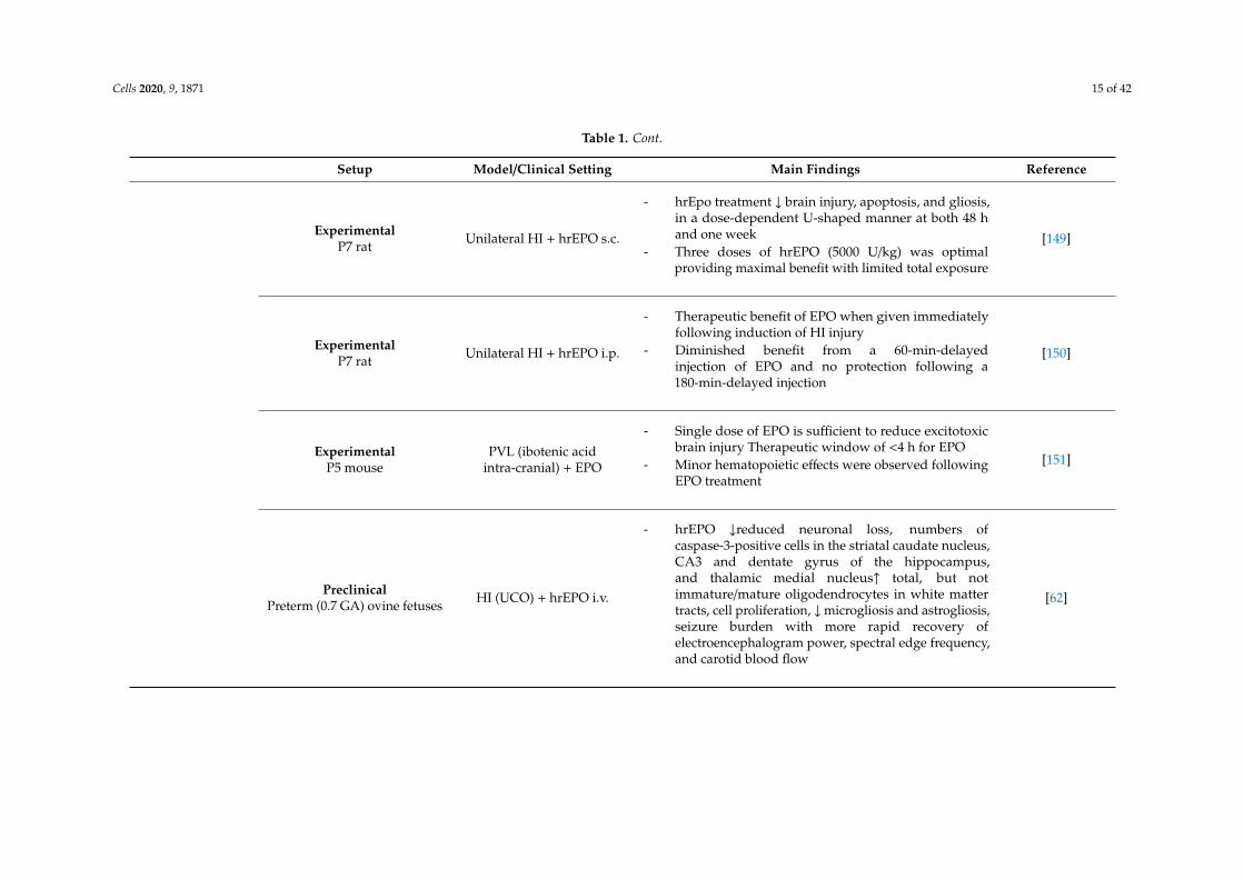
Cells 2020, 9, 1871 15 of 42
Table 1. Cont.
Setup Model/Clinical Setting Main Findings Reference
ExperimentalP7 rat Unilateral HI + hrEPO s.c.
- hrEpo treatment ↓ brain injury, apoptosis, and gliosis,in a dose-dependent U-shaped manner at both 48 hand one week
- Three doses of hrEPO (5000 U/kg) was optimalproviding maximal benefit with limited total exposure
[149]
ExperimentalP7 rat Unilateral HI + hrEPO i.p.
- Therapeutic benefit of EPO when given immediatelyfollowing induction of HI injury
- Diminished benefit from a 60-min-delayedinjection of EPO and no protection following a180-min-delayed injection
[150]
ExperimentalP5 mouse
PVL (ibotenic acidintra-cranial) + EPO
- Single dose of EPO is sufficient to reduce excitotoxicbrain injury Therapeutic window of <4 h for EPO
- Minor hematopoietic effects were observed followingEPO treatment
[151]
PreclinicalPreterm (0.7 GA) ovine fetuses HI (UCO) + hrEPO i.v.
- hrEPO ↓reduced neuronal loss, numbers ofcaspase-3-positive cells in the striatal caudate nucleus,CA3 and dentate gyrus of the hippocampus,and thalamic medial nucleus↑ total, but notimmature/mature oligodendrocytes in white mattertracts, cell proliferation, ↓microgliosis and astrogliosis,seizure burden with more rapid recovery ofelectroencephalogram power, spectral edge frequency,and carotid blood flow
[62]
Cells 2020, 9, 1871 16 of 42
Table 1. Cont.
Setup Model/Clinical Setting Main Findings Reference
PreclinicalPreterm (0.7 GA) ovine fetuses LPS i.v. + hrEPO i.v.
- hrEPO did not improve fetal hypoxemia, hypotensioninduced by LPS
- hrEPO ↓ brain injury ↑myelination in the corticospinaltract and the optic nerve
[152]
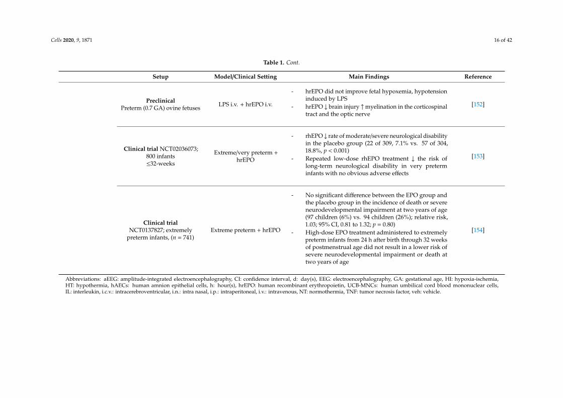
Clinical trial NCT02036073;800 infants≤32-weeks
Extreme/very preterm +hrEPO
- rhEPO ↓ rate of moderate/severe neurological disabilityin the placebo group (22 of 309, 7.1% vs. 57 of 304,18.8%, p < 0.001)
- Repeated low-dose rhEPO treatment ↓ the risk oflong-term neurological disability in very preterminfants with no obvious adverse effects
[153]
Clinical trialNCT0137827; extremely
preterm infants, (n = 741)Extreme preterm + hrEPO
- No significant difference between the EPO group andthe placebo group in the incidence of death or severeneurodevelopmental impairment at two years of age(97 children (6%) vs. 94 children (26%); relative risk,1.03; 95% CI, 0.81 to 1.32; p = 0.80)
- High-dose EPO treatment administered to extremelypreterm infants from 24 h after birth through 32 weeksof postmenstrual age did not result in a lower risk ofsevere neurodevelopmental impairment or death attwo years of age
[154]
Abbreviations: aEEG: amplitude-integrated electroencephalography, CI: confidence interval, d: day(s), EEG: electroencephalography, GA: gestational age, HI: hypoxia-ischemia,HT: hypothermia, hAECs: human amnion epithelial cells, h: hour(s), hrEPO: human recombinant erythropoietin, UCB-MNCs: human umbilical cord blood mononuclear cells,IL: interleukin, i.c.v.: intracerebroventricular, i.n.: intra nasal, i.p.: intraperitoneal, i.v.: intravenous, NT: normothermia, TNF: tumor necrosis factor, veh: vehicle.
Cells 2020, 9, 1871 17 of 42
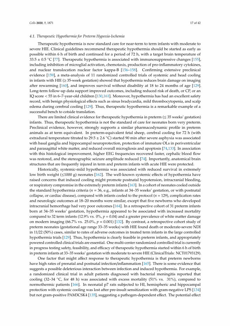
4.1. Therapeutic Hypothermia for Preterm Hypoxia-Ischemia
Therapeutic hypothermia is now standard care for near-term to term infants with moderate tosevere HIE. Clinical guidelines recommend therapeutic hypothermia should be started as early aspossible within 6 h of birth and continued for a period of 72 h, with a target brain temperature of33.5 ± 0.5 ◦C [77]. Therapeutic hypothermia is associated with immunosuppressive changes [155],including inhibition of microglial activation, chemotaxis, production of pro-inflammatory cytokines,and nuclear translocation nuclear factor kappa-B [156–158]. Confirming extensive preclinicalevidence [159], a meta-analysis of 11 randomized controlled trials of systemic and head coolingin infants with HIE (≥ 35-week gestation) showed that hypothermia reduces brain damage on imagingafter rewarming [160], and improves survival without disability at 18 to 24 months of age [129].Long-term follow-up data support improved outcomes, including reduced risk of death, or CP, or anIQ score < 55 in 6–7-year-old children [130,161]. Moreover, hypothermia has had an excellent safetyrecord, with benign physiological effects such as sinus bradycardia, mild thrombocytopenia, and scalpedema during cerebral cooling [129]. Thus, therapeutic hypothermia is a remarkable example of asuccessful bench to cotside translation.
There are limited clinical evidence for therapeutic hypothermia in preterm (≤ 35 weeks’ gestation)infants. Thus, therapeutic hypothermia is not the standard of care for neonates born very preterm.Preclinical evidence, however, strongly supports a similar pharmacodynamic profile in pretermanimals as at term equivalent. In preterm-equivalent fetal sheep, cerebral cooling for 72 h (withextradural temperature titrated to 29.5 ± 2.6 ◦C) started 90 min after severe asphyxia was associatedwith basal ganglia and hippocampal neuroprotection, protection of immature OLs in periventricularand parasagittal white matter, and reduced overall microgliosis and apoptosis [74,133]. In associationwith this histological improvement, higher EEG frequencies recovered faster, cephalic blood flowwas restored, and the stereographic seizure amplitude reduced [74]. Importantly, anatomical brainstructures that are frequently injured in term and preterm infants with acute HIE were protected.
Historically, systemic-mild hypothermia was associated with reduced survival in extremelylow birth weight (≤1000 g) neonates [162]. The well-known systemic effects of hypothermia haveraised concerns that induced cooling might promote postnatal hypotension, intracranial bleeding,or respiratory compromise in the extremely preterm infants [163]. In a cohort of neonates cooled outsidethe standard hypothermia criteria (n = 36, e.g., infants at 34–35 weeks’ gestation, or with postnatalcollapse, or cardiac disease), compared with infants cooled to the protocol (n = 129), complication ratesand neurologic outcomes at 18–20 months were similar, except that five newborns who developedintracranial hemorrhage had very poor outcomes [164]. In a retrospective cohort of 31 preterm infantsborn at 34–35 weeks’ gestation, hypothermia appeared to be associated with increased mortalitycompared to 32 term infants (12.9% vs. 0%, p = 0.04) and a greater prevalence of white matter damageon modern imaging (66.7% vs. 25.0%, p = 0.001) [132]. By contrast, a retrospective cohort study ofpreterm neonates (gestational age range 33–35 weeks) with HIE found death or moderate-severe NDIin 11/22 (50%) cases, similar to rates of adverse outcomes in treated term infants in the large controlledhypothermia trials [129]. Thus, hypothermia is clearly feasible in preterm infants, and appropriatelypowered controlled clinical trials are essential. One multi-center randomized controlled trial is currentlyin progress testing safety, feasibility, and efficacy of therapeutic hypothermia started within 6 h of birthin preterm infants at 33–35 weeks’ gestation with moderate to severe HIE (ClinicalTrials: NCT01793129).
One factor that might affect response to therapeutic hypothermia is that preterm newbornshave high rates of prenatal and postnatal infection/inflammation [165]. There is some evidence thatsuggests a possible deleterious interaction between infection and induced hypothermia. For example,a randomized clinical trial in adult patients diagnosed with bacterial meningitis reported thatcooling (32–34 ◦C, for 48 h) was associated with excess mortality (51% vs. 31%), compared tonormothermic patients [166]. In neonatal p7 rats subjected to HI, hemispheric and hippocampalprotection with systemic cooling was lost after pre-insult sensitization with gram-negative LPS [134]but not gram-positive PAM3CSK4 [135], suggesting a pathogen-dependent effect. The potential effect

Cells 2020, 9, 1871 18 of 42
of infection in encephalopathic infants treated with hypothermia is unclear, as known infection was anexclusion criterion in the hypothermia trials [129].
4.2. Cell-Based Therapies
4.2.1. Stem Cells
Cell-based interventions as a therapeutic strategy for injury to the (neonatal) brain have attractedmuch attention in the past decade [136,167–174]. Many different types of stem cells, derived fromfetal, placental, and adult tissues, are currently under investigation [175–180]. Stromal cells,including mesenchymal stem cells and multipotent adult progenitor cells (MAPC) are a subsetof progenitors that have been shown to differentiate into multiple lineages (i.e., osteoblasts, adipocytes,and chondrocytes) [175–178]. They have been a particular focus of research as they are easily obtainable(e.g., from cord blood, Wharton’s jelly and bone marrow) and do not have the ethical and safetyconcerns of embryonic stem cells [172,174]. Moreover, bone marrow-derived adherent stromal cellshave low immunogenicity due to a lack of expression of MHC class II antigens, allowing their use forallogenic therapy [174]. The therapeutic potential of mesenchymal stem cells (MSCs) has mainly beenattributed to their immune-modulatory and regenerative potential [172,173,177,181]. MSCs modulateinnate and adaptive immune responses from a pro-inflammatory status towards an anti-inflammatorystatus, thereby reducing tissue injury and creating an environment supporting tissue repair andregeneration [177,178,182].
Besides immune modulation in favor of regeneration, MSCs directly affect the injured CNSthrough secretion of neurotrophic factors that stimulate and maintain neurogenesis of the endogenousneural stem cell population and its subsequent differentiation into neuronal and oligodendrogliallineages [173,177,181,183–185].
The neuroprotective potential of systemically administered MSCs and Multipotent AdultProgenitor Cells (MAPC) was shown in a preclinical preterm ovine model of global hypoxia-HIinjury [65,67]. MSC administration during the latent phase following global HI resulted in functionalimprovement and prevented hypomyelination of the subcortical white matter at seven days postHI. These protective effects were attributed to anti-inflammatory effects since neuroinflammation,and peripheral immune activation were significantly reduced [65,67]. Specifically, intravenous MSCtreatment reduced the proliferative capacity of splenic T lymphocytes with concomitant reduced cerebralT cell infiltration. In addition, systemic administration of MSCs ameliorated splenic involution causedby global HI, implicating a key role of the spleen in the protective mechanisms of stem cell therapy.The results from this study were supported and extended by studies from Walker et al. [186,187] andDepaul et al. [188], demonstrating a crucial role for the spleen in (1) the pathophysiology of CNS injuryand (2) providing mechanistic insight for the beneficial effects of stem cell therapy. Moreover, there isincreasing evidence for the effectiveness of stem cell administration via the intranasal route, which is afeasible route for the pharmacological treatment of neonates. This concept of intranasal administrationis based on the passage of stem cells over the cribiform plate along the olfactory nerve, allowing forrapid dispersion throughout the brain, where local effects can be exerted [73,189–193]. In a study by vanVelthoven et al. delayed (10 days post HI injury, induced at P9), nasal administration of MSC therapyimproved neurological outcomes 28 days post HI. Interestingly, improved outcomes were attributed tothe regenerative potential of MSC rather than inhibition of injurious processes or prevention of injurysince cells were administered when injury was readily established [192]. The delayed intravenousMSC administration in fetal sheep five days after global HI was less effective than acute administration12 h after reperfusion [72]. These results were attributed to systemic and neuro-immunomodulatoryeffects [72]. Collectively, the administration route in relation to the timing of administration appearsto be crucial for the action of MSCs (immune modulation and regeneration) to establish optimalneuroprotection. As such, repeated dosing by different administration routes throughout different
Cells 2020, 9, 1871 19 of 42
phases (latent, secondary, and even tertiary) of disease progression might ultimately lead to thegreatest benefit.
4.2.2. Clinical Stem Cell Trials
Therapeutic hypothermia has improved intact survival and neurodevelopmental outcome ininfants with moderate-severe neonatal encephalopathy [129–131]. However, neuroprotection ispartial, and a significant proportion of asphyxiated infants still die or suffer life-long consequences,including CP, cognitive deficits, and epilepsy [129–131]. Encouragingly, preclinical and clinical studiesare now investigating therapeutic hypothermia as a potential treatment for extending over several daysafter the insult infants with mild encephalopathy [194]; however, hypothermia is counter-indicated forpreterm infants as discussed above, and there is currently no neurotherapeutic treatment for thesepatients. Critically, additional therapeutic strategies that can augment the neuroprotective effect oftherapeutic hypothermia in infants with moderate-severe encephalopathy, and reduce the neurologicalburden in (extreme) preterm infants are under active investigation [167]. In light of our discussion onchorioamnionitis, the potential adjuvant therapies should target the detrimental neuroinflammatoryresponse that occur during and after the reperfusion phase after birth asphyxia, while also promotingneuronal regeneration. As reviewed above, MSCs and MAPC meet those criteria and are emerging asa promising therapeutic intervention for multiple diseases in neonatal medicine. A recent clinical trialin adult ischemic stroke patients showed that MSCs [195] and MAPC cells were safe and improvedneurological symptoms after one year of follow-up [195,196].
More recent clinical trials have also demonstrated that stem cells are safe, with promisingprotective results in multiple neonatal diseases, including HIE [197], bronchopulmonary dysplasia(BPD) [198–200], IVH [137,138], and established CP [201].
Within this context, recent findings indicated that MSCs may interact either synergistically [202]or antagonistically [203] with therapeutic hypothermia in experimental models of neonatal HIE,depending on the timing of administration. These conflicting findings indicate that the translationalgap between preclinical research and the clinical application still needs to be addressed before stemcells could be safely translated into clinical practice for neonatal diseases. Several clinical trials testingthe protective potential of stem cells are still recruiting or pending, including for NCT04255147 forBPD, NCT03356821 for perinatal stroke, and NCT02612155 for HIE.
4.2.3. Stem Cell Therapy—Timing Is Key
Although a large body of experimental animal studies have demonstrated the beneficial effects ofcell-based therapies for preterm brain damage, (pre)clinical studies confirming these data are limited.In part, this mismatch can be attributed to different methodological approaches between animalmodels and clinical practice in terms of the use of single-hit animal models whilst clinical etiology ismultifactorial, use of inadequately characterized and heterogeneous stem cell populations, route ofadministration, and dosing strategies. At the same time, clinical trials often use top down approachesin which stem cells are administered at trivial time-points and the clinical outcomes are measured.Therefore, future studies testing stem cells should closely align with the underlying pathophysiologyand stage of injury to address the multi-factorial nature of preterm brain injury.
In addition, the stem cell secretome, which is modulated by micro-environmental cues duringthe different phases leading to preterm brain injury, defines efficacy of administered stem cells. Thus,a detailed analysis of the biodistribution of stem cells over time combined with detailed secretomeanalysis might (1) unravel novel potential pathways involved in the pathophysiology preterm braininjury, (2) and enable adjunctive cell-free therapies comprising specific trophic and immunomodulatoryfactors, and other regulators (such as miRNA), and most importantly (3) provide insight into timing,which appears to be a crucial determinant for optimal therapeutic efficacy.
Cells 2020, 9, 1871 20 of 42
4.2.4. Stem Cell-Derived Extracellular Vesicles
Despite that immunomodulatory and regenerative effects have been shown, the underlyingmechanisms of action of stem cell therapies remain largely unknown. It was initially thought thatthe therapeutic action of stem cells relied on direct replacement of dead and injured cells. However,since the number of cells that reach the site of injury is minimal, with marginal engraftment andshort cell survival, this theory has been largely discredited [139,188]. Consistent with this, we andothers have been unable to identify stem cells within the cerebral parenchyma seven days afterintravenous administration [65,67,139]. Meanwhile, there is growing evidence that the beneficialeffects of stem cell therapy are mediated at least in part via paracrine mechanisms as studieshave shown comparable therapeutic effects in MSC-conditioned medium compared to its cellularcomponent [204,205]. In addition, we and others have demonstrated potent neuroprotective effectswith mesenchymal stem cell-derived extracellular vesicles (MSC-EV) in animal models of pretermbrain injury [66,140,141,206].
Remarkably, these therapeutic effects could not be explained by the known anti-inflammatoryeffects of the MSC-EVs, as observed after MSC treatment [66]. This prompted us to focus on alternativeexplanations for the pharmacologic effects of MSC-EVs, in particular, restoration of the injuredBBB after global HI. There is accumulating evidence that the BBB becomes functional during thesecond trimester [85,207]. Nevertheless, a global HI insult would result in the release of reactiveoxygen species and excitotoxic molecules into the extracellular environment (i.e., the direct effects ofHI), with increased cytokine release by the peripheral and local innate immune system (secondaryinflammatory component), which leads to BBB dysfunction [208]. In turn, increased BBB permeabilityallows intracranial infiltration of peripheral immune cells (e.g., macrophages, leukocytes, and T-cells)that aggravate white matter injury via the release of pro-inflammatory mediators. Thus, strengtheningor restoring BBB integrity by enforcing endothelial cells which would attenuate the degree of whitematter injury.
4.3. Pharmacological Interventions
4.3.1. AnnexinA1
ANXA1 (37 kDa), formerly known as macrocortin, renocortin, lipomodulin, or lipocortin-1,was first described in the 1980s, and initially known for its anti-inflammatory effects as a downstreammediator of glucocorticoids [209]. It is a calcium-dependent phospholipid-binding protein that hasreceived more research attention in recent years due to its multimodal function that extends beyondsuppressing inflammation [210].
Increased BBB permeability in ANXA1 knock-out (KO) mice and progressive loss of endogenousANXA1 in the cerebrovasculature and plasma of patients with multiple sclerosis led to betterunderstanding of the function of ANXA1 [211]. ANXA1 strengthens BBB integrity by 1) binding ofextracellular ANXA1 to the FPR2 (formyl peptide receptor 2), inhibiting Rho A kinase, and therebystabilizing tight junctions; 2) direct interaction of intracellular ANXA1 with actin molecules, and therebypromoting cytoskeleton stability and tight junction formation between endothelial cells [211,212].The intravenous administration of MSC-derived extracellular vesicles containing ANXA1 preventedthe previously described HI-induced depletion of endogenous ANXA1 in the cerebrovasculature,thereby preventing BBB leakage [81]. Given the time-dependent drop of ANXA1 in the courseof antenatal infection, the ANXA1 administration in this infectious context might be a promisingtherapeutic strategy. Collectively, our data support the notion that the ANXA1/FPR axis is a therapeutictarget to treat fetuses exposed to HI and indicate a pharmacological window of opportunity forANXA1 supplementation.
Cells 2020, 9, 1871 21 of 42
4.3.2. Cytokine Treatment
Clinical studies and preclinical animal models have reported acute increases in systemic levelsof cytokines such as tumor necrosis factor (TNF)-α, IL-1β, and IL-6 after HI [142–144,213,214] andintra-amniotic exposure to inflammation [54,56]. High CSF/serum ratios indicate that local productionof cytokines within the CNS also contributes to increased cytokine levels [213]. Thus, decreasing thelevels of pro-inflammatory cytokines represents a promising strategy to suppress neuroinflammationand ensure neuroprotection. In preclinical fetal ovine models, neutralizing antibodies against IL-1βand IL-6 or pharmacological antagonism of TNF-α have already been tested and shown promisingshort-term effects [142–145]. One day after HI, the systemic infusion of anti–IL-6 mAb attenuated BBBdysfunction and decreased cerebral IL-6 levels. Similarly, IL-1β neutralizing antibodies improvedBBB integrity, lowered IL-1β levels in the brain, and reduced IL-1β transport across the BBB [144,146].Further, histological studies have shown that infusion of anti-IL1β decreased short-term ischemicreperfusion-related parenchymal brain injury [145].
The abovementioned studies have shown promising results, but mainly focused on the short-termeffects after HI. Determining the long-term effects is crucial to reduce the detrimental processes thatoccur during the tertiary phase of the brain injury after HI. As IL-1β and IL-6 expression changesthroughout gestation, and they fulfill physiologic functions that are not detrimental by definition,caution has to be paid to the right dosing and timing [214].
4.3.3. Recombinant Human Erythropoietin for Preterm Neonates
Recombinant human erythropoietin (rEpo) is a common treatment for anemia in preterm infantsand pediatric patients with chronic renal disease. In addition to stimulating erythropoiesis, rEpo hasshown anti-apoptotic, anti-inflammation, and anti-excitotoxic effects after HI and infection-inducedperinatal brain injury [215,216], suggesting rEpo could be a promising neuroprotectant forencephalopathic infants. In the long-term, it could promote OL and neuronal maturation andreplacement [147,148], and so might promote neurorestoration after injury. Compelling preclinicalevidence has shown that early administration of rEpo is neuroprotective over a broad dose range,from 1000 to 30,000 IU/kg, and that continued exposure to high-dose rEpo is more effective, but thatoptimal treatment regimens are likely to be paradigm-specific.
For example, after maternal LPS injection in rats at 18–19 days gestation, peripheral rEpo(5000 UI/kg) injection at birth was associated with reduced IL6, IL1, and TNF-α concentrations,and apoptosis and demyelination at p7 [217]. Critically, after HI in P7 rats, repeated rEpo injections(5000 IU/kg, at days 1, 2, and 3) provided greater protection than either a single (5000 IU/kg) dose or threeinjections with 2500 or 30,000 IU/kg rEpo [149], but brain protection was largely lost when the treatmentwas delayed until 1–3 h after HI [150]. By contrast, P5 mice treated with ibotenic acid (an excitotoxin)had fewer white matter lesions after a single rEpo injection (5000 IU/kg, at 1 h), but additionalinjections did not augment protection [151]. For post-HI treatment, these data are consistent with theexperience with therapeutic hypothermia where optimal protection was achieved when brain coolingwas started as soon as possible during the latent phase and continued until the delayed secondaryevents, such as overt seizures had resolved, after ~72 h, with loss of protection if the treatment isdelayed more than ~6 h after global ischemia. Supporting this concept, in preterm-equivalent fetalsheep, prolonged rEpo (5000 IU/kg, from 30 min to 72 h) infusion after asphyxia was associated withpartial subcortical white matter and neuroprotection, and improved electrophysiological EEG recovery,in association with reduced apoptosis and inflammation [62]. Similarly, intravenous rEpo (5000 IU/kg)boluses administered once daily for three days to preterm fetal sheep with endotoxin-induced braindamage, reduced axonal damage, microglial, and astrocytic responses in white matter, and improvedmyelination [152].
There is strong clinical evidence that rEpo treatment is safe in neonatal cohorts and shows indicativeevidence of possible benefit. A meta-analyses in preterm infants suggested that rEpo/Darbepoetinstarted within eight days from birth reduced rates of IVH, PVL, and necrotizing enterocolitis,
Cells 2020, 9, 1871 22 of 42
and improved neurologic deficits, without adverse effects, at 18–22 months’ corrected age (fourrandomized controlled trials, 1130 cases, relative risk (RR) 0.62, 95% Confidence interval (CI) 0.48 to 0.80;risk difference −0.08, 95% CI −0.12 to −0.04) [218]. However, disappointingly, a more recent largemulti-center, randomized, trial that assessed high-dose rEpo treatment for perinatal brain damagein extremely preterm infants (741 patients, 24 to 27 weeks and six days) found that repeated rEpo(1000 IU/kg, i.v.) doses at 48 h intervals for a total of six doses, followed with maintenance doses(400 IU/kg, s.c.) three times per week through 32 completed weeks of postmenstrual age, did notreduce death or severe neurodevelopmental impairment, compared to placebo (26% vs. 26%, RR 1.03,95% CI 0.81–1.32, p = 0.80) at 22–26 months of age [154]. There were also no significant differencesbetween the treatment groups in the rates of intracranial hemorrhage, sepsis, NEC, death, or frequencyof serious complications.
These negative results are disappointing; however, there were several limitations that should bekept in mind. First, the optimal rEpo regimen for preterm brain damage is still unknown. Treatmentwas started from 24 h after birth, which might be too late for benefit. Second, the treatment durationcovered the period when PVL is prominent (i.e., 24–32 weeks’ gestation), but it is possible that rEpotreatment should have been continued for longer. There is extensive evidence that chronic microgliosisand extracellular matrix disturbance continue to contribute to disrupted myelination later in gestation,as discussed above [219]. Third, cognitive testing for mild impairments is more sensitive later inlife [220], and so potentially long-term follow-up might still show small but beneficial effects. Thus,further studies are required to conclusively determine whether timely and well-targeted treatmentwith rEpo could alleviate perinatal brain damage in preterm infants.
5. Future Perspectives—Closing the Translational Gap
Despite many years of research into preterm brain injury and treatment, there is still an unmetclinical need for patients with preterm brain injury. The, often complex, multi-factorial nature ofpreterm brain damage, comprising both pre-and postnatal hits, which are not accurately reproduced incurrent translational models (most models use single hit paradigms), is considered to be paramountfor this translational gap.
5.1. Multi-Factorial Nature of Preterm Brain Injury
Over the past decade, (pre)clinical evidence demonstrated that the preterm brain couldsensitize/desensitize to a second injurious hit after pre-exposure to inflammation [221–223]. This conceptof preconditioning is supported by clinical data, which shows that the combination of antenatalinfection and a hypoxic-ischemic insult around birth dramatically increases the risk of CP (OR 78)when compared to either HI (OR 2.5) or infection (OR 7.2) alone [221]. Besides this specificallycombined insult, multiple ante-, peri- or postnatal factors can contribute to the risk of developing braininjury in the preterm infant, including being small for gestational age and having impaired placentalgrowth [224–226]. Furthermore, the evidence is accumulating that postnatal ventilation-inducedwhite matter injury, barotrauma around birth, (par)enteral feeding, standard-care medication(glucocorticoids), operative procedures, all may contribute to the development of preterm braininjury [227]. Clinically, exposure to multiple hits is associated with an enormous increase in the riskof and severity of white matter abnormalities [41,224,228]. The combined impact of these factors onpreterm brain development is determined by the specific interaction of individual insults and can bemodulated by post-insult treatments.
5.2. Biomarkers
An important obstacle for the translation of preclinical findings from standardized models isthe much higher heterogeneity of the clinical population. Consequently, with the current diagnosticapproaches, only a proportion of at risk cases are identified. As such, increasing sensitivity ofexisting biomarkers, extending the ‘diagnostic toolbox’ with novel biomarkers, or the composition of

Cells 2020, 9, 1871 23 of 42
biomarker panels are essential to identify and stratify preterm infants with brain injury. Improvedsensitivity will enable the identification of specific windows of therapeutic opportunity and, therefore,optimize clinical decision-making, leading to more targeted and individualized therapy. Clinical andpreclinical evidence for the use of biomarkers to identify preterm brain injury is provided in Table 2.
Cells 2020, 9, 1871 24 of 42
Table 2. Overview of the clinical and preclinical studies of diagnostic modalities (biomarker, imaging) for vulnerable neonates born preterm.
Setup Model/Clinical Setting Main Findings Reference
Plasma BiomarkersClinical
n = 155 <37 weeks GACord blood IL-6
Preterm birth- Cord blood IL-6 concentrations > 11 pg/mL
associated with increased rate of severeneonatal morbidity
[43]
Clinical94 patients with preterm labor
Amniotic fluid IL-6, TNF-α, IL-1βPreterm birth
- IL-6, TNF-α, IL-1β ↑ in amniotic fluid of pretermneonates with white matter injury vs. pretermneonates without white matter lesions
- ↑ of IL-6, TNF-α, IL-1β levels still significantwhen adjusted for GA and birth weight
[44]
Clinical315 patients with preterm labor
(20–35 weeks’ GA)Cord blood IL-6
Preterm birth- A cord blood IL-6≥17.5 pg/mL has a sensitivity of
70% and a specificity of 78% in the identificationof funisitis
[229]
PreclinicalIntra-amniotic LPS exposure of
fetal sheepIL-6, IL-8
(0.7–0.8 GA)
Inflammation(LPS exposure)
- Fetal plasma IL-6 transiently increased from 5 huntil 24 h after intra-amniotic LPS exposure.
- Fetal plasma IL-8 increased at four- andeight-days post-intra-amniotic LPS
[56]
VOCPreclinical
Ovine chorioamnionitis model(0.8 GA)
Infection (Ureaplasmaparvum)
- Changes in the VOC profile of ewes are detectablewith good accuracy > 72 h post-infection
- VOC profile changed as the duration ofinfection progresses
[230]
Imaging
ClinicalInfants < 31 weeks GA
(n = 77)MRI
Very preterm
- Identification of infants with abnormal motoroutcome based on the FA data from early MRIwith mean sensitivity 70%, mean specificity 74%,mean AUC 72%, mean F1 score of 68% and meanaccuracy 73%.
- Identification patches around the motor cortexand somatosensory regions with high precision(74%).
- Part of the cerebellum, and occipital and frontallobes were also highly associated with abnormalNSMDA/motor outcome.
[231]
Cells 2020, 9, 1871 25 of 42
Table 2. Cont.
Setup Model/Clinical Setting Main Findings Reference
EEGClinical
Very low birth weight infants (n =95)
Extreme preterm/verypreterm
- No significant difference between conventionalEEG amplitude and intensity for infants with orwithout evidence of white matter injury
- Premature infants with increasingly severe whitematter injury had progressively lower SEFs vs.infants who did not exhibit white matter injury→ SEF-based measures are useful for definingthe presence and severity of white matter injury
[232]
ClinicalMedian GA 25 weeks (22–30
weeks; n = 94)
Extreme preterm/verypreterm
- Poor outcome was associated with depressedaEEG/EEG already during the first 12 h andwith prolonged interburst intervals and higherinterburst percentage at 24 h
- Long-term outcome can be predicted byaEEG/EEG with 75–80% accuracy already at 24postnatal hours in very preterm infants, also ininfants with no early indication of brain injury
[233]
ClinicalGA between 27 and 32 weeks (n =
12)
Very preterm/moderatepreterm
- Absent cyclicity on aEEG within 24 h of age isassociated with poor outcome in preterm infants
[234]
ClinicalGA < 30 weeks GA (n = 65) Very preterm
- Data-driven approach to quantify EEGmaturational deviations in preterms with normaland abnormal neurodevelopmental outcomes.
- Abnormal outcome trajectories were associatedwith clinically defined dysmature anddisorganized EEG patterns.
[235]
Abbreviations: aEEG: amplitude-integrated electroencephalography, AUC: area under the curve, CUS: cerebral ultrasound, EEG: electroencephalography, GA: gestational age, h: hour(s),IL: interleukin, MRI: Magnetic resonance imaging, NSMDA: neurosensory motor developmental assessment, PPROM: preterm premature rupture of the membranes, SEF: spectral edgefrequency, TNF: tumor necrosis factor, VOC: volatile organic compounds.
Cells 2020, 9, 1871 26 of 42
5.2.1. Plasma Biomarkers
Patient stratification based on reliable biomarkers is key to provide the most optimal treatmentstrategy. Multiple clinical studies have shown that fetal involvement with the induction of FIRS,characterized by elevated plasma IL-6 levels, during chorioamnionitis correlates with higher ratesof neonatal sepsis, multi-organ dysfunction, perinatal brain injury, and CP [43,44,229]. Moreover,increased plasma levels of IL-6 in the acute phase (<24 h) after an ischemic insult or traumatic braininjury have been shown to be a reliable prognostic marker for adverse cerebral outcomes [236,237].
Thus, it is appealing to consider systemic IL-6 levels as a prognostic marker to predictneurodevelopmental outcomes with infection. However, caution is required for severalreasons: Firstly, increased plasma IL-6 levels are also observed after non-infection-related insults,including Rh-alloimmunization [238], trauma [239], and HI insults [236]. Secondly, we havedemonstrated in the preclinical ovine model that IL-6 concentrations were only transiently increasedwithin the first 24 h after intra-uterine exposure to infection [54,56]. Collectively, the timing of IL-6detection appears to be essential to interpret IL-6 levels as a marker for cerebral prognosis and soneeds to be incorporated into the clinical decision model. Further studies should focus on the useof IL-6 in combination with other promising systemic inflammatory biomarkers (C-reactive protein,procalcitonin).
However, the successful development and implementation of neurotherapeutics in clinical careare currently impeded by a lack of diagnostic tools for early (prenatal) detection and surveillanceof intra-amniotic infection. Recently, we demonstrated in a proof of concept that intra-amnioticUP infection induces a distinct time-dependent volatile organic compound (VOC)-signature in theexpiration breath of pregnant sheep. Changes in the VOC profile were detectable with good accuracyas soon as 72 h post-infection. Moreover, the VOC profile changed as the duration of the infectionprogressed [230]. However, these promising results need to be validated in a less standardized clinicalcohort, in which the origin of infection is, most often, poly-microbial and confounding factors, such asdiet and unbounded environment might influence the sensitivity and specificity of the VOC profile.
5.2.2. Imaging
In contrast to the previously common cystic white matter lesions that were easily detectableby ultrasound and magnetic resonance imaging (MRI), the new diffuse WMI with microscopic,punctate lesions, and altered white matter development are difficult to detect by ultrasound andconventional MRI (T1, T2) [240]. As such, diffuse WMI in preterm infants is detected only in 20%of all cases by conventional MRI at term equivalent age [241], thereby underlining the clinical needfor modalities that could accurately and specifically assess brain structure and function in pretermneonates. In recent years, brain-imaging techniques, such as Diffusion Tensor Imaging (DTI) andfunctional magnetic resonance imaging (fMRI), have allowed scientists to study the brain in morestructural detail and detect deteriorating neural networks in the brain [242]. To optimize the sensitivityand specificity of these new imaging modalities, its correlation with pathological changes in the fetalbrain is essential. Early therapeutic strategies have been proven to be the most effective in promotingneurodevelopment, thereby highlighting the need for the early prediction of outcome, evaluated byspecific measures; MRI [231], especially DWI and DTI, or techniques that could readily be deployed ina neonatal intensive care unit, such as ultrasound [243–245], ultrafast Doppler [246], EEG, or functionalultrasound imaging [247] could help develop and translate new treatment strategies and prognosticmarkers to improve long-term outcomes.
The detection of additional clinical improvement in functional neurological outcomes in the contextof therapeutic hypothermia after birth asphyxia requires large numbers of participants. Using predictiveneuroimaging and neuromonitoring biomarkers as surrogate endpoints in experimental andproof-of-concept clinical trials may help reduce sample sizes. For example, thalamic n-acetylaspartate(NAA), in term infants, is a highly predictive neuronal marker that could be measured with magneticresonance spectroscopy (MRS). In asphyxiated and cooled newborns, thalamic NAA acquired days
Cells 2020, 9, 1871 27 of 42
after birth, accurately predicts adverse neurodevelopmental outcome at two years [248–250]. Due toits high predictive value and clinical relevance, thalamic NAA may, therefore, be a robust surrogatemarker for neurological outcomes in future clinical trials testing the add-on effect of regenerativetherapy in addition to therapeutic hypothermia. Since MR imaging is part of standard clinical care inneonates, adding more precise imaging protocols seems obvious and would be feasible. This wouldalso add to the current knowledge mostly based on selected cohorts and may improve the predictivevalue for early brain MRI.
5.2.3. EEG
Continuous electrophysiological monitoring with conventional (video) electroencephalography(EEG) and amplitude-integrated electroencephalography (aEEG) is an important diagnostic toolin neonatal intensive care [251], especially in the encephalopathic term neonate [252]. However,compared with near-term and full-term newborns, accurate prediction of neurodevelopmental outcomesin preterm neonates with EEG is complicated as electrophysiological characteristics are still developing.Several EEG features have shown promise. For example, Roland sharp waves have been recognized as anearly predictor for white matter injury and neurologic outcome [250–252]. Further, recent developmentsin neural network analytics indicate the development of specific cortical networks, and their disruptionhas been suggested as an early marker of perinatal brain damage [232]. In the high-risk preterm neonate,the suppression of amplitude and high frequencies [232], prolonged inter-burst interval [233], seizures,and disrupted sleep-wake cycling [234,253], are also associated with poor outcomes [153]. EEG andaEEG are generally applied as a diagnostic tool in the neonatal population, even in very prematureinfants. Currently, the interpretation is based on visual inspection and quantitative measures that arenot systematically applied. Since all data are acquired digitally, a systematical quantitative approachcan be easily added and has a clear predictive value [235]. Thus, aEEG could be a promising prognostictool in clinical trials, testing potential regenerative therapies (Table 2). Overview of clinical andpreclinical studies of diagnostic modalities (biomarker, imaging) for vulnerable neonates born preterm.
5.3. Postnatal Factors
Following birth, preterm infants receive supportive care, including adequate respiratory support,blood pressure support, normalization of fluid-electrolyte balance and blood glucose levels, feeding,and, if necessary, antibiotic treatment and treatment of seizures [254].
5.3.1. Ventilation-Induced Brain Injury
Ventilation is a key aspect of the postnatal management of preterm neonates. Clinical advancessuch as antenatal steroids and surfactant replacement therapy have proven successful in preventingrespiratory distress syndrome and improved postnatal survival. In addition, antenatal steroids areassociated with a reduced risk for intraventricular hemorrhage [255]. However, repeated antenataldosing is still under debate as it is associated with decreased fetal growth and does not reducemortality. In addition, data on long-term adverse effects are currently lacking [256]. Nonetheless,a large proportion (92%) of preterm neonates, especially those younger than 28 weeks gestational age,cannot maintain proper oxygen levels and require respiratory support, albeit mostly non-invasively(e.g., continuous positive airway pressure) [227]. Still, much low birth weights for gestational ageneonates will initially require mechanical ventilation.
Mechanical ventilation in preterm infants is associated with increased risk for brain injury andsubsequent adverse neurodevelopmental outcomes, especially when pre-existing inflammation ispresent [257]. Mechanisms considered responsible are induction of a systemic immune response andcardiopulmonary and hemodynamic instability leading to a neuroinflammatory response [257,258].Collectively, mechanical ventilation, even at low tidal volumes or for short periods, is an importantfactor in the etiology of preterm brain injury and should be taken into account when designing(pre)clinical studies.
Cells 2020, 9, 1871 28 of 42
5.3.2. Feeding
During the last trimester of pregnancy, the fetal undergoes rapid growth; total brain volumeincreases by 140% over these 10 weeks [259]. During this period of rapid growth, the fetal brainis particularly vulnerable to nutrient deficiencies. Antenatal exposure to undernutrition has beenassociated with long-lasting adverse neurodevelopmental outcomes [260]. Greater postnatal intakeof specific nutrients, as reviewed by Hortensius and colleagues, is positively related to improvedneurodevelopmental outcomes, including fractional anisotropy, higher motor, and cognitive scoresand improved white matter integrity [259].
5.3.3. Postnatal Medication
Although often lifesaving, many of the medications administered to preterm infants, such ascorticosteroids and narcotics have known adverse effects on the CNS and may compromiseneurodevelopment. Postnatal administration, especially of potent synthetic glucocorticoids is associatedwith impaired long-term neurodevelopment [261]. Importantly, the administration of hydrocortisoneas an alternative for dexamethasone did not appear to be associated with neurological deficits [262].As such, the effects of prenatal (as briefly discussed in Section 5.3.1) and postnatal administration ofspecific medications should be taken into account when modeling preterm brain injury, especially forlong-term studies.
6. Conclusions
The etiology of preterm brain injury is complex and multifactorial. In this review, we mainlyfocused on antenatal risk factors that directly cause preterm brain injury and potentially sensitize toinjury when combined with additional triggers around the time of birth. Preterm infants are the mostvulnerable of babies and additional adverse events after birth are compounded by clinical interventionssuch as mechanical ventilation, hyperoxia as well as and neonatal infection, which contributes todisturbed brain development, white and grey matter injury, and increased predisposition to shortterm and long term adverse neurodevelopmental outcomes. As such, every preterm neonate hasits own “fingerprint” of risk factors that potentially contribute to the development of preterm braininjury. We propose that achieving the correct combination of interventions at the correct time inrelation to the onset, nature, and stage of injury will be of great clinical importance to support optimalneurodevelopmental outcomes. We propose that future studies should (1) address the multifactorialnature of preterm brain injury, including postnatal hits, thereby allowing (2) appropriate patientstratification based on reliable biomarkers that provide insight into disease onset and progression, andcan also be used to monitor therapeutic treatment efficacy. (3) In this regard, a clinical and experimentalfocus on understanding the impact of dose, route, and timing of treatment is paramount; (4) to exploresynergistic protection treatments, as a single therapy is unlikely to provide complete protection orendogenous repair required for normal neurodevelopmental outcome; and (5) to complete long-termfollow-up on these studies.
Author Contributions: D.R.M.G.O., R.G., L.K., and T.G.A.M.W. conceptualized this manuscripts and preparedthe first draft. All authors: D.R.M.G.O., R.G., L.K., R.K.J., R.J.J.W., M.C.H., J.V. and G.W., A.J.G., T.G.A.M.W.revised and edited the manuscript. All authors have read and agreed to the published version of the manuscript.
Funding: This project has received funding from the European Union’s Horizon 2020 Research and Innovationprogram under Grant Agreement No 874721/PREMSTEM.
Conflicts of Interest: The authors declare no conflict of interest.
References
1. Back, S.A.; Miller, S.P. Brain injury in premature neonates: A primary cerebral dysmaturation disorder?Ann. Neurol. 2014, 75, 469–486. [CrossRef] [PubMed]
Cells 2020, 9, 1871 29 of 42
2. D’Apremont, I.; Marshall, G.; Musalem, C.; Mariani, G.; Musante, G.; Bancalari, A.; Fabres, J.; Mena, P.;Zegarra, J.; Tavosnanska, J. Trends in Perinatal Practices and Neonatal Outcomes of VLBW Infants during a16-year Period at NEOCOSUR Centers. J. Pediatr. 2020. [CrossRef]
3. Van Haastert, I.C.; Groenendaal, F.; Uiterwaal, C.S.; Termote, J.U.; van der Heide-Jalving, M.; Eijsermans, M.J.;Gorter, J.W.; Helders, P.J.; Jongmans, M.J.; de Vries, L.S. Decreasing incidence and severity of cerebral palsyin prematurely born children. J. Pediatr. 2011, 159, 86–91. [CrossRef] [PubMed]
4. Logitharajah, P.; Rutherford, M.A.; Cowan, F.M. Hypoxic-ischemic encephalopathy in preterm infants:Antecedent factors, brain imaging, and outcome. Pediatric Res. 2009, 66, 222–229. [CrossRef] [PubMed]
5. Rutherford, M.A.; Supramaniam, V.; Ederies, A.; Chew, A.; Bassi, L.; Groppo, M.; Anjari, M.; Counsell, S.;Ramenghi, L.A. Magnetic resonance imaging of white matter diseases of prematurity. Neuroradiology 2010,52, 505–521. [CrossRef] [PubMed]
6. Alexandrou, G.; Martensson, G.; Skiold, B.; Blennow, M.; Aden, U.; Vollmer, B. White matter microstructureis influenced by extremely preterm birth and neonatal respiratory factors. Acta Paediatr. 2014, 103,48–56. [CrossRef]
7. Jakovcevski, I.; Zecevic, N. Sequence of oligodendrocyte development in the human fetal telencephalon.Glia 2005, 49, 480–491. [CrossRef]
8. Batalle, D.; O’Muircheartaigh, J.; Makropoulos, A.; Kelly, C.J.; Dimitrova, R.; Hughes, E.J.; Hajnal, J.V.;Zhang, H.; Alexander, D.C.; Edwards, A.D. Different patterns of cortical maturation before and after 38weeks gestational age demonstrated by diffusion MRI in vivo. NeuroImage 2019, 185, 764–775. [CrossRef]
9. Deoni, S.C.; Dean III, D.C.; Remer, J.; Dirks, H.; O’Muircheartaigh, J. Cortical maturation and myelination inhealthy toddlers and young children. Neuroimage 2015, 115, 147–161. [CrossRef]
10. Grydeland, H.; Walhovd, K.B.; Tamnes, C.K.; Westlye, L.T.; Fjell, A.M. Intracortical myelin links withperformance variability across the human lifespan: Results from T1-and T2-weighted MRI myelin mappingand diffusion tensor imaging. J. Neurosci. 2013, 33, 18618–18630. [CrossRef]
11. Ouyang, M.; Dubois, J.; Yu, Q.; Mukherjee, P.; Huang, H. Delineation of early brain development from fetusesto infants with diffusion MRI and beyond. Neuroimage 2019, 185, 836–850. [CrossRef] [PubMed]
12. Van Tilborg, E.; de Theije, C.G.M.; van Hal, M.; Wagenaar, N.; de Vries, L.S.; Benders, M.J.; Rowitch, D.H.;Nijboer, C.H. Origin and dynamics of oligodendrocytes in the developing brain: Implications for perinatalwhite matter injury. Glia 2018, 66, 221–238. [CrossRef] [PubMed]
13. Volpe, J.J.; Kinney, H.C.; Jensen, F.E.; Rosenberg, P.A. The developing oligodendrocyte: Key cellular target inbrain injury in the premature infant. Int. J. Dev. Neurosci. 2011, 29, 423–440. [CrossRef]
14. Back, S.A.; Han, B.H.; Luo, N.L.; Chricton, C.A.; Xanthoudakis, S.; Tam, J.; Arvin, K.L.; Holtzman, D.M.Selective vulnerability of late oligodendrocyte progenitors to hypoxia-ischemia. J. Neurosci. 2002, 22, 455–463.[CrossRef] [PubMed]
15. Back, S.A.; Gan, X.; Li, Y.; Rosenberg, P.A.; Volpe, J.J. Maturation-dependent vulnerability of oligodendrocytesto oxidative stress-induced death caused by glutathione depletion. J. Neurosci. 1998, 18, 6241–6253. [CrossRef]
16. Segovia, K.N.; McClure, M.; Moravec, M.; Luo, N.L.; Wan, Y.; Gong, X.; Riddle, A.; Craig, A.; Struve, J.;Sherman, L.S.; et al. Arrested oligodendrocyte lineage maturation in chronic perinatal white matter injury.Ann. Neurol. 2008, 63, 520–530. [CrossRef]
17. Back, S.A.; Rosenberg, P.A. Pathophysiology of glia in perinatal white matter injury. Glia 2014, 62,1790–1815. [CrossRef]
18. Keunen, K.; Kersbergen, K.J.; Groenendaal, F.; Isgum, I.; de Vries, L.S.; Benders, M.J. Brain tissue volumes inpreterm infants: Prematurity, perinatal risk factors and neurodevelopmental outcome: A systematic review.J. Matern. Fetal. Neonatal Med. 2012, 25, 89–100. [CrossRef]
19. Volpe, J.J. Dysmaturation of Premature Brain: Importance, Cellular Mechanisms, and Potential Interventions.Pediatr. Neurol. 2019. [CrossRef]
20. Galinsky, R.; Lear, C.A.; Dean, J.M.; Wassink, G.; Dhillon, S.K.; Fraser, M.; Davidson, J.O.; Bennet, L.;Gunn, A.J. Complex interactions between hypoxia-ischemia and inflammation in preterm brain injury.Dev. Med. Child. Neurol. 2018, 60, 126–133. [CrossRef]
21. Riddle, A.; Maire, J.; Gong, X.; Chen, K.X.; Kroenke, C.D.; Hohimer, A.R.; Back, S.A. Differential susceptibilityto axonopathy in necrotic and non-necrotic perinatal white matter injury. Stroke 2012, 43, 178–184.[CrossRef] [PubMed]

Cells 2020, 9, 1871 30 of 42
22. Van Tilborg, E.; Achterberg, E.J.M.; van Kammen, C.M.; van der Toorn, A.; Groenendaal, F.; Dijkhuizen, R.M.;Heijnen, C.J.; Vanderschuren, L.; Benders, M.; Nijboer, C.H.A. Combined fetal inflammation and postnatalhypoxia causes myelin deficits and autism-like behavior in a rat model of diffuse white matter injury. Glia2018, 66, 78–93. [CrossRef] [PubMed]
23. McClendon, E.; Chen, K.; Gong, X.; Sharifnia, E.; Hagen, M.; Cai, V.; Shaver, D.C.; Riddle, A.; Dean, J.M.;Gunn, A.J.; et al. Prenatal cerebral ischemia triggers dysmaturation of caudate projection neurons. Ann. Neurol.2014, 75, 508–524. [CrossRef] [PubMed]
24. McClendon, E.; Shaver, D.C.; Degener-O’Brien, K.; Gong, X.; Nguyen, T.; Hoerder-Suabedissen, A.; Molnar, Z.;Mohr, C.; Richardson, B.D.; Rossi, D.J.; et al. Transient Hypoxemia Chronically Disrupts Maturation ofPreterm Fetal Ovine Subplate Neuron Arborization and Activity. J. Neurosci 2017, 37, 11912–11929. [CrossRef]
25. Fowke, T.M.; Galinsky, R.; Davidson, J.O.; Wassink, G.; Karunasinghe, R.N.; Prasad, J.D.; Bennet, L.;Gunn, A.J.; Dean, J.M. Loss of interneurons and disruption of perineuronal nets in the cerebral cortexfollowing hypoxia-ischaemia in near-term fetal sheep. Sci. Rep. 2018, 8, 1–13.
26. Van Kooij, B.J.; Benders, M.J.; Anbeek, P.; Van Haastert, I.C.; De Vries, L.S.; Groenendaal, F. Cerebellar volumeand proton magnetic resonance spectroscopy at term, and neurodevelopment at 2 years of age in preterminfants. Dev. Med. Child. Neurol. 2012, 54, 260–266. [CrossRef]
27. Stoodley, C.J.; Limperopoulos, C. Structure-function relationships in the developing cerebellum: Evidencefrom early-life cerebellar injury and neurodevelopmental disorders. Semin. Fetal Neonat. Med. 2016, 21,356–364. [CrossRef]
28. Pieterman, K.; White, T.J.; van den Bosch, G.E.; Niessen, W.J.; Reiss, I.K.M.; Tibboel, D.; Hoebeek, F.E.;Dudink, J. Cerebellar Growth Impairment Characterizes School-Aged Children Born Preterm withoutPerinatal Brain Lesions. Ajnr Am. J. Neuroradiol. 2018, 39, 956–962. [CrossRef]
29. Limperopoulos, C.; Bassan, H.; Gauvreau, K.; Robertson, R.L., Jr.; Sullivan, N.R.; Benson, C.B.; Avery, L.;Stewart, J.; Soul, J.S.; Ringer, S.A.; et al. Does cerebellar injury in premature infants contribute to the highprevalence of long-term cognitive, learning, and behavioral disability in survivors? Pediatrics 2007, 120,584–593. [CrossRef]
30. Volpe, J.J. Cerebellum of the premature infant: Rapidly developing, vulnerable, clinically important.J. Child. Neurol. 2009, 24, 1085–1104. [CrossRef]
31. Wu, Y.; Stoodley, C.; Brossard-Racine, M.; Kapse, K.; Vezina, G.; Murnick, J.; du Plessis, A.J.; Limperopoulos, C.Altered local cerebellar and brainstem development in preterm infants. NeuroImage 2020, 116702. [CrossRef][PubMed]
32. Hutton, L.C.; Yan, E.; Yawno, T.; Castillo-Melendez, M.; Hirst, J.J.; Walker, D.W. Injury of the developingcerebellum: A brief review of the effects of endotoxin and asphyxial challenges in the late gestation sheepfetus. Cerebellum 2014, 13, 777–786. [CrossRef] [PubMed]
33. Galinsky, R.; Polglase, G.R.; Hooper, S.B.; Black, M.J.; Moss, T.J. The consequences of chorioamnionitis:Preterm birth and effects on development. J. Pregnancy 2013, 2013, 412831. [CrossRef] [PubMed]
34. Yap, V.; Perlman, J.M. Mechanisms of brain injury in newborn infants associated with the fetal inflammatoryresponse syndrome. Semin. Fetal Neonat. Med. 2020. [CrossRef]
35. Kim, C.J.; Romero, R.; Chaemsaithong, P.; Chaiyasit, N.; Yoon, B.H.; Kim, Y.M. Acute chorioamnionitisand funisitis: Definition, pathologic features, and clinical significance. Am. J. Obs. Gynecol 2015, 213,S29–S52. [CrossRef]
36. Goldenberg, R.L.; Hauth, J.C.; Andrews, W.W. Intrauterine infection and preterm delivery. N. Engl. J. Med.2000, 342, 1500–1507. [CrossRef]
37. Romero, R.; Espinoza, J.; Goncalves, L.F.; Kusanovic, J.P.; Friel, L.A.; Nien, J.K. Inflammation in preterm andterm labour and delivery. Semin Fetal Neonatal Med. 2006, 11, 317–326. [CrossRef]
38. Elovitz, M.A.; Brown, A.G.; Breen, K.; Anton, L.; Maubert, M.; Burd, I. Intrauterine inflammation, insufficientto induce parturition, still evokes fetal and neonatal brain injury. Int. J. Dev. Neurosci. 2011, 29,663–671. [CrossRef]
39. Arayici, S.; Kadioglu Simsek, G.; Oncel, M.Y.; Eras, Z.; Canpolat, F.E.; Oguz, S.S.; Uras, N.; Zergeroglu, S.;Dilmen, U. The effect of histological chorioamnionitis on the short-term outcome of preterm infants≤ 32 weeks:A single-center study . J. Matern. Fetal Neonatal. Med. 2014, 27, 1129–1133. [CrossRef]
Cells 2020, 9, 1871 31 of 42
40. Lu, H.Y.; Zhang, Q.; Wang, Q.X.; Lu, J.Y. Contribution of Histologic Chorioamnionitis and Fetal InflammatoryResponse Syndrome to Increased Risk of Brain Injury in Infants With Preterm Premature Rupture ofMembranes. Pediatr. Neurol. 2016, 61, 94–98 e91. [CrossRef]
41. Korzeniewski, S.J.; Romero, R.; Cortez, J.; Pappas, A.; Schwartz, A.G.; Kim, C.J.; Kim, J.S.; Kim, Y.M.;Yoon, B.H.; Chaiworapongsa, T.; et al. A “multi-hit” model of neonatal white matter injury: Cumulativecontributions of chronic placental inflammation, acute fetal inflammation and postnatal inflammatory events.J. Perinat. Med. 2014, 42, 731–743. [CrossRef] [PubMed]
42. Soraisham, A.S.; Trevenen, C.; Wood, S.; Singhal, N.; Sauve, R. Histological chorioamnionitis andneurodevelopmental outcome in preterm infants. J. Perinatol 2013, 33, 70–75. [CrossRef] [PubMed]
43. Gomez, R.; Romero, R.; Ghezzi, F.; Yoon, B.H.; Mazor, M.; Berry, S.M. The fetal inflammatory responsesyndrome. Am. J. Obs. Gynecol 1998, 179, 194–202. [CrossRef]
44. Yoon, B.H.; Jun, J.K.; Romero, R.; Park, K.H.; Gomez, R.; Choi, J.H.; Kim, I.O. Amniotic fluid inflammatorycytokines (interleukin-6, interleukin-1beta, and tumor necrosis factor-alpha), neonatal brain white matterlesions, and cerebral palsy. Am. J. Obs. Gynecol 1997, 177, 19–26. [CrossRef]
45. Pappas, A.; Kendrick, D.E.; Shankaran, S.; Stoll, B.J.; Bell, E.F.; Laptook, A.R.; Walsh, M.C.; Das, A.; Hale, E.C.;Newman, N.S.; et al. Chorioamnionitis and early childhood outcomes among extremely low-gestational-ageneonates. Jama Pediatr 2014, 168, 137–147. [CrossRef]
46. Malaeb, S.; Dammann, O. Fetal inflammatory response and brain injury in the preterm newborn.J. Child. Neurol 2009, 24, 1119–1126. [CrossRef]
47. Douglas-Escobar, M.; Weiss, M.D. Hypoxic-ischemic encephalopathy: A review for the clinician. JAMA Pediatr.2015, 169, 397–403. [CrossRef]
48. Manuck, T.A.; Rice, M.M.; Bailit, J.L.; Grobman, W.A.; Reddy, U.M.; Wapner, R.J.; Thorp, J.M.; Caritis, S.N.;Prasad, M.; Tita, A.T. Preterm neonatal morbidity and mortality by gestational age: A contemporary cohort.Am. J. Obstet. Gynecol. 2016, 215, 103.e101–103.e114. [CrossRef]
49. Graham, E.M.; Ruis, K.A.; Hartman, A.L.; Northington, F.J.; Fox, H.E. A systematic review of the role ofintrapartum hypoxia-ischemia in the causation of neonatal encephalopathy. Am. J. Obstet. Gynecol. 2008, 199,587–595. [CrossRef]
50. Rees, S.; Inder, T. Fetal and neonatal origins of altered brain development. Early Hum. Dev. 2005, 81,753–761. [CrossRef]
51. Wang, Q.; Lv, H.; Lu, L.; Ren, P.; Li, L. Neonatal hypoxic-ischemic encephalopathy: Emerging therapeuticstrategies based on pathophysiologic phases of the injury. J. Matern. Fetal Neonatal. Med. 2018, 1–8.[CrossRef] [PubMed]
52. Roelfsema, V.; Bennet, L.; George, S.; Wu, D.; Guan, J.; Veerman, M.; Gunn, A.J. Window of opportunity ofcerebral hypothermia for postischemic white matter injury in the near-term fetal sheep. J. Cereb. Blood FlowMetab. 2004, 24, 877–886. [CrossRef] [PubMed]
53. Fleiss, B.; Gressens, P. Tertiary mechanisms of brain damage: A new hope for treatment of cerebral palsy?Lancet Neurol. 2012, 11, 556–566. [CrossRef]
54. Gussenhoven, R.; Ophelders, D.R.M.G.; Kemp, M.W.; Payne, M.S.; Spiller, O.B.; Beeton, M.L.; Stock, S.J.;Cillero-Pastor, B.; Barré, F.P.Y.; Heeren, R.M.A.; et al. The Paradoxical Effects of Chronic Intra-AmnioticUreaplasma parvum Exposure on Ovine Fetal Brain Development. Dev. Neurosci. 2017, 39, 472–486.[CrossRef] [PubMed]
55. Ophelders, D.R.; Gussenhoven, R.; Lammens, M.; Küsters, B.; Kemp, M.W.; Newnham, J.P.; Payne, M.S.;Kallapur, S.G.; Jobe, A.H.; Zimmermann, L.J. Neuroinflammation and structural injury of the fetal ovinebrain following intra-amniotic Candida albicans exposure. J. Neuroinflamm. 2016, 13, 1–12. [CrossRef]
56. Gussenhoven, R.; Westerlaken, R.J.J.; Ophelders, D.; Jobe, A.H.; Kemp, M.W.; Kallapur, S.G.;Zimmermann, L.J.; Sangild, P.T.; Pankratova, S.; Gressens, P.; et al. Chorioamnionitis, neuroinflammation,and injury: Timing is key in the preterm ovine fetus. J. Neuroinflamm. 2018, 15, 113. [CrossRef]
57. Carson, M.J.; Thrash, J.C.; Walter, B. The cellular response in neuroinflammation: The role of leukocytes,microglia and astrocytes in neuronal death and survival. Clin. Neurosci. Res. 2006, 6, 237–245. [CrossRef]
58. Volpe, J.J. Neurobiology of periventricular leukomalacia in the premature infant. Pediatr. Res. 2001, 50,553–562. [CrossRef]
Cells 2020, 9, 1871 32 of 42
59. Prout, A.P.; Frasch, M.G.; Veldhuizen, R.A.; Hammond, R.; Ross, M.G.; Richardson, B.S. Systemic andcerebral inflammatory response to umbilical cord occlusions with worsening acidosis in the ovine fetus.Am. J. Obs. Gynecol. 2010, 202, 82.e81–89. [CrossRef]
60. Brew, N.; Azhan, A.; den Heijer, I.; Boomgardt, M.; Davies, G.I.; Nitsos, I.; Miller, S.L.; Walker, A.M.;Walker, D.W.; Wong, F.Y. Dopamine treatment during acute hypoxia is neuroprotective in the developingsheep brain. Neuroscience 2016, 316, 82–93. [CrossRef]
61. Brew, N.; Nakamura, S.; Hale, N.; Azhan, A.; Davies, G.I.; Nitsos, I.; Miller, S.L.; Walker, D.W.; Wong, F.Y.Dobutamine treatment reduces inflammation in the preterm fetal sheep brain exposed to acute hypoxia.Pediatr. Res. 2018, 84, 442–450. [CrossRef] [PubMed]
62. Wassink, G.; Davidson, J.O.; Dhillon, S.K.; Fraser, M.; Galinsky, R.; Bennet, L.; Gunn, A.J. Partial white andgrey matter protection with prolonged infusion of recombinant human erythropoietin after asphyxia inpreterm fetal sheep. J. Cereb. Blood Flow Metab. 2017, 37, 1080–1094. [CrossRef] [PubMed]
63. Jellema, R.K.; Passos, V.L.; Zwanenburg, A.; Ophelders, D.R.; De Munter, S.; Vanderlocht, J.; Germeraad, W.T.;Kuypers, E.; Collins, J.J.; Cleutjens, J.P. Cerebral inflammation and mobilization of the peripheralimmune system following global hypoxia-ischemia in preterm sheep. J. Neuroinflamm. 2013, 10, 1–19.[CrossRef] [PubMed]
64. Jellema, R.K.; Lima Passos, V.; Ophelders, D.R.M.G.; Wolfs, T.G.A.M.; Zwanenburg, A.; De Munter, S.;Nikiforou, M.; Collins, J.J.P.; Kuypers, E.; Bos, G.M.J.; et al. Systemic G-CSF attenuates cerebralinflammation and hypomyelination but does not reduce seizure burden in preterm sheep exposed toglobal hypoxia–ischemia. Exp. Neurol. 2013, 250, 293–303. [CrossRef] [PubMed]
65. Jellema, R.K.; Wolfs, T.G.; Lima Passos, V.; Zwanenburg, A.; Ophelders, D.R.; Kuypers, E.; Hopman, A.H.;Dudink, J.; Steinbusch, H.W.; Andriessen, P.; et al. Mesenchymal stem cells induce T-cell tolerance andprotect the preterm brain after global hypoxia-ischemia. PLoS ONE 2013, 8, e73031. [CrossRef]
66. Ophelders, D.R.M.G.; Wolfs, T.G.A.M.; Jellema, R.K.; Zwanenburg, A.; Andriessen, P.; Delhaas, T.;Ludwig, A.-K.; Radtke, S.; Peters, V.; Janssen, L.; et al. Mesenchymal Stromal Cell-Derived ExtracellularVesicles Protect the Fetal Brain After Hypoxia-Ischemia. Stem Cells Transl. Med. 2016, 5, 754–763. [CrossRef]
67. Jellema, R.K.; Ophelders, D.R.G.; Zwanenburg, A.; Nikiforou, M.; Delhaas, T.; Andriessen, P.; Mays, R.W.;Deans, R.; Germeraad, W.T.; Wolfs, T.G. Multipotent adult progenitor cells for hypoxic-ischemic injury in thepreterm brain. J. Neuroinflamm. 2015, 12, 1–14. [CrossRef]
68. Davidson, J.O.; Drury, P.P.; Green, C.R.; Nicholson, L.F.; Bennet, L.; Gunn, A.J. Connexin hemichannelblockade is neuroprotective after asphyxia in preterm fetal sheep. PLoS ONE 2014, 9, e96558. [CrossRef]
69. Abbasi, H.; Drury, P.P.; Lear, C.A.; Gunn, A.J.; Davidson, J.O.; Bennet, L.; Unsworth, C.P. EEG sharp wavesare a biomarker of striatal neuronal survival after hypoxia-ischemia in preterm fetal sheep. Sci. Rep. 2018,8, 16312. [CrossRef]
70. Yawno, T.; Mahen, M.; Li, J.; Fahey, M.C.; Jenkin, G.; Miller, S.L. The beneficial effects of melatonin administrationfollowing hypoxia-ischemia in preterm fetal sheep. Front. Cell. Neurosci. 2017, 11, 296. [CrossRef]
71. Li, J.; Yawno, T.; Sutherland, A.E.; Gurung, S.; Paton, M.; McDonald, C.; Tiwari, A.; Pham, Y.;Castillo-Melendez, M.; Jenkin, G.; et al. Preterm umbilical cord blood derived mesenchymal stem/stromalcells protect preterm white matter brain development against hypoxia-ischemia. Exp. Neurol. 2018, 308,120–131. [CrossRef] [PubMed]
72. Li, J.; Yawno, T.; Sutherland, A.; Loose, J.; Nitsos, I.; Bischof, R.; Castillo-Melendez, M.; McDonald, C.A.;Wong, F.Y.; Jenkin, G. Preterm white matter brain injury is prevented by early administration of umbilicalcord blood cells. Exp. Neurol. 2016, 283, 179–187. [CrossRef] [PubMed]
73. van den Heuij, L.G.; Fraser, M.; Miller, S.L.; Jenkin, G.; Wallace, E.M.; Davidson, J.O.; Lear, C.A.; Lim, R.;Wassink, G.; Gunn, A.J. Delayed intranasal infusion of human amnion epithelial cells improves whitematter maturation after asphyxia in preterm fetal sheep. J. Cereb. Blood Flow Metab. 2019, 39, 223–239.[CrossRef] [PubMed]
74. Bennet, L.; Roelfsema, V.; George, S.; Dean, J.M.; Emerald, B.S.; Gunn, A.J. The effect of cerebral hypothermiaon white and grey matter injury induced by severe hypoxia in preterm fetal sheep. J. Physiol. 2007, 578,491–506. [CrossRef]
75. Riddle, A.; Dean, J.; Buser, J.R.; Gong, X.; Maire, J.; Chen, K.; Ahmad, T.; Cai, V.; Nguyen, T.; Kroenke, C.D.Histopathological correlates of magnetic resonance imaging–defined chronic perinatal white matter injury.Ann. Neurol. 2011, 70, 493–507. [CrossRef]
Cells 2020, 9, 1871 33 of 42
76. Buser, J.R.; Maire, J.; Riddle, A.; Gong, X.; Nguyen, T.; Nelson, K.; Luo, N.L.; Ren, J.; Struve, J.; Sherman, L.S.Arrested preoligodendrocyte maturation contributes to myelination failure in premature infants. Ann. Neurol.2012, 71, 93–109. [CrossRef]
77. Davies, A.; Wassink, G.; Bennet, L.; Gunn, A.J.; Davidson, J.O. Can we further optimize therapeutichypothermia for hypoxic-ischemic encephalopathy? Neural Regen. Res. 2019, 14, 1678.
78. Lee, W.L.A.; Michael-Titus, A.T.; Shah, D.K. Hypoxic-Ischaemic Encephalopathy and the Blood-Brain Barrierin Neonates. Dev. Neurosci. 2017, 39, 49–58. [CrossRef]
79. Malaeb, S.N.; Sadowska, G.B.; Stonestreet, B.S. Effects of maternal treatment with corticosteroids on tightjunction protein expression in the cerebral cortex of the ovine fetus with and without exposure to in uterobrain ischemia. Brain Res. 2007, 1160, 11–19. [CrossRef]
80. Chen, X.; Threlkeld, S.W.; Cummings, E.E.; Juan, I.; Makeyev, O.; Besio, W.G.; Gaitanis, J.; Banks, W.A.;Sadowska, G.B.; Stonestreet, B.S. Ischemia–reperfusion impairs blood–brain barrier function and alters tightjunction protein expression in the ovine fetus. Neuroscience 2012, 226, 89–100. [CrossRef]
81. Gussenhoven, R.; Klein, L.; Ophelders, D.; Habets, D.H.J.; Giebel, B.; Kramer, B.W.; Schurgers, L.J.;Reutelingsperger, C.P.M.; Wolfs, T. Annexin A1 as Neuroprotective Determinant for Blood-Brain BarrierIntegrity in Neonatal Hypoxic-Ischemic Encephalopathy. J. Clin. Med. 2019, 8. [CrossRef] [PubMed]
82. Loiola, R.A.; Wickstead, E.S.; Solito, E.; McArthur, S. Estrogen Promotes Pro-resolving Microglial Behaviorand Phagocytic Cell Clearance Through the Actions of Annexin A1. Front. Endocrinol. 2019, 10.[CrossRef] [PubMed]
83. Ries, M.; Loiola, R.; Shah, U.N.; Gentleman, S.M.; Solito, E.; Sastre, M. The anti-inflammatory AnnexinA1 induces the clearance and degradation of the amyloid-beta peptide. J. Neuroinflamm. 2016, 13, 234.[CrossRef] [PubMed]
84. McArthur, S.; Cristante, E.; Paterno, M.; Christian, H.; Roncaroli, F.; Gillies, G.; Solito, E. Annexin A1: ACentral Player in the Anti-Inflammatory and Neuroprotective Role of Microglia. J. Immunol. 2010, 185,6317–6328. [CrossRef]
85. Mallard, C.; Ek, C.J.; Vexler, Z.S. The myth of the immature barrier systems in the developing brain: Role inperinatal brain injury. J. Physiol. 2018, 596, 5655–5664. [CrossRef]
86. Gelderblom, M.; Leypoldt, F.; Steinbach, K.; Behrens, D.; Choe, C.-U.; Siler, D.A.; Arumugam, T.V.; Orthey, E.;Gerloff, C.; Tolosa, E. Temporal and spatial dynamics of cerebral immune cell accumulation in stroke.Stroke 2009, 40, 1849–1857. [CrossRef]
87. Fathali, N.; Ostrowski, R.P.; Hasegawa, Y.; Lekic, T.; Tang, J.; Zhang, J.H. Splenic immune cells in experimentalneonatal hypoxia–ischemia. Transl. Stroke Res. 2013, 4, 208–219. [CrossRef]
88. Baburamani, A.A.; Supramaniam, V.G.; Hagberg, H.; Mallard, C. Microglia toxicity in preterm brain injury.Reprod. Toxicol. 2014, 48, 106–112. [CrossRef]
89. Hagberg, H.; Mallard, C.; Ferriero, D.M.; Vannucci, S.J.; Levison, S.W.; Vexler, Z.S.; Gressens, P. The role ofinflammation in perinatal brain injury. Nat. Rev. Neurol. 2015, 11, 192–208. [CrossRef]
90. Czeh, M.; Gressens, P.; Kaindl, A.M. The yin and yang of microglia. Dev. Neurosci. 2011, 33, 199–209. [CrossRef]91. Lai, J.C.Y.; Rocha-Ferreira, E.; Ek, C.J.; Wang, X.; Hagberg, H.; Mallard, C. Immune responses in perinatal
brain injury. Brain Behav. Immun. 2017, 63, 210–223. [CrossRef] [PubMed]92. Ziemka-Nalecz, M.; Jaworska, J.; Zalewska, T. Insights Into the Neuroinflammatory Responses After Neonatal
Hypoxia-Ischemia. J. Neuropathol. Exp. Neurol. 2017, 76, 644–654. [CrossRef] [PubMed]93. Tsuji, S.; Di Martino, E.; Mukai, T.; Tsuji, S.; Murakami, T.; Harris, R.A.; Blomgren, K.; Åden, U. Aggravated
brain injury after neonatal hypoxic ischemia in microglia-depleted mice. J. Neuroinflamm. 2020, 17, 111.[CrossRef] [PubMed]
94. Lalancette-Hébert, M.; Gowing, G.; Simard, A.; Weng, Y.C.; Kriz, J. Selective ablation of proliferatingmicroglial cells exacerbates ischemic injury in the brain. J. Neurosci. 2007, 27, 2596–2605. [CrossRef]
95. Faustino, J.V.; Wang, X.; Johnson, C.E.; Klibanov, A.; Derugin, N.; Wendland, M.F.; Vexler, Z.S. Microglialcells contribute to endogenous brain defenses after acute neonatal focal stroke. J. Neurosci. 2011, 31,12992–13001. [CrossRef]
96. Colton, C.A. Heterogeneity of microglial activation in the innate immune response in the brain.J. Neuroimmune Pharmacol. 2009, 4, 399–418. [CrossRef]
Cells 2020, 9, 1871 34 of 42
97. Chhor, V.; Le Charpentier, T.; Lebon, S.; Oré, M.-V.; Celador, I.L.; Josserand, J.; Degos, V.; Jacotot, E.;Hagberg, H.; Sävman, K.; et al. Characterization of phenotype markers and neuronotoxic potential ofpolarised primary microglia in vitro. BrainBehav. Immun. 2013, 32, 70–85. [CrossRef]
98. Hellström Erkenstam, N.; Smith, P.L.P.; Fleiss, B.; Nair, S.; Svedin, P.; Wang, W.; Boström, M.; Gressens, P.;Hagberg, H.; Brown, K.L.; et al. Temporal Characterization of Microglia/Macrophage Phenotypes in a MouseModel of Neonatal Hypoxic-Ischemic Brain Injury. Front. Cell. Neurosci. 2016, 10. [CrossRef]
99. Serdar, M.; Kempe, K.; Rizazad, M.; Herz, J.; Bendix, I.; Felderhoff-Müser, U.; Sabir, H. Early Pro-inflammatoryMicroglia Activation After Inflammation-Sensitized Hypoxic-Ischemic Brain Injury in Neonatal Rats.Front. Cell. Neurosci. 2019, 13, 237. [CrossRef]
100. Jenkins, D.D.; Rollins, L.G.; Perkel, J.K.; Wagner, C.L.; Katikaneni, L.P.; Bass, W.T.; Kaufman, D.A.;Horgan, M.J.; Languani, S.; Givelichian, L.; et al. Serum cytokines in a clinical trial of hypothermiafor neonatal hypoxic-ischemic encephalopathy. J. Cereb Blood Flow Metab 2012, 32, 1888–1896. [CrossRef]
101. Johnston, M.V.; Fatemi, A.; Wilson, M.A.; Northington, F. Treatment advances in neonatal neuroprotectionand neurointensive care. Lancet Neurol 2011, 10, 372–382. [CrossRef]
102. Smith, P.L.P.; Mottahedin, A.; Svedin, P.; Mohn, C.-J.; Hagberg, H.; Ek, J.; Mallard, C. Peripheral myeloidcells contribute to brain injury in male neonatal mice. J. Neuroinflamm. 2018, 15, 301. [CrossRef] [PubMed]
103. Zachariae, C.O.C.; Larsen, C.G.; Matsushima, K. Monocyte Chemoattractant Protein 1 (MCP-1). In Encyclopediaof Immunology, 2nd ed.; Delves, P.J., Ed.; Elsevier: Oxford, UK, 1998; pp. 1748–1750.
104. Garcia-Bonilla, L.; Brea, D.; Benakis, C.; Lane, D.A.; Murphy, M.; Moore, J.; Racchumi, G.; Jiang, X.;Iadecola, C.; Anrather, J. Endogenous Protection from Ischemic Brain Injury by Preconditioned Monocytes.J. Neurosci. 2018, 38, 6722. [CrossRef] [PubMed]
105. Hudome, S.; Palmer, C.; Roberts, R.L.; Mauger, D.; Housman, C.; Towfighi, J. The role of neutrophils in theproduction of hypoxic-ischemic brain injury in the neonatal rat. Pediatr. Res. 1997, 41, 607–616. [CrossRef]
106. Palmer, C.; Roberts, R.L.; Young, P.I. Timing of neutrophil depletion influences long-term neuroprotection inneonatal rat hypoxic-ischemic brain injury. Pediatr. Res. 2004, 55, 549–556. [CrossRef]
107. Revuelta, M.; Elicegui, A.; Moreno-Cugnon, L.; Bührer, C.; Matheu, A.; Schmitz, T. Ischemic stroke in neonataland adult astrocytes. Mech. Ageing Dev. 2019, 183, 111147. [CrossRef]
108. Disdier, C.; Stonestreet, B.S. Hypoxic-ischemic-related cerebrovascular changes and potential therapeuticstrategies in the neonatal brain. J. Neurosci. Res. 2020, 98, 1468–1484. [CrossRef]
109. Sweeney, M.D.; Ayyadurai, S.; Zlokovic, B.V. Pericytes of the neurovascular unit: Key functions and signalingpathways. Nat. Neurosci. 2016, 19, 771–783. [CrossRef]
110. El-Khoury, N.; Braun, A.; Hu, F.; Pandey, M.; Nedergaard, M.; Lagamma, E.F.; Ballabh, P. Astrocyte End-Feetin Germinal Matrix, Cerebral Cortex, and White Matter in Developing Infants. Pediatr. Res. 2006, 59,673–679. [CrossRef]
111. Bhalala, U.S.; Koehler, R.C.; Kannan, S. Neuroinflammation and neuroimmune dysregulation after acutehypoxic-ischemic injury of developing brain. Front. Pediatr 2015, 2, 144. [CrossRef]
112. Leonardo, C.C.; Eakin, A.K.; Ajmo, J.M.; Collier, L.A.; Pennypacker, K.R.; Strongin, A.Y.; Gottschall, P.E.Delayed administration of a matrix metalloproteinase inhibitor limits progressive brain injury afterhypoxia-ischemia in the neonatal rat. J. Neuroinflamm. 2008, 5, 34. [CrossRef]
113. Colombo, E.; Farina, C. Astrocytes: Key regulators of neuroinflammation. Trends Immunol. 2016, 37, 608–620.[CrossRef] [PubMed]
114. Liddelow, S.A.; Guttenplan, K.A.; Clarke, L.E.; Bennett, F.C.; Bohlen, C.J.; Schirmer, L.; Bennett, M.L.;Münch, A.E.; Chung, W.-S.; Peterson, T.C. Neurotoxic reactive astrocytes are induced by activated microglia.Nature 2017, 541, 481–487. [CrossRef]
115. Shiow, L.R.; Favrais, G.; Schirmer, L.; Schang, A.-L.; Cipriani, S.; Andres, C.; Wright, J.N.; Nobuta, H.;Fleiss, B.; Gressens, P.; et al. Reactive astrocyte COX2-PGE2 production inhibits oligodendrocyte maturationin neonatal white matter injury. Glia 2017, 65, 2024–2037. [CrossRef] [PubMed]
116. Disdier, C.; Awa, F.; Chen, X.; Dhillon, S.K.; Galinsky, R.; Davidson, J.O.; Lear, C.A.; Bennet, L.; Gunn, A.J.;Stonestreet, B.S. Lipopolysaccharide-induced changes in the neurovascular unit in the preterm fetal sheepbrain. J. Neuroinflamm. 2020, 17, 167. [CrossRef] [PubMed]
117. Robel, S.; Sontheimer, H. Glia as drivers of abnormal neuronal activity. Nat. Neurosci. 2016, 19, 28–33.[CrossRef] [PubMed]
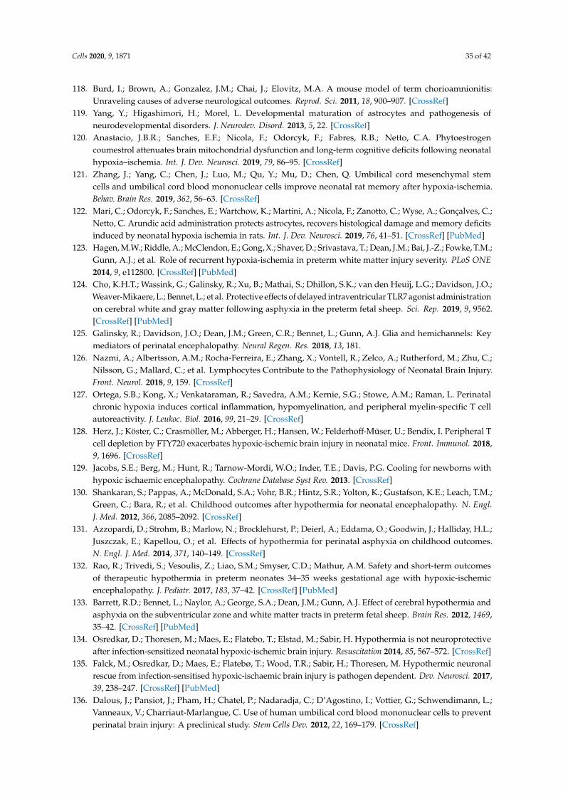
Cells 2020, 9, 1871 35 of 42
118. Burd, I.; Brown, A.; Gonzalez, J.M.; Chai, J.; Elovitz, M.A. A mouse model of term chorioamnionitis:Unraveling causes of adverse neurological outcomes. Reprod. Sci. 2011, 18, 900–907. [CrossRef]
119. Yang, Y.; Higashimori, H.; Morel, L. Developmental maturation of astrocytes and pathogenesis ofneurodevelopmental disorders. J. Neurodev. Disord. 2013, 5, 22. [CrossRef]
120. Anastacio, J.B.R.; Sanches, E.F.; Nicola, F.; Odorcyk, F.; Fabres, R.B.; Netto, C.A. Phytoestrogencoumestrol attenuates brain mitochondrial dysfunction and long-term cognitive deficits following neonatalhypoxia–ischemia. Int. J. Dev. Neurosci. 2019, 79, 86–95. [CrossRef]
121. Zhang, J.; Yang, C.; Chen, J.; Luo, M.; Qu, Y.; Mu, D.; Chen, Q. Umbilical cord mesenchymal stemcells and umbilical cord blood mononuclear cells improve neonatal rat memory after hypoxia-ischemia.Behav. Brain Res. 2019, 362, 56–63. [CrossRef]
122. Mari, C.; Odorcyk, F.; Sanches, E.; Wartchow, K.; Martini, A.; Nicola, F.; Zanotto, C.; Wyse, A.; Gonçalves, C.;Netto, C. Arundic acid administration protects astrocytes, recovers histological damage and memory deficitsinduced by neonatal hypoxia ischemia in rats. Int. J. Dev. Neurosci. 2019, 76, 41–51. [CrossRef] [PubMed]
123. Hagen, M.W.; Riddle, A.; McClendon, E.; Gong, X.; Shaver, D.; Srivastava, T.; Dean, J.M.; Bai, J.-Z.; Fowke, T.M.;Gunn, A.J.; et al. Role of recurrent hypoxia-ischemia in preterm white matter injury severity. PLoS ONE2014, 9, e112800. [CrossRef] [PubMed]
124. Cho, K.H.T.; Wassink, G.; Galinsky, R.; Xu, B.; Mathai, S.; Dhillon, S.K.; van den Heuij, L.G.; Davidson, J.O.;Weaver-Mikaere, L.; Bennet, L.; et al. Protective effects of delayed intraventricular TLR7 agonist administrationon cerebral white and gray matter following asphyxia in the preterm fetal sheep. Sci. Rep. 2019, 9, 9562.[CrossRef] [PubMed]
125. Galinsky, R.; Davidson, J.O.; Dean, J.M.; Green, C.R.; Bennet, L.; Gunn, A.J. Glia and hemichannels: Keymediators of perinatal encephalopathy. Neural Regen. Res. 2018, 13, 181.
126. Nazmi, A.; Albertsson, A.M.; Rocha-Ferreira, E.; Zhang, X.; Vontell, R.; Zelco, A.; Rutherford, M.; Zhu, C.;Nilsson, G.; Mallard, C.; et al. Lymphocytes Contribute to the Pathophysiology of Neonatal Brain Injury.Front. Neurol. 2018, 9, 159. [CrossRef]
127. Ortega, S.B.; Kong, X.; Venkataraman, R.; Savedra, A.M.; Kernie, S.G.; Stowe, A.M.; Raman, L. Perinatalchronic hypoxia induces cortical inflammation, hypomyelination, and peripheral myelin-specific T cellautoreactivity. J. Leukoc. Biol. 2016, 99, 21–29. [CrossRef]
128. Herz, J.; Köster, C.; Crasmöller, M.; Abberger, H.; Hansen, W.; Felderhoff-Müser, U.; Bendix, I. Peripheral Tcell depletion by FTY720 exacerbates hypoxic-ischemic brain injury in neonatal mice. Front. Immunol. 2018,9, 1696. [CrossRef]
129. Jacobs, S.E.; Berg, M.; Hunt, R.; Tarnow-Mordi, W.O.; Inder, T.E.; Davis, P.G. Cooling for newborns withhypoxic ischaemic encephalopathy. Cochrane Database Syst Rev. 2013. [CrossRef]
130. Shankaran, S.; Pappas, A.; McDonald, S.A.; Vohr, B.R.; Hintz, S.R.; Yolton, K.; Gustafson, K.E.; Leach, T.M.;Green, C.; Bara, R.; et al. Childhood outcomes after hypothermia for neonatal encephalopathy. N. Engl.J. Med. 2012, 366, 2085–2092. [CrossRef]
131. Azzopardi, D.; Strohm, B.; Marlow, N.; Brocklehurst, P.; Deierl, A.; Eddama, O.; Goodwin, J.; Halliday, H.L.;Juszczak, E.; Kapellou, O.; et al. Effects of hypothermia for perinatal asphyxia on childhood outcomes.N. Engl. J. Med. 2014, 371, 140–149. [CrossRef]
132. Rao, R.; Trivedi, S.; Vesoulis, Z.; Liao, S.M.; Smyser, C.D.; Mathur, A.M. Safety and short-term outcomesof therapeutic hypothermia in preterm neonates 34–35 weeks gestational age with hypoxic-ischemicencephalopathy. J. Pediatr. 2017, 183, 37–42. [CrossRef] [PubMed]
133. Barrett, R.D.; Bennet, L.; Naylor, A.; George, S.A.; Dean, J.M.; Gunn, A.J. Effect of cerebral hypothermia andasphyxia on the subventricular zone and white matter tracts in preterm fetal sheep. Brain Res. 2012, 1469,35–42. [CrossRef] [PubMed]
134. Osredkar, D.; Thoresen, M.; Maes, E.; Flatebo, T.; Elstad, M.; Sabir, H. Hypothermia is not neuroprotectiveafter infection-sensitized neonatal hypoxic-ischemic brain injury. Resuscitation 2014, 85, 567–572. [CrossRef]
135. Falck, M.; Osredkar, D.; Maes, E.; Flatebø, T.; Wood, T.R.; Sabir, H.; Thoresen, M. Hypothermic neuronalrescue from infection-sensitised hypoxic-ischaemic brain injury is pathogen dependent. Dev. Neurosci. 2017,39, 238–247. [CrossRef] [PubMed]
136. Dalous, J.; Pansiot, J.; Pham, H.; Chatel, P.; Nadaradja, C.; D’Agostino, I.; Vottier, G.; Schwendimann, L.;Vanneaux, V.; Charriaut-Marlangue, C. Use of human umbilical cord blood mononuclear cells to preventperinatal brain injury: A preclinical study. Stem Cells Dev. 2012, 22, 169–179. [CrossRef]
Cells 2020, 9, 1871 36 of 42
137. Ahn, S.Y.; Chang, Y.S.; Sung, D.K.; Sung, S.I.; Yoo, H.S.; Lee, J.H.; Oh, W.I.; Park, W.S. Mesenchymal stemcells prevent hydrocephalus after severe intraventricular hemorrhage. Stroke 2013, 44, 497–504. [CrossRef]
138. Ahn, S.Y.; Chang, Y.S.; Sung, S.I.; Park, W.S. Mesenchymal Stem Cells for Severe Intraventricular Hemorrhagein Preterm Infants: Phase I Dose-Escalation Clinical Trial. Stem Cells Transl Med. 2018, 7, 847–856. [CrossRef]
139. Yasuhara, T.; Hara, K.; Maki, M.; Mays, R.W.; Deans, R.J.; Hess, D.C.; Carroll, J.E.; Borlongan, C.V. Intravenousgrafts recapitulate the neurorestoration afforded by intracerebrally delivered multipotent adult progenitorcells in neonatal hypoxic-ischemic rats. J. Cereb Blood Flow Metab 2008, 28, 1804–1810. [CrossRef]
140. Drommelschmidt, K.; Serdar, M.; Bendix, I.; Herz, J.; Bertling, F.; Prager, S.; Keller, M.; Ludwig, A.-K.;Duhan, V.; Radtke, S. Mesenchymal stem cell-derived extracellular vesicles ameliorate inflammation-inducedpreterm brain injury. Brain Behav. Immun. 2017, 60, 220–232. [CrossRef]
141. Thomi, G.; Joerger-Messerli, M.; Haesler, V.; Muri, L.; Surbek, D.; Schoeberlein, A. Intranasally administeredexosomes from umbilical cord stem cells have preventive neuroprotective effects and contribute to functionalrecovery after perinatal brain injury. Cells 2019, 8, 855. [CrossRef]
142. Zhang, J.; Sadowska, G.B.; Chen, X.; Park, S.Y.; Kim, J.-E.; Bodge, C.A.; Cummings, E.; Lim, Y.-P.; Makeyev, O.;Besio, W.G. Anti–IL-6 neutralizing antibody modulates blood-brain barrier function in the ovine fetus.FASEB J. 2015, 29, 1739–1753. [CrossRef] [PubMed]
143. Galinsky, R.; Dhillon, S.K.; Dean, J.M.; Davidson, J.O.; Lear, C.A.; Wassink, G.; Nott, F.; Kelly, S.B.; Fraser, M.;Yuill, C. Tumor necrosis factor inhibition attenuates white matter gliosis after systemic inflammation inpreterm fetal sheep. J. Neuroinflamm. 2020, 17, 1–16. [CrossRef] [PubMed]
144. Chen, X.; Sadowska, G.B.; Zhang, J.; Kim, J.-E.; Cummings, E.E.; Bodge, C.A.; Lim, Y.-P.; Makeyev, O.;Besio, W.G.; Gaitanis, J. Neutralizing anti-interleukin-1β antibodies modulate fetal blood–brain barrierfunction after ischemia. Neurobiol. Dis. 2015, 73, 118–129. [CrossRef] [PubMed]
145. Chen, X.; Hovanesian, V.; Naqvi, S.; Lim, Y.-P.; Tucker, R.; Donahue, J.E.; Stopa, E.G.; Stonestreet, B.S. Systemicinfusions of anti-interleukin-1β neutralizing antibodies reduce short-term brain injury after cerebral ischemiain the ovine fetus. BrainBehav. Immun. 2018, 67, 24–35. [CrossRef] [PubMed]
146. Patra, A.; Chen, X.; Sadowska, G.B.; Zhang, J.; Lim, Y.-P.; Padbury, J.F.; Banks, W.A.; Stonestreet, B.S.Neutralizing anti-interleukin-1β antibodies reduce ischemia-related interleukin-1β transport across theblood–brain barrier in fetal sheep. Neuroscience 2017, 346, 113–125. [CrossRef] [PubMed]
147. Iwai, M.; Cao, G.; Yin, W.; Stetler, R.A.; Liu, J.; Chen, J. Erythropoietin promotes neuronal replacement throughrevascularization and neurogenesis after neonatal hypoxia/ischemia in rats. Stroke 2007, 38, 2795–2803. [CrossRef]
148. Iwai, M.; Stetler, R.A.; Xing, J.; Hu, X.; Gao, Y.; Zhang, W.; Chen, J.; Cao, G. Enhanced oligodendrogenesis andrecovery of neurological function by erythropoietin after neonatal hypoxic/ischemic brain injury. Stroke 2010,41, 1032–1037. [CrossRef]
149. Kellert, B.A.; McPherson, R.J.; Juul, S.E. A comparison of high-dose recombinant erythropoietin treatmentregimens in brain-injured neonatal rats. Pediatric Res. 2007, 61, 451–455. [CrossRef]
150. Alexander, M.; Hill, C.; Rosenkrantz, T.; Fitch, R. Evaluation of the therapeutic benefit of delayedadministration of erythropoietin following early hypoxic-ischemic injury in rodents. Dev. Neurosci.2012, 34, 515–524. [CrossRef]
151. Keller, M.; Yang, J.; Griesmaier, E.; Gorna, A.; Sarkozy, G.; Urbanek, M.; Gressens, P.; Simbruner, G.Erythropoietin is neuroprotective against NMDA-receptor-mediated excitotoxic brain injury in newbornmice. Neurobiol. Dis. 2006, 24, 357–366. [CrossRef]
152. Rees, S.; Hale, N.; De Matteo, R.; Cardamone, L.; Tolcos, M.; Loeliger, M.; Mackintosh, A.; Shields, A.;Probyn, M.; Greenwood, D. Erythropoietin is neuroprotective in a preterm ovine model of endotoxin-inducedbrain injury. J. Neuropathol. Exp. Neurol. 2010, 69, 306–319. [CrossRef] [PubMed]
153. Song, J.; Sun, H.; Xu, F.; Kang, W.; Gao, L.; Guo, J.; Zhang, Y.; Xia, L.; Wang, X.; Zhu, C. Recombinanthuman erythropoietin improves neurological outcomes in very preterm infants. Ann. Neurol. 2016, 80, 24–34.[CrossRef] [PubMed]
154. Juul, S.E.; Comstock, B.A.; Wadhawan, R.; Mayock, D.E.; Courtney, S.E.; Robinson, T.; Ahmad, K.A.;Bendel-Stenzel, E.; Baserga, M.; LaGamma, E.F. A Randomized Trial of Erythropoietin for Neuroprotectionin Preterm Infants. N. Engl. J. Med. 2020, 382, 233–243. [CrossRef] [PubMed]
155. Cho, K.H.; Davidson, J.O.; Dean, J.M.; Bennet, L.; Gunn, A.J. Cooling and immunomodulation for treatinghypoxic-ischemic brain injury. Pediatrics Int. 2020. [CrossRef] [PubMed]
Cells 2020, 9, 1871 37 of 42
156. Wassink, G.; Gunn, E.R.; Drury, P.P.; Bennet, L.; Gunn, A.J. The mechanisms and treatment of asphyxialencephalopathy. Front. Neurosci. 2014, 8, 40. [CrossRef]
157. Han, H.S.; Karabiyikoglu, M.; Kelly, S.; Sobel, R.A.; Yenari, M.A. Mild hypothermia inhibits nuclear factor-κBtranslocation in experimental stroke. J. Cereb. Blood Flow Metab. 2003, 23, 589–598. [CrossRef]
158. Ceulemans, A.-G.; Zgavc, T.; Kooijman, R.; Hachimi-Idrissi, S.; Sarre, S.; Michotte, Y. Mild hypothermia causesdifferential, time-dependent changes in cytokine expression and gliosis following endothelin-1-inducedtransient focal cerebral ischemia. J. Neuroinflamm. 2011, 8, 60. [CrossRef]
159. Gunn, A.J.; Gunn, T.R.; de Haan, H.H.; Williams, C.E.; Gluckman, P.D. Dramatic neuronal rescue withprolonged selective head cooling after ischemia in fetal lambs. J. Clin. Investig. 1997, 99, 248–256. [CrossRef]
160. Shankaran, S.; Barnes, P.D.; Hintz, S.R.; Laptook, A.R.; Zaterka-Baxter, K.M.; McDonald, S.A.;Ehrenkranz, R.A.; Walsh, M.C.; Tyson, J.E.; Donovan, E.F. Brain injury following trial of hypothermiafor neonatal hypoxic–ischaemic encephalopathy. Arch. Dis. Child. -Fetal Neonatal Ed. 2012, 97, F398–F404.
161. Azzopardi, D.; Group, T.S. Predictive value of the amplitude integrated EEG in infants with hypoxicischaemic encephalopathy: Data from a randomised trial of therapeutic hypothermia. Arch. Dis. Child. -FetalNeonatal Ed. 2014, 99, F80–F82. [CrossRef]
162. Silverman, W.A.; Fertig, J.W.; Berger, A.P. The influence of the thermal environment upon the survival ofnewly born premature infants. Pediatrics 1958, 22, 876–886. [PubMed]
163. Gunn, A.J.; Bennet, L. Brain cooling for preterm infants. Clin. Perinatol. 2008, 35, 735–748. [CrossRef] [PubMed]164. Smit, E.; Liu, X.; Jary, S.; Cowan, F.; Thoresen, M. Cooling neonates who do not fulfil the standard cooling
criteria–short-and long-term outcomes. Acta Paediatr. 2015, 104, 138–145. [CrossRef] [PubMed]165. Kuban, K.C.; Joseph, R.M.; O’Shea, T.M.; Heeren, T.; Fichorova, R.N.; Douglass, L.; Jara, H.; Frazier, J.A.;
Hirtz, D.; Rollins, J.V. Circulating inflammatory-associated proteins in the first month of life and cognitiveimpairment at age 10 years in children born extremely preterm. J. Pediatr. 2017, 180, 116–123.e111. [CrossRef]
166. Mourvillier, B.; Tubach, F.; Van de Beek, D.; Garot, D.; Pichon, N.; Georges, H.; Lefevre, L.M.; Bollaert, P.-E.;Boulain, T.; Luis, D. Induced hypothermia in severe bacterial meningitis: A randomized clinical trial.Jama 2013, 310, 2174–2183. [CrossRef] [PubMed]
167. Bennet, L.; Tan, S.; Van den Heuij, L.; Derrick, M.; Groenendaal, F.; van Bel, F.; Juul, S.; Back, S.A.;Northington, F.; Robertson, N.J. Cell therapy for neonatal hypoxia–ischemia and cerebral palsy. Ann. Neurol.2012, 71, 589–600. [CrossRef]
168. Titomanlio, L.; Kavelaars, A.; Dalous, J.; Mani, S.; El Ghouzzi, V.; Heijnen, C.; Baud, O.; Gressens, P. Stem celltherapy for neonatal brain injury: Perspectives and challenges. Ann. Neurol. 2011, 70, 698–712. [CrossRef]
169. Van Velthoven, C.T.; Kavelaars, A.; van Bel, F.; Heijnen, C.J. Regeneration of the ischemic brain by engineeredstem cells: Fuelling endogenous repair processes. Brain Res. Rev. 2009, 61, 1–13. [CrossRef]
170. Gortner, L.; Felderhoff-Müser, U.; Monz, D.; Bieback, K.; Klüter, H.; Jellema, R.; Kramer, B.; Keller, M.; Reiss, I.;Horn, P. Regenerative therapies in neonatology: Clinical perspectives. Klin. Pädiatrie 2012, 224, 233. [CrossRef]
171. Borlongan, C.V.; Weiss, M.D. Baby STEPS: A giant leap for cell therapy in neonatal brain injury. Pediatric Res.2011, 70, 3–9. [CrossRef]
172. Donega, V.; van Velthoven, C.T.; Nijboer, C.H.; Kavelaars, A.; Heijnen, C.J. The endogenous regenerativecapacity of the damaged newborn brain: Boosting neurogenesis with mesenchymal stem cell treatment.J. Cereb. Blood Flow Metab. 2013, 33, 625–634. [CrossRef] [PubMed]
173. Van Velthoven, C.T.; Kavelaars, A.; Heijnen, C.J. Mesenchymal stem cells as a treatment for neonatal ischemicbrain damage. Pediatric Res. 2012, 71, 474–481. [CrossRef] [PubMed]
174. Fleiss, B.; Guillot, P.V.; Titomanlio, L.; Baud, O.; Hagberg, H.; Gressens, P. Stem cell therapy for neonatalbrain injury. Clin. Perinatol. 2014, 41, 133–148. [CrossRef] [PubMed]
175. Jiang, Y.; Jahagirdar, B.N.; Reinhardt, R.L.; Schwartz, R.E.; Keene, C.D.; Ortiz-Gonzalez, X.R.; Reyes, M.;Lenvik, T.; Lund, T.; Blackstad, M. Pluripotency of mesenchymal stem cells derived from adult marrow.Nature 2002, 418, 41–49. [CrossRef] [PubMed]
176. Méndez-Ferrer, S.; Michurina, T.V.; Ferraro, F.; Mazloom, A.R.; MacArthur, B.D.; Lira, S.A.; Scadden, D.T.;Ma’ayan, A.; Enikolopov, G.N.; Frenette, P.S. Mesenchymal and haematopoietic stem cells form a uniquebone marrow niche. nature 2010, 466, 829–834. [CrossRef] [PubMed]
177. Uccelli, A.; Moretta, L.; Pistoia, V. Mesenchymal stem cells in health and disease. Nat. Rev. Immunol. 2008, 8,726–736. [CrossRef]
Cells 2020, 9, 1871 38 of 42
178. Le Blanc, K.; Mougiakakos, D. Multipotent mesenchymal stromal cells and the innate immune system.Nat. Rev. Immunol. 2012, 12, 383–396. [CrossRef]
179. Bieback, K.; Schallmoser, K.; Klüter, H.; Strunk, D. Clinical protocols for the isolation and expansion ofmesenchymal stromal cells. Transfus. Med. Hemotherapy 2008, 35, 286–294. [CrossRef]
180. Portmann-Lanz, C.B.; Schoeberlein, A.; Huber, A.; Sager, R.; Malek, A.; Holzgreve, W.; Surbek, D.V. Placentalmesenchymal stem cells as potential autologous graft for pre-and perinatal neuroregeneration. Am. J.Obstet. Gynecol. 2006, 194, 664–673. [CrossRef]
181. Uccelli, A.; Benvenuto, F.; Laroni, A.; Giunti, D. Neuroprotective features of mesenchymal stem cells.Best Pract. Res. Clin. Haematol. 2011, 24, 59–64. [CrossRef]
182. Siegel, G.; Schäfer, R.; Dazzi, F. The immunosuppressive properties of mesenchymal stem cells.Transplantation 2009, 87, S45–S49. [CrossRef] [PubMed]
183. Van Velthoven, C.T.; Kavelaars, A.; van Bel, F.; Heijnen, C.J. Repeated mesenchymal stem cell treatmentafter neonatal hypoxia–ischemia has distinct effects on formation and maturation of new neurons andoligodendrocytes leading to restoration of damage, corticospinal motor tract activity, and sensorimotorfunction. J. Neurosci. 2010, 30, 9603–9611. [CrossRef] [PubMed]
184. Rivera, F.J.; Couillard-Despres, S.; Pedre, X.; Ploetz, S.; Caioni, M.; Lois, C.; Bogdahn, U.; Aigner, L.Mesenchymal stem cells instruct oligodendrogenic fate decision on adult neural stem cells. Stem Cells 2006,24, 2209–2219. [CrossRef] [PubMed]
185. Steffenhagen, C.; Dechant, F.-X.; Oberbauer, E.; Furtner, T.; Weidner, N.; Küry, P.; Aigner, L.; Rivera, F.J.Mesenchymal stem cells prime proliferating adult neural progenitors toward an oligodendrocyte fate.Stem Cells Dev. 2011, 21, 1838–1851. [CrossRef]
186. Walker, P.A.; Shah, S.K.; Jimenez, F.; Gerber, M.H.; Xue, H.; Cutrone, R.; Hamilton, J.A.; Mays, R.W.; Deans, R.;Pati, S. Intravenous multipotent adult progenitor cell therapy for traumatic brain injury: Preserving theblood brain barrier via an interaction with splenocytes. Exp. Neurol. 2010, 225, 341–352. [CrossRef]
187. Walker, P.A.; Bedi, S.S.; Shah, S.K.; Jimenez, F.; Xue, H.; Hamilton, J.A.; Smith, P.; Thomas, C.P.; Mays, R.W.;Pati, S. Intravenous multipotent adult progenitor cell therapy after traumatic brain injury: Modulation of theresident microglia population. J. Neuroinflamm. 2012, 9, 228. [CrossRef]
188. DePaul, M.A.; Palmer, M.; Lang, B.T.; Cutrone, R.; Tran, A.P.; Madalena, K.M.; Bogaerts, A.; Hamilton, J.A.;Deans, R.J.; Mays, R.W. Intravenous multipotent adult progenitor cell treatment decreases inflammationleading to functional recovery following spinal cord injury. Sci. Rep. 2015, 5, 16795. [CrossRef]
189. Donega, V.; Nijboer, C.H.; van Tilborg, G.; Dijkhuizen, R.M.; Kavelaars, A.; Heijnen, C.J. Intranasallyadministered mesenchymal stem cells promote a regenerative niche for repair of neonatal ischemic braininjury. Exp. Neurol. 2014, 261, 53–64. [CrossRef]
190. Donega, V.; van Velthoven, C.T.; Nijboer, C.H.; van Bel, F.; Kas, M.J.; Kavelaars, A.; Heijnen, C.J. Intranasalmesenchymal stem cell treatment for neonatal brain damage: Long-term cognitive and sensorimotorimprovement. PLoS ONE 2013, 8. [CrossRef]
191. Wagenaar, N.; Nijboer, C.H.; van Bel, F. Repair of neonatal brain injury: Bringing stem cell-based therapyinto clinical practice. Dev Med. Child. Neurol 2017, 59, 997–1003. [CrossRef]
192. Van Velthoven, C.T.; Kavelaars, A.; Van Bel, F.; Heijnen, C.J. Nasal administration of stem cells: A promisingnovel route to treat neonatal ischemic brain damage. Pediatric Res. 2010, 68, 419–422. [CrossRef] [PubMed]
193. Oppliger, B.; Joerger-Messerli, M.; Mueller, M.; Reinhart, U.; Schneider, P.; Surbek, D.V.; Schoeberlein, A.Intranasal delivery of umbilical cord-derived mesenchymal stem cells preserves myelination in perinatalbrain damage. Stem Cells Dev. 2016, 25, 1234–1242. [CrossRef] [PubMed]
194. Finder, M.; Boylan, G.B.; Twomey, D.; Ahearne, C.; Murray, D.M.; Hallberg, B. Two-Year NeurodevelopmentalOutcomes After Mild Hypoxic Ischemic Encephalopathy in the Era of Therapeutic Hypothermia.Jama Pediatr. 2019. [CrossRef]
195. Kabat, M.; Bobkov, I.; Kumar, S.; Grumet, M. Trends in mesenchymal stem cell clinical trials 2004–2018: Isefficacy optimal in a narrow dose range? Stem Cells Transl. Med. 2020, 9, 17–27. [CrossRef] [PubMed]
196. Hess, D.C.; Wechsler, L.R.; Clark, W.M.; Savitz, S.I.; Ford, G.A.; Chiu, D.; Yavagal, D.R.; Uchino, K.;Liebeskind, D.S.; Auchus, A.P.; et al. Safety and efficacy of multipotent adult progenitor cells in acuteischaemic stroke (MASTERS): A randomised, double-blind, placebo-controlled, phase 2 trial. Lancet Neurol2017, 16, 360–368. [CrossRef]
Cells 2020, 9, 1871 39 of 42
197. Cotten, C.M.; Murtha, A.P.; Goldberg, R.N.; Grotegut, C.A.; Smith, P.B.; Goldstein, R.F.; Fisher, K.A.;Gustafson, K.E.; Waters-Pick, B.; Swamy, G.K.; et al. Feasibility of autologous cord blood cells for infantswith hypoxic-ischemic encephalopathy. J. Pediatr 2014, 164, 973–979. [CrossRef] [PubMed]
198. Ahn, S.Y.; Chang, Y.S.; Kim, J.H.; Sung, S.I.; Park, W.S. Two-Year Follow-Up Outcomes of Premature InfantsEnrolled in the Phase I Trial of Mesenchymal Stem Cells Transplantation for Bronchopulmonary Dysplasia.J. Pediatr 2017, 185, 49–54. [CrossRef]
199. Chang, Y.S.; Ahn, S.Y.; Yoo, H.S.; Sung, S.I.; Choi, S.J.; Oh, W.I.; Park, W.S. Mesenchymal stem cells forbronchopulmonary dysplasia: Phase 1 dose-escalation clinical trial. J. Pediatr 2014, 164, 966–972. [CrossRef]
200. Powell, S.B.; Silvestri, J.M. Safety of Intratracheal Administration of Human Umbilical Cord Blood DerivedMesenchymal Stromal Cells in Extremely Low Birth Weight Preterm Infants. J. Pediatr. 2019. [CrossRef]
201. Sun, J.M.; Song, A.W.; Case, L.E.; Mikati, M.A.; Gustafson, K.E.; Simmons, R.; Goldstein, R.; Petry, J.;McLaughlin, C.; Waters-Pick, B.; et al. Effect of Autologous Cord Blood Infusion on Motor Function and BrainConnectivity in Young Children with Cerebral Palsy: A Randomized, Placebo-Controlled Trial. Stem CellsTransl Med. 2017, 6, 2071–2078. [CrossRef]
202. Bi, M.; Wang, J.; Zhang, Y.; Li, L.; Wang, L.; Yao, R.; Duan, S.; Tong, S.; Li, J. Bone mesenchymal stemcells transplantation combined with mild hypothermia improves the prognosis of cerebral ischemia in rats.PLoS ONE 2018, 13, e0197405. [CrossRef] [PubMed]
203. Herz, J.; Koster, C.; Reinboth, B.S.; Dzietko, M.; Hansen, W.; Sabir, H.; van Velthoven, C.; Bendix, I.;Felderhoff-Muser, U. Interaction between hypothermia and delayed mesenchymal stem cell therapy inneonatal hypoxic-ischemic brain injury. Brain Behav Immun 2018, 70, 118–130. [CrossRef] [PubMed]
204. Lai, R.C.; Arslan, F.; Lee, M.M.; Sze, N.S.; Choo, A.; Chen, T.S.; Salto-Tellez, M.; Timmers, L.; Lee, C.N.; ElOakley, R.M.; et al. Exosome secreted by MSC reduces myocardial ischemia/reperfusion injury. Stem Cell Res.2010, 4, 214–222. [CrossRef] [PubMed]
205. Lai, R.C.; Chen, T.S.; Lim, S.K. Mesenchymal stem cell exosome: A novel stem cell-based therapy forcardiovascular disease. Regen Med. 2011, 6, 481–492. [CrossRef] [PubMed]
206. Dabrowska, S.; Andrzejewska, A.; Strzemecki, D.; Muraca, M.; Janowski, M.; Lukomska, B. Human bonemarrow mesenchymal stem cell-derived extracellular vesicles attenuate neuroinflammation evoked by focalbrain injury in rats. J. Neuroinflamm. 2019, 16, 216. [CrossRef]
207. Saunders, N.R.; Daneman, R.; Dziegielewska, K.M.; Liddelow, S.A. Transporters of the blood-brain andblood-CSF interfaces in development and in the adult. Mol Asp. Med. 2013, 34, 742–752. [CrossRef]
208. Moretti, R.; Pansiot, J.; Bettati, D.; Strazielle, N.; Ghersi-Egea, J.F.; Damante, G.; Fleiss, B.; Titomanlio, L.; Gressens, P.Blood-brain barrier dysfunction in disorders of the developing brain. Front. Neurosci 2015, 9, 40. [CrossRef]
209. Smith, S.F. Lipocortin 1: Glucocorticoids caught in the act? Thorax 1996, 51, 1057–1059. [CrossRef] [PubMed]210. Purvis, G.S.D.; Solito, E.; Thiemermann, C. Annexin-A1: Therapeutic Potential in Microvascular Disease.
Front. Immunol 2019, 10, 938. [CrossRef] [PubMed]211. Cristante, E.; McArthur, S.; Mauro, C.; Maggioli, E.; Romero, I.A.; Wylezinska-Arridge, M.; Couraud, P.O.;
Lopez-Tremoleda, J.; Christian, H.C.; Weksler, B.B.; et al. Identification of an essential endogenous regulatorof blood-brain barrier integrity, and its pathological and therapeutic implications. Proc. Natl. Acad. Sci. USA2013, 110, 832–841. [CrossRef]
212. McArthur, S.; Loiola, R.A.; Maggioli, E.; Errede, M.; Virgintino, D.; Solito, E. The restorative role of annexinA1 at the blood-brain barrier. Fluids Barriers Cns 2016, 13, 17. [CrossRef] [PubMed]
213. Aly, H.; Khashaba, M.T.; El-Ayouty, M.; El-Sayed, O.; Hasanein, B.M. IL-1beta, IL-6 and TNF-alpha andoutcomes of neonatal hypoxic ischemic encephalopathy. Brain Dev. 2006, 28, 178–182. [CrossRef] [PubMed]
214. Disdier, C.; Chen, X.; Kim, J.-E.; Threlkeld, S.W.; Stonestreet, B.S. Anti-Cytokine Therapy to AttenuateIschemic-Reperfusion Associated Brain Injury in the Perinatal Period. Brain Sci 2018, 8, 101.[CrossRef] [PubMed]
215. Oorschot, D.E.; Sizemore, R.J.; Amer, A.R. Treatment of Neonatal Hypoxic-Ischemic Encephalopathy withErythropoietin Alone, and Erythropoietin Combined with Hypothermia: History, Current Status, and FutureResearch. Int. J. Mol. Sci. 2020, 21, 1487. [CrossRef] [PubMed]
216. Rees, S.; Harding, R.; Walker, D. The biological basis of injury and neuroprotection in the fetal and neonatalbrain. Int. J. Dev. Neurosci. 2011, 29, 551–563. [CrossRef] [PubMed]
Cells 2020, 9, 1871 40 of 42
217. Kumral, A.; Baskin, H.; Yesilirmak, D.C.; Ergur, B.U.; Aykan, S.; Genc, S.; Genc, K.; Yilmaz, O.; Tugyan, K.;Giray, O. Erythropoietin attenuates lipopolysaccharide-induced white matter injury in the neonatal rat brain.Neonatology 2007, 92, 269–278. [CrossRef] [PubMed]
218. Ohlsson, A.; Aher, S.M. Early erythropoiesis-stimulating agents in preterm or low birth weight infants.Cochrane Database Syst. Rev. 2017, 2017. [CrossRef]
219. Back, S.A. White matter injury in the preterm infant: Pathology and mechanisms. Acta Neuropathol. 2017,134, 331–349. [CrossRef]
220. Wong, H.S.; Santhakumaran, S.; Cowan, F.M.; Modi, N.; Group, M.f.N.I. Developmental assessments inpreterm children: A meta-analysis. Pediatrics 2016, 138, e20160251. [CrossRef]
221. Fleiss, B.; Tann, C.J.; Degos, V.; Sigaut, S.; Van Steenwinckel, J.; Schang, A.L.; Kichev, A.; Robertson, N.J.;Mallard, C.; Hagberg, H.; et al. Inflammation-induced sensitization of the brain in term infants. Dev Med.Child. Neurol 2015, 57 Suppl. 3, 17–28. [CrossRef]
222. Hagberg, H.; Dammann, O.; Mallard, C.; Leviton, A. Preconditioning and the developing brain. Semin Perinatol2004, 28, 389–395. [CrossRef] [PubMed]
223. Gravina, G.; Svedin, P.; Ardalan, M.; Levy, O.; Ek, C.J.; Mallard, C.; Lai, J.C. Staphylococcus epidermidissensitizes perinatal hypoxic-ischemic brain injury in male but not female mice. Front. Immunol. 2020, 11, 516.[CrossRef] [PubMed]
224. Leviton, A.; Fichorova, R.N.; O’Shea, T.M.; Kuban, K.; Paneth, N.; Dammann, O.; Allred, E.N.;Investigators, E.S. Two-hit model of brain damage in the very preterm newborn: Small for gestational ageand postnatal systemic inflammation. Pediatr Res. 2013, 73, 362–370. [CrossRef] [PubMed]
225. Khwaja, O.; Volpe, J.J. Pathogenesis of cerebral white matter injury of prematurity. Arch. Dis Child. FetalNeonatal Ed. 2008, 93, F153–F161. [CrossRef]
226. Miller, S.L.; Huppi, P.S.; Mallard, C. The consequences of fetal growth restriction on brain structure andneurodevelopmental outcome. J. Physiol 2016, 594, 807–823. [CrossRef]
227. Barton, S.K.; Tolcos, M.; Miller, S.L.; Christoph-Roehr, C.; Schmolzer, G.M.; Moss, T.J.; Hooper, S.B.;Wallace, E.M.; Polglase, G.R. Ventilation-Induced Brain Injury in Preterm Neonates: A Review of PotentialTherapies. Neonatology 2016, 110, 155–162. [CrossRef]
228. Barnett, M.L.; Tusor, N.; Ball, G.; Chew, A.; Falconer, S.; Aljabar, P.; Kimpton, J.A.; Kennea, N.; Rutherford, M.;David Edwards, A.; et al. Exploring the multiple-hit hypothesis of preterm white matter damage usingdiffusion MRI. Neuroimage Clin 2018, 17, 596–606. [CrossRef]
229. Yoon, B.H.; Romero, R.; Park, J.S.; Kim, M.; Oh, S.Y.; Kim, C.J.; Jun, J.K. The relationship among inflammatorylesions of the umbilical cord (funisitis), umbilical cord plasma interleukin 6 concentration, amniotic fluidinfection, and neonatal sepsis. Am. J. Obs. Gynecol 2000, 183, 1124–1129. [CrossRef]
230. Ophelders, D.R.M.G.; Boots, A.W.; Hütten, M.C.; Al-Nasiry, S.; Jellema, R.K.; Spiller, O.B.; van Schooten, F.J.;Smolinska, A.; Wolfs, T.G.A.M. Screening of chorioamnionitis using volatile organic compound (VOC)detection in exhaled breath: A pre-clinical proof of concept study. In preparation.
231. Saha, S.; Pagnozzi, A.; Bourgeat, P.; George, J.M.; Bradford, D.; Colditz, P.B.; Boyd, R.N.; Rose, S.E.; Fripp, J.;Pannek, K. Predicting motor outcome in preterm infants from very early brain diffusion MRI using a deeplearning convolutional neural network (CNN) model. NeuroImage 2020, 116807. [CrossRef]
232. Inder, T.E.; Buckland, L.; Williams, C.E.; Spencer, C.; Gunning, M.I.; Darlow, B.A.; Volpe, J.J.; Gluckman, P.D.Lowered electroencephalographic spectral edge frequency predicts the presence of cerebral white matterinjury in premature infants. Pediatrics 2003, 111, 27–33. [CrossRef]
233. Wikström, S.; Pupp, I.H.; Rosén, I.; Norman, E.; Fellman, V.; Ley, D.; Hellström-Westas, L. Early single-channelaEEG/EEG predicts outcome in very preterm infants. Acta Paediatr. 2012, 101, 719–726. [CrossRef] [PubMed]
234. Kidokoro, H.; Kubota, T.; Hayashi, N.; Hayakawa, M.; Takemoto, K.; Kato, Y.; Okumura, A. Absent cyclicityon aEEG within the first 24 h is associated with brain damage in preterm infants. Neuropediatrics 2010, 41,241–245. [CrossRef] [PubMed]
235. Pillay, K.; Dereymaeker, A.; Jansen, K.; Naulaers, G.; De Vos, M. Applying a data-driven approach to quantifyeeG maturational deviations in preterms with normal and abnormal neurodevelopmental outcomes. Sci. Rep.2020, 10, 1–14.
236. Chiesa, C.; Pellegrini, G.; Panero, A.; De Luca, T.; Assumma, M.; Signore, F.; Pacifico, L. Umbilical cordinterleukin-6 levels are elevated in term neonates with perinatal asphyxia. Eur. J. Clin Invest. 2003, 33,352–358. [CrossRef] [PubMed]
Cells 2020, 9, 1871 41 of 42
237. Waje-Andreassen, U.; Krakenes, J.; Ulvestad, E.; Thomassen, L.; Myhr, K.M.; Aarseth, J.; Vedeler, C.A. IL-6: Anearly marker for outcome in acute ischemic stroke. Acta Neurol Scand 2005, 111, 360–365. [CrossRef] [PubMed]
238. Vaisbuch, E.; Romero, R.; Gomez, R.; Kusanovic, J.P.; Mazaki-Tovi, S.; Chaiworapongsa, T.; Hassan, S.S. Anelevated fetal interleukin-6 concentration can be observed in fetuses with anemia due to Rh alloimmunization:Implications for the understanding of the fetal inflammatory response syndrome. J. Matern Fetal Neonatal Med.2011, 24, 391–396. [CrossRef]
239. Rodney, T.; Osier, N.; Gill, J. Pro- and anti-inflammatory biomarkers and traumatic brain injury outcomes: Areview. Cytokine 2018, 110, 248–256. [CrossRef]
240. Jin, C.; Londono, I.; Mallard, C.; Lodygensky, G.A. New means to assess neonatal inflammatory brain injury.J. Neuroinflamm. 2015, 12, 180. [CrossRef]
241. Kersbergen, K.J.; Benders, M.J.; Groenendaal, F.; Koopman-Esseboom, C.; Nievelstein, R.A.; van Haastert, I.C.;de Vries, L.S. Different patterns of punctate white matter lesions in serially scanned preterm infants. PLoS ONE2014, 9, e108904. [CrossRef]
242. Ment, L.R.; Vohr, B.R. Preterm birth and the developing brain. Lancet Neurol 2008, 7, 378–379. [CrossRef]243. Annink, K.V.; de Vries, L.S.; Groenendaal, F.; Vijlbrief, D.C.; Weeke, L.C.; Roehr, C.C.; Lequin, M.; Reiss, I.;
Govaert, P.; Benders, M.J. The development and validation of a cerebral ultrasound scoring system for infantswith hypoxic-ischaemic encephalopathy. Pediatric Res. 2020, 87, 59–66. [CrossRef] [PubMed]
244. Govaert, P.; Roehr, C.C.; Gressens, P. Cranial ultrasound by neonatologists. Pediatr. Res. 2020, 87, 1–2.[CrossRef] [PubMed]
245. Dudink, J.; Steggerda, S.J.; Horsch, S. State-of-the-art neonatal cerebral ultrasound: Technique and reporting.Pediatric Res. 2020, 87, 3–12. [CrossRef] [PubMed]
246. Demené, C.; Mairesse, J.; Baranger, J.; Tanter, M.; Baud, O. Ultrafast Doppler for neonatal brain imaging.Neuroimage 2019, 185, 851–856. [CrossRef] [PubMed]
247. Deffieux, T.; Demene, C.; Pernot, M.; Tanter, M. Functional ultrasound neuroimaging: A review of thepreclinical and clinical state of the art. Curr. Opin. Neurobiol. 2018, 50, 128–135. [CrossRef] [PubMed]
248. Robertson, N.J.; Thayyil, S.; Cady, E.B.; Raivich, G. Magnetic resonance spectroscopy biomarkers in termperinatal asphyxial encephalopathy: From neuropathological correlates to future clinical applications.Curr. Pediatr. Rev. 2014, 10, 37–47. [CrossRef]
249. Thayyil, S.; Chandrasekaran, M.; Taylor, A.; Bainbridge, A.; Cady, E.B.; Chong, W.K.; Murad, S.; Omar, R.Z.;Robertson, N.J. Cerebral magnetic resonance biomarkers in neonatal encephalopathy: A meta-analysis.Pediatrics 2010, 125, e382–e395. [CrossRef]
250. Lally, P.J.; Montaldo, P.; Oliveira, V.; Soe, A.; Swamy, R.; Bassett, P.; Mendoza, J.; Atreja, G.; Kariholu, U.;Pattnayak, S.; et al. Magnetic resonance spectroscopy assessment of brain injury after moderate hypothermiain neonatal encephalopathy: A prospective multicentre cohort study. Lancet Neurol. 2019, 18, 35–45. [CrossRef]
251. Glass, H.C.; Kan, J.; Bonifacio, S.L.; Ferriero, D.M. Neonatal seizures: Treatment practices among term andpreterm infants. Pediatric Neurol. 2012, 46, 111–115. [CrossRef]
252. Azzopardi, D. Clinical applications of cerebral function monitoring in neonates. Semin. Fetal Neonatal Med.2015, 20, 154–163. [CrossRef]
253. Uria-Avellanal, C.; Marlow, N.; Rennie, J.M. Outcome following neonatal seizures. Semin. Fetal Neonatal Med.2013, 18, 224–232. [CrossRef] [PubMed]
254. Neil, J.J.; Volpe, J.J. Encephalopathy of prematurity: Clinical-neurological features, diagnosis, imaging,prognosis, therapy. In Volpe’s Neurology of the Newborn; Elsevier: Amsterdam, The Netherlands, 2018;pp. 425–457.
255. Roberts, D.; Brown, J.; Medley, N.; Dalziel, S.R. Antenatal corticosteroids for accelerating fetal lung maturationfor women at risk of preterm birth. Cochrane Database Syst. Rev. 2017. [CrossRef] [PubMed]
256. Sweet, D.G.; Carnielli, V.; Greisen, G.; Hallman, M.; Ozek, E.; Te Pas, A.; Plavka, R.; Roehr, C.C.; Saugstad, O.D.;Simeoni, U. European consensus guidelines on the management of respiratory distress syndrome–2019update. Neonatology 2019, 115, 432–450. [CrossRef] [PubMed]
257. Polglase, G.R.; Nitsos, I.; Baburamani, A.A.; Crossley, K.J.; Slater, M.K.; Gill, A.W.; Allison, B.J.; Moss, T.J.;Pillow, J.J.; Hooper, S.B. Inflammation in utero exacerbates ventilation-induced brain injury in preterm lambs.J. Appl. Physiol. 2012, 112, 481–489. [CrossRef] [PubMed]
Cells 2020, 9, 1871 42 of 42
258. Barton, S.K.; Moss, T.J.; Hooper, S.B.; Crossley, K.J.; Gill, A.W.; Kluckow, M.; Zahra, V.; Wong, F.Y.; Pichler, G.;Galinsky, R. Protective ventilation of preterm lambs exposed to acute chorioamnionitis does not reduceventilation-induced lung or brain injury. PLoS ONE 2014, 9, e112402. [CrossRef]
259. Hortensius, L.M.; Van Elburg, R.M.; Nijboer, C.H.; Benders, M.J.; De Theije, C.G.M. Postnatal nutrition toimprove brain development in the preterm infant: A systematic review from bench to bedside. Front. Physiol.2019, 10, 961. [CrossRef]
260. Keunen, K.; Van Elburg, R.M.; Van Bel, F.; Benders, M.J. Impact of nutrition on brain development and itsneuroprotective implications following preterm birth. Pediatr. Res. 2015, 77, 148–155. [CrossRef]
261. Doyle, L.W.; Ehrenkranz, R.A.; Halliday, H.L. Dexamethasone treatment in the first week of life forpreventing bronchopulmonary dysplasia in preterm infants: A systematic review. Neonatology 2010, 98,217–224. [CrossRef]
262. Kersbergen, K.J.; de Vries, L.S.; van Kooij, B.J.; Išgum, I.; Rademaker, K.J.; van Bel, F.; Hüppi, P.S.; Dubois, J.;Groenendaal, F.; Benders, M.J. Hydrocortisone treatment for bronchopulmonary dysplasia and brain volumesin preterm infants. J. Pediatr. 2013, 163, 666–671. [CrossRef]
© 2020 by the authors. Licensee MDPI, Basel, Switzerland. This article is an open accessarticle distributed under the terms and conditions of the Creative Commons Attribution(CC BY) license (http://creativecommons.org/licenses/by/4.0/).