Presentation1.pptx acute abdomen.
115
DR/ABD ALLAH NAZEER.MD. Radiological imaging of acute abdomen.
-
Upload
abdellah-nazeer -
Category
Documents
-
view
269 -
download
0
Transcript of Presentation1.pptx acute abdomen.

DR/ABD ALLAH NAZEER.MD.
Radiological imaging of acute abdomen.


ileocolic intussusception caused by mesenteric lymphadenitis. a. Transverse ultrasonography shows several enlarged mesenteric lymph nodes (arrowheads). b. Transverse ultrasonography shows the classic “doughnut sign” of the concentric rings of intussusception (arrow). c-e. Venous phase of CT scan axial (c, d) and coronal (e) scans show a target lesion in the right lower abdomen with inner intussusceptum (open arrows) and outer intussuscipiens (arrows). f, g. Air reduction shows a soft tissue mass (arrows) representing an intussusception.

Intussusception in a 69-year-old woman with partial small bowel obstruction caused by metastatic lung cancer. a-f. Venous phase of axial CT scans show multiple small bowel wall thickening and enteroenteric intussusceptions (arrows). Small bowel wall thickening represents metastasis of adenocarcinoma of lung cancer.

a-c. Axial CT scans show classic CT appearance of enteroenteric (jejunojejunal) intussusception (arrows). There is no evidence of obstruction, although small bowel was minimally dilated. d. Coronal reformatted CT scan shows invagination (arrows) of bowel and mesenteric vessels.


Portal venous gas due to small bowel infarction.


Sigmoid volvulus.


Plain abdominal radiograph of toxic megacolon that shows distention of the transverse colon associated with mucosal edema. The maximum transverse diameter of the transverse colon is 7.5 cm.

Toxic megacolon secondary to Clostridium infection.


Ischemic colitis with accordion sign-alternating edematous haustral folds separated by transverse mucosal ridges filled with oral contrast material simulating the appearance of accordion.
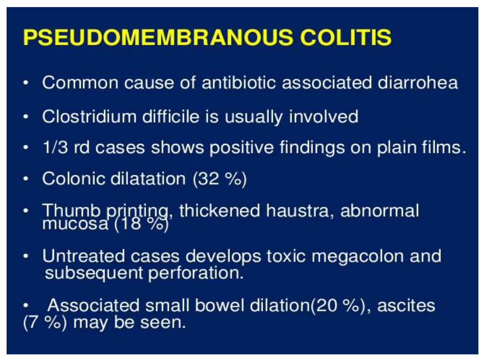


Pseudomembranous colitis. (Left) Axial CT scan of the midabdomen utilizing oral but not intravenous contrast demonstrates marked thickening of the colonic wall (white arrows) producing the so-called "accordion sign." There is a small amount of pericolonic stranding (red arrow) and ascites (green arrow). (Right) Axial CT scan through the pelvis shows marked thickening of the wall of the rectum (yellow arrows) indicating this is a pan-colitis.


CT images for acute appendicitis.

CT images for acute appendicitis.


Acute calcular cholecystitis.


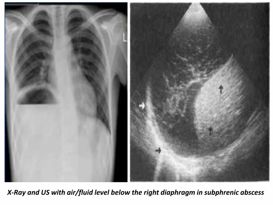
X-Ray and US with air/fluid level below the right diaphragm in subphrenic abscess

CT images for subphrenic abscess.


Left ureteric stone with hydronephrosis.


Advanced stages of salpingitis. a and b) Transvaginal US shows Elongated and dilated fallopian tube with echogenic contents (“string sign”)c and d) Contrast-enhanced CT scan shows dilated fallopian tube with wall thickening and enhancement

Coronal and axial CT imaged shows pelvic fluid collections with wall enhancement (abdominal abscess). c) Axial CT image shows pelvic fluid collection with gas bubbles.


Calcified abdominal aortic aneurysm.


Thank You.