Preparation and Characterization of YAG:Ce Phosphors …€¦ · Preparation and Characterization...
5
Preparation and Characterization of YAG:Ce 3+ Phosphors by Sol-solvothermal Process Ran Huang 1 , Sai Li 1 , Shaolin Xue 2 + , Zhixing Jiang 2 , Shuxian Wu 2 1 State Key Laboratory for Modification of Chemical Fibers and Polymer Materials, College of Material Science and Engineering, Donghua University, Shanghai 201620, P. R. China 2 College of Science, Donghua University, Shanghai 201620, P. R. China Abstract. White LEDs, which have the potential of replacing the conventional incandescent and fluorescent lamps due to its advantages of low operating voltages, longer lifetimes, small size, and absence of mercury are beneficial to energy conservation and green lighting industry. YAG:Ce 3+ phosphors are used as color converter in white LEDs because of its high luminescence efficiency, short luminescence lifetime and a green-yellow emission. In this study, we have prepared YAG:Ce 3+ phosphors by a sol-solvothermal process. YAG:Ce 3+ phosphors can be formed at the sintering temperature of 850°C. The average particle size is about 40 nm and the morphology can be described as spherical. The YAG:Ce 3+ samples show a broad emission band located in the range from 500 nm to 700 nm. The fluorescence intensity is exponential decay, and the decay time is about 62.28 ns. Keywords: YAG:Ce 3+ phosphors, synthesis method, luminescence, nano-particle, decay time. 1. Introduction During the last decades, cerium doped yttrium aluminum garnet (YAG:Ce 3+ ) which had advantages of high irradiance and interesting luminescent properties was applied widely in many scientific, technical and industrial applications including, e.g., solid-state lasers, fluorescent lamps, flying spot scanners, scintillators [1-3]. In recent years, YAG:Ce 3+ has received renewed interest related to yet another application: the use as color converter in white LEDs [4]. The white LEDs have the advantages of low applied voltage, long life, small size, and absence of mercury [5]. And the white LEDs technology is based on the principle of integrating two complementary luminescent emissions viz., blue and yellow emissions to generate white light suitable for lighting applications [6]. Part of the blue light from the GaN LED is absorbed by a thin layer of YAG:Ce 3+ phosphors and is converted into yellow light. The combination of blue and yellow gives a bright white light source. Schematic structure of dichromatic white LEDs is shown in Fig. 1. Fig. 1: Schematic structure of dichromatic white LEDs. + Corresponding author. Tel.: +86 021 67792089; fax: +86 021 67792085. E-mail address: [email protected] 2012 International Conference on Future Environment and Energy IPCBEE vol.28(2012) © (2012)IACSIT Press, Singapoore 85
Transcript of Preparation and Characterization of YAG:Ce Phosphors …€¦ · Preparation and Characterization...
Preparation and Characterization of YAG:Ce3+ Phosphors by Sol-solvothermal Process
Ran Huang 1, Sai Li 1, Shaolin Xue 2 +, Zhixing Jiang 2, Shuxian Wu 2 1 State Key Laboratory for Modification of Chemical Fibers and Polymer Materials, College of Material
Science and Engineering, Donghua University, Shanghai 201620, P. R. China 2 College of Science, Donghua University, Shanghai 201620, P. R. China
Abstract. White LEDs, which have the potential of replacing the conventional incandescent and fluorescent lamps due to its advantages of low operating voltages, longer lifetimes, small size, and absence of mercury are beneficial to energy conservation and green lighting industry. YAG:Ce3+ phosphors are used as color converter in white LEDs because of its high luminescence efficiency, short luminescence lifetime and a green-yellow emission. In this study, we have prepared YAG:Ce3+ phosphors by a sol-solvothermal process. YAG:Ce3+ phosphors can be formed at the sintering temperature of 850°C. The average particle size is about 40 nm and the morphology can be described as spherical. The YAG:Ce3+ samples show a broad emission band located in the range from 500 nm to 700 nm. The fluorescence intensity is exponential decay, and the decay time is about 62.28 ns.
Keywords: YAG:Ce3+ phosphors, synthesis method, luminescence, nano-particle, decay time.
1. Introduction During the last decades, cerium doped yttrium aluminum garnet (YAG:Ce3+) which had advantages of
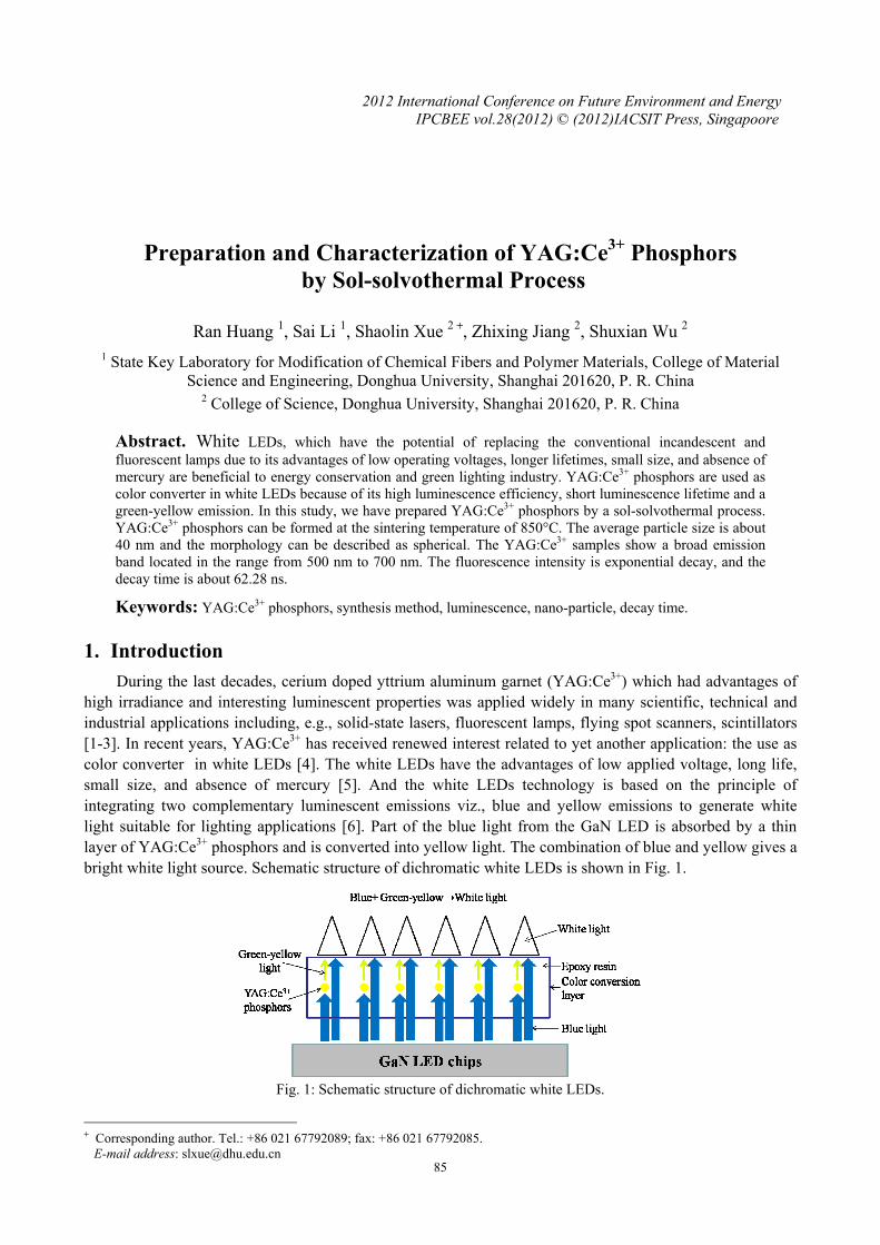
high irradiance and interesting luminescent properties was applied widely in many scientific, technical and industrial applications including, e.g., solid-state lasers, fluorescent lamps, flying spot scanners, scintillators [1-3]. In recent years, YAG:Ce3+ has received renewed interest related to yet another application: the use as color converter in white LEDs [4]. The white LEDs have the advantages of low applied voltage, long life, small size, and absence of mercury [5]. And the white LEDs technology is based on the principle of integrating two complementary luminescent emissions viz., blue and yellow emissions to generate white light suitable for lighting applications [6]. Part of the blue light from the GaN LED is absorbed by a thin layer of YAG:Ce3+ phosphors and is converted into yellow light. The combination of blue and yellow gives a bright white light source. Schematic structure of dichromatic white LEDs is shown in Fig. 1.
Fig. 1: Schematic structure of dichromatic white LEDs.
+ Corresponding author. Tel.: +86 021 67792089; fax: +86 021 67792085. E-mail address: [email protected]
2012 International Conference on Future Environment and EnergyIPCBEE vol.28(2012) © (2012)IACSIT Press, Singapoore
85
YAG:Ce3+ phosphors were synthesized conventionally via a solid-state reaction process, because it is comparatively simple and very suitable for mass production. But using solid-state reaction process to obtain YAG:Ce3+ phosphors with designed compositions and desired performances, the relatively high sintering temperature is required. Additionally, the particle size of YAG:Ce3+ phosphor prepared by this method is relatively large (micrometer-scale) and less controllable [7]. Yuexiao Pan et al. had successfully synthesized YAG:Ce3+ phosphors by a solid-state reaction method. In their work, the raw materials were milled thoroughly and transferred into a furnace for crystallization firstly at 1300°C for 10 hours under CO atmosphere, and then at 1500°C for 10 hours to enhance both the degree of crystallization and the luminescence intensity of the products. And repeated milling was needed between the two treatments for higher homogeneity [8]. To reduce the sintering temperature and produce smaller (submicron or nanometer-scale) products, various wet-chemical methods such as hydrothermal synthesis, co-precipitation method, sol-gel process and combustion synthesis have attracted widespread attention over the past several years. The wet-chemical metheds offered the possibilities for controlling homogeneity, purity of phase, size distribution, surface topography, and microstructural uniformity of the phosphors [7].
In this article, we have prepared YAG:Ce3+ phosphors by a sol-solvothermal process, and investigated the luminescence properties of YAG:Ce3+ samples.
2. Experimental detail
2.1. Samples prepared The raw materials are cerium nitrate (Ce(NO3)3·6H2O, analytical pure), yttrium nitrate (Y(NO3)3·6H2O,
analytical pure), and aluminum isopropoxide (C9H21AlO3, chemical pure). The detailed experimental processes are as follows: firstly, all of the raw materials were dispersed uniformly in isopropyl alcohol (C3H8O, analytical pure) by using an ultrasound device (KQ218, Kunshan, China). Secondly, the well-mixed solution was made into sol by using magnetic heating stirrer and then placed in a high pressure autoclave. The autoclave was heated at 190°C for 20 hours. And the precursor was obtained by washing and drying the products in the autoclave. Finally, we can obtain the YAG:Ce3+ phosphors after sintered the precursor at 850°C for 4 hours in a tubular furnace.
2.2. Characterization The prepared YAG:Ce3+ phosphors were characterized by X-ray diffraction (XRD, D/Max-2550 PC,
Rigaku, Tokyo, Japan) with a Cu Kα radiation source. The morphology and size of these phosphors were investigated using field emission scanning electron microscopy (FESEM, S-4800, Hitachi, Japan). The luminescent properties were measured by using the luminescence spectrophotometer (FLS920, Edinburgh Instrument, England) with Xe-lamp (450W) as an excitation source.
3. Results and discussion
3.1. XRD analysis
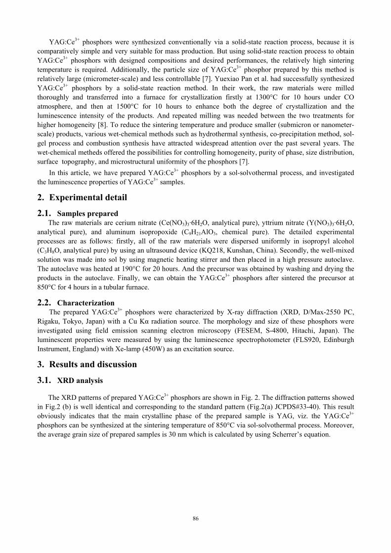
The XRD patterns of prepared YAG:Ce3+ phosphors are shown in Fig. 2. The diffraction patterns showed in Fig.2 (b) is well identical and corresponding to the standard pattern (Fig.2(a) JCPDS#33-40). This result obviously indicates that the main crystalline phase of the prepared sample is YAG, viz. the YAG:Ce3+ phosphors can be synthesized at the sintering temperature of 850°C via sol-solvothermal process. Moreover, the average grain size of prepared samples is 30 nm which is calculated by using Scherrer’s equation.
86
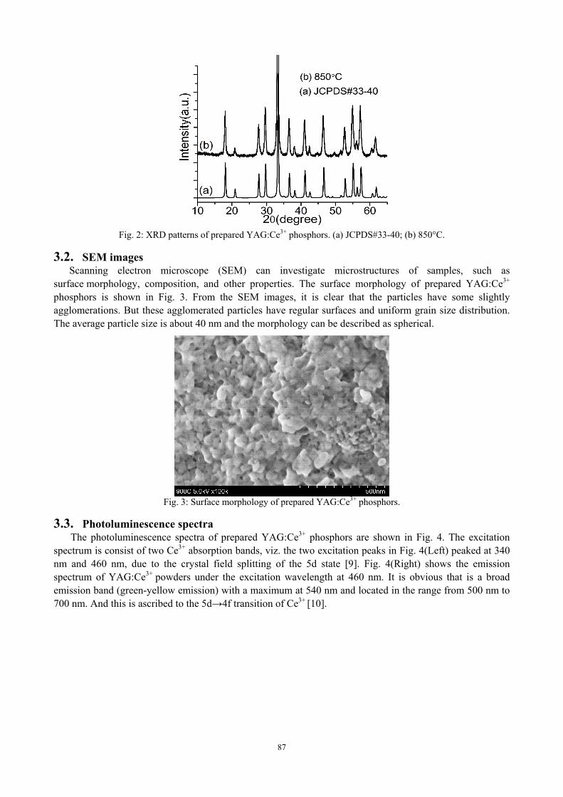
Fig. 2: XRD patterns of prepared YAG:Ce3+ phosphors. (a) JCPDS#33-40; (b) 850°C.
3.2. SEM images Scanning electron microscope (SEM) can investigate microstructures of samples, such as
surface morphology, composition, and other properties. The surface morphology of prepared YAG:Ce3+ phosphors is shown in Fig. 3. From the SEM images, it is clear that the particles have some slightly agglomerations. But these agglomerated particles have regular surfaces and uniform grain size distribution. The average particle size is about 40 nm and the morphology can be described as spherical.
Fig. 3: Surface morphology of prepared YAG:Ce3+ phosphors.
3.3. Photoluminescence spectra The photoluminescence spectra of prepared YAG:Ce3+ phosphors are shown in Fig. 4. The excitation
spectrum is consist of two Ce3+ absorption bands, viz. the two excitation peaks in Fig. 4(Left) peaked at 340 nm and 460 nm, due to the crystal field splitting of the 5d state [9]. Fig. 4(Right) shows the emission spectrum of YAG:Ce3+ powders under the excitation wavelength at 460 nm. It is obvious that is a broad emission band (green-yellow emission) with a maximum at 540 nm and located in the range from 500 nm to 700 nm. And this is ascribed to the 5d→4f transition of Ce3+ [10].
87
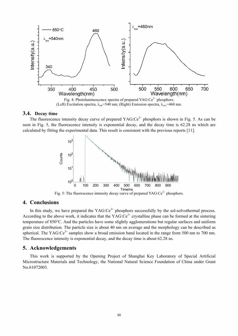
Fig. 4: Photoluminescence spectra of prepared YAG:Ce3+ phosphors.
(Left) Excitation spectra, λem=540 nm; (Right) Emission spectra, λexc=460 nm.
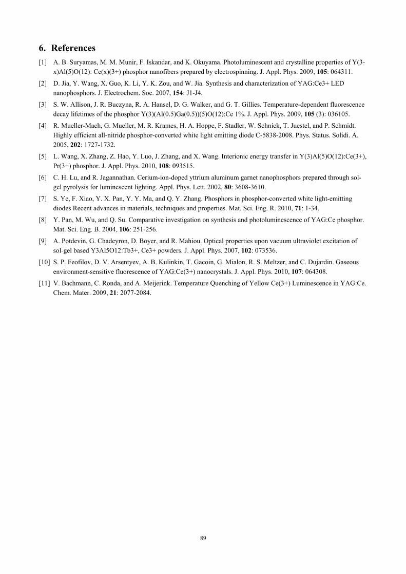
3.4. Decay time The fluorescence intensity decay curve of prepared YAG:Ce3+ phosphors is shown in Fig. 5. As can be
seen in Fig. 5, the fluorescence intensity is exponential decay, and the decay time is 62.28 ns which are calculated by fitting the experimental data. This result is consistent with the previous reports [11].
010
110
210
310
0 100 200 300 400 500 600 700 800 900
Cou
nts
Time/ns Fig. 5: The fluorescence intensity decay curve of prepared YAG:Ce3+ phosphors.
4. Conclusions In this study, we have prepared the YAG:Ce3+ phosphors successfully by the sol-solvothermal process.
According to the above work, it indicates that the YAG:Ce3+ crystalline phase can be formed at the sintering temperature of 850°C. And the particles have some slightly agglomerations but regular surfaces and uniform grain size distribution. The particle size is about 40 nm on average and the morphology can be described as spherical. The YAG:Ce3+ samples show a broad emission band located in the range from 500 nm to 700 nm. The fluorescence intensity is exponential decay, and the decay time is about 62.28 ns.
5. Acknowledgements This work is supported by the Opening Project of Shanghai Key Laboratory of Special Artificial
Microstructure Materials and Technology, the National Natural Science Foundation of China under Grant No.61072003.
88
6. References [1] A. B. Suryamas, M. M. Munir, F. Iskandar, and K. Okuyama. Photoluminescent and crystalline properties of Y(3-
x)Al(5)O(12): Ce(x)(3+) phosphor nanofibers prepared by electrospinning. J. Appl. Phys. 2009, 105: 064311.
[2] D. Jia, Y. Wang, X. Guo, K. Li, Y. K. Zou, and W. Jia. Synthesis and characterization of YAG:Ce3+ LED nanophosphors. J. Electrochem. Soc. 2007, 154: J1-J4.
[3] S. W. Allison, J. R. Buczyna, R. A. Hansel, D. G. Walker, and G. T. Gillies. Temperature-dependent fluorescence decay lifetimes of the phosphor Y(3)(Al(0.5)Ga(0.5))(5)O(12):Ce 1%. J. Appl. Phys. 2009, 105 (3): 036105.
[4] R. Mueller-Mach, G. Mueller, M. R. Krames, H. A. Hoppe, F. Stadler, W. Schnick, T. Juestel, and P. Schmidt. Highly efficient all-nitride phosphor-converted white light emitting diode C-5838-2008. Phys. Status. Solidi. A. 2005, 202: 1727-1732.
[5] L. Wang, X. Zhang, Z. Hao, Y. Luo, J. Zhang, and X. Wang. Interionic energy transfer in Y(3)Al(5)O(12):Ce(3+), Pr(3+) phosphor. J. Appl. Phys. 2010, 108: 093515.
[6] C. H. Lu, and R. Jagannathan. Cerium-ion-doped yttrium aluminum garnet nanophosphors prepared through sol-gel pyrolysis for luminescent lighting. Appl. Phys. Lett. 2002, 80: 3608-3610.
[7] S. Ye, F. Xiao, Y. X. Pan, Y. Y. Ma, and Q. Y. Zhang. Phosphors in phosphor-converted white light-emitting diodes Recent advances in materials, techniques and properties. Mat. Sci. Eng. R. 2010, 71: 1-34.
[8] Y. Pan, M. Wu, and Q. Su. Comparative investigation on synthesis and photoluminescence of YAG:Ce phosphor. Mat. Sci. Eng. B. 2004, 106: 251-256.
[9] A. Potdevin, G. Chadeyron, D. Boyer, and R. Mahiou. Optical properties upon vacuum ultraviolet excitation of sol-gel based Y3Al5O12:Tb3+, Ce3+ powders. J. Appl. Phys. 2007, 102: 073536.
[10] S. P. Feofilov, D. V. Arsentyev, A. B. Kulinkin, T. Gacoin, G. Mialon, R. S. Meltzer, and C. Dujardin. Gaseous environment-sensitive fluorescence of YAG:Ce(3+) nanocrystals. J. Appl. Phys. 2010, 107: 064308.
[11] V. Bachmann, C. Ronda, and A. Meijerink. Temperature Quenching of Yellow Ce(3+) Luminescence in YAG:Ce. Chem. Mater. 2009, 21: 2077-2084.
89