PRENATAL DIAGNOSIS OF CONGENITAL …©1987-2002 Romero-Pilu-Jeanty-Ghidini-Hobbins PRENATAL...
81
©1987-2002 Romero-Pilu-Jeanty-Ghidini-Hobbins PRENATAL DIAGNOSIS OF CONGENITAL ANOMALIES Roberto Romero, M.D. Associate Professor of Obstetrics and Gynecology Director of Perinatal Research Yale University School of Medicine New Haven, Connecticut Gianluigi Pilu, M.D. Attending Physician Section of Prenatal Pathophysiology Second Department of Obstetrics and Gynecology University of Bologna School of Medicine Bologna, Italy Philippe Jeanty, M.D. Assistant Professor Department of Radiology Vanderbilt University Nashville, Tennessee Alessandro Ghidini, M.D. Research Fellow Department of Obstetrics and Gynecology Yale University School of Medicine New Haven, Connecticut John C. Hobbins, M.D. Professor of Obstetrics and Gynecology and Diagnostic Imaging Yale University School of Medicine Director of Obstetrics Yale-New Haven Hospital New Haven, Connecticut APPLETON & LANGE Norwalk, Connecticut/San Mateo, Califomia
Transcript of PRENATAL DIAGNOSIS OF CONGENITAL …©1987-2002 Romero-Pilu-Jeanty-Ghidini-Hobbins PRENATAL...

©1987-2002 Romero-Pilu-Jeanty-Ghidini-Hobbins
PRENATAL DIAGNOSIS OF CONGENITAL ANOMALIES
Roberto Romero, M.D.
Associate Professor of Obstetrics and Gynecology Director of Perinatal Research
Yale University School of Medicine New Haven, Connecticut
Gianluigi Pilu, M.D. Attending Physician
Section of Prenatal Pathophysiology Second Department of Obstetrics and Gynecology
University of Bologna School of Medicine Bologna, Italy
Philippe Jeanty, M.D.
Assistant Professor Department of Radiology
Vanderbilt University Nashville, Tennessee
Alessandro Ghidini, M.D.
Research Fellow Department of Obstetrics and Gynecology
Yale University School of Medicine New Haven, Connecticut
John C. Hobbins, M.D.
Professor of Obstetrics and Gynecology and Diagnostic Imaging
Yale University School of Medicine Director of Obstetrics
Yale-New Haven Hospital New Haven, Connecticut
APPLETON & LANGE Norwalk, Connecticut/San Mateo, Califomia

©1987-2002 Romero-Pilu-Jeanty-Ghidini-Hobbins
0-8385-7921-3 Notice: Our knowledge in clinical sciences is constantly changing. As new information becomes available, changes in treatment and in the use of drugs become necessary. The author(s) and the publisher of this volume have taken care to make certain that the doses of drugs and schedules of treatment are correct and compatible with the standards generally accepted at the time of publication. The reader is advised to consult carefully the instruction and information material included in the package insert of each drug or therapeutic agent before administration. This advice is especially important when using new or infrequently used drugs. Copyright © 1988 by Appleton & Lange A Publishing Division of Prentice Hall All rights reserved. This book, or any parts thereof, may not be used or reproduced in any manner without written permission. For information, address Appleton & Lange, 25 Van Zant Street, East Norwalk, Connecticut 06855.
88 89 90 91 92 / 10 9 8 7 6 5 4 3 2 1 Prentice-Hall of Australia, Pty. Ltd., Sydney Prentice-Hall Canada, Inc. Prentice-Hall Hispanoamericana, S.A., Mexico Prentice-Hall of India Private Limited, New Delhi Prentice-Hall International (UK) Limited, London Prentice-Hall of Japan, Inc., Tokyo Prentice-Hall of Southeast Asia (Pte.) Ltd., Singapore Whitehall Books Ltd., Wellington, New Zealand Editora Prentice-Hall do Brasil Ltda., Rio de Janeiro
Library of Congress Cataloging-in-Publication Data
Prenatal diagnosis of congenital anomalies. Includes index.
1. Prenatal diagnosis. 2. Fetus---Abnormalities --- Diagnosis. I. Romero, Roberto. [DNLM: 1. Abnormalities ---diagnosis. 2. Fetal Diseases--diagnosis.
3. Prenatal Diagnosis---methods. QS 675 P926] RG628.P74 1987 618.2'2 87-14557 ISBN 0-8385-7921-3 Production Editor: Donald L. Delauter Design: M. Chandler Martylewski PRINTED IN THE UNITED STATES OF AMERICA

©1987-2002 Romero-Pilu-Jeanty-Ghidini-Hobbins
The Central Nervous System Normal Sonographic Anatomy of the Porencephaly/ 50 Fetal Central Nervous System/ 1 Hydranencephaly/ 52 HYDROCEPHALUS/ 21 Microcephaly/ 54 Aqueductal Stenosis/ 24 Holoprosencephaly/ 59 Communicating Hydrocephalus/ 27 Inienecphaly/ 65 Dandy-Walker Malformation/ 30 Agenesis of the Corpus Callosum/ 67 Choroid Plexus Papilloma/ 34 Lissencephaly/ 70 NEURAL TUBE DEFECTS/ 36 Intracranial Arachnoid Cysts/ 71 Spina Bifida/ 36 Intracranial Tumors/ 73 Anencephaly/ 43 Acrania/ 75 Cephalocele/ 46 Choroid Plexus Cyst/ 76 Aneurysm of the Vein of Galen/ 77
Normal Sonographic Anatomy of the Fetal Central Nervous System INTRACRANIAL ANATOMY The objective of the sonographic examination of the fetal central nervous system (CNS) is to reconstruct with a two-dimensional tool a complex three-dimensional structure.In this effort, the larger the number of scanning planes obtained, the more accurate the representation will appear.The three planes traditionally used for such an evaluation are the axial, sagittal, and coronal (Fig. 1-1). The sonographer should be aware that important developmental changes occur in the fetal brain well after the end of embryogenesis and up to the third trimester. The lateral ventricles and subarachnoid cisterns decrease steadily in size throughout gestation, resulting not only in a geometric modification of the cerebral structures but also in important changes in the sonographic appearance of the fetal brain. During the early second trimester, the fluid-filled lateral ventricles are large. This causes enhancement of sound transmission, and the distal cerebral cortex appears more echoic than later in gestation. Familiarity with the normal ultrasound appearance of the fetal brain in different scanning planes and at different gesta-
tional ages is critical for the recognition of congenital anomalies. Axial Planes Axial planes are obtained by scanning the head of the fetus at an angle of about 20 degrees to the canthomeatal line.9 Four different levels are commonly used (Fig. 1-2), The first scanning plane passes through the bodies of the lateral ventricles. In Figure 1-3A, the different appearances of this view throughout gestation can be seen. At 16 weeks, the lateral ventricles occupy most of the relative hemispheres and are partially filled with the echogenic choroid plexuses. At midgestation, the size of the lateral ventricles has considerable diminished, but in many cases it is still possible to observe the two walls that line the ventricular cavity on both sides. During the third trimester, only the lateral wall can be visualized. The distance between the midline echo and the lateral wall of the ventricle is now approximately one third of the hemispheric width. This value will remain constant throughout life (Fig. 1-3B). The axial view has been used to derive nomograms for the normal size of the ventricles The
©1987-2002 Romero-Pilu-Jeanty-Ghidini-Hobbins
2 THE CENTRAL NERVOUS SYSTEM
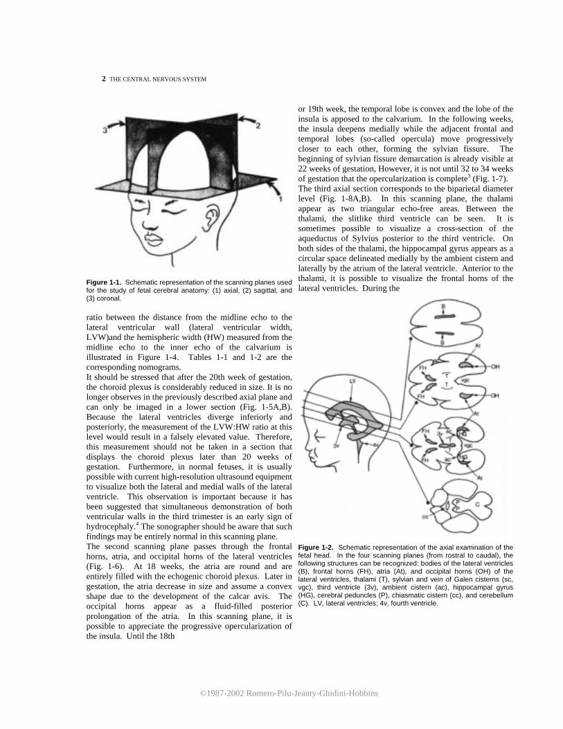
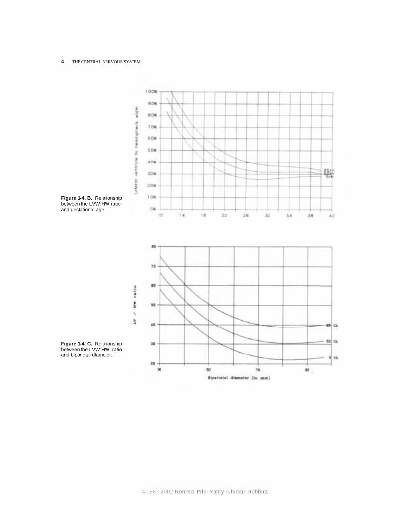
Figure 1-1. Schematic representation of the scanning planes used for the study of fetal cerebral anatomy: (1) axial, (2) sagittal, and (3) coronal. ratio between the distance from the midline echo to the lateral ventricular wall (lateral ventricular width, LVW)and the hemispheric width (HW) measured from the midline echo to the inner echo of the calvarium is illustrated in Figure 1-4. Tables 1-1 and 1-2 are the corresponding nomograms. It should be stressed that after the 20th week of gestation, the choroid plexus is considerably reduced in size. It is no longer observes in the previously described axial plane and can only be imaged in a lower section (Fig. 1-5A,B). Because the lateral ventricles diverge inferiorly and posteriorly, the measurement of the LVW:HW ratio at this level would result in a falsely elevated value. Therefore, this measurement should not be taken in a section that displays the choroid plexus later than 20 weeks of gestation. Furthermore, in normal fetuses, it is usually possible with current high-resolution ultrasound equipment to visualize both the lateral and medial walls of the lateral ventricle. This observation is important because it has been suggested that simultaneous demonstration of both ventricular walls in the third trimester is an early sign of hydrocephaly.4 The sonographer should be aware that such findings may be entirely normal in this scanning plane. The second scanning plane passes through the frontal horns, atria, and occipital horns of the lateral ventricles (Fig. 1-6). At 18 weeks, the atria are round and are entirely filled with the echogenic choroid plexus. Later in gestation, the atria decrease in size and assume a convex shape due to the development of the calcar avis. The occipital horns appear as a fluid-filled posterior prolongation of the atria. In this scanning plane, it is possible to appreciate the progressive opercularization of the insula. Until the 18th
or 19th week, the temporal lobe is convex and the lobe of the insula is apposed to the calvarium. In the following weeks, the insula deepens medially while the adjacent frontal and temporal lobes (so-called opercula) move progressively closer to each other, forming the sylvian fissure. The beginning of sylvian fissure demarcation is already visible at 22 weeks of gestation, However, it is not until 32 to 34 weeks of gestation that the opercularization is complete3 (Fig. 1-7). The third axial section corresponds to the biparietal diameter level (Fig. 1-8A,B). In this scanning plane, the thalami appear as two triangular echo-free areas. Between the thalami, the slitlike third ventricle can be seen. It is sometimes possible to visualize a cross-section of the aqueductus of Sylvius posterior to the third ventricle. On both sides of the thalami, the hippocampal gyrus appears as a circular space delineated medially by the ambient cistern and laterally by the atrium of the lateral ventricle. Anterior to the thalami, it is possible to visualize the frontal horns of the lateral ventricles. During the
Figure 1-2. Schematic representation of the axial examination of the fetal head. In the four scanning planes (from rostral to caudal), the following structures can be recognized: bodies of the lateral ventricles (B), frontal horns (FH), atria (At), and occipital horns (OH) of the lateral ventricles, thalami (T), sylvian and vein of Galen cisterns (sc, vgc), third ventricle (3v), ambient cistern (ac), hippocampal gyrus (HG), cerebral peduncles (P), chiasmatic cistern (cc), and cerebellum (C). LV, lateral ventricles; 4v, fourth ventricle.

©1987-2002 Romero-Pilu-Jeanty-Ghidini-Hobbins
NORMAL SONOGRAPHIC ANATOMY OF THE FETAL CENTRAL NERVOUS SYSTEM 3
Figure 1-3. A. Axial scans at the level of the bodies (B) of the lateral ventricles at 16, 23, and 30 weeks. Note the prominent choroid plexus (CP) in the 16-week fetus and the progressive shrinking of the ventricular cavity. The arrowheads indicate the medial and lateral walls of the ventricle.
Figure 1-3. B. Anatomic specimen from an adult brain correspond-ing to the axial section shown in Figure 1-3A. Note the similarity in ventricular versus hemispheric size with the ultrasound image of the 30-week fetus. (Reproduced with permission from Matsui, Irano : An Atlas of the Human Brain for Computed Tomography. Tokyo, Igaku Shoin, 1978.)
Figure 1-4. A. Measurement of the LVW:HW ratio.
©1987-2002 Romero-Pilu-Jeanty-Ghidini-Hobbins
4 THE CENTRAL NERVOUS SYSTEM Figure 1-4. B. Relationship between the LVW:HW ratio and gestational age. Figure 1-4. C. Relationship between the LVW:HW ratio and biparietal diameter.

©1987-2002 Romero-Pilu-Jeanty-Ghidini-Hobbins
NORMAL SONOGRAPHIC ANATOMIY OF THE FETAL CENTRAL NERVOUS SYSTEM 5
©1987-2002 Romero-Pilu-Jeanty-Ghidini-Hobbins
6 THE CENTRAL NERVOUS SYSTEM
A B
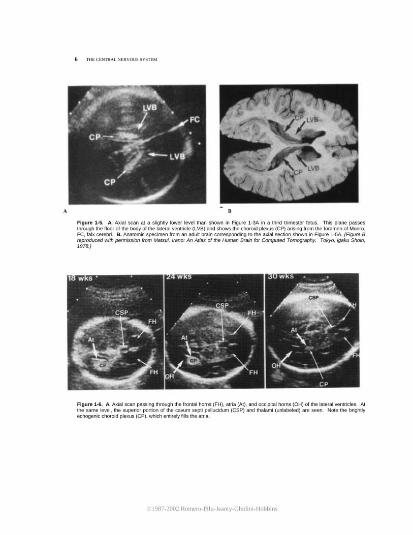
Figure 1-5. A. Axial scan at a slightly lower level than shown in Figure 1-3A in a third trimester fetus. This plane passes through the floor of the body of the lateral ventricle (LVB) and shows the choroid plexus (CP) arising from the foramen of Monro. FC, falx cerebri. B. Anatomic specimen from an adult brain corresponding to the axial section shown in Figure 1-5A. (Figure B reproduced with permission from Matsui, Irano: An Atlas of the Human Brain for Computed Tomography. Tokyo, lgaku Shoin, 1978.)
Figure 1-6. A. Axial scan passing through the frontal horns (FH), atria (At), and occipital horns (OH) of the lateral ventricles. At the same level, the superior portion of the cavum septi pellucidum (CSP) and thalami (unlabeled) are seen. Note the brightly echogenic choroid plexus (CP), which entirely fills the atria.
©1987-2002 Romero-Pilu-Jeanty-Ghidini-Hobbins
NORMAL SONOGRAPHIC ANATOMIY OF THE FETAL CENTRAL NERVOUS SYSTEM 7
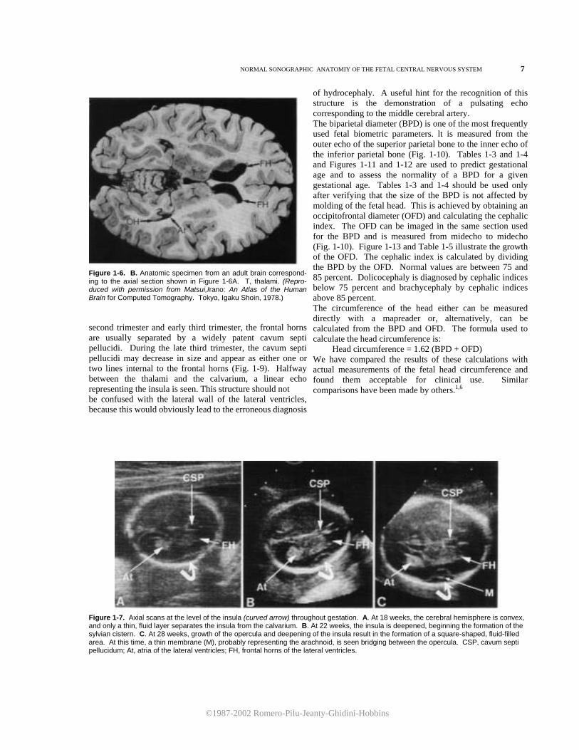
Figure 1-6. B. Anatomic specimen from an adult brain correspond-ing to the axial section shown in Figure 1-6A. T, thalami. (Repro-duced with permission from Matsui,Irano: An Atlas of the Human Brain for Computed Tomography. Tokyo, Igaku Shoin, 1978.) second trimester and early third trimester, the frontal horns are usually separated by a widely patent cavum septi pellucidi. During the late third trimester, the cavum septi pellucidi may decrease in size and appear as either one or two lines internal to the frontal horns (Fig. 1-9). Halfway between the thalami and the calvarium, a linear echo representing the insula is seen. This structure should not be confused with the lateral wall of the lateral ventricles, because this would obviously lead to the erroneous diagnosis
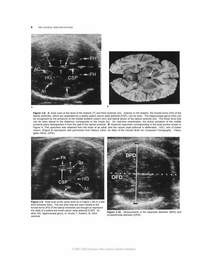
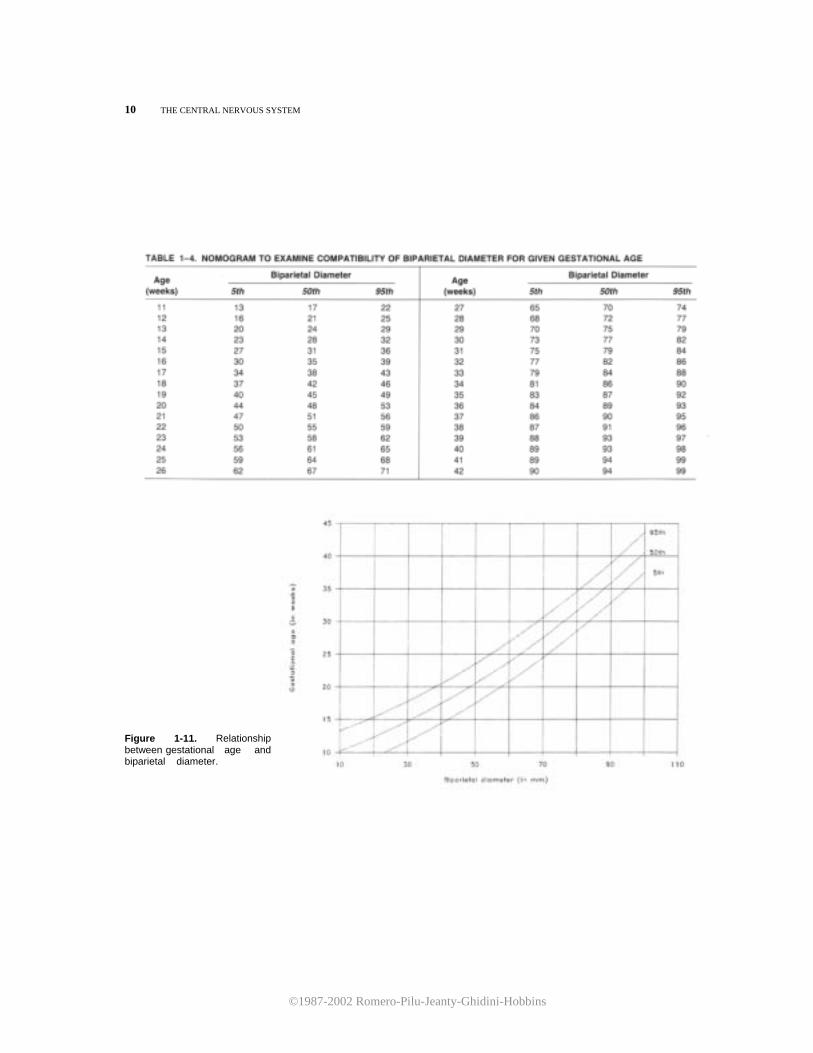
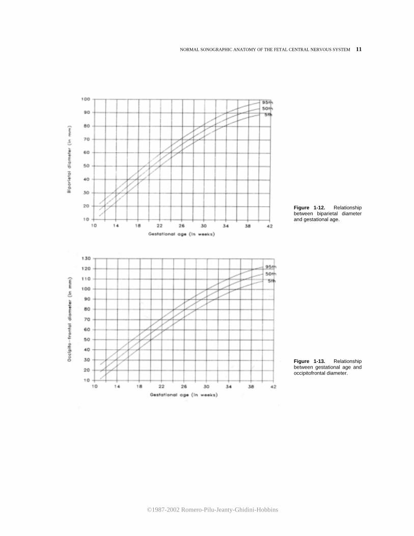
of hydrocephaly. A useful hint for the recognition of this structure is the demonstration of a pulsating echo corresponding to the middle cerebral artery. The biparietal diameter (BPD) is one of the most frequently used fetal biometric parameters. lt is measured from the outer echo of the superior parietal bone to the inner echo of the inferior parietal bone (Fig. 1-10). Tables 1-3 and 1-4 and Figures 1-11 and 1-12 are used to predict gestational age and to assess the normality of a BPD for a given gestational age. Tables 1-3 and 1-4 should be used only after verifying that the size of the BPD is not affected by molding of the fetal head. This is achieved by obtaining an occipitofrontal diameter (OFD) and calculating the cephalic index. The OFD can be imaged in the same section used for the BPD and is measured from midecho to midecho (Fig. 1-10). Figure 1-13 and Table 1-5 illustrate the growth of the OFD. The cephalic index is calculated by dividing the BPD by the OFD. Normal values are between 75 and 85 percent. Dolicocephaly is diagnosed by cephalic indices below 75 percent and brachycephaly by cephalic indices above 85 percent. The circumference of the head either can be measured directly with a mapreader or, alternatively, can be calculated from the BPD and OFD. The formula used to calculate the head circumference is:
Head circumference = 1.62 (BPD + OFD) We have compared the results of these calculations with actual measurements of the fetal head circumference and found them acceptable for clinical use. Similar comparisons have been made by others.1,6
Figure 1-7. Axial scans at the level of the insula (curved arrow) throughout gestation. A. At 18 weeks, the cerebral hemisphere is convex, and only a thin, fluid layer separates the insula from the calvarium. B. At 22 weeks, the insula is deepened, beginning the formation of the sylvian cistern. C. At 28 weeks, growth of the opercula and deepening of the insula result in the formation of a square-shaped, fluid-filled area. At this time, a thin membrane (M), probably representing the arachnoid, is seen bridging between the opercula. CSP, cavum septi pellucidum; At, atria of the lateral ventricles; FH, frontal horns of the lateral ventricles.
©1987-2002 Romero-Pilu-Jeanty-Ghidini-Hobbins
8 THE CENTRAL NERVOIJS SYSTEM
A
B
Figure 1-8. A. Axial scan at the level of the thalami (T) and third ventricle (3v). Anterior to the thalami, the frontal horns (FH) of the lateral ventricles, which are separated by a widely patent cavum septi pellucidi (CSP), can be seen. The hippocampal gyrus (HG) can be recognized by the presence of the medial ambient cistern (AC) and lateral atrium of the lateral ventricle (At). The linear echo that can be seen lateral to the thalamus corresponds to the insula (in). On real-time examination, the active pulsation of the middle cerebral artery distinguishes it from the wall of the lateral ventricle. B. Anatomic specimen corresponding to the axial section shown in Figure A. This specimen was obtained from the brain of an adult, and the cavum septi pellucidi is obliterated. VGC, vein of Galen cistern. (Fígure B reproduces with permission from Matsui, Irano: An Atlas of the Human Brain for Computed Tomography. Tokyo, Igaku Shoin, 1978.)
Figure 1-9. Axial scan at the same level as in Figure 1-8A in a late third trimester fetus. The two lines that are seen medial to the frontal horns (Fh) of the lateral ventricles are thought to represent the walls of a patent but small cavum septi pellucidi (CSP). At, atria; HG, hippocampal gyrus; In, insula; T, thalami; 3v, third ventricle
Figure 1-10. Measurement of the biparietal diameter (BPD) and occipitofrontal diameter (OFD).

©1987-2002 Romero-Pilu-Jeanty-Ghidini-Hobbins
NORMAL SONOGRAPHIC ANATOMY OF THE FETAL CENTRAL NERVOUS SYSTEM 9
©1987-2002 Romero-Pilu-Jeanty-Ghidini-Hobbins
10 THE CENTRAL NERVOUS SYSTEM
Figure 1-11. Relationship between gestational age and biparietal diameter.
©1987-2002 Romero-Pilu-Jeanty-Ghidini-Hobbins
NORMAL SONOGRAPHIC ANATOMY OF THE FETAL CENTRAL NERVOUS SYSTEM 11
Figure 1-12. Relationship between biparietal diameter and gestational age.
Figure 1-13. Relationship between gestational age and occipitofrontal diameter.
©1987-2002 Romero-Pilu-Jeanty-Ghidini-Hobbins
12 THE CENTRAL NERVOUS SYSTEM
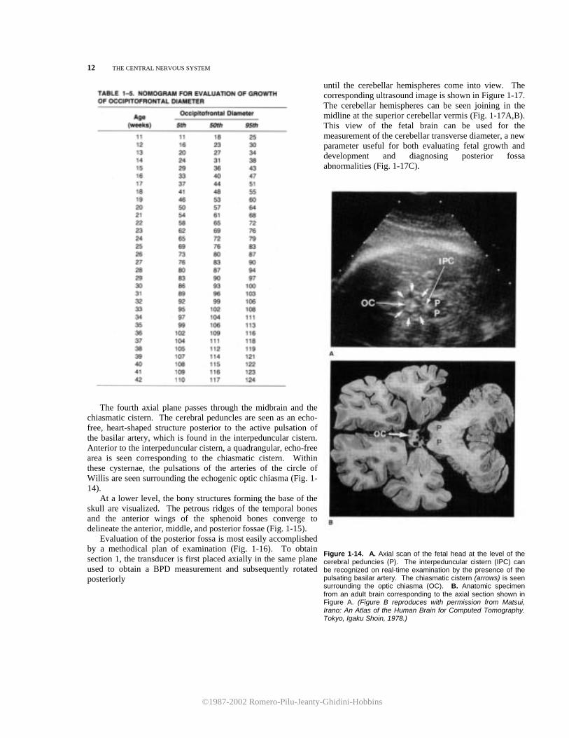
The fourth axial plane passes through the midbrain and the chiasmatic cistern. The cerebral peduncles are seen as an echo-free, heart-shaped structure posterior to the active pulsation of the basilar artery, which is found in the interpeduncular cistern. Anterior to the interpeduncular cistern, a quadrangular, echo-free area is seen corresponding to the chiasmatic cistern. Within these cysternae, the pulsations of the arteries of the circle of Willis are seen surrounding the echogenic optic chiasma (Fig. 1-14).
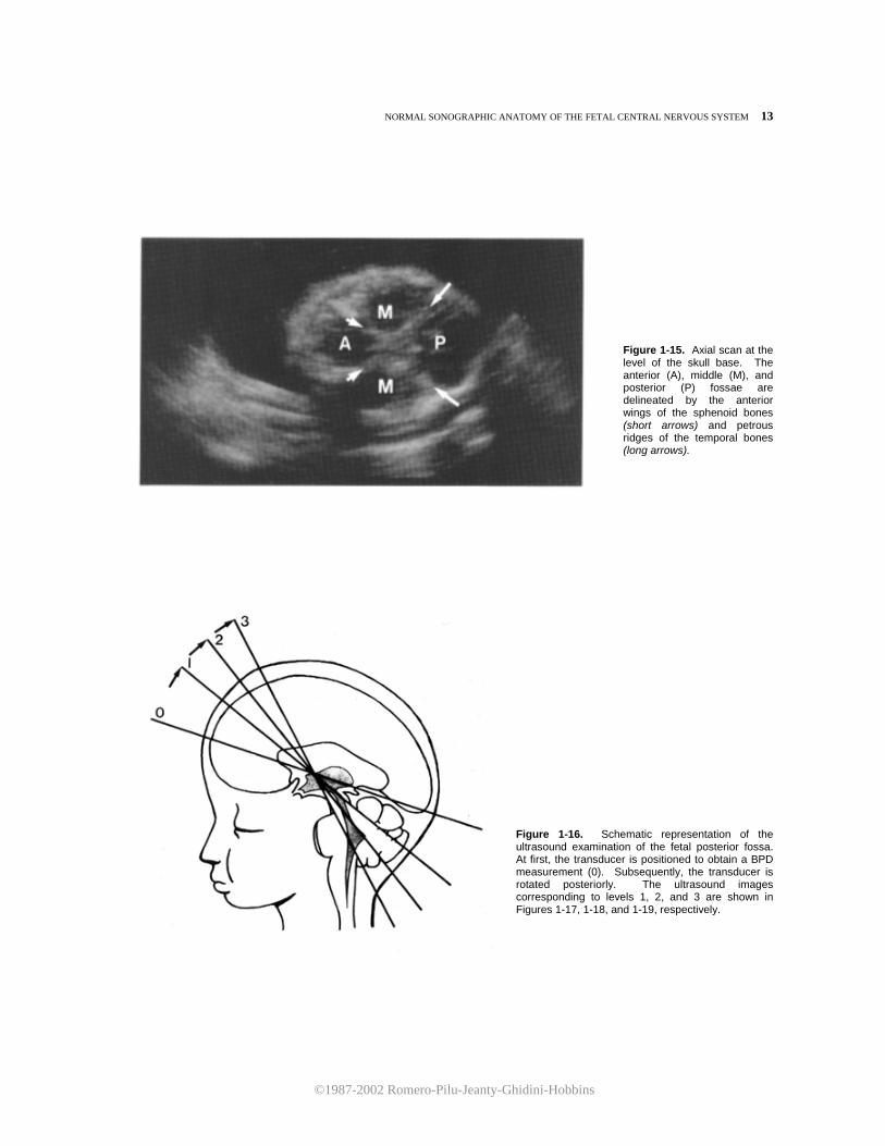
At a lower level, the bony structures forming the base of the skull are visualized. The petrous ridges of the temporal bones and the anterior wings of the sphenoid bones converge to delineate the anterior, middle, and posterior fossae (Fig. 1-15).
Evaluation of the posterior fossa is most easily accomplished by a methodical plan of examination (Fig. 1-16). To obtain section 1, the transducer is first placed axially in the same plane used to obtain a BPD measurement and subsequently rotated posteriorly
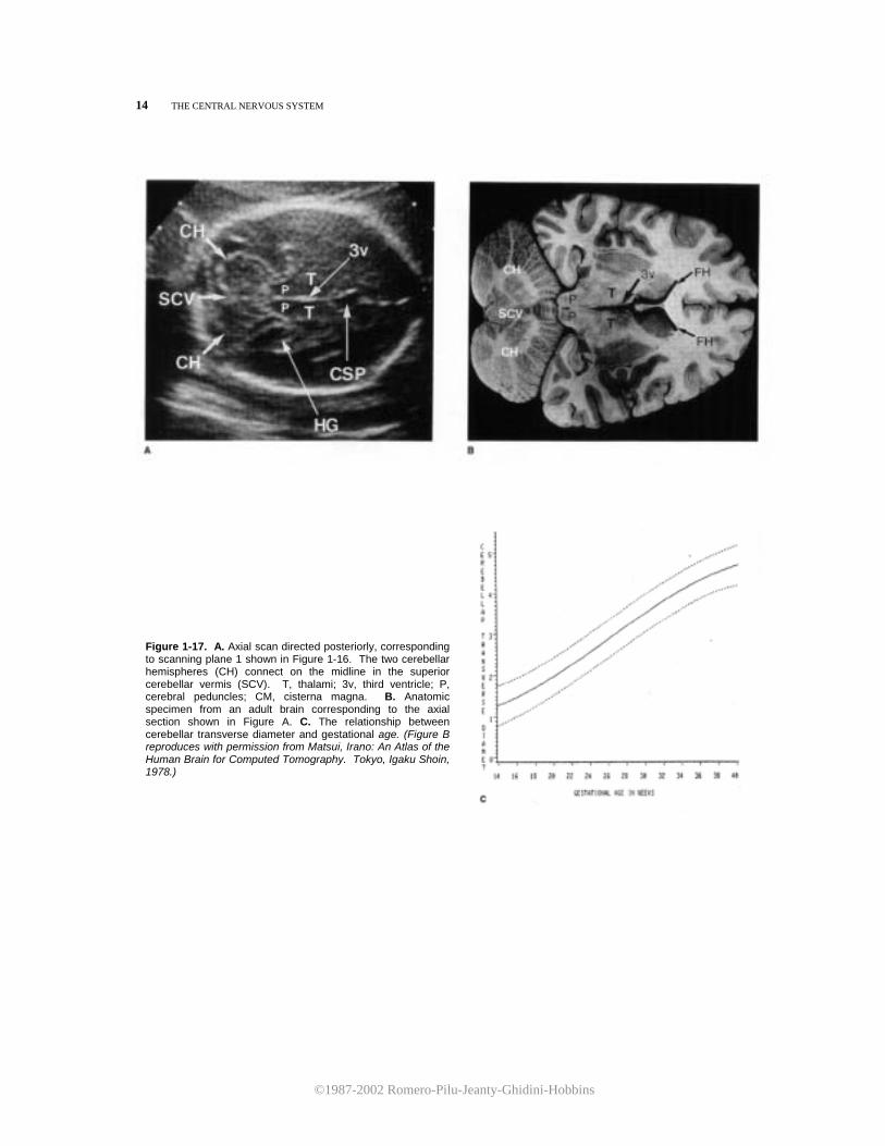
until the cerebellar hemispheres come into view. The corresponding ultrasound image is shown in Figure 1-17. The cerebellar hemispheres can be seen joining in the midline at the superior cerebellar vermis (Fig. 1-17A,B). This view of the fetal brain can be used for the measurement of the cerebellar transverse diameter, a new parameter useful for both evaluating fetal growth and development and diagnosing posterior fossa abnormalities (Fig. 1-17C).
Figure 1-14. A. Axial scan of the fetal head at the level of the cerebral peduncies (P). The interpeduncular cistern (IPC) can be recognized on real-time examination by the presence of the pulsating basilar artery. The chiasmatic cistern (arrows) is seen surrounding the optic chiasma (OC). B. Anatomic specimen from an adult brain corresponding to the axial section shown in Figure A. (Figure B reproduces with permission from Matsui, Irano: An Atlas of the Human Brain for Computed Tomography. Tokyo, Igaku Shoin, 1978.)
©1987-2002 Romero-Pilu-Jeanty-Ghidini-Hobbins
NORMAL SONOGRAPHIC ANATOMY OF THE FETAL CENTRAL NERVOUS SYSTEM 13
Figure 1-15. Axial scan at the level of the skull base. The anterior (A), middle (M), and posterior (P) fossae are delineated by the anterior wings of the sphenoid bones (short arrows) and petrous ridges of the temporal bones (long arrows).
Figure 1-16. Schematic representation of the ultrasound examination of the fetal posterior fossa. At first, the transducer is positioned to obtain a BPD measurement (0). Subsequently, the transducer is rotated posteriorly. The ultrasound images corresponding to levels 1, 2, and 3 are shown in Figures 1-17, 1-18, and 1-19, respectively.
©1987-2002 Romero-Pilu-Jeanty-Ghidini-Hobbins
14 THE CENTRAL NERVOUS SYSTEM
Figure 1-17. A. Axial scan directed posteriorly, corresponding to scanning plane 1 shown in Figure 1-16. The two cerebellar hemispheres (CH) connect on the midline in the superior cerebellar vermis (SCV). T, thalami; 3v, third ventricle; P, cerebral peduncles; CM, cisterna magna. B. Anatomic specimen from an adult brain corresponding to the axial section shown in Figure A. C. The relationship between cerebellar transverse diameter and gestational age. (Figure B reproduces with permission from Matsui, Irano: An Atlas of the Human Brain for Computed Tomography. Tokyo, Igaku Shoin, 1978.)
©1987-2002 Romero-Pilu-Jeanty-Ghidini-Hobbins
NORMAL SONOGRAPHIC ANATOMY OF THE FETAL CENTRAL NERVOTJS SYSTEM 15
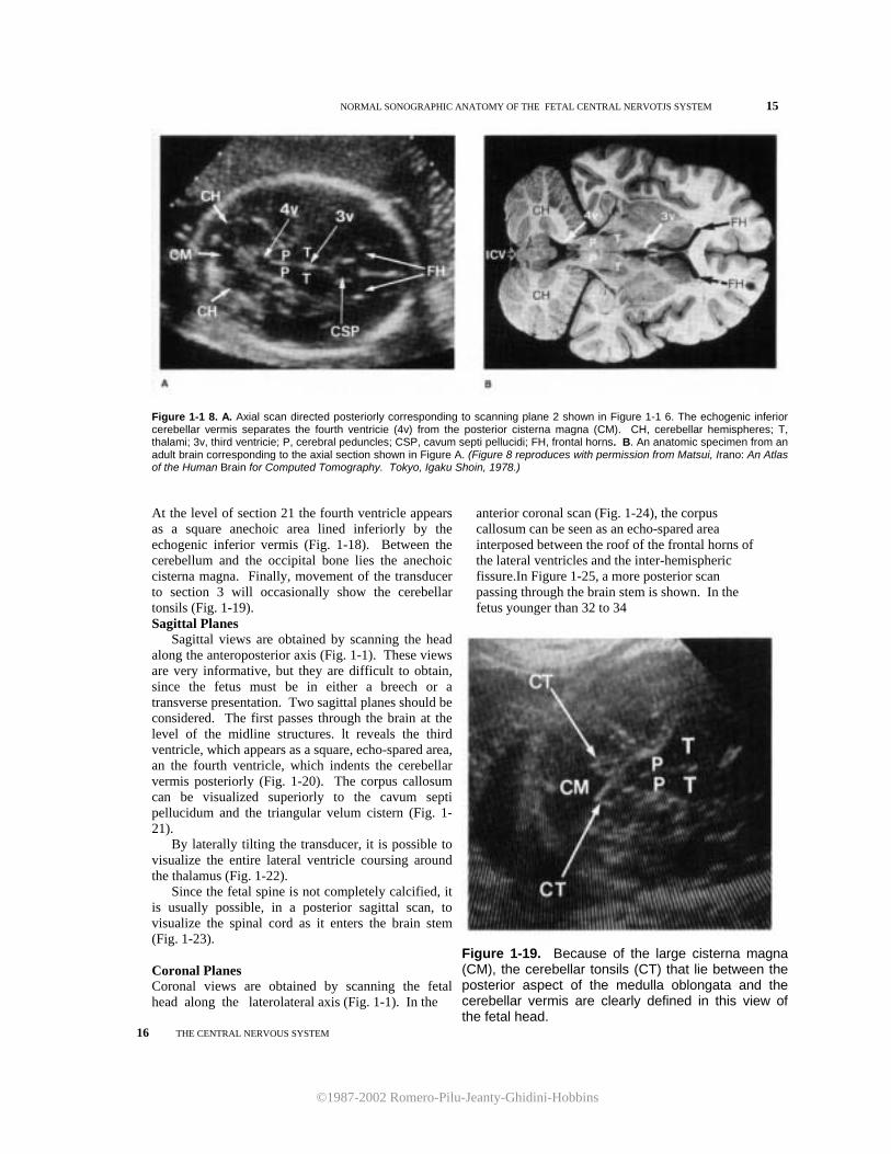
Figure 1-1 8. A. Axial scan directed posteriorly corresponding to scanning plane 2 shown in Figure 1-1 6. The echogenic inferior cerebellar vermis separates the fourth ventricie (4v) from the posterior cisterna magna (CM). CH, cerebellar hemispheres; T, thalami; 3v, third ventricie; P, cerebral peduncles; CSP, cavum septi pellucidi; FH, frontal horns. B. An anatomic specimen from an adult brain corresponding to the axial section shown in Figure A. (Figure 8 reproduces with permission from Matsui, Irano: An Atlas of the Human Brain for Computed Tomography. Tokyo, Igaku Shoin, 1978.)
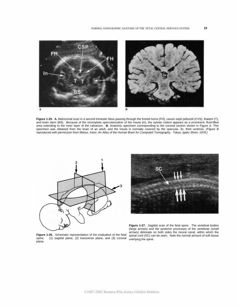
anterior coronal scan (Fig. 1-24), the corpus callosum can be seen as an echo-spared area interposed between the roof of the frontal horns of the lateral ventricles and the inter-hemispheric fissure.In Figure 1-25, a more posterior scan passing through the brain stem is shown. In the fetus younger than 32 to 34
At the level of section 21 the fourth ventricle appears as a square anechoic area lined inferiorly by the echogenic inferior vermis (Fig. 1-18). Between the cerebellum and the occipital bone lies the anechoic cisterna magna. Finally, movement of the transducer to section 3 will occasionally show the cerebellar tonsils (Fig. 1-19). Sagittal Planes
Sagittal views are obtained by scanning the head along the anteroposterior axis (Fig. 1-1). These views are very informative, but they are difficult to obtain, since the fetus must be in either a breech or a transverse presentation. Two sagittal planes should be considered. The first passes through the brain at the level of the midline structures. lt reveals the third ventricle, which appears as a square, echo-spared area, an the fourth ventricle, which indents the cerebellar vermis posteriorly (Fig. 1-20). The corpus callosum can be visualized superiorly to the cavum septi pellucidum and the triangular velum cistern (Fig. 1-21).
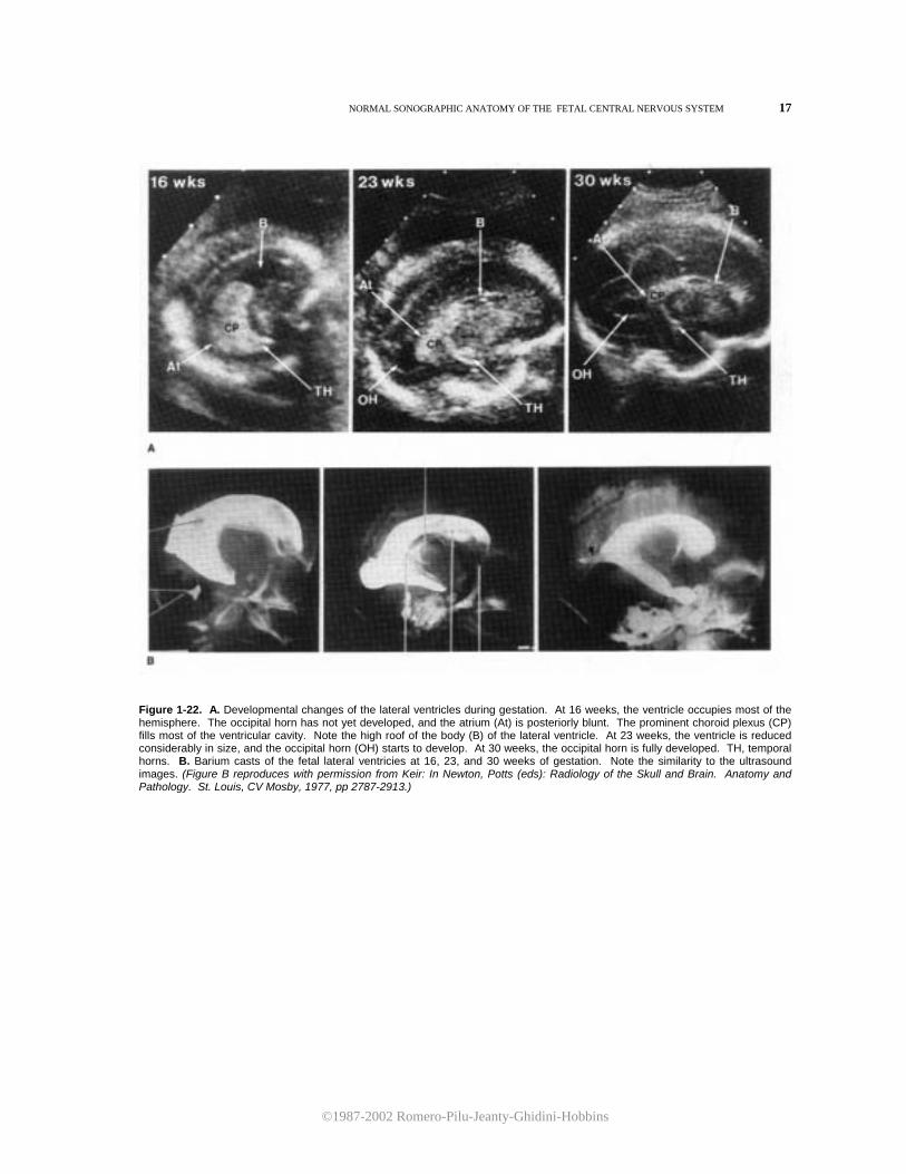
By laterally tilting the transducer, it is possible to visualize the entire lateral ventricle coursing around the thalamus (Fig. 1-22).
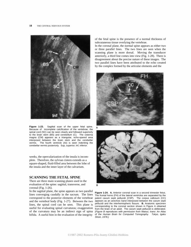
Since the fetal spine is not completely calcified, it is usually possible, in a posterior sagittal scan, to visualize the spinal cord as it enters the brain stem (Fig. 1-23). Coronal Planes Coronal views are obtained by scanning the fetal head along the laterolateral axis (Fig. 1-1). In the
Figure 1-19. Because of the large cisterna magna (CM), the cerebellar tonsils (CT) that lie between the posterior aspect of the medulla oblongata and the cerebellar vermis are clearly defined in this view of the fetal head.
16 THE CENTRAL NERVOUS SYSTEM

©1987-2002 Romero-Pilu-Jeanty-Ghidini-Hobbins
Figure 1-20. A. A midsagittal scan of the fetal brain at 30 weeks of gestation, demonstrating the third ventricie (3v) and the fourth ventricie (4v). A widely patent cavum septi pellucidi (CSP) is seen above the roof of the third ventricie. Note the echogenic cerebellar vermis (C). B. Anatomic specimen from a 30-week-old fetus corresponding to the sagittal section shown in Figure A. CC, corpus callosum; SP, septum pellucidum. (Figure 8 reproduces with permission from Keir: In Newton, Potts (eds): Radiology of the Skull and Brain, Anatomy and Pathology. St Louis, CV Mosby, 1977, pp 2787-2913.) Figure 1-21. A midsagittal scan of the fetal brain at 26 weeks. The corpus callosum is the thin anechoic area interposed between the hyper-echogenic triangular velum cistern (TVC) and the large cavum septi pellucidum (CSP),which is posteri- orly continuous with a patent cavum vergae (unlabeled).The arrows indi- cate the continuity between the trian- gular velum cistern and the postero- inferior vein of Galen cistern within which the vein of Galen is seen (VG). 3v, 3rd ventricie; Ant, anterior; Post, posterior.
©1987-2002 Romero-Pilu-Jeanty-Ghidini-Hobbins
NORMAL SONOGRAPHIC ANATOMY OF THE FETAL CENTRAL NERVOUS SYSTEM 17
Figure 1-22. A. Developmental changes of the lateral ventricles during gestation. At 16 weeks, the ventricle occupies most of the hemisphere. The occipital horn has not yet developed, and the atrium (At) is posteriorly blunt. The prominent choroid plexus (CP) fills most of the ventricular cavity. Note the high roof of the body (B) of the lateral ventricle. At 23 weeks, the ventricle is reduced considerably in size, and the occipital horn (OH) starts to develop. At 30 weeks, the occipital horn is fully developed. TH, temporal horns. B. Barium casts of the fetal lateral ventricies at 16, 23, and 30 weeks of gestation. Note the similarity to the ultrasound images. (Figure B reproduces with permission from Keir: In Newton, Potts (eds): Radiology of the Skull and Brain. Anatomy and Pathology. St. Louis, CV Mosby, 1977, pp 2787-2913.)
©1987-2002 Romero-Pilu-Jeanty-Ghidini-Hobbins
18 THE CENTRAL NERVOUS SYSTEM
Figure 1-23. Sagittal scan of the upper fetal spine. Because of incomplete calcification of the vertebrae, the spinal cord (SC) can be seen clearly and followed superiorly to the brain stem (BS) and cerebellum (C). The cisterna magna (CM) appears as a triangular, echo-spared area interposed between the brain stem and the cerebellar vermis. The fourth ventricle (4v) is seen indenting the cerebellar vermis posteriorly. Sup, superior; lnf, inferior. weeks, the opercularization of the insula is incom- plete. Therefore, the sylvian cistern extends as a square-shaped, fluid-filled area between the lobe of the insula and the inner layer of the calvarium. SCANNING THE FETAL SPINE There are three main scanning planes used in the evaluation of the spine: sagittal, transverse, and coronal (Fig. 1-26). In the sagittal plane, the spine appears as two parallel lines converging caudally in the sacrum. The lines correspond to the posterior elements of the vertebrae and the vertebral body (Fig. 1-27). Between the two lines, the spinal cord can be seen. This plane is useful for evaluating spinal curvaturas; exaggeration of the curvatura may be an indirect sign of spina bifida. A useful hint in the evaluation of the integrity
of the fetal spine is the presence of a normal thickness of subcutaneous tissue overlying the vertebrae. In the coronal plane, the normal spine appears as either two or three parallel lines. The two lines are seen when the scanning plane is more dorsal. Moving the transducer anteriorly, a third line comes into view (Fig. 1-28). There is disagreement about the precise nature of these images. The two parallel lines have been attributed to the echo created by the complex formed by the articular elements and the
Figure 1-24. A. Anterior coronal scan in a second trimester fetus. The frontal horns (FH) of the lateral ventricles are separated by the patent cavum septi pellucidi (CSP). The corpus callosum (CC) appears as an anechoic band interposed between the cavum septi pellucidi and the interhemispheric fissure. B. Anatomic specimen corresponding to the coronal section shown in Figure A obtained from the brain of an adult. The cavum septi pellucidi is obliterated. (Figure B reproduces with permission from Matsui, Irano: An Atlas of the Human Brain for Computed Tomography. Tokyo, Igaku Shoin, 1978.)
©1987-2002 Romero-Pilu-Jeanty-Ghidini-Hobbins
NORMAL SONOGRAPHIC ANATOMY OF THE FETAL CENTRAL NERVOUS SYSTEM 19
Figure 1-25. A. Midcoronal scan in a second trimester fetus passing through the frontal horns (FH), cavum septi pellucidi (CVS), thalami (T), and brain stem (BS). Because of the incomplete opercularization of the insula (in), the sylvian cistern appears as a prominent, fluid-filled area extending to the inner layer of the calvarium. B. Anatomic specimen corresponding to the coronal section shown in Figure A. This specimen was obtained from the brain of an adult, and the insula is normally covered by the opercula. 3v, third ventricie. (Figure B reproduced with permission from Matsui, Irano: An Atlas of the Human Brain for Computed Tomography. Tokyo, Igaku Shoin, 1978.)
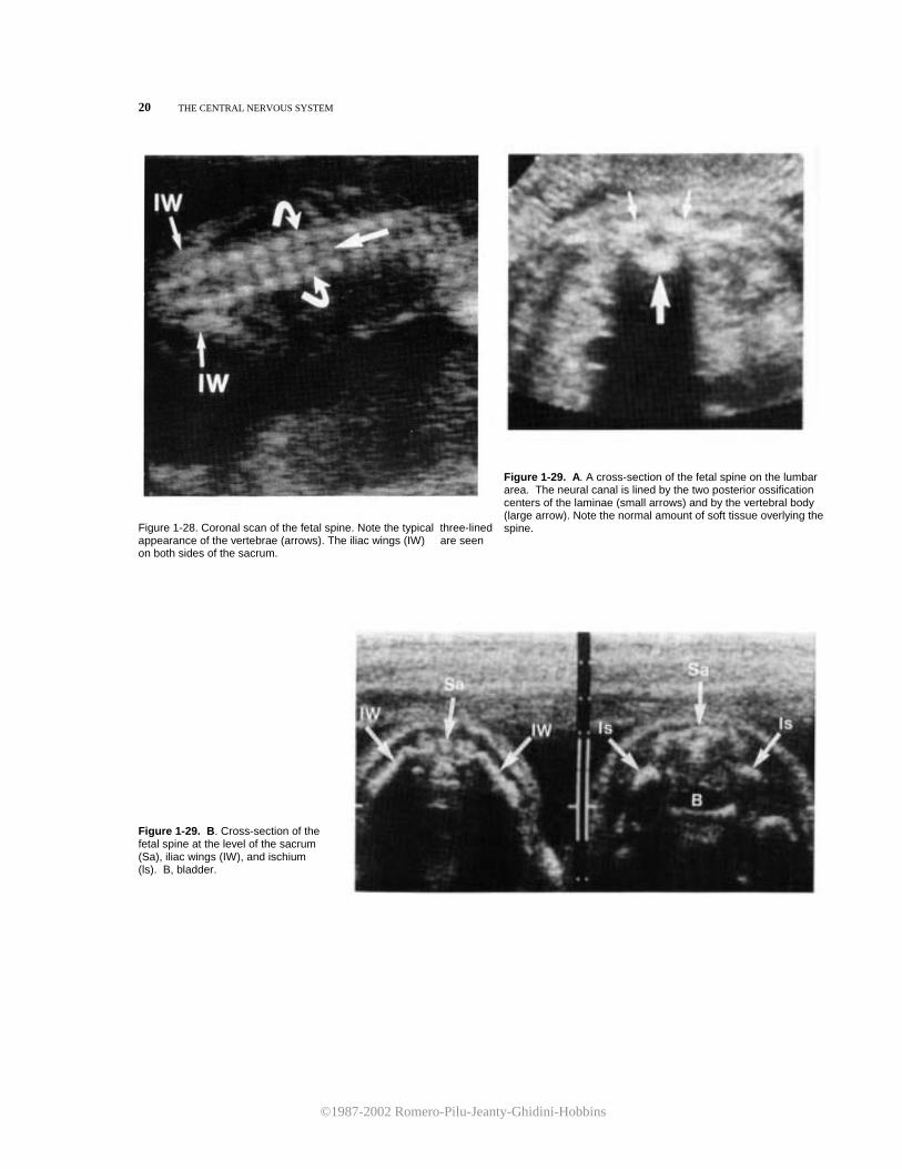
Figure 1-26. Schematic representation of the evaluation of the fetal spine: (1) sagittal plane, (2) transverse plane, and (3) coronal plane.
Figure 1-27. Sagittal scan of the fetal spine. The vertebral bodies (large arrows) and the posterior processes of the vertebrae (small arrows) delineate on both sides the neural canal, within which the spinal cord (SC) can be seen. Note the normal amount of soft tissue overlying the spine.
©1987-2002 Romero-Pilu-Jeanty-Ghidini-Hobbins
20 THE CENTRAL NERVOUS SYSTEM
Figure 1-28. Coronal scan of the fetal spine. Note the typical three-lined appearance of the vertebrae (arrows). The iliac wings (IW) are seen on both sides of the sacrum.
Figure 1-29. A. A cross-section of the fetal spine on the lumbar area. The neural canal is lined by the two posterior ossification centers of the laminae (small arrows) and by the vertebral body (large arrow). Note the normal amount of soft tissue overlying the spine.
Figure 1-29. B. Cross-section of the fetal spine at the level of the sacrum (Sa), iliac wings (IW), and ischium (ls). B, bladder.
©1987-2002 Romero-Pilu-Jeanty-Ghidini-Hobbins
HYDROCEPHALUS 21
lamina of the vertebrae. The third line probably
corresponds to the vertebral body. Coronal planes should not be confused with oblique
sections. A helpful hint in this regard is to examine the amount of tissue on both sides of the fetus. A correct coronal plane requires equal amounts of soft tissue on both sides of the spine. Oblique sections can be recognized by the asymmetry of the fetal trunk.
In transverse sections, the neural canal appears as a closed circle. It is lined anteriorly by the ossification center in the body of the vertebrae and posteriorly by the two ossification centers of the laminae (Fig. 1-29). REFERENCES
1. Christenson D, McCown RB: The elusive ellipse. Am J Obstet Gynecol 152:114, 1985.
2. Denkhaus H, Winsberg F: Ultrasonic measurement of the fetal ventricular system. Radiology 131:781., 1979.
3. Dorovini-Zis K, Dolman CL: Gestational development of brain. Arch Pathol Lab Med 101:192, 1977.
4. Fiske CE, Filly RA, Callen PW: Sonographic measure-ment of lateral ventricular width in early ventricular dilation. J Clin Ultrasound 9:303, 1981.
5. Hadlock FP, Deter RL, Park SK: Real-time sonography: Ventricular and vascular anatomy of the fetal brain in utero. AJR 136:133, 1981.
6. Hadlock FP, Kent WR, Loyd JL, et al.: An evaluation of two methods for measuring fetal head and body circum-ferences. J Ultrasound Med 1:359, 1982.
7. Jeanty P, Dramaix-Wilmet M, Delbeke D, et al.: Ultra-sonic evaluation of fetal ventricular growth. Neuroradi-ology 21:127, 1981.
8. Johnson ML, Dunne MG, Mack LA, et al.: Evaluation of fetal intracranial anatomy by static and real-time ultra-sound. J Clin Ultrasound 8:311, 1980.
9. Shepard M, Filly RA: A standarized plane for biparietal diameter measurement. J Ultrasound Med 1:145, 1982.
HYDROCEPHALUS
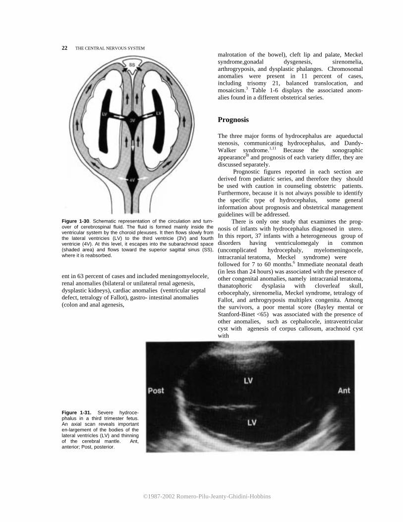
Hydrocephalus is commonly defined as an increased intracranial content of cerebrospinal fluid (CSF). Even though many disorders of the CNS share this condition, the term "hydrocephalus" is generally used to refer to a situation in which an abnormal accumulation of CSF results in enlargement of the ventricular system. Figure 1-30 shows the origin, circulation, and drainage of CSF. CSF is formed mainly at the level of the choroid plexuses inside the ventricular system and flows slowly from the lateral ventricles to the third ventricle and from there to the fourth ventricle. At this level, CSF passes through the foramina of Luschka and Magendie inside the subarachnoid space that externally bathes the cerebral structures. Flowing along the subarachnoid cisterns, the fluid is then reabsorbed by the granulations of Pacchioni that are mainly distributed along the supe-rior sagittal sinus.
In the majority of cases, congenital hydrocephalus is the consequence of an obstruction along the normal pathway of the CSF (obstructive hydrocephaly). Hydrocephalus is one of the most common congenital anomalies, with an incidence of 0.3 to 0.8 per 1000 births.12
The diagnosis of hydrocephalus has traditionally relied on the demonstration of enlarged lateral ventricles (Fig. 1-31). Several nomograms have been developed to quantify the dimensions of the lateral
ventricles.9,13-15 As previously described (see p. 2), the LVW:HW ratio is the parameter most frequently used for this assessment. However, several false negative diagnoses in early pregnancy have been reported 4,10,14 and they raise questions about the sensitivity of the measurement of the LVW:HW ratio in diagnosing early or mild ventricular dilatation. Morphologic, rather than purely biometric, criteria have been suggested for the early detection of hydrocephalus, including the simultaneous visualization of the medial and lateral wall of the lateral ventriclel0 and the anterior displacement of the choroid plexus7 (Fig. 1-32). Recently, measurement of the atria of the lateral ventricle has been suggested.2 At present, the problem of early detection of hydrocephalus remains unsolved. We have found that from 16 to 20 weeks of pregnancy, a combination of morphologic and biometric criteria allows for either a specific diagnosis or a questionable diagnosis in the majority of cases. Associated Anomalies
Hydrocephalus is commonly associated with other congenital anomalies. Associated intracranial anom-alies have been reported in 37 percent of hydroceph-alus cases. They include hypoplasia of the corpus callosum, cephalocele, arteriovenous malformation, and arachnoid cyst. Extracranial anomalies were pres-
©1987-2002 Romero-Pilu-Jeanty-Ghidini-Hobbins
22 THE CENTRAL NERVOUS SYSTEM
Figure 1-30. Schematic representation of the circulation and turn- over of cerebrospinal fluid. The fluid is formed mainly inside the ventricular system by the choroid plexuses. It then flows slowly from the lateral ventricies (LV) to the third ventricie (3V) and fourth ventricie (4V). At this level, it escapes into the subarachnoid space (shaded area) and flows toward the superior sagittal sinus (SS), where it is reabsorbed. ent in 63 percent of cases and included meningomyelocele, renal anomalies (bilateral or unilateral renal agenesis, dysplastic kidneys), cardiac anomalies (ventricular septal defect, tetralogy of Fallot), gastro- intestinal anomalies (colon and anal agenesis,
malrotation of the bowel), cleft lip and palate, Meckel syndrome,gonadal dysgenesis, sirenomelia, arthrogryposis, and dysplastic phalanges. Chromosomal anomalies were present in 11 percent of cases, including trisomy 21, balanced translocation, and mosaicism.3 Table 1-6 displays the associated anom- alies found in a different obstetrical series. Prognosis The three major forms of hydrocephalus are aqueductal stenosis, communicating hydrocephalus, and Dandy-Walker syndrome.1,11 Because the sonographic appearancel8 and prognosis of each variety differ, they are discussed separately. Prognostic figures reported in each section are derived from pediatric series, and therefore they should be used with caution in counseling obstetric patients. Furthermore, because it is not always possible to identify the specific type of hydrocephalus, some general information about prognosis and obstetrical management guidelines will be addressed. There is only one study that examimes the prog- nosis of infants with hydrocephalus diagnosed in utero. In this report, 37 infants with a heterogeneous group of disorders having ventriculomegaly in common (uncomplicated hydrocephaly, myelomeningocele, intracranial teratoma, Meckel syndrome) were followed for 7 to 60 months.6 Immediate neonatal death (in less than 24 hours) was associated with the presence of other congenital anomalies, namely intracranial teratoma, thanatophoric dysplasia with cloverleaf skull, cebocephaly, sirenomelia, Meckel syndrome, tetralogy of Fallot, and arthrogryposis multiplex congenita. Among the survivors, a poor mental score (Bayley mental or Stanford-Binet <65) was associated with the presence of other anomalies, such as cephalocele, intraventricular cyst with agenesis of corpus callosum, arachnoid cyst with
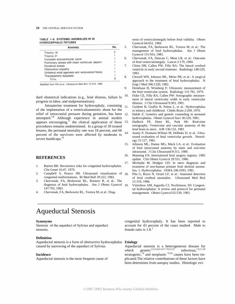
Figure 1-31. Severe hydroce-phalus in a third trimester fetus. An axial scan reveals important en-largement of the bodies of the lateral ventricles (LV) and thinning of the cerebral mantle. Ant, anterior; Post, posterior.

©1987-2002 Romero-Pilu-Jeanty-Ghidini-Hobbins
HYDROCEPHALUS 23
Figure 1-32. A. In this 30-week fetus, both medial and lateral walls of the body of the lateral ventricle (arrows) are simultaneously visualized. The fetus was found to have spina bifida and subsequently developed marked ventriculomegaly. agenesis of corpus callosum, microcephaly, and ring chromosome 18. On the other hand, all cases with normal intelligence (Bayley mental or Stanford-Binet score >80) did not have associated anomalies or they had meningomyelocele. Therefore, the most important prognostic consideration is the presence and nature of the associated anomalies. Pediatric data suggest that a correlation exists between cortical mantle thickness before shunting and long-term intellectual performances. Thickness of less than 1 cm has been associated with a poor outcome.19 However, this correlation is imperfect and excellent neurologic outcomes have been observed
after early shunting with mantle thickness of less than 1 cm. This parameter, therefore, should not be used for obstetrical management decisions. Obstetrical Management A search for associated congenital anomalies and a workup for congenital infections associated with hydrocephaly (i.e., toxoplasmosis, cytomegalovirus, rubella) is indicated. Amniocentesis should be performed for alphafetoprotein, fetal karyotype, and viral cultures. Before viability, the option of pregnancy termination should be offered to the parents. After viability, the management issues are the role of intrauterine treatment with ventriculo-amniotic shunt, time and mode of delivery, and cephalocentesis. Little data exist to support any specific manage-ment plan. Our general recommendations include delaying delivery until fetal lung maturity is documented, avoiding cephalocentesis, and using cesarean section for obstetrical indications only. Fetal lung maturity is determined by performing weekly amniocenteses beginning at 36 weeks of gestation. Cephalocentesis is associated with a perinatal mortality in excess of 90 percent 5,6 and its use should be limited to those instances in which hydrocephaly is associated with anomalies carrying a dismal prognosis (e.g., thanatophoric dysplasia and Meckel syn-drome). This procedure should be performed under sonographic guidance. Macrocrania or overt hydrocephaly (head circumference above the 98th percentile for gestational age) in the absence of any other associated anomaly suggesting poor prognosis is not an indication for cephalocentesis. Most infants with hydrocephaly do not have macrocrania, and therefore a trial of labor is indicated in vertex presen-tation. Cesarean section should be reserved for stan-
Figure 1-32. B. Early hydrocephalus in an 18-week fetus with spina bifida. Although the LVW:HW ratio is within normal limits, ventriculomegaly is inferred by the anterior displacement of the choroid plexus (CP), which does not entirely fill the atrium (At). The body of the lateral ventricle (B) is within normal limits.
©1987-2002 Romero-Pilu-Jeanty-Ghidini-Hobbins
24 THE CENTRAL NERVOUS SYSTEM
dard obstetrical indications (e.g., fetal distress, failure lo progress in labor, and malpresentations). Intrauterine treatment for hydrocephaly, consisting of the implantation of a ventriculoamniotic shunt for the relief of intracranial pressure during gestation, has been attempted.5,8 Although experience in animal models appears encouraging,17 the clinical application of these procedures remains undetermined. In a group of 39 treated fetuses, the perinatal mortality rate was 18 percent, and 66 percent of the survivors were affected by moderate to severe handicaps.16 REFERENCES 1. Burton BK: Recurrence risks for congenital hydrocephalus.
Clin Genet 16:47, 1979. 2. Campbell S, Pearce JM: Ultrasound visualization of
congenital malformations. Br Med Bull 39:322, 1983. 3. Chervenak, FA, Berkowitz RL, Romero R, et al.: The
diagnosis of fetal hydrocephalus. Am J Obstet Gynecol 147:703, 1983.
4. Chervenak, FA, Berkowitz RL, Tortora M, et al.: Diag-
nosis of ventriculomegaly before fetal viability. Obstet Gynecol 64:652, 1984.
5. Chervenak, FA, Berkowitz RL, Tortora M, et al.: The management of fetal hydrocephalus. Am J Obstet Gynecol 151:933, 1985.
6. Chervenak, FA, Duncan C, Ment LR, et al.: Outcome of fetal ventriculomegaly. Lancet 2:179, 1984.
7. Chinn DH, Callen PW, Filly RA: The lateral cerebral ventricle in early second trimester. Radiology 148:529, 1983.
8. Clewell WH, Johnson ML, Meier PR, et al.: A surgical approach to the treatment of fetal hydrocephalus. N Engl J Med 306:1320, 1982.
9. Denkhaus H, Winsberg F: Ultrasonic measurement of the fetal ventricular system. Radiology 131:781, 1979.
10. Fiske CE, Filly RA, Callen PW: Sonographic measure-ment of lateral ventricular width in early ventricular dilation. J Clin Ultrasound 9:303, 1981.
11. Guidetti B, Giuffre R, Palma L, et al.: Hydrocephalus in infancy and childhood. Childs Brain 2:209, 1976.
12. Habib Z: Genetics and genetic counseling in neonatal hydrocephalus. Obstet Gynecol Surv 36:529, 1981.
13. Hadlock FP, Deter RL, Park SK: Real-time sonography: Ventricular and vascular anatomy of the fetal brain in utero. AJR 136:133, 1981.
14. Jeanty P, Dramaix-Wilmet M, Delbeke D, et al.: Ultra-sound evaluation of fetal ventricular growth. Neurol-ogy 21:127, 1981.
15. Johnson ML, Dunne MG, Mack LA, et al.: Evaluation of fetal intracranial anatomy by static and real-time ultrasound. J Clin Ultrasound 8:311, 1980.
16. Manning FA: International fetal surgery registry: 1985 update. Clin Obstet Gynecol 29:551, 1986.
17. Michejda M, Hodgen GD: In utero diagnosis and treatment of non-human primate fetal skeletal anoma-lies. 1. Hydrocephalus. JAMA 246:1093, 1981.
18. Pilu G, Rizzo N, Orsini LF, et al.: Antenatal detection of fetal cerebral anomalies. Ultrasound Med Biol 12:319, 1986.
19. Vintzileos AM, Ingardia CJ, Nochimson, DJ: Congeni-tal hydrocephalus: A review and protocol for perinatal management. Obstet Gynecol 62:539, 1983.
Aqueductal Stenosis Synonyms Stenosis of the aqueduct of Sylvius and aqueduct stenosis. Definition Aqueductal stenosis is a form of obstructive hydrocephalus caused by narrowing of the aqueduct of Sylvius. Incidence Aqueductal stenosis is the most frequent cause of
congenital hydrocephaly. It has been reported to account for 43 percent of the cases studied. Male to female ratio is 1.8.3
Etiology Aqueductal stenosis is a heterogeneous disease for which genetic,2,3,5,6,8,14,17-19,21,22 infectious,1,9,11,20 teratogenic,18 and neoplastic l3,18 causes have been im-plicated.The relative contributions of these factors have been determines from autopsy studies. Histologic evi-
©1987-2002 Romero-Pilu-Jeanty-Ghidini-Hobbins
AQUEDUCTAL STENOSIS 25
dence of inflammation (gliosis) has been found in approximately 50 percent of the cases studied.13 Toxo-plasmosis, syphilis, cytomegalovirus, mumps, and in-fluenza virus have caused aqueductal stenosis in ani-mals.18 In cases without evidence of inflammation, the disease appears to be the consequence of maldevelop-ment for an unknown reason. This maldevelopment is histologically expressed by forking (see Pathology) or simple narrowing of the aqueduct. Genetic transmis-sion has been postulated to account for some of these cases. Many familial studies have demonstrated that aqueductal stenosis can be inherited as an X-linked recessive trait.2,3,5,6,8,14,17,19,21,22 Sex-linked transmission was thought to be a rare cause of the disease, because only 1 case was found among 200 siblings of probands with hydrocephalus.6 However, it has been suggested that this mode of inheritance involves 25 percent of affected male infants.3 The possibility of a coexistent polygenic pattern of inheritance has been suggested by case reports of families in which both females and males were affected.3 Teratogenic agents, such as radiation, have been implicated in animal models, but the relevance of these observations to humans is uncertain. 18 Such tumors as gliomas, pinealomas, meningiomas, and other conditions (neurofibromatosis and tuberous sclerosis) may cause aqueductal stenosis by a compressive mechanism.13 However, the prevalence of these entities in the prenatal period is extremely low. It has also been suggested that communicating hydrocephalus may lead to secondary aqueductal stenosis, causing white matter edema and extrinsic compression. 1,5
Embryology The aqueduct of Sylvius is the portion of the ventric-ular system that connects the third and fourth ventricles (Fig. 1-30). The aqueduct develops from a narrowing of the primitive ventricular cavity between the prosencephalon and rhomboencephalon at about the sixth week (conceptional age). Pathology Aqueductal stenosis may result from an inflammatory process or a developmental anomaly. "Gliosis" is the term used to describe the inflammatory reaction seen in the CNS. This reaction is characterized by a mononuclear-microglial response and a repair process conducted by astrocytes.13 Malformations include forking, narrowing, and the presence of a transverse septum.18 Forking describes the substitution of the aqueduct by multiple narrow channels. Narrowing may be of variable degree and is usually accompanied by an irregular outline of the ependymal wall. When a septum is responsible for the stenosis of the aqueduct, it is usually located in its posterior portion.
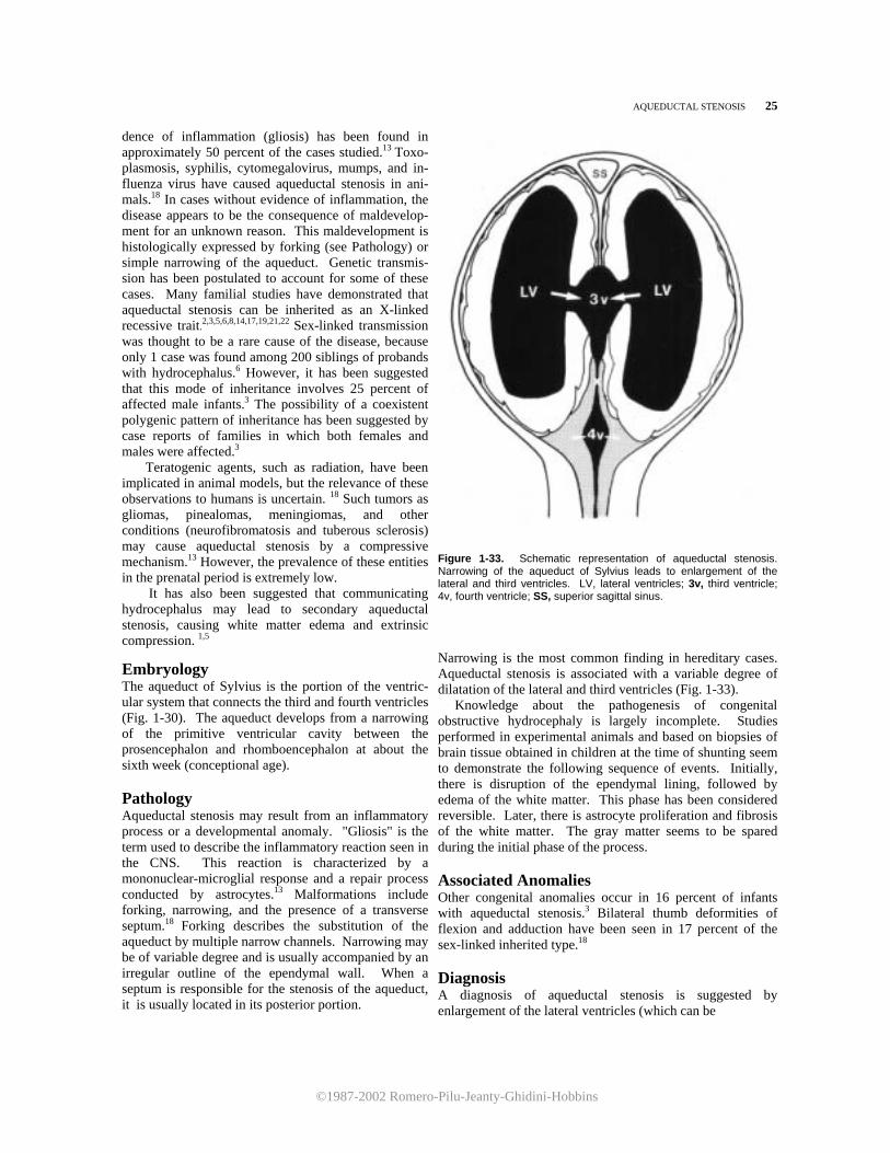
Figure 1-33. Schematic representation of aqueductal stenosis. Narrowing of the aqueduct of Sylvius leads to enlargement of the lateral and third ventricles. LV, lateral ventricles; 3v, third ventricle; 4v, fourth ventricle; SS, superior sagittal sinus. Narrowing is the most common finding in hereditary cases. Aqueductal stenosis is associated with a variable degree of dilatation of the lateral and third ventricles (Fig. 1-33). Knowledge about the pathogenesis of congenital obstructive hydrocephaly is largely incomplete. Studies performed in experimental animals and based on biopsies of brain tissue obtained in children at the time of shunting seem to demonstrate the following sequence of events. Initially, there is disruption of the ependymal lining, followed by edema of the white matter. This phase has been considered reversible. Later, there is astrocyte proliferation and fibrosis of the white matter. The gray matter seems to be spared during the initial phase of the process. Associated Anomalies Other congenital anomalies occur in 16 percent of infants with aqueductal stenosis.3 Bilateral thumb deformities of flexion and adduction have been seen in 17 percent of the sex-linked inherited type.18 Diagnosis A diagnosis of aqueductal stenosis is suggested by enlargement of the lateral ventricles (which can be
©1987-2002 Romero-Pilu-Jeanty-Ghidini-Hobbins
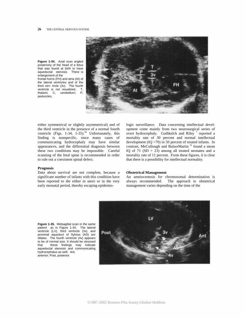
26 THE CENTRAL NERVOUS SYSTEM Figure 1-34. Axial scan angled posterioriy of the head of a fetus that was found at birth to have aqueductal stenosis. There is enlargement of the frontal horns (FH) and atria (At) of the lateral ventricles and of the third ven- tricle (3v). The fourth ventricle is not visualized. T, thalami; C, cerebellum; P, peduncles.
either symmetrical or slightly asymmetrical) and of the third ventricle in the presence of a normal fourth ventricle (Figs. 1-34, 1-35).16 Unfortunately, this finding is nonspecific, since many cases of communicating hydrocephaly may have similar appearances, and the differential diagnosis between these two conditions may be impossible. Careful scanning of the fetal spine is recommended in order to rule out a coexistent spinal defect. Prognosis Data about survival are not complete, because a significant number of infants with this condition have been reported to die either in utero or in the very early neonatal period, thereby escaping epidemio-
logic surveillance. Data concerning intellectual devel-opment come mainly from two neurosurgical series of overt hydrocephaly. Guthkelch and Riley 7 reported a mortality rate of 30 percent and normal intellectual development (IQ >70) in 50 percent of treated infants. In contrast, MeCullough and BalzerMartin l2 found a mean IQ of 71 (SD = 23) among all treated neonates and a mortality rate of 11 percent. From these figures, it is clear that there is a possibility for intellectual normality. Obstetrical Management An amniocentesis for chromosomal determination is always recommended. The approach to obstetrical management varies depending on the time of the
Figure 1-35. Midsagittal scan in the same patient as in Figure 1-34. The lateral ventricle (LV), third ventricle (3v), and proximal aqueduct of Sylvius (AS) are dilates. The fourth ventricle (4v) appears to be of normal size. It should be stressed that these findings may indicate aqueductal stenosis and communicating hydrocephalus as well. Ant, anterior; Post, posterior.

©1987-2002 Romero-Pilu-Jeanty-Ghidini-Hobbins
COMMUNICATING HYDROCEPHALUS 27
diagnosis. Before viability, the option of pregnancy ation should be offered lo the mother. The mode of delivery depends purely on obstetrical indications. Cephalocentesis should not be used in cases of isolated aqueductal stenosis. Cesarean section is only indicated for macrocephaly, fetal distress, or other obstetrical indications. If another congenital anomaly invariably associated with neonatal death is present, cesarean section should be avoided.4 The role of intrauterine shunting is experimental at the present time (see also p. 23). REFERENCES 1. Adams RD, Kubik CS, Bonner FJ: The clinical and
pathological aspects of influenzal meningitis. Arch Pediatr 65:354, 1948.
2. Bickers DS, Adams RD: Hereditary stenosis of the aqueduct of Sylvius as a cause of congenital hydroceph-alus. Brain 72:246, 1949.
3. Burton BK: Recurrence risks for congenital hydrocephalus. Clin Genet 16:47, 1979.
4. Chervenak FA, Berkowitz RL, Tortora M, et al.: The management of fetal hydrocephalus. Am J Obstet Gynecol 151:933, 1985.
5. Edwards JH: The syndrome of sex-linked hydrocephalus. Arch Dis Child 36:486, 1961.
6. Edwards JH, Norman RM, Roberts JM: Sex-linked hydrocephalus: Report of a family with 15 affected members. Arch Dis Child 36:481, 1961.
7. Guthkelch AN, Riley NA: Influence of aetiology on prognosis in surgically treated infantile hydrocephalus. Arch Dis Child 44:29, 1969.
8. Holmes LB, Nash A, ZuRhein GM, et al.: X-Iinked aqueductal stenosis: Clinical and neuropathological findings in two families. Pediatrics 51:697, 1973.
9. johnson RT, johnson KP, Edmonds Cj: Virus-induced hydrocephalus: Development of aqueductal stenosis in
hamsters after mumps infection. Science 157:1066, 1967.
10. Lorber j: Resuits of treatment of myelomeningocele: An analysis of 524 unselected cases, with special reference to possible selection for treatment. Dev Med Child Neurol 13:279, 1971.
11. Margolis G, Kilham L: Hydrocephalus in hamsters, ferrets, rats and mice following inoculations with reovirus Type 1. J Clin Invest 21:183, 1969.
12. McCullough DC, Balzer-Martin LA: Current prognosis in overt neonatal hydrocephalus. J Neurosurg 57:378, 1982.
13. Milhorat TH: Hydrocephalus and the Cerebrospinal
Fluid. Baltimore, Williams & Wilkins, 1972. 14. Needleman HL, Root AW: Sex-linked hydrocephalus.
Report of 2 families with chromosomal study of 2 cases. Pediatries 31:396, 1963.
15. Nugent GR, Al-Mefty 0, Chou S: Communicating
hydrocephalus as a cause of aqueductal stenosis. j Neurosurg 51:812, 1979.
16. Pilu C, Rizzo N, Orsini CF, et al.: Antenatal detection
of cerebral anomalies. Ultrasound Med Biol 12:319, 1986.
17. Price JR, Horne BM: Family history indicating heredi-tary factors in hydrocephalus. Ment Retard 6:40, 1968.
18. Salam MZ: Stenosis of the aqueduct of Sylvius. In:
Vinken Pj, Bruyn GW (eds): Handbook of Clinical Neurology. Amsterdam, Elsevier/North Holland Bio-medical Press, 1977, Vol 30, pp 609-622.
19. Shannon MW, Nadler HL: X-Iinked hydrocephalus. J Med Genet 5:326, 1968.
20. Timmons GD, Johiison KP: Aqueductal stenosis and hydrocephalus after mumps encephalitis. N Engl J Med 283:1505, 1970.
21. Warren MC, Lu AT, Ziering WH: Sex-linked hydro-
cephalus with aqueductal stenosis. j Pediatr 63:1104, 1963.
22. Williamson EM: Incidence and family aggregation of major congenital malformations of central nervous sys-tem. J Med Genet 2:161, 1965.
Communicating Hydrocephalus
Synonym External hydrocephalus. Definition Communicating hydrocephalus is a form of enlarge-ment of the ventricles and subarachnoid system
caused by an obstruction to CSF flow outside the ventricular system. Incidence Communicating hydrocephalus is the second major form of congenital hydrocephalus. It accounts for 38 percent of all cases.1
©1987-2002 Romero-Pilu-Jeanty-Ghidini-Hobbins
28 THE CENTRAL NERVOUS SYSTEM
Etiology In most cases, the etiology is unknown. Communicating hydrocephalus is found in infants with spinal defects and has also been seen in association with obliteration of the superior sagittal sinus3 ,subarachnoid hemorrhage2, absence of Pacchioni granulations 4 and choroid plexus papilloma.8.Subarachnoid hemorrhage is probably the most common cause of infantile communicating hydrocephalus, but it is probably rare in the prenatal period. Familial transmission is rare; only 1 affected individual was found among 154 siblings of 77 probands.l However, the recurrence rate quoted for this condition is 1 to 2 percent, which is higher than the incidence in the general population.1 Pathology The basic cause of communicating hydrocephalus is either a mechanical obstruction outside the ventricular system or an impaired reabsorption of cerebrospi-
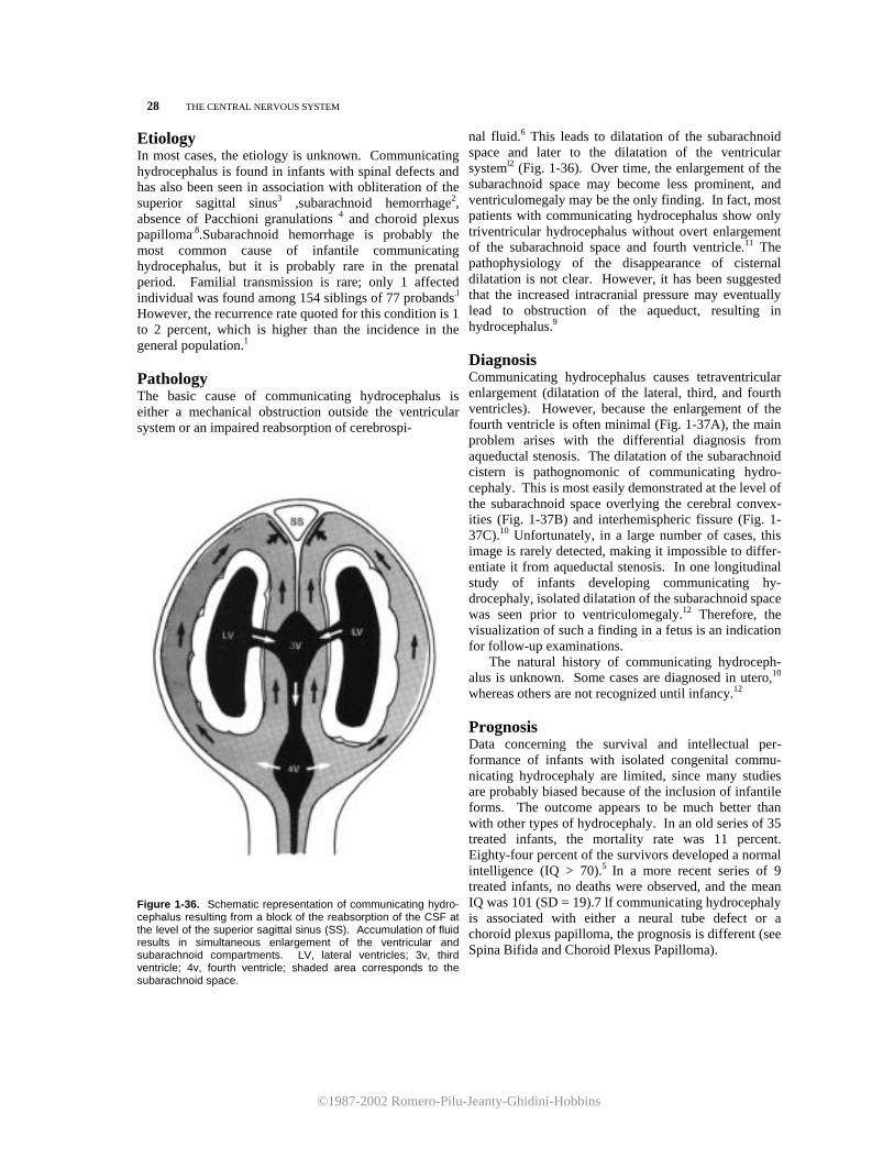
Figure 1-36. Schematic representation of communicating hydro-cephalus resulting from a block of the reabsorption of the CSF at the level of the superior sagittal sinus (SS). Accumulation of fluid results in simultaneous enlargement of the ventricular and subarachnoid compartments. LV, lateral ventricles; 3v, third ventricle; 4v, fourth ventricle; shaded area corresponds to the subarachnoid space.
nal fluid.6 This leads to dilatation of the subarachnoid space and later to the dilatation of the ventricular systeml2 (Fig. 1-36). Over time, the enlargement of the subarachnoid space may become less prominent, and ventriculomegaly may be the only finding. In fact, most patients with communicating hydrocephalus show only triventricular hydrocephalus without overt enlargement of the subarachnoid space and fourth ventricle.11 The pathophysiology of the disappearance of cisternal dilatation is not clear. However, it has been suggested that the increased intracranial pressure may eventually lead to obstruction of the aqueduct, resulting in hydrocephalus.9
Diagnosis Communicating hydrocephalus causes tetraventricular enlargement (dilatation of the lateral, third, and fourth ventricles). However, because the enlargement of the fourth ventricle is often minimal (Fig. 1-37A), the main problem arises with the differential diagnosis from aqueductal stenosis. The dilatation of the subarachnoid cistern is pathognomonic of communicating hydro-cephaly. This is most easily demonstrated at the level of the subarachnoid space overlying the cerebral convex-ities (Fig. 1-37B) and interhemispheric fissure (Fig. 1-37C).10 Unfortunately, in a large number of cases, this image is rarely detected, making it impossible to differ-entiate it from aqueductal stenosis. In one longitudinal study of infants developing communicating hy-drocephaly, isolated dilatation of the subarachnoid space was seen prior to ventriculomegaly.12 Therefore, the visualization of such a finding in a fetus is an indication for follow-up examinations. The natural history of communicating hydroceph-alus is unknown. Some cases are diagnosed in utero,10 whereas others are not recognized until infancy.12 Prognosis Data concerning the survival and intellectual per-formance of infants with isolated congenital commu-nicating hydrocephaly are limited, since many studies are probably biased because of the inclusion of infantile forms. The outcome appears to be much better than with other types of hydrocephaly. In an old series of 35 treated infants, the mortality rate was 11 percent. Eighty-four percent of the survivors developed a normal intelligence (IQ > 70).5 In a more recent series of 9 treated infants, no deaths were observed, and the mean IQ was 101 (SD = 19).7 lf communicating hydrocephaly is associated with either a neural tube defect or a choroid plexus papilloma, the prognosis is different (see Spina Bifida and Choroid Plexus Papilloma).

©1987-2002 Romero-Pilu-Jeanty-Ghidini-Hobbins
COMMUNICATING HYDROCEPHALLUS 29
Figure 1-37. Communicating hydrocephaly in a 30-week-old fetus. A. An axial scan angled posteriorly reveals the dilatation of the frontal horns (FH) and occipital horns (OH) of the lateral ventricles and of the third ventricle (3v). There is a questionable enlargement of the fourth ventricle (4v). B. An anterior coronal scan reveals the simultaneous enlargement of the frontal horns (FH) and of the supracortical cisterns (curved arrows). F, falx cerebri. C. A slightly posterior coronal scan reveals a prominent interhemispheric fissure (arrows). F, falx cerebri. (Figures 8 and C reproduces with permission from Pilu et al: J Ultrasound Med 5:365, 1986.)
©1987-2002 Romero-Pilu-Jeanty-Ghidini-Hobbins
30 THE CENTRAL NERVOUS SYSTEM
Obstetrical Management The approach does not differ from that outlined for aqueductal stenosis (see pp. 23, 27). REFERENCES 1. Burton BK: Recurrence risks for congenital hydroceph-
alus. Clin Genet 16:47, 1979. 2. Ellington E, Margolis G: Block of arachnoid villus by
subarachnoid hemorrhage. J Neurosurg 30:651, 1969. 3. Emery JL, Zachary RB: Hydrocephalus associated with
obliteration of the longitudinal sinus. Arch Dis Child 31:288, 1956.
4. Gutierrez Y, Friede RL, Kaliney WJ: Agenesis of arachnoid granulations and its relationship to commu-nicating hydrocephalus. J Neurosurg 43:553, 1975.
5. Guthkelch AN, Riley NA: Influence of aetiology on prognosis in surgically treated infantile hydrocephalus. Arch Dis Child 44:29, 1969.
6. McComb JG: Recent research into the nature of cere-brospinal fluid formation and absorption. J Neurosurg 59:369, 1983.
7. McCullough DC, Balzer-Martin LA: Current prognosis in overt neonatal hydrocephalus. J Neurosurg 57:378, 1982.
8. Milhorat TH, Hammock MK, Davis DA, et al.: Choroid plexus papilloma. 1. Proof of cerebrospinal fluid over-production. Childs Brain 2:273, 1976.
9. Nugent GR, Al-Mefty 0, Chou S: Communicating hydrocephalus as a cause of aqueductal stenosis. J Neurosurg 51:812, 1979.
10. Pilu G, DePalma L, Romero R, et al.: The fetal sub-
arachnoid cisterns: An ultrasound study. With report of a case of communicating hydrocephalus. J Ultrasound Med 5:365, 1986.
11. Raybaud C, Bamberger-Bozo C, Laffont J, et al.: Inves-tigation of nontumoral hydrocephalus in children. Neuroradiology 16:24, 1978.
12. Robertson WC, Gomez MR: External hydrocephalus. Early finding in congenital communicating hydroceph-alus. Arch Neurol 35:541, 1978.
Dandy-Walker Malformation Synonym Dandy-Walker syndrome. Definition Dandy-Walker malformation (DWM) is characterized by the association of (1) hydrocephalus of variable degree, (2) a cyst in the posterior fossa, and (3) a defect in the cerebellar vermis through which the cyst communicates with the fourth ventricle
Incidence DWM accounts for 12 percent of all cases of congenital hydrocephalus.5 However, this figure may represent an underestimation of the real incidence because cases without hydrocephalus and without significant symptoms have also been reported..2-3 Etiology Unknown. DWM may occur as a part of mendelian disorders, such as Meckel syndrome and Warburg
syndrome. It has been found in chromosomal aberra-tions, such as Turner syndrome, 6p -, 9qh +, trisomy 9, and triploidy. Environmental factors, such as viral in-fection, alcohol, and diabetes, have been suggested as playing a role in its etiology.29 When DWM is not associated with mendelian disorders, the recurrence risk is 1 to 5 percent.29 In rare cases, the disease is probably inherited as an autosomal recessive trait, with a recurrence risk of 25 percent.23 A cerebral anomaly similar to DWM, Joubert syndrome, is also inherited as an autosomal recessive trait.24 History DWM was formally describes by Dandy and Blackfan at the beginning of the century .7,8 They postulated this condition to be secondary to congenital atresia of the foramina of Luschka and Magendie, which provide an exit to the CSF from the fourth ventricle to the subarachnoid space. Walker was the physician who describes the first surgical treatment.35 Although Benda2 proved that the pathogenetic hypothesis suggested by these authors was untenable, he suggested the eponym, Dandy-Walker syndrome.
©1987-2002 Romero-Pilu-Jeanty-Ghidini-Hobbins
DANDY-WALKER MALFORMATION 31
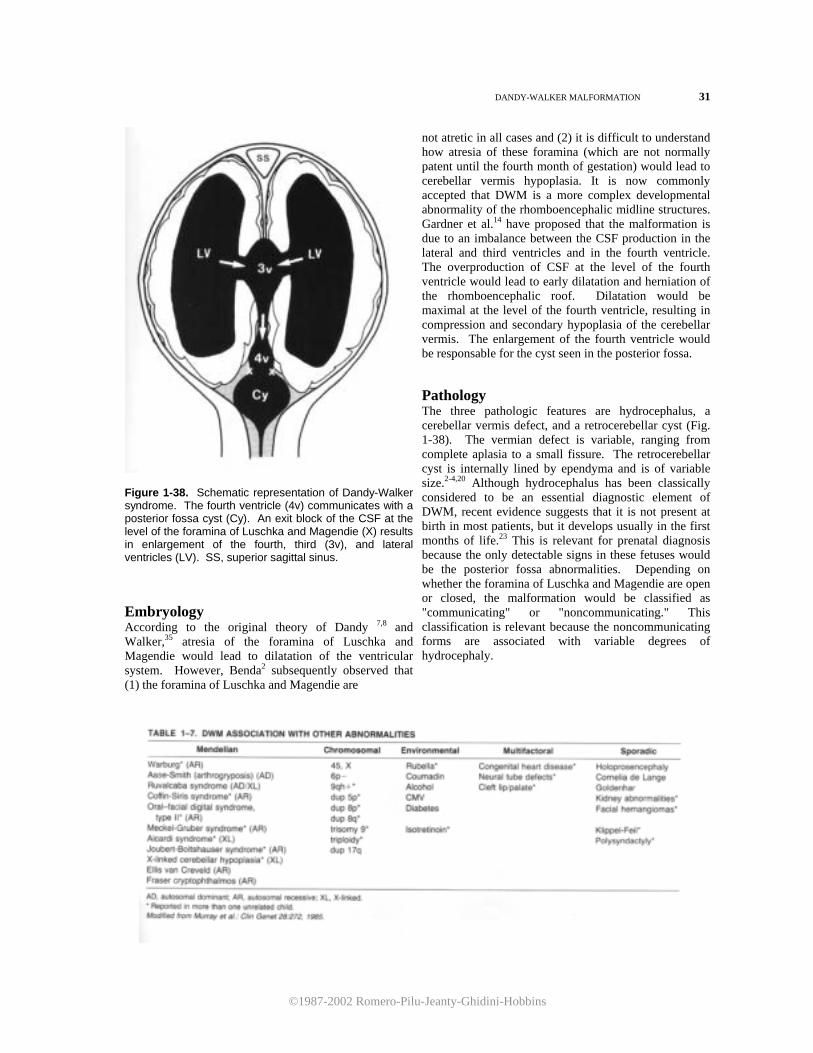
Figure 1-38. Schematic representation of Dandy-Walker syndrome. The fourth ventricle (4v) communicates with a posterior fossa cyst (Cy). An exit block of the CSF at the level of the foramina of Luschka and Magendie (X) results in enlargement of the fourth, third (3v), and lateral ventricles (LV). SS, superior sagittal sinus. Embryology According to the original theory of Dandy 7,8 and Walker,35 atresia of the foramina of Luschka and Magendie would lead to dilatation of the ventricular system. However, Benda2 subsequently observed that (1) the foramina of Luschka and Magendie are
not atretic in all cases and (2) it is difficult to understand how atresia of these foramina (which are not normally patent until the fourth month of gestation) would lead to cerebellar vermis hypoplasia. It is now commonly accepted that DWM is a more complex developmental abnormality of the rhomboencephalic midline structures. Gardner et al.14 have proposed that the malformation is due to an imbalance between the CSF production in the lateral and third ventricles and in the fourth ventricle. The overproduction of CSF at the level of the fourth ventricle would lead to early dilatation and herniation of the rhomboencephalic roof. Dilatation would be maximal at the level of the fourth ventricle, resulting in compression and secondary hypoplasia of the cerebellar vermis. The enlargement of the fourth ventricle would be responsable for the cyst seen in the posterior fossa. Pathology The three pathologic features are hydrocephalus, a cerebellar vermis defect, and a retrocerebellar cyst (Fig. 1-38). The vermian defect is variable, ranging from complete aplasia to a small fissure. The retrocerebellar cyst is internally lined by ependyma and is of variable size.2-4,20 Although hydrocephalus has been classically considered to be an essential diagnostic element of DWM, recent evidence suggests that it is not present at birth in most patients, but it develops usually in the first months of life.23 This is relevant for prenatal diagnosis because the only detectable signs in these fetuses would be the posterior fossa abnormalities. Depending on whether the foramina of Luschka and Magendie are open or closed, the malformation would be classified as "communicating" or "noncommunicating." This classification is relevant because the noncommunicating forms are associated with variable degrees of hydrocephaly.

©1987-2002 Romero-Pilu-Jeanty-Ghidini-Hobbins
32 THE CENTRAL NERVOUS SYSTEM
Figure 1-39. A. In this fetus with hydrocephaly, an axial scan directed posteriorly demonstrates the pathognomonic findings of Dandy-Walker syndrome: A posterior fossa cyst (Cy) is seen to communicate with the grossly enlarged fourth ventricle (4v) through a vermian defect. CH, cerebellar hemispheres; LV, enlarged lateral ventricle. B. In this midsagittal scan of fetus in Figure A, the enlarged third ventricle (3v) communicates through a typically dilated and kinked aqueduct (curved arrow) with the posterior fossa cyst (Cy). LV, lateral ventricie; Ant, anterior; Post, posterior. (Reproduced with permission from Pilu et al.: J Reprod Med 31:1017, 1986.)
Associated Anomalies DWM is frequently associated with other CNS abnor-malities. Clinical studies have found an incidence of 50 percent of associated anomalies.34 Agenesis of the corpus callosum has been reported to occur in between 723 and 1734 percent of patients studied. Pathologic studies have demonstrated an incidence of cerebral defects as high as 68 percent.20 However, it should be stressed that most of these anomalies (polymicrogyria, agyria, microgyria, malformation of the inferior olives) are not sonographically detectable in utero. Other anomalies include encephaloceles, polycystic kidneys, and cardiovascular de-
fects (mainly ventricular septal defects).4,23,31,34 A detailed list of genetic and nongenetic conditions associated with DWM is given in Table 1-7. Diagnosis The diagnosis of DWM should be considered whenever a cystic mass is seen in the posterior fossa.9-11,22,25,26,30,32,36 The differential diagnosis includes an arachnoid cyst and dilatation of the cisterna magna. A defect in the vermis, through which the cyst communicates with the fourth ventricle, is pathognomonic of DWM. Such a finding is well documented in both computed tomo-
Figure 1-40. Dandy-Walker syn drome with a large posterior fossa cyst (Cy). Note the widely separated cerebellar hemispheres (CH). The prenatal ultrasound study is compared with a postnatal computed tomographic scan. T, thalami. (Reproduced with permission from Pilu et al.: J Reprod Med 31:1017, 1986.)

©1987-2002 Romero-Pilu-Jeanty-Ghidini-Hobbins
DANDY-WALKER MALFORMATION 33
Figure 1-41. In this 21-week fetus, Dandy-Walker syndrome is revealed onLy by the presence of a defect of the inferior vermis (affow) at the level of the 4th ventricle (4v). CH, cerebellar hemispheres; T, thalami. (Reproduced with permissíon from Pilu et al: J Reprod Med 31:1017, 1986.) graphic l3,17,21,23,27,34 and ultrasound studies l5-17,36 in the postnatal period, and it can be demonstrated in the fetus as well,32 (Fig, 1-39). The defect may vary in size from a small fissure to a large tunnel with widely separated cerebellar hemispheres (Fig. 1-40). Extreme care is necessary because, in some cases, the superior vermis is intact and the defect can only be demonstrated by careful examination of the inferior vermis (Fig. 1-41). Differentiation from an arachnoid cyst or enlarged cisterna magna may be difficult, however. This difficulty can be encountered even in the neonatal period despite the use of computed tomography. There is controversy in the radiologic literature about the optimal means of making a diagnosis. Some authors are concerned about the limitations of computed tomography and recommend that a pneumoencephalogram be performed.1 Other authors believe that pneumoencephalography may be misleading and recommend contrast studies (metrimazide, radionucleotides) when noncontrast computed tomography is equivocal.27,34 Traditionally, DWM has been considered a cause of intrauterine hydrocephalus. However, the evidence indicates that this association is not frequent in the fetus.23,32 Therefore, we recommend a careful study of the posterior fossa as part of a routine survey of the intracranial anatomy.32 Prognosis and Obstetrical Management Data on the prognosis of DWM are controversial. The first in-depth large series concerning the treatment of infants affected by DWM indicated a uniformly poor prognosis.12,18,19,33 The mortality rate was about 50 percent, and 50 to 60 percent of the survivors were intellectually impaired. In two recent series, survival
rates of 74 percent34 and 88 percent23 with an IQ above 80 in 30 and 60 percent of survivors, respectively, have been reported. Consequently, we believe that if a positive diagnosis is made before viability, the option of pregnancy termination should be offered to the parents. lt is difficult to provide guidelines for the management of fetuses diagnosed in the third trimester. Fetal karyotyping is indicated because of the occasional association with chromosomal aberrations.29 Depp et al.10 reported intrauterino shunting in a case of DWM. The role of intrauterine treatment for this disease is experimental, and its efficacy is yet to be proven.
REFERENCES 1. Archer CR, Darwish H, Smith K: Enlarged cisternae
magnae and posterior fossa cysts simulating Dandy-Walker syndrome on computed tomography. Radiol-ogy 127:681, 1978.
2. Benda CE: The Dandy-Walker syndrome or the so-called atresia of the foramen Magendie. J Neuropathol Exp Neurol 13:14, 1954.
3. Brodal A, Hauglie-Hanssen E: Congenital hydroceph-alus with defective development of the cerebellar vermis (Dandy-Walker syndrome): Clinical and ana-tomical findings in two cases with particular reference to the so-called atresia of the foramina of Magendie and Luschka. J Neurol Neurosurg Psychiatry 22:99, 1959.
4. Brown JR: The Dandy-Walker syndrome. In: Vinken PJ, Bruyn GW (eds): Handbook of Clinical Neurology. Amsterdam, Elsevier/North Holland Biomedical Press, 1977, Vol 30, pp 623-646.
5. Burton BK: Recurrence risks for congenital hydroceph-alus. Clin Genet 16:47, 1979.
6. Chervenak FA, Romero R: Is there a role for fetal cephalocentesis in modern obstetrics? Am J Perinatol 1:170, 1984.
7. Dandy WE: The diagnosis and treatment of hydroceph-alus due to occlusion of the foramina of Magendie and Luschka. Surg Gynecol Obstet 32:112, 1921.
8. Dandy WE, Blackfan KD: Intemal hydrocephalus: an experimental, clinical and pathological study. Am J Dis Child 8:406, 1914.
9. Dempsey PJ, Koch HJ: In utero diagnosis of the Dandy-Walker syndrome: Differentiation from extra-axial posterior fossa cyst. J Clin Ultrasound 9:403, 1981.
10. Depp R, Sabbagha RE, Brown T, et al.: Fetal surgery for hydrocephalus: Successful in utero ventriculoamniotic shunt for Dandy-Walker syndrome. Obstet Gynecol 61:710, 1983.
11. Fileni A, Colosimo C, Mirk P, et al.: Dandy-Walker syndrome: Diagnosis in utero by means of ultrasound and CT correlations. Neuroradiology 24:233, 1983.
12. Fischer EG: Dandy-Walker syndrome: An evaluation of surgical treatment. J Neurosurg 39:615, 1973.
13. Fitz CR: Midline anomalies of the brain and spine. Radiol Clin North Am 20:95, 1982.
14. Gardner E, O'Rahilly R, Prolo D: The Dandy~Walker

©1987-2002 Romero-Pilu-Jeanty-Ghidini-Hobbins
34 THE CENTRAL NERVOUS SYSTEM
and Arnold-Chiari malformations: Clinical, develop-mental and teratological considerations. Arch Neurol 32:393, 1975.
15. Goodwin V, Quisling RG: The neonatal cisterna magna:Ultrasonic evaluation. Radiology 149:691, 1983.
16. Grant EG, Schellinger D, Richardson JD: Real-time ultrasonography of the posterior fossa. J Ultrasound Med 2:73, 1983.
17. Groenhout CM, Gooskens RH, Veiga-Pires JA, et al.: Value of sagittal sonography and direct sagittal CT of the Dandy-Walker syndrome. AJNR 5:476, 1984.
18. Guidetti B, Giuffre R, Palma L, et al.: Hydrocephalus in infancy and childhood. Childs Brain 2:209, 1976.
19. Guthkelch AN, Riley NA: Influence of aetiology on prognosis in surgically treated infantile hydrocephalus. Arch Dis Child 44:29, 1969.
20. Hart MN, Malamud N, Ellis WG: The Dandy-Walker syndrome: A clinicopathological study based on 28 cases. Neurology 22:771, 1972.
21. Harwood-Nash DC, Fitz CR: Congenital anomalies of the brain. In: Neuroradiology in Infants and Children. St. Louis, Mosby, 1976, Vol 3, pp 998-1053.
22. Hatjis CG, Horbar JD, Anderson GG: The in utero diagnosis of a posterior fossa intracranial cyst (Dandy-Walker cyst). Am J Obstet Gynecol 140:473, 1981.
23. Hirsch JF, Pierre-Kahn A, Renier D, et al.: The Dandy-Walker malformation: A review of 40 cases. J Neurosurg 61:515, 1984.
24. Joubert M, Eisenring JJ, Robb JP, et al.: Familial agenesis of the cerebellar vermis: A syndrome of epi-sodic hyperpnea, abnormal eye movements, ataxia and retardation. Neurology 19:813, 1969.
25. Kirkinen P, Jouppila P, Valkeakari T, et al.: Ultrasonic evaluation of the Dandy-Walker syndrome. Obstet Gynecol 59:18S, 1982.
26. Mahony BS, Callen PW, Filly RA, et al.: The fetal cisterna magna. Radiology 153:773, 1984.
27. Masdeu JC, Dobben GD, Azar-Kia B: Dandy-Walker syndrome studied by computed tomography and pneumoencephalography, Radiology 147:109, 1983.
28. McCullough DC, Balzer-Martin LA: Current prognosis in overt neonatal hydrocephalus. J Neurosurg 57:378, 1982.
29. Murray JC, Johnson JA, Bird TD: Dandy-Walker mal- formation: Etiologic heterogeneity and empiric recur-rence risks. Clin Genet 28:272, 1985.
30. Newman GC, Buschi Al, Sugg NK, et al.: DandyWalker syndrome diagnosed in utero by ultrasonography. Neurology 32:180, 1982.
31. Olson GS, Halpe DC, Kaplan AM et al.: Dandy-Walker malformation and associated cardiac anomalies. Childs Brain 8:173, 1981.
32. Pilu G, Romero R, DePalma L, et al.: Antenatal diagnosis and obstetrical management of Dandy-Walker syndrome. J Reprod Med 31:1017, 1986.
33. Raimondi AJ, Samuelson G, Yarzagaray L, et al.: Atresia of the foramina of Luschka and Magendie: The Dandy-Walker cyst. J Neurosurg 31:202, 1969.
34. Sawaya R, McLaurin RL: Dandy-Walker syndrome: Clinical analysis of 23 cases. J Neurosurg 55:89, 1981.
35. Taggart JK, Walker AE: Congenital atresia of the fora-mens of Luschka and Magendie. AMA Arch Neurol Psychiatr 48:583, 1942.
36. Taylor GA, Sanders RC: Dandy-Walker syndrome: Rec-ognition by sonography. AJNR 4:1203, 1983.
Choroid Plexus Papilloma Definition Choroid plexus papilloma (CPP) is a generally benign tumor of the choroid plexus. Incidence CPP is an exceedingly rare intracranial neoplasm that accounts for 0.6 percent of all brain tumors found in adults and 3 percent in children.6,7 Etiology Unknown. This tumor has been reported in four patients with Aicardi syndrome, an X-Iinked disorder characterized by agenesis of the corpus callosum, chorioretinal lacunae, vertebral abnormalities, seizure disorder, and mental retardation.2,13,15 Pathology Choroid plexuses are the main source of CSF. They
are normally located inside the lateral, third, and fourth ventricles. Papillomas may occur in any of these sites,1,4,11 but they occur most frequently at the level of the atria of the lateral ventricles.7,16 The lesion is unilateral in the overwhelming rnajority of cases. Only a few cases of bilateral papilloma have been reported.18 In most instances, these tumors are benign and are formed by villi that are histologically similar to normal choroid plexus. Malignancy may occur and can be recognized by invasion of adjacent nervous tissue and histologic departure from the normal cellular pattern, with mitosis and pleomorphism.6-8 CPPs are usually associated with hydrocephalus. This may be caused either by overproduction of CSF, leading to communicating hydrocephalus, or by an obstruction to the flow of CSF, resulting in dilatation of different portions of the ventricular system.3,6,7,9,14
©1987-2002 Romero-Pilu-Jeanty-Ghidini-Hobbins
CHOROID PLEXUS PAPILLOMA 35
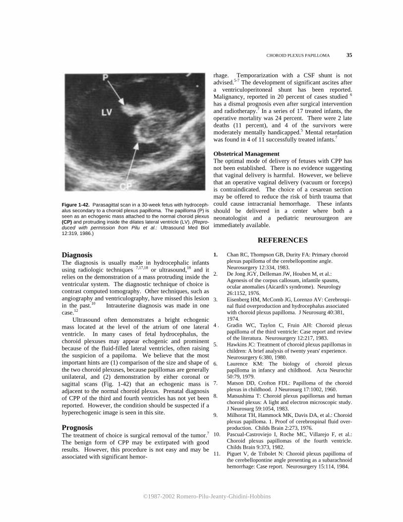
Figure 1-42. Parasagittal scan in a 30-week fetus with hydroceph-alus secondary to a choroid plexus papilloma. The papilloma (P) is seen as an echogenic mass attached to the normal choroid plexus (CP) and protruding inside the dilates lateral ventricle (LV). (Repro-duced with permission from Pilu et al.: Ultrasound Med Biol 12:319, 1986.) Diagnosis The diagnosis is usually made in hydrocephalic infants using radiologic techniques 7,17,18 or ultrasound,18 and it relies on the demonstration of a mass protruding inside the ventricular system. The diagnostic technique of choice is contrast computed tomography. Other techniques, such as angiography and ventriculography, have missed this lesion in the past.10 Intrauterine diagnosis was made in one case.12 Ultrasound often demonstrates a bright echogenic mass located at the level of the atrium of one lateral ventricle. In many cases of fetal hydrocephalus, the choroid plexuses may appear echogenic and prominent because of the fluid-filled lateral ventricles, often raising the suspicion of a papiloma. We believe that the most important hints are (1) comparison of the size and shape of the two choroid plexuses, because papillomas are generally unilateral, and (2) demonstration by either coronal or sagittal scans (Fig. 1-42) that an echogenic mass is adjacent to the normal choroid plexus. Prenatal diagnosis of CPP of the third and fourth ventricles has not yet been reported. However, the condition should be suspected if a hyperechogenic image is seen in this site. Prognosis The treatment of choice is surgical removal of the tumor.7 The benign form of CPP may be extirpated with good results. However, this procedure is not easy and may be associated with significant hemor-
rhage. Temporarization with a CSF shunt is not advised.5-7 The development of significant ascites after a ventriculoperitoneal shunt has been reported. Malignancy, reported in 20 percent of cases studied 6 has a dismal prognosis even after surgical intervention and radiotherapy.7 In a series of 17 treated infants, the operative mortality was 24 percent. There were 2 late deaths (11 percent), and 4 of the survivors were moderately mentally handicapped.5 Mental retardation was found in 4 of 11 successfully treated infants.7 Obstetrical Management The optimal mode of delivery of fetuses with CPP has not been established. There is no evidence suggesting that vaginal delivery is harmful. However, we believe that an operative vaginal delivery (vacuum or forceps) is contraindicated. The choice of a cesarean section may be offered to reduce the risk of birth trauma that could cause intracranial hemorrhage. These infants should be delivered in a center where both a neonatologist and a pediatric neurosurgeon are immediately available.
REFERENCES
1. Chan RC, Thompson GB, Durity FA: Primary choroid
plexus papilloma of the cerebellopontine angle. Neurosurgery 12:334, 1983.
2. De Jong JGY, Delleman JW, Houben M, et al.: Agenesis of the corpus callosum, infantile spasms, ocular anomalies (Aicardi's syndrome). Neurology 26:1152, 1976.
3. Eisenberg HM, McComb JG, Lorenzo AV: Cerebrospi-nal fluid overproduction and hydrocephalus associated with choroid plexus papilloma. J Neurosurg 40:381, 1974.
4 . Gradin WC, Taylon C, Fruin AH: Choroid plexus papilloma of the third ventricle: Case report and review of the literatura. Neurosurgery 12:217, 1983.
5. Hawkins JC: Treatment of choroid plexus papillomas inchildren: A brief analysis of twenty years' experience. Neurosurgery 6:380, 1980.
6. Laurence KM: The biology of choroid plexus papilloma in infancy and childhood. Acta Neurochir 50:79, 1979.
7. Matson DD, Crofton FDL: Papilloma of the choroid plexus in childhood. J Neurosurg 17:1002, 1960.
8. Matsushima T: Choroid plexus papillornas and human choroid plexus: A light and electron microscopic study. J Neurosurg 59:1054, 1983.
9. Milhorat TH, Hammock MK, Davis DA, et al.: Choroid plexus papilloma. 1. Proof of cerebrospinal fluid over-production. Childs Brain 2:273, 1976.
10. Pascual-Castroviejo I, Roche MC, Villarejo F, et al.: Choroid plexus papillomas of the fourth ventricle. Childs Brain 9:373, 1982.
11. Piguet V, de Tribolet N: Choroid plexus papilloma of the cerebellopontine angle presenting as a subarachnoid hemorrhage: Case report. Neurosurgery 15:114, 1984.
©1987-2002 Romero-Pilu-Jeanty-Ghidini-Hobbins
36 THE CENTRAL NERVOUS SYSTEM 12. Pilu G, De Palma L, Romero R, et al.: The fetal
subarachnoid cisterns: An ultrasound study with report of a case of congenital communicating hydrocephalus. J Ultrasound Med 5:365, 1986.
13. Robinow M, Johnson FG, Minella PA: Aicardi syn-drome, papilloma of the choroid plexus, cleft lip, and cleft of the posterior palate. J Pediatr 104:404, 1984.
14. Sahar A, Feinsod M, Beller AJ: Choroid plexus papil- loma: Hydrocephalus and cerebroespinal fluid dynam- ics.Surg Neurol 13:476, 1980. 15. Tachibana H, Matsui A, Takeshita K: Aicardi's syn-
drome with multiple papilloma of choroid plexus. Arch Neurol 39:194, 1982.
16. Turcotte JF, Copty M, Bedard F, et al.: Lateral ventricle choroid plexus papilloma and communicating hydro-cephalus. Surg Neurol 13:143, 1980.
17. Veiga-Pires JA, Dossetor RS, van Nieuwenhuizen 0: CT seanning for papilloma of choroid plexus. Neuro-radiology 17:13, 1978.
18. Weich K, Strand R, Bresnan M, et al.: Congenital hydro-cephalus due to villous hypertrophy of the telencephalic choroid plexuses. J Neurosurg 59:172, 1983.
NEURAL TUBE DEFECTS The term "neural tube defects" refers to a group of malformations including anencephaly, cephaloceles, and spina bifida.
anomalies, or result from teratogenic exposure. Table 1-9 lists the recognized causes of neural tube defects.3,6,38 Table 1-10 describes the recurrence risk for neural tube defects according to different risk factors.
Embryology Most of the CNS derives from the neural plate by means of a process called "neurulation." The chronology of this event is depicted in Figure 1-43. The
Spina Bifida Synonyms Spinal dysraphism, rachischisis, meningocele, and myelomeningocele. Definition Spina bifida can be defined as a midline defect of the vertebrae resulting in exposure of the contents of the neural canal. In the vast majority of cases, the defect is localizad to the posterior arch (dorsal) of the vertebrae. In rare cases, the defect consists of a splitting of the vertebral body. Incidence Spina bifida is the most common malformation of the CNS. The incidence varies according to many factors, such as geographical area, ethnic differences, and seasonal variation.2,11,15,21,40,43 Typically, these anom-alies are very common in the British Isles and uncom-mon in the eastern world (Table 1-8). Spinal defects are more frequent in Caucasians than in Orientals or blacks. These differences seem to persist even after migration, suggesting a genetic rather than an envi-ronmental effect. Etiology Neural tube defects are most commonly inherited with a multifactorial pattern. They could also occur as part of a mendelian syndrome or chromosomal
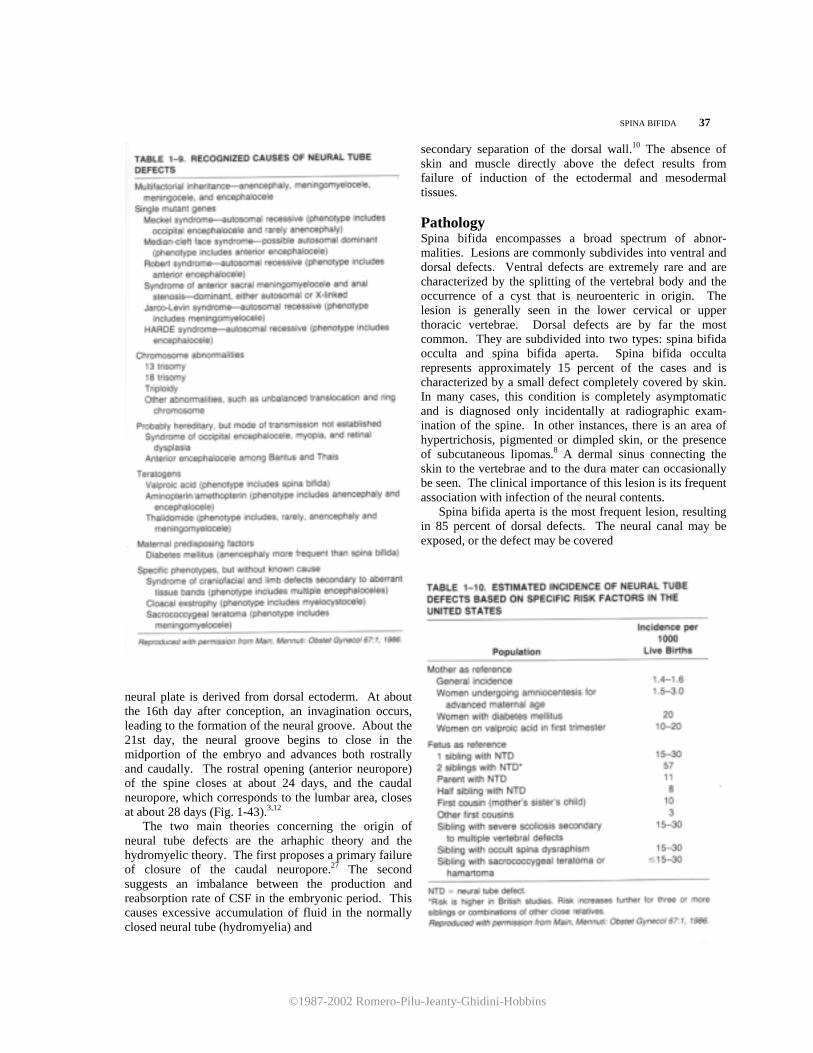
©1987-2002 Romero-Pilu-Jeanty-Ghidini-Hobbins
SPINA BIFIDA 37
neural plate is derived from dorsal ectoderm. At about the 16th day after conception, an invagination occurs, leading to the formation of the neural groove. About the 21st day, the neural groove begins to close in the midportion of the embryo and advances both rostrally and caudally. The rostral opening (anterior neuropore) of the spine closes at about 24 days, and the caudal neuropore, which corresponds to the lumbar area, closes at about 28 days (Fig. 1-43).3,12
The two main theories concerning the origin of neural tube defects are the arhaphic theory and the hydromyelic theory. The first proposes a primary failure of closure of the caudal neuropore.27 The second suggests an imbalance between the production and reabsorption rate of CSF in the embryonic period. This causes excessive accumulation of fluid in the normally closed neural tube (hydromyelia) and
secondary separation of the dorsal wall.10 The absence of skin and muscle directly above the defect results from failure of induction of the ectodermal and mesodermal tissues. Pathology Spina bifida encompasses a broad spectrum of abnor-malities. Lesions are commonly subdivides into ventral and dorsal defects. Ventral defects are extremely rare and are characterized by the splitting of the vertebral body and the occurrence of a cyst that is neuroenteric in origin. The lesion is generally seen in the lower cervical or upper thoracic vertebrae. Dorsal defects are by far the most common. They are subdivided into two types: spina bifida occulta and spina bifida aperta. Spina bifida occulta represents approximately 15 percent of the cases and is characterized by a small defect completely covered by skin. In many cases, this condition is completely asymptomatic and is diagnosed only incidentally at radiographic exam-ination of the spine. In other instances, there is an area of hypertrichosis, pigmented or dimpled skin, or the presence of subcutaneous lipomas.8 A dermal sinus connecting the skin to the vertebrae and to the dura mater can occasionally be seen. The clinical importance of this lesion is its frequent association with infection of the neural contents.
Spina bifida aperta is the most frequent lesion, resulting in 85 percent of dorsal defects. The neural canal may be exposed, or the defect may be covered
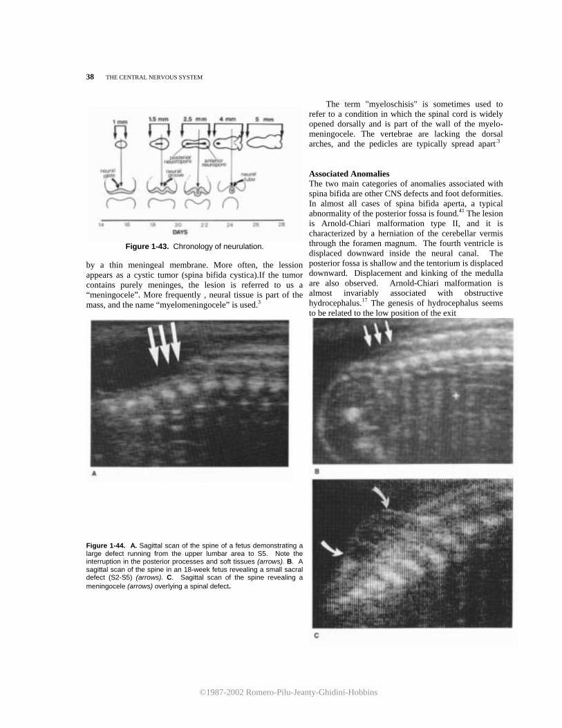
©1987-2002 Romero-Pilu-Jeanty-Ghidini-Hobbins
38 THE CENTRAL NERVOUS SYSTEM
Figure 1-43. Chronology of neurulation.
by a thin meningeal membrane. More often, the lession appears as a cystic tumor (spina bifida cystica).If the tumor contains purely meninges, the lesion is referred to us a “meningocele”. More frequently , neural tissue is part of the mass, and the name “myelomeningocele” is used.3
Figure 1-44. A. Sagittal scan of the spine of a fetus demonstrating a large defect running from the upper lumbar area to S5. Note the interruption in the posterior processes and soft tissues (arrows). B. A sagittal scan of the spine in an 18-week fetus revealing a small sacral defect (S2-S5) (arrows). C. Sagittal scan of the spine revealing a meningocele (arrows) overlying a spinal defect.
The term "myeloschisis" is sometimes used to refer to a condition in which the spinal cord is widely opened dorsally and is part of the wall of the myelo-meningocele. The vertebrae are lacking the dorsal arches, and the pedicles are typically spread apart.3 Associated Anomalies The two main categories of anomalies associated with spina bifida are other CNS defects and foot deformities. In almost all cases of spina bifida aperta, a typical abnormality of the posterior fossa is found.41 The lesion is Arnold-Chiari malformation type II, and it is characterized by a herniation of the cerebellar vermis through the foramen magnum. The fourth ventricle is displaced downward inside the neural canal. The posterior fossa is shallow and the tentorium is displaced downward. Displacement and kinking of the medulla are also observed. Arnold-Chiari malformation is almost invariably associated with obstructive hydrocephalus.17 The genesis of hydrocephalus seems to be related to the low position of the exit
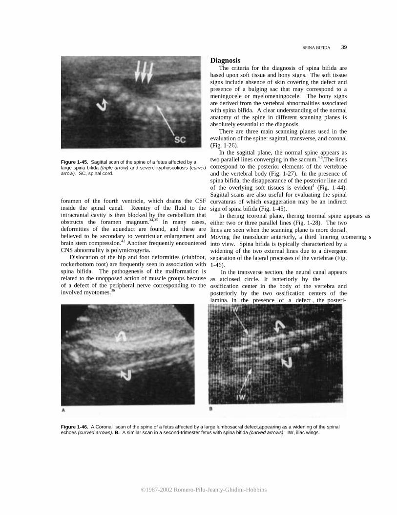
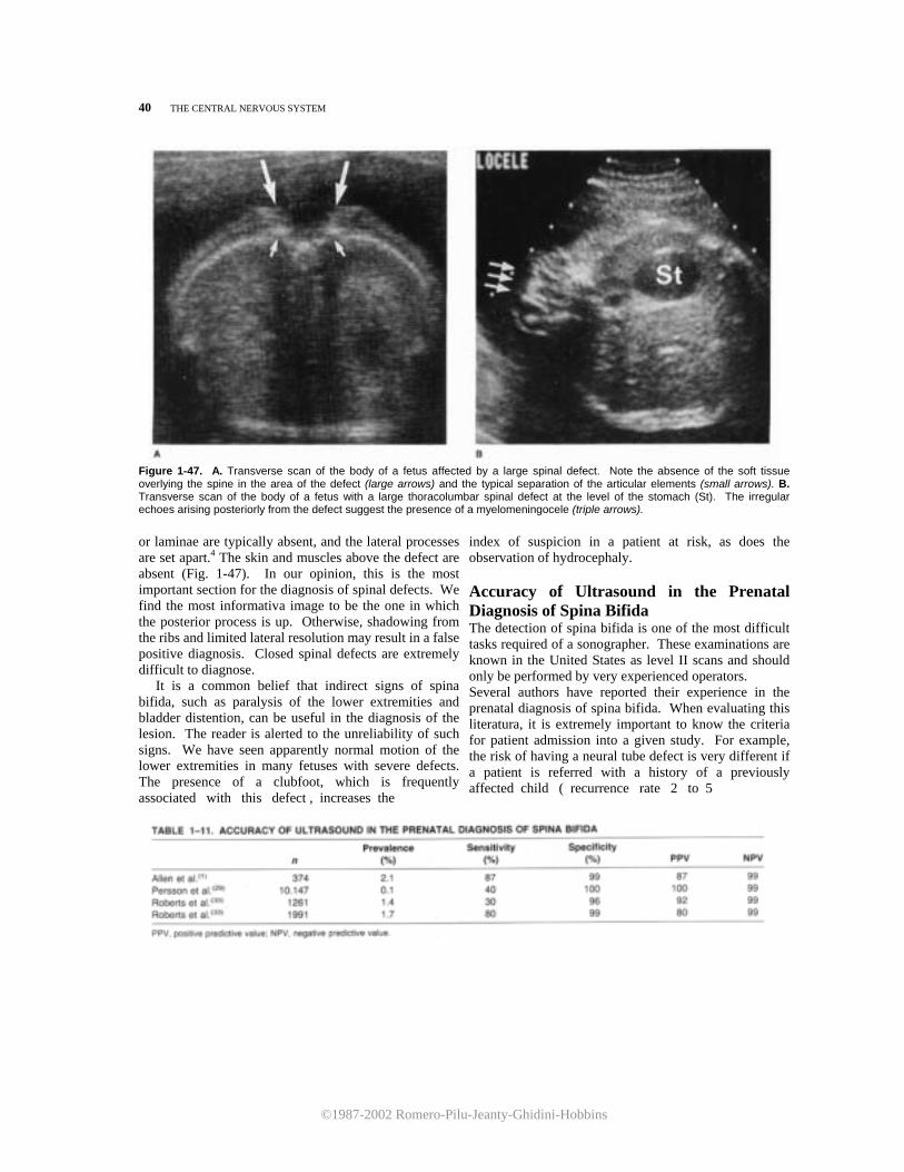
©1987-2002 Romero-Pilu-Jeanty-Ghidini-Hobbins
40 THE CENTRAL NERVOUS SYSTEM
Figure 1-47. A. Transverse scan of the body of a fetus affected by a large spinal defect. Note the absence of the soft tissue overlying the spine in the area of the defect (large arrows) and the typical separation of the articular elements (small arrows). B. Transverse scan of the body of a fetus with a large thoracolumbar spinal defect at the level of the stomach (St). The irregular echoes arising posteriorly from the defect suggest the presence of a myelomeningocele (triple arrows).
or laminae are typically absent, and the lateral processes are set apart.4 The skin and muscles above the defect are absent (Fig. 1-47). In our opinion, this is the most important section for the diagnosis of spinal defects. We find the most informativa image to be the one in which the posterior process is up. Otherwise, shadowing from the ribs and limited lateral resolution may result in a false positive diagnosis. Closed spinal defects are extremely difficult to diagnose. It is a common belief that indirect signs of spina bifida, such as paralysis of the lower extremities and bladder distention, can be useful in the diagnosis of the lesion. The reader is alerted to the unreliability of such signs. We have seen apparently normal motion of the lower extremities in many fetuses with severe defects. The presence of a clubfoot, which is frequently associated with this defect , increases the
index of suspicion in a patient at risk, as does the observation of hydrocephaly. Accuracy of Ultrasound in the Prenatal Diagnosis of Spina Bifida The detection of spina bifida is one of the most difficult tasks required of a sonographer. These examinations are known in the United States as level II scans and should only be performed by very experienced operators. Several authors have reported their experience in the prenatal diagnosis of spina bifida. When evaluating this literatura, it is extremely important to know the criteria for patient admission into a given study. For example, the risk of having a neural tube defect is very different if a patient is referred with a history of a previously affected child ( recurrence rate 2 to 5
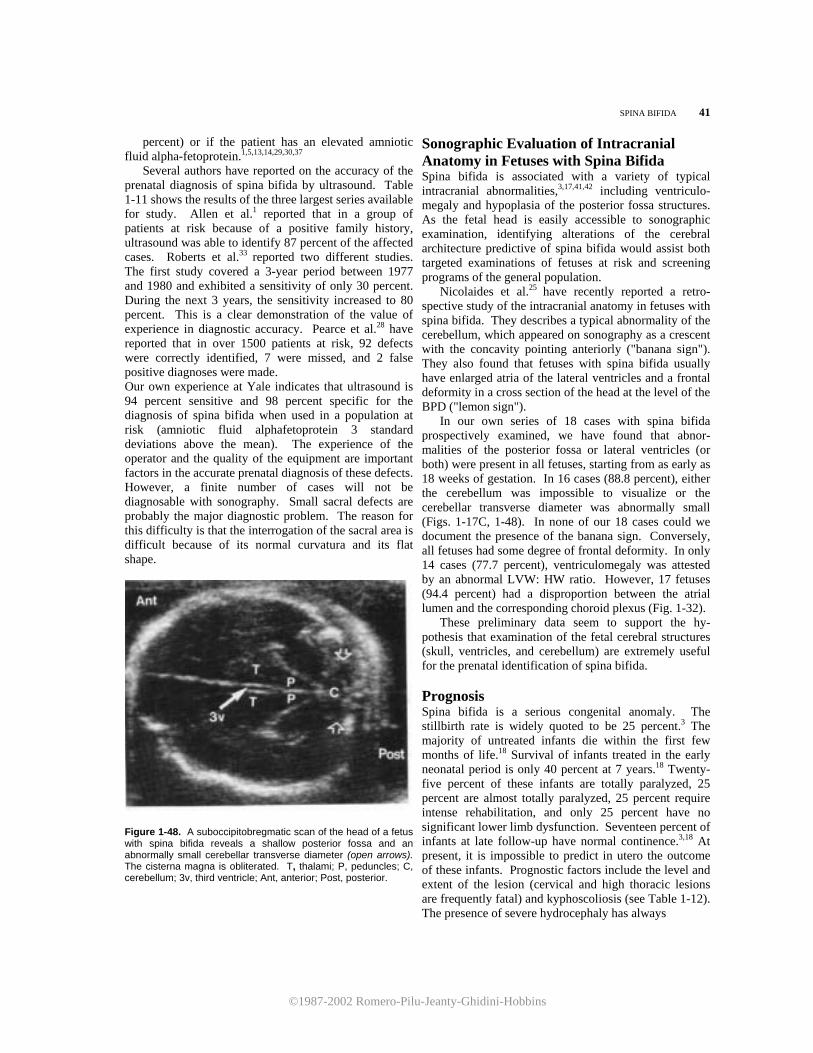
©1987-2002 Romero-Pilu-Jeanty-Ghidini-Hobbins
SPINA BIFIDA 41
percent) or if the patient has an elevated amniotic
fluid alpha-fetoprotein.1,5,13,14,29,30,37 Several authors have reported on the accuracy of the
prenatal diagnosis of spina bifida by ultrasound. Table 1-11 shows the results of the three largest series available for study. Allen et al.1 reported that in a group of patients at risk because of a positive family history, ultrasound was able to identify 87 percent of the affected cases. Roberts et al.33 reported two different studies. The first study covered a 3-year period between 1977 and 1980 and exhibited a sensitivity of only 30 percent. During the next 3 years, the sensitivity increased to 80 percent. This is a clear demonstration of the value of experience in diagnostic accuracy. Pearce et al.28 have reported that in over 1500 patients at risk, 92 defects were correctly identified, 7 were missed, and 2 false positive diagnoses were made. Our own experience at Yale indicates that ultrasound is 94 percent sensitive and 98 percent specific for the diagnosis of spina bifida when used in a population at risk (amniotic fluid alphafetoprotein 3 standard deviations above the mean). The experience of the operator and the quality of the equipment are important factors in the accurate prenatal diagnosis of these defects. However, a finite number of cases will not be diagnosable with sonography. Small sacral defects are probably the major diagnostic problem. The reason for this difficulty is that the interrogation of the sacral area is difficult because of its normal curvatura and its flat shape.
Figure 1-48. A suboccipitobregmatic scan of the head of a fetus with spina bifida reveals a shallow posterior fossa and an abnormally small cerebellar transverse diameter (open arrows). The cisterna magna is obliterated. T, thalami; P, peduncles; C, cerebellum; 3v, third ventricle; Ant, anterior; Post, posterior.
Sonographic Evaluation of Intracranial Anatomy in Fetuses with Spina Bifida Spina bifida is associated with a variety of typical intracranial abnormalities,3,17,41,42 including ventriculo-megaly and hypoplasia of the posterior fossa structures. As the fetal head is easily accessible to sonographic examination, identifying alterations of the cerebral architecture predictive of spina bifida would assist both targeted examinations of fetuses at risk and screening programs of the general population.
Nicolaides et al.25 have recently reported a retro-spective study of the intracranial anatomy in fetuses with spina bifida. They describes a typical abnormality of the cerebellum, which appeared on sonography as a crescent with the concavity pointing anteriorly ("banana sign"). They also found that fetuses with spina bifida usually have enlarged atria of the lateral ventricles and a frontal deformity in a cross section of the head at the level of the BPD ("lemon sign").
In our own series of 18 cases with spina bifida prospectively examined, we have found that abnor-malities of the posterior fossa or lateral ventricles (or both) were present in all fetuses, starting from as early as 18 weeks of gestation. In 16 cases (88.8 percent), either the cerebellum was impossible to visualize or the cerebellar transverse diameter was abnormally small (Figs. 1-17C, 1-48). In none of our 18 cases could we document the presence of the banana sign. Conversely, all fetuses had some degree of frontal deformity. In only 14 cases (77.7 percent), ventriculomegaly was attested by an abnormal LVW: HW ratio. However, 17 fetuses (94.4 percent) had a disproportion between the atrial lumen and the corresponding choroid plexus (Fig. 1-32).
These preliminary data seem to support the hy-pothesis that examination of the fetal cerebral structures (skull, ventricles, and cerebellum) are extremely useful for the prenatal identification of spina bifida. Prognosis Spina bifida is a serious congenital anomaly. The stillbirth rate is widely quoted to be 25 percent.3 The majority of untreated infants die within the first few months of life.18 Survival of infants treated in the early neonatal period is only 40 percent at 7 years.18 Twenty-five percent of these infants are totally paralyzed, 25 percent are almost totally paralyzed, 25 percent require intense rehabilitation, and only 25 percent have no significant lower limb dysfunction. Seventeen percent of infants at late follow-up have normal continence.3,18 At present, it is impossible to predict in utero the outcome of these infants. Prognostic factors include the level and extent of the lesion (cervical and high thoracic lesions are frequently fatal) and kyphoscoliosis (see Table 1-12). The presence of severe hydrocephaly has always
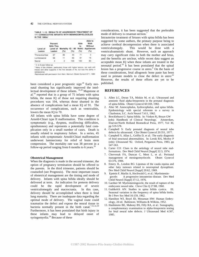
©1987-2002 Romero-Pilu-Jeanty-Ghidini-Hobbins
42 THE CENTRAL NERVOUS SYSTEM
been considered a poor prognostic sign.18 Early neo-natal shunting has significantly improved the intel-lectual development of these infants. 16,20
Mapstone et al.19 reported that in a group of 75 infants with spina bifida, the mean IQ of those not requiring shunting procedures was 104, whereas those shunted in the absence of complications had a mean IQ of 91. The occurrence of complications, such as ventriculitis, lowers the mean IQ to 70. All infants with spina bifida have some degree of Arnold-Chiari type II malformation. This condition is symptomatic (e.g., dyspnea, swallowing difficulties, opisthotonos) and represents a potentially fatal com-plication only in a small number of cases. Death is usually related to respiratory failure. In a series, 45 infants with symptomatic Arnold-Chiari malformation underwent laminectomy for relief of brain stem compression. The mortality rate was 38 percent in a follow-up period ranging from 6 months to 6 years.26 Obstetrical Management When the diagnosis is made in the second trimester, the option of pregnancy termination should be offered to the parents. In the third trimester, patients should be counseled (see Prognosis). The most important issues of obstetrical management are the timing and mode of delivery. Infants with spina bifida ideally should be delivered at term. An indication for preterm delivery could be the rapid development of severe ventriculomegaly and macrocrania. In this case, delivery should be accomplished when there is fetal lung maturity. There are inadequate data regarding the optimal mode of delivery. The vaginal route could traumatize the defect and expose the neural tissue to bacteria normally present in the birth canal.7,31,32,39
Furthermore, it has been postulated that birth injury in these infants may lead to delayed onset of syringomyelia.24 Because of these
considerations, it has been suggested that the preferable mode of delivery is cesarean section.7 Intrauterine treatment of fetuses with spina bifida has been suggested by some authors, the primary purpose being to achieve cerebral decompression when there is associated ventriculomegaly. This would be done with a ventriculoamniotic shunt. However, such an approach may carry significant risks to both the mother and fetus, and the benefits are unclear, while recent data suggest an acceptable mean IQ when these infants are treated in the neonatal period.19 lt has been postulated that the spinal lesion has a progressive course in utero.9 On the basis of these considerations, fetal allogeneic bone paste has been used in primate models to close the defect in utero.22 However, the results of these efforts are yet to be published. REFERENCES
1. Allen LC, Doran TA, Miskin M, et al.: Ultrasound and amniotic fluid alpha-fetoprotein in the prenatal diagnosis of spina bifida. Obstet Gynecol 60:169, 1982.
2. Alter M: Anencephalus, hydrocephalus, and spina bifida. Epidemiology with special referente to a survey in Charleston, S.C. Arch Neurol 7:411, 1962.
3. Brocklehurst G: Spina bifida. In: Vinken Pj, Bruyn GW (eds): Handbook of Clinical Neurology. Amsterdam,
Elsevier/North Holland Biomedical Press, 1977, Vol 30, pp 519-578.
4. Campbell S: Early prenatal diagnosis of neural tube defects by ultrasound. Clin Obstet Gynecol 20:351, 1977.
5. Campbell S, Allan L, Griffin D, et al.: The early diagnosis of fetal structural abnormalities. In: Lerski RA, Morley P (eds): Ultrasound '82. Oxford, Pergamon Press, 1983, pp 547-563.
6. Carter CO: Clues to the aetiology of neural tube mal-formations. Dev Med Child Neurol [Suppl] 32:3, 1974.
7. Chervenak FA, Duncan C, Ment L, et al.: Perinatal management of meningomyelocele. Obstet Gynecol 63:376, 1984.
8. Emery JL, Lendon RG: Lipomas of the cauda equina and other fatty tumours related to neurospinal dysraphism. Dev Med Child Neurol [Suppl] 20:62, 1969.
9. Epstein F, Marlin A, Hochwald G, et al.: Myelomenin- gocele: A progressive intrauterino disease. Dev Med
Child Neurol [Suppl] 37:12, 1976. 10. Gardner M: Myelomeningocele, the result of rupture of the
embryonic neural tube. Cleve Clin Q 27:88, 1960. 11. Guthkelch AN: Studies in spina bifida cystica. III.
Seasonal variation in the frequency of spina bifida births. Br J Prev Soc Med 16:159, 1962.
12. Hamilton WJ, Boyd JD, Mossman HW: Human Embry-ology, 2d ed. Baltimore, Williams & Wilkins, 1952.
13. Hashimoto BE, Mahony BS, Filly RA, et al.: Sonography, a complementary examination to alpha-fetoprotein testing for fetal neural tube defects. J Ultrasound Med 4:307, 1985.
©1987-2002 Romero-Pilu-Jeanty-Ghidini-Hobbins
ANENCEPHALY 43
14. Hobbins JC, Venus 1, Tortora M, et al.: Stage II ultra-sound examination for the diagnosis of fetal abnormal~ ities with an elevated amniotic fluid alpha-fetoprotein concentration. Am J Obstet Gynecol 142:1026, 1982.
15.Laurence KM, Carter CO, David PA: Major central nervous system malformations in South Wales. 1. Inci-dence, local variations and geographical factors. Br J Prev Soc Med 22:146, 1968.
16.Leonard CO, Freeman JM: Spina bifida: A new disease. Pediatrics 68:136, 1981.
17.Lorber J: Systematic ventriculographic studies in infants born with meningomyelocele and encephalocele. The incidence and development of hydrocephalus. Arch Dis Child 36:381, 1961.
18.Lorber J: Results of treatment of myelomeningocele. An analysis of 524 unselected cases, with special reference to possible selection for treatment. Dev Med Child Neurol 13:279, 1971.
19.Mapstone TB, Rekate HL, Nulsen FE, et al.: Relationship of CSF shunting and IQ in children with myelo-
meningocele: A retrospectiva analysis. Childs Brain 11:112, 1984.
20 -W. McCullough DC, Balzer-Martin LA: Current prognosis in overt neonatal hydrocephalus. J Neurosurg 57:378, 1982.
21.-McKeown T, Record RG: Malformations in a popula-tion observed for 5 years after birth. In: Wolstenholme GEW, O'Connor CM (eds): Ciba Foundation Symposium on Congenital Malformations. London, Churchill, 1960, pp 2-14.
22.-Michejda M, McCullough D, Bacher J, et al.: Investiga-tional approaches in fetal neurosurgery. Concepts Pediatr Neurosurg 4:44, 1983.
23 .Neel JV: A study of major congenital defects in Japanese infants. Am J Hum Genet 10:398, 1958.
24.-Newman PW, Terenty TR, Foster JB: Some observations on the pathogenesis of syringomyelia. J Neurol Neurosurg Psychiatry 44:964, 1981.
25.Nicolaides KH, Campbell S, Gabbe SG, et al.: Ultrasound screening for spina bifida: Cranial and cerebellar signs. Lancet 2:72, 1986.
26.Park TS, Hoffman HJ, Hendrick EB, et al.: Experience with surgical decompression of the Arnold-Chiari mal-formation in young infants with myelomeningocele. Neurosurgery 13:147, 1983.
27.Patten BM: Embryological stages in the establishing of myeloschisis with spina bifida. Am J Anat 93:365, 1953.
28.Pearce JM, Little D, Campbell S: The diagnosis of
abnormalities of the fetal central nervous system. In: Sanders RC, James AE (eds): The Principles and Prac-tice of Ultrasonography in Obstetrics and Gynecology, 3d ed. Norwalk, CT, Appleton-Century-Crofts, 1985, pp 243-256.
29. Persson PH, Kullander S, Gennser G, et al.: Screening for fetal malformations using ultrasound and measure-ments of alpha-fetoprotein in maternal serum. Br Med J 286:747, 1983.
30. Polanska N, Burgess DE, Hill P, et al.: Screening for neural tube defect: False positive findings on ultra-sound and in amniotic fluid. Br Med J 287:24, 1983.
31. Ralis ZA: Traumatizing effect of breech delivery on infants with spina bifida. J Pediatr 87:613, 1975.
32. Ralis Z, Ralis HM: Morphology of peripheral nerves in children with spina bifida. Dev Med Child Neurol [Suppl] 27:109, 1972.
33. Roberts CJ, Hibbard BM, Roberts EE, et al.: Diagnostic effectiveness of ultrasound in detection of neural tube defect. The South Wales experience of 2509 scans (1977-1982) in high-risk mothers. Lancet 2:1068, 1983.
34. Russell DS: Observations en the pathology of hydro-cephalus. Med Res Counc Spec Sev 265:1, 1949.
35. Russell DS, Donald C: The mechanism of internal hydrocephalus in spina bifida. Brain 58:203, 1935.
36. Sharrard WJW: The mechanism of paralytic deformity in spina bifida. Dev Med Child Neurol 4:310, 1962.
37. Slotnick N, Filly RA, Callen PW, et al.: Sonography as a procedure complementary to alpha-fetoprotein testing for neural tube defects. J Ultrasound Med 1:319, 1982.
38. Smith C: Computer programme to estimate recurrence risks for multifactorial familiar disease. Br Med J 1:495, 1972.
39. Stark G, Drummond M: Spina bifida as an obstetric problem. Dev Med Child Neurol [Suppl] 22:157, 1970.
40. Stevenson AC, Johnston HA, Stewart MIP, et al.: Congenital malformations. A report study of a series of consecutiva births in 24 centres. Bull WHO 34 [Suppl 9]:127, 1966.
41. Variend S, Emery JL: The weight of the cerebellum in children with myelomeningocele. Dev Med Child Neurol 15 [Suppl 29]:77, 1973.
42. Williams B: Is aqueduct stenosis a result of hydrocephalus? Brain 96:399, 1973.
43. Williamson EM: Incidence and family aggregation of major congenital malformations of central nervous system. J Med Genet 2:161, 1965.
Anencephaly Synonyms Pseudoencephaly, extracranial disencephaly, and acleidencephaly.
Definition Anencephaly is an anomaly characterized by the absence of cerebral hemispheres and cranial vault.
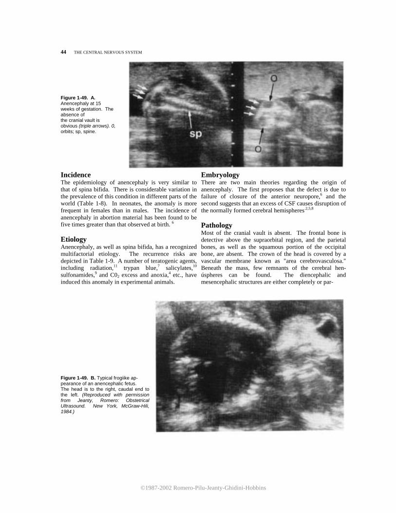
©1987-2002 Romero-Pilu-Jeanty-Ghidini-Hobbins
44 THE CENTRAL NERVOUS SYSTEM Figure 1-49. A. Anencephaly at 15 weeks of gestation. The absence of the cranial vault is obvious (triple arrows). 0, orbits; sp, spine.
Incidence The epidemiology of anencephaly is very similar to that of spina bifida. There is considerable variation in the prevalence of this condition in different parts of the world (Table 1-8). In neonates, the anomaly is more frequent in females than in males. The incidence of anencephaly in abortion material has been found to be five times greater than that observed at birth. 6 Etiology Anencephaly, as well as spina bifida, has a recognized multifactorial etiology. The recurrence risks are depicted in Table 1-9. A number of teratogenic agents, including radiation,11 trypan blue,7 salicylates,10 sulfonamides,9 and C02 excess and anoxia,4 etc., have induced this anomaly in experimental animals.
Embryology There are two main theories regarding the origin of anencephaly. The first proposes that the defect is due to failure of closure of the anterior neuropore,6 and the second suggests that an excess of CSF causes disruption of the normally formed cerebral hemispheres.2,5,8
Pathology Most of the cranial vault is absent. The frontal bone is detective above the supraorbital region, and the parietal bones, as well as the squamous portion of the occipital bone, are absent. The crown of the head is covered by a vascular membrane known as "area cerebrovasculosa." Beneath the mass, few remnants of the cerebral hen-úspheres can be found. The diencephalic and mesencephalic structures are either completely or par-
Figure 1-49. B. Typical frogiike ap- pearance of an anencephalic fetus. The head is to the right, caudal end to the left. (Reproduced with permission from Jeanty, Romero: Obstetrical Ultrasound. New York, McGraw-Hili, 1984.)
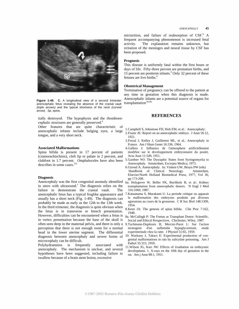
©1987-2002 Romero-Pilu-Jeanty-Ghidini-Hobbins
ANENCEPHALY 45
Figure 1-49. C. A longitudinal view of a second trimester anencephalic fetus revealing the absence of the cranial vault (triple arrows) and the typical shortness of the neck (curved arrow). Sp, spine. tially destroyed. The hypophysis and the rhomboen-cephalic structures are generally preserved.6 Other features that are quite characteristic ofanencephalic infants include bulging eyes, a largetongue, and a very short neck. Associated Malformations Spina bifida is present in 17 percent of patients(craniorachischisis), cleft lip or palate in 2 percent, andclubfoot in 1.7 percent. Omphaloceles have also beendescribes in some cases.3,6
Diagnosis Anencephaly was the first congenital anomaly identifiedin utero with ultrasound.1 The diagnosis relies on thefailure to demonstrate the cranial vault. Theanencephalic fetus has a typical froglike appearance andusually has a short neck (Fig. 1-49). The diagnosis canprobably be made as early as the 12th to the 13th week.In the third trimester, the diagnosis is quite obvious whenthe fetus is in transverse or breech presentation.However, difficulties can be encountered when a fetus isin vertex presentation because the base of the skull isoften seen deep in the maternal pelvis, and there is only aperception that there is not enough room for a normalhead in the lower uterine segment. The differentialdiagnosis between anencephaly and severe forms ofmicrocephaly can be difficult. Polyhydramnios is frequently associated withanencephaly. The mechanism is unclear, and severalhypotheses have been suggested, including failure toswallow because of a brain stem lesion, excessive
micturition, and failure of reabsorption of CSF.6 A frequent accompanying phenomenon is increased fetal activity. The explanation remains unknown, but irritation of the meninges and neural tissue by CSF has been proposed. Prognosis This disease is uniformly fatal within the first hours or days of life. Fifty-three percent are premature births, and 15 percent are postterm infants.3 Only 32 percent of these fetuses are live births.6 Obstetrical Management Termination of pregnancy can be offered to the patient at any time in gestation when this diagnosis is made. Anencephalic infants are a potential source of organs for transplantation.6a,8a
REFERENCES
1. Campbell S, Johnstone FD, Holt EM, et al.: Anencephaly:2. Frazer JE: Report on an anencephalic embryo. J Anat 56:12,
1921. 3. Frezal J, Kelley J, Guillemot ML, et al.: Anencephaly in
France. Am J Hum Genet 16:336, 1964. 4. Gallera J: Influence de l'atmosphere artificiellement
modifiee sur le developpement embryonnaire du poulet. Acta Anat 11:549, 1951.
5. Gardner WJ: The Dysraphic States from Syringomyelia to Anencephaly. Amsterdam, Excerpta Medica, 1973.
6. Giroud A: Anencephaly. In: Vinken GW, Bruyn PW (eds) Handbook of Clinical Neurology. Amsterdam,
Elsevier/North Holland Biomedical Press, 1977, Vol 30, pp 173-208.
6a. Holzgreve W, Beller FK, Buchholz B, et al.: Kidney transplantation from anencephalic donors. N Engl J Med 316:1069, 1987.
7. Katsunuma S, Murakami U: La periode critique ou apparait la malformation des embryons produite par diverses agressions au cours de la grossesse. C R Soc Biol 148:1309, 1954.
8. Keen JA: The genesis of spina bifida. Clin Proc 7:162, 1948.
8a. McCullagh P: The Foetus as Transplant Donor: Scientific, Social and Ethical Perspectives. Chichester, Wiley, 1987.
9. Tuchmann-Duplessis H, Mercier-Parot L: Sur l'action teratogene d'un sulfamida hypoglycemiant, etude experimentale chez la ratte. J Physiol 51:65, 1959.
10. Warkany J, Takacs E: Experimental production of con-genital malformations in rats by salicylate poisoning. Am J Pathol 35:315, 1959.
11.Wilson JG, Karr JW: Effects of irradiation on embryonic development. 1. X-rays en the 10th day of gestation in the rat. Am j Anat 88:1, 1951.
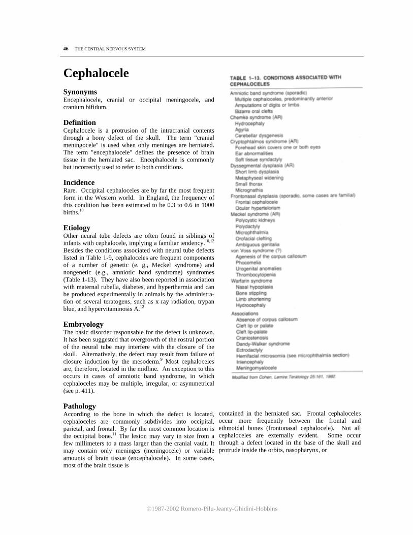
©1987-2002 Romero-Pilu-Jeanty-Ghidini-Hobbins
46 THE CENTRAL NERVOUS SYSTEM
Cephalocele Synonyms Encephalocele, cranial or occipital meningocele, and cranium bifidum. Definition Cephalocele is a protrusion of the intracranial contents through a bony defect of the skull. The term "cranial meningocele" is used when only meninges are herniated. The term "encephalocele" defines the presence of brain tissue in the herniated sac. Encephalocele is commonly but incorrectly used to refer to both conditions. Incidence Rare. Occipital cephaloceles are by far the most frequent form in the Western world. In England, the frequency of this condition has been estimated to be 0.3 to 0.6 in 1000 births.10 Etiology Other neural tube defects are often found in siblings of infants with cephalocele, implying a familiar tendency.10,12 Besides the conditions associated with neural tube defects listed in Table 1-9, cephaloceles are frequent components of a number of genetic (e. g., Meckel syndrome) and nongenetic (e.g., amniotic band syndrome) syndromes (Table 1-13). They have also been reported in association with maternal rubella, diabetes, and hyperthermia and can be produced experimentally in animals by the administra-tion of several teratogens, such as x-ray radiation, trypan blue, and hypervitaminosis A.12 Embryology The basic disorder responsable for the defect is unknown. It has been suggested that overgrowth of the rostral portion of the neural tube may interfere with the closure of the skull. Alternatively, the defect may result from failure of closure induction by the mesoderm.9 Most cephaloceles are, therefore, located in the midline. An exception to this occurs in cases of amniotic band syndrome, in which cephaloceles may be multiple, irregular, or asymmetrical (see p. 411). Pathology According to the bone in which the defect is located, cephaloceles are commonly subdivides into occipital, parietal, and frontal. By far the most common location is the occipital bone.11 The lesion may vary in size from a few millimeters to a mass larger than the cranial vault. It may contain only meninges (meningocele) or variable amounts of brain tissue (encephalocele). In some cases, most of the brain tissue is
contained in the herniated sac. Frontal cephaloceles occur more frequently between the frontal and ethmoidal bones (frontonasal cephalocele). Not all cephaloceles are externally evident. Some occur through a defect located in the base of the skull and protrude inside the orbits, nasopharynx, or

©1987-2002 Romero-Pilu-Jeanty-Ghidini-Hobbins
CEPHALOCELE 47
Figure 1-50. A. Longitudinal scan of the upper torso and head in a 21-week fetus with a small occipital meningocele (M). Sp, spine. B. Pathologic specimen obtained from the same fetus. The arrow points to the bony defect in the skull that connected the intracranial cavity to the meningocele (M). The diameter of the orifice was 2 mm. C. Occipital meningocele (arrowheads). A small amount of neural tissue is seen protruding inside the meningeal sac (curved arrow). A small bony defect is inferred by the presence of a pencil-like sound enhancement (straight arrows).
oropharynx. Frontal cephaloceles almost always contain brain tissue.12 Associated Anomalies As previously mentioned, cephaloceles can be found as part of a number of specific syndromes (Table 1-13). In addition, both meningoceles and encephaloceles are associated with other CNS abnormalities. Hydrocephalus has been reported in 80 percent of occipital meningoceles, 65 percent of occipital encephaloceles, l0 and 15 percent of frontal cephaloceles.5 Spina bifida is found in 7 to 15 percent of all cephaloceles.1 Microcephaly was observed in 20
percent of cases studied.10 By definition, the herniation of the cerebellum inside the cephalocele is termed "Chiari type III deformity." This deformity, combined with aqueductal stenosis, is the major cause of hydrocephalus in these infants.12 Frontal cephaloceles are often associated with the median cleft face syndrome, characterized by hypertelorism and median cleft lip or palate.3 Diagnosis Traditionally, the diagnosis of cephalocele relies on the demonstration of a paracranial mass.1,4,6,8,13,14 However, this criterion is insufficient to distinguish
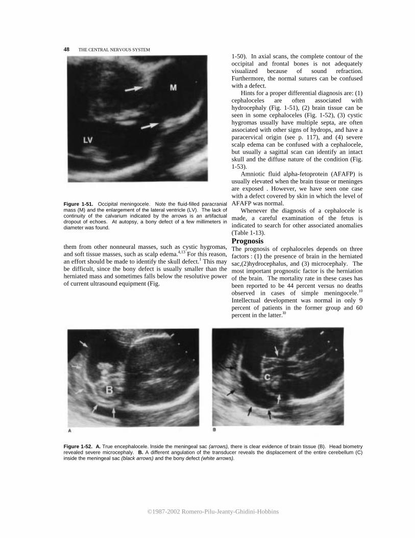
©1987-2002 Romero-Pilu-Jeanty-Ghidini-Hobbins
48 THE CENTRAL NERVOUS SYSTEM
Figure 1-51. Occipital meningocele. Note the fluid-filled paracranial mass (M) and the enlargement of the lateral ventricle (LV). The lack of continuity of the calvarium indicated by the arrows is an artifactual dropout of echoes. At autopsy, a bony defect of a few millimeters in diameter was found. them from other nonneural masses, such as cystic hygromas, and soft tissue masses, such as scalp edema.4,13 For this reason, an effort should be made to identify the skull defect.1 This may be difficult, since the bony defect is usually smaller than the herniated mass and sometimes falls below the resolutive power of current ultrasound equipment (Fig.
1-50). In axial scans, the complete contour of the occipital and frontal bones is not adequately visualized because of sound refraction. Furthermore, the normal sutures can be confused with a defect.
Hints for a proper differential diagnosis are: (1) cephaloceles are often associated with hydrocephaly (Fig. 1-51), (2) brain tissue can be seen in some cephaloceles (Fig. 1-52), (3) cystic hygromas usually have multiple septa, are often associated with other signs of hydrops, and have a paracervical origin (see p. 117), and (4) severe scalp edema can be confused with a cephalocele, but usually a sagittal scan can identify an intact skull and the diffuse nature of the condition (Fig. 1-53).
Amniotic fluid alpha-fetoprotein (AFAFP) is usually elevated when the brain tissue or meninges are exposed . However, we have seen one case with a defect covered by skin in which the level of AFAFP was normal.
Whenever the diagnosis of a cephalocele is made, a careful examination of the fetus is indicated to search for other associated anomalies (Table 1-13). Prognosis The prognosis of cephaloceles depends on three factors : (1) the presence of brain in the herniated sac,(2)hydrocephalus, and (3) microcephaly. The most important prognostic factor is the herniation of the brain. The mortality rate in these cases has been reported to be 44 percent versus no deaths observed in cases of simple meningocele.10 Intellectual development was normal in only 9 percent of patients in the former group and 60 percent in the latter.l0
Figure 1-52. A. True encephalocele. lnside the meningeal sac (arrows), there is clear evidence of brain tissue (B). Head biometry revealed severe microcephaly. B. A different angulation of the transducer reveals the displacement of the entire cerebellum (C) inside the meningeal sac (black arrows) and the bony defect (white arrows).
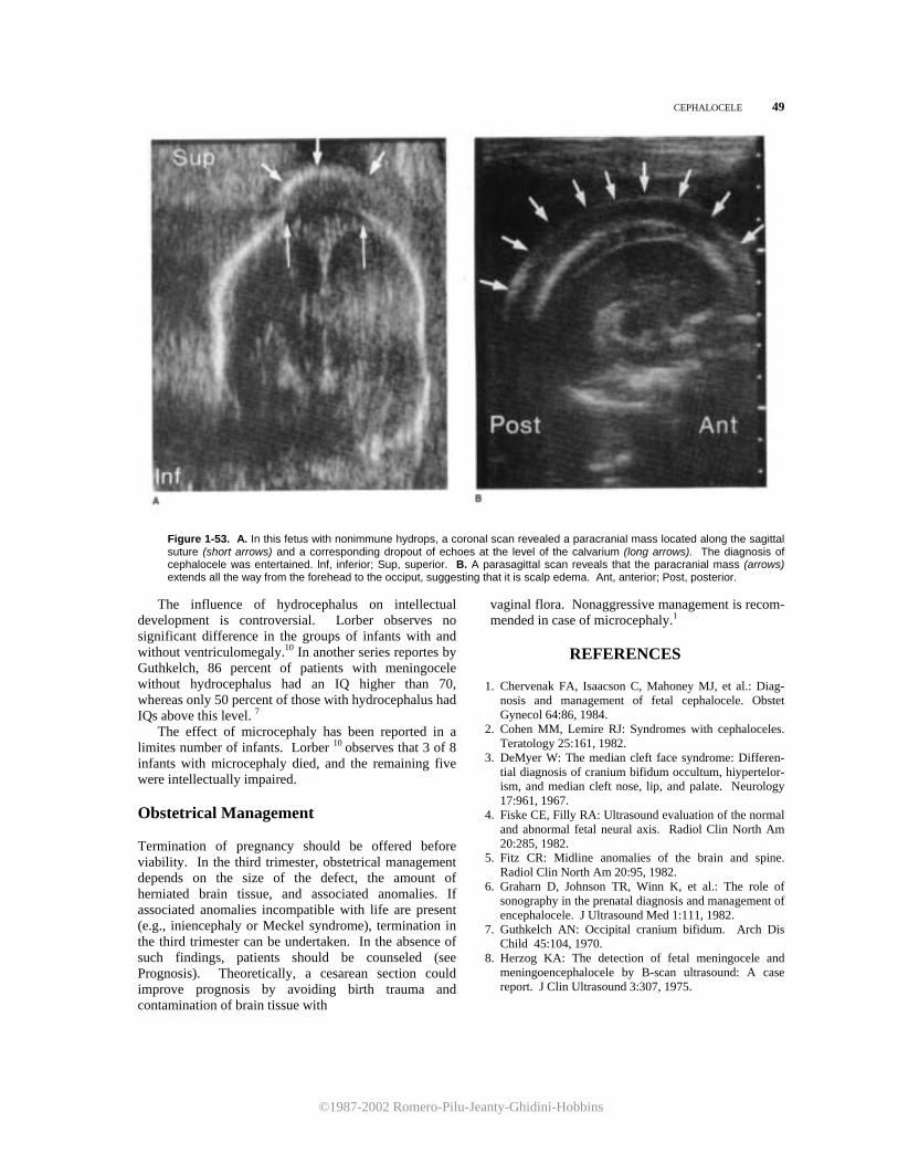
©1987-2002 Romero-Pilu-Jeanty-Ghidini-Hobbins
CEPHALOCELE 49
Figure 1-53. A. In this fetus with nonimmune hydrops, a coronal scan revealed a paracranial mass located along the sagittal suture (short arrows) and a corresponding dropout of echoes at the level of the calvarium (long arrows). The diagnosis of cephalocele was entertained. lnf, inferior; Sup, superior. B. A parasagittal scan reveals that the paracranial mass (arrows) extends all the way from the forehead to the occiput, suggesting that it is scalp edema. Ant, anterior; Post, posterior.
The influence of hydrocephalus on intellectual
development is controversial. Lorber observes no significant difference in the groups of infants with and without ventriculomegaly.10 In another series reportes by Guthkelch, 86 percent of patients with meningocele without hydrocephalus had an IQ higher than 70, whereas only 50 percent of those with hydrocephalus had IQs above this level. 7
The effect of microcephaly has been reported in a limites number of infants. Lorber 10 observes that 3 of 8 infants with microcephaly died, and the remaining five were intellectually impaired. Obstetrical Management Termination of pregnancy should be offered before viability. In the third trimester, obstetrical management depends on the size of the defect, the amount of herniated brain tissue, and associated anomalies. If associated anomalies incompatible with life are present (e.g., iniencephaly or Meckel syndrome), termination in the third trimester can be undertaken. In the absence of such findings, patients should be counseled (see Prognosis). Theoretically, a cesarean section could improve prognosis by avoiding birth trauma and contamination of brain tissue with
vaginal flora. Nonaggressive management is recom-mended in case of microcephaly.1
REFERENCES
1. Chervenak FA, Isaacson C, Mahoney MJ, et al.: Diag-
nosis and management of fetal cephalocele. Obstet Gynecol 64:86, 1984.
2. Cohen MM, Lemire RJ: Syndromes with cephaloceles. Teratology 25:161, 1982.
3. DeMyer W: The median cleft face syndrome: Differen-tial diagnosis of cranium bifidum occultum, hiypertelor-ism, and median cleft nose, lip, and palate. Neurology 17:961, 1967.
4. Fiske CE, Filly RA: Ultrasound evaluation of the normal and abnormal fetal neural axis. Radiol Clin North Am 20:285, 1982.
5. Fitz CR: Midline anomalies of the brain and spine. Radiol Clin North Am 20:95, 1982.
6. Graharn D, Johnson TR, Winn K, et al.: The role of sonography in the prenatal diagnosis and management of encephalocele. J Ultrasound Med 1:111, 1982.
7. Guthkelch AN: Occipital cranium bifidum. Arch Dis Child 45:104, 1970.
8. Herzog KA: The detection of fetal meningocele and meningoencephalocele by B-scan ultrasound: A case report. J Clin Ultrasound 3:307, 1975.

©1987-2002 Romero-Pilu-Jeanty-Ghidini-Hobbins
50 THE CENTRAL NERVOUS SYSTEM
9. Leong AS, Shaw CM: The pathology of occipital en-cephalocele and a discussion of the pathogenesis.Pa- thology 11:223, 1979.
10. Lorber J: The prognosis of occipital encephalocele. Dev Med Child Neurol [Suppl] 13:75, 1966.
11. Matson DD: Neurosurgery of Infancy and Childhood. Springfield, IL, Charles C Thomas, 1969.
12. McLaurin RL: Cranium bifidum and cranial ceph-aloceles. In: Vinken GW, Bruyn PW (eds): Hand-
book of Clinical Neurology. Amsterdam, Elsevier/ North Holland Biomedical Press, 1977, Vol 30, pp 209-218.
13. Nicolini U, Ferrazzi E, Massa E, et al.: Prenatal diagnosis of cranial masses by ultrasound: Report of five cases. JCU 11:170, 1983.
14. Pilu, G, Rizzo N, Orsini LF, et al.: Antenatal recogni-tion of cerebral anomalies. Ultrasound Med Biol 12:319, 1986.
Porencephaly
Synonyms Porencephalic cyst, schizencephaly, and congenital brain clefts.
Definition The term "porencephaly" describes an intracerebral, CSF-containing cystic cavity, which may or may not communicate with the ventricular system and the subarachnoid space.
Incidence True porencephaly is an extremely rare disease. In an autopsy study of 1000 cases of infantile brain damage, 25 infants (2.5 percent) had this condition. 5 The prevalence of congenital pseudoporencephaly is unknown.
Etiopathogenesis Porencephalic disorders are generally subdivided into two types: true porencephaly and pseudo-porencephaly. True porencephaly (schizencephaly) is a developmental anomaly caused by a failure in the migration of cells destined to form the cerebral cortex. This anomaly causes a local defect in both gray and white matter. In the absence of neural tissue, the subarachnoid space expands to fill the void, and, hence, the appearance of a porous cyst occurs. The designation "porous" alludes to the frequently seen communication of the brain with the subarachnoid space. Pseudoporencephaly is a consequence of local destruction of the cerebral parenchyma by a vascular, infectious, or traumatic cause that may occur either in utero or any time after birth.1,2,5 Examples of this acquired pseudoporencephaly are the cysts that de-veloped after repeated needling of the ventricular system in hydrocephalic infants before early neonatal shunting procedures were introduced.7 There is no evidence of familiar occurrence of porencephaly. The cytoarchitectural disorders due to failure of
migration comprise a broad spectrum from microgyria to porencephaly.6 In the former, the migration disorder is mild, and there are small cerebral gyri. The spectrum continues through macrogyria (large and fewer cerebral gyri), lissencephaly or agyria (absence of cerebral gyri), to porencephaly, where the defect is so severe that it affects a local portion of both cortex and white matter.5,10,11
Pathology True porencephaly is characterized by cystic cavities of variable size usually localizad around the Sylvian fissure and, in many cases, symmetrical in shape. They are frequently associated with other localized cytoarchitectural disorders, such as micropolygyria
Figure 1-54. Axial scan of the head of a 32-week fetus with severe porencephaly. The cerebral hemisphere that is closer to the transducer is entirely replaced by a cystic structure (Cy). Note the marked shift of the midline and the mildly enlarged contralateral ventricle (LV). (Reproduced with permission from Pilu et al.: Ultrasound Med Biol 12.-319, 1986.)
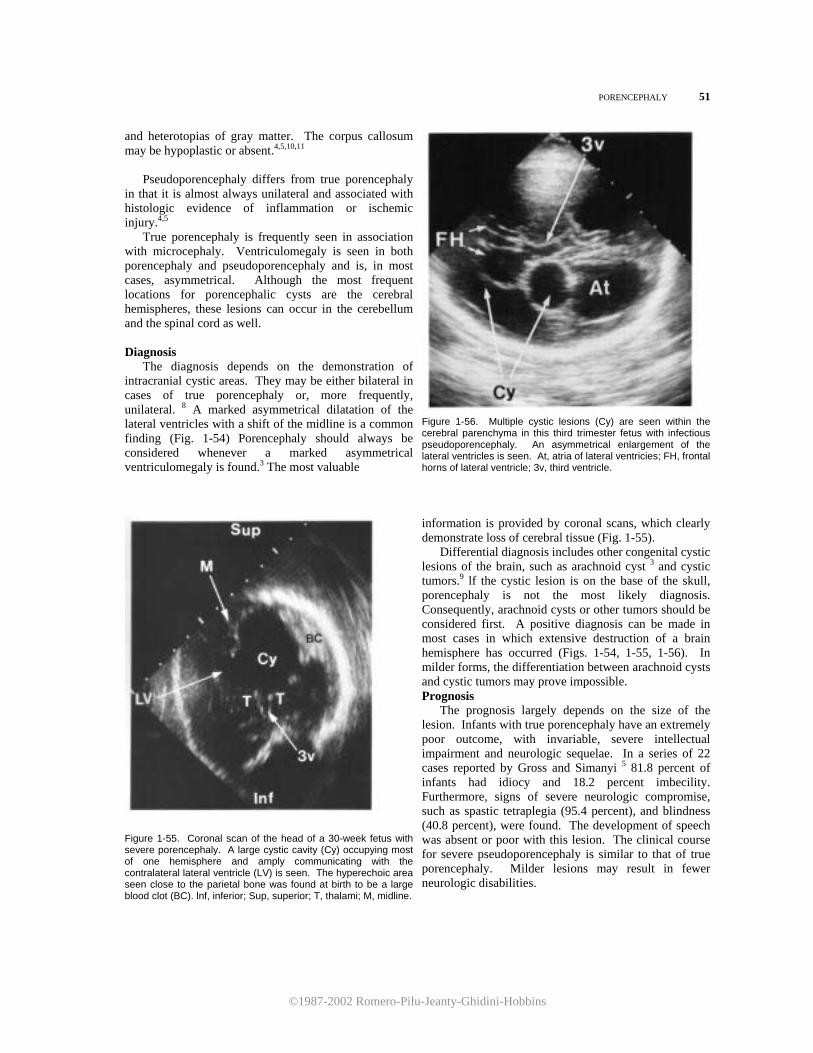
©1987-2002 Romero-Pilu-Jeanty-Ghidini-Hobbins
PORENCEPHALY 51
and heterotopias of gray matter. The corpus callosum may be hypoplastic or absent.4,5,10,11
Pseudoporencephaly differs from true porencephaly in that it is almost always unilateral and associated with histologic evidence of inflammation or ischemic injury.4,5
True porencephaly is frequently seen in association with microcephaly. Ventriculomegaly is seen in both porencephaly and pseudoporencephaly and is, in most cases, asymmetrical. Although the most frequent locations for porencephalic cysts are the cerebral hemispheres, these lesions can occur in the cerebellum and the spinal cord as well. Diagnosis
The diagnosis depends on the demonstration of intracranial cystic areas. They may be either bilateral in cases of true porencephaly or, more frequently, unilateral. 8 A marked asymmetrical dilatation of the lateral ventricles with a shift of the midline is a common finding (Fig. 1-54) Porencephaly should always be considered whenever a marked asymmetrical ventriculomegaly is found.3 The most valuable
Figure 1-56. Multiple cystic lesions (Cy) are seen within the cerebral parenchyma in this third trimester fetus with infectious pseudoporencephaly. An asymmetrical enlargement of the lateral ventricles is seen. At, atria of lateral ventricies; FH, frontal horns of lateral ventricle; 3v, third ventricle.
Figure 1-55. Coronal scan of the head of a 30-week fetus with severe porencephaly. A large cystic cavity (Cy) occupying most of one hemisphere and amply communicating with the contralateral lateral ventricle (LV) is seen. The hyperechoic area seen close to the parietal bone was found at birth to be a large blood clot (BC). lnf, inferior; Sup, superior; T, thalami; M, midline.
information is provided by coronal scans, which clearly demonstrate loss of cerebral tissue (Fig. 1-55).
Differential diagnosis includes other congenital cystic lesions of the brain, such as arachnoid cyst 3 and cystic tumors.9 lf the cystic lesion is on the base of the skull, porencephaly is not the most likely diagnosis. Consequently, arachnoid cysts or other tumors should be considered first. A positive diagnosis can be made in most cases in which extensive destruction of a brain hemisphere has occurred (Figs. 1-54, 1-55, 1-56). In milder forms, the differentiation between arachnoid cysts and cystic tumors may prove impossible. Prognosis
The prognosis largely depends on the size of the lesion. Infants with true porencephaly have an extremely poor outcome, with invariable, severe intellectual impairment and neurologic sequelae. In a series of 22 cases reported by Gross and Simanyi 5 81.8 percent of infants had idiocy and 18.2 percent imbecility. Furthermore, signs of severe neurologic compromise, such as spastic tetraplegia (95.4 percent), and blindness (40.8 percent), were found. The development of speech was absent or poor with this lesion. The clinical course for severe pseudoporencephaly is similar to that of true porencephaly. Milder lesions may result in fewer neurologic disabilities.
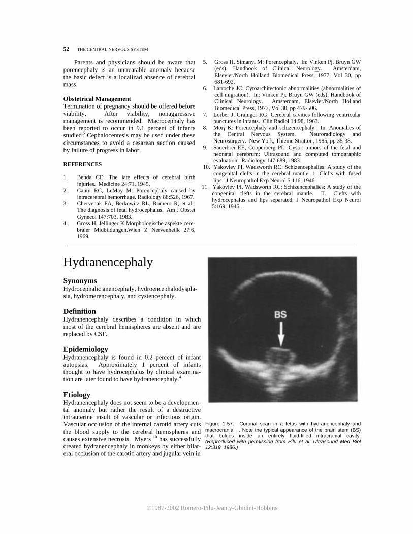
©1987-2002 Romero-Pilu-Jeanty-Ghidini-Hobbins
52 THE CENTRAL NERVOUS SYSTEM Parents and physicians should be aware that porencephaly is an untreatable anomaly because the basic defect is a localizad absence of cerebral mass. Obstetrical Management Termination of pregnancy should be offered before viability. After viability, nonaggressive management is recommended. Macrocephaly has been reported to occur in 9.1 percent of infants studied.5 Cephalocentesis may be used under these circumstances to avoid a cesarean section caused by failure of progress in labor. REFERENCES 1. Benda CE: The late effects of cerebral birth
injuries. Medicine 24:71, 1945. 2. Cantu RC, LeMay M: Porencephaly caused by
intracerebral hemorrhage. Radiology 88:526, 1967. 3. Chervenak FA, Berkowitz RL, Romero R, et al.:
The diagnosis of fetal hydrocephalus. Am J Obstet Gynecol 147:703, 1983.
4. Gross H, Jellinger K:Morphologische aspekte cere-braler Midbildungen.Wien Z Nervenheilk 27:6, 1969.
5. Gross H, Simanyi M: Porencephaly. In: Vinken Pj, Bruyn GW (eds): Handbook of Clinical Neurology. Amsterdam, Elsevier/North Holland Biomedical Press, 1977, Vol 30, pp 681-692.
6. Larroche JC: Cytoarchitectonic abnormalities (abnormalities of cell migration). In: Vinken Pj, Bruyn GW (eds); Handbook of Clinical Neurology. Amsterdam, Elsevier/North Holland Biomedical Press, 1977, Vol 30, pp 479-506.
7. Lorber J, Grainger RG: Cerebral cavities following ventricular punctures in infants. Clin Radiol 14:98, 1963.
8. Mor¡ K: Porencephaly and schizencephaly. In: Anomalies of the Central Nervous System. Neuroradiology and Neurosurgery. New York, Thieme Stratton, 1985, pp 35-38.
9. Sauerbrei EE, Cooperberg PL: Cystic tumors of the fetal and neonatal cerebrum: Ultrasound and computed tomographic evaluation. Radiology 147:689, 1983.
10. Yakovlev PI, Wadsworth RC: Schizencephalies: A study of the congenital clefts in the cerebral mantle. 1. Clefts with fused lips. J Neuropathol Exp Neurol 5:116, 1946.
11. Yakovlev PI, Wadsworth RC: Schizencephalies: A study of the congenital clefts in the cerebral mantle. II. Clefts with hydrocephalus and lips separated. J Neuropathol Exp Neurol 5:169, 1946.
Hydranencephaly Synonyms Hydrocephalic anencephaly, hydroencephalodyspla- sia, hydromerencephaly, and cystencephaly. Definition Hydranencephaly describes a condition in which most of the cerebral hemispheres are absent and are replaced by CSF. Epidemiology Hydranencephaly is found in 0.2 percent of infant autopsias. Approximately 1 percent of infants thought to have hydrocephalus by clinical examina-tion are later found to have hydranencephaly.4 Etiology Hydranencephaly does not seem to be a developmen-tal anomaly but rather the result of a destructive intrauterine insult of vascular or infectious origin. Vascular occlusion of the internal carotid artery cuts the blood supply to the cerebral hemispheres and causes extensive necrosis. Myers 10 has successfully created hydranencephaly in monkeys by either bilat-eral occlusion of the carotid artery and jugular vein in
Figure 1-57. Coronal scan in a fetus with hydranencephaly and macrocrania . . Note the typical appearance of the brain stem (BS) that bulges inside an entirely fluid-filled intracranial cavity. (Reproduced with permission from Pilu et al: Ultrasound Med Biol 12:319, 1986.)
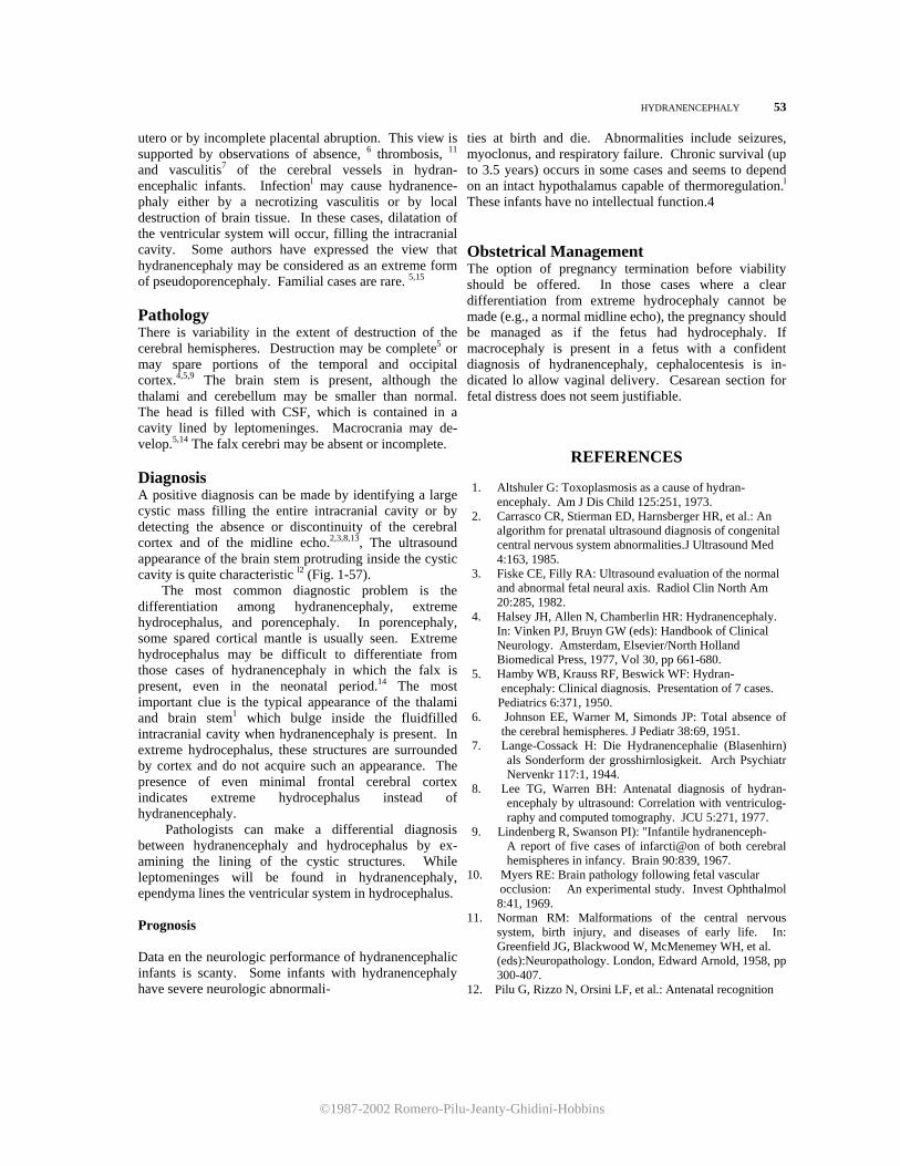
©1987-2002 Romero-Pilu-Jeanty-Ghidini-Hobbins
HYDRANENCEPHALY 53 utero or by incomplete placental abruption. This view is supported by observations of absence, 6 thrombosis, 11
and vasculitis7 of the cerebral vessels in hydran-encephalic infants. Infectionl may cause hydranence-phaly either by a necrotizing vasculitis or by local destruction of brain tissue. In these cases, dilatation of the ventricular system will occur, filling the intracranial cavity. Some authors have expressed the view that hydranencephaly may be considered as an extreme form of pseudoporencephaly. Familial cases are rare. 5,15 Pathology There is variability in the extent of destruction of the cerebral hemispheres. Destruction may be complete5 or may spare portions of the temporal and occipital cortex.4,5,9 The brain stem is present, although the thalami and cerebellum may be smaller than normal. The head is filled with CSF, which is contained in a cavity lined by leptomeninges. Macrocrania may de-velop.5,14 The falx cerebri may be absent or incomplete. Diagnosis A positive diagnosis can be made by identifying a large cystic mass filling the entire intracranial cavity or by detecting the absence or discontinuity of the cerebral cortex and of the midline echo.2,3,8,13, The ultrasound appearance of the brain stem protruding inside the cystic cavity is quite characteristic l2 (Fig. 1-57). The most common diagnostic problem is the differentiation among hydranencephaly, extreme hydrocephalus, and porencephaly. In porencephaly, some spared cortical mantle is usually seen. Extreme hydrocephalus may be difficult to differentiate from those cases of hydranencephaly in which the falx is present, even in the neonatal period.14 The most important clue is the typical appearance of the thalami and brain stem1 which bulge inside the fluidfilled intracranial cavity when hydranencephaly is present. In extreme hydrocephalus, these structures are surrounded by cortex and do not acquire such an appearance. The presence of even minimal frontal cerebral cortex indicates extreme hydrocephalus instead of hydranencephaly. Pathologists can make a differential diagnosis between hydranencephaly and hydrocephalus by ex-amining the lining of the cystic structures. While leptomeninges will be found in hydranencephaly, ependyma lines the ventricular system in hydrocephalus. Prognosis Data en the neurologic performance of hydranencephalic infants is scanty. Some infants with hydranencephaly have severe neurologic abnormali-
ties at birth and die. Abnormalities include seizures, myoclonus, and respiratory failure. Chronic survival (up to 3.5 years) occurs in some cases and seems to depend on an intact hypothalamus capable of thermoregulation.l These infants have no intellectual function.4 Obstetrical Management The option of pregnancy termination before viability should be offered. In those cases where a clear differentiation from extreme hydrocephaly cannot be made (e.g., a normal midline echo), the pregnancy should be managed as if the fetus had hydrocephaly. If macrocephaly is present in a fetus with a confident diagnosis of hydranencephaly, cephalocentesis is in-dicated lo allow vaginal delivery. Cesarean section for fetal distress does not seem justifiable.
REFERENCES 1. Altshuler G: Toxoplasmosis as a cause of hydran-
encephaly. Am J Dis Child 125:251, 1973. 2. Carrasco CR, Stierman ED, Harnsberger HR, et al.: An
algorithm for prenatal ultrasound diagnosis of congenital central nervous system abnormalities.J Ultrasound Med 4:163, 1985.
3. Fiske CE, Filly RA: Ultrasound evaluation of the normal and abnormal fetal neural axis. Radiol Clin North Am 20:285, 1982.
4. Halsey JH, Allen N, Chamberlin HR: Hydranencephaly. In: Vinken PJ, Bruyn GW (eds): Handbook of Clinical Neurology. Amsterdam, Elsevier/North Holland Biomedical Press, 1977, Vol 30, pp 661-680.
5. Hamby WB, Krauss RF, Beswick WF: Hydran- encephaly: Clinical diagnosis. Presentation of 7 cases.
Pediatrics 6:371, 1950. 6. Johnson EE, Warner M, Simonds JP: Total absence of
the cerebral hemispheres. J Pediatr 38:69, 1951. 7. Lange-Cossack H: Die Hydranencephalie (Blasenhirn)
als Sonderform der grosshirnlosigkeit. Arch Psychiatr Nervenkr 117:1, 1944.
8. Lee TG, Warren BH: Antenatal diagnosis of hydran-encephaly by ultrasound: Correlation with ventriculog-raphy and computed tomography. JCU 5:271, 1977.
9. Lindenberg R, Swanson PI): "Infantile hydranenceph- A report of five cases of infarcti@on of both cerebral hemispheres in infancy. Brain 90:839, 1967.
10. Myers RE: Brain pathology following fetal vascular occlusion: An experimental study. Invest Ophthalmol 8:41, 1969.
11. Norman RM: Malformations of the central nervous system, birth injury, and diseases of early life. In: Greenfield JG, Blackwood W, McMenemey WH, et al. (eds):Neuropathology. London, Edward Arnold, 1958, pp 300-407.
12. Pilu G, Rizzo N, Orsini LF, et al.: Antenatal recognition
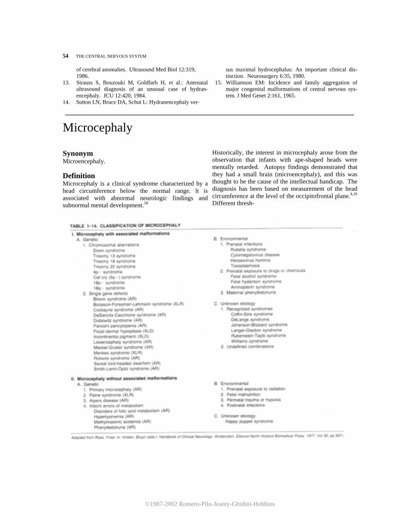
©1987-2002 Romero-Pilu-Jeanty-Ghidini-Hobbins
54 THE CENTRAL NERVOUS SYSTEM
of cerebral anomalies. Ultrasound Med Biol 12:319, 1986.
13. Strauss S, Bouzouki M, Goldfarb H, et al.: Antenatal ultrasound diagnosis of an unusual case of hydran-encephaly. JCU 12:420, 1984.
14. Sutton LN, Bruce DA, Schut L: Hydranencephaly ver-
sus maximal hydrocephalus: An important clinical dis-tinction. Neurosurgery 6:35, 1980.
15. Williamson EM: Incidence and family aggregation of major congenital malformations of central nervous sys-tem. J Med Genet 2:161, 1965.
Microcephaly Synonym Microencephaly.
Definition Microcephaly is a clinical syndrome characterized by a head circumference below the normal range. lt is associated with abnormal neurologic findings and subnormal mental development.18
Historically, the interest in microcephaly arose from the observation that infants with ape-shaped heads were mentally retarded. Autopsy findings demonstrated that they had a small brain (microencephaly), and this was thought to be the cause of the intellectual handicap. The diagnosis has been based on measurement of the head circumference at the level of the occipitofrontal plane.4,16 Different thresh-
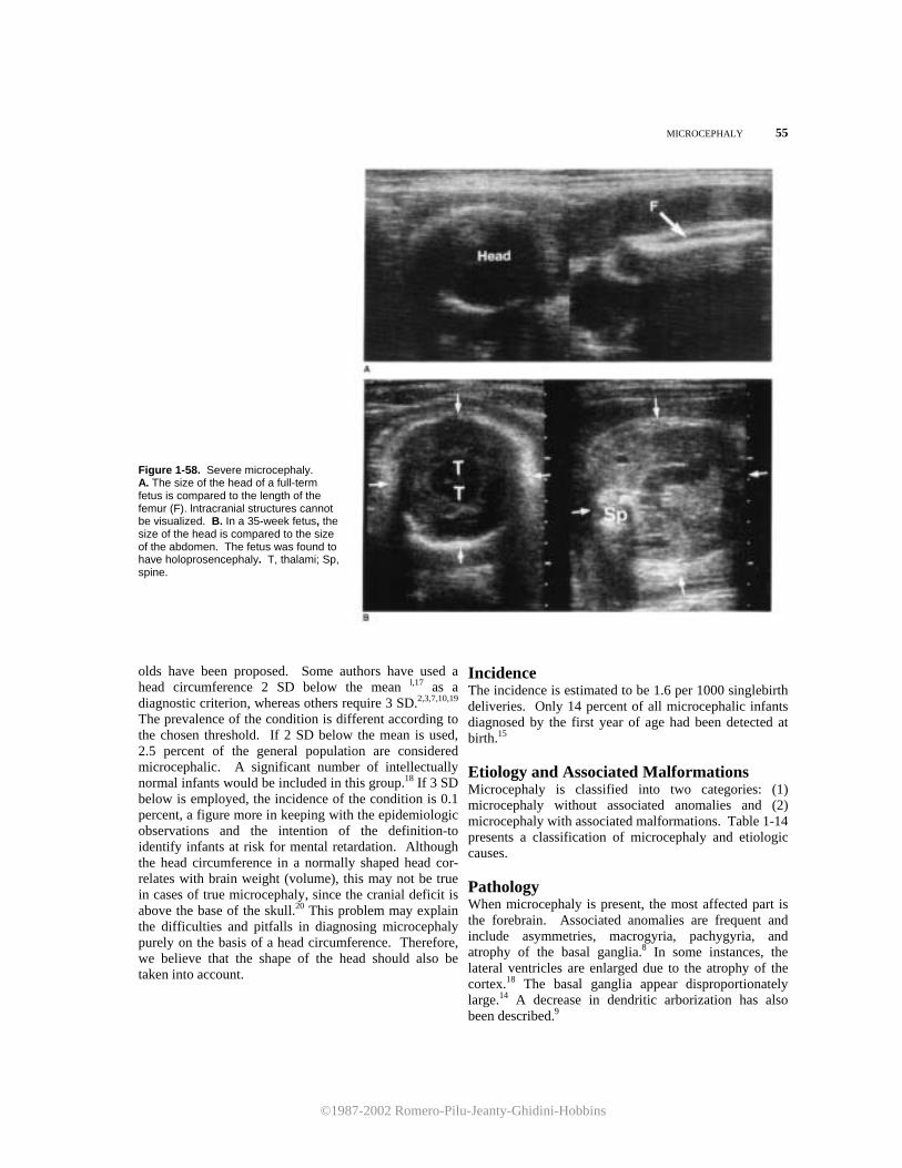
©1987-2002 Romero-Pilu-Jeanty-Ghidini-Hobbins
MICROCEPHALY 55 Figure 1-58. Severe microcephaly. A. The size of the head of a full-term fetus is compared to the length of the femur (F). lntracranial structures cannot be visualized. B. In a 35-week fetus, the size of the head is compared to the size of the abdomen. The fetus was found to have holoprosencephaly. T, thalami; Sp, spine.
olds have been proposed. Some authors have used a head circumference 2 SD below the mean l,17 as a diagnostic criterion, whereas others require 3 SD.2,3,7,10,19 The prevalence of the condition is different according to the chosen threshold. If 2 SD below the mean is used, 2.5 percent of the general population are considered microcephalic. A significant number of intellectually normal infants would be included in this group.18 If 3 SD below is employed, the incidence of the condition is 0.1 percent, a figure more in keeping with the epidemiologic observations and the intention of the definition-to identify infants at risk for mental retardation. Although the head circumference in a normally shaped head cor-relates with brain weight (volume), this may not be true in cases of true microcephaly, since the cranial deficit is above the base of the skull.20 This problem may explain the difficulties and pitfalls in diagnosing microcephaly purely on the basis of a head circumference. Therefore, we believe that the shape of the head should also be taken into account.
Incidence The incidence is estimated to be 1.6 per 1000 singlebirth deliveries. Only 14 percent of all microcephalic infants diagnosed by the first year of age had been detected at birth.15 Etiology and Associated Malformations Microcephaly is classified into two categories: (1) microcephaly without associated anomalies and (2) microcephaly with associated malformations. Table 1-14 presents a classification of microcephaly and etiologic causes. Pathology When microcephaly is present, the most affected part is the forebrain. Associated anomalies are frequent and include asymmetries, macrogyria, pachygyria, and atrophy of the basal ganglia.8 In some instances, the lateral ventricles are enlarged due to the atrophy of the cortex.18 The basal ganglia appear disproportionately large.14 A decrease in dendritic arborization has also been described.9
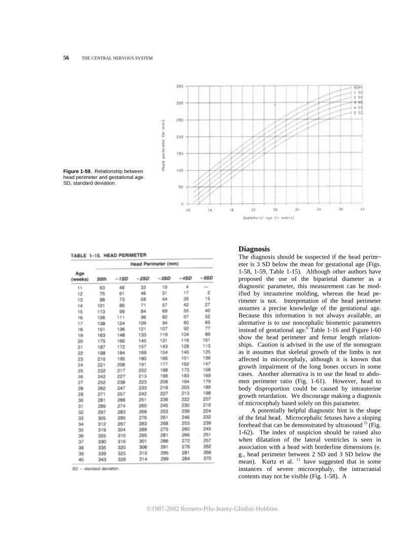
©1987-2002 Romero-Pilu-Jeanty-Ghidini-Hobbins
56 THE CENTRAL NERVOUS SYSTEM Figure 1-59. Relationship between head perimeter and gestational age. SD, standard deviation.
Diagnosis The diagnosis should be suspected if the head perim~ eter is 3 SD below the mean for gestational age (Figs. 1-58, 1-59, Table 1-15). Although other authors have proposed the use of the biparietal diameter as a diagnostic parameter, this measurement can be mod-ified by intrauterine molding, whereas the head pe-rimeter is not. Interpretation of the head perimeter assumes a precise knowledge of the gestational age. Because this information is not always available, an altemative is to use noncephalic biometric parameters instead of gestational age.6 Table 1-16 and Figure l-60 show the head perimeter and femur length relation-ships. Caution is advised in the use of the nomogram as it assumes that skeletal growth of the limbs is not affected in microcephaly, although it is known that growth impairment of the long bones occurs in some cases. Another alternativa is to use the head to abdo-men perimeter ratio (Fig. 1-61). However, head to body disproportion could be caused by intrauterine growth retardation. We discourage making a diagnosis of microcephaly based solely on this parameter. A potentially helpful diagnostic hint is the shape of the fetal head. Microcephalic fetuses have a sloping forehead that can be demonstrated by ultrasound l3 (Fig. 1-62). The index of suspicion should be raised also when dilatation of the lateral ventricles is seen in association with a head with borderline dimensions (e. g., head perimeter between 2 SD and 3 SD below the mean). Kurtz et al. 11 have suggested that in some instances of severe microcephaly, the intracranial contents may not be visible (Fig. 1-58). A
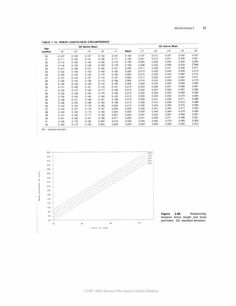
©1987-2002 Romero-Pilu-Jeanty-Ghidini-Hobbins
MICROCEPHALY 57
Figure 1-60. Relationship between femur length and head perimeter. SD, standard deviation.
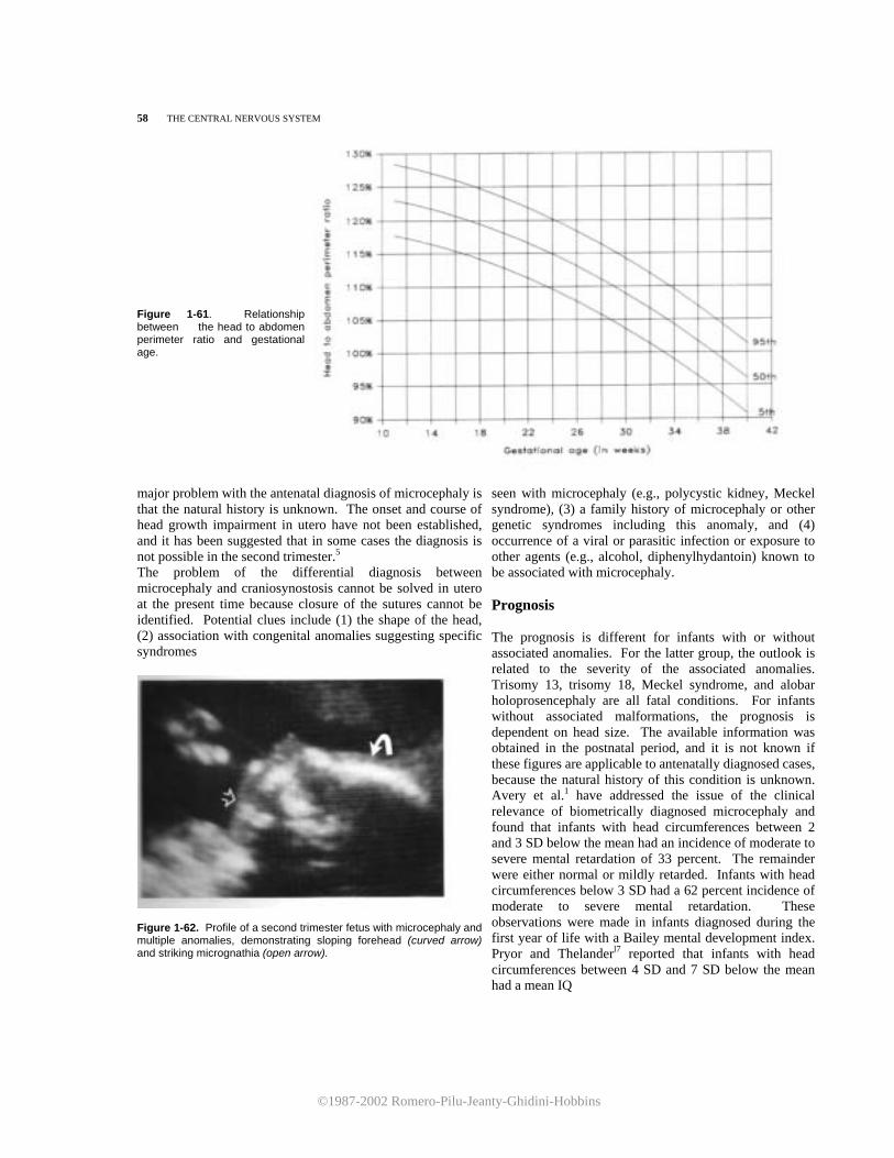
©1987-2002 Romero-Pilu-Jeanty-Ghidini-Hobbins
58 THE CENTRAL NERVOUS SYSTEM Figure 1-61. Relationship between the head to abdomen perimeter ratio and gestational age.
major problem with the antenatal diagnosis of microcephaly is that the natural history is unknown. The onset and course of head growth impairment in utero have not been established, and it has been suggested that in some cases the diagnosis is not possible in the second trimester.5 The problem of the differential diagnosis between microcephaly and craniosynostosis cannot be solved in utero at the present time because closure of the sutures cannot be identified. Potential clues include (1) the shape of the head, (2) association with congenital anomalies suggesting specific syndromes
Figure 1-62. Profile of a second trimester fetus with microcephaly and multiple anomalies, demonstrating sloping forehead (curved arrow) and striking micrognathia (open arrow).
seen with microcephaly (e.g., polycystic kidney, Meckel syndrome), (3) a family history of microcephaly or other genetic syndromes including this anomaly, and (4) occurrence of a viral or parasitic infection or exposure to other agents (e.g., alcohol, diphenylhydantoin) known to be associated with microcephaly. Prognosis The prognosis is different for infants with or without associated anomalies. For the latter group, the outlook is related to the severity of the associated anomalies. Trisomy 13, trisomy 18, Meckel syndrome, and alobar holoprosencephaly are all fatal conditions. For infants without associated malformations, the prognosis is dependent on head size. The available information was obtained in the postnatal period, and it is not known if these figures are applicable to antenatally diagnosed cases, because the natural history of this condition is unknown. Avery et al.1 have addressed the issue of the clinical relevance of biometrically diagnosed microcephaly and found that infants with head circumferences between 2 and 3 SD below the mean had an incidence of moderate to severe mental retardation of 33 percent. The remainder were either normal or mildly retarded. Infants with head circumferences below 3 SD had a 62 percent incidence of moderate to severe mental retardation. These observations were made in infants diagnosed during the first year of life with a Bailey mental development index. Pryor and Thelanderl7 reported that infants with head circumferences between 4 SD and 7 SD below the mean had a mean IQ
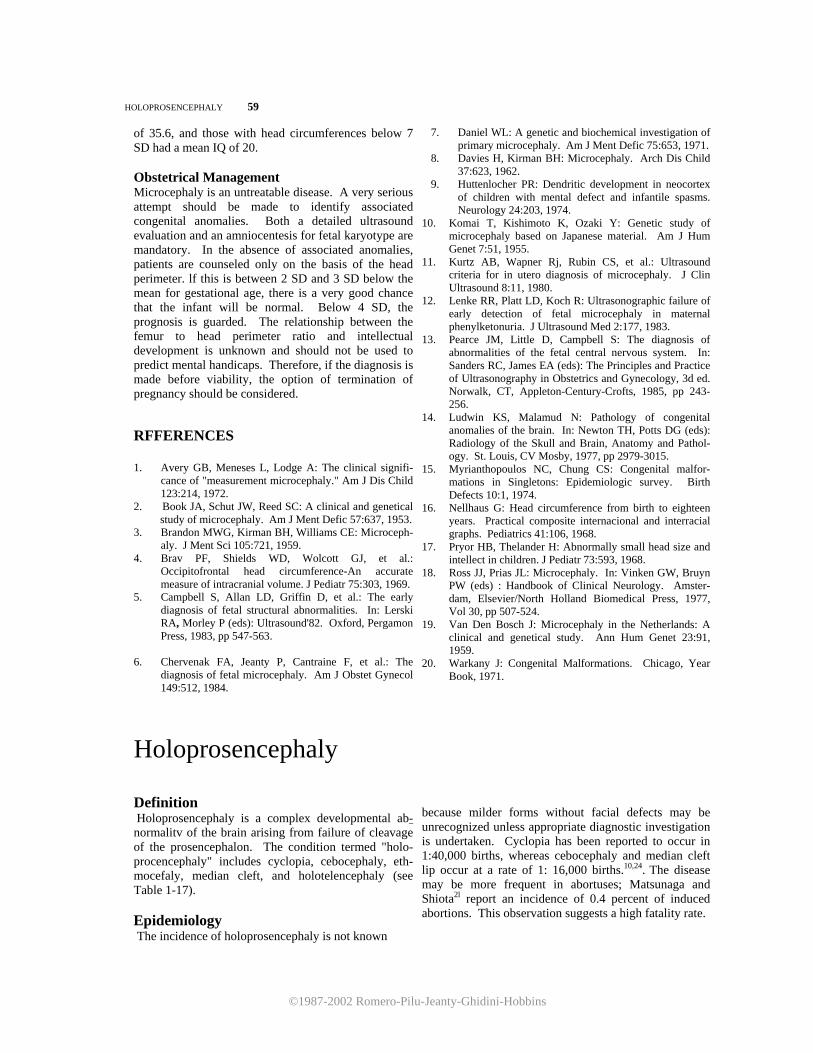
©1987-2002 Romero-Pilu-Jeanty-Ghidini-Hobbins
HOLOPROSENCEPHALY 59
of 35.6, and those with head circumferences below 7 SD had a mean IQ of 20.
Obstetrical Management Microcephaly is an untreatable disease. A very serious attempt should be made to identify associated congenital anomalies. Both a detailed ultrasound evaluation and an amniocentesis for fetal karyotype are mandatory. In the absence of associated anomalies, patients are counseled only on the basis of the head perimeter. lf this is between 2 SD and 3 SD below the mean for gestational age, there is a very good chance that the infant will be normal. Below 4 SD, the prognosis is guarded. The relationship between the femur to head perimeter ratio and intellectual development is unknown and should not be used to predict mental handicaps. Therefore, if the diagnosis is made before viability, the option of termination of pregnancy should be considered. RFFERENCES
1. Avery GB, Meneses L, Lodge A: The clinical signifi-
cance of "measurement microcephaly." Am J Dis Child 123:214, 1972.
2. Book JA, Schut JW, Reed SC: A clinical and genetical study of microcephaly. Am J Ment Defic 57:637, 1953.
3. Brandon MWG, Kirman BH, Williams CE: Microceph-aly. J Ment Sci 105:721, 1959.
4. Brav PF, Shields WD, Wolcott GJ, et al.: Occipitofrontal head circumference-An accurate measure of intracranial volume. J Pediatr 75:303, 1969.
5. Campbell S, Allan LD, Griffin D, et al.: The early diagnosis of fetal structural abnormalities. In: Lerski RA, Morley P (eds): Ultrasound'82. Oxford, Pergamon Press, 1983, pp 547-563.
6. Chervenak FA, Jeanty P, Cantraine F, et al.: The
diagnosis of fetal microcephaly. Am J Obstet Gynecol 149:512, 1984.
7. Daniel WL: A genetic and biochemical investigation of primary microcephaly. Am J Ment Defic 75:653, 1971.
8. Davies H, Kirman BH: Microcephaly. Arch Dis Child 37:623, 1962.
9. Huttenlocher PR: Dendritic development in neocortex of children with mental defect and infantile spasms. Neurology 24:203, 1974.
10. Komai T, Kishimoto K, Ozaki Y: Genetic study of microcephaly based on Japanese material. Am J Hum Genet 7:51, 1955.
11. Kurtz AB, Wapner Rj, Rubin CS, et al.: Ultrasound criteria for in utero diagnosis of microcephaly. J Clin Ultrasound 8:11, 1980.
12. Lenke RR, Platt LD, Koch R: Ultrasonographic failure of early detection of fetal microcephaly in maternal phenylketonuria. J Ultrasound Med 2:177, 1983.
13. Pearce JM, Little D, Campbell S: The diagnosis of abnormalities of the fetal central nervous system. In: Sanders RC, James EA (eds): The Principles and Practice of Ultrasonography in Obstetrics and Gynecology, 3d ed. Norwalk, CT, Appleton-Century-Crofts, 1985, pp 243-256.
14. Ludwin KS, Malamud N: Pathology of congenital anomalies of the brain. In: Newton TH, Potts DG (eds): Radiology of the Skull and Brain, Anatomy and Pathol-ogy. St. Louis, CV Mosby, 1977, pp 2979-3015.
15. Myrianthopoulos NC, Chung CS: Congenital malfor-mations in Singletons: Epidemiologic survey. Birth Defects 10:1, 1974.
16. Nellhaus G: Head circumference from birth to eighteen years. Practical composite internacional and interracial graphs. Pediatrics 41:106, 1968.
17. Pryor HB, Thelander H: Abnormally small head size and intellect in children. J Pediatr 73:593, 1968.
18. Ross JJ, Prias JL: Microcephaly. In: Vinken GW, Bruyn PW (eds) : Handbook of Clinical Neurology. Amster-dam, Elsevier/North Holland Biomedical Press, 1977, Vol 30, pp 507-524.
19. Van Den Bosch J: Microcephaly in the Netherlands: A clinical and genetical study. Ann Hum Genet 23:91, 1959.
20. Warkany J: Congenital Malformations. Chicago, Year Book, 1971.
Holoprosencephaly
Definition Holoprosencephaly is a complex developmental ab-normalitv of the brain arising from failure of cleavage of the prosencephalon. The condition termed "holo-procencephaly" includes cyclopia, cebocephaly, eth-mocefaly, median cleft, and holotelencephaly (see Table 1-17).
Epidemiology
The incidence of holoprosencephaly is not known
because milder forms without facial defects may be unrecognized unless appropriate diagnostic investigation is undertaken. Cyclopia has been reported to occur in 1:40,000 births, whereas cebocephaly and median cleft lip occur at a rate of 1: 16,000 births.10,24. The disease may be more frequent in abortuses; Matsunaga and Shiota2l report an incidence of 0.4 percent of induced abortions. This observation suggests a high fatality rate.
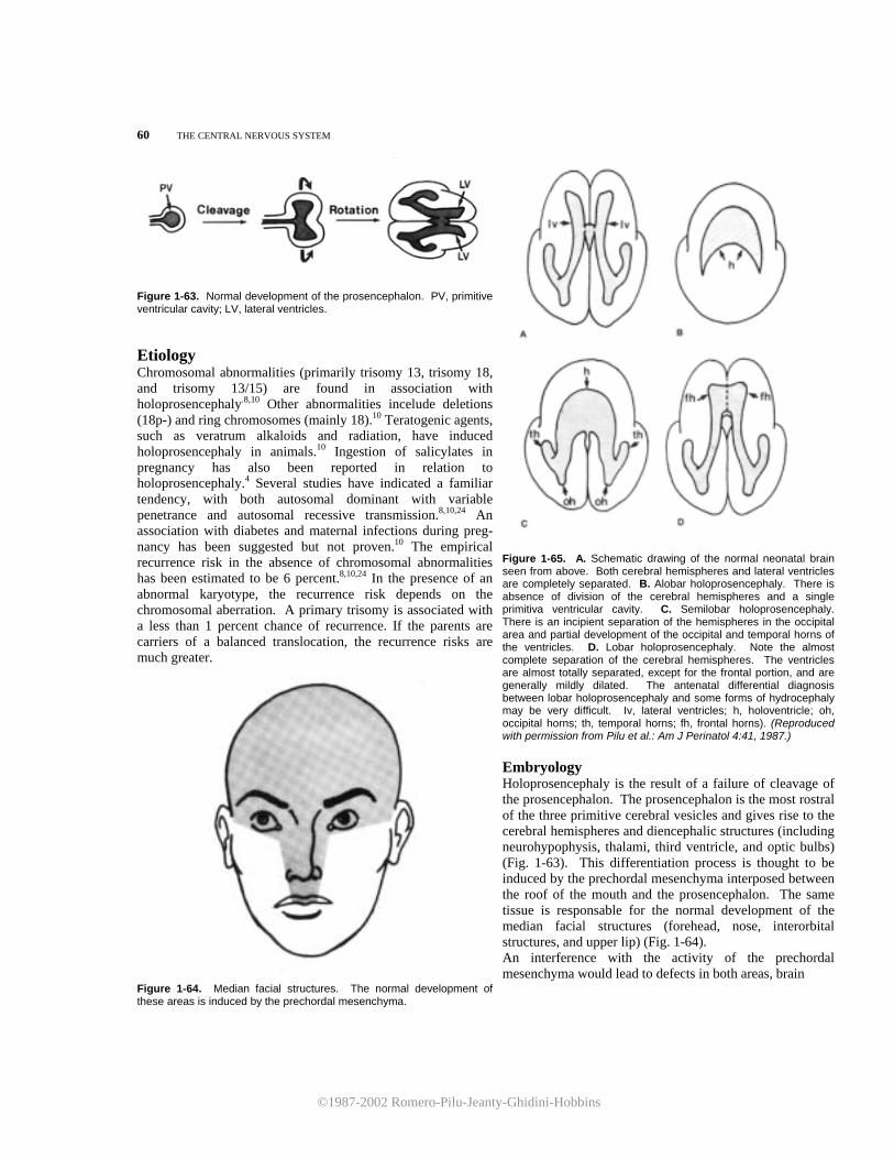
©1987-2002 Romero-Pilu-Jeanty-Ghidini-Hobbins
60 THE CENTRAL NERVOUS SYSTEM
Figure 1-63. Normal development of the prosencephalon. PV, primitive ventricular cavity; LV, lateral ventricles.
Etiology Chromosomal abnormalities (primarily trisomy 13, trisomy 18, and trisomy 13/15) are found in association with holoprosencephaly.8,10 Other abnormalities incelude deletions (18p-) and ring chromosomes (mainly 18).10 Teratogenic agents, such as veratrum alkaloids and radiation, have induced holoprosencephaly in animals.10 Ingestion of salicylates in pregnancy has also been reported in relation to holoprosencephaly.4 Several studies have indicated a familiar tendency, with both autosomal dominant with variable penetrance and autosomal recessive transmission.8,10,24 An association with diabetes and maternal infections during preg-nancy has been suggested but not proven.10 The empirical recurrence risk in the absence of chromosomal abnormalities has been estimated to be 6 percent.8,10,24 In the presence of an abnormal karyotype, the recurrence risk depends on the chromosomal aberration. A primary trisomy is associated with a less than 1 percent chance of recurrence. If the parents are carriers of a balanced translocation, the recurrence risks are much greater.
Figure 1-65. A. Schematic drawing of the normal neonatal brain seen from above. Both cerebral hemispheres and lateral ventricles are completely separated. B. Alobar holoprosencephaly. There is absence of division of the cerebral hemispheres and a single primitiva ventricular cavity. C. Semilobar holoprosencephaly. There is an incipient separation of the hemispheres in the occipital area and partial development of the occipital and temporal horns of the ventricles. D. Lobar holoprosencephaly. Note the almost complete separation of the cerebral hemispheres. The ventricles are almost totally separated, except for the frontal portion, and are generally mildly dilated. The antenatal differential diagnosis between lobar holoprosencephaly and some forms of hydrocephaly may be very difficult. Iv, lateral ventricles; h, holoventricle; oh, occipital horns; th, temporal horns; fh, frontal horns). (Reproduced with permission from Pilu et al.: Am J Perinatol 4:41, 1987.)
Figure 1-64. Median facial structures. The normal development of these areas is induced by the prechordal mesenchyma.
Embryology Holoprosencephaly is the result of a failure of cleavage of the prosencephalon. The prosencephalon is the most rostral of the three primitive cerebral vesicles and gives rise to the cerebral hemispheres and diencephalic structures (including neurohypophysis, thalami, third ventricle, and optic bulbs) (Fig. 1-63). This differentiation process is thought to be induced by the prechordal mesenchyma interposed between the roof of the mouth and the prosencephalon. The same tissue is responsable for the normal development of the median facial structures (forehead, nose, interorbital structures, and upper lip) (Fig. 1-64). An interference with the activity of the prechordal mesenchyma would lead to defects in both areas, brain
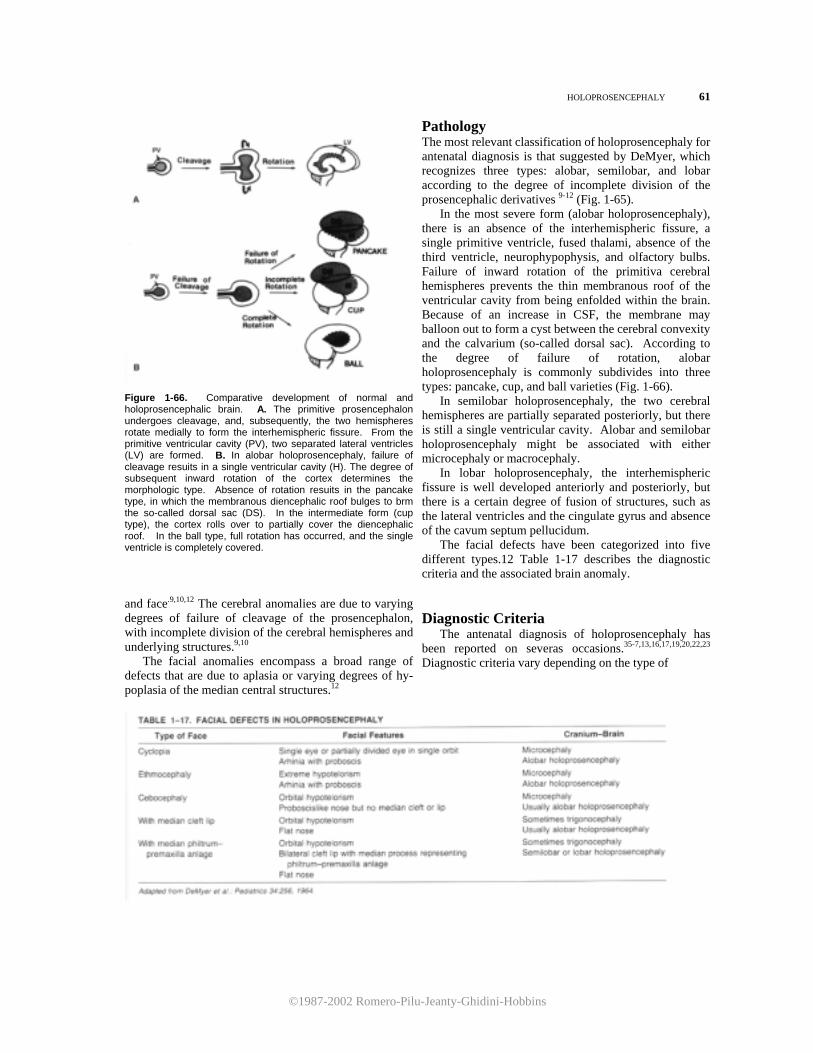
©1987-2002 Romero-Pilu-Jeanty-Ghidini-Hobbins
HOLOPROSENCEPHALY 61
Figure 1-66. Comparative development of normal and holoprosencephalic brain. A. The primitive prosencephalon undergoes cleavage, and, subsequently, the two hemispheres rotate medially to form the interhemispheric fissure. From the primitive ventricular cavity (PV), two separated lateral ventricles (LV) are formed. B. In alobar holoprosencephaly, failure of cleavage resuits in a single ventricular cavity (H). The degree of subsequent inward rotation of the cortex determines the morphologic type. Absence of rotation resuits in the pancake type, in which the membranous diencephalic roof bulges to brm the so-called dorsal sac (DS). In the intermediate form (cup type), the cortex rolls over to partially cover the diencephalic roof. In the ball type, full rotation has occurred, and the single ventricle is completely covered. and face.9,10,12 The cerebral anomalies are due to varying degrees of failure of cleavage of the prosencephalon, with incomplete division of the cerebral hemispheres and underlying structures.9,10
The facial anomalies encompass a broad range of defects that are due to aplasia or varying degrees of hy-poplasia of the median central structures.12
Pathology The most relevant classification of holoprosencephaly for antenatal diagnosis is that suggested by DeMyer, which recognizes three types: alobar, semilobar, and lobar according to the degree of incomplete division of the prosencephalic derivatives 9-12 (Fig. 1-65).
In the most severe form (alobar holoprosencephaly), there is an absence of the interhemispheric fissure, a single primitive ventricle, fused thalami, absence of the third ventricle, neurophypophysis, and olfactory bulbs. Failure of inward rotation of the primitiva cerebral hemispheres prevents the thin membranous roof of the ventricular cavity from being enfolded within the brain. Because of an increase in CSF, the membrane may balloon out to form a cyst between the cerebral convexity and the calvarium (so-called dorsal sac). According to the degree of failure of rotation, alobar holoprosencephaly is commonly subdivides into three types: pancake, cup, and ball varieties (Fig. 1-66).
In semilobar holoprosencephaly, the two cerebral hemispheres are partially separated posteriorly, but there is still a single ventricular cavity. Alobar and semilobar holoprosencephaly might be associated with either microcephaly or macrocephaly.
In lobar holoprosencephaly, the interhemispheric fissure is well developed anteriorly and posteriorly, but there is a certain degree of fusion of structures, such as the lateral ventricles and the cingulate gyrus and absence of the cavum septum pellucidum.
The facial defects have been categorized into five different types.12 Table 1-17 describes the diagnostic criteria and the associated brain anomaly. Diagnostic Criteria
The antenatal diagnosis of holoprosencephaly has been reported on severas occasions.35-7,13,16,17,19,20,22,23 Diagnostic criteria vary depending on the type of

©1987-2002 Romero-Pilu-Jeanty-Ghidini-Hobbins
62 THE CENTRAL NERVOUS SYSTEM
Figure 1-67. Axial scan in a fetus with alobar holoprosen-cephaly,revealing the sickle-shaped holoventricle (H) lined anteriorly by the undivided cortex and posteriorly by the prominent uncleft thalami (T). Both the midline echo and the third ventricle are absent. P, cerebral peduncles. (Reproduced with permission from Pilu et al: Am J Perinatol 4:41, 1987)
Figure 1-69. Midcoronal scan in a hoIoprosencephalic fetus. The cortex (arrows) is only partially enfolded over the holoventricle (H), which amply communicates with the superior dorsal sac (DS). Note the uncleft thalami (T) on the floor of the ventricular cavity. Sup, superior; lnf, inferior. (Reproduced with permission from Pilu et al: Am J Perinatol 4:41, 1987.)
Figure 1-68. Axial scan at the level of the large dorsal sac (DS) in a fetus with alobar holoprosencephaly, cup variety. Note the crescent-shaped cortex and the absence of the midline echo. H, holoventricle. (Reproduced with permission from Pilu et al: Am J Perinatol 4.-41, 1987.)
Figure 1-70. Axial scans at the level of the orbits in a holoprosencephalic fetus, revealing hypotelorism and absence of the nasal bridge. (Reproduced with permission from Pilu et al.: Am J Perinatol 4:41, 1987.)
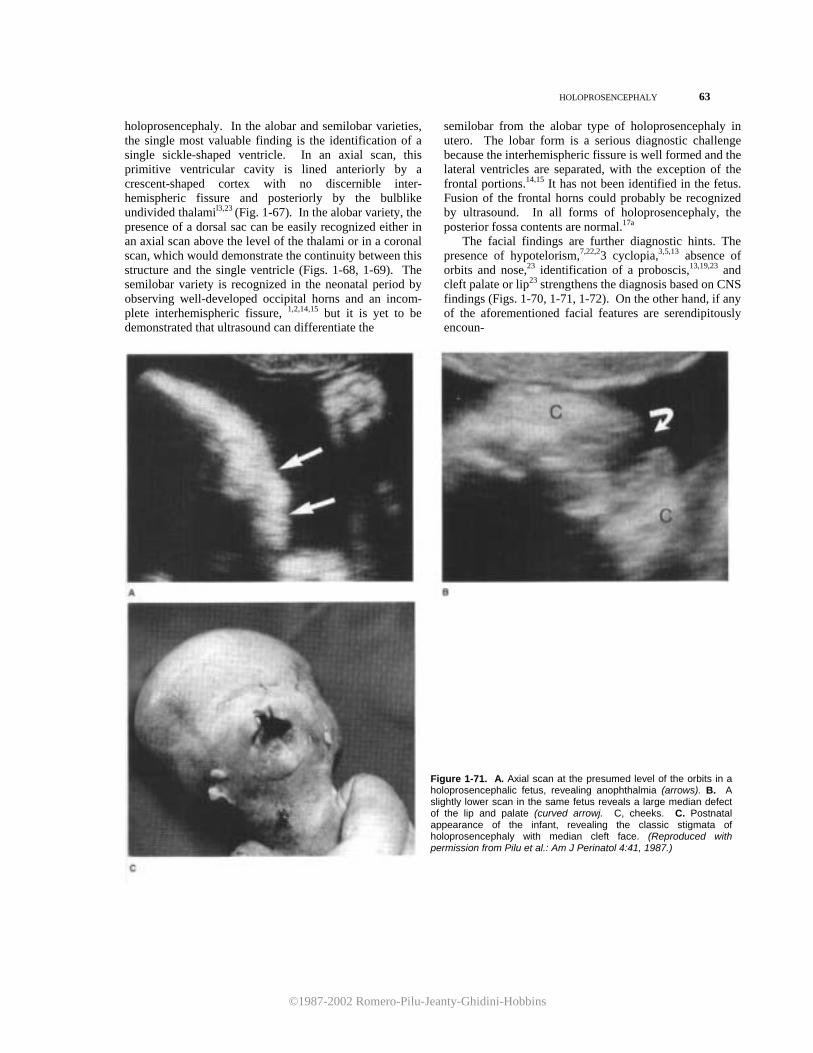
©1987-2002 Romero-Pilu-Jeanty-Ghidini-Hobbins
HOLOPROSENCEPHALY 63
holoprosencephaly. In the alobar and semilobar varieties, the single most valuable finding is the identification of a single sickle-shaped ventricle. In an axial scan, this primitive ventricular cavity is lined anteriorly by a crescent-shaped cortex with no discernible inter-hemispheric fissure and posteriorly by the bulblike undivided thalamil3,23 (Fig. 1-67). In the alobar variety, the presence of a dorsal sac can be easily recognized either in an axial scan above the level of the thalami or in a coronal scan, which would demonstrate the continuity between this structure and the single ventricle (Figs. 1-68, 1-69). The semilobar variety is recognized in the neonatal period by observing well-developed occipital horns and an incom-plete interhemispheric fissure, 1,2,14,15 but it is yet to be demonstrated that ultrasound can differentiate the
semilobar from the alobar type of holoprosencephaly in utero. The lobar form is a serious diagnostic challenge because the interhemispheric fissure is well formed and the lateral ventricles are separated, with the exception of the frontal portions.14,15 It has not been identified in the fetus. Fusion of the frontal horns could probably be recognized by ultrasound. In all forms of holoprosencephaly, the posterior fossa contents are normal.17a The facial findings are further diagnostic hints. The presence of hypotelorism,7,22,23 cyclopia,3,5,13 absence of orbits and nose,23 identification of a proboscis,13,19,23 and cleft palate or lip23 strengthens the diagnosis based on CNS findings (Figs. 1-70, 1-71, 1-72). On the other hand, if any of the aforementioned facial features are serendipitously encoun-
Figure 1-71. A. Axial scan at the presumed level of the orbits in a holoprosencephalic fetus, revealing anophthalmia (arrows). B. A slightly lower scan in the same fetus reveals a large median defect of the lip and palate (curved arrowj. C, cheeks. C. Postnatal appearance of the infant, revealing the classic stigmata of holoprosencephaly with median cleft face. (Reproduced with permission from Pilu et al.: Am J Perinatol 4:41, 1987.)
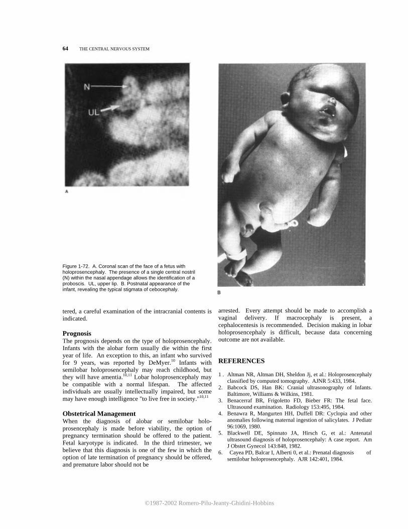
©1987-2002 Romero-Pilu-Jeanty-Ghidini-Hobbins
64 THE CENTRAL NERVOUS SYSTEM
Figure 1-72. A. Coronal scan of the face of a fetus with holoprosencephaly. The presence of a single central nostril (N) within the nasal appendage allows the identification of a proboscis. UL, upper lip. B. Postnatal appearance of the infant, revealing the typical stigmata of cebocephaly.
tered, a careful examination of the intracranial contents is indicated. Prognosis The prognosis depends on the type of holoprosencephaly. Infants with the alobar form usually die within the first year of life. An exception to this, an infant who survived for 9 years, was reported by DeMyer.10 Infants with semilobar holoprosencephaly may reach childhood, but they will have amentia.10,11 Lobar holoprosencephaly may be compatible with a normal lifespan. The affected individuals are usually intellectually impaired, but some may have enough intelligence "to live free in society."10,11 Obstetrical Management When the diagnosis of alobar or semilobar holo-prosencephaly is made before viability, the option of pregnancy termination should be offered to the patient. Fetal karyotype is indicated. In the third trimester, we believe that this diagnosis is one of the few in which the option of late termination of pregnancy should be offered, and premature labor should not be
arrested. Every attempt should be made to accomplish a vaginal delivery. If macrocephaly is present, a cephalocentesis is recommended. Decision making in lobar holoprosencephaly is difficult, because data concerning outcome are not available. REFERENCES 1 . Altman NR, Altman DH, Sheldon Jj, et al.: Holoprosencephaly
classified by computed tomography. AJNR 5:433, 1984. 2. Babcock DS, Han BK: Cranial ultrasonography of Infants.
Baltimore, Williams & Wilkins, 1981. 3. Benacerraf BR, Frigoletto FD, Bieber FR: The fetal face.
Ultrasound examination. Radiology 153:495, 1984. 4. Benawra R, Mangurten HH, Duffell DR: Cyclopia and other
anomalies following maternal ingestion of salicylates. J Pediatr 96:1069, 1980.
5. Blackwell DE, Spinnato JA, Hirsch G, et al.: Antenatal ultrasound diagnosis of holoprosencephaly: A case report. Am J Obstet Gynecol 143:848, 1982.
6. Cayea PD, Balcar I, Alberti 0, et al.: Prenatal diagnosis of semilobar holoprosencephaly. AJR 142:401, 1984.

©1987-2002 Romero-Pilu-Jeanty-Ghidini-Hobbins
INIENCEPHAI,Y 65 7. Chervenak FA, Isaacson G, Mahoney Mj, et al.: The
obstetric significance of holoprosencephaly. Obstet Gynecol 63:115, 1984.
8. Cohen MM: An update on the holoprosencephalic disorders. J. Pediatr 101:865, 1982.
9. DeMyer W: Classification of cerebral malformations. Birth Defects 7:78, 1971.
10. DeMyer W: Holoprosencephaly (cyclopia-arhinencephaly). In: Vinken PJ, Bruyn GW (eds): Handbook of Clinical Neurology. Amsterdam, Elsevier/North Holland Biomedical Press, 1977, Vol 30, pp 431-478.
11. DeMyer W, Zeman W: Alobar holoprosencephaly (ar-hinencephaly) with median cleft lip and palate: Clini-cal, electroencephalographic and nosologic consider-ations. Confin Neurol 23:1, 1963.
12. DeMyer W, Zeman W, Palmer CG: The face predicts the brain: Diagnostic significance of median facial anomalies for holoprosencephaloy (arhinencephaly). Pediatrics 34:256, 1964.
13. Filly RA, Chinn DH, Callen PW: Alobar holoprosence- phaly: Ultrasonographic prenatal diagnosis. Radiology 151:455, 1984.
14. Fitz CR: Midline anomalies of the brain and spine. Radiol Clin North Am 29:95, 1982.
15. Fitz CR: Holoprosencephaly and related entities. Neuroradiology 25:225, 1983.
16. Hidalgo H, Bowie J, Rosenberg ER, et al.: In utero sonographic diagnosis of fetal cerebral anomalies. AJR 139:143, 1982.
17. Hill LM, Breckle R, Bonebrake CR: Ultrasonic findings with holoprosencephaly. J Reprod Med 27:172, 1982.
17a.Hoffman-Tretin JC, Horoupian DS, Koenigsberg M, et al.: Lobar holoprosencephaly with hydrocephalus: Antenatal demonstration and differential diagnosis. J Ultrasound Med 5:691, 1986.
18. Kurtz AB, Wapner Rj, Rubin CS, et al.: Ultrasound criteria for in utero diagnosis of microcephaly. JCU 8:11, 1980.
19. Lev-Gur M, Maklad NF, Patel S: Ultrasonic findings in fetal cyclopia. A case report. J Reprod Med 28:554, 1983.
20. Pearce JM, Little DJ, Campbell S: The diagnosis of abnormalities of the fetal central nervous system. In: Sanders RC, james AE (eds): The Principles and Practice of Ultrasonography in Obstetrics and Gynecology, 3d ed. Norwalk, CT, Appleton-Century Crofts, 1985, pp 243-256.
21. Matsunaga E, Shiota K: Holeprosencephaly in human embryos: Epidemiologic studies of 150 cases. Teratol-ogy 16:261, 1977.
22. Mayden KL, Tortora M, Berkowitz RL, et al: Orbital diameters: A new parameter for prenatal diagnosis and
dating. Am J Obstet Gynecol 144:289, 1982. 23. Pilu G, Romero R, Jeanty P, et al.: Criteria for the
antenatal diagnosis of holoprosencephaly. Am Perinatol 4:41, 1987.
24. Roach E, DeMyer W, Conneally PM, et al.: Holopros-encephaly. Birth data, genetie and demographic analyses of 30 families. Birth Defects 11:294, 1975.
Iniencephaly
Definition Iniencephaly is a complex developmental abnormality characterized by an exaggerated lordosis of the spine, usually associated with spina bifida and cephalocele. Epidemiology I is an extremely rare condition. The reported fre-quency has varied from 1:896 in England15 to 1:65,000 in India." Etiology Occurrence in siblings has been observed in only 1 patient of 57.3 Females are more frequently affected than males (M:F ratio = 0.28).14 Iniencephaly has been reported in association with matemal syphilis1,10 and with sedative intake.12 It can be produced in animals by the administration of vinblastine,5 streptonigrin,19 and triparanol.16
Embryology Different hypotheses have been postulated. Persistence of the embryonic cervical lordosis at the third week, leading to failure of closure of the neural tube, or abnormal development of the rostral portion of the notocord and somites of the cervicooccipital region are the most widely accepted theories.14 Pathology The criteria for the diagnosis of iniencephaly are (1) imperfect formation of the base of the skull, particularly at the level of the foramen magnum, (2) rachischisis, and (3) exaggerated lordosis of the spine. The spine is short and grossly abnormal, with kyphoscoliosis. Associated Anomalies Eighty-four percent of iniencephalic infants have other associated anomalies,8 including anencephaly, cephaloceles, hydrocephaly, cyclopia, absence of mandible, cleft lip and palate, cardiovascular anomalies, diaphragmatic hernia, single umbilical artery,
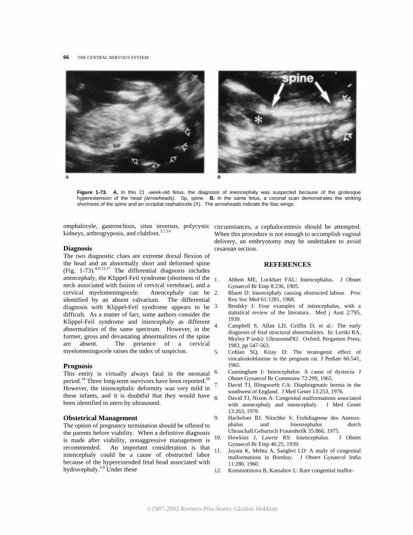
©1987-2002 Romero-Pilu-Jeanty-Ghidini-Hobbins
66 THE CENTRAL NERVOUS SYSTEM
Figure 1-73. A. In this 21 -week-old fetus, the diagnosis of iniencephaly was suspected because of the grotesque hyperextension of the head (arrowheads). Sp, spine. B. In the same fetus, a coronal scan demonstrates the striking shortness of the spine and an occipital cephalocele (X). The arrowheads indicate the iliac wings.
omphalocele, gastroschisis, situs inversus, polycystic kidneys, arthrogryposis, and clubfoot.3,7,14 Diagnosis The two diagnostic clues are extreme dorsal flexion of the head and an abnormally short and deformed spine (Fig. 1-73).4,9,13,17 The differential diagnosis includes anencephaly, the Klippel-Feil syndrome (shortness of the neck associated with fusion of cervical vertebrae), and a cervical myelomeningocele. Anencephaly can be identified by an absent calvarium. The differential diagnosis with Klippel-Feil syndrome appears to be difficult. As a matter of fact, some authors consider the Klippel-Feil syndrome and iniencephaly as different abnormalities of the same spectrum. However, in the former, gross and devastating abnormalities of the spine are absent. The presence of a cervical myelomeningocele raises the index of suspicion. Prognosis This entity is virtually always fatal in the neonatal period.14 Three long-term survivors have been reported.18 However, the iniencephalic deformity was very mild in these infants, and it is doubtful that they would have been identified in utero by ultrasound. Obstetrical Management The option of pregnancy termination should be offered to the parents before viability. When a definitive diagnosis is made after viability, nonaggressive management is recommended. An important consideration is that iniencephaly could be a cause of obstructed labor because of the hyperextended fetal head associated with hydrocephaly.2,6 Under these
circumstances, a cephalocentesis should be attempted. When this procedure is not enough to accomplish vaginal delivery, an embryotomy may be undertaken to avoid cesarean section.
REFERENCES 1 . Abbott ME, Lockhart FAL: Iniencephalus. J Obstet
Gynaecol Br Emp 8:236, 1905. 2. Bluett D: Iniencephaly causing obstructed labour. Proc
Roy Soc Med 61:1281, 1968. 3. Brodsky I: Four examples of iniencephalus, with a
statistical review of the literatura. Med j Aust 2:795, 1939.
4. Campbell S, Allan LD, Griffin D, et al.: The early diagnosis of fetal structural abnormalities. In: Lerski RA, Morley P (eds): Ultrasound'82. Oxford, Pergamon Press, 1983, pp 547-563.
5. Cohlan SQ, Kitay D: The teratogenic effect of vincaleukoblastine in the pregnant rat. J Pediatr 66:541, 1965.
6. Cunningham I: Iniencephalus: A cause of dystocia. J Obstet Gynaecol Br Commonw 72:299, 1965.
7. David TJ, Illingworth CA: Diaphragmatic hernia in the southwest of England. J Med Genet 13:253, 1976.
8. David TJ, Nixon A: Congenital malformations associated with anencephaly and iniencephaly. J Med Genet 13:263, 1976
9. Hackeloer BJ, Nitschke S: Fruhdiagnose des Anenze-phalus und Inienzephalus durch Ultraschall.Geburtsch Frauenheilk 35:866, 1975.
10. Howkins J, Lawrie RS: Iniencephalus. J Obstet Gynaecol Br Emp 46:25, 1939.
11. Jayant K, Mehta A, Sanghvi LD: A study of congenital malformations in Bombay. J Obstet Gynaecol India 11:280, 1960.
12. Konstantinova B, Kassabov L: Rare congenital malfor-
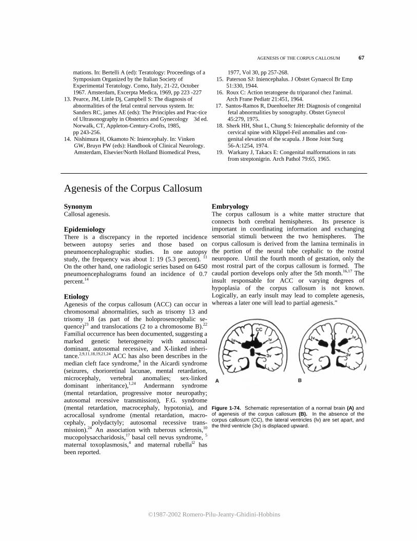
©1987-2002 Romero-Pilu-Jeanty-Ghidini-Hobbins
AGENESIS OF THE CORPUS CALLOSUM 67
mations. In: Bertelli A (ed): Teratology: Proceedings of a Symposium Organized by the Italian Society of Experimental Teratology. Como, Italy, 21-22, October 1967. Amsterdam, Excerpta Medica, 1969, pp 223 -227
13. Pearce, JM, Little Dj, Campbell S: The diagnosis of abnormalities of the fetal central nervous system. In: Sanders RC, james AE (eds): The Principles and Prac-tice of Ultrasonography in Obstetrics and Gynecology 3d ed. Norwalk, CT, Appleton-Century-Crofts, 1985, pp 243-256.
14. Nishimura H, Okamoto N: Iniencephaly. In: Vinken GW, Bruyn PW (eds): Handbook of Clinical Neurology. Amsterdam, Elsevier/North Holland Biomedical Press,
1977, Vol 30, pp 257-268. 15. Paterson SJ: Iniencephalus. J Obstet Gynaecol Br Emp 51:330, 1944. 16. Roux C: Action teratogene du triparanol chez l'animal. Arch Frane Pediatr 21:451, 1964.
17. Santos-Ramos R, Duenhoelter JH: Diagnosis of congenital fetal abnormalities by sonography. Obstet Gynecol 45:279, 1975.
18. Sherk HH, Shut L, Chung S: Iniencephalic deformity of the cervical spine with Klippel-Feil anomalies and con- genital elevation of the scapula. J Bone Joint Surg
56-A:1254, 1974. 19. Warkany J, Takacs E: Congenital malformations in rats
from streptonigrin. Arch Pathol 79:65, 1965.
Agenesis of the Corpus Callosum
Embryology The corpus callosum is a white matter structure that connects both cerebral hemispheres. Its presence is important in coordinating information and exchanging sensorial stimuli between the two hemispheres. The corpus callosum is derived from the lamina terminalis in the portion of the neural tube cephalic to the rostral neuropore. Until the fourth month of gestation, only the most rostral part of the corpus callosum is formed. The caudal portion develops only after the 5th month.16,17 The insult responsable for ACC or varying degrees of hypoplasia of the corpus callosum is not known. Logically, an early insult may lead to complete agenesis, whereas a later one will lead to partial agenesis."
Synonym Callosal agenesis. Epidemiology There is a discrepancy in the reported incidence between autopsy series and those based on pneumoencephalographic studies. In one autopsy study, the frequency was about 1: 19 (5.3 percent). 11 On the other hand, one radiologic series based on 6450 pneumoencephalograms found an incidence of 0.7 percent.14
Etiology Agenesis of the corpus callosum (ACC) can occur in chromosomal abnormalities, such as trisomy 13 and trisomy 18 (as part of the holoprosencephalic se-quence)23 and translocations (2 to a chromosome B).22 Familial occurrence has been documented, suggesting a marked genetic heterogeneity with autosomal dominant, autosomal recessive, and X-linked inheri-tance.2,9,11,18,19,21,24 ACC has also been describes in the median cleft face syndrome,8 in the Aicardi syndrome (seizures, chorioretinal lacunae, mental retardation, microcephaly, vertebral anomalies; sex-linked dominant inheritance),1,24 Andermann syndrome (mental retardation, progressive motor neuropathy; autosomal recessive transmission), F.G. syndrome (mental retardation, macrocephaly, hypotonia), and acrocallosal syndrome (mental retardation, macro-cephaly, polydactyly; autosomal recessive trans-mission).24 An association with tuberous sclerosis,10 mucopolysaccharidosis,17 basal cell nevus syndrome, 5 maternal toxoplasmosis,4 and maternal rubellal2 has been reported.
Figure 1-74. Schematic representation of a normal brain (A) and of agenesis of the corpus callosum (B). In the absence of the corpus callosum (CC), the lateral ventricles (lv) are set apart, and the third ventricle (3v) is displaced upward.
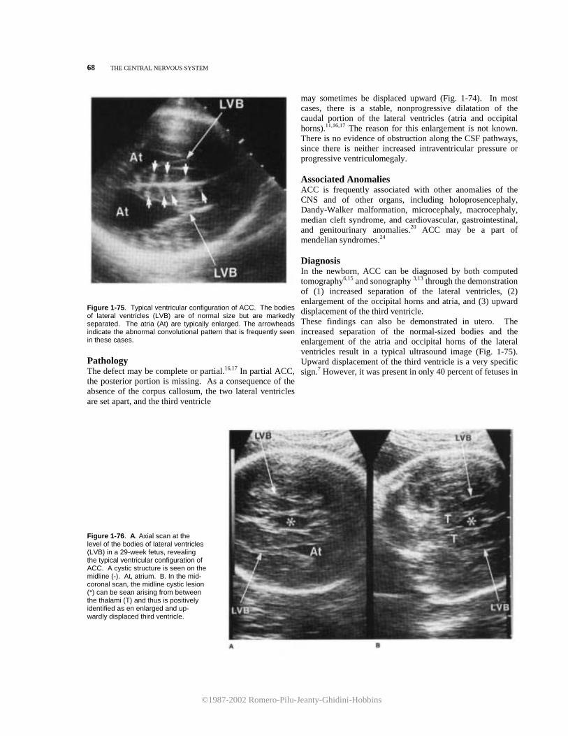
©1987-2002 Romero-Pilu-Jeanty-Ghidini-Hobbins
68 THE CENTRAL NERVOUS SYSTEM
Figure 1-75. Typical ventricular configuration of ACC. The bodies of lateral ventricles (LVB) are of normal size but are markedly separated. The atria (At) are typically enlarged. The arrowheads indicate the abnormal convolutional pattern that is frequently seen in these cases. Pathology The defect may be complete or partial.16,17 In partial ACC, the posterior portion is missing. As a consequence of the absence of the corpus callosum, the two lateral ventricles are set apart, and the third ventricle
may sometimes be displaced upward (Fig. 1-74). In most cases, there is a stable, nonprogressive dilatation of the caudal portion of the lateral ventricles (atria and occipital horns).11,16,17 The reason for this enlargement is not known. There is no evidence of obstruction along the CSF pathways, since there is neither increased intraventricular pressure or progressive ventriculomegaly. Associated Anomalies ACC is frequently associated with other anomalies of the CNS and of other organs, including holoprosencephaly, Dandy-Walker malformation, microcephaly, macrocephaly, median cleft syndrome, and cardiovascular, gastrointestinal, and genitourinary anomalies.20 ACC may be a part of mendelian syndromes.24 Diagnosis In the newborn, ACC can be diagnosed by both computed tomography6,15 and sonography 3,13 through the demonstration of (1) increased separation of the lateral ventricles, (2) enlargement of the occipital horns and atria, and (3) upward displacement of the third ventricle. These findings can also be demonstrated in utero. The increased separation of the normal-sized bodies and the enlargement of the atria and occipital horns of the lateral ventricles result in a typical ultrasound image (Fig. 1-75). Upward displacement of the third ventricle is a very specific sign.7 However, it was present in only 40 percent of fetuses in
Figure 1-76. A. Axial scan at the level of the bodies of lateral ventricles (LVB) in a 29-week fetus, revealing the typical ventricular configuration of ACC. A cystic structure is seen on the midline (-). At, atrium. B. In the mid- coronal scan, the midline cystic lesion (*) can be sean arising from between the thalami (T) and thus is positively identified as en enlarged and up- wardly displaced third ventricle.
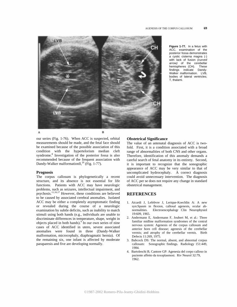
©1987-2002 Romero-Pilu-Jeanty-Ghidini-Hobbins
AGENESIS OF THE CORPUS CALLOSUM 69
Figure 1-77. In a fetus with ACC, examination of the posterior fossa demonstrates a cystic cisterna magna (-) with lack of fusion (curved arrow) of the cerebellar hemispheres (CH). These findings indicate Dandy-Walker malformation. LVB, bodies of lateral ventricles; T, thalami.
our series (Fig. 1-76). When ACC is suspected, orbital measurements should be made, and the fetal face should be examined because of the possible association of this condition with the hypertelorism median cleft syndrome.8 Investigation of the posterior fossa is also recommended because of the frequent association with Dandy-Walker malformationll,20 (Fig. 1-77). Prognosis The corpus callosum is phylogenetically a recent structure, and its absence is not essential for life functions. Patients with ACC may have neurologic problems, such as seizures, intellectual impairment, and psychosis.11,16,17 However, these conditions are believed to be caused by associated cerebral anomalies. Isolated ACC may be either a completely asymptomatic finding or revealed during the course of a neurologic examination by subtle deficits, such as inability to match stimuli using both hands (e.g., individuals are unable to discriminate differences in temperature, shape, weight in objects placed in both hands).9 In our own series of nine cases of ACC identified in utero, severe associated anomalies were found in three (Dandy-Walker malformation, microcephaly, diaphragmatic hernia). Of the remaining six, one infant is affected by moderate paraparesis and five are developing normally.
Obstetrical Significance The value of an antenatal diagnosis of ACC is two-fold. First, it is a condition associated with a broad range of abnormalities of both CNS and other organs. Therefore, identification of this anomaly demands a careful search of fetal anatomy in its entirety. Second, it is important to recognize that the sonographic appearance of ACC may be very similar to that of uncomplicated hydrocephaly. A correct diagnosis could avoid unnecessary intervention. The diagnosis of ACC per se does not require any change in standard obstetrical management. REFERENCES 1. Aicardi J, Lefebvre J, Lerique-Koechlin A: A new
syn:Spasm in flexion, callosal agenesis, ocular ab-normalities. Electroencephalogr Clin Neurophysiol 19:609, 1965.
2. Andermann E, Andermann F, Joubert M, et al.: Three familiar midline malformation syndromes of the central nervous system: Agenesis of the corpus callosum and anterior horn cell disease; agenesis of the cerebellar vermis; and atrophy of the cerebellar vermis. Birth Defects 11:269, 1975.
3. Babcock DS: The normal, absent, and abnormal corpus callosum: Sonographic findings. Radiology 151:449, 1984.
4. Bartoleschi B, Cantore GP: Agenesia del corpo calloso in paziente affetto da toxoplasmosi. Riv Neurol 32:79, 1962.
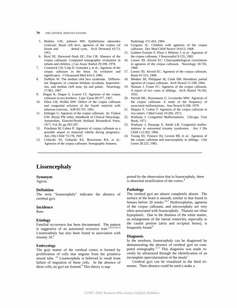
©1987-2002 Romero-Pilu-Jeanty-Ghidini-Hobbins
70 THE CENTRAL NERVOUS SYSTEM
5. Binkley GW, johnson HH: Epithelioma adenoides
cysticum: Basal cell nevi, agenesis of the corpus cal losum and dental cysts. Arch Dermatol 63:73, 1951.
6. Byrd SE, Harwood~Nash DC, Fitz CR: Absence of the corpus callosum: Computed tomographic evaluation in infants and children. j Can Assoc Radiol 29:108, 1978.
7. Comstock CH, Culp D, Gonzalez j, et al.: Agenesis of the corpus callosum in the fetus: Its evolution and significance. J Ultrasound Med 4:613, 1985.
8. DeMyer W: The median cleft face syndrome. Differen-tial diagnosis of cranium bifidum occultum, hypertelor-ism, and median cleft nose, lip and palate. Neurology 17:961, 1967.
9. Dogan K, Dogan S, Louren CI: Agenesis of the corpus callosum in two brothers. Lijee Vjesn 89:377, 1967.
10. Elliot GB, Wollin DW: Defect of the corpus callosum and congenital oclusion of the fourth ventricle with tuberous sclerosis. AJR 85:701, 1961.
11. Ettlinger G: Agenesis of the corpus callosum. In: Vinken GW, Bruyn PW (eds): Handbook of Clinical Neurology. Amsterdam, Elsevier/North Holland Biomedical Press, 1977, Vol 30, pp 285-297.
12. Friedman M, Cohen P: Agenesis of corpus callosum as a possible sequel to maternal rubella during pregnancy. Am j Dis Child 73:178, 1947.
13. Cebarski SS, Gebarski KS, Bowerman RA, et al.: Agenesis of the corpus callosum: Sonographic features.
Radiology 151:443, 1984. 14. Grogono JL: Children with agenesis of the corpus
callosum. Dev Med Child Neurol 10:613, 1968. 15. Guibert-Trainier F, Piton J, Billerey J, et al.: Agenesis of
the corpus callosum. J Neuroradiol 9:135, 1982. 16. Loeser JD, Alvord EC: Clinicopathological correlations
in agenesis of the corpus callosum. Neurology 18:745, 1968.
17. Loeser JD, Alvord EC: Agenesis of the corpus callosum. Brain 91:553, 1968.
18. Menkes JH, Philippart M, Clark DB: Hereditary partial agenesis of corpus callosum. Arch Neurol 11:198, 1964.
19. Naiman J, Fraser FC: Agenesis of the corpus callosum. A report of two cases in siblings. Arch Neurol 74:182, 1955.
20. Parrish ML, Roessmann U, Levinsohn MW: Agenesis of the corpus callosum: A study of the frequency of associated malformations. Ann Neurol 6:349, 1979.
21. Shapira Y, Cohen T: Agenesis of the corpus callosum in two sisters. J Med Genet 10:266, 1973.
22. Warkany J: Congenital Malformations. Chicago, Year Book, 1971.
23. Warkany J, Passarge E, Smith LB: Congenital malfor-mations in autosomal trisomy syndromes. Am J Dis Child 112:502, 1966.
24. Young ID, Trounce JQ, Levene MI, et al.: Agenesis of the corpus callosum and macrocephaly in siblings. Clin Genet 28:225, 1985.
Lissencephaly Synonym Agyria. Definition The term "lissencephaly" indicates the absence of cerebral gyri. Incidence Rare. Etiology Familial occurrence has been documented. The pattern is suggestive of an autosomal recessive trait.2,4,5,9,10,13 Lissencephaly has also been found in association with trisomy 18.8 Embryology The gray matter of the cerebral cortex is formed by proliferation of cells that migrate from the primitiva neural tube. 14 Lissencephaly is believed lo result from failure of migration of these cells. In the absence of these cells, no gyri are formed.8 This theory is sup-
ported by the observation that in lissencephaly, there is abnormal stratification of the cortex.8
Pathology The cerebral gyri are almost completely absent. The surface of the brain is smooth, similar to that found in fetuses before 20 weeks.3,6,7 Hydrocephalus, agenesis of the corpus callosum, and microcephaly are very often associated with lissencephaly. Thalami are often hypoplastic. Due to the thinness of the white matter, an enlargement of the lateral ventricles, especially in the caudal portion (atria and occipital horns), is frequently found.6 Diagnosis In the newborn, lissencephaly can be diagnosed by demonstrating the absence of cerebral gyri en com-puted tomography.5,11 This diagnosis was made re-cently by ultrasound through the identification of an incomplete opercularization of the insula.l Cerebral gyri can be visualized in the third tri-mester. Their absence could be used o make a
©1987-2002 Romero-Pilu-Jeanty-Ghidini-Hobbins
INTRACRANIAL ARACHNOID CYSTS 71
diagnosis. However, prenatal diagnosis based upon this criterion has not been reported. We have made this diagnosis in a patient at risk because of a positive family history by demonstrating the associated ventriculomegaly. Other findings that can be documented with ultrasound include agenesis of the corpus callosum and microcephaly. Failure to visualize the thalamic structures can raise the index of suspicien. Associated Anomalies Lissencephaly is commonly associated with other anomalies such as micromelia, club foot, polydactyly, camptodactyly, syndactyly, duodenal atresia, micro-gnathia, omphalocele, hepatosplenomegaly, and cardiac and renal anomalies. Polyhydramnios is present in 50 percent of cases.12 Prognosis Lissencephaly is invariably fatal by infancy or childhood and it is always associated with severe intellectual impairment (IQ <35). Some infants show neurologic signs of decerebration, seizures, and spastic diplegia.4,7 Obstetrical Management It is unclear if the diagnosis can be made before viability. If ventriculomegaly is identified in a patient at risk because of a previously affected infant, the option of pregnancy termination should be offered to the parents. Lissencephaly is a condition for which pregnancy termination may be offered in the third trimester if a confident diagnosis can be made. This latter requirement has not been met.
REFERENCES
1. Babcock DS: Sonographic demonstration of lissencephaly (agyria). J Ultrasound Med 2:465, 1983.
2. Barth PG, Mullaart R, Stam FC, et al.: Familial lis-sencephaly with extreme neopallial hypoplasia. Brain Dev 4:145, 1982.
3. Daube JR, Chou SM: Lissencephaly. Two cases. Neu-rology 16:179, 1966.
4. Dieker H, Edwards RH, Zu Rhein G, et al.: The lissencephaly syndrome. Birth Defects 5:53, 1969.
5. Garcia CA, Dunn D, Trevor R: The lissencephaly (agyria) syndrome in siblings: Computerized tomographic and neuropathologic findings. Arch Neurol 35: 608, 1978.
6. Jellinger K, Rett A: Agyria-pachygyria (lissencephaly syndrome). Neuropaediatrie 7:66, 1976.
7. Larroche JC: Cytoarchitectonic abnormalities (abnor-malities of cell migration). In: Vinken GW, Bruyn PW
Handbook of Clinical Neurology. Amsterdam, Elsevier/North Holland Biomedical Press, 1977, Vol 30, pp 479-505.
8. Ludwin SK, Malamud N: Pathology of congenital anomalies of the brain. In: Newton TH, Potts DG (eds): Radiology of the Skull and Brain. Anatomy and Pathol-ogy. St. Louis, CV Mosby, 1977, pp 2979-3015.
9. Miller JQ: Lissencephaly in 2 siblings. Neurology 13: 841, 1963.
10. Norman MG, Roberts J, Siroid J, et al.: Lissencephaly. J Can Soc Neurol 3:39, 1976.
11. Ohno K, Enomoto T, Imamoto J, et al.: Lissencephaly (agyria) on computed tomography. J Comput Assist Tomogr 3:92, 1979.
12. Opitz JM: Lissencephaly syndrome. In: Bergsma D (ed) Birth Defects Compendium, 2d ed. New York, Alan R
Liss, 1979, p 658. 13. Reznik M, Alberca RS: Hypertelorisme et lis- sencephalie: Etude d'une forme familiale.(Famille Ma ... ).
Acta Neurol Belg 63:970, 1963. 14. Sidman RL, Rakie P: Neuronal migration, with special
reference to developing human brain: A review. Brain Res 62:1, 1973.
Intracranial Arachnoid Cysts Definition Arachnoid cysts are fluid-filled cavities lined completely or partially by the arachnoid membrane. Epidemiology The frequency of this disorder is not known. In most cases, the diagnosis is made at autopsy in an otherwise asymptomatic individual.
Etiology Arachnoid cysts are classified as primary or secondary. Secondary or acquired cysts result from trauma, meningitis, infarction, or bleeding. Necrotic remnants or hematomas formed after the initial insult are subsequently reabsorbed, and a cyst is formed. In the absence of any obvious cause, the cyst can be considered as primary and regarded to be the consequence
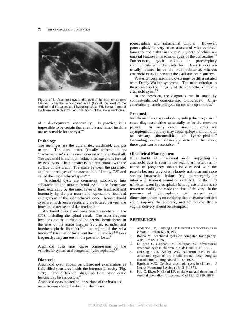
©1987-2002 Romero-Pilu-Jeanty-Ghidini-Hobbins
72 THE CENTRAL NERVOUS SYSTEM
Figure 1-78. Arachnoid cyst at the level of the interhemispheric fissure. Note the echo-spared area (Cy) at the level of the midline and the associated hydrocephalus. FH, frontal horns of the lateral ventricles; OH, occipital horns of the lateral ventricles. of a developmental abnormality. In practice, it is impossible to be certain that a remote and minor insult is not responsable for the cyst.10 Pathology The meninges are the dura mater, arachnoid, and pia mater. The dura mater (usually referred to as "pachymeninge") is the most external and lines the skull. The arachnoid is the intermediate meninge and is formed by two layers. The pia mater is in direct contact with the surface of the brain. The space between the pia mater and the inner layer of the arachnoid is filled by CSF and called the "subarachnoid space" 10 Arachnoid cysts are commonly subdivided into subarachnoid and intraarachnoid cysts. The former are lined externally by the inner layer of the arachnoid and internally by the pia mater and represent a localizad enlargement of the subarachnoid space. Intraarachnoid cysts are much less frequent and are located between the inner and outer layer of the arachnoid.10 Arachnoid cysts have been found anywhere in the CNS, including the spinal canal. The most frequent locations are the surface of the cerebal hemispheres in the sites of the major fissures (sylvian, rolandic, and interhemispheric fissures),12,13 the region of the sella turcica,5,8 the anterior fossa, and the middle fossa.4,11 Less frequently, they are seen in the posterior fossa.3 Arachnoid cysts may cause compression of the ventricular system and congenital hydrocephalus.1,10 Diagnosis Arachnoid cysts appear on ultrasound examination as fluid-filled structures inside the intracranial cavity (Fig. 1-78). The differential diagnosis from other cystic lesions may he impossible.6 Arachnoid cysts located on the surface of the brain and main fissures should be distinguished from
porencephaly and intracranial tumors. However, porencephaly is very often associated with ventricu-lomegaly and a shift in the midline, both of which are unusual features in arachnoid cysts of the convexities.10 Furthermore, cystic cavities in porencephaly communicate with the ventricles. Brain tumors are usually located inside the brain substance, whereas arachnoid cysts lie between the skull and brain surface. Posterior fossa arachnoid cysts must be differentiated from Dandy-Walker syndrome. The main criterion in these cases is the integrity of the cerebellar vermis in arachnoid cysts.7,9 In the newborn, the diagnosis can be made by contrast-enhanced computerized tomography. Char-acteristically, arachnoid cysts do not take up contrast.2 Prognosis Insufficient data are available regarding the prognosis of cases diagnosed either antenatally or in the newborn period. In many cases, arachnoid cysts are asymptomatic, but they may cause epilepsy, mild motor or sensory abnormalities, or hydrocephalus.10 Depending on the location and extent of the lesion, these cysts can be resectable.1,10
Obstetrical Management If a fluid-filled intracranial lesion suggesting an arachnoid cyst is seen in the second trimester, termi-nation of pregnancy should be discussed with the parents because prognosis is largely unknown and more serious intracranial lesions (e.g., porencephaly or intracranial tumors) cannot be excluded. In the third trimester, when hydrocephalus is not present, there is no reason to modify the mode and time of delivery. In the presence of hydrocephalus with normal skull dimensions, there is no evidence that a cesarean section could improve the outcome, and we believe that a vaginal delivery should be attempted.
REFERENCES
1. Anderson FM, Landing BH: Cerebral arachnoid cysts in
infants. 1 Pediatr 69:88, 1966. 2. Banna M: Arachnoid cysts on computed tomography.
AJR 127:979, 1976. 3. DiRocco C, Caldarelli M, DiTrapani G: Infratentorial
arachnoid cysts in children. Childs Brain 8:119, 1981. 4. Geissinger JD, Kohler WC, Robinson BW, et al.:
Arachnoid cysts of the middle cranial fossa: Surgical considerations. Surg Neurol 10:27, 1978.
5. Harrison MJG: Cerebral arachnoid cysts in children. J Neurol Neurosurg Psychiatry 34:316, 1971.
6. Pilu G, Rizzo N, Orsini LF, et al.: Antenatal detection of cerebral anomalies. Ultrasound Med Biol 12:319, 1986.
©1987-2002 Romero-Pilu-Jeanty-Ghidini-Hobbins
INTRACRANIAL TUMORS 73
7. Pilu G, Romero R, DePalma L, et al.: Antenatal diagnosis
and obstetrical management of Dandy-Walker syndrome. J Reprod Med 31:1017, 1986.
8. Ring BA, Waddington M: Primary arachnoid cyst of the sella turcica. AJR 98:611, 1966.
9. Roach ES, Laster DW, Summer TE, et al.: Posterior fossa arachnoid cyst demonstrated by ultra-sound. J Clin Ultrasound 10:88, 1982.
10. Shaw CM, Alvord EC: Congenital arachnoid cysts and their differential diagnosis. In: Vinken PJ, Bruyn GW
(eds): Handbook of Clinical Neurology. Amsterdam, Elsevier/North Holland Biomedical Press, 1977, Vol 30, pp 75-135.
13. Smith RA, Smith WA: Arachnoid cysts of the middle cranial fossa. Surg Neurol 5 :246, 1976.
14. Starkman SP, Brown TC, Linell EA: Cerebral arachnoid cysts. J Neuropathol Exp Neurol 17 :484, 1958.
15. Zingesser L, Schechter M, Gonatas N, et al.: Agenesis of the corpus callosum associated with an interhemispheric arachnoid cyst. Br J Radiol 37:905, 1964.
Intracranial Tumors
Intracranial tumors include epidermoid, dermoid, teratoma, germinoma, medulloblastoma, tuberous sclerosis (Bourneville's disease), neurofibromatosis (Von Recklinghausen's disease), and systemic angiomatosis of the central nervous system and eye (Von Hippel-Lindau's disease). Incidence Fetal intracranial tumors are rare. There are obvious difficulties in assessing the incidence of congenital brain neoplasms, because some lesions are asymptomatic or become symptomatic during childhood, adolescence, or even adulthood. Malignancies of the CNS were found to account for 0.04 to 0.18 percent of the total deaths of infants under 1 year of age.3 lt should be stressed that only a very small portion of brain tumors in children seem to arise during fetal life. In a series of 730 neoplasms diagnosed between
1 and 16 years of age, only 56 (7.8 percent) were thought to be congenital.3 Etiology Embryonic tumors are thought to derive from embry-ologically displaced cells. Brain tumors have been produced in animals by the use of chemical2 and viral teratogens.7 The relevance of these experiments to human brain neoplasms is unclear. Pathology There are severas classifications of congenital brain tumors.3,5 A commonly used system is shown in Table 1-18. Epidermoid tumors (also known as cholesteatomas") derive from epithelial cells and frequently appear as cystic lesions, containing a leaflike material, that originate from the desquamation of the internar epithelial lining. They are most commonly located at the level of the cerebellopontine
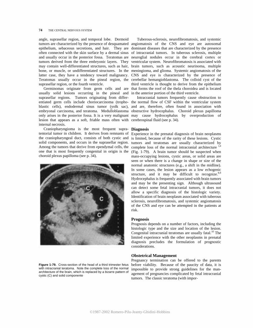
©1987-2002 Romero-Pilu-Jeanty-Ghidini-Hobbins
74 THE CENTRAL NERVOUS SYSTEM
angle, suprasellar region, and temporal lobe. Dermoid tumors are characterized by the presence of desquamated epithelium, sebaceous secretions, and hair. They are often connected with the skin surface by a dermal sinus and usually occur in the posterior fossa. Teratomas are tumors derived from the three embryonic layers. They may contain well-differentiated structures, such as hair, bone, or muscle, or undifferentiated structures. In the latter case, they have a tendency toward malignancy. Teratomas usually occur in the pineal region, the suprasellar region, or the fourth ventricle. Germinomas originate from germ cells and are usually solid lesions occurring in the pineal and suprasellar regions. Tumors originating from differ-entiated germ cells include choriocarcinoma (tropho-blastic cells), endodermal sinus tumor (yolk sac), embryonal carcinoma, and teratoma. Medulloblastoma only arises in the posterior fossa. It is a very malignant lesion that appears as a soft, friable mass often with internal necrosis. Craniopharyngioma is the most frequent supra-tentorial tumor in children. It derives from remnants of the craniopharyngeal duct, consists of both cystic and solid components, and occurs in the suprasellar region. Among the tumors that derive from ependymal cells, the one that is most frequently congenital in origin is the choroid plexus papilloma (see p. 34).
Figure 1-79. Cross-section of the head of a third trimester fetus with intracranial teratoma. Note the complete loss of the normal architecture of the brain, which is replaced by a bizarre pattern of cystic (C) and solid componente
Tuberous-sclerosis, neurofibromatosis, and systemic angiomatosis of the CNS and eye are autosomal dominant diseases that are characterized by the presence of intracranial tumors. In tuberous sclerosis, multiple neuroglial nodules occur in the cerebral cortex or ventricular system. Neurofibromatosis is associated with brain tumors, such as acoustic neurinoma, multiple meningioma, and glioma. Systemic angiomatosis of the CNS and eye is characterized by the presence of cerebellar hemangioblastoma. The colloid cyst of the third ventricle is thought to derive from the epithelium that forms the roof of the thela choroidea and is located in the anterior portion of the third ventricle. Intracranial tumors frequently cause obstruction to the normal flow of CSF within the ventricular system and are, therefore, often found in association with obstructive hydrocephalus. Choroid plexus papilloma may cause hydrocephalus by overproduction of cerebrospinal fluid (see p. 34). Diagnosis Experience in the prenatal diagnosis of brain neoplasms is limited, because of the rarity of these lesions. Cystic tumors and teratomas are usually characterized by complete loss of the normal intracranial architecture l,4 (Fig. 1-79). A brain tumor should be suspected when mass-occupying lesions, cystic areas, or solid areas are seen or when there is a change in shape or size of the normal anatomic structures (e.g., a shift in the midline). In some cases, the lesion appears as a low echogenic structure, and it may be difficult to recognize.8,9 Hydrocephalus is frequently associated with brain tumors and may be the presenting sign. Although ultrasound can detect some fetal intracranial tumors, it does not allow a specific diagnosis of the histologic variety. Identification of brain neoplasm associated with tuberous sclerosis, neurofibromatosis, and systemic angiomatosis of the CNS and eye can be attempted in the patients at risk. Prognosis Prognosis depends on a number of factors, including the histologic type and the size and location of the lesion. Congenital intracranial teratomas are usually fatal.10 The limited experience with the other neoplasms in prenatal diagnosis precludes the formulation of prognostic considerations. Obstetrical Management Pregnancy termination can be offered to the parents before viability. Because of the paucity of data, it is impossible to provide strong guidelines for the man-agement of pregnancies complicated by fetal intracranial tumors. The classic teratoma (with impor-
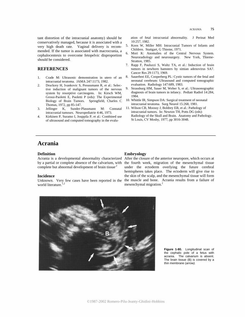
©1987-2002 Romero-Pilu-Jeanty-Ghidini-Hobbins
ACRANIA 75 tant distortion of the intracranial anatomy) should be conservatively managed, because it is associated with a very high death rate. Vaginal delivery is recom-mended. If the tumor is associated with macrocrania, a cephalocentesis to overcome fetopelvic disproportion should be considered. REFERENCES 1. Crade M: Ultrasonic demonstration in utero of an
intracranial teratoma. JAMA 247:1173, 1982. 2. Druckrey H, Ivankovic S, Preussmann R, et al.: Selec-
tive induction of malignant tumors of the nervous system by resorptive carcinogens. In: Kirsch WM, Grossi-Paoletti E, Paoletti P (eds): The Experimental Biology of Brain Tumors. Springfield, Charles C Thomas, 1972, pp 85-147.
3. Jellinger K, Sunder-Plassmann M: Connatal intracranial tumours. Neuropediatrie 4:46, 1973.
4. Kirkinen P, Suramo I, Jouppila P, et al.: Combined use of ultrasound and computed tomography in the evalu-
ation of fetal intracranial abnormality. J Perinat Med 10:257, 1982.
5. Koos W, Miller MH: Intracranial Tumors of Infants and Children. Stuttgart, G Thieme, 1971.
6. Mori K: Anomalies of the Central Nervous System. Neuroradiology and neurosurgery. New York, Thieme-Stratton, 1985.
7. Rapp F, Pauluzzi S, Waltz TA, et al.: Induction of brain tumors in newborn hamsters by simian adenovirus SA7. Cancer Res 29:1173, 1969.
8. Sauerbrei EE, Cooperberg PL: Cystic tumors of the fetal and neonatal cerebrum: Ultrasound and computed tomographic evaluation. Radiology 147:689, 1983.
9. Strassburg HM, Sauer M, Weber S, et al.: Ultrasonographic diagnosis of brain tumors in infancy. Pediatr Radiol 14:284, 1984.
10. Whittle IR, Simpson DA: Surgical treatment of neonatal intracranial teratoma. Surg Neurol 15:268, 1981.
11. Wilson CB, Moossy J, Boldrey EB, et al.: Pathology of intracranial tumors. In: Newton TH, Potts DG (eds): Radiology of the Skull and Brain. Anatomy and Pathology. St Louis, CV Mosby, 1977, pp 3016-3048.
Acrania Definition Acrania is a developmental abnormality characterized by a partial or complete absence of the calvarium, with complete but abnormal development of brain tissue.2 Incidence Unknown. Very few cases have been reported in the world literature.1,2
Embryology After the closure of the anterior neuropore, which occurs at the fourth week, migration of the mesenchymal tissue under the ectoderm overlying the future cerebral hemispheres takes place. The ectoderm will give rise to the skin of the scalp, and the mesenchymal tissue will form the muscle and bone. Acrania results from a failure of mesenchymal migration.2
Figure 1-80. Longitudinal scan of the cephalic pole of a fetus with acrania. The calvarium is absent. The brain tissue (B) is covered by a thin membrane (arrow).

©1987-2002 Romero-Pilu-Jeanty-Ghidini-Hobbins
76 THE CENTRAL NERVOUS SYSTEM
Etiology Unknown. Only sporadic cases have been reported.1,2 We have seen pathologic findings very similar to those pathognomonic of acrania in three fetuses with amniotic band syndrome. Pathology The calvarian dermal bones of the skull, the related musculatura, and dura mater are absent. The hemi-spheres are present but grossly abnormal and are covered by a thin membrane. Cerebellum, brain stem, and cranial nerves are normal. Associated Anomalies Cleft lip and palate, and talipes.2 Diagnosis The condition is identified by the absence of the calvarium. The cerebral hemispheres are surrounded by a thin membrane2 (Fig. 1-80). Differential diagno-sis includes anencephaly and large encephaloceles. In the former case, cerebral tissue is completely absent. In the latter case, some remnant of the cranial vault can always be detected. A distinction should also be
made between acrania and conditions characterized by lack of mineralization of the skull bones (hypophosphatasia, osteogenesis imperfecta). In these skeletal dysplasias, the intracranial anatomy is normal, and the brain is surrounded by a thick layer of tissue representing soft tissues and unossified bone. Bowing or shortening of long bones is generallv found. Prognosis Acrania is uniformly lethal. Obstetrical Management Pregnancy termination can be offered to the parents any time the condition is diagnosed. REFERENCES 1. Kristoffersen K, Pedersen BN, Secher Nj, et al.: Akrani og
spina bifida diagnosticeret ved bestemmelse af alfa fotoprotein i 16. graviditetsuge. Ugeskr Laeger 137:1719, 1975.
2. Mannes EJ, Crelin ES, Hobbins JC, et al.: Sonographic demonstration of fetal acrania. AJR 139:181, 1982.
Choroid Plexus Cyst Incidence Cysts of the choroid plexus are found in 50 percent of brains serially autopsied."
Etiopathogenesis These cysts are thought to arise from neuroepithelial folds within the choroid plexus that become fil-
Figure 1-81. A. Axial scan of the head of a 21 -week fetus. The arrows point to a cyst inside the choroid plexus. B. Three weeks later, the cyst has considerably diminished in size. Note the normal size of the ventricles. A further scan performed 2 weeks later failed to reveal any anomaly of the choroid plexus. The infant was normal at birth. FH, frontal horns.

©1987-2002 Romero-Pilu-Jeanty-Ghidini-Hobbins
ANEURYSM OF THE VEIN OF GALEN 77 led with fluid and cellular debris.11,12 In autopsy specimens, they are usually less than 1 cm in diam-eter.11,12 Pathology Cysts are lined by the ependyma and deeply enfolded within the choroid plexus. They may be bilateral. Generally, they are asymptomatic unless they obstruct the flow of CSF, causing hydrocephaly. 1,2,4,5,8,9 Diagnosis A round hypoechogenic area can be seen within the texture of the choroid plexus, most frequently at the level of the atrium of the lateral ventricle 3,6,7 (Fig. 1-81). This condition should be differentiated from choroid plexus papilloma, which generally produces an echogenic image and subependymal hemorrhages, which are rare in the fetus and are located below the choroid plexus. Prognosis Ten cases of fetal choroid plexus cysts have been reported.3,7,10 In six cases this was the only anomaly identified. In four, the choroid plexus cyst was bilateral and associated with severe anomalies, such as trisomy 18 (three cases), omphalocele, obstructive uropathy, and ventricular septal defect.10 In the cases of isolated choroid plexus cysts, the lesion spontaneously disappeared before the 28th week (in five cases before the 24th week). The infants were neurologically normal at birth. Hydrocephalus has been reported occasionally in infants and adults with choroid plexus cysts.1,2,4,5,8,9 Obstetrical Management The limited experience available with these lesions suggests that they are clinically benign. Serial scans to monitor their status and exclude the development of hydrocephalus are indicated. There is no reason to modify standard obstetrical management.3 However, the occurrence of hydrocephalus, chromosomal ab-normalities, and other associated anomalies has been
reported, and, therefore, a careful survey of the fetal anatomy and a fetal karyotype seem indicated.l0a REFERENCES
1. Andreussi L, Cama A, Cozzutto C, et al.: Cyst of the choroid plexus of the left lateral ventricle. Surg Neurol 12:53, 1979.
2. Baker GS, Gottlieb CM: Cyst of the choroid plexus of the lateral ventricle causing disabling headache and unconsciousness:Report of case. Mayo Clin Proc 31:95, 1956.
3. Chudleigh P, Pearce JM, Campbell S: Short communi-cations. The prenatal diagnosis of transient cysts of the fetal choroid plexus. Prenatal Diagn 4:135, 1984.
4. DeLaTorre E, Alexander E, Courtland HD, et al.: Tumors of the lateral ventricles of the brain: Report of eight cases, with suggestions for clinical management. J Neurosurg 20:461, 1963.
5. Dempsey RJ, Chandler WF: Choroid plexus cyst in the lateral ventricle causing obstructive symptoms in an adult. Surg Neurol 15:116, 1981.
6. Fakhry J, Schechter A, Tenner MS, et al.: Cysts of the choroid plexus in neonates: Documentation and review of the literatura. J Ultrasound Med 4:561, 1985.
7. Friday RO, Schwartz DB, Tuffli GA: Spontaneous in-trauterine resolution of intraventricular cystic masses. J Ultrasound Med 4:385, 1985.
8. Giorgi C: Symptomatic cyst of the choroid plexus of the lateral ventricle. Neurosurgery 5:53, 1979.
9. Neblett CR, Robertson JW: Symptomatic cysts of the telencephalic choroid plexus. J Neurol Neurosurg Psy-chiatry 34:324, 1971.
10.Nicolaides KH, Rodeck CH, Gosden CM: Rapid karyotyping in non-lethal fetal malformations. Lancet 1:283, 1986.
10a. Ostlere SJ, Irving HC, Lilford RJ: Choroid plexus cysts in the fetus. Lancet 1:1491, 1987.
11. Shuangshoti S, Netsky MG: Neuroepithelial (colloid) cysts of the nervous system: Further observations on pathogenesis, location, incidence and histochemistry. Neurology 16:887, 1966.
12. Shuangshoti S, Netsky MG: Histogenesis of choroid plexus in man. Am J Anat 118:283, 1966.
Aneurysm of the Vein of Galen Synonym Varix of the vein of Galen. Definition Aneurysm of the vein of Galen (AVG) is a complex
arteriovenous malformation ranging in appearance from a gigantic aneurysmal enlargement of the vein of Galen to multiple communications between the system of the vein of Galen and the cerebral arteries (carotid or vertebrobasilar systems).

©1987-2002 Romero-Pilu-Jeanty-Ghidini-Hobbins
78 THE CENTRAL NERVOUS SYSTEM
Figure 1-82. Axial scan of the fetal head, showing the normal anatomy of the vein of Galen (VG). Straight ar rows point to the medial contour of the occipital lobes. Curved arrows point to the ambient cisterns. T, Thalami; At, atria of ventriculi; Ant, anterior; Post, posterior.
Incidence Unknown. Less than 200 cases had been reported in the literatura up to 1984.9 It is more common in males than females (M: F ratio = 2:1).7
Embryology The cerebral vessels derive from a primitive plexus that differentiate in both arteries and veins. There is controversy about the chronology of the derangement giving rise to cerebral arteriovenous malformations (AVM). According to some authors, the primary
Figure 1-83. Axial scan of the fetal head showing the aneurysm of the vein of Galen as a large interhemispheric cystic mass.
defect occurs early, during the phase of differentiation of the angioblasts to form primitiva capillaries, arteries, and veins.6 Others believe that AVM arises during the late histologic differentiation of the primitive vessels into adult vessels.5 Pathology and Clinical Presentation The vein of Galen is the major cerebral vein. It runs superoposteriorly to the thalami within a subarachnoid space known as the "vein of Galen cystern." It joins the inferior sagittal sinus, which runs along the lower edge of the cerebral falx to form the straight sinus (Fig. 1-82). In cases of AVG, an aneurysmal dilatation of the vein of Galen is usually found in association with varying patterns of arteriovenous communication.3 The clinical presentation of AVG depends on the type of lesion. Large arteriovenous communications result in significant intracranial shunt and usually appear in the neonatal period with high output congestive heart failure. In some patients, up to 80 percent of the cardiac output is diverted to the cerebral circulation.5 Diversion of blood from the cerebral circulation may lead to infarction of the brain and porencephaly. High output heart failure may also result in reduction of coronary blood flow, myocardial ischemia, and infarction.3 Hydrocephalus is frequently found, and it is thought to result from either compression of the aqueduct of Sylvius by the dilated vessel or from increased intracranial venous pressure. In other cases characterized by milder arteriovenous communication, AVG may occur during the first year of age with macrocrania, subarachnoidal hemorrhages, and seizures. In a third group of patients, the condition becomes symptomatic later in
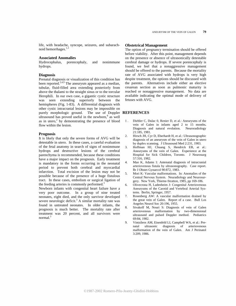
©1987-2002 Romero-Pilu-Jeanty-Ghidini-Hobbins
ANEURYSM OF THE VEIN OF GALEN 79
life, with headache, syncope, seizures, and subarach-noid hemorrhages.1,3
Associated Anomalies Hydrocephalus, porencephaly, and nonimmune hydrops.
Diagnosis Prenatal diagnosis or visualization of this condition has been reported.2,4,9 The aneurysm appeared as a median, tubular, fluid-filled area extending posteriorly from above the thalami to the straight sinus or to the torcular Herophili. In our own case, a gigantic cystic structure was seen extending superiorly between the hemispheres (Fig. 1-83). A differential diagnosis with other cystic intracranial lesions may be impossible on purely morphologic ground. The use of Doppler ultrasound has proved useful in the newborn,8 as well as in utero,2 by demonstrating the presence of blood flow within the lesion.
Prognosis It is likely that only the severe forms of AVG will be detestable in utero. In these cases, a careful evaluation of the fetal anatomy in search of signs of nonimmune hydrops and destructive lesions of the cerebral parenchyma is recommended, because these conditions have a major impact on the prognosis. Early treatment is mandatory in the forms occurring in the neonatal period to prevent both cerebral and myocardial infarction. Total excision of the lesion may not be possible because of the presence of a huge fistulous tract. In these cases, embolism or surgical ligation of the feeding arteries is commonly performed.3 Newborn infants with congenital heart failure have a very poor outcome. In a group of nine treated neonates, eight died, and the only survivor developed severe neurologic deficit.3 A similar mortality rate was found in untreated neonates. In older infants, the prognosis is much better. The mortality rate after treatment was 20 percent, and all survivors were normal.3
Obstetrical Management The option of pregnancy termination should be offered before viability. After this point, management depends on the presence or absence of ultrasonically detestable cerebral damage or hydrops. If severe porencephaly is found, we feel that a nonaggressive management should be offered to the parents. Because the mortality rate of AVG associated with hydrops is very high despite treatment, the options should be discussed with the parents. Alternatives include either an elective cesarean section as soon as pulmonic maturity is reached or nonaggressive management. No data are available indicating the optimal mode of delivery of fetuses with AVG.
REFERENCES
1 . Diebler C, Dulac 0, Renier D, et al.: Aneurysms of the vein of Galen in infants aged 2 to 15 months. Diagnosis and natural evolution. Neuroradiology 21:185, 1981.
2. Hirsch JH, Cyr D, Eberhardt H, et al.: Ultrasonographic diagnosis of an aneurysm of the vein of Galen in utero by duplex scanning. J Ultrasound Med 2:231, 1983.
3. Hoffman HJ, Chuang S, Hendrick EB, et al.: Aneurysms of the vein of Galen. Experience at the Hospital for Sick Children, Toronto. J Neurosurg 57:316, 1982.
4. Mao K, Adams 1: Antenatal diagnosis of intracranial arteriovenous fistula by ultrasonography. Case report. Br J Obstet Gynaecol 90:872, 1983.
5. Mori K: Vascular malformations. In: Anomalies of the Central Nervous System. Neuradiology and Neurosur-gery. New York, Thieme-Stratton, 1985, pp 169-186.
6. Olivecrona H, Ladenheim J: Congenital Arteriovenous Aneurysms of the Carotid and Vertebral Arterial Sys-tems. Berlin, Springer, 1957.
7. Rosenberg AW: A vascular malformation drained by the great vein of Galen. Report of a case. Bull Los Angeles Neurol Soc 20:196, 1955.
8. Sivakoff M, Nouri S: Diagnosis of vein of Galen arteriovenous malformation by two-dimensional ultrasound and pulsed Doppler method. Pediatrics 69:84, 1982.
9. Vintzileos AM, Eisenfeld LI, Campbell WA, et al.: Pre-natal ultrasonic diagnosis of arteriovenous malformation of the vein of Galen. Am J Perinatol 3:209, 1986.