PREMENOPAUSAL ADNEXAL MASS · DIFFERENTIAL DIAGNOSIS Functional Cyst Leiomyomata Endometrioma...
15
PREMENOPAUSAL ADNEXAL MASS FLAME LECTURE: 230 RANDALL / BURNS 12.24.15
Transcript of PREMENOPAUSAL ADNEXAL MASS · DIFFERENTIAL DIAGNOSIS Functional Cyst Leiomyomata Endometrioma...

PREMENOPAUSAL ADNEXAL MASS FLAME LECTURE: 230
RANDALL / BURNS 12.24.15

Learning Objectives
u Outline the approach to a patient with an adnexal mass
u Compare the characteristics of functional cysts, benign ovarian neoplasms and ovarian cancers
u Prerequisites:
u NONE
u See also – for closely related topics u FLAME LECTURE 231 – Postmenopausal Adnexal Mass
u FLAME LECTURE 232 – Ovarian Cancer

ADNEXAL MASSES OVERVIEW u What’s the most likely cause?
u Premenopausal women: functional cyst
u Postmenopausal women: serous cystadenoma
u What’s the most concerning cause? OVARIAN CANCER
u < 15 years old: most common ovarian cancer - yolk sac tumor
( =endodermal sinus tumor)
u 15 – 30 yo: most common ovarian cancer - germ cell tumor
u > 55 yo: most common ovarian cancer - epithelial tumor
u There is NO effective screening method for ovarian cancer at this time
u Thus, even though the majority of ovarian masses are benign, we have a low threshold for escalating work up to rule out cancer
u Most adnexal masses are discovered incidentally on exam or imaging and investigation must be broadened beyond gynecologic causes
Navigate: Malignant Masses Evaluation Methods Workup Introduction Benign Masses Masses in Pregnancy

DIFFERENTIAL DIAGNOSIS
Functional Cyst
Leiomyomata
Endometrioma
Tubo-ovarian abscess
Ectopic pregnancy
Mature teratoma
Serous cystadenoma
Hydrosalpinx
Paratubal cyst
Germ cell tumor
Sex cord/stromal tumor
Epithelial carcinoma
Diverticular abscess
Appendicitis
Nerve sheath tumor
Ureter diverticulum
Pelvic kidney
Bladder diverticulum
GI cancers
Retroperitoneal sarcomas
Metastases
Gynecologic Non-Gynecologic
BENIGN BENIGN MALIGNANT MALIGNANT
Navigate: Malignant Masses Evaluation Methods Workup Introduction Benign Masses Masses in Pregnancy

OVARIAN CANCER SYMPTOMS u The symptoms of ovarian cancer are very insidious and
should always be asked of any patient of any age with a known pelvic mass u Are you experiencing any abdominal or pelvic pain?
u Have you been having any abnormal vaginal bleeding?
u Have you noticed you get full quickly, have a decreased appetite, or feel bloated?
u Have you experienced any weight loss over the past three to six months that you haven’t been trying to lose?
Navigate: Malignant Masses Evaluation Methods Workup Introduction Benign Masses Masses in Pregnancy

OVARIAN CANCER u Woman’s lifetime risk: 1 in 70
u If diagnosed at Stage I, 5-year survival is 90% BUT:
u Most women are diagnosed at an advanced stage 2/2 the insidious nature of their symptoms, and 5-year survival is 30-55%!
u Risk factors: u Family hx is the strongest risk factor: BRCA 1 carriers have 60-
fold increased risk, BRCA 2 carriers have 30-fold increased risk, and Lynch carrier has 13-fold increased risk
u Anything that causes increased ovarian epithelium turnover causes more repair and more opportunities for cancer development
u Conversely, factors that decrease ovulation, thereby decreasing ovarian epithelium disruption, are considered protective
u Most ovarian cancers are diagnosed in postmenopausal women
RISK FACTORS
� Familial ovarian cancer syndrome (BRCA, Lynch)
� Ovarian cancer family hx
Personal hx of breast cancer
Early menarche / late menopause
Infertility / nulliparity
Increasing age
PROTECTIVE FACTORS
Oral contraceptives
Breastfeeding
Multiparity
Chronic anovulation (ex PCOS)
BTL / salpingectomy / hysterectomy
Navigate: Malignant Masses Evaluation Methods Workup Introduction Benign Masses Masses in Pregnancy

BENIGN MASSES u In premenopausal women, benign gynecologic
masses are most commonly functional cysts u Follicular cyst – most common
u Failure of follicular rupture during follicular phase, usually unilateral (3-8cm)
u Resolves in 60-90 days
u Corpus luteum cyst – CL that is >3cm or hemorrhagic u Over-enlargement of corpus luteum during luteal phase of
cycle
u Causes delayed menstruation
Navigate: Malignant Masses Evaluation Methods Workup Introduction Benign Masses Masses in Pregnancy

BENIGN MASSES – CONT’D u Theca lutein cyst
u Due to abnormally high ß-HCg (pregnancy,
IVF, complete molar pregnancy)
u Endometrioma – “Chocolate cyst” u Ectopic endometrial tissue + pelvic pain, dysmenorrhea,
dyspareunia, infertility
u Mature teratoma – “Dermoid” u Complications: 4-10cm cyst can cause ovarian torsion
u <5cm, twisted ovary would self resolve
u >10cm, too large to torse
u Dermoid cysts at particularly high risk of torsion
Navigate: Malignant Masses Evaluation Methods Workup Introduction Benign Masses Masses in Pregnancy
EVALUATION OF ADNEXAL MASS u Physical exam:
u Is beneficial for discovering masses incidentally, however has low sensitivity and is generally difficult to differentiate between benign and malignant conditions
u Has especially limited ability to detect masses in patients with high BMI
u Transvaginal ultrasound:
u Pros: highly available and tolerable, cost-effective, most effective routine imaging
u Cons: lacks specificity and positive predictive value for cancer, especially in premenopausal women
u Concerning findings: mixed or solid consistency, + septations,
mural nodules, papillary excrescences (outgrowths), ascites
u Benign findings:
u Benign cysts: Round, unilocular, thin-walled sonolucent
cysts with smooth, regular borders
u Mature teratoma (dermoid cyst): hypoechoic attenuating
component with multiple small homogeneous interfaces
Navigate: Malignant Masses Evaluation Methods Workup Introduction Benign Masses Masses in Pregnancy

Hemorrhagic Non-hemorrhagic
BENIGN CYST EVALUATION - TVUS Pre-menarche
Pre-menopausal
Post-menopausal
< 2cm > 2cm
Repeat US in 6-12 weeks
Surgical evaluation
< 5 cm > 10 cm
No follow-up Surgical evaluation
5 – 10 cm
Repeat US in 6-12 weeks
Repeat US in 1 year
Hemorrhagic Non- hemorrhagic
Repeat US in 6-12 weeks
< 3cm 3 – 5 cm
No follow-up Repeat US in 1 year
> 10 cm Surgical evaluation
5 – 10 cm
Repeat US in 6-12 weeks
*While waiting for repeat US, provide OCP’s (won’t resolve current cyst but will prevent future cysts)
Navigate: Malignant Masses Evaluation Methods Workup Introduction Benign Masses Masses in Pregnancy

EVALUATION OF ADNEXAL MASS u CT, MRI, PET:
u Not recommended for initial evaluation and don’t add significantly to TVUS
u CT best used to look for metastases after all other workup suggests possible malignancy
u MRI useful for distinguishing origin of non-adnexal masses but not for ovarian tumors
u Serum CA-125:
u Low sensitivity because only elevated in 50% of stage I cancers and can also be elevated due to other conditions (fibroids, endometriosis, PID, ascites, pregnancy)
u Higher sensitivity in postmenopausal women because less incidence of alternative causes of elevation
u CA-125 is NOT diagnostic. Usually measured to trend responses to treatment and recurrence
u However CA-125 should still be measured in evaluation of adnexal masses because extreme values are still helpful (e.g. Mass in premenopausal woman with CA-125 of 300 is suspicious)
Navigate: Malignant Masses Evaluation Methods Workup Introduction Benign Masses Masses in Pregnancy

BENIGN CYST EVALUATION - LABS
Navigate: Malignant Masses Evaluation Methods Workup Introduction Benign Masses Masses in Pregnancy
Tumor marker: Typically elevated in: CA-125 Epithelial ovarian cancer
Inihibin A Granulosa cell tumor
Anti-mullerian hormone Granulosa cell tumor
LDH Dysgerminoma, endodermal sinus
AFP Endodermal sinus, Embryonal
HCG Choriocarcinoma
Testosterone/DHEA Sertoli-Leydig cell tumor
In the premenopausal patient, laboratory evaluation is generally reserved for complex masses > 5 cm and/or associated symptoms (AUB, virulization, hirsutism).
** Mixed cell-type tumors, such as gonadoblastomas or mixed germ-cell tumors, display a range and mix of tumor markers.
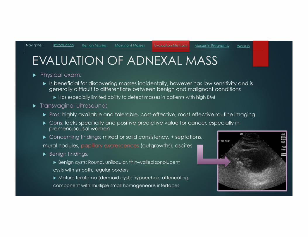
SUMMARY OF WORK-UP ADNEXAL MASS IN PREMENOPAUSAL FEMALE
SYMPTOMATIC ASYMPTOMATIC
History, ß-hCG, CBC, GC-CT,
Must first rule-out acute conditions:
ectopic pregnancy,
ovarian torsion, PID, ruptured cyst
Transvaginal US
Transvaginal US
< 10 cm mass > 10 cm mass
Ascites No Ascites
REFER TO GYN/ONC
Acute condition
Mass
CA-125 < 200u/mL CA-125 > 200u/mL
BENIGN CYST EVALUATION TREAT CONDITION
Navigate: Malignant Masses Evaluation Methods Workup Introduction Benign Masses Masses in Pregnancy

ADNEXAL MASSES IN PREGNANCY u Up to 3% of pregnant women have an identified adnexal mass at the time of
delivery
u Most common pathologic causes include: mature teratomas, theca lutein cysts, corpus luteum cysts. Malignancies are very rare.
u Changes to evaluation process:
u TVUS is still preferred but may need to add abdominal ultrasound if at high gestational age
u MRI is preferred to CT for additional imaging to avoid fetal radiation
u CA-125 levels are elevated in pregnancy, peaking in the first trimester
u Complications: adnexal masses do not pose risk to the pregnancy, however masses >5 cm are at risk for torsion following delivery because the space created by the shrinking uterus allows more opportunity for twisting
u Treatment: expectant management with close postpartum follow-up
u If symptomatic then can safely perform surgery during pregnancy
Navigate: Malignant Masses Evaluation Methods Workup Introduction Benign Masses Masses in Pregnancy

IMPORTANT LINKS / REFERENCES
u ACOG Practice Bulletin 83, July 2007 (“Management of Adnexal Masses”)
1. UpToDate.com 2. Callahan & Caughey Blueprints: Obstetrics
& Gynecology 6th ed. 2013
![Innovation in fertility treatment - fertilityvision.cz · [Murray, Fertil steril, 1998]. TAKE HOME MESSAGE •Improvement in IVF results may have reduced the negative impact of hydrosalpinx.](https://static.fdocuments.us/doc/165x107/5cf7ab1f88c99346318d81a1/innovation-in-fertility-treatment-murray-fertil-steril-1998-take-home.jpg)