Lack of Long-Lasting Hydrosalpinx in A/J Mice Correlates ... fileLack of Long-Lasting Hydrosalpinx...
9
Lack of Long-Lasting Hydrosalpinx in A/J Mice Correlates with Rapid but Transient Chlamydial Ascension and Neutrophil Recruitment in the Oviduct following Intravaginal Inoculation with Chlamydia muridarum Hongbo Zhang, a,d Zhou Zhou, a Jianlin Chen, a,d Ganqiu Wu, a,d Zhangsheng Yang, a Zhiguang Zhou, d Joel Baseman, a Jin Zhang, c Robert Lee Reddick, b Guangming Zhong a Departments of Microbiology and Immunology a and Pathology, b University of Texas Health Science Center at San Antonio, San Antonio, Texas, USA; Department of Pathology, Metropolitan Methodist Hospital, San Antonio, Texas, USA c ; Departments of Pathology, Obstetrics and Gynecology, and Medicine, Second Xiangya Hospital, Department of Histology, Xiangya Medical School, Central South University, Changsha, Hunan, China d Lower genital tract infection with Chlamydia trachomatis and C. muridarum can induce long-lasting hydrosalpinx in the upper genital tract of women and female mice, respectively. However, A/J mice were highly resistant to induction of long-lasting hydro- salpinx by C. muridarum. We further compared host inflammatory responses and chlamydial infection courses between the hy- drosalpinx-resistant A/J mice and CBA/J mice known to be susceptible to hydrosalpinx induction. Both mouse strains developed robust pyosalpinx during the acute phase followed by hydrosalpinx during the chronic phase. However, the hydrosalpinges dis- appeared in A/J mice by day 60 after infection, suggesting that some early hydrosalpinges are reversible. Although the overall inflammatory responses were indistinguishable between CBA/J and A/J mice, we found significantly more neutrophils in oviduct lumen of A/J mice on days 7 and 10, which correlated with a rapid but transient oviduct invasion by C. muridarum with a peak infection on day 7. In contrast, CBA/J mice developed a delayed and extensive oviduct infection. These comparisons have re- vealed an important role of the interactions of oviduct infection with inflammatory responses in chlamydial induction of long- lasting hydrosalpinx, suggesting that a rapid but transient invasion of oviduct by chlamydial organisms can prevent the develop- ment of the long-lasting hydrosalpinges. L ong-lasting hydrosalpinx is a pathological hallmark of tubal factor infertility associated with Chlamydia trachomatis infec- tion in women (1) and C. muridarum infection in female mice (2–4). The precise mechanism of Chlamydia-induced long-lasting hydrosalpinx remains unknown, although Chlamydia-triggered inflammatory responses have been hypothesized to contribute significantly to chlamydial pathogenesis (5, 6). For example, it is not clear whether live organism infection in the fallopian tube is necessary for induction of hydrosalpinx, how chlamydial organ- isms trigger hydrosalpinx-causing inflammation, and when hy- drosalpinx becomes irreversible. Since it is difficult to directly ad- dress these questions during C. trachomatis infection in women, a murine model of C. muridarum infection has been used exten- sively to study pathogenic mechanisms and immune responses of C. trachomatis (7, 8). This is because genital tract infection of mice with C. muridarum can lead to long-lasting hydrosalpinx that closely mimics hydrosalpinx found in C. trachomatis-infected women under laparoscopy (9–11). To delineate the host inflammatory mechanism of chlamydial induction of long-lasting hydrosalpinx, Shah et al. carefully com- pared the genital tract inflammatory pathological responses be- tween the chlamydial infection-resistant C57BL/6J and the sus- ceptible C3H/HeN mice, which led the authors to correlate a robust acute inflammatory response with the development of long-lasting hydrosalpinx observed on day 56 after infection (3). However, it is not known whether the intensity of the acute in- flammatory response can always contribute to chlamydial induc- tion of long-lasting hydrosalpinx, since both C57BL/6L and C3H/ HeN can develop significant long-lasting hydrosalpinx, although with different incidence rates (3, 12). It is also unclear whether live organism infection in the oviduct is necessary for sustaining hy- drosalpinx-causing inflammation, since live organism recovery was monitored in the lower but not upper genital tract (3). Dar- ville et al. further reported that mice deficient in TLR2 failed to develop a robust acute inflammatory response with decreased ovi- duct dilation (13). However, these observations were made under a microscope on day 35 after infection (13). It is not clear whether the proposed TLR2-mediated signaling pathway is sufficient for C. muridarum induction of long-lasting hydrosalpinx. More impor- tantly, the TLR2 knockout mice developed an equivalent level of chronic inflammation in the oviduct on day 35 after infection (13), and mice deficient in MyD88, a critical adaptor molecule of the TLR2-mediated signaling pathway, developed more severe long-lasting hydrosalpinx (14). These findings question the role of TLR2 signaling pathways in chlamydial induction of long-lasting hydrosalpinx. The role of neutrophils in chlamydial infection and pathogenesis has been extensively studied. While some found a protective role of early neutrophil infiltration in protection against chlamydial pathogenicity (15, 16), others correlated an increased infiltration of neutrophils with chlamydial pathogenic- Received 11 January 2014 Returned for modification 31 January 2014 Accepted 26 March 2014 Published ahead of print 7 April 2014 Editor: R. P. Morrison Address correspondence to Guangming Zhong, [email protected]. Copyright © 2014, American Society for Microbiology. All Rights Reserved. doi:10.1128/IAI.00055-14 2688 iai.asm.org Infection and Immunity p. 2688 –2696 July 2014 Volume 82 Number 7 on August 24, 2019 by guest http://iai.asm.org/ Downloaded from
Transcript of Lack of Long-Lasting Hydrosalpinx in A/J Mice Correlates ... fileLack of Long-Lasting Hydrosalpinx...

Lack of Long-Lasting Hydrosalpinx in A/J Mice Correlates with Rapidbut Transient Chlamydial Ascension and Neutrophil Recruitment inthe Oviduct following Intravaginal Inoculation with Chlamydiamuridarum
Hongbo Zhang,a,d Zhou Zhou,a Jianlin Chen,a,d Ganqiu Wu,a,d Zhangsheng Yang,a Zhiguang Zhou,d Joel Baseman,a Jin Zhang,c
Robert Lee Reddick,b Guangming Zhonga
Departments of Microbiology and Immunologya and Pathology,b University of Texas Health Science Center at San Antonio, San Antonio, Texas, USA; Department ofPathology, Metropolitan Methodist Hospital, San Antonio, Texas, USAc; Departments of Pathology, Obstetrics and Gynecology, and Medicine, Second Xiangya Hospital,Department of Histology, Xiangya Medical School, Central South University, Changsha, Hunan, Chinad
Lower genital tract infection with Chlamydia trachomatis and C. muridarum can induce long-lasting hydrosalpinx in the uppergenital tract of women and female mice, respectively. However, A/J mice were highly resistant to induction of long-lasting hydro-salpinx by C. muridarum. We further compared host inflammatory responses and chlamydial infection courses between the hy-drosalpinx-resistant A/J mice and CBA/J mice known to be susceptible to hydrosalpinx induction. Both mouse strains developedrobust pyosalpinx during the acute phase followed by hydrosalpinx during the chronic phase. However, the hydrosalpinges dis-appeared in A/J mice by day 60 after infection, suggesting that some early hydrosalpinges are reversible. Although the overallinflammatory responses were indistinguishable between CBA/J and A/J mice, we found significantly more neutrophils in oviductlumen of A/J mice on days 7 and 10, which correlated with a rapid but transient oviduct invasion by C. muridarum with a peakinfection on day 7. In contrast, CBA/J mice developed a delayed and extensive oviduct infection. These comparisons have re-vealed an important role of the interactions of oviduct infection with inflammatory responses in chlamydial induction of long-lasting hydrosalpinx, suggesting that a rapid but transient invasion of oviduct by chlamydial organisms can prevent the develop-ment of the long-lasting hydrosalpinges.
Long-lasting hydrosalpinx is a pathological hallmark of tubalfactor infertility associated with Chlamydia trachomatis infec-
tion in women (1) and C. muridarum infection in female mice(2–4). The precise mechanism of Chlamydia-induced long-lastinghydrosalpinx remains unknown, although Chlamydia-triggeredinflammatory responses have been hypothesized to contributesignificantly to chlamydial pathogenesis (5, 6). For example, it isnot clear whether live organism infection in the fallopian tube isnecessary for induction of hydrosalpinx, how chlamydial organ-isms trigger hydrosalpinx-causing inflammation, and when hy-drosalpinx becomes irreversible. Since it is difficult to directly ad-dress these questions during C. trachomatis infection in women, amurine model of C. muridarum infection has been used exten-sively to study pathogenic mechanisms and immune responses ofC. trachomatis (7, 8). This is because genital tract infection of micewith C. muridarum can lead to long-lasting hydrosalpinx thatclosely mimics hydrosalpinx found in C. trachomatis-infectedwomen under laparoscopy (9–11).
To delineate the host inflammatory mechanism of chlamydialinduction of long-lasting hydrosalpinx, Shah et al. carefully com-pared the genital tract inflammatory pathological responses be-tween the chlamydial infection-resistant C57BL/6J and the sus-ceptible C3H/HeN mice, which led the authors to correlate arobust acute inflammatory response with the development oflong-lasting hydrosalpinx observed on day 56 after infection (3).However, it is not known whether the intensity of the acute in-flammatory response can always contribute to chlamydial induc-tion of long-lasting hydrosalpinx, since both C57BL/6L and C3H/HeN can develop significant long-lasting hydrosalpinx, althoughwith different incidence rates (3, 12). It is also unclear whether live
organism infection in the oviduct is necessary for sustaining hy-drosalpinx-causing inflammation, since live organism recoverywas monitored in the lower but not upper genital tract (3). Dar-ville et al. further reported that mice deficient in TLR2 failed todevelop a robust acute inflammatory response with decreased ovi-duct dilation (13). However, these observations were made undera microscope on day 35 after infection (13). It is not clear whetherthe proposed TLR2-mediated signaling pathway is sufficient for C.muridarum induction of long-lasting hydrosalpinx. More impor-tantly, the TLR2 knockout mice developed an equivalent level ofchronic inflammation in the oviduct on day 35 after infection(13), and mice deficient in MyD88, a critical adaptor molecule ofthe TLR2-mediated signaling pathway, developed more severelong-lasting hydrosalpinx (14). These findings question the role ofTLR2 signaling pathways in chlamydial induction of long-lastinghydrosalpinx. The role of neutrophils in chlamydial infection andpathogenesis has been extensively studied. While some found aprotective role of early neutrophil infiltration in protectionagainst chlamydial pathogenicity (15, 16), others correlated anincreased infiltration of neutrophils with chlamydial pathogenic-
Received 11 January 2014 Returned for modification 31 January 2014Accepted 26 March 2014
Published ahead of print 7 April 2014
Editor: R. P. Morrison
Address correspondence to Guangming Zhong, [email protected].
Copyright © 2014, American Society for Microbiology. All Rights Reserved.
doi:10.1128/IAI.00055-14
2688 iai.asm.org Infection and Immunity p. 2688 –2696 July 2014 Volume 82 Number 7
on August 24, 2019 by guest
http://iai.asm.org/
Dow
nloaded from

ity (17). Many other host molecules have been shown to contrib-ute to chlamydial pathogenicity in the upper genital tract, includ-ing matrix metalloproteinases (18), inducible nitric oxidesynthase (19), interleukin-1 (IL-1) receptor (20), caspase-1 (6,21), IL-17 (16), CD28 (22), and tumor necrosis factor alpha(TNF-�) (23). However, none of these previous studies have suf-ficiently addressed the questions on the mechanism, location, du-ration, and extent of inflammatory signaling pathways activatedduring chlamydial infection as they influence chlamydial induc-tion of long-lasting hydrosalpinx. Recently, we correlated the liveorganism infection in the oviduct with chlamydial induction oflong-lasting hydrosalpinx (12), which is consistent with the obser-vation that epithelial cells actively infected with chlamydial organ-isms are more inflammatory than cells stimulated with noninfec-tious chlamydial antigens (5, 24, 25). The current study extendsour previous observations by further defining the relationship be-tween oviduct infection and oviduct inflammatory responses inchlamydial pathogenesis.
In the current study, we unexpectedly found that A/J mice werehighly resistant to induction of long-lasting hydrosalpinx by C.muridarum. We took advantage of this mouse property and care-fully compared its host inflammatory responses and chlamydialinfection courses with that of CBA/J mice that are known to besusceptible to hydrosalpinx induction (12). We found that bothmouse strains developed robust pyosalpinx during the acute phasefollowed by hydrosalpinx during the chronic phase. However, inA/J mice, the hydrosalpinges disappeared by day 60 after infection,demonstrating that some early hydrosalpinges are reversible. Al-though the overall inflammatory responses were indistinguishablebetween CBA and A/J mice, an accelerated exudation of neutro-phils into the oviduct lumen coupled with a rapid but transientoviduct infection within the first week after intravaginal inocula-tion was identified in the hydrosalpinx-resistant A/J mice. In con-trast, a more extensive oviduct infection beyond 2 weeks afterintravaginal inoculation was found in the hydrosalpinx-suscepti-ble CBA/J mice. These observations have revealed an importantrelationship between oviduct infection and inflammatory re-sponses in chlamydial induction of long-lasting hydrosalpinx,correlating a rapid but transient invasion of oviduct by chlamydialorganisms followed by an enhanced neutrophil exudation withthe prevention of the irreversible hydrosalpinges.
MATERIALS AND METHODSChlamydial organisms and infection. Chlamydia muridarum (Niggstrain) organisms used in the current study were propagated in HeLa cells(human cervical carcinoma epithelial cells; ATCC catalog no. CCL2.1),purified, aliquoted, and stored as described previously (26). Female CBA/J(stock number 000656) and A/J mice (000646) were purchased at the ageof 5 to 6 weeks old from Jackson Laboratories (Bar Harbor, ME). Eachmouse was inoculated intravaginally with 2 � 105 inclusion-formingunits (IFUs) of C. muridarum organisms in 20 �l of SPG (sucrose-phos-phate-glutamate buffer). Five days prior to infection, each mouse wasinjected subcutaneously with 2.5 mg depot medroxyprogesterone acetate(Depo-Provera; Pharmacia Upjohn, Kalamazoo, MI) to synchronize theestrus cycle and increase mouse susceptibility to chlamydial infection. Forin vitro infection of HeLa cells, HeLa cells grown on coverslips in 24-wellplates containing Dulbecco’s modified Eagle medium (DMEM) (GIBCOBRL, Rockville, MD) with 10% fetal calf serum (FCS; GIBCO BRL) at37°C in an incubator supplied with 5% CO2 were inoculated with C.muridarum organisms as described previously (6). The infected cultureswere examined by immunofluorescence as described below.
Monitoring live C. muridarum organism recovery from swabs andgenital tract tissues. To monitor live organism shedding, vaginal swabswere taken on different days after intravaginal infection. Each swab wassuspended in 500 �l of ice-cold SPG followed by vortexing with glassbeads, and the released organisms were titrated on HeLa cell monolayersin duplicates as described previously (14). To monitor upper genital tractinfection, the genital tract tissue was harvested sterilely from each mouseon different days after infection as indicated in individual experiments.Each tissue was cut into 3 segments, including vagina/cervix, uterus/uter-ine horn (both sides from the same mouse were combined as a singlesegment), and oviducts/ovaries (both sides from the same mouse werepooled as a single tissue sample). Each segment sample was homogenizedin 300 �l of SPG using a 2-ml mini tissue grinder (Fisher Scientific, Pitts-burgh, PA). After brief sonication, the released live organisms weretitrated as described above. The total number of IFUs per swab/tissue wascalculated based on the number of IFUs per field, number of fields percoverslip, dilution factors, and inoculation and total sample volumes. Anaverage was taken from the serially diluted and duplicate samples for anygiven swab/tissue. The calculated total number of IFUs/swab or tissue wasconverted into log10 values, and the log10 IFUs were used to calculatemeans and standard deviations for each group at each time point.
Evaluating mouse genital tract tissue pathologies and histologicalscoring. Mice were sacrificed on different days postinfection as indicatedin individual experiments, and urogenital tract tissues were isolated. Be-fore the tissues were removed, in situ gross examination was performedfor evidence of oviduct pathologies, including pyosalpinx and hydrosal-pinx. Mice with pyosalpinx or hydrosalpinx on either side of oviductswere determined to be positive when calculating the percentage of micewith positive oviduct pathology. The severity of hydrosalpinx was scoredbased on the following criteria: 0, no pyosalpinx or hydrosalpinx; 1, pyo-salpinx or hydrosalpinx barely visible, requiring confirmation under ste-reoscope or microscope examination; 2, pyosalpinx or hydrosalpinxclearly visible by the naked eye but smaller in size than the ovary on thesame side; 3, pyosalpinx or hydrosalpinx size equal to that of the ovary onthe same side; and 4, pyosalpinx or hydrosalpinx size larger than that ofthe ovary on the same side. Scores from both sides of oviducts from thesame mouse were combined as the score for that mouse. Obviously, bothhydrosalpinx incidence rates and hydrosalpinx severity scores were deter-mined by the naked eye by observing the isolated genital tract organs.Thus, they represent gross pathology.
For histological scoring and inflammatory cell counting, the excisedmouse genital tract tissues, after photographing, were fixed in 10% neutralformalin, embedded in paraffin, and serially sectioned longitudinally(with 5 �m/each section). Efforts were made to include cervix, both uter-ine horns, and oviducts, as well as lumenal structures of each tissue in eachsection. The sections were stained with hematoxylin and eosin (H&E) asdescribed elsewhere (3). The H&E-stained sections were scored for sever-ity of inflammation and pathologies based on the modified schemes es-tablished previously (3, 14) by board-certified pathologists who wereblinded as to mouse group designation. The following values were usedfor scoring the dilatation of the oviduct: 0, no significant dilatation; 1,mild dilatation of a single cross-section; 2, one to three dilated cross-sections; 3, more than three dilated cross-sections; and 4, confluent pro-nounced dilation. Inflammatory cell infiltrates were scored for oviductlumen and interstitial tissue (oviduct wall tissue) separately: 0, no signif-icant infiltration; 1, infiltration at a single focus; 2, infiltration at two tofour foci; 3, infiltration at more than four foci; and 4, confluent infiltra-tion. Scores from both sides of the oviducts were added to represent theoviduct pathology for a given mouse, and the individual mouse scoreswere calculated as medians for each group. Thus, oviduct lumenal dilationscores and oviduct lumen or wall tissue inflammation scores representmicroscopic observations. Together with the gross pathology parametershydrosalpinx incidence and severity, the combination of the 4 parametersallow us to more accurately describe the oviduct pathology.
In some experiments, the individual inflammatory cells were identi-
Oviduct Infection and Inflammation in Hydrosalpinx
July 2014 Volume 82 Number 7 iai.asm.org 2689
on August 24, 2019 by guest
http://iai.asm.org/
Dow
nloaded from

fied and counted from oviduct lumenal and wall tissue areas. For example,both total inflammatory cells and neutrophils and mononuclear cells werecounted in individual 100� objective lens views. Inflammatory cells from5 to 10 random 100� objective lens views were counted from each oviductcross-section from both sides. An average number of total and individualinflammatory cells per view from each mouse was used to calculate mousegroup means and standard deviations.
Immunofluorescence assay. HeLa cells grown on coverslips withchlamydial infection were fixed and permeabilized for immunostaining asdescribed previously (27, 28). Hoechst dye (blue; Sigma) was used tovisualize nuclear DNA. For titrating IFUs from swab and tissue homoge-nate samples, a mouse antichlamydial lipopolysaccharide (LPS) antibody(clone MB5H9; unpublished observations) plus a goat anti-mouse IgGconjugated with Cy3 (red; Jackson ImmunoResearch Laboratories, Inc.,West Grove, PA) were used to visualize chlamydial inclusions. Hoechstdye (Sigma-Aldrich, St. Louis, MO) was used to label DNA (blue). Fordetecting C. muridarum inclusions in mouse genital tract tissue sections, arabbit anti-C. muridarum antibody (raised with purified C. muridarumelementary bodies; unpublished data) was used to label chlamydial inclu-sions followed by a goat anti-rabbit IgG conjugated with Cy2 (green;Jackson ImmunoResearch Laboratories, Inc.). All immunofluorescence-labeled samples were observed under an Olympus AX-70 fluorescencemicroscope (Olympus, Melville, NY). The images were taken using a Sim-ple PCI and processed using Adobe Photoshop as described previously(29, 30).
Statistical analyses. The differences in IFUs recovered from mouseswabs and tissues and differences in inclusion and inflammatory cellcounting were analyzed using Kruskal-Wallis or analysis of variance(ANOVA) followed by Student t test. The pathology scores (both grossand microscopic) were analyzed with a Wilcoxon rank-sum test. Fisher’sexact test was used to analyze category data, including incidence rates.
RESULTSFailure of A/J mice to develop long-lasting hydrosalpinx follow-ing C. muridarum intravaginal infection. We previously re-
ported that five strains of mice, including CBA/J, were induced todevelop long-lasting hydrosalpinx upon intravaginal infectionwith C. muridarum (12). When we tested A/J mice, the result wasunexpected. The A/J mice were highly resistant to hydrosalpinxinduction (Fig. 1). With CBA/J mice as a control, both strains (n �10 for CBA/J and n � 10 for A/J) were sacrificed for observinghydrosalpinx on day 60 after intravaginal infection with C. muri-darum. A/J mice failed to develop any significant long-lasting hy-drosalpinx. Both hydrosalpinx incidence rates (Fisher’s exact test)and severity scores (Wilcoxon rank-sum test) were significantlylower in A/J than CBA/J mice (P � 0.01 for both).
Early pyohydrosalpinges in both CBA/J and A/J but long-lasting hydrosalpinx only in CBA/J mice. To investigate themechanism of Chlamydia-induced hydrosalpinx, we comparedoviduct gross pathology following C. muridarum infection in bothCBA/J and A/J mice (Fig. 2). Mice infected with C. muridarumwere sacrificed for visual observation of pyosalpinx and hydrosal-pinx on days 3, 7, 10, 14, 21, 28, 35, and 60 after infection. Pyosal-pinx and hydrosalpinx were visually identified in both CBA/J andA/J mice. These gross pathologies were validated under a micro-scope. Oviducts with pyosalpinx were filled with inflammatoryinfiltrates, while those with hydrosalpinx showed empty lumenalspace suggestive of clear solution accumulation. Note that inflam-mation infiltration was also observed under microscopy in someoviducts without visually detectable pathology. Obviously, micro-scopic observation is more sensitive than visual observation. Nooviduct gross pathology was detected in mice sacrificed on days 3and 7. Thus, images from these 2 time points were not included.Both the gross pathology incidence rates and severity scores weredetermined for each mouse and calculated from each group ateach time point (Fig. 3). Extensive pyosalpinx was observed in
FIG 1 Development of long-lasting hydrosalpinx in CBA/J but not A/J mice following C. muridarum infection. Both CBA/J (n � 10) and A/J (n � 10) mice wereintravaginally infected with 2 � 105 IFUs of C. muridarum, and 60 days after infection all mice were sacrificed for observing hydrosalpinx. (A) Images of wholegenital tracts from all 20 mice are presented in the left columns (a to j for CBA/J, k to t for A/J) with vagina on the left and oviduct/ovary on the right sides. Theimages for areas covering the oviduct/ovary portions were magnified and are shown to the right of the corresponding whole-genital-tract images, withhydrosalpinx marked with red arrows and hydrosalpinx severity scores indicated in white numbers. (B) The hydrosalpinx severity scores and incidence rates (datanot shown) are listed along the y axis. Mice with hydrosalpinx in either or both oviducts were considered positive for hydrosalpinx. The severity of bothhydrosalpinges from a given mouse was scored separately and added together as the severity score assigned to the particular mouse. Note that A/J mice failed todevelop any significant hydrosalpinx, with both hydrosalpinx incidence (a, Fisher’s exact test) and severity (b, Wilcoxon rank-sum test) significantly lower thanthose of CBA/J mice (**, P � 0.01 for both).
Zhang et al.
2690 iai.asm.org Infection and Immunity
on August 24, 2019 by guest
http://iai.asm.org/
Dow
nloaded from

both strains of mice during the first 4 weeks after infection, fol-lowed by or overlapping hydrosalpinx in both strains of mice.However, by day 60, no significant hydrosalpinx was detected inA/J mice.
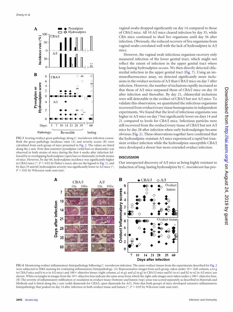
Rapid neutrophil recruitment into oviduct lumen of A/J dur-ing acute phase. We first compared the overall inflammatory in-filtration in oviduct lumen and tissues separately between CBA/Jand A/J mice (Fig. 4). The inflammatory scores were largely sim-ilar between these two strains of mice along the entire infectioncourse. The oviduct lumenal inflammatory scores peaked on day14, while the tissue scores peaked on days 10 and 14 after infection.The only significant differences between CBA/J and A/J mice werethe tissue inflammatory scores on day 60. There was no overallsignificant difference. These observations suggest that the micro-scopic assessment of overall inflammation infiltration was notsensitive enough for identifying correlates associated with eitherhydrosalpinx resistance or susceptibility.
We then counted individual types of inflammatory cells, in-cluding neutrophils and mononuclear cells (mainly consisting ofmacrophages, lymphocytes, and plasma cells), in both oviductlumen and tissue sections (Fig. 5). Under a 100� objective lens,neutrophils and mononuclear cells can be distinguished. Wefound that during the first 10 days of infection, most inflamma-tory cells from either oviduct lumen or tissue sections were neu-trophils for both CBA/J and A/J mice. Interestingly, the level ofneutrophil exudation in the oviduct lumen was significantlyhigher in the hydrosalpinx-resistant A/J than the susceptible
CBA/J mice on both days 7 and 10. The tissue neutrophil infiltra-tion peaked on day 7, while the lumenal neutrophils peaked onday 10. By day 14, the neutrophils started to drop and continued todecline thereafter, which marked the onset of chronic infection.Consistent with the chronic infection, significant numbers of in-flammatory cells became mononuclear cells on day 14. The mono-nuclear cells continued to increase and peaked on day 28. By day60, only a minimal number of inflammatory cells was detected inA/J mice, while a significant number of inflammatory cells was stillmaintained in CBA/J mice, which is consistent with the overallinflammation assessment described above for Fig. 4. Together, theabove-described observations demonstrated that the hydrosal-pinx-resistant A/J mice responded to chlamydial infection with amore rapid recruitment of neutrophils into the oviduct lumenduring the acute phase with significantly more neutrophil infiltra-tion on days 7 and 10 after infection compared to those of thehydrosalpinx-susceptible CBA/J mice. These results suggested acorrelation between rapid neutrophil recruitment during theacute phase and lack of long-lasting hydrosalpinx.
Oviduct infection peaks earlier but resolves faster in A/J thanCBA/J mice. Since both infection courses and host responses cancontribute to chlamydial induction of hydrosalpinx, we furthercompared chlamydial infection courses in CBA/J and A/J mice bymonitoring the live organism recovery from vaginal swabs follow-ing intravaginal inoculation with C. muridarum (Fig. 6). Wefound that A/J mice displayed a significantly reduced infectiontime course. The level of live organisms recovered from A/J mouse
FIG 2 Monitoring oviduct gross pathology along C. muridarum infection course. CBA/J and A/J mice were infected with C. muridarum as described in the legendto Fig. 1 and sacrificed for visual observation of oviduct pathology, including both pyosalpinx and hydrosalpinx, on days 3 (n � 5 for CBA/J and A/J groups,respectively), 7 (n � 5), 10 (n � 6), 14 (n � 5), 21 (n � 5), 28 (n � 5), 35 (n � 5), and 60 (n � 10) after inoculation. One or two representative images ofoviduct/ovary from both sides of a mouse genital tract are presented for both strains of mice at each time point, except for days 3 and 7 (no gross pathology couldbe detected on these early days). The visually observable pyosalpinx (Pyo) was marked with yellow and hydrosalpinx (Hydro) with red arrows, while the grosspathology severity was scored according to the criteria described in Materials and Methods and marked with numbers in white in corresponding images. Tovalidate the visually observable gross pathology, H&E-stained sections were prepared from the corresponding genital tract tissues (Histopath.). Representativeimages from each strain of mice, taken under a 10� and 100� objective lens, respectively, are shown with pyosalpinx, hydrosalpinx, or normal oviducts markedwith P (in yellow), H (red), or N (blue), respectively. White rectangles in images of a 10� objective lens indicate the areas where the images were further magnifiedunder the 100� objective lens. Note that oviducts with pyosalpinx (yellow P) were filled with inflammatory infiltrates, while those with hydrosalpinx (red H)showed empty lumenal space suggestive of clear solution accumulation. Inflammation was also observed under microscopy in oviducts without visuallydetectable pathology.
Oviduct Infection and Inflammation in Hydrosalpinx
July 2014 Volume 82 Number 7 iai.asm.org 2691
on August 24, 2019 by guest
http://iai.asm.org/
Dow
nloaded from
vaginal swabs dropped significantly on day 14 compared to thoseof CBA/J mice. All 10 A/J mice cleared infection by day 35, whileCBA mice continued to shed live organisms until day 56 afterinfection. Obviously, the reduced recovery of live organisms fromvaginal swabs correlated well with the lack of hydrosalpinx in A/Jmice.
However, the vaginal swab infectious organism recovery onlymeasured infection of the lower genital tract, which might notreflect the extent of infection in the upper genital tract wherelong-lasting hydrosalpinx occurs. We then directly detected chla-mydial infection in the upper genital tract (Fig. 7). Using an im-munofluorescence assay, we detected significantly more inclu-sions in the oviduct sections of A/J than CBA/J mice on day 7 afterinfection. However, the number of inclusions rapidly increased sothat those of A/J mice surpassed those of CBA/J mice on day 10after infection and thereafter. By day 21, chlamydial inclusionswere still detectable in the oviduct of CBA/J but not A/J mice. Tovalidate this observation, we quantitated the infectious organismsrecovered from oviduct/ovary tissue homogenates in independentexperiments. We found that the level of infectious organisms washigher in A/J mice on day 7 but significantly lower on days 14 and21 compared to levels for CBA/J mice. Infectious particles werestill recovered from the oviduct/ovary tissue of CBA/J but not A/Jmice by day 28 after infection when early hydrosalpinges becameobvious (Fig. 2). These observations together have confirmed thatthe hydrosalpinx-resistant A/J mice experienced a rapid but tran-sient oviduct infection while the hydrosalpinx-susceptible CBA/Jmice developed a slower but more extended oviduct infection.
DISCUSSION
Our unexpected discovery of A/J mice as being highly resistant toinduction of long-lasting hydrosalpinx by C. muridarum has pro-
FIG 3 Scoring oviduct gross pathology along C. muridarum infection course.Both the gross pathology incidence rates (A) and severity scores (B) werecalculated from each group of mice presented in Fig. 2. The values are listedalong the y axis. Note that extensive pyosalpinx (solid bars or diamonds) wasobserved in both strains of mice during the first 4 weeks after infection fol-lowed by or overlapping hydrosalpinx (open bars or diamonds) in both strainsof mice. However, by day 60, hydrosalpinx incidence was significantly higherin CBA/J mice (*, P � 0.01 by Fisher’s exact; also see the legend to Fig. 1), andby days 35 and 60, hydrosalpinx severity was significantly lower in A/J mice (*,P � 0.01 by Wilcoxon rank-sum test).
FIG 4 Monitoring oviduct inflammatory histopathology following C. muridarum infection. The same oviduct tissues from the experiments described for Fig. 2were subjected to H&E staining for evaluating inflammatory histopathology. (A) Representative images from each group, taken under 10� (left column, a to gin CBA/J mice and h to n in A/J mice) and 100� objective lenses (right column, a1 to g1 and a2 to g2 in CBA/1J mice and h1 to n1 and h2 to n2 in A/J mice) areshown. White rectangles in images from the 10� objective lens indicate the same areas from which the right-side images were taken under a 100� objective lens.(B) The severity of inflammatory infiltration or exudation in oviduct tissue (bottom) and lumen (top) areas was scored separately as described in Materials andMethods and is listed along the y axis (solid diamonds for CBA/J, open diamonds for A/J). Note that both groups of mice developed extensive inflammatoryhistopathology that peaked on day 14 after infection in both oviduct tissue and lumen (*, P � 0.05 by Wilcoxon rank-sum test).
Zhang et al.
2692 iai.asm.org Infection and Immunity
on August 24, 2019 by guest
http://iai.asm.org/
Dow
nloaded from

vided us a unique tool for further investigating the mechanisms ofchlamydial pathogenic mechanisms in the murine model. By si-multaneously comparing host inflammatory responses and chla-mydial infection courses between the hydrosalpinx-resistant A/Jand CBA/J mice known to be susceptible to hydrosalpinx induc-tion, we have further defined the relationship between oviductinfection and inflammatory responses in chlamydial pathogene-sis. Although both mouse strains developed robust pyosalpinxduring the acute phase followed by hydrosalpinx during thechronic phase, the hydrosalpinges disappeared in A/J mice by day60 after infection. The lack of long-lasting hydrosalpinx in A/Jmice correlated with an accelerated exudation of neutrophils intothe oviduct lumen during early stages of infection. As a result, theA/J mice only experienced a transient oviduct infection. In con-trast, the hydrosalpinx-susceptible CBA/J mice developed moreextended mononuclear cell infiltration with a more extensive ovi-duct infection, which may significantly contribute to the develop-ment of long-lasting hydrosalpinx.
FIG 6 Monitoring C. muridarum intravaginal infection time course. Thesame CBA/J (solid diamonds) and A/J (open diamonds) mice intravaginallyinfected with C. muridarum as described in the legend to Fig. 1 were monitoredfor live organism recovery from vaginal swabs taken after infection as indicatedalong the x axis. Swab samples were titrated on HeLa cell monolayers, and theinclusion-forming units (IFUs) calculated from each group were expressed aslog10 IFUs as shown along the y axis. Note that the hydrosalpinx-resistant A/Jmice displayed a reduced level of live organism recovery and shortened shed-ding time course (*, P � 0.05; **, P � 0.01; Student t test).
FIG 5 Detecting oviduct inflammatory cells following C. muridarum infection. The H&E-stained sections described in the legend to Fig. 4 were reexamined foridentifying neutrophils and mononuclear cells. (A) Example images marked with either neutrophils (red arrows) or mononuclear cells (yellow arrows) areshown. In the image taken from an oviduct tissue section with an acute infection, more neutrophils were detected (a, A/J mice), while at the chronic stage, moremononuclear inflammatory cells, including plasma cells, lymphocytes, and macrophages, were detected (b, CBA/J mice). Total inflammatory cells and neutro-phil and mononuclear cell subpopulations were counted in individual 100� objective lens views (for high-density infiltration) or the entire oviduct tissue orlumen section (for scarce infiltration). (B) The number of neutrophils (a and b) and mononuclear cells (c and d) per view from either oviduct lumen (a and c)or tissue (b and d) areas of each mouse was used to calculate means and standard deviations for each group as shown along the y axis. Note that both CBA/J (solidbar) and A/J (open bar) mice were dominated by neutrophils in both oviduct lumen and tissues during the first 10 days of infection (a and b), followed bymononuclear cells (c and d). Interestingly, levels of oviduct lumenal neutrophils were significantly higher in A/J than CBA/J mice on days 7 and 10.
Oviduct Infection and Inflammation in Hydrosalpinx
July 2014 Volume 82 Number 7 iai.asm.org 2693
on August 24, 2019 by guest
http://iai.asm.org/
Dow
nloaded from

A second surprise finding in the current study was that thehydrosalpinx-resistant A/J mice also developed robust pyosalpinxand maintained hydrosalpinx during the first 5 weeks. More than50% of A/J mice developed visible hydrosalpinx by days 28 or 35after infection. This is certainly an underestimate, since visual ob-servation is not nearly as sensitive as microscope examination foridentifying hydrosalpinx. Microscopic examination of the sameA/J mouse oviducts validated the hydrosalpinges visually identi-fied. The hydrosalpinges revealed under microscopy were as se-vere as those previously identified microscopically on day 35 (13)or 42 (31) after infection. However, by day 60 after infection, mostof these early hydrosalpinges disappeared in A/J mice. These ob-servations have demonstrated that the hydrosalpinges detectedusing microscopy during the first 5 or 6 weeks of infection arereversible and do not always develop into long-lasting hydrosal-pinx. Indeed, fibrosis is known to be reversible (32). Tubal fibrosis
responsible for causing these early hydrosalpinges may be reversedin the late stages of infection. In addition, it has been reported thatnot all mouse hydrosalpinges represent complete fibrotic blockageof the oviducts (3). However, it is the long-lasting hydrosalpinxthat is most medically relevant, since fallopian tubal occlusion isdetected in most women with tubal-factor infertility by hystero-salpingography (33). Thus, it is necessary to ensure that the ob-served hydrosalpinx is long-lasting and represents irreversibletubal blockage when investigating chlamydial pathogenic mecha-nisms using the C. muridarum murine infection model, which canbe achieved by either using a dye solution to directly measure theoviduct patency (3) or visually observing hydrosalpinx 60 days orlonger after infection (12, 14, 22, 27, 34, 35).
A third surprise finding in the current manuscript was ourobservation that significantly more chlamydial inclusions in theoviduct sections and higher numbers of infectious organisms in
FIG 7 Monitoring oviduct infection following C. muridarum intravaginal inoculation. (A) The oviduct sections from genital tract tissues harvested from miceon days 3 (n � 5 for CBA/J and A/J mice, respectively), 7 (n � 10), 10 (n � 10), 14 (n � 5), and 21 (n � 5) after infection, as described in the legend to Fig. 2,plus additional mice for immunofluorescence assay. The sections were labeled with a DNA dye (blue) and a rabbit antichlamydial antibody (raised with C.muridarum elementary bodies; green). Representative images taken under 10� (a to j) and 100� (a1 to j1) magnification from both CBA/J (a to e and a1 to e1)and A/J (f to j and f1 to j1) at each time as indicated on top of each image are presented with representative inclusions marked with green arrows. White rectanglesin images from a 10� objective lens indicate the same areas viewed under the 100� objective lens. (B) The inclusions were counted and semiquantitated asdescribed in Materials and Methods, as shown along the y axis. The number of inclusions per slide varied considerably from 0 to �300 (image a). To showdifferences between the two groups of mice, the region covering samples with 25 or fewer inclusions was magnified as image a1. Note that on day 7 after infection,significantly more inclusions were detected in the oviduct sections of A/J mice (a1, open diamonds). The number of inclusions rapidly increased in CBA/J mice(solid), surpassing the A/J mice on day 10 postinfection or thereafter. (C) The microscopic observations described here were validated by titrating infectiouschlamydial organisms from oviduct/ovary tissue homogenates harvested from mice on days 3, 7, 10, 14, 21, and 28, as indicated along the x axis. The recoveredIFUs, expressed in log10 values, are shown along the y axis. Note that the IFUs were higher in A/J oviduct/ovary tissue samples on day 7 but significantly lower ondays 14 and 21 (*, P � 0.05; Student t test). The IFU recovery study was carried out with 5 mice per group.
Zhang et al.
2694 iai.asm.org Infection and Immunity
on August 24, 2019 by guest
http://iai.asm.org/
Dow
nloaded from

the homogenate of oviduct/ovary were detected in the hydrosal-pinx-resistant A/J mice on day 7 after infection compared toCBA/J mice. This rapid ascension should have made the A/J micemore susceptible to the development of more severe upper genitalpathology. Although the A/J mice developed pyosalpinx andmaintained hydrosalpinx during the first 5 weeks, by day 60, theseearly hydrosalpinges disappeared. The question is why? One hy-pothesis is that the rapid ascension triggered an accelerated acuteinflammation, as evidenced by the significantly enhanced neutro-phil exudation into oviduct lumen of the A/J mice on days 7 and10 after infection. The enhanced acute inflammatory neutrophilsmay be effective in limiting chlamydial infection in the oviduct.This hypothesis is supported by the finding that the rapid oviductinfection peaked on day 7, followed by a significantly suppressedchlamydial organism count on days 10 and 14 and no infectiousorganisms detectable by day 28. The roles of early neutrophil in-filtration in protection against chlamydial infection and pathoge-nicity have been demonstrated previously (15, 16). Although thesignificantly reduced oviduct infection may have induced theacute pyosalpinx and early hydrosalpinx, it may not be able tomaintain inflammation that drives the development of long-last-ing hydrosalpinx. This hypothesis is also supported by our recentfindings that long-lasting hydrosalpinx require significant and ex-tended oviduct infection (12). Alternatively, a second hypothesisis that A/J mice are genetically defective in responding to chlamyd-ial infection with a hydrosalpinx-causing prolonged inflamma-tory response, which is inconsistent with the equally robust pyo-salpinx we detected in both A/J and CBA/J mice. Interestingly, A/Jmice are known to be deficient in complement component 5 (C5)(36, 37). Since C5 has been found to play significant roles in var-ious inflammatory pathologies and anti-C5 therapy has beenshown to reduce the severity of autoimmune pathologies (38, 39),it is possible that A/J mice are not able to develop long-lastinghydrosalpinx if C5 is required for the conversion of early hydro-salpinx into long-lasting hydrosalpinx. Although complementcomponent 3 (C3) has been shown to be protective in controllingC. muridarum lung infection (40), the role of C5 in either chla-mydial infection or pathogenesis has not been evaluated. Since C5can be activated independently of C3 (41), we are currently exam-ining the role of C5 in chlamydial induction of hydrosalpinx,which may help explain the pronounced pathological differencesobserved between A/J and CBA/J mice and provide insights indevising effective therapeutic interventions.
Although we have revealed a distinct difference in long-lastinghydrosalpinx development between CBA/J and A/J mice, it is notclear whether these two strains also display such a distinct differ-ence in infertility during mating experiments. Thus, besides usingthe CBA/J and A/J mouse platform to further understand the cel-lular and molecular mechanisms involved in hydrosalpinx devel-opment, it will be worth testing whether A/J mice can also main-tain largely normal fertility 60 days after C. muridarum infectionwhen CBA/J mice are induced to develop significant infertility.
REFERENCES1. Sherman KJ, Daling JR, Stergachis A, Weiss NS, Foy HM, Wang SP,
Grayston JT. 1990. Sexually transmitted diseases and tubal pregnancy. Sex.Transm. Dis. 17:115–121. http://dx.doi.org/10.1097/00007435-199007000-00001.
2. de la Maza LM, Pal S, Khamesipour A, Peterson EM. 1994. Intravaginalinoculation of mice with the Chlamydia trachomatis mouse pneumonitisbiovar results in infertility. Infect. Immun. 62:2094 –2097.
3. Shah AA, Schripsema JH, Imtiaz MT, Sigar IM, Kasimos J, Matos PG,Inouye S, Ramsey KH. 2005. Histopathologic changes related to fibroticoviduct occlusion after genital tract infection of mice with Chlamydiamuridarum. Sex. Transm. Dis. 32:49 –56. http://dx.doi.org/10.1097/01.olq.0000148299.14513.11.
4. Murthy AK, Li W, Guentzel MN, Zhong G, Arulanandam BP. 2011.Vaccination with the defined chlamydial secreted protein CPAF inducesrobust protection against female infertility following repeated genitalchlamydial challenge. Vaccine 29:2519 –2522. http://dx.doi.org/10.1016/j.vaccine.2011.01.074.
5. Stephens RS. 2003. The cellular paradigm of chlamydial pathogenesis. TrendsMicrobiol. 11:44–51. http://dx.doi.org/10.1016/S0966-842X(02)00011-2.
6. Cheng W, Shivshankar P, Li Z, Chen L, Yeh IT, Zhong G. 2008. Caspase-1contributes to Chlamydia trachomatis-induced upper urogenital tract in-flammatory pathologies without affecting the course of infection. Infect. Im-mun. 76:515–522. http://dx.doi.org/10.1128/IAI.01064-07.
7. Cotter TW, Meng Q, Shen ZL, Zhang YX, Su H, Caldwell HD. 1995.Protective efficacy of major outer membrane protein-specific immuno-globulin A (IgA) and IgG monoclonal antibodies in a murine model ofChlamydia trachomatis genital tract infection. Infect. Immun. 63:4704 –4714.
8. Pal S, Peterson EM, de la Maza LM. 2005. Vaccination with the Chla-mydia trachomatis major outer membrane protein can elicit an immuneresponse as protective as that resulting from inoculation with live bacteria.Infect. Immun. 73:8153– 8160. http://dx.doi.org/10.1128/IAI.73.12.8153-8160.2005.
9. Rodgers AK, Budrys NM, Gong S, Wang J, Holden A, Schenken RS,Zhong G. 2011. Genome-wide identification of Chlamydia trachomatisantigens associated with tubal factor infertility. Fertil. Steril. 96:715–721.http://dx.doi.org/10.1016/j.fertnstert.2011.06.021.
10. Chanelles O, Ducarme G, Sifer C, Hugues JN, Touboul C, Poncelet C.2011. Hydrosalpinx and infertility: what about conservative surgical man-agement? Eur. J. Obstet. Gynecol. Reprod. Biol. 159:122–126. http://dx.doi.org/10.1016/j.ejogrb.2011.07.004.
11. Budrys NM, Gong S, Rodgers AK, Wang J, Louden C, Shain R, SchenkenRS, Zhong G. 2012. Chlamydia trachomatis antigens recognized in womenwith tubal factor infertility, normal fertility, and acute infection. Obstet. Gy-necol. 119:1009–1016. http://dx.doi.org/10.1097/AOG.0b013e3182519326.
12. Lei L, Chen J, Hou S, Ding Y, Yang Z, Zeng H, Baseman J, Zhong G.2014. Reduced live organism recovery and lack of hydrosalpinx in miceinfected with plasmid-free Chlamydia muridarum. Infect. Immun. 82:983–992. http://dx.doi.org/10.1128/IAI.01543-13.
13. Darville T, O’Neill JM, Andrews CW, Jr, Nagarajan UM, Stahl L, OjciusDM. 2003. Toll-like receptor-2, but not Toll-like receptor-4, is essentialfor development of oviduct pathology in chlamydial genital tract infec-tion. J. Immunol. 171:6187– 6197.
14. Chen L, Lei L, Chang X, Li Z, Lu C, Zhang X, Wu Y, Yeh IT, Zhong G.2010. Mice deficient in MyD88 develop a Th2-dominant response andsevere pathology in the upper genital tract following Chlamydia muri-darum infection. J. Immunol. 184:2602–2610. http://dx.doi.org/10.4049/jimmunol.0901593.
15. Barteneva N, Theodor I, Peterson EM, de la Maza LM. 1996. Role ofneutrophils in controlling early stages of a Chlamydia trachomatis infec-tion. Infect. Immun. 64:4830 – 4833.
16. Andrew DW, Cochrane M, Schripsema JH, Ramsey KH, Dando SJ,O’Meara CP, Timms P, Beagley KW. 2013. The duration of Chlamydiamuridarum genital tract infection and associated chronic pathologicalchanges are reduced in IL-17 knockout mice but protection is not in-creased further by immunization. PLoS One 8:e76664. http://dx.doi.org/10.1371/journal.pone.0076664.
17. Darville T, Andrews CW, Jr, Sikes JD, Fraley PL, Rank RG. 2001. Earlylocal cytokine profiles in strains of mice with different outcomes fromchlamydial genital tract infection. Infect. Immun. 69:3556 –3561. http://dx.doi.org/10.1128/IAI.69.6.3556-3561.2001.
18. Imtiaz MT, Schripsema JH, Sigar IM, Kasimos JN, Ramsey KH. 2006.Inhibition of matrix metalloproteinases protects mice from ascending in-fection and chronic disease manifestations resulting from urogenitalChlamydia muridarum infection. Infect. Immun. 74:5513–5521. http://dx.doi.org/10.1128/IAI.00730-06.
19. Ramsey KH, Sigar IM, Rana SV, Gupta J, Holland SM, Byrne GI. 2001.Role for inducible nitric oxide synthase in protection from chronic Chla-mydia trachomatis urogenital disease in mice and its regulation by oxygen
Oviduct Infection and Inflammation in Hydrosalpinx
July 2014 Volume 82 Number 7 iai.asm.org 2695
on August 24, 2019 by guest
http://iai.asm.org/
Dow
nloaded from

free radicals. Infect. Immun. 69:7374 –7379. http://dx.doi.org/10.1128/IAI.69.12.7374-7379.2001.
20. Nagarajan UM, Sikes JD, Yeruva L, Prantner D. 2012. Significant role ofIL-1 signaling, but limited role of inflammasome activation, in oviductpathology during Chlamydia muridarum genital infection. J. Immunol.188:2866 –2875. http://dx.doi.org/10.4049/jimmunol.1103461.
21. Igietseme JU, Omosun Y, Partin J, Goldstein J, He Q, Joseph K,Ellerson D, Ansari U, Eko FO, Bandea C, Zhong G, Black CM. 2013.Prevention of Chlamydia-induced infertility by inhibition of local caspaseactivity. J. Infect. Dis. 207:1095–1104. http://dx.doi.org/10.1093/infdis/jit009.
22. Chen L, Cheng W, Shivshankar P, Lei L, Zhang X, Wu Y, Yeh IT, ZhongG. 2009. Distinct roles of CD28- and CD40 ligand-mediated costimula-tion in the development of protective immunity and pathology duringChlamydia muridarum urogenital infection in mice. Infect. Immun. 77:3080 –3089. http://dx.doi.org/10.1128/IAI.00611-08.
23. Murthy AK, Li W, Chaganty BK, Kamalakaran S, Guentzel MN, SeshuJ, Forsthuber TG, Zhong G, Arulanandam BP. 2011. Tumor necrosisfactor alpha production from CD8� T cells mediates oviduct pathologicalsequelae following primary genital Chlamydia muridarum infection. In-fect. Immun. 79:2928 –2935. http://dx.doi.org/10.1128/IAI.05022-11.
24. Cheng W, Shivshankar P, Zhong Y, Chen D, Li Z, Zhong G. 2008.Intracellular interleukin-1alpha mediates interleukin-8 production in-duced by Chlamydia trachomatis infection via a mechanism independentof type I interleukin-1 receptor. Infect. Immun. 76:942–951. http://dx.doi.org/10.1128/IAI.01313-07.
25. Rasmussen SJ, Eckmann L, Quayle AJ, Shen L, Zhang YX, Anderson DJ,Fierer J, Stephens RS, Kagnoff MF. 1997. Secretion of proinflammatorycytokines by epithelial cells in response to Chlamydia infection suggests acentral role for epithelial cells in chlamydial pathogenesis. J. Clin. Investig.99:77– 87. http://dx.doi.org/10.1172/JCI119136.
26. Chen C, Chen D, Sharma J, Cheng W, Zhong Y, Liu K, Jensen J, ShainR, Arulanandam B, Zhong G. 2006. The hypothetical protein CT813 islocalized in the Chlamydia trachomatis inclusion membrane and is im-munogenic in women urogenitally infected with C. trachomatis. Infect.Immun. 74:4826 – 4840. http://dx.doi.org/10.1128/IAI.00081-06.
27. Li Z, Lu C, Peng B, Zeng H, Zhou Z, Wu Y, Zhong G. 2012. Inductionof protective immunity against Chlamydia muridarum intravaginal infec-tion with a chlamydial glycogen phosphorylase. PLoS One 7:e32997. http://dx.doi.org/10.1371/journal.pone.0032997.
28. Zhong G, Reis e Sousa C, Germain RN. 1997. Production, specificity,and functionality of monoclonal antibodies to specific peptide-major his-tocompatibility complex class II complexes formed by processing of ex-ogenous protein. Proc. Natl. Acad. Sci. U. S. A. 94:13856 –13861. http://dx.doi.org/10.1073/pnas.94.25.13856.
29. Fan T, Lu H, Hu H, Shi L, McClarty GA, Nance DM, Greenberg AH,Zhong G. 1998. Inhibition of apoptosis in chlamydia-infected cells: block-ade of mitochondrial cytochrome c release and caspase activation. J. Exp.Med. 187:487– 496. http://dx.doi.org/10.1084/jem.187.4.487.
30. Zhong G, Fan P, Ji H, Dong F, Huang Y. 2001. Identification of achlamydial protease-like activity factor responsible for the degradation of
host transcription factors. J. Exp. Med. 193:935–942. http://dx.doi.org/10.1084/jem.193.8.935.
31. O’Connell CM, Ingalls RR, Andrews CW, Jr, Scurlock AM, Darville T.2007. Plasmid-deficient Chlamydia muridarum fail to induce immunepathology and protect against oviduct disease. J. Immunol. 179:4027–4034.
32. Pellicoro A, Ramachandran P, Iredale JP. 2012. Reversibility of liverfibrosis. Fibrogenesis Tissue Repair 5(Suppl 1):S26.
33. Broeze KA, Opmeer BC, Van Geloven N, Coppus SF, Collins JA, DenHartog JE, Van der Linden PJ, Marianowski P, Ng EH, Van der SteegJW, Steures P, Strandell A, Van der Veen F, Mol BW. 2011. Are patientcharacteristics associated with the accuracy of hysterosalpingography indiagnosing tubal pathology? An individual patient data meta-analysis.Hum. Reprod. Update 17:293–300. http://dx.doi.org/10.1093/humupd/dmq056.
34. Chen L, Lei L, Zhou Z, He J, Xu S, Lu C, Chen J, Yang Z, Wu G, YehIT, Zhong G, Wu Y. 2013. Contribution of interleukin-12 p35 (IL-12p35)and IL-12p40 to protective immunity and pathology in mice infected withChlamydia muridarum. Infect. Immun. 81:2962–2971. http://dx.doi.org/10.1128/IAI.00161-13.
35. Lu C, Zeng H, Li Z, Lei L, Yeh IT, Wu Y, Zhong G. 2012. Protectiveimmunity against mouse upper genital tract pathology correlates withhigh IFNgamma but low IL-17 T cell and anti-secretion protein antibodyresponses induced by replicating chlamydial organisms in the airway. Vac-cine 30:475– 485. http://dx.doi.org/10.1016/j.vaccine.2011.10.059.
36. Tuite A, Elias M, Picard S, Mullick A, Gros P. 2005. Genetic control ofsusceptibility to Candida albicans in susceptible A/J and resistantC57BL/6J mice. Genes Immun. 6:672– 682. http://dx.doi.org/10.1038/sj.gene.6364254.
37. Mashruwala MA, Smith AK, Lindsey DR, Moczygemba M, Wetsel RA,Klein JR, Actor JK, Jagannath C. 2011. A defect in the synthesis ofinterferon-gamma by the T cells of complement-C5 deficient mice leads toenhanced susceptibility for tuberculosis. Tuberculosis 91(Suppl 1):S82–S89. http://dx.doi.org/10.1016/j.tube.2011.10.016.
38. Copland DA, Hussain K, Baalasubramanian S, Hughes TR, Morgan BP,Xu H, Dick AD, Nicholson LB. 2010. Systemic and local anti-C5 therapyreduces the disease severity in experimental autoimmune uveoretinitis.Clin. Exp. Immunol. 159:303–314. http://dx.doi.org/10.1111/j.1365-2249.2009.04070.x.
39. Durigutto P, Macor P, Ziller F, De Maso L, Fischetti F, Marzari R,Sblattero D, Tedesco F. 2013. Prevention of arthritis by locally synthe-sized recombinant antibody neutralizing complement component C5.PLoS One 8:e58696. http://dx.doi.org/10.1371/journal.pone.0058696.
40. Bode J, Dutow P, Sommer K, Janik K, Glage S, Tummler B, Munder A,Laudeley R, Sachse KW, Klos A. 2012. A new role of the complementsystem: C3 provides protection in a mouse model of lung infection withintracellular Chlamydia psittaci. PLoS One 7:e50327. http://dx.doi.org/10.1371/journal.pone.0050327.
41. Ramos TN, Darley MM, Weckbach S, Stahel PF, Tomlinson S, BarnumSR. 2012. The C5 convertase is not required for activation of the terminalcomplement pathway in murine experimental cerebral malaria. J. Biol.Chem. 287:24734 –24738. http://dx.doi.org/10.1074/jbc.C112.378364.
Zhang et al.
2696 iai.asm.org Infection and Immunity
on August 24, 2019 by guest
http://iai.asm.org/
Dow
nloaded from