Pregnancy and the Nephrotic Syndrome - bmj.com · PregnancyandNephrotic Syndrome-StuddandBlainey in...
5
276 1 February 1969 Rubella Vaccines-Dudgeon et al. Cooper, L. Z., Giles, J. P., and Krugman, S. (1968). Amer. 7. Dis. Child., 115, 655. Dudgeon, J. A. (1967). Arch. Dis. Childh., 42, 110. DudgCon, J. A., Butler, N. R., and Plotkin, S. A. (1964). Brit. med. 7., 2, 155. Du Pan, R. M., Huygelen, C., Peetermans, J., and Prinzie, A. (1968a). Pediat. Res., 2, 38. Dii Pan, R. M., Huygelen, C., Peetermans, J., and Prinzie, A. (1968b). Amer. 7. Dis. Child., 115, 658. Gregg, N. McA. (1941). Trans. ophthal. Soc. Aust., 3, 35. Huygelen, C., and Peetermans, J. (1967). Arch. ges. Virusforsch., 21, 357. Lepow, M. L., Veronelli, J. A., Hostetler, D. D., and Robbins, P. C. (1968). Amer. 7. Dis. Child., 115, 639. McCarthy, K., and Taylor-Robinson, C. H. (1967). Brit. med. Bull., 23, 185. Meyer, H. M., Parkman, P. D., and Panos, T. C. (1966). New Engl. 7. Med., 275, 575. Meyer, H. M., and Parkman, P. D. (1969). Proceedings of the 23rd Symposium on Microbiological Standardization-Rubella Vaccines. London, November 1968. In press. Meyer, x M., Parkman, P. D., Hobbins, T. E., Larson, H. E., and Hopps, H. E. (1968a). To be published. Meyer, H. M., Parkman, P. D., Hobbins, T. E., and Ennis, F. A. (1968b). Amer. 7. Dis. Child., 115, 648. SParkman, P. D., Meyer, H. M., Kirschstein, R. L., and Hopps, H. E. (1966). New Engl. 7. Med., 275, 569. Plotkin, S. A., Dudgeon, J. A., and Ransay, A. M. (1963). Brit. med. 7., 2, 1296. Plotkin, S. A., Farquhar, J., Katz, M., and Ingalls, T. H. (1967). Amer. 7. Epidem., 86, 468. Public Health Laboratory Service (1968). Brit. med. 7., 3, 203. Sever J. L., et al. (1966). Proc. Soc. exp. Biol. (N.Y.), 122, 513. Stewart, G. L., Parkman, P. D., Hopps, H. E,, Douglas, R. D., Hamilton, J. P., and Meyer, H. M. (1967). New Engl. 7. Med., 276, 554. Pregnancy and the Nephrotic Syndrome J. W. W. STUDD,* M.R.C.O.G.; J. D. BLAINEYt M.D., F.R.C.P. Brit. med. J., 1969, 1, 276-280 Summary: Nineteen patients with nephrotic syndrome, 13 with histological diagnosis, were studied through- out 31 pregnancies. Eight were diagnosed for the first time during pregnancy. Antenatal problems due to severe oedema, urinary tract infection, and refractory orthochromic anemia were encountered. t patients were hypertensive at book- g, and in two of these pregnancy was terminated; three others had a significant increase in blood pressure. In 12 of the remaining pregnancies a rise in blood pressure of 20 mm, Hg or more occurred towards term. There were 29 live births (including one set of twins), one stillbirth due to a cord accident, and one neonatal death. The infant birth weight, apart from being affected by hypertension, was related to the maternal serum albumin level. The patients have been under observation for up to 20 years. Fifteen have not shown any deterioration of renal function during the prolonged period of observation. One developed oliguric renal failure immediately post partum and three others died, two, four, and 12 years after their pregnancies. Introduction The relationship between pre-eclampsia and the nephrotic syndrome has remained confused and poorly understood, largely owing to the varied morbidity and clinical course of both con- ditions. Without renal biopsy or long-term clinical studies the distinction may be extremely difficult. This similarity has led to the misdiagnosis and underreporting of the coexistence o nephrotic syndrome with pregnancy, estimated by Wegner (1937) as occurring in 0 028% of pregnancies. The nephrotic syndrome has been claimed to result from severe pre-eclampsia (Hopper et al., 1961 ; Sarles et al., 1964), and cyclical nephrotic syndrome has been reported as occurring only during preg- nancy (Schreiner, 1963). *Research Senior Registrar. Consultant Physician. Lkpartments of Obstetrics and Gynaecology and Experimental Pathology, Queen Elizabeth Hospital, Birmingham 15. The established view of Dieckmann (1936) and Wegner (1937) that pregnancy had a deleterious effect on chronic renal disease has been challenged by Seftel and Schewitz (1957), Silberman and Adams (1962), Marcus (1963), and Johnston et a!. (1963), who have stressed the good maternal prognosis during pregnancy and the low rate of foetal loss. This com- mumication is a study of 31 pregnancies occurring in 19 patients with the nephrotic syndrome. Many of these patients have been under careful long-term surveillance for more than 10 years. These pregnancies can therefore be viewed as isolated occurrences against the natural history and pathology of the renal disease, thus correcting the lack of long-term follow-up available in previous publications. The cases have been selected by rigid biochemical criteria (Squire et a!., 1957), all having had at some time a serum albumin of less than 2 g./100 ml. and a proteinuria of more than 5 g./day. In eight patients (Table I) the nephrotic syndrome was first diagnosed during pregnancy, though in two of these there was a past history of previous acute renal disease. In the remaining 11 patients (Table II) renal disease had been present from 1 to 15 years before the pregnancies studied. Five patients in this group were receiving steroid therapy throughout eight pregnancies and two others had already completed successful courses of steroids before the onset of their three pregnancies. Two of these patients (Cases 10 and 11) and one patient (Case 18) who exhibited a progressive spontaneous recovery after the first pregnancy were the only patients who did not show severe clinical and biochemical evidence of the nephrotic syndrome at the time of the preg- nancies. Two pregnancies were terminated at 16 weeks by hysterotomy on account of increasing oedema and hypetten- sion. Apart from the 31 pregnancies in the present study, there were two spontaneous abortions without biochemical data. Histological Diagnosis Renal biopsy was performed before or after pregnancy in 13 cases. Case 5 had a second biopsy one year after the preg- nancy and Case 2 had two renal biopsies following the initial biopsy taken at the time of caesarean section. The staining techniques and criteria of histological diagnosis were those used by Brewer (1964). The clinical history and progress, bio-
Transcript of Pregnancy and the Nephrotic Syndrome - bmj.com · PregnancyandNephrotic Syndrome-StuddandBlainey in...

276 1 February 1969 Rubella Vaccines-Dudgeon et al.Cooper, L. Z., Giles, J. P., and Krugman, S. (1968). Amer. 7. Dis.
Child., 115, 655.Dudgeon, J. A. (1967). Arch. Dis. Childh., 42, 110.DudgCon, J. A., Butler, N. R., and Plotkin, S. A. (1964). Brit. med. 7.,
2, 155.Du Pan, R. M., Huygelen, C., Peetermans, J., and Prinzie, A. (1968a).
Pediat. Res., 2, 38.Dii Pan, R. M., Huygelen, C., Peetermans, J., and Prinzie, A. (1968b).
Amer. 7. Dis. Child., 115, 658.Gregg, N. McA. (1941). Trans. ophthal. Soc. Aust., 3, 35.Huygelen, C., and Peetermans, J. (1967). Arch. ges. Virusforsch., 21,
357.Lepow, M. L., Veronelli, J. A., Hostetler, D. D., and Robbins, P. C.
(1968). Amer. 7. Dis. Child., 115, 639.McCarthy, K., and Taylor-Robinson, C. H. (1967). Brit. med. Bull., 23,
185.Meyer, H. M., Parkman, P. D., and Panos, T. C. (1966). New Engl. 7.
Med., 275, 575.
Meyer, H. M., and Parkman, P. D. (1969). Proceedings of the 23rdSymposium on Microbiological Standardization-Rubella Vaccines.London, November 1968. In press.
Meyer, x M., Parkman, P. D., Hobbins, T. E., Larson, H. E., andHopps, H. E. (1968a). To be published.
Meyer, H. M., Parkman, P. D., Hobbins, T. E., and Ennis, F. A.(1968b). Amer. 7. Dis. Child., 115, 648.
SParkman, P. D., Meyer, H. M., Kirschstein, R. L., and Hopps, H. E.(1966). New Engl. 7. Med., 275, 569.
Plotkin, S. A., Dudgeon, J. A., and Ransay, A. M. (1963). Brit. med. 7.,2, 1296.
Plotkin, S. A., Farquhar, J., Katz, M., and Ingalls, T. H. (1967). Amer.7. Epidem., 86, 468.
Public Health Laboratory Service (1968). Brit. med. 7., 3, 203.Sever J. L., et al. (1966). Proc. Soc. exp. Biol. (N.Y.), 122, 513.Stewart, G. L., Parkman, P. D., Hopps, H. E,, Douglas, R. D., Hamilton,
J. P., and Meyer, H. M. (1967). New Engl. 7. Med., 276, 554.
Pregnancy and the Nephrotic Syndrome
J. W. W. STUDD,* M.R.C.O.G.; J. D. BLAINEYt M.D., F.R.C.P.
Brit. med. J., 1969, 1, 276-280
Summary: Nineteen patients with nephrotic syndrome,13 with histological diagnosis, were studied through-
out 31 pregnancies. Eight were diagnosed for the firsttime during pregnancy.
Antenatal problems due to severe oedema, urinarytract infection, and refractory orthochromic anemia wereencountered. t patients were hypertensive at book-g, and in two of these pregnancy was terminated; threeothers had a significant increase in blood pressure. In 12of the remaining pregnancies a rise in blood pressure of20 mm, Hg or more occurred towards term.There were 29 live births (including one set of twins),
one stillbirth due to a cord accident, and one neonataldeath. The infant birth weight, apart from being affectedby hypertension, was related to the maternal serumalbumin level.The patients have been under observation for up to
20 years. Fifteen have not shown any deterioration ofrenal function during the prolonged period of observation.One developed oliguric renal failure immediately postpartum and three others died, two, four, and 12 yearsafter their pregnancies.
Introduction
The relationship between pre-eclampsia and the nephroticsyndrome has remained confused and poorly understood, largelyowing to the varied morbidity and clinical course of both con-ditions. Without renal biopsy or long-term clinical studiesthe distinction may be extremely difficult. This similarity hasled to the misdiagnosis and underreporting of the coexistenceo nephrotic syndrome with pregnancy, estimated by Wegner(1937) as occurring in 0 028% of pregnancies. The nephroticsyndrome has been claimed to result from severe pre-eclampsia(Hopper et al., 1961 ; Sarles et al., 1964), and cyclical nephroticsyndrome has been reported as occurring only during preg-nancy (Schreiner, 1963).
*Research Senior Registrar.Consultant Physician.
Lkpartments of Obstetrics and Gynaecology and Experimental Pathology,Queen Elizabeth Hospital, Birmingham 15.
The established view of Dieckmann (1936) and Wegner(1937) that pregnancy had a deleterious effect on chronic renaldisease has been challenged by Seftel and Schewitz (1957),Silberman and Adams (1962), Marcus (1963), and Johnstonet a!. (1963), who have stressed the good maternal prognosisduring pregnancy and the low rate of foetal loss. This com-mumication is a study of 31 pregnancies occurring in 19 patientswith the nephrotic syndrome. Many of these patients havebeen under careful long-term surveillance for more than 10years. These pregnancies can therefore be viewed as isolatedoccurrences against the natural history and pathology of therenal disease, thus correcting the lack of long-term follow-upavailable in previous publications.The cases have been selected by rigid biochemical criteria
(Squire et a!., 1957), all having had at some time a serumalbumin of less than 2 g./100 ml. and a proteinuria of morethan 5 g./day. In eight patients (Table I) the nephroticsyndrome was first diagnosed during pregnancy, though intwo of these there was a past history of previous acute renaldisease. In the remaining 11 patients (Table II) renal diseasehad been present from 1 to 15 years before the pregnanciesstudied. Five patients in this group were receiving steroidtherapy throughout eight pregnancies and two others hadalready completed successful courses of steroids before the onsetof their three pregnancies. Two of these patients (Cases 10and 11) and one patient (Case 18) who exhibited a progressivespontaneous recovery after the first pregnancy were the onlypatients who did not show severe clinical and biochemicalevidence of the nephrotic syndrome at the time of the preg-nancies. Two pregnancies were terminated at 16 weeks byhysterotomy on account of increasing oedema and hypetten-sion. Apart from the 31 pregnancies in the present study,there were two spontaneous abortions without biochemical data.
Histological Diagnosis
Renal biopsy was performed before or after pregnancy in13 cases. Case 5 had a second biopsy one year after the preg-nancy and Case 2 had two renal biopsies following the initialbiopsy taken at the time of caesarean section. The stainingtechniques and criteria of histological diagnosis were those usedby Brewer (1964). The clinical history and progress, bio-

1 February 1969 Pregnancy and Nephrotic Syndrome-Studd and BlaineyTABLE I.-Nephrotic Syndrome Presenting During Pregnancy
B.P. atBooking
130/90
130/80
140/80140/80160/90
120/70120/80
N.K.
120/80
120/80
140/90
B.P. in Late S. Alb Urea MaturityPregnancy (g./100 ml.) (mg./100 ml.) in Weeks
150/100
190/120150/100150/100200/120120/70110/70140/90
200/110
140/80
120/70
2-5
1-8
N.K.1-62-4
1-41-3
N.K.
0 9
1-9
2-2
15
61
242814
2030
40
110
30
28
36
33
383737
3740
40
33
40
38
Onset
Spontaneous
C.S. + steril.
InducedInducedInduced
C.S.Induced
SpontaneousSpontaneousInduced
Induced
* Case 6 received steroid therapy for a trial period of 4 weeks during the pregnancy.
Weight(g.)19132055
1,7003,5152,0982,7202,5802,8912,7501,633
2,8203,136
Creatinine Clearancein 1968 (ml./min.)
Died 1963 malignanthypertension
80
86
88
Died 1964 renal failure
Anuric-chronic dialysis
56
102
TABLE II.-Pregnancies in Patients with Diagnosed Nephrotic Syndrome
Creatinine B.PClearance
(Before Pregnancy)
117
71
142
65
62
23
108
94
80
N.K.
N.K.
110/70
110/60110/70
130/85
130/80
160/100130/70
250/150
160/100150/100
110/70130/90
140/90190/95
120/80
120/70
Date of B.P. in Late UreaPregnancy Pregnancy (0 ml.)
1961*1964*
1966t1967t
1960t1966*
19611965
1952
1960*1966
1963*1965*
1963*1965*
1955195819601963
1947
110/70110/70110/70100/60
130/80
160/100
180/1101501100
210/130180/100130/90
150/100190/1101501100190/110
120/80140/90150/90130/90
130/70
3320
3024
15
68
N.K.30
N.K.
5028
1611
2426
18352013
28
S. Alb. Maturity
100 ml.) in Weeks
2-04-2
3.93-8
4*01-4
N.K.2-3
N.K.
1-93-4
1.91-7
2-12-9
2-14 03-84 0
2-6
3740
4040
40
37
1634
16
3636
4035
3833
40404039
37
* Steroids administered during pregnancy. t Completed successful steroid course before pregnancies.
chemistry, and differential protein clearances as described byBlainey et al. (1960) enabled a fairly confident diagnosis to bemade in three cases without renal biopsy (Table III). Threefurther cases, also without biopsy, were of an undeterminedtype of gloInerulonephritis. Kark et al. (1958) described themany renal lesions that may produce the nephrotic syndromein adults, but only patients with the basic pathology ofglomerulonephritis were encountered in this series.
TABLE III
Diagnosis by
Minimal change glomerulonephritis ..Proliferative glomerulonephritis .
Membranous glomerulonephritis .Undetermined type ..
Bipy Protein:Biopsy jeIClearances3
46
Biochemical Changes During Pregnancy
Data concerning the quantity of proteinuria during preg-
nancy are often inadequate, sometimes unrecorded, or expressedas + + +, or by the grossly inaccurate Esbach technique.Urinary protein loss was estimated serially in the four mostrecent patients who had a gradual and variable increase inproteinuria in the first half of pregnancy. There did not seem
to be a further increase towards term or with the advent ofhypertension. In Case 9 massive increases in proteinuria
occurred on two occasions when an attempt was made to lowerthe steroid dose, but with a return to 60 mg. of prednisolonedaily the pregnancy was able to continue for 37 weeks withprotein loss of only 2 g./day. The quantity of proteinuriausually decreased markedly in the puerperium and was followedby a diuresis and a considerable loss in weight.The lowest serum albumin levels observed during each
pregnancy are recorded in Tables I and II. The oedema waspronounced in all patients, except during the seven pregnanciesthat occurred when the renal disease was in remission and inthe second pregnancy in Case 9, when the renal lesion wasbeing successfully treated with steroids. Three patientsdeveloped severe vulval oedema: Case 2 had a caesarean sectionat 33 weeks and has subsequently made virtually a completespontaneous recovery; Case 12 required intravenous infusionof albumin as well as the more usual treatment of high-proteinand low-salt diet, diuretics, and steroids, and had a successfulpregnancy. The other (Case 6) is described in full below.
Clinical Manifestations During PregnancyRefractory orthochromic anaemia of less than 10 g./100 ml.
occurred in 11 pregnancies, including one set of twins.The blood pressure at the booking clinic was compared with
that obtained during the week preceding delivery. In eightpregnancies a blood pressure of 140/90 or higher was recorded,
CaseNo.
1
2
3
4
5
6*
7
8
Pathology
Membranous
Membranous
Membranous
?Membranous
Proliferative
Proliferative
?Proliferative
BRsTrMEDICAL JOURNAL
Date ofPregnancy
1959
1958
195519591966
19641966
1962
1967
1964
1967
277
Case Onset Pathology
Minimal change
Minimal change
Minimal change
? Minimal change
Membranous
Membranous
Membranous
Proliferative
Proliferative
9
10
11
12
13
14
15
16
17
18
1960
1960
1958
1959
1953
1950
1952
1956
1957
1952
19 1933
Onset
InducedInduced
SpontaneousSpontaneousSpontaneousInduced
TerminatedC.S. + steril.
Terminated
C.S.C.S. + steril.
SpontaneousInduced
C.S.C.S.
SpontaneousSpontaneousSpontaneousSpontaneousC.S. + steril.
Weight(g.)
1,9283,1753,5443,2984,054
2,495
2,210
2,7352,976
3,1181,475
2,7781,360
3,2983,2043,7143,2882,608
CreatinineClearancein 1968
(ml./min.)
162
77
127
59
142IDied 1964
105
83
88
180
60
-1.. -i.. _ __1 _
!-..l ..l -1-!~- !1l ll~~~~~~~~~~~~~~~~~~~~~~~~~~~~~~~~~~~~~~~~~~~~~~~~~~~~~~
-I ~~~I _il1 --- -_ _
- -I
-~
111

Pregnancy and Nephrotic Syndrome-Studd and Blainey
in three of these the systolic or diastolic pressure had increasedby at least 20 mm. Hg towards term, and two cases (Nos. 13and 14) with severe hypertension were terminated soon aftertheir initial antenatal visit. Twenty-two pregnancies werenormotensive at booking; 12 of these showed similar significantincreases in blood pressure in the final weeks of pregnancy.The remaining patient (Case 5) was first seen in labour witha blood pressure of 140/90.A urinary tract infection occurred in 11 pregnancies, in one
patient producing a transient rise in blood urea to 90 mg./100 ml. One patient suffered from a chronic paronychia andboils. Five of these 11 patients were undergoing steroidtherapy at the time, and one of this steroid group was admittedin her second pregnancy with bronchopneumonia as well as arecurrent urinary infection. There were no cases of severeinfection in the puerperium.
There were 29 live births and one stillbirth. Labour occurredspontaneously from 33 to 40 weeks in 11 pregnancies. Fifteenwere induced or underwent caesarean section before term.Three pregnancies were induced at term. No pregnancywas allowed to become postmature. In all, 11 pregnancieswent to term. The stillbirth was caused by a cord around theneck at birth after the foetal heart had been clearly heard duringlabour. One infant delivered abdominally at 33 weeks wasnoted to be oedematous and died after two days.The birth weights and gestational ages of the 30 infants
documented in Tables I and II are expressed in Table IV aspercentiles from the charts of Lubchenco et al. (1963).
TABLE IV
One set of twins of 10 and 25 percentile birth weight are notincluded. It is noteworthy that four of the singleton infantswere dysmature, taking the 10th percentile as a definition, andthe overall pattern is one of infants of slightly less than averagebirth weight, with a preponderance of small babies born tohypertensive mothers. The four infants from mothers in arecovered phase of the nephrotic syndrome were all aboveaverage size, whereas the infants of the hypoalbuminaemic(Cases 4, 7, and 9) even in the absence of maternal hyperten-sion, were below the 25 percentile. Fig. 1 shows that in 26pregnancies the foetal birth weight was clearly related to thelevel of serum albumin in the mother.
4
2
E
8
-0
as
E
E3
0
S
S
BRiTIsHMEDICAL JOURNAL
Five patients had eight pregnancies while being treated withsteroids. Two of these patients in three pregnancies weredelivered by caesarean section and the remainder per vaginam.There were no congenital abnormalities, but two infants ofhypertensive mothers were below the 10th percentile.
Puerperium
No patient had any thromboembolic problem, though thesuggestion of Marcus (1963), that patients with the nephroticsyndrome have a tendency towards hypercoagulation states inpregnancy and the puerperium, persuaded us to anticoagulatethe three most recent patients.Most patients underwent a heavy diuresis in the first post-
partum week, the weight of the oedematous patients falling byas much as 10 kg. in that time. The failure of diuresis inCase 6 was unexpected and heralded the onset of oliguric renalfailure.
Long-term Follow-up of Renal Conditions
Six patients (two on steroids) showed a marked improvementof renal function following the pregnancy. One of this groupspontaneously improved through all of three pregnancies, andhad no proteinuria in her fourth pregnancy. Another patient,with a diagnosis of membranous glomerulonephritis discoveredduring pregnancy, had an elective caesarean section and tuballigation at 33 weeks for increasing oedema and nitrogen reten-tion, and has made an almost complete spontaneous recoveryover the course of 10 years with proteinuria of 1 g./day, normalrenal function, and a biopsy picture of lobular basementmembrane thickening.
Only in Case 6 (see Fig. 2) was there obvious deteriorationof renal function during pregnancy.
Creatininea, clearance
e0 E E E*.
' > E E -0 Lcs-
oQ u U
* 5-
* 0
.
.
S
0
* *o
* S
.
.
0
0
75 90
FIG. 1.-Association of low foetal birthweight with severityof maternal hypoalbuminaemia during pregnancy.
15 20 25 30 35Duration of pregnancy (weeks)
FIG. 2.-Further data on Case 6. Patient with nephrotic syndromediagnosed for the first time during the pregnancy which resulted in
oliguric renal failure.
Case 6.-A 26-year-old primigravida had no symptoms of renaldisease when she first attended the antenatal clinic at 14 weeks'gestation. She gave a history of acute glomerulonephritis andrheumatic fever when 10 years old, remaining well when follow-upwas discontinued five years later. She had a blood pressure of140/90 mm. Hg and a proteinuria of 7 g./day. Ankle oedemaoccurred at the twenty-first week of pregnancy, becoming moresevere with increasing maturity despite high-protein and low-saltdiet, rest, and diuretics. She was admitted with severe vulval
278 1 February 1969
10 25 50Percentile birth weight

1 February 1969 Pregnancy and Nephrotic Syndrome-Studd and Blainey munm 279
oedema 10 days later, her weight having increased by 16 kg. sincethe first antenatal clinic visit. The mild hypertension of 140/95was treated with 1 g. of methyldopa daily. She was given twounits of whole blood, Prednisone 20 mg. daily did not decreasethe quantity of the unselective proteinuria and was discontinuedafter four weeks. Bed rest, dietary control, and alternate coursesof chlorothiazide and spironolactone did not produce a diuresis.Other data are given in Table V. She remained normotensive andthe oedema persisted. She was allowed home for a short time, tobe readmitted in spontaneous labour 10 days later, at 33 weeks'maturity. Her blood pressure was 200/110. Haemoglobin con-centration of 6-8 g./100 ml. was corrected during labour withpacked cells. She had a normal vertex delivery of a prematureinfant weighing 1,633 g. The placenta was retained and the bloodloss during labour of 1 litre was replaced. Post-partum diuresisdid not occur and peritoneal dialysis was begun on the twelfthpost-partum day. Renal venography excluded a renal veinthrombosis. L.E. cells were not found. Renal biopsy showed asevere proliferative glomerulonephritis with organizing epithelialcrescents. The patient was accepted for chronic dialysis and atthe time of writing was dialysed three times a week at home. Ninemonths after delivery she was normotensive with a urinary volumeof 50 ml./day and proteinuria 1 g./day.
TABLE V
Maturity in Weeks: 14 26Serum creatinine (mg./100 ml.) ...0 2-25Serum albumin (g./100 ml.) ..2-0 0-86Haemoglobin (g./100 ml.) .. 11 9 10-2Complement (units/ml.) (normal 35:k 5) .. 1311
One of the two patients terminated at 16 weeks as a result ofincreasing oedema and hypertension died of renal failure andmalignant hypertension 12 years later; the other terminationwas followed by a successful pregnancy four years later. Herrenal function and blood pressure continued to improve. Onepatient (Case 1) with m branous glomerulonephritis and one(Case 5) with proliferative glomerulonephritis died of renalfailure two and four years after their pregnancies respectively.Though the death in Case 5 was hastened by poor social con-ditions and self-neglect, it is believed that the deterioration inthese patients represented the expected deterioration of thechronic renal disease.Ten patients reviewed after from 1 to 10 years have shown
no progression of their renal disease following pregnancy,though one has hypertension. The only patient followed for20 years has shown a gradual deterioration with moderatehypertension consistent with the expected natural history ofthe disease.
Discussion
The satisfactory result of pregnancy and long-term follow-upin the cases reviewed endorse our view that, with adequatesupervision, the majority of patients with nephrotic syndromepass through pregnancy without great danger. The data donot reproduce the experience of Kincaid-Smith et al. (1967),who found that one patient died of renal failure shortly aftershe had developed a severe nephrotic syndrome in early preg-nancy, another required a renal graft, serial renal biopsiesshowing a considerable increase in the severity of the renallesions, and five other patients with nephrotic syndrome showed" definite impairment of renal function persisting after preg-nancy." Though 1 of the 19 patients in the present seriesdeveloped renal failure, it is not possible to indict pregnancyfor this deterioration. Rapid worsening does occur in the non-pregnant state, and renal biopsy in this case showed long-standing proliferative glomerulonephritis with crescent forma-tion. There were no characteristic anteriolar thromboses thatRobson et al. (1968) described in four cases of post-partumrenal failure. Apart from her hypertension, uncontrolledlatterly by large doses of antihypertensives, she had laboratorysigns of activity of the renal disease-for example, rising bloodurea which we ascribed to her high-protein diet, low comple-
ment, and grossly increased red and white cell excretion inthe urine.McKay (1963), in an account of 142 patients with chronic
renal disease, including eight nephrotics, stated that there israrely an indication for termination, even if the proteinuria isheavy, but in the presence of hypertension he suggested a " trialof pregnancy" with recourse to termination if renal impair-ment or hypertension becomes more severe. This has muchto commend it if criteria of activity can be established. Increas-ing blood pressure, urinary red cell excretion, and a low serumcomplement are of bad omen, but a moderate rise of bloodurea may result from the necessary high-protein diet or anacute urinary tract infection. The oedema, of multifactorialcausation, is more dependent on changes in serum albumin andbody sodium due to normal pregnancy adaptations than toglomerular fibrosis and destruction. Alarming as gross oedemamay appear, therefore, it is not considered evidence of deteriora-tion if it is not associated with nitrogen retention or hyper-tension and is to some extent responsive to the diuretics andraised protein diet.Browne and Browne (1960) and Kincaid-Smith et al. (1967)
suggested that the development of pre-eclampsia is a strongcase for termination. Hypertension in these complicated preg-nancies may be pre-eclampsia, but this is usually an impossibleclinical diagnosis to make in the presence of renal disease, itselfmanifested by oedema, hypertension, and proteinuria, the usualsigns of pre-eclampsia. This may be just a question ofsemantics, as we also believe that uncontrolled "h tenionof pregnancy," can only do harm to a damaged kidney.Anaemia can be severe and must add to any placental insuffi-
ciency present. Retained placenta and post-partum haemor-rhage will be tolerated badly by patients who are both anaemicand hypovolaemic, owing to the abnormal distribution of bodyfluids. Infection, being attributed to hypogammaglobulinaemia(Fishberg, 1954), can be troublesome in pregnancy and thepuerperium. Urinary cultures must be performed frequentlyduring the antenatal course, and Barnes (1965) recommendedprophylactic antibiotics during labour. Unlike Wegner (1937)or Seftel and Sohewitz (1957), who have reported post-partumsepsis, there were no cases of severe infection in the puerperium.The range of infant birth weight was below average, par-
ticularly affected by hypertension. Hytten and Paintin (1963)showed that foetal birth weight is largely proportional to thenormal increase in circulatory blood volume; it would appearthat the decreased blood volume in severe pre-eclampsia andnephrotic syndrome may also be a factor in the causation ofthe small-for-dates baby. The association of low birth weightwith a severe depletion of serum albumin in this study supportsthis observation. The decision to induce labour in cases ofplacental insufficiency associated with massive proteinuria ismore difficult than usual, as one is denied the aid of assessmentby urinary oestriol, which is unfortunately removed in a largeand variable proportion with the protein before the oestriolassay is made.
We wish to express our thanks to our obstetric colleagues in the.Midlands for allowing us to publish data concerning patients undertheir care and to Professor D. B. Brewer for his studies of therenal biopsies.
This study was supported by a United Birmingham HospitalsEndowment Research Fellowship and the Mary Clift MemorialFund.
REFERENCES
Barnes, C. G. (1965). Medical Disorders in Obstetric Practice, 2nd ed.,p. 69. Oxford.
Blamney, J. D., Brewer, D. B., Hardwicke, J., and Soothffl, J. F. (1960).Quart. 7. Med., 29, 235.
Brewer, D. B. (1964). Renal Biopsy. London.Browne, F. J., and Browne, J. C. Mc. (1960). Antenatal and Postnatal
Care, 9th ed., p. 251. London.
280 1 February 1969. Pregnancy and Nephrotic Syndrome-Studd and Blainey MEDICBRNALDieckmann, W. J. (1936). Amer. 7. Obstet. Gynec., 32, 227.Fis1hberg, A. M. (1954). Hypertension and Nephritis, 5th ed. Phila-
delphia.Hopper, J., Farqubar, M. G., Yamauchi, H., Moon, H. D., and Page,
E. W. (1961). Obstet. and Gynec., 17, 271.Hymen, F. E., and Paintn, D. B. (1963). 7. Obstet. Gynaec. Brit. Cwlth,
70, 402.Johnston, C. I., Johnson, J. R., and Reader, R. (1963). Aust. Ann. Med.,
12, 342.Kark, R. M., Pireni, C. L., Pollak, V. E., Muehrcke, R. C., and Blainey,
J. D. (1958). Ann. intern. Med., 49, 751.Kincaid-Smith, P., Fairley, K. F., and Bullen, M. (1967). Med. 7. Aust.,
2, 1155.Lubchenco, L. O., Hansman, C., Dressler, M., and Boyd, E. (1963).
Pediatrics, 32, 793.
McKay, E. V. (1963). Aust. N.Z. 7. Obstet. Gynaec., 3, 21.Marcus, S. L. (1963). Obstet. Gynec. Surv., 18, 511.Robson, J. S., Martin, A. M., Ruckley, V. A., and Macdonald, M. K.
(1968). Quart. 7. Med., 37, 423.Sarles, H. E., Herring, M. B., Watson, T. F., Gregory, R. L., and
Remmers, A. R. (1964). Ann. intern. Med., 61, 300.Schreiner, G. E. (1963). In Diseases of the Kidney, edited by M. B.
Strauss and L. G. Welt, p. 335. London.Seftel, H. C., and Schewitz, L. J. (1957). 7. Obstet. Gynaec. Brit. Emp.,
64, 862.Silberman, I. A., and Adams, D. A. (1962). New Engl. med. 7., 267,
1286.Squire, J. R., Blainey, J. D., and Hardwicke, J. (1957). Brit. med. Bull.,
13, 43.Wegner, C. R. (1937). Amer. 7. Obstet. Gynec., 33, 51.
Awareness during Caesarean Section under General AnaesthesiaJAMES WILSON,* M.B., F.F.A. R.C.S., D.OBST.R.C.O.G.; DAVID J. TURNER, M.B., F.F.A. R.C.S., D.A.
Brit. med.J., 1969, 1, 281-283
Summary: Investigation of a series of 150 obstetricpatients, the majority undergoing caesarean section,
showed the expected figure of 2% with factual recall.There was, however, a 17-3% occurrence of unpleasantrecall-associated in 10 cases (6.6% of the total) withrecall of pain. There was a negative correlation betweenthe giving of a narcotic within six hours of the operationand the occurrence of unpleasant recall. Several otheraetiological factors-age, parity, preoperative emotionaltension, ventilation, nitrous oxide wash-out with oxygen,and nitrous oxide concentration-were investigated andno relation was found between them and unpleasant recall.It is suggested, therefore, that premedication still has animportant function in light anaesthesia, using musclerelaxants, to prevent any form of unpleasant operativeawareness.
Introduction
The problem of awareness during operation has been with ussince the beginning of anaesthesia, and has been accentuatedduring the past 25 years, following the introduction of musclerelaxants combined with light anaesthesia (Parkhouse, 1960;Lancet, 1968). This paper presents a prospective study of theseproblems and suggestions for prevention and further study.
Method
The patients studied were those considered to be especiallyat risk of being aware because of light anaesthesia and lackof sedation-namely, obstetric patients anaesthetized at or nearterm in the Royal Infirmary of Edinburgh. The anaesthetistsvaried from consultants to registrars, but their technique wasfairly standardized, being similar to that described by HamerHodges et al. (1959).A cyclostyled form was completed by each anaesthetist, giving
details of preoperative emotional state, premedication includingsedation given during labour, anaesthetic drugs and dosage, gasflows and concentration of inhalational agents when used, theduration of nitrous oxide wash-out if performed, and anyvariation on the basic technique such as the administration ofintravenous analgesics.
* Lecturer.t Senior Registrar.Department of Anaesthetics, University of Edinburgh, and Royal Infirm-
ary of Edinburgh.
During the operation any evidence of awareness was recordedin the three periods (a) before delivery, (b) during nitrous oxidewash-out, and (c) after delivery of the child. The criteriaused to assess possible awareness were opening of the eyes andlimb movements.The degree of recovery from anaesthesia on leaving the
theatre was noted and the patient was then visited twice-onthe first occasion within a few hours of the anaesthetic, andsecondly within 36 hours. The first visit was usually carriedout by the administering anaesthetist and the second by one ofus. At both interviews the same questionary was used-namely: (1) What was the last thing you remember beforegoing to sleep ? (2) What was the next thing you were awareof ? (3) Did you dream ? (4) Did you feel any pain beforeyou woke up ? (5) When did you first feel pain ? (6) Wouldyou object to a similar anaesthetic again ?The second interview was more searching and was used
to separate recall of true dreams from the vague recollectionsof the immediate post-anaesthetic period.
In all cases where the recollection suggested factual recall,the patient's story was carefully compared with that of theobstetrician and with that of the anaesthetist present during theanaesthetic.
Results
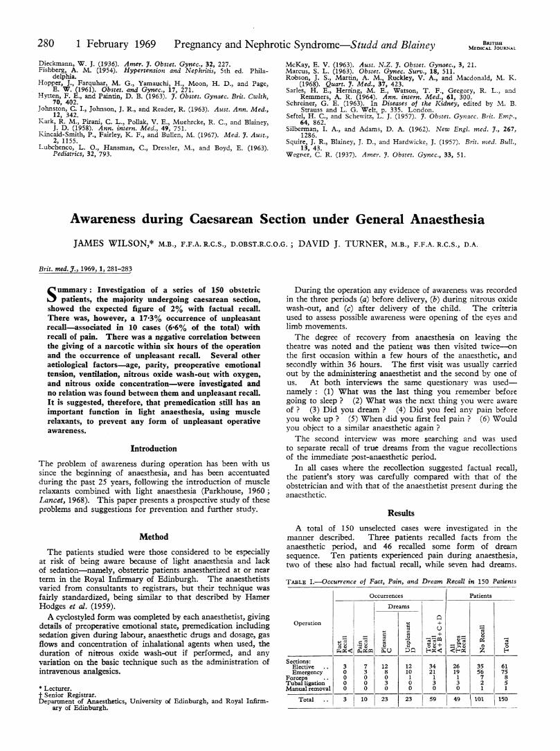
A total of 150 unselected cases were investigated in themanner, described. Three patients recalled facts from theanaesthetic period, and 46 recalled some form of dreamsequence. Ten patients experienced pain during anaesthesia,two of these also had factual recall, while seven had dreams.
TABLE I.-Occurrence of Fact, Pain, and Dream Recall in 150 Patients
Occurrences Patients
Dreams
Operation +
a .
Sections:Elective .. 3 7 12 12 34 26 35 61Emergency 0 '3 8 10 21 19 56 75'
Forceps.. 0 0 0 1 1 1 7 8Tubllgaton 0 0 3 0 3 3 2 5
Manual removal 0 0 0 0 0 0 1 1
Total .. 3 10 23 23 59 49 101 150