Practice CMAJ · Practice CMAJ CMAJ • JANUARY 6 ... mendations for treatment are based on limited...
3
Practice CMAJ CMAJ • JANUARY 6, 2009 • 180(1) © 2009 Canadian Medical Association or its licensors 72 The case: A 52-year-old woman was admitted with painful swelling in her left thigh. She had a 12-year history of type 2 diabetes mellitus and hypertension, and she was a smoker (40 pack-year smoking history). At her last exami- nation, her HbA 1C level was 7.7%, her serum total choles- terol level was 8.65 (normal < 5.17) mmol/L and her triglyc- eride level was 3.77 (normal < 2.26) mmol/L. She had diabetic retinopathy and microalbuminuria, and she had a myocardial infarction 12 years earlier. Two months before admission, the patient underwent a left femoropopliteal bypass for peripheral vascular disease. Her medications at the time of admission included acetylsalicylic acid, enalapril, metformin, glibenclamide and simvastatin. On admission, the patient complained of continuous severe pain in her anterior thigh that had developed grad- ually. The pain was not reduced by nonsteroidal anti- inflammatory drugs taken orally. She reported that the pain was very intense, and that it was present at rest but became worse with movement. The pain had increased over the 4 weeks before presentation. She denied any trauma, fever or chills. The patient had lost 4 kg, and she was unable to walk because of the pain. A physical examination showed a firm swelling in the anterior part of her thigh. An area that measured 10 × 5 cm was hard and felt warm, and there was mild ery- thema and tenderness. The patient’s muscle strength and motion were markedly limited by pain. Her acute phase reactants were elevated: her erythro- cyte sedimentation rate was 153 (normal 0–30) mm/h, and her C-reactive protein level was 115 (normal 0.2–3.0) mg/L. She was anemic (hemoglobin 6.2 mmol/L) and had mild leukocytosis (11.2 [normal 3.8–9.8] × 10 9 /L, with 78% poly- morphonuclear cells). She also had a mildly reduced level of serum albumin (31 [normal 41–53] g/L) and a normal globu- lin level (30 [normal 20–35] g/L). The results of liver function tests were normal. Doppler ultrasonography showed no ab- scess or deep vein thrombosis. Her femoropopliteal bypass graft and larger arteries were patent. Magnetic resonance imaging showed increased signal intensity on T 2 -weighted images of her left quadricep, with diffuse subcutaneous and fat edema. There was ex- tensive swelling of the quadriceps vastus me- dialis and rectus with intermuscular edema (Figure 1, Figure 2). Despite 3 weeks of bed rest, opiates, enoxaparin and clopidogrel, there was only a slight clinical improvement. However, her C- reactive protein level gradually decreased. An open muscle biopsy was performed to rule out a tumour. Instead, diabetic muscle infarction was confirmed (Figure 3). One week after the biopsy, there was significant worsening of her pain and severe edema of DOI:10.1503/cmaj.080572 Painful swelling in the thigh: diabetic muscle infarction Ami Schattner MD, Taiba Zornitzki MD, Meital Adi MD, Joshua Friedman MD Teaching cases From the Departments of Medicine (Schattner, Zornitzki, Friedman) and Radiology (Adi), Kaplan Medical Centre, Rehovot; and the Faculty of Medicine (Schattner, Zor- nitzki, Adi, Friedman), Hebrew University and Hadassah Medical School, Jerusalem, Israel Key points • Patients with longstanding diabetes and microvascular complications may develop diabetic muscle infarction. • Patients usually present with a painful, tender muscle mass, often in the thigh. • Constitutional symptoms are usually absent. • Levels of muscle enzymes may not be increased but levels of acute phase reactants often are. • The diagnosis is clinical, and an MRI can confirm the diagnosis. • Diagnostic tests (imaging, laboratory investigations) can be used to rule out other causes. • Slow, spontaneous resolution usually occurs; muscle biopsy or surgery can delay recovery. Box 1: Differential diagnosis of diabetic muscle infarction Infection Tumour • Pyomyositis • Lymphoma, primary muscle • Necrotizing fasciitis • Sarcoma, soft-tissue • Cellulitis • Benign muscle tumour • Abscess Inflammatory • Osteomyelitis • Myositis, focal inflammatory Trauma • Polymyositis or dermatomyositis • Hematoma Diabetes-related condition • Exertional rupture of muscle • Diabetic amyotrophy • Myositis ossificans Other Vascular • Ruptured Baker cyst • Deep vein thrombosis • Adverse reaction to simvastatin • Acute compartment syndrome • Arterial graft complication
Transcript of Practice CMAJ · Practice CMAJ CMAJ • JANUARY 6 ... mendations for treatment are based on limited...

Practice CMAJ
CMAJ • JANUARY 6, 2009 • 180(1)© 2009 Canadian Medical Association or its licensors
72
The case: A 52-year-old woman was admitted with painfulswelling in her left thigh. She had a 12-year history of type2 diabetes mellitus and hypertension, and she was asmoker (40 pack-year smoking history). At her last exami-nation, her HbA1C level was 7.7%, her serum total choles-terol level was 8.65 (normal < 5.17) mmol/L and her triglyc-eride level was 3.77 (normal < 2.26) mmol/L. She haddiabetic retinopathy and microalbuminuria, and she had amyocardial infarction 12 years earlier. Two months beforeadmission, the patient underwent a left femoropoplitealbypass for peripheral vascular disease. Her medications atthe time of admission included acetylsalicylic acid,enalapril, metformin, glibenclamide and simvastatin.
On admission, the patient complained of continuous severe pain in her anterior thigh that had developed grad-ually. The pain was not reduced by nonsteroidal anti-inflammatory drugs taken orally. She reported that thepain was very intense, and that it was present at rest butbecame worse with movement. The pain had increasedover the 4 weeks before presentation. She denied anytrauma, fever or chills. The patient had lost 4 kg, and shewas unable to walk because of the pain.
A physical examination showed a firm swelling in theanterior part of her thigh. An area that measured10 × 5 cm was hard and felt warm, and there was mild ery-thema and tenderness. The patient’s muscle strength andmotion were markedly limited by pain.
Her acute phase reactants were elevated: her erythro-cyte sedimentation rate was 153 (normal 0–30) mm/h, andher C-reactive protein level was 115 (normal 0.2–3.0) mg/L.She was anemic (hemoglobin 6.2 mmol/L) and had mildleukocytosis (11.2 [normal 3.8–9.8] × 109/L, with 78% poly-morphonuclear cells). She also had a mildly reduced level ofserum albumin (31 [normal 41–53] g/L) and a normal globu-lin level (30 [normal 20–35] g/L). The results of liver functiontests were normal. Doppler ultrasonography showed no ab-scess or deep vein thrombosis. Her femoropopliteal bypassgraft and larger arteries were patent. Magnetic resonanceimaging showed increased signal intensity on T2-weighted
images of her left quadricep, with diffusesubcutaneous and fat edema. There was ex-tensive swelling of the quadriceps vastus me-dialis and rectus with intermuscular edema(Figure 1, Figure 2).
Despite 3 weeks of bed rest, opiates,enoxaparin and clopidogrel, there was only aslight clinical improvement. However, her C-reactive protein level gradually decreased.An open muscle biopsy was performed torule out a tumour. Instead, diabetic muscleinfarction was confirmed (Figure 3). Oneweek after the biopsy, there was significantworsening of her pain and severe edema of
DO
I:10
.150
3/cm
aj.0
8057
2
Painful swelling in the thigh: diabetic muscle infarction
Ami Schattner MD, Taiba Zornitzki MD, Meital Adi MD, Joshua Friedman MD
Teaching cases
From the Departments of Medicine (Schattner, Zornitzki,Friedman) and Radiology (Adi), Kaplan Medical Centre,Rehovot; and the Faculty of Medicine (Schattner, Zor-nitzki, Adi, Friedman), Hebrew University and HadassahMedical School, Jerusalem, Israel
Key points
• Patients with longstanding diabetes and microvascularcomplications may develop diabetic muscle infarction.
• Patients usually present with a painful, tender musclemass, often in the thigh.
• Constitutional symptoms are usually absent.• Levels of muscle enzymes may not be increased but levels
of acute phase reactants often are.• The diagnosis is clinical, and an MRI can confirm the diagnosis.• Diagnostic tests (imaging, laboratory investigations) can be
used to rule out other causes.• Slow, spontaneous resolution usually occurs; muscle biopsy
or surgery can delay recovery.
Box 1: Differential diagnosis of diabetic muscle infarction
Infection Tumour
• Pyomyositis • Lymphoma, primary muscle
• Necrotizing fasciitis • Sarcoma, soft-tissue
• Cellulitis • Benign muscle tumour
• Abscess Inflammatory
• Osteomyelitis • Myositis, focal inflammatory
Trauma • Polymyositis or dermatomyositis
• Hematoma Diabetes-related condition
• Exertional rupture of muscle • Diabetic amyotrophy
• Myositis ossificans Other
Vascular • Ruptured Baker cyst
• Deep vein thrombosis • Adverse reaction to simvastatin
• Acute compartment syndrome • Arterial graft complication

Practice
CMAJ • JANUARY 6, 2009 • 180(1) 73
the whole thigh. After 2 months of bed rest, tramadol forpain and a combination of clopidogrel and low-molecular-weight heparin, all of her complaints and physical findingsgradually dissipated. The results of her blood tests normal-ized. The patient resumed working and was well 10months later.
Our patient presented with long-term intense leg pain and dys-function, a large tender mass in her thigh, elevation of acute-phase proteins and a prolonged unrelenting course. A long listof differential diagnoses was proposed (Box 1). Although dia-betic muscle infarction seemed the most likely diagnosis, amuscle biopsy was judged necessary to rule out a primarymuscle tumour. The biopsy was followed by clinical deteriora-tion, similar to that reported in other cases.1
About 100 cases of diabetic muscle infarction have beenreported since the first report in 1965.1,2 Thus, idiopathic mus-
cle infarction is rare and occurs specifically among patientswith diabetes. Although the exact pathogenesis is unknown, itis assumed to be a microvascular complication of diabetes.Most affected patients have a long history of diabetes (eithertype 1 or type 2) and about 97% have other microvascularcomplications, most often nephropathy.2
Patients typically present with intense, nonremitting and dis-abling pain in a lower limb with associated edema and mass.Normal temperature and normal levels of muscle enzymes arealso typical.2 Muscles in the thigh are affected 4 times more fre-quently than the calf. The quadriceps are the most commonlyaffected muscles. Involvement of the arms is rare.
Magnetic resonance imaging (MRI) is the diagnostic testof choice, and T2-weighted axial plane images are useful fordiagnosis.2,3 Other diagnostic modalities may help to rule outother diagnoses, rather than help to make the correct diagno-sis of diabetic muscle infarction. The clinical presentation andMRI findings are distinctive (Figure 1, Figure 2); thus, abiopsy is rarely indicated. Muscle biopsy and surgery shouldbe avoided despite the severe presentation because these in-terventions can prolong resolution or temporarily worsen thepatient’s symptoms.
Whether this rare complication of diabetes can be pre-vented is unknown. Diabetic muscle infarction is self-limiting, and full recovery can be expected over time. Recom-mendations for treatment are based on limited evidence andexpert opinion. We recommend supportive treatments, such
Figure 2: Magnetic resonance imaging of the patient’s thigh.A: Coronal image showing diffuse, heterogeneous, intense sig-nal of the involved muscle (arrow), which suggests edema. B:Sagittal image showing a heterogeneously enhanced mass (ar-row) with peripheral enhancement after adminstration ofgadolinium.
Figure 1: Magnetic resonance imaging of the patient’s thighs.A: T1-weighted images showing swelling and hypointensity (ar-row) of the involved muscle. B: T2-weighted images showingdiffuse, heterogeneous signal in the involved muscle (arrow),suggesting edema. C: A focal area of heterogeneously en-hanced mass (arrow) with peripheral enhancement was visibleafter adminstration of gadolinium.
Practice
CMAJ • JANUARY 6, 2009 • 180(1)74
as bed rest, analgesics and, if required, a cautious use of anti-inflammatory medications. As a possible microvascular com-plication of diabetes, antiplatelets are theoretically effective,and advice such as optimizing glycemic control and smokingcessation seems prudent. In one analysis of cases reportedfrom several sources, patients who underwent surgery had arecovery period that was more than double that of those whoreceived conservative treatments such as bed rest, analgesicsand anti-inflammatory medications (13 weeks v. 5.5 weeks,p < 0.05).4
Unfortunately, diabetic muscle infarction recurs in aboutone-half of patients.1,4 The overall prognosis for these patientsmay be related to the severity of their underlying diabetes and
the degree of macrovascular and especially microvascularcomplications.
In conclusion, diabetic muscle infarction is a rare compli-cation of diabetes. The incidence is likely to increase becauseof the increasing global prevalence of diabetes. Physiciansshould consider diabetic muscle infarction in the differentialdiagnosis for patients with diabetes who have a painful,swollen muscle. Conservative treatment strategies should bechosen over aggressive invasive measures when possible.
REFERENCES1. Kapur S, Brunet JA, McKendry RJ. Diabetic muscle infarction: case report and re-
view. J Rheumatol 2004;31:190-4.2. Trujillo-Santos AJ. Diabetic muscle infarction. An underdiagnosed complication
of long-standing diabetes. Diabetes Care 2003;26:211-5.3. Kattapuram TM, Suri R, Rosol MS, et al. Idiopathic and diabetic skeletal muscle
necrosis: evaluation by magnetic resonance imaging. Skeletal Radiol 2005;34:203-9.
4. Kapur S, McKendry RJ. Treatment and outcomes of diabetic muscle infarction. JClin Rheumatol 2005;11:8-12.
Teaching cases are brief case reports that convey clear,practical lessons. Preference is given to common presentationsof important rare conditions, and important unusualpresentations of common problems. Articles start with a briefsummary (100 words) outlining the case and its relevance to ageneral audience. The case presentation follows (500 wordsmaximum) as well as a discussion of the underlying condition(1000 words maximum). Up to 5 references are permitted andvisual elements (e.g., tables of the differential diagnosis,clinical features or diagnostic approach) are encouraged.Written consent from patients for publication of their story is anecessity and should accompany submissions. See informationfor authors at www.cmaj.ca.
Competing interests: None declared.
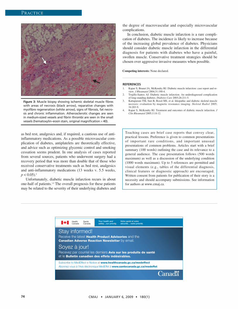
Figure 3: Muscle biopsy showing ischemic skeletal muscle fibreswith areas of necrosis (black arrow), reparative changes withmyofibre regeneration (white arrow), signs of fibrosis, fat necro-sis and chronic inflammation. Atherosclerotic changes are seenin medium-sized vessels and fibrin thrombi are seen in the smallvessels (hematoxylin–eosin stain, original magnification × 40).
Stay informed! Receive the latest Health Product Advisories and the Canadian Adverse Reaction Newsletter by email.
Soyez à jour! Recevez par courriel les derniers Avis sur les produits de santéet le Bulletin canadien des effets indésirables.
Subscribe to MedEffect e-Notice at www.healthcanada.gc.ca/medeffectAbonnez-vous à l’Avis électronique MedEffet à www.santecanada.gc.ca/medeffet


![2010 Guidelines Papaioannou A, et al. CMAJ 2010 Oct 12. [Epub ahead of print]. 2010 Clinical Practice Guidelines for the Diagnosis and Management of Osteoporosis.](https://static.fdocuments.us/doc/165x107/56649c775503460f9492c143/2010-guidelines-papaioannou-a-et-al-cmaj-2010-oct-12-epub-ahead-of-print.jpg)

![Nationale VersorgungsLeitlinie Nicht-spezifischer ... · mendations, Assessment, Development and Evaluation) bewertet [3]. Genauere Informationen zur Recher- Genauere Informationen](https://static.fdocuments.us/doc/165x107/5d616d4788c993bb0b8be145/nationale-versorgungsleitlinie-nicht-spezifischer-mendations-assessment.jpg)