
Practical 8 Gram negative rods (Enterobacteriaceae)...
15
Manal Al khulaifi 1 Practical 8 Gram negative rods (Enterobacteriaceae) Objective: 1- To identify Enterobacteriaceae from other Gram negative rods and to differentiate between lactose fermenter and non lactose fermenter by: A. Detection of the presence of cytochrome oxidase in Gram-negative rods. B. To determine the ability of bacteria to metabolize glucose oxidatively or fermentatively. C. To know Gram reaction of Enterobacteriaceae. D. Use of MacConkey agar to differentiate between Lactose fermenter and non lactose fermenter Enterobacteriaceae. Required materials: 1- Overnight cultures of E.coli, K. pneumoniae, Shigella, Salmonella, Pseudomons. 2- Gram stains dyes and reagents, filter paper, slides. 3- plates of MacConkey agar 4- tubes containing 5ml OF media 5- 10 ml of oxidase reagent Experimental: 1- Gram stain of Enterobacteriaceae. 2- Oxidase teat. 3- OF test. 4- Inoculation of MacConkey agar with lactose fermenter and non lactose fermenter Enterobacteriaceae. Oxidase and Oxidative-Fermentative (OF) tests can be used in differentiation Enterobacteriaceae from other Gram negative rods as showed in the table: Microorganism Oxidase test OF test Enterobacteriaceae Negative Fermentative Pseudomonads Positive Oxidative Vibrio Positive Fermentative Gram stain of Enterobacteriaceae
Transcript of Practical 8 Gram negative rods (Enterobacteriaceae)...

Manal Al khulaifi
1
Practical 8
Gram negative rods (Enterobacteriaceae)
Objective:
1- To identify Enterobacteriaceae from other Gram negative rods and to differentiate
between lactose fermenter and non lactose fermenter by:
A. Detection of the presence of cytochrome oxidase in Gram-negative rods.
B. To determine the ability of bacteria to metabolize glucose oxidatively or fermentatively.
C. To know Gram reaction of Enterobacteriaceae.
D. Use of MacConkey agar to differentiate between Lactose fermenter and non lactose fermenter
Enterobacteriaceae.
Required materials:
1- Overnight cultures of E.coli, K. pneumoniae, Shigella, Salmonella, Pseudomons.
2- Gram stains dyes and reagents, filter paper, slides.
3- plates of MacConkey agar
4- tubes containing 5ml OF media
5- 10 ml of oxidase reagent
Experimental:
1- Gram stain of Enterobacteriaceae.
2- Oxidase teat.
3- OF test.
4- Inoculation of MacConkey agar with lactose fermenter and non lactose fermenter
Enterobacteriaceae.
Oxidase and Oxidative-Fermentative (OF) tests can be used in differentiation
Enterobacteriaceae from other Gram negative rods as showed in the table:
Microorganism Oxidase test OF test
Enterobacteriaceae Negative Fermentative
Pseudomonads Positive Oxidative
Vibrio Positive Fermentative
Gram stain of Enterobacteriaceae

Manal Al khulaifi
2
Oxidase Test:
1. Principle - To determine the presence of cytochrome oxidase in bacteria. Recall that in
aerobic bacteria the cytochrome serve as electron carriers during aerobic respiration. The
detection of cytochrome is extremely beneficial in differentiating many groups of
bacteria. All members of Enterobacteriaceae are oxidase negative, while other Gram-
negative rods, such as Pseudomonas, are oxidase positive. The oxidase test is based on
the ability of bacteria to turn (oxidize) a reagent (tetramethyl-p-phenylenediamine),
which serves as an alternate substrate for the cytochrome oxidase, to a purple color.
2. Materials:
o Overnight cultures of E. coli and Pseudomonas aeruginosa.
o Oxidase reagent "tetramethyl-p-phenylenediamine".
o Pieces of filter paper.
3. Method - hold a piece of the oxidase test paper with forceps and touch onto an area of
heavy growth
4. Results - Color change to purple within:
o 10 seconds = positive
o 10 - 60 seconds = delayed positive
o >60 seconds = negative
5. Special Features
o Useful in differentiation of
Enterobacteriaceae (-) and Pseudomonas
(+).
o An oxidase positive organism will be
catalase positive.
o Strict anaerobe organisms are oxidase
negative.
Oxidation-Fermentation test (O/F):
1. Principle - To determine the ability of bacteria to breakdown glucose oxidatively or
fermentatively. The OF test is used to determine whether a bacterium has the enzymes
necessary for the aerobic breakdown of glucose (i.e. oxidation) and/or for the
fermentation of glucose.
2. Materials:
a- Overnight cultures of E. coli and Pseudomonas aeruginosa.
b- Tubes of OF medium/each student.
c- Sterile liquid paraffin.
3. Method
a- Inoculate two tubes of OF medium for each organism being tested. Inoculation is carried
out as a stab to within 1 cm of the bottom of the tube.
b- Overlay one tube (covered) only with sterile paraffin oil to exclude all oxygen.
Not overlay the second tube (open).
c- Incubate at 37°C for 24 hours.
(+) (-)

Manal Al khulaifi
3
4. Results
5. Special Features - The test differentiates Enterobacteriaceae (F) from the Pseudomonas
sp. (O), and Micrococcus sp. (O) from Staphylococcus sp. (F).
6. Precautions in Interpretation - Some organisms require prolonged incubation before
acid production is visible.
Members of Enterobacteriaceae are characterized by:
Small Gram-negative non-spore-forming enteric bacilli
All Enterobacteriaciae:
1. ferment glucose with acid production
2. reduce nitrates into nitrites (NO3 to NO2 or all the way to N2)
3. are oxidase negative
All are aerobic but can be facultative anaerobic
Motile via peritrichous flagella except Shigella and Klebsiella which are non-motile
Non-capsulated except Klebsiella
Grow on ordinary medium i.e. non-fastidious as well as grow on bile containing media as
MacConkey's medium, which used in primary classification depends on lactose fermentation on
MacConkey's agar medium
Open tube Covered tube Result
Yellow Green oxidation (O)
Yellow Yellow fermentation (F)
Green Green No action on glucose

Manal Al khulaifi
4
Some members of the Enterobacteriaceae are true pathogens such as:
Salmonella spp.
Shigella spp.
Yersinia spp.
Certain strains of Escherichia coli :
ETEC = enterotoxigenic E. coli
EIEC = enteroinvasive E. coli
EPEC = enteropathogenic E. coli
EHEC = enterohemorrhagic E. coli
EaggEC = enteroaggregative E. coli
UPEC = uropathogenic E. coli
Most members of the Enterobacteriaceae are opportunistic or cause secondary infections of
wounds, the urinary and respiratory tracts, and the circulatory system e.g. E. coli.
*Primary classification of Enterobacteriaceae depends on lactose fermentation:
Laboratory Identification:
Specimens whether pus, sputum, urine, feces, CSF should be cultured immediately or placed
on special media to prevent overgrowth
Culture:
Colony morphology: moist, gray (except Serratia marcescans which appears red) smooth
colonies on non-selective media
Special differential and selective media used for separation of genera and species
Enterobacteriaceae
Lactose fermenter e.g. E. coli, Klebsiella,
Enterobacter, Citrobacter
Lactose non fermenters e.g. Salmonella, Shigella,
Proteus

Manal Al khulaifi
5
I- MacConkey agar medium:
MacConkey agar is selective and differential medium for isolation of
Enterobacteriaceae. The medium is made selective by the incorporation of bile and
crystal violet, which inhibit gram-positive bacteria, especially staphylococci and
enterococci. The medium is differential by use of combination of neutral red (in
acidic pH the color turns red) and lactose. When an organism ferment lactose, the
drop in pH (due to acid formation as end product of fermentation of lactose) causes
the colony to take on a pink-red appearance whereas organism which unable to
ferment lactose appears as pale yellow colonies.
II Eosin Methylene Blue (EMB) agar medium:
EMB agar is a selective and differential medium used for the isolation and differentiation
of enteric pathogens from contaminated clinical specimens. Eosin and methylene blue
are the selective agents and inhibit gram-positive organisms. EMB contains lactose.
Organisms that ferment lactose binds to dyes under acidic conditions and appear as
blue-black colonies with a metallic sheen. Under less acidic conditions, other
coliforms, such as Klebsiella, Enterobacter, and Citrobacter, appear as brown-pink
"dark" colonies. Nonfermenters, such as Salmonella, Shigella, and Proteus, appear as
the color of medium or transparent and colorless.
Lactose fermintor
( pink colony)
Lactose Non- fermintor
Colorless colony
Manal Al khulaifi
6
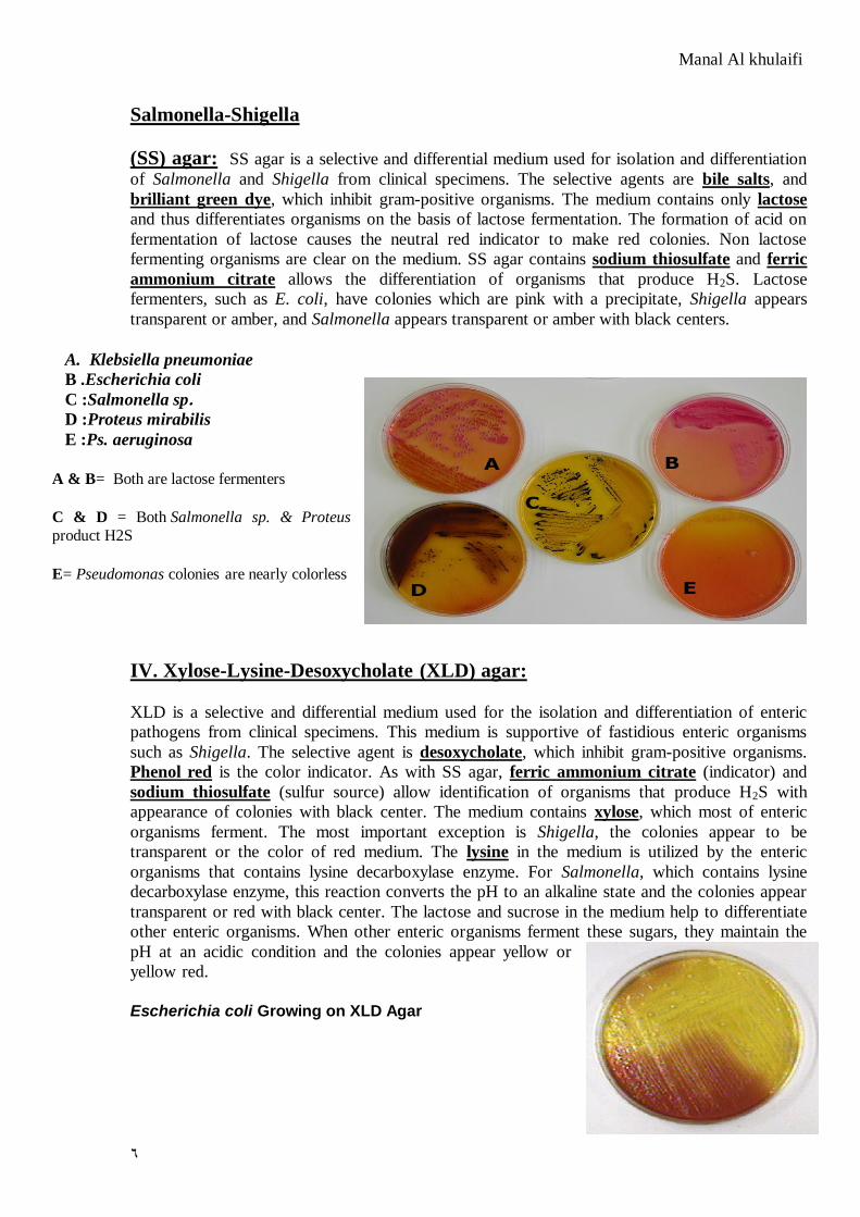
Salmonella-Shigella
(SS) agar: SS agar is a selective and differential medium used for isolation and differentiation
of Salmonella and Shigella from clinical specimens. The selective agents are bile salts, and
brilliant green dye, which inhibit gram-positive organisms. The medium contains only lactose
and thus differentiates organisms on the basis of lactose fermentation. The formation of acid on
fermentation of lactose causes the neutral red indicator to make red colonies. Non lactose
fermenting organisms are clear on the medium. SS agar contains sodium thiosulfate and ferric
ammonium citrate allows the differentiation of organisms that produce H2S. Lactose
fermenters, such as E. coli, have colonies which are pink with a precipitate, Shigella appears
transparent or amber, and Salmonella appears transparent or amber with black centers.
A. Klebsiella pneumoniae B . Escherichia coli
C :Salmonella sp. D :Proteus mirabilis
E :Ps. aeruginosa
A & B= Both are lactose fermenters
C & D = Both Salmonella sp. & Proteus
product H2S
E= Pseudomonas colonies are nearly colorless
IV. Xylose-Lysine-Desoxycholate (XLD) agar:
XLD is a selective and differential medium used for the isolation and differentiation of enteric
pathogens from clinical specimens. This medium is supportive of fastidious enteric organisms
such as Shigella. The selective agent is desoxycholate, which inhibit gram-positive organisms.
Phenol red is the color indicator. As with SS agar, ferric ammonium citrate (indicator) and
sodium thiosulfate (sulfur source) allow identification of organisms that produce H2S with
appearance of colonies with black center. The medium contains xylose, which most of enteric
organisms ferment. The most important exception is Shigella, the colonies appear to be
transparent or the color of red medium. The lysine in the medium is utilized by the enteric
organisms that contains lysine decarboxylase enzyme. For Salmonella, which contains lysine
decarboxylase enzyme, this reaction converts the pH to an alkaline state and the colonies appear
transparent or red with black center. The lactose and sucrose in the medium help to differentiate
other enteric organisms. When other enteric organisms ferment these sugars, they maintain the
pH at an acidic condition and the colonies appear yellow or
yellow red.
Escherichia coli Growing on XLD Agar

Manal Al khulaifi
7
Acid from fermentation lowers the pH and turns the phenol red from red (alkaline) to yellow
(acid). No hydrogen sulfide is produced.
Enterobacteriaceae: I- Lactose Fermenters
Biochemical tests for identification of Enterobacteriaceae:
All members of Enterobacteriaceae are ferment glucose, oxidase negative, and reduce nitrate
into nitrites.
Nitrate Reduction:
1. Principle - To determine the ability of an organism to reduce nitrate to nitrites or free
nitrogen gas
2. Method - Inoculate a nitrate broth and incubate for 5 days at 37°C.
3. Result
1. To each nitrate broth culture add 1 ml of sulphanilic acid and 1 ml of -
naphtylamine. The production of a red color occurs in the presence of nitrite indicates
the ability of the organism to reduce nitrate to nitrite.
2. To broths showing a negative reaction add a few particles of zinc. The appearance
of a red color indicates that nitrate is still present and hence has not been reduced
by the organism. If the solution does not change color the organism has reduced
the nitrate through nitrite to nitrogen gas.
4. Special Features - Used in identification of Enterobacteriaceae (usually +).
5. Precautions in Interpretation - Interpret results immediately as the color produced in a
positive reaction may fade quickly.
Triple Sugar Iron Agar (TSI) and H2S production:
1. Principle - To determine the ability of an organism to attack a specific carbohydrate
incorporated into a basal growth medium, with or without the production of gas, along
with the determination of possible hydrogen sulphide (H2S) production.
2. Method - Inoculate TSI medium with an inoculating needle by stabbing the butt and
streaking the slant. Incubate at 37°C for 24 hours.

Manal Al khulaifi
8
3.Results
H2S production is indicated by the presence of a black precipitate
4. Special Features - H2S production and carbohydrate fermentation patterns are generally
characteristic for specific bacterial groups, especially the Enterobacteriaceae.
5. Precautions in Interpretation - An H2S organism may produce so much of the black
precipitate (ferrous sulphide) that the acidity produced in the butt is completely masked.
However, if H2S is produced, an acid condition does exist in the butt even if it is not
observable.
Indole, Methyl Red, Voges-Prosakaur, Citrate (IMViC) Tests:
o The following four tests comprise a series of important determinations that are
collectively called the IMViC series of reactions (I= indole; M=methyl red;
V=Voges Proskauer; and C=citrate). The IMViC series of reactions allows for the
differentiation of the various members of Enterobacteriaceae.
Indole "Tryptophan Hydrolysis" Test:
1. Principle – Certain microorganisms can metabolize the amino acid tryptophan through
the action of the enzyme tryptophanase. Once again, the activity of the enzyme is not
measured directly, but rather, one of the end products (Indole) is detected. The enzymatic
Butt color
Slant color
H2S Result
Example
Yellow Red Negative
Glucose only fermented Lactose non fermenter
e.g. Shigella
Yellow Red Positive/blackening in
butt Glucose only fermented
with H2S production Lactose non fermenter
e.g. Salmonella & Proteus
Yellow Yellow Negative Glucose fermented, also
lactose and/or sucrose Lactose fermenter
e.g. E. coli
Red Red Negative No action on glucose,
lactose or sucrose Non fermenter organisms
e.g. Pseudomonas

Manal Al khulaifi
9
degradation leads to the formation of pyruvic acid, indole and ammonia. The presence of
indole is detected by addition of Kovac's reagent.
2. Materials 1. Tube of tryptone water
2. Tested microorganism (such as E. coli (+) and Klebsiella (-)
3. Kovac's reagent
3. Method 1. Inoculate tryptone water with the tested microorganism
2. Incubate at 37°C for 24- 48 hours
3. After incubation interval, add 1 ml Kovacs reagent, shake the tube gently and read
immediately.
Kovacs reagent
p-dimethylaminobenzaldehyde 50 g
amyl or butyl alcohol 750 ml
HCl (conc.) 250 ml
4. Result - a bright pink color in the top layer (ring) indicates
the presence of indole. The absence of color means that
indole was not produced, and that the organism does not
possess the tryptophanase enzyme i.e. indole is negative
5. Special Features - Used in the differentiation of genera and species. e.g. E. coli (+) from
Enterobacter (-).
6. Precautions in Interpretation
o Cultures to be tested for indole production must be incubated aerobically
o The optimum pH for tryptophanase activity is one that is slightly alkaline (ph 7.4
- 7.8); a decrease in pH results in decreased indole production and a possible false
negative.
Methyl Red "Mixed acid Fermentation" Test:
1. Principle – All enteric bacteria can utilize sugar for their energy demands. Mixed acid
fermenters such as E. coli ferment glucose to produce large amounts of acetic, formic and
succinic acids as end products. The large amount of acid produced lowers the pH of the
medium to below 5.0. By using the indicator methyl red (MR), the production of these
acids as the end product of fermentation can be monitored. If pH drops below 4.5, the
color of MR indicator will be red. If the pH is above 6.0, the color will be yellow/orange.
2. Material 1. tubes of MRVP medium (Phosphate buffered glucose peptone water)
2. Tested microorganism
3. Methyl red indicator
3. Method
1. Inoculate the tested organism into a tube of MRVP broth.
2. Incubate the tube at 37°C for 24 hours.
3. After incubation, add a few drops of MR solution to the culture. Read immediately.
(+) (-)
Manal Al khulaifi
11
4- Results
4. Special features - The test is used to differentiate between genera.
e.g. E. coli and Citrobacter (+) from Klebsiela and Enterobacter (-)
5. Precautions in Interpretation - If the MR test is performed too
early, the results may be a false positive since MR negative
organisms may not have had time to completely metabolize the
initial acid products that accumulated from the glucose
fermentation
Voges Proskauer "Butanediol Fermentation" Test:
1. Principle – Some bacteria, rather than producing abundant acid in the fermentation of glucose,
produce other products such as alcohols. All species of Enterobacter and Klebsiella will form
products such as alcohol and 2,3-butanediol rather than the large amount of acid, as does E.
coli and Citrobacter. A test for acetylmethylcarbinol (a precursor of 2-3, butanediol that
appear in the growth medium) is performed. If Barrit's reagent is added, a pink color
developing after few minutes indicates the presence of acetylmethylcarbinol and the test is
positive.
2. Materials 1. Tubes of MRVP
2. Tested microorganism
3. Barrit's solution A "40%KOH"
4. Barrit's solution B "-Naphthol"
3. Method 1. Inoculate the tube by organism
2. Incubate the inoculated tube at 37°C for 24 h.
3. After it has become apparent that growth has occurred, add approximately 0.5 ml
of -naphthol, followed by 0.5 ml of 40% KOH.
4. Shake vigorously and let the tube stand for 1 to 2 h.
5. Results
Special Features - The test is used to differentiate between genera.
e.g. K. pneumoniae (+) and Enterobacter (+) from E. coli (-) and
Citrobacter.
6. Precautions in Interpretation - After exposure to the reagents for over 2 hour, a negative
VP culture may show a copper-like color due to the action of the KOH on the alpha
naphthol. This is not a positive reaction.
Red MR positive
Yellow/orange MR negative
Pink VP(+)
No change VP(-)
(-) (+)
(-) (+)

Manal Al khulaifi
11
Citrate Utilization Test
1. Objective – determine whether microorganism can utilize citrate as a sole source of
carbon.
2. Principle – Some microorganisms can metabolize citrate as a sole of carbon source. Like
many of the nutrients already used, citrate needs to be transported into the bacterial cells
before it can be metabolized. This transport depends upon the enzyme citrate permease.
Once inside the cell, the citrate is broken down to pyurvate and carbon dioxide. The
carbon dioxide produced combines with sodium in the medium and water to form sodium
carbonate, an alkaline product. The rise in pH is detected by the color change in
bromothymol blue indicator present in the medium from green to deep blue.
Citrate →Pyruvate →CO2 + Na + H2O →Na2CO3 →↑ pH
3. Method - Streak a Simmon's Citrate agar slant with the
organism and incubate at 37°C for 24 hours.
4. Result - Examine for growth (+). Growth on the medium is
accompanied by a rise in pH to change the medium from its
initial green color to deep blue.
5. Special Features - Aids in the differentiation of genera and
species.
6. Precautions in Interpretation - The medium must be lightly
inoculated (from plate cultures, not from a broth) to avoid a
carry over of nutrients, which may lead to a false positive
result.
Urea Hydrolysis (Urease):
1. Principle – Urease is an enzyme that catalyzes the conversion of urea to CO2 and NH3.
The urease test is particularly useful in identifying Proteus, Klebsiella and Enterobacter.
The urea media contain the indicator phenol red. In the presence of urease, urea split and
ammonia and carbon dioxide are produced. Ammonia combines with water to produce
ammonium hydroxide, a strong base which raises the pH of the medium. This rise in the
pH causes the phenol red indicator to turn a deep pink. This is indicative of a positive
reaction for urease.
2. Method - Streak a urea agar tube with the organism and incubate at
37°C for 24 h
3. Result –If color of medium turns from yellow to pink indicates
positive test. Proteus give positive reaction after 4 h while
Kelebsiella and Enterobacter gave positive results after 24 h...
4. Special Features - Aids in the differentiation of members of
Proteus also differentiates between E. coli (-) from Klebsiella (+)
and Enterobacter (+).
(+) (-)
(+) (-)

Manal Al khulaifi
12
5. Precautions in Interpretation - The test must be read after 4 h and 24 h
Item Result Example
Non-urease producer Negative E. coli
Week urease producer Positive after 18 hrs Klebsiella
Strong urease producer Positive after 2-6 hrs Proteus
E.coli:
Most significant species in the genus
Important potential pathogen in humans
Common isolate from colon flora
Ferments glucose, lactose, trehalose, & xylose
Positive indole and methyl red tests
Does NOT produce H2S
Simmons citrate negative
Usually motile
Voges-Proskauer test negative
Cause Gastrointestinal Infections, urinary tract infection, Septicemia & Meningitis
E.coli on nutrient agar
E.coli on EMB agar ( metallic greenish colonies)
Klebsiella
* Large gram negative bacilli
* Usually found in GI tract
* K. pneumoniae is mostly commonly isolated species
*the colonies are large, moist and mucoid, due to possesses a
polysaccharide capsule, which protects against phagocytosis
*Has a distinctive “yeasty” odor
Frequent cause of nosocomial pneumonia
Lactose positive(grow on MacConkey’s agar &
produce rose pink colonies)

Manal Al khulaifi
13
Most are urease positive
Non-motile
Prominent polysaccaride capsule
Facultative anaerobes,.
The differences between Lactose fermenter are summarized in the following table:
Items E. coli Citrobacter Klebsiella Enterobacter
MacConkey's Rose pink colonies
Rose pink colonies
Rose pink colonies
Rose pink colonies
EMB Metallic sheen Dark colonies Dark colonies Dark colonies
Indole + + - -
MR + + - -
VP - - + +
CIT - + + +
Urease - + + +
Motility Motile Motile Non motile Motile
IMVIC test results
IMVIC test Result for E.coli

Manal Al khulaifi
14
IMVIC test Result for Proteus sp.
Motility Urease Citrate VP MR Indole TSI
Motile -ve -ve -ve +ve +ve A/A/- E. coli
Motile -ve +ve -ve +ve +ve A/A/- Citrobacter
freundi
Non motile +ve +ve +ve -ve -ve A/A/- Klebsiella
pneumoniae
Motile +ve +ve +ve -ve -ve A/A/- Enterobacter
cloacae
Motile -ve +ve -ve +ve -ve A/Alk/+ Salmonella
typhi
Non motile -ve -ve -ve +ve -ve A/Alk/- Shigella boydii
Motile
Swarwing
+ve +ve -ve +ve -ve A/Alk/+ Proteus mirabilis
EMB SS MacConkey O/F Nitrate
reductase
Oxidase Gram
stain
Metallic
sheen
LF LF O+/F+ +ve -ve -ve rod E. coli
Dark LF LF O+/F+ +ve -ve -ve
rods Citrobacter
Dark LF LF O+/F+ +ve -ve -ve
rods Klebsiella
Dark LF LF O+/F+ +ve -ve -ve
rods Enterobacter
Colorless NLF/H2S NLF O+/F+ +ve -ve -ve
rods Salmonella
Colorless NLF NLF O+/F+ +ve -ve -ve
rods Shigella
Colorless NLF/H2S NLF O+/F+ +ve -ve -ve
rods Proteus
Summary of Enterobacteriaceae
+ +
-
-

Manal Al khulaifi
15


![[Micro] gram positive spore bearing rods](https://static.fdocuments.us/doc/165x107/55d6fd2abb61eb344d8b45f4/micro-gram-positive-spore-bearing-rods.jpg)


