PowerPoint 簡報 - Academia Sinica
30
A Genetically Encoded Tag for Correlated Light and Electron Microscopy of Intact Cells, Tissues, and Organisms Speaker: Bo-Hua Chen Coach: Dr. Wei-Yuan Yang Sit-in: Dr. Cheng-Chung Wang
Transcript of PowerPoint 簡報 - Academia Sinica

A Genetically Encoded Tag for Correlated Light and Electron
Microscopy of Intact Cells, Tissues, and Organisms
Speaker: Bo-Hua Chen
Coach: Dr. Wei-Yuan Yang
Sit-in: Dr. Cheng-Chung Wang

awarded the 2008 Nobel Prize in chemistry for his development of the eGFP
invited to speak as an Academia Sinica Lecturer in December 2009
Academician in 2010
Heim, R., Cubitt, A., and Tsien, R. 1995. Improved green fluorescence. Nature 373 (6516): 663–664.
S65T shifted the absorption maximum from 395 to 488 nm and increased fluorescence, photostability

Subcellular localization is important
The dynamic behavior of cells is
a consequence of the elaborate
interactions between
complexes/organelles.
Microscopy to reveal the
important information to
understand complex cellular
function.

Microscopy Fluorescence light microscopy <identity>
Sample preparation is easy and relatively inexpensive.
appropriate tags provides the ability to track specific proteins of interest in living
cells
limitation: resolve two objects separated by ~200 nm
Objects that are closer than 200 nm are blurred into a single spot.
Electron microscopy <resolution>
EM provides the unique “space” where all objects (labeled and un-labeled) can
be visually explored at high resolution.
You can get up to a hundred-fold higher useful magnification from EM than from
light microscopy.
ultrastructures: mitochondria, Golgi, lysosome, membrane with high
curvature, …..
provides nanometer spatial resolution, but is not available in live-cell imaging.
lacks good tag to identify protein of interest
Annu Rev Biophys Biomol Struct. 2006;35:199-224

Identifying the protein in EM immunogold labeling
fixation to preserve the ultrastructure inhibits the diffusion of
antibodies and impairs antigenicity
Triton-100 facilitating the diffusion of antibodies degrades
membrane.
Membrane is not vivid
high specificity to the cross-linked antigen
expensive
distance from epitope (30nm)
Not all targets can be labeled.
LAMP1 localization in human HepG2 cells
10 nm gold
Nature Reviews Molecular Cell Biology 10, 623-635 (2009) Nature Protocols 3, 144 - 152 (2008)

Genetically encodable tag
benefit: produced in cells
Can GFP be visible to electron microscopy? generation of endogenous singlet oxygen by photoactivated GFP
Biol Chem. 2000 Dec;381(12):1251-8
Oxygen radicals generated during the GFP bleaching process can
photooxidize DAB into an electron-dense precipitate that can be visualized
by routine electron microscopy and electron tomography.
Nature Methods 2, 857 - 862 (2005)
human Golgi resident glycosylation enzyme, N-acetylgalactosaminyltransferase-
2 fused to eGFP

ROS generated by illumination can polymerize DAB to a light brown precipitate which can be stained by Osmium and imaged in EM.
Photo-oxidation for EM

Bond order=(8-4)/2=2
Ground state oxygen, O2, is a triplet diradical.
http://www.meta-synthesis.com/webbook/16_diradical/diradical.html
Degeneracy=2S+1 S is the total electron spin angular momentum
Energy level diagram Linear combination of 2 Oxygen atomic orbitals
2[ (1/2)+(1/2) ]+1=3 2[ (1/2)+(-1/2) ]+1=1
Singlet oxygen
disobey Hund’s Rule

Genetically encodable tags GFP
1O2 quantum yield (1O2 /photon absorbed) is low and unquantifiable. Biophys J 94: 168–172
Tetracysteine-ReAsH system
12-residue peptide tag
1O2 quantum yield is 0.024 (the best previous genetically targetable generator)
Adding of biarsenical dye ReAsH into cell is required.
Deep tissue or organism labeling is difficult.
nonspecific background signal
As is toxic

Aim
engineer fluorescent protein with high efficiency
of production of singlet oxygen

Arabidopsis thaliana phototropin 2
binds to its chromophore, flavin mononuceotide (FMN) very tightly (dissociation constant ~0.1 nM)
FMN can generate singlet oxygen efficiently. (quantum yield=0.51)
The only cofactor which is necessary for the mitochondrial electron transport chain is thus present in nearly all cells.
LOV2 domain
106 amino acids
less than half the size of GFP (248a.a.)
http://www-ijpb.versailles.inra.fr/en/arabido/arabido.htm
http://pubchem.ncbi.nlm.nih.gov/summary/summary.cgi?cid=8965
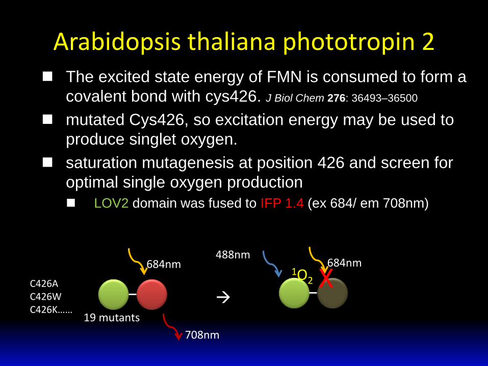
Arabidopsis thaliana phototropin 2 The excited state energy of FMN is consumed to form a
covalent bond with cys426. J Biol Chem 276: 36493–36500
mutated Cys426, so excitation energy may be used to
produce singlet oxygen.
saturation mutagenesis at position 426 and screen for
optimal single oxygen production
LOV2 domain was fused to IFP 1.4 (ex 684/ em 708nm)
684nm
708nm
488nm
1O2 684nm
X 19 mutants
C426A C426W C426K……

Screen for better 1O2 generator E. coli colonies were imaged before and after 488nm
illumination
~70% decrease …C426G
C426X

MiniSOG increase brightness of the C426G mutant
Saturation mutagenesis of other residues surrounding the
chromophore binding site
plus random mutagenesis
for mini Singlet Oxygen Generator
Predicted structure

MiniSOG Excitation
fluorescence quantum yield: 0.37
448nm: (16.7±0.7) X103 M-1cm-1
473nm: (13.6±0.5) X103 M-1cm-1
Emission
500nm
528nm
Singlet oxygen quantum yield: 0.47
Quantum yield: 0.
singlet-oxygen fluorescent probe
FMN=0.51

Size exclusion chromatography of miniSOG
MiniSOG was determined to be monomeric in solution

Successful localizing
HeLa
miniSOG labeled proteins and organelles appeared to have correct localizations in cultured mammalian cells.
10μm
concentrates at focal adhesions

Schematic diagram

Procedure transfected cells
fixation 2% glutaraldehyde pH7.4
blocking 50mM glycine, 10mM KCN, 5mM aminotriazole (reduce background reaction)
confocal microscope (identify transfected cell)
ice cold DAB solution bubbled with oxygen and freshed
illuminate and stop as soon as light brown DAB polymer appear (with FITC filtered light from xenon lamp, 2-10mins)
post-fixation with 1% osmium tetroxide 30min on ice
stained with 2% uranyl acetate at 4˚C (protein, nucleic acid)
dehydrated in ethanol and infiltrated by resin
C. Elegans injection of cDNAs 50ng/μL
3ug SynCAM2-miniSOG DNA delivered into lateral ventricel of embryos by in utero electroporation
p7, p21 brain removed, fixed by perfusion with 4% formaldehyde
sliced to 100 μm sections
area of interest identified by confocal
postfix with 2% glutaraldehyde
blocking 50mM glycine, 10mM KCN, 5 mM aminotriazole (reduce background reaction)

Procedure
Photooxidized areas of resin-embedded transfected cells or tissue identified by transmit light and sawed out (cut)
80keV TEM
Ultra-section (50-70nm)
Resin embedded H2B-miniSOG expressing cells
0.5μm thick section
400keV Electron tomography

transmitted light imaging
α-Actinin cross-links actin
bundles and attaches actin
filaments to focal adhesions
confocal image prior to photooxidation
α-actinin-miniSOG image is consistent with published observation.
The Cell: A Molecular Approach. 2nd edition. Cooper GM. Copyright © 2000, Geoffrey M Cooper.

H2B in nucleus
3 nm thick computed slice from an electron tomogram
Nuclear pore Fibrillar chromatin structures near the nuclear envelope and nuclear pores were also observable at high resolution

transmitted light imaging
differential contrast
well-preserved morphology of outer and inner membranes of mitochondria
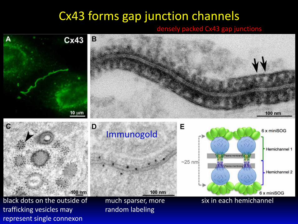
Cx43 forms gap junction channels
six in each hemichannel black dots on the outside of trafficking vesicles may represent single connexon
densely packed Cx43 gap junctions
much sparser, more random labeling
Immunogold

C. elegan
targeted to the mitochondria in body wall muscles
adjacent unlabeled mitochondria in a different cell type
miniSOG is under muscle-cell specific promoter

Could miniSOG reveal new molecular details of
the organization of neuronal synapses?
J Neurosci. 2007 Nov 14;27(46):12516-30.

SynCAM1
SynCAM2
Synaptic vesicles
http://cognitivephilosophy.net/brain-research/neuroplasticity-in-brief/
SynCAM1-miniSOG was found only at presynaptic terminals, identified by the presence of synaptic vesicles.
SynCAM2 localized to postsynaptic sites.

Conclusion
The utility of miniSOG was demonstrated by correctly labeling
several already well-understood proteins in mammalian cells,
nematodes and rodents.
Correlated confocal and EM imaging could be performed with
miniSOG, producing excellent EM contrast, efficient labeling, and
good preservation of ultrastructure.
SynCAM1 and SynCAM2 can be precisely indicated by miniSOG
to the presynaptic and postsynaptic sides of mammalian brain
synapses respectively.

Discussion
Although the method is limited in that one can tag only one kind of protein at a time, the tag is useful and will be a valued tool for cell biologists.
MiniSOG will grant new powers to electron microscopy, permitting scientists to pursue answers to questions previously impossible to ask.

Thank you for your attention!