
PLA Enhanced via Plasma Technology: A Review€¦ · A Review Pieter Cools, Nathalie De Geyter and...
32
In: New Developments in Polylactic Acid Research ISBN: 978-1-63463-054-2 Editor: Courtney Winthrop © 2015 Nova Science Publishers, Inc. Chapter 3 PLA Enhanced via Plasma Technology: A Review Pieter Cools, Nathalie De Geyter and Rino Morent Research Unit Plasma Technology, Department of Applied Physics, Ghent University, Ghent, Belgium Abstract In the last 50 years, it has numerously been evidenced that plasma treatments can effectively change the surface properties of conventional polymers (PP, PET, PE, PA…). Only recently the influence of plasmas on biodegradable polymers, such as polylactic acid (PLA) has been reported. PLA can have important biomedical applications in tissue engineering as three-dimensional porous structures (scaffolds). But their low surface energy leads to poor cell attachment and proliferation and this limits the success of these biodegradable scaffolds. The response it elicits from the surrounding biological environment is crucial and this response is mainly governed by the scaffold surface properties. Therefore, PLA surface properties need to be modified to introduce additional functional groups, which can be recognized as adhesion sites for surrounding cells. Different methods have been developed to obtain the wanted surface properties, however, in the past decade, the use of non-equilibrium plasmas for selective surface modification has been a rapidly growing research field. This chapter therefore presents a critical overview on recent advances in plasma-assisted surface modification of PLA. 1. Introduction Amongst other biodegradable materials, PLA can have important biomedical applications in tissue engineering as three-dimensional porous structures (scaffolds). On the one hand it exhibits excellent mechanical properties and a relatively fast degradation rate, making it an excellent choice of material. On the other hand it suffers from a low surface free energy, leading to a reduced cell adhesion and proliferation, drastically limiting its use as a scaffold No part of this digital document may be reproduced, stored in a retrieval system or transmitted commercially in any form or by any means. The publisher has taken reasonable care in the preparation of this digital document, but makes no expressed or implied warranty of any kind and assumes no responsibility for any errors or omissions. No liability is assumed for incidental or consequential damages in connection with or arising out of information contained herein. This digital document is sold with the clear understanding that the publisher is not engaged in rendering legal, medical or any other professional services.
Transcript of PLA Enhanced via Plasma Technology: A Review€¦ · A Review Pieter Cools, Nathalie De Geyter and...

In: New Developments in Polylactic Acid Research ISBN: 978-1-63463-054-2
Editor: Courtney Winthrop © 2015 Nova Science Publishers, Inc.
Chapter 3
PLA Enhanced via Plasma Technology:
A Review
Pieter Cools, Nathalie De Geyter and Rino Morent Research Unit Plasma Technology, Department of Applied Physics,
Ghent University, Ghent, Belgium
Abstract
In the last 50 years, it has numerously been evidenced that plasma treatments can
effectively change the surface properties of conventional polymers (PP, PET, PE, PA…).
Only recently the influence of plasmas on biodegradable polymers, such as polylactic
acid (PLA) has been reported. PLA can have important biomedical applications in tissue
engineering as three-dimensional porous structures (scaffolds). But their low surface
energy leads to poor cell attachment and proliferation and this limits the success of these
biodegradable scaffolds. The response it elicits from the surrounding biological
environment is crucial and this response is mainly governed by the scaffold surface
properties. Therefore, PLA surface properties need to be modified to introduce additional
functional groups, which can be recognized as adhesion sites for surrounding cells.
Different methods have been developed to obtain the wanted surface properties, however,
in the past decade, the use of non-equilibrium plasmas for selective surface modification
has been a rapidly growing research field. This chapter therefore presents a critical
overview on recent advances in plasma-assisted surface modification of PLA.
1. Introduction
Amongst other biodegradable materials, PLA can have important biomedical applications
in tissue engineering as three-dimensional porous structures (scaffolds). On the one hand it
exhibits excellent mechanical properties and a relatively fast degradation rate, making it an
excellent choice of material. On the other hand it suffers from a low surface free energy,
leading to a reduced cell adhesion and proliferation, drastically limiting its use as a scaffold
No part of this digital document may be reproduced, stored in a retrieval system or transmitted commercially in any form or by any means. The publisher has taken reasonable care in the preparation of this digital document, but makes no expressed or implied warranty of any kind and assumes no responsibility for any errors or omissions. No liability is assumed for incidental or consequential damages in connection with or arising out of information contained herein. This digital document is sold with the clear understanding that the publisher is not engaged in rendering legal, medical or any other professional services.

Pieter Cools, Nathalie De Geyter and Rino Morent 80
material. The response it elicits from the surrounding biological environment is crucial and
this response is mainly governed by the scaffold surface properties.
In the last few decades, numerous surface modification techniques have been developed
that introduce extra functionalities on the substrate surface, leading to an increase in surface
free energy. When applying a surface modification in general, it is critical that bulk
properties, such as mechanical strength and structural integrity, remain unaffected. When it
comes to biomedical applications specifically, the use of solvents and chemicals based
surface treatment techniques are reduced to a limited set approved by EU and US legislation.
Therefore solvent free techniques such as γ-radiation, UV treatments, corona discharges etc.
have gained a lot of popularity in the field of tissue engineering. One of those techniques that
has been around for a long time has recently found its way into the biomedical field: non-
thermal plasma technology.
In the last 50 years, it has numerously been evidenced that non-thermal plasma
technology can effectively change the surface properties of conventional polymers (PP, PET,
PE, etc.) [1-5]. Although biodegradable polymers are already commercially available for
more than 35 years, the studies on plasma modification of these materials situate themselves
more recently. The first publications started around the period Langer and Vacanti introduced
the concept of tissue engineering and their number has kept steadily growing ever since [6].
At the end of last century, literature was dominated by oxygen plasma treatments in low
pressure RF discharges and the corresponding response on cell adhesion, growth and
proliferation. Although today the majority of publications still focuses on these low pressure
oxygen plasma, there is a growing field around atmospheric pressure plasma treatments.
Both topics, as well as other discharge gasses, plasma polymerization, grafting, biomedical
applications and applications outside the biomedical field will be discussed in the following
paragraphs.
2. Non-Thermal Plasma Technology
2.1. History and Definitions
Plasma is a gaseous mixture of radicals, ions, electrons and neutrals that is also known as
the fourth state of matter and was first defined by Langmuir in 1929. Over 99% of the known
universe consists of plasma, earth being one of the few exceptions. In nature, plasma occurs
under the form of lighting or aura borealis (northern lights), but the majority of plasma on
earth are man-made. In the lab plasmas are normally generated by supplying energy to a
neutral gas, via thermal energy (flames) or by applying an electrical field causing the
formation of charge carriers [7].
An important distinction has to be made between thermal and non-thermal plasma. In
thermal plasma, both electrons and heavy particles have the same temperature situated around
4000-5000 K or higher and are considered to be in a thermal equilibrium. For non-thermal
plasma, only the electrons are accelerated by the electrical field, causing a thermal difference
between the light and heavy particles, resulting in plasma that is operating at lower
temperatures. Due to this difference in operating temperature, they are often referred to as
„hot‟ and „cold‟ plasma respectively. For the cold plasma this still can result in a temperature
PLA Enhanced via Plasma Technology: A Review 81
of a few 1000 K. For biomedical applications, only non-thermal plasma treatments are
applied where the degree of ionization is 1% or lower, resulting in plasma operating at room
temperature range (290-330 K) thus avoiding thermal degradation of the (biodegradable)
polymers. In the field of non-thermal plasma there is still a wide variety in the way plasma
can be generated.
2.2. Plasma Sources
Each of the sources discussed in the following section has its advantages and
disadvantages. Over time they have found applications in all branches of the industry:
automotive, packaging, textiles, aerospace, (bio)medical etc. The plasma reactor designs that
have been developed are numerous and a whole book could be written on that topic alone, as
for each new applications design changes are made to optimize the plasma treatment for that
specific process. But almost all of these designs can be linked to one of the plasma sources
discussed here.
DC Discharge
Non-thermal plasma generated via a DC discharge are normally formed in a closed
reactor between electrodes at low pressures (10-1
– 10 pa) [7, 8]. Depending on the used
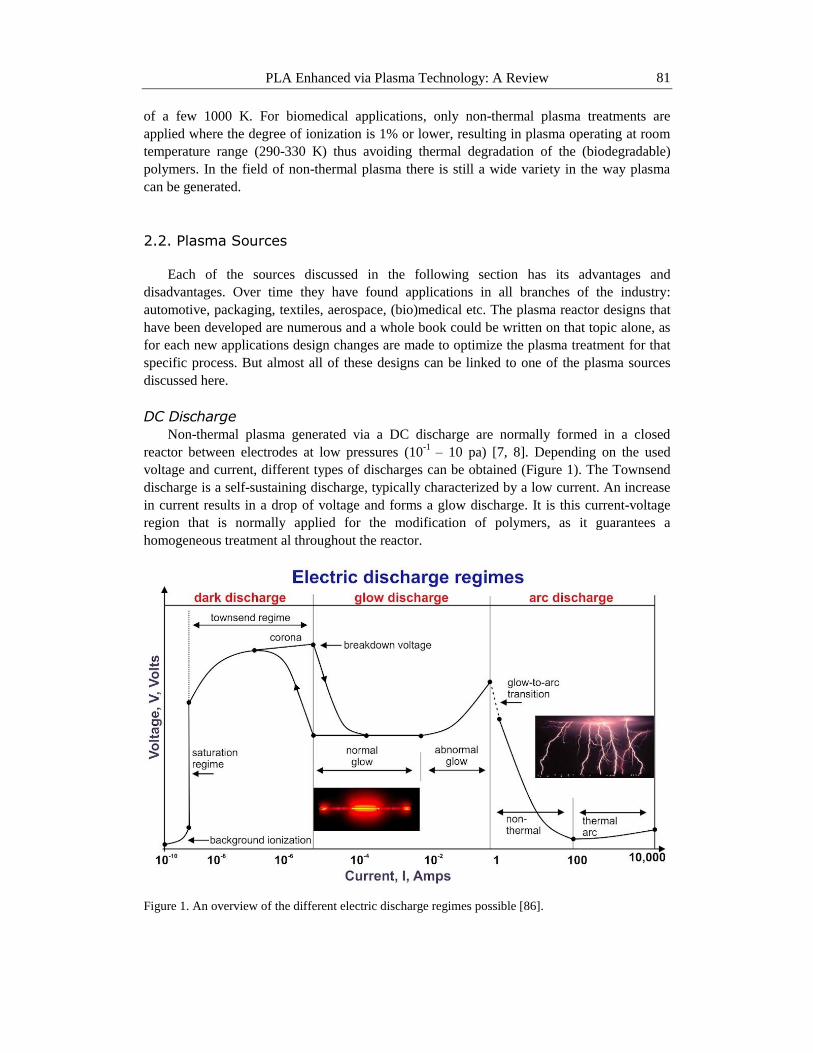
voltage and current, different types of discharges can be obtained (Figure 1). The Townsend
discharge is a self-sustaining discharge, typically characterized by a low current. An increase
in current results in a drop of voltage and forms a glow discharge. It is this current-voltage
region that is normally applied for the modification of polymers, as it guarantees a
homogeneous treatment al throughout the reactor.
Figure 1. An overview of the different electric discharge regimes possible [86].
Pieter Cools, Nathalie De Geyter and Rino Morent 82
A further increase of the discharge current results in the build-up of the corresponding
voltage until an arc is formed and the voltage drops almost completely. In general it can be
concluded that all processes going on in a DC discharge are well-known, giving a high
control over the process.
Next to the continuous DC discharge, also pulsed systems are available. Two of the main
advantages of pulsed systems, concerning biomedical applications, are the possibility of using
higher discharge powers without thermally damaging the substrates and if used for the
deposition of thin films, it applies a more homogeneous coating.
Alongside the limited pressure range, a major disadvantage of the DC discharge
technology is the direct exposure of the metal electrodes to the plasma medium, making them
vulnerable to corrosion when exposed to certain compounds formed in the plasma.
RF and Microwave Discharges
Compared to DC discharges, radiofrequency and microwave discharges are formed and
sustained using high frequency electromagnetic fields [7-9]. RF discharges can operate at a
range between 1 – 100 MHz, but in most cases a fixed frequency of 13.56 MHz is applied.
Concerning the operating pressure, a wider range (1-103 Pa) is possible, but with the
exception of a few, high-vacuum equipment is needed, which is expensive and drastically
increases treatment times. For biomedical purposes, this is probably the most applied
discharge, as it is the plasma technique of choice for oxygen plasma treatments and several
reactors are commercially available on the market.
For microwave discharges, the frequency range is an order of magnitude higher and is
normally fixed at 2.45 GHz. In contrast to RF and DC discharges, microwave reactors can be
operated in a very broad pressure range starting from 1 Pa up to atmospheric pressure.
Operating at higher pressures does result in an increase of heat transfer to the substrate,
limiting the practical pressure range for biomedical applications, giving cause to the same
limitations for the vacuum equipment as for the RF discharge.
Dielectric Barrier Discharge
A dielectric barrier discharge (DBD) or silent discharge is using high frequency AC or
RF as a discharge source and together with its high pressure operating range (5-100 kPa), it
differs somewhat from the previous described techniques [7, 8]. Already in 1857, Siemens
used a DBD for the generation of ozone and to this day it is still one of its most important
industrial applications [10].
A DBD reactor typically consists of 2 electrodes, which are usually a few mm apart, of
which at least 1 is covered with a dielectric material such as glass, quartz or a ceramic
material. The voltage applied ranges typically from 0.5 kV up to a few 100 kV. The discharge
formed consists out of numerous small streamers, called micro-discharges. Due to the
dielectric material, the discharge current is limited, giving cause to very short-lived micro-
discharges (1-10 ns) that are distributing themselves homogeneously across the electrode.
The major advantage of the DBD compared to other plasma techniques, is the possibility
to operate at pressures that do not require extensive vacuum equipment, making it a cheaper
and more time-effective. Furthermore they allow for a wider range of applications such as
plasma chemistry, polymerization, etching, cleaning etc. due to their very low heat
generation. Many of these applications are not always possible using other techniques as will
be discussed in the next paragraph.

PLA Enhanced via Plasma Technology: A Review 83
Atmospheric Pressure Plasma Jets
In the previous sections, the most important power supplies have been discussed with
their pros and cons and as has been mentioned, they can be used for any kind of reactor
design. In this paragraph some special attention will be given to a kind of set-up that has
hugely gained in popularity for the last decade: the atmospheric pressure plasma jet (APPJ).
The atmospheric pressure plasma jet (APPJ) is typically a variation of the parallel plate
set-up. Through 2 concentric electrodes, whether or not covered with a dielectric material, a
mixture of different gasses is send. The high voltage is applied and the ionized gas is send
through a nozzle, forming a plasma plume. The substrate is typically placed a few mm
underneath the nozzle, in the so-called afterglow, where it is exposed to the reactive species
of the plasma [8].
In 2007 Laroussi et al., wrote an excellent review on the topic of arc-free APPJ‟s that
covers all the essentials [11]. Discussing each of them here would lead this chapter too far out
of its scope and would not add any crucial information to understand its applicability for
biomedical research, which will be discussed in chapter part 4.2.
2.3. Plasma Material Interactions
Before discussing the applications of plasma technology in the biomedical field, it is of
the essence to understand the possible interactions of the ions, radicals and electrons present
in the plasma and the substrate exposed to them.
Plasma Cleaning and Etching
Contamination is everywhere. During the production process and storage of
(bio)materials, they are exposed to a number of solvents, greases, volatiles etc. These
products often adsorb on the material surface, resulting in a reduced product performance. A
known example in the biomedical field is the adsorption of low molecular weight carbon
species onto a pristine titanium sample, when exposed to ambient air. When used as an
implant material, this surface pollution results in a reduced cell adhesion, proliferation and
growth and in some cases even results in cell death [12, 13].
When exposing these contaminated surfaces to the mixture of highly reactive species
generated by the plasma, they remove this contamination in a matter of seconds, without
having any major influence on the underlying surface [14]. When further increasing the
treatment time and/or discharge power, the plasma is capable of not only cleaning the surface,
but also of etching away the top layers of the sample [15-18]. For hard materials, higher
discharge powers and/or a prolonged exposure are essential to obtain a notable etching effect.
Polymer surfaces on the other hand, are inevitably etched when exposed to non-thermal
plasma. For biomedical applications this is considered a benign effect, as possible changes in
nano-roughness can amplify the positive effects of plasma treatments concerning cell growth
and adhesion [19, 20].

Pieter Cools, Nathalie De Geyter and Rino Morent 84
Plasma Activation
The main reason for exposing (biodegradable) polymers, such as PLA, to plasma, is to
enhance its surface properties. The charged particles in the plasma interact with the substrate
in such a way that reactive radical sites are generated. Depending on the gas feed, these sites
will react (in)directly with O/N di-radicals, forming a broad variety of functional groups on
the surface. This incorporation of polar groups, as evidenced via x-ray photoelectron
spectroscopy (XPS), result in a drastic change in wettability, which in turn has a positive
effect on material –cell interactions.
The traditional use of plasma treatments on biodegradable materials is not always
considered the right course of action. For some applications such as the inside of artificial
stents or heart valves an increase in cell adhesion has to be avoided at all cost, as it will lead
to a premature failure of the implant. Instead of using the traditional gas feeds for plasma
treatment (noble gasses, oxygen, dry air, nitrogen…) different research groups have used
fluorinated gasses such as CF4 that result in the formation of super hydrophobic surfaces with
water contact angles of 150° and higher. These fluorinated surfaces prevent cells from
effectively adhering on the surface and thus guaranteeing an optimal performance of the
implant material [4, 21, 22].
When plotting the static water contact angle (WCA) as a function of plasma treatment
time, while fixating all other parameters, a graph is generated that depicts the treatment
effectiveness (see figure 2). In most cases a plasma treatment results in a progressive
decrease/increase of the WCA followed by a saturation plateau. Using these plots allows a
fast and easy fine-tuning of the surface free energy needed for a specific application.
One of the advantages of plasma activation compared to other techniques introducing
functional groups on the surface is that it falls under the category of non-invasive techniques,
only modifying the outermost surface layers. This guarantees the preservation of the
structural integrity and chemical and mechanical properties of the bulk.
Plasma Grafting and Polymerization
An alternative way of using non-thermal plasma is as an initiation medium for the
polymerization of thin films. Almost always preceded by plasma activation, the
polymerization process can follow two pathways: plasma grafting is very much the same as
radical polymer grafting in traditional chemistry. The radicals introduced on the surface by
plasma activation are used as initiation sites to start the chain reaction. Before the monomer is
introduced into the plasma chamber, the plasma is deactivated. Therefore the functionalities
present in the monomer are preserved and a “traditional” polymer chain is grown.
For plasma polymerization this is not the same case. This time the plasma is used as an
initiation medium and remains active during the polymerization reaction. The active radicals
present in the plasma form initiation sites on both the substrate surface and the monomer
molecules that are mixed in the feed. In contrast to chemical initiation, plasmas are not as
specific as to where the radicals are introduced, using the functional groups of the polymer
precursor as well to initiate the chain reaction, resulting in a highly cross-linked, pinhole free
and completely amorphous thin film that significantly differs from its traditional counterpart
and adheres to almost any surface. Varying the discharge power gives a high control over the
amount of functionalities preserved in the film. From a biomedical viewpoint this is an
interesting application, as the functional group density plays a critical role in the growth and
proliferation of cells and differs for the type of cells used.

PLA Enhanced via Plasma Technology: A Review 85
2.4. Surface Characterization Techniques
As mentioned in section 2.3, when applying non-thermal plasma to a biodegradable
polymer such as PLA, a number of changes are induced onto the material surface. In order to
understand material-cell interactions, it is critical to thoroughly characterise the induced
surface changes. To do so, a wide variety of analysing techniques are available.
Contact Angle Goniometry
The usual strategy is to start with a contact angle goniometry study to map the changes in
wettability and surface energy. This is the logical choice to commence with, as it is both an
extremely fast technique and gives very sensitive results. For the changes in wettability a
droplet of water is deposited onto the surface and the angle between the droplet and the
surface is measured (see figure 2). To determine the changes in surface free energy, multiple
liquids (usually water and/or diodomethane) are deposited onto the surface and the contact
angles are determined. These results are then combined in the harmonic mean equations and
the surface free energy is calculated [23]. These results often give a first indication what kind
of changes the plasma treatment induces and to what extent, but it gives no chemical
information.
Figure 2. Example of changes in water contact angle as a function of energy density for different
discharge gasses (measurements performed on PET, using a DBD discharge at medium pressure) [87].
X-Ray Photoelectron Spectroscopy
The method of choice for a surface chemical analysis is XPS. An X-ray beam with a
fixed energy radiates the surface. This excites the inner shells of the atoms and photons with
an energy characteristic for those elements are emitted. Via a hemispherical analyser the
photons of a certain energy can be focused and collected. Scanning all energies in one
spectrum gives you information on the elemental composition of the substrate. The real power

Pieter Cools, Nathalie De Geyter and Rino Morent 86
of the technique lies in the fact that chemical bonds cause a shift in the energy at which the
photon is emitted. This allows obtaining information about the chemical bonds present in the
material analyzed via the deconvolution of the measured peaks. One of the major reasons
XPS is preferred over other similar techniques is because of the analysis depth (+/-10 nm)
lying in the same range as the plasma penetration depth [24]. The downside would be that it
only gives information about the chemical bonds present on the surface, making it difficult to
distinct certain sets of functional groups. In some cases derivatisation techniques can solve
this problem, but they can easily lead to misinterpretation [25-27].
Fourier Transform Infrared Spectroscopy
An alternative technique that can be applied is FT-IR (Fourier transform infrared)
spectroscopy. An IR laser beam is send through the sample surface, partially absorbing the IR
light. The absorbed light causes molecular vibrations characteristic for the analysed material
at very specific frequencies. Upon collection of the light going through the sample and
subtracting it from the original beam, a spectrum is formed that is unique for a specific
molecule (so-called molecular fingerprint). Peaks at a certain frequency present in the spectra,
give more information on the different functional groups present. When used for plasma
technology, if applied in the right way, it can give more information about new functional
groups that are introduced via plasma treatment and expose the modifications that have been
done to the functional groups already present. The downside lies in the fact that the analysis
depth (500-1000 nm) is an order of magnitude higher, thus resulting in a low, often
insufficient sound to noise ratio, making it hard to perform a reliable analysis.
Atomic Force Microscopy
Besides the chemical composition, also the surface morphology plays a crucial role in
cell adhesion and proliferation processes. Changes in roughness as small as 5 nm can make
the difference between success and failure of a tissue engineered system. There are several
methods available for visualisation and quantification of the surface morphology. One of the
most popular methods for quantifying the surface roughness and generating visual images up
to the nm level is atomic force microscopy (AFM).
AFM measurements are based on a laser beam that is reflected on an oscillating
cantilever onto a detector. As the tip of the cantilever goes over the sample surface, it will be
subjected to changes in interatomic forces. These forces bend the cantilever, giving cause to a
shift in the reflection spot of the laser beam. By linking the shift of the reflection spot to a
change in sample surface height, a very accurate mapping of the surface is possible (see
figure 3). Next to imaging the technique can also be used for quantifying the roughness and
measuring attractive and repulsive forces.
Secondary Electron Microscopy
One of the most popular visualisation techniques used in the biomedical industry is
secondary electron microscopy (SEM). A beam of electrons is focused via a number of
mirrors onto the substrate. The impact of the electrons on the sample surface causes the
emission of secondary electrons which are collected onto a detector. These secondary
electrons can form high resolution images up to the nm-scale. Not only is it used for mapping
the surface morphology, it is also an important technique for cell characterization.

PLA Enhanced via Plasma Technology: A Review 87
Figure 3. AFM images depicting the effects of low pressure plasma treatment on the surface topography
[88].
3. PLA and Low Pressure Plasma: The Biomedical Applications
When combing through the literature for papers on the combination of PLA and plasma,
a wide variety of biomedical applications has been studied by labs all over the world.
Numerous reactor designs, based on the sources described in the above sections, are used for
plasma treatment in order to benefit from one or more of the plasma induced effects on the
PLA surface. In order to keep an overview on all the different applications, the chapter will
start with those publications focusing solely on the physical changes plasma induces on the
PLA surface. This will be followed by the papers dealing with the direct effect of the plasma
on the PLA‟s histological properties. Finally there will be an overview on the use of plasma

Pieter Cools, Nathalie De Geyter and Rino Morent 88
as a grafting and polymerization technique or to covalently bond specific (macro)molecules
on PLA substrates for biomedical purposes.
3.1. Low Pressure Oxygen Plasma
Oxygen is the most popular feed gas for low pressure treatment of PLA, followed directly
by argon and anhydrous ammonia. The popularity of these three discharge gasses has been
stimulated by the availability of commercial systems working solely with these 3 [28-41].
Wettability
Most papers using an oxygen discharge have performed a surface characterization,
starting with a static water contact angle (WCA) study. For non-treated PLA most studies
agree on a WCA of about 80°. During treatment, the contact angle decreases progressively to
values between 10° and 50° for treatment times between 2-20 min and discharge powers
between 10-90 W, which is quite a broad region [20, 21, 28-30, 36, 37, 42-44]. Ferreira et al.,
mention this difference explicitly by stating that for histological studies, the goal is not
looking for the lowest contact angle, or highest roughness, but for the parameters resulting in
the best cell response [30].
Surface Morphology
The WCA study is usually followed by a visualisation of the surface, either with AFM,
SEM or both. Oxygen plasma in general are well-known for their excellent surface etching
properties, introducing a sub-micron roughness which is considered to boost cell growth and
proliferation. Mattioli et al., describe the changes in surface morphology as the apparent
formation of nano-pillars [21]. Most other papers simply state that there is an increase in
surface roughness upon visual analysis of the recorded images. According to Riccardi et al,
the plasma will preferentially sputter the amorphous regions of the polymer film, thus
resulting in a distinct increase in roughness [45]. When treated for longer periods of time,
again a decrease in surface roughness is noted, due to the eventual etching of the crystalline
regions [30] (See figure 3).
Chemical Composition
The physical characterization, using the WCA, AFM and SEM results, is linked to a
chemical surface analysis (see section 2.3.) involving XPS and IR measurements in only a
few papers. For the untreated samples a theoretical O/C ratio of 0.5 would be expected, which
is experimentally confirmed by Zhao et al., [29]. After treatment, all studies agree on an
increase in oxygen content on the surface, but there is no consensus on the amount of oxygen
incorporated, nor as to what kind of function groups and to what extent they are incorporated
[20, 28-30, 42, 44].
In-Vitro
When browsing the literature on PLA and non-thermal plasma, it becomes evident that
the research is mainly focused on the histological impact of the treatment, rather than on the
treatment itself. For the analysed studies using oxygen as a feeding gas, 6 different cell types
PLA Enhanced via Plasma Technology: A Review 89
were seeded onto the samples for cell adhesion and proliferation essays: MG63, FRC, CHO,
B65, M3T3, hMSC… [20, 28, 36, 38, 40-42, 44, 46].
Although it could be interesting to discuss each article, it would lead to far and the
conclusions for all of them are more or less the same. The adhesion essays show that in the
first 24 hours, the cell density for the plasma treated samples lies significantly higher for the
plasma activated PLA compared to the untreated samples. The study using CHO, performed
by Yamaguchi et al, shows that the plasma activation results in similar cell adhesion
compared to the standard tissue culture PS dishes [44]. After the first 24 hours and up to 7
days, the proliferation rate is more or less the same for all cells tested, with the exception of
the hMSC cells, which show an overall superior proliferation rate.
When comparing the cell morphology after 7 days of incubation, there is a major
difference: the cells on the untreated samples are small, round and no network is formed
between different cells. For the cells proliferated on activated samples, cells flatten out and
form an intercellular web. One of the direct consequences of this difference in cell
morphology has been studied by Wan et al., they exposed the PLA, seeded with M3T3, to
shear stress. For the untreated polymer films this resulted in extensive detachment of the cells
form the surface, while for the treated PLA the cells extrusion was retracted to the body and
al changes happened very slowly, indicating a more stable cell culture [36].
Overall it can be concluded that the changes in surface- chemistry, wettability and
roughness induced by the oxygen plasma, result in benign histological properties.
3.2. Low Pressure Ammonia Plasma
The big difference between oxygen and ammonia plasma is the introduction of nitrogen
containing species into the plasma and a possible incorporation of amines onto the PLA
surface, which are considered beneficial for cell adhesion and proliferation.
Wettability
In contrast to the WCA results obtained from the oxygen treated PLA, there is more of a
consensus on the contact angles for the ammonia treatments. Using an RF discharge, WCA
are found starting from 60° up to 20° [28, 32, 36, 47-49]. In contrast to other gasses,
prolonged exposure to the NH3 plasma does not lead to a fixed angle, but a significant re-
increase in WCA is noted. The lowest contact angle is obtained at a power of 50 W and an
exposure of 2 min in all studies. Jiao et al., contribute these higher angles at higher energy
densities to the thermal degradation of PLA, causing a decrease in the incorporation of polar
groups [47]. Compared to the oxygen plasma, the contact angles are slightly higher, but as
mentioned before, it is not always the highest density in polar groups that lead to the best
histological results.
Surface Morphology
The literature describing the changes in surface morphology is limited, but those papers
that have a surface morphology study included come to the same conclusion as the oxygen
plasma: there is an increase surface roughness and a fine grain structure is introduced [48].
More interesting is the SEM study of Wan et al., that showed a visible degradation of a PLA
scaffold when exposed to powers higher than 20W [36]. This was confirmed by the AFM

Pieter Cools, Nathalie De Geyter and Rino Morent 90
study of Jiao et al., (50 W-2min) that showed the first signs of film destruction and the
softening of the borders between crystals [47]. Linking the changes in surface topography
with the wettability studies, confirms that a careful selection of discharge power and
treatment time is advised.
Chemical Composition
As mentioned in the beginning of this section, ammonia plasma are mainly used to
incorporate nitrogen functionalities into the PLA surface. The focus of the chemical surface
composition therefore lies onto the analysis of the nitrogen peaks. In this case no literature
was found using IR as an analysis technique, so all results presented are measured via XPS
[28, 32, 33, 47-50].
In general, the effectiveness of the nitrogen incorporation can be linked again to the
changes in surface morphology and WCA: an overexposure (Power and time) of the PLA
leads to a reduction of nitrogen functionalities compared to more moderate operating
conditions. Different papers report an incorporation of 5-10% of nitrogen associated with a
decrease of both oxygen and carbon. The deconvolution of the nitrogen peak at 400 eV learns
that the majority of the nitrogen is incorporated as primary amines (75%) and equal amounts
of imines and amides (10-15%) and this for a power of 50W and a treatment time of 2
min(See figure 4) [33]. As mentioned before, an increase in power leads to a decrease in
nitrogen content and causes a shift from primary amines to amides.
Figure 4. Deconvolution of the C1s and N1s XPS peaks, proving the presence of Amine and Amide
functional groups [89].

PLA Enhanced via Plasma Technology: A Review 91
Jiao et al., performed a chemical reaction using an acidic orange 7 solution to form a
complex between the primary amines and the acid dye [47]. This was followed by washing
the samples with a NaOH solution, desorbing the dye. The NaOH solution is exposed to light
with a wavelength of 492 nm and an absorbance spectrum was recorded. Based on this
procedure an amino group density on the PLA surface of 1.4 10-9
mol/cm2 was found.
Combined with the XPS analyses it is proven that ammonia plasma are able to incorporate
primary amines onto a PLA surface.
Biomedical Applications
In contrast to the oxygen treatments, not only histological studies were performed, but
also protein and peptide adhesion was investigated.
The histological studies show a similar enhancement in the adhesion essays compared to
the oxygen plasma for all cell types tested: M3T3, HUVEC, rbMVEC, BAEC, BSMC,
fibronectin, H1444 and rat calvarial cells [28, 31, 32, 47, 49-52]. The proliferation essays on
the other hand differ compared to the oxygen proliferation essays in such a way that the
ammonia plasma also significantly enhances the proliferation process, with the exception of
the M3T3 cells, up to and beyond the growth rate of tissue culture PS dishes.
Several groups went beyond the 2D-films and started investigating cell properties on 3D-
scaffolds [31, 32, 50]. Not only were they interested in adhesion and proliferation essays on
the scaffold surfaces, but even more so in the migration efficiency of the cells into the
scaffolds. Cheng et al., performed a very thorough study of the cell infiltration into the
scaffold, both in-vitro and in-vivo [50]. 20 µm thick slices were cut from the scaffolds and
cells were coloured with a fluorescent dye. In figure 5, 3 different cross sections are included.
The untreated one at the top shows a very low density of cells present and almost no
infiltration of the cells. The cross section in the middle, showing the effects of an Ar
treatment, depicts a much higher cell density and reasonable infiltration. The lower one, being
the result from the ammonia treatment, shows both excellent cell proliferation and infiltration.
The same study was performed in vivo on rats and the same results were obtained, though
with a lower efficiency. Therefore they concluded that an excellent infiltration in-vitro is
required in order to have success in-vivo and that the combination might lie in a combined
use of plasma treatment and the incorporation of growth factors into the scaffold.
The protein and peptide adsorption studies found give mixed result on the plasma
treatment of PLA. Xu et al., investigated the anchorage of GRGDS peptides onto PLA
scaffolds before and after an ammonia plasma treatment. After the treatment they saw an
excellent conjugation between the peptide and the polymer surface, caused by the amide
bonding between the incorporated primary amines and the carboxyl functionalities present in
the peptide [33].
Sarapirom et al., did some research on the adsorption of the HSA protein before and after
treatment and came to the conclusion that the protein prefers non-polar, untreated, surfaces.
This result should be stressed, because it points out that not every treatment is successful for
every application, emphasizing the need for application tailored research [48].
Finally Yang et al., used an ammonia plasma treatment for the immobilisation of collagen
onto a PLA surface. Compared to a collagen coating, the M3T3 cell proliferation showed no
significant differences, but once the samples were rinsed in PBS solution for some minutes,
some interesting changes were noted. Before and after rinsing, the cells number on
immobilized collagen did not differ significantly. For the normal collagen coating on the

Pieter Cools, Nathalie De Geyter and Rino Morent 92
other hand, most of the cells were washed away. This proves that plasma technology is able to
anchor collagen tightly onto the surface, making the treated scaffold a better match for tissue
engineering purposes.
Overall it can be concluded that ammonia plasma treatments are able to incorporate
primary amines onto a PLA surface, making it a more versatile treatment and resulting in a
better proliferation compared to oxygen treatments. Although it leads to positive effects in
most cases, a case to case study is required to guarantee its success.
Figure 5. Different fluorescent cell essay cross-sections of: A) untreated scaffold, B) Ar plasma treated
scaffold and C) ammonia plasma treated scaffold [90].
3.3. Low Pressure Argon Plasma
Of the 3 most used low pressure plasmas, Ar plasma is considered to be the mildest
treatment technique. Via a cascade of secondary processes, reactive sites are introduced onto
the surface that, once exposed to ambient air, result in the incorporation of a whole set of
oxygen containing functional groups [53].
Wettability
The more mild conditions of the Ar plasma treatment are clearly reflected in the WCA
results. Whereas oxygen and ammonia plasma reached contact angles below 20°, the Ar
treatment results have a saturation contact angle between 25° and 50°, depending on the

PLA Enhanced via Plasma Technology: A Review 93
discharge system used [29, 35, 50, 53-56]. Rather than differences in surface topography, this
difference of 30°, in some cases, is most likely caused by the differences in the kind of
functionalities that are incorporated, which will be discussed in one of the following
paragraphs.
Surface Morphology
The changes in surface morphology that are introduced by the argon plasma are
comparable to the oxygen and ammonia treatments. Different papers describe the
phenomenon of different etching rates between the crystalline and amorphous regions of the
film resulting in the exposure of the crystallites present in the film [29, 35, 54, 56]. Zhao et
al., did a comparative study between Ar and oxygen plasma treatments and for the surface
morphology they found that the etching of the PLA is less pronounced compared to the
oxygen etching [29]. Slepicka et al., found an increase in roughness from 6.9 nm to 11 nm,
which was in line with the expectations. More surprisingly, a gravimetrical analysis of the
treated samples showed that, using a DC discharge of 10 W, the etching is capable of
removing up to 75 nm of material over a period of 4 minutes. This shows that the top layers
are etched away completely and that the underlying layer is etched at different rates,
depending on the crystallinity, as stated before [56].
Figure 6. A) Schematic representation of the experimental process to produce superhydrophobic
surfaces. B) SEM image of the smooth PLLA surface. C) SEM image of the rough surface. D)
Magnified SEM image of a protrusion on the superhydrophobic surface. Insets: Water drops on the
corresponding surfaces [91].
In the next paragraph, special attention will be given to an article published by Song et
al., In their study they synthesized super-hydrophobic PLA films using a novel polymer
solution-air casting method (See figure 6) [57]. In their study, they investigated the effect of

Pieter Cools, Nathalie De Geyter and Rino Morent 94
argon plasma on the wettability properties of the super-hydrophobic film. Starting from a
WCA above 150° the plasma treatment results in a progressive decrease of the contact angle
up to and under 5°. Compared to the treatments on smooth films, the effect of the Ar plasma
treatment is greatly enhanced. The reason for mentioning this research here, is to stress on the
combined effect surface roughness and plasma treatments have on the wettability of a PLA
surface. In the application section, more attention will be given to the histological results
found in this study.
Chemical Composition
As mentioned in the introduction, the formation of radical sites onto the sample surface
occurs through a cascade of secondary reactions as can be seen in figure 7. Once exposed to
the ambient air, oxygen containing functional groups can be formed at the reactive sites.
When browsing through the literature dealing on the chemical surface analysis of Ar
plasma treated PLA, it becomes apparent that most studies do not result into the incorporation
of an extra amount of oxygen containing functionalities, nor are they able to introduce new
functionalities [34, 35, 53, 54, 56, 58]. Different groups even note a decrease in oxygen
content, suggesting that degradation of the PLA takes place. Linking this to the wettability
and surface morphology analyses, it would suggest that the Ar plasma etching and the
resulting increase in surface roughness are the main causes of the decrease in WCA.
Those treatments that were able to increase the oxygen incorporation, find similar results
as the oxygen treatment, with the biggest increase of oxygen being contributed to carboxyl
functional groups [29, 55].
In general, the surface analysis study clearly shows that treatment parameters have to be
carefully selected in order to avoid degradation of the polymer material and to obtain
significant changes in chemical composition.
Biomedical Applications
The articles dealing on the histological effects and adhesion of peptides are limited and
just as the surface analysis, the results are somewhat mixed. Slepicka et al., seeded VSMC
cells onto the Ar plasma treated surface, and although the first 24 hours suggest a better
adhesion, after 7 days no differences were found between treated and untreated samples,
indicating that PLA is an excellent material to grow smooth muscle cells to begin with [54,
56]. Cheng et al., whom have been discussed before for their excellent study on the cell
adhesion and proliferation of BAEC and BSMC cells on ammonia plasma treated, also used
Ar plasma for treating their PLA scaffolds and their comparison between treatments showed
that on the scaffold surface Ar plasma would result in a significantly better proliferation.
For the cell infiltration, as depicted in figure 5, excellent cell infiltration was found, similar to
the NH3 treatment and superior compared to the untreated PLA. In the in-vivo studies, the
differences between the in-vitro treatments were no longer detected, showing again the
importance of in-vivo testing [50].
Ding et al., used the Ar plasma treatment to successfully immobilize chitosan onto the
PLLA surface. The cell tests, using L929 and L02 cell lines, gave no significant difference in
proliferation rate compared to the cells seeded onto glass plates. Also the cell morphology
was round shaped, indicating the lack of cell differentiation [55].

PLA Enhanced via Plasma Technology: A Review 95
Figure 7. Possible reactions occurring on the PLA film surface on Ar-plasma irradiation [92].
Ho et al., used plasma treatment as a tool for the immobilization of RGDS and RGES
peptides. They compared the adhesion, growth and proliferation of ROS cells onto untreated,
plasma treated and peptide immobilized surfaces. After the first 24 hours, the surfaces that
were having RGDS anchored onto them resulted in the highest cell density. The RGES and
plasma treated samples gave similar results, indicating that the RGES had no significant
influence and that the changes in surface chemistry and morphology induced by the plasma
were the main contributors to the enhanced growth compared to the untreated samples. After
4 weeks the same trends were observed and after calculating the growth rate curves they
concluded that RGDS coated surfaces not only lead to a better adhesion but also to a faster
cell growth and division [59].
As mentioned in the surface morphology section, Song et al., also performed cell
adhesion and proliferation tests, using a L929 cell line, on their super-hydrophobic and
hydrophilic PLA surfaces. Results show that on the hydrophobic surfaces cell growth is poor
and the cell remain small and round. On the Ar plasma treated surface, a significant increase
of cell growth is noted and the cells exhibit a more flattened and extended morphology [57].

Pieter Cools, Nathalie De Geyter and Rino Morent 96
Overall, this research shows that combining surface structuring with plasma treatment allows
tuning of biomaterial surfaces to their specific application.
It can be concluded that Ar plasma treatment can be a viable alternative, if the reaction
conditions have to be mild and if changes in surface morphology play a more important role
than differences in surface chemical composition. Again one has to be careful for sample
degradation and what kind of biomedical application it is used for.
3.4. Hydrophobic Surfaces
As mentioned multiple times in the previous parts of the chapter, the increase of surface
hydrophilicity does not always result in better cell growth and adhesion. In other cases, the
adhesion of cells (crf heart valves, insides of stents, dialysis equipment…) has to be avoided
at all cost, as it would lead to a reduced functionality or complete failure of the material
application. The method of choice to generate (super)hydrophobic surfaces is by using a
fluorinated gasses to form the discharge. The most popular gas would be CF4, but also other
gasses such as SF6 have been used to treat PLA. It is even possible to generate super
hydrophobic coatings using non-fluorinated gasses, but articles, using these techniques on
PLA, have yet to be published.
Wettability
Treating the surfaces with CF4 or SF6 plasma both have the same effect on the wettability
of PLA. An increase between 20° and 40° in WCA up to values of 115° were found,
depending on the treatment time and power used [21, 60-62]. As fluorinated species are
known for their extensive etching, their effect on the surface morphology and thus the indirect
effect on the wettability should not be neglected and will be discussed in the next section.
Surface Morphology
Of all the articles describing the changes in surface morphology, the publication of
Mattioli et al., was the most clarifying, as it compares the topography after oxygen and CF4
plasma treatments [21]. In the chapter part on low pressure oxygen plasma, the technique is
praised for its excellent etching properties. Putting the pictures underneath one another, as can
be found in figure 2, it clearly shows that the etching effect is more profound for the CF4
treatment. The same increase in surface roughness can be found in the other articles [60-62].
Chemical Composition
The main difference between fluorinated gas plasma treatments and other plasma
treatments is self-evidently the incorporation of fluorine onto the PLA surface. XPS
measurements show an increase up to 25% of fluorine. Whereas the elemental composition
shows no real changes in carbon concentration, it is mainly the oxygen that is being replaced.
Chaiwong et al., performed ATR-FTIR spectroscopy after treatment with SF6 plasma and
found C-F stretch vibrations, indicating that the fluorine is covalently bond onto the PLA
surface. Ageing studies show that after 4 weeks, the amount of fluorine present on the surface
does not change significantly, supporting the statement that a stable bond is formed between
fluorine and the PLA surface [60].

PLA Enhanced via Plasma Technology: A Review 97
Biomedical Applications
Although the fluorination technique has been extensively used on other biomaterials [37],
only 1 article was found describing the antibacterial properties of the fluorinated coatings.
Boonyawan et al., tried to grow E. Coli bacteria onto treated PLA surfaces. After fluorination
they saw a decrease in bacterial adhesion of almost 40%, while an antibacterial efficiency of
more than 92% was achieved [62].
Although the haemocompatibility of fluorine coatings has been studied on other
substrates, the biocompatibility of CF4 plasma treated PLA has yet to be tested in-vitro and
in-vivo [63]. The first results indicate that these kinds of coatings can be used for antibacterial
purposes.
3.5. Other Low Pressure Discharges
Next to the conventional discharge gasses, research groups have been experimenting with
a number of other gasses and tested the histological effects. In what follows, each gas will be
discussed briefly and will be linked to the discharge gas it best resembles.
Low Pressure Air Plasma
Air plasma, being a mixture of oxygen and nitrogen, can be considered as a solid
alternative for the oxygen plasma treatment. Even though the discharge gas contains around
80 % of nitrogen, very little nitrogen functionalities will be incorporated, as oxygen is the far
more reactive species and dominates the radical formation and functional group incorporation
processes.
When comparing the changes in wettability between air and oxygen plasma treatments,
similar values, lying between 10° and 15°, are found [64]. No AFM measurements were
found, so no surface morphology information can be given. XPS measurements were
performed and a slight decrease in oxygen content was noted, that was compensated by the
incorporation of 3-5% of nitrogen containing functionalities. As the number of polar groups
did not change significantly, Demina et al., contributed the changes in wettability and surface
energy to an increase in surface charge density. Upon performing a histological study, using
mouse fibroblasts (L929), they found that the cells grew and differentiated better after
treatment [64].
Low Pressure CO2 Plasma
Another gas that is commonly available and is considered waste in most industrial
processes is CO2. 2 papers were found using CO2 as a discharge gas, of which only 1 focussed
on biomedical applications [61, 65]. After treatment, the contact angle decreased from 84° to
48°, similar to Ar plasma treatment. FTIR spectroscopy resulted in a completely different
fingerprint, indicating that CO2 is incorporated onto the surface. The characteristic ester peak
at 1730 cm-1
increased dramatically in intensity and together with a more subtle appearance of
the OH band at 3300 cm-1
this spectrum suggests an increase of different oxygen containing
functionalities, similar to air and oxygen plasma treatments. For their histological studies
Khorasani et al., used 2 different cell-lines, B65 and L929, and examined their adhesion and
proliferation. For the B65 cell line, better adhesion and proliferation was noted, cells were

Pieter Cools, Nathalie De Geyter and Rino Morent 98
flattened out and an extensive network was found. For the L929 line, no significant
differences were found compared to the untreated PLA.
If an overview is made of the different treatments were the L929 cell line is used, it
becomes clear that different discharge gasses and parameters result in extreme differences
concerning cell adhesion, proliferation and morphology, making it not always straightforward
as to what treatment should be considered [55, 57, 65].
4. Medium/Atmospheric Pressure Plasma
Atmospheric pressure plasmas were already used by Siemens in the second half of the
19th century for the ozone generation, but only in the last 25 years there has been a renewed
interest in using the technique for material surface modification, as an alternative to low-
pressure plasma treatment systems. The lack of extensive vacuum equipment makes it time
and cost-effective and allows easy incorporation in production lines.
As mentioned in the general introduction, plasma modification at elevated pressure has
proven its effectiveness on aliphatic and aromatic polymers of all kind and together with the
growing interest in biodegradable materials, there have been a number of publications using
atmospheric pressure plasma treatments on polylactic acid of whom a selection will be
discussed in the following chapter section.
Due to the limited amount of studies available in literature dealing on the plasma
treatment of PLA at atmospheric pressure conditions, a different subdivision will be applied.
The first part of this chapter section will give an overview of parallel plate systems used at
atmospheric pressure. In the second part, special attention will be given to a special set-up
that has gained a lot of interest in the field of plasma technology: the plasma jet.
4.1. Parallel Plate Systems
Most systems operating at atmospheric pressure conditions are based on the DBD
principle (see section 2.2). The most basic set-up available consists of 2 planar electrodes, of
which 1 or 2 are covered with a dielectric material. This system can be operated in a closed
reactor, giving control over the atmosphere present, or in ambient air, making it more easily
applicable.
The discharge gasses used for treatment are similar to the low pressure systems, with the
exception of helium. Helium is quite popular, as it allows for very stable glow-like plasma.
The downsides of helium as a discharge gas, is that it is less effective in introducing polar
functionalities as the surface and due to impurities present, often results in a mediocre
reproducibility.
Before starting with the analysis of results, it is important to make the distinction between
atmospheric pressure and medium pressure treatment. Medium pressure treatments, operating
at a few kPa, do not need the extensive vacuum equipment used for low pressure systems (a
rotary vane or membrane pump is still required), while at the same time more stable plasma
are obtained compared to atmospheric plasma treatments. Due to the presence of a pumping
procedure, the treatment is not as time efficient as its atmospheric pressure counterpart [66].

PLA Enhanced via Plasma Technology: A Review 99
Morent et al., performed a comparative surface characterization study on plasma treated
PLA at medium pressure (5kPa), using He, Ar, air and N2 as discharge gasses [67, 68]. The
WCA study shows a progressive decrease in contact angle as a function of energy density.
Big differences are found between gasses: 59° and 60° for air and argon treatments, 35° and
31° for helium and nitrogen treatments. AFM measurements showed small differences
between treatments, therefore these WCA values primarily have to originate from differences
in surface chemical composition. XPS measurements showed that the decrease in WCA, due
to air and argon plasma treatments, was caused by the incorporation of extra oxygen
containing functionalities (+ 5-7% O) in the films. For the helium and nitrogen treatments no
significant increase in oxygen was noted, but for both treatments the incorporation of nitrogen
functionalities was detected (+3-6% N), hence the difference in WCA. Similar results were
found by Hergelova et al., for treatment in ambient air (55°-60°), by Vergne et al., for
treatment in nitrogen (27+/-3°), both at atmospheric pressure [69-71].
Comparing these results with the low pressure plasma treatments one could conclude,
based on the WCA, that a higher pressure results in a reduced treatment efficiency. The XPS
results on the other hand indicate that the incorporation of functional groups is at least as
favourable. In this case in-vitro or in-vivo studies would be able to make a distinction in
efficiency between treatments, but for the parallel-plate set-up, only 2 histological studies
have been performed. Jacobs et al., used HFF-1, human foreskin fibroblasts, cells, seeded on
PLA plasma treated films, using 3 different discharge gasses (Ar, He and air) [22]. The study
finds very similar results compared to low-pressure treatments, showing a better cell adhesion
in the first 24 hours. After 7 days, the proliferation study shows no significant differences
anymore between untreated and treated PLA. Phase contrast and fluorescent microscopy
indicate superior morphology after 24 hours, independent of discharge gas. After 7 days both
untreated and treated samples show the same morphological features. Nakagawa et al., seeded
MC3T3 cells on air and CO2 plasma treated samples and found similar results compared to
similar studies performed at low pressure [72].
Two histological studies are insufficient to draw any major conclusions. Therefore, the
chapter will continue with an overview of that other, maybe even more popular set-up: the
plasma jet.
4.2. Atmospheric Pressure Plasma Jet
The biggest advantage of the jet compared to all other treatments is its mobility, making
it possible to homogeneously treat complex 3D structures. Furthermore, there is no pumping
procedure, making it cost and time effective.
As just mentioned, plasma jets are often used for treatment of 3D structures. In this
perspective, the electrospinning of scaffolds is by far the most popular application in the
biomedical field. To analyse these materials, contact angle goniometry cannot be used in the
same way, making it difficult to make an objective comparison on the wettability. Other
techniques (AFM, SEM, XPS etc.) do have the needed spatial resolution to give an adequate
analysis of the changes caused by APPJ treatment.
AFM analysis of the surface morphology results in images that are identical to the ones
found for other plasma treatment techniques (see section 3). By changing the movement
Pieter Cools, Nathalie De Geyter and Rino Morent 100
speed of the jet, the surface morphology can be tailored from completely flat up to the same
topography found for fully saturated samples [73, 74].
XPS analysis reveals that plasma jets have fewer problems with the incorporation of
oxygen into the sample, up to a point were O/C ratios higher than 1 are found. This indicates
that the presence of oxygen in the surrounding atmosphere can have a beneficial influence on
the incorporation of polar functional groups [73-77].
The in-vitro studies again indicate results that are similar to other treatment techniques:
the seeding of MC3T3 fibroblasts leads to a better adhesion in the first 24 hours, and a similar
proliferation rate after 7 days with enhanced morphological properties [73]. Other groups
used the plasma to generate anchor sites for chitosan/gelatine. Both in-vitro and in-vivo
studies show excellent stability of the immobilized proteins and superior cell adhesion
(C2C12 and nerve regeneration respectively) and proliferation compared to untreated and
uncoated samples [74, 76].
In general it can be concluded that medium and atmospheric pressure systems form a
valid alternative for low-pressure treatments, giving similar results concerning wettability,
polar group incorporation, morphology modification and histological response.
5. Plasma Coating
The main principles behind plasma grafting and polymerization are already explained in
part 2.3.
One of the major disadvantages of non-thermal plasma treatments is the limited stability
of the treatment. Over time, the surface will try to restore its initial surface energy, resulting
in a decreased wettability and the migration of polar groups. To overcome this instability,
research groups have turned their attention to using the formed reactive sites (grafting) and
the plasma itself (polymerization) as initiation sites for polymerisation reactions.
5.1. Plasma Grafting
In parts 3 and 4, the use of plasma treatments for immobilization of polysaccharides and
proteins such as chitosan, cellulose and RGD have been mentioned. Some authors would also
consider this a form of plasma induced grafting, being limited to a 1 step reaction. In what
follows next, an overview will be given on the use of plasma technology for the grafting and
polymerization onto polylactic acid substrates.
Single-Step Grafting
Some research on the immobilization of macromolecules has been discussed before and
will not be gone through again in detail, but as most of them analyse the histological effects
both on plasma treated and plasma grafted samples, some general trend can be found [28, 29,
55, 78]. In every single case the grafted substrates are equal or superior in performance
compared to plasma treated samples. This is no real surprise, as the macromolecular structure
of proteins and sugars is well-liked by cells and stimulate adhesion, proliferation and

PLA Enhanced via Plasma Technology: A Review 101
differentiation. Therefore they have been the choice of material or coating for many tissue
engineering labs.
In this next paragraph there is a special mention for the work of Yang et al., [28]. In their
research, a comparative histological study was done between PLA samples that were coated
with collagen, with or without ammonium plasma pre-treatment. 7 days after the seeding of
the MC3T3 cells, no real differences in proliferation were found. After rinsing with a PBS
solution, the plasma grafted samples showed no significant difference in cell count. The
samples that had been immersed in a collagen solution without pre-treatment gave cause to a
significant decrease in cell count after rinsing. This research proves that the plasma pre-
treatment results in a tight bonding between the collagen and the PLA, therefore guaranteeing
an optimal functionality of the biomaterial in a more active environment.
Traditional Grafting
For plasma grafting in the traditional sense, acrylic acid and allylamine are two popular
precursors, as the functional end-groups of the resulting polymers, carboxylic acid and
primary amine respectively, are well liked by a great variety of cells and can be used as
intermediates for follow-up reactions [78-81].
Barry et al., performed both plasma grafting and plasma polymerization using allylamine
as a precursor to deposit polyallylamine on porous PLA scaffolds and compared the
distribution of the coating as well as the histological performance [81]. In what follows here,
only the grafting part will be discussed, while in the section on plasma polymerization, a
comparison will be made between the different approaches. The XPS analysis shows a
homogeneous incorporation of nitrogen al throughout the scaffold, though at concentrations
of only 2 percent, indicating a low grafting efficiency (see figure 8).
Figure 8. Nitrogen concentration at the surface as determined by XPS at set intervals across the internal
diameter of grafted and ppAAm-deposited scaffolds (20 W unwashed). These were calculated at set
intervals along the internal diameter (shown as a dotted line in the computed tomography image of the
PLA scaffold, following mechanical sectioning. The photoelectrons were collected from a 0.3 x 0.7 mm
rectangle oriented with the short side parallel to the direction of translation [93].

Pieter Cools, Nathalie De Geyter and Rino Morent 102
Deconvolution of the peaks shows that the amine functionality is preserved, even after
rinsing with water. The cell essay shows that 24 hours after seeding, there is an increase of
about 40% in cell adhesion compared to the untreated samples. SEM imaging showed a
mixed cell morphology, both round and flat, and no visible infiltration of the cells into the
inner parts of the scaffold.
Hydroxylapatite
Another interesting application that has not yet been discussed in this chapter is the
enhanced growth of apatite on PLA. (Hydroxyl)apatite is a mineral consisting out of calcium
phosphate and plays a central role in tissue engineering for implant applications as it
stimulates osteointegration [82]. Yokoyama et al., used an oxygen plasma to introduce extra
functional groups onto the PLA surface, after which the samples were alternatingly dipped in
Ca2+
and (HPO4)2-
ion solutions [79]. Due to electrostatic forces, the ions were adsorbed.
After the dipping process, the samples were stored in a simulated body fluid (SBF) solution.
After 24 hours, analysis showed the growth of a dense apatite layer onto the PLA surface and
this in contrast to the samples that were not exposed to plasma. Adhesion tests showed that
the grown apatite layer adhered better to the PLA surface compared to commercially
available apatite (HAPEXTM
). Park et al., used a different approach where the PLA was
submitted to acrylic acid plasma grafting, followed immediately by immersion in SBF
solution. After different time periods (5-10-15 days) due to the interaction between the
COOH functional end groups of the grafted polyacrylic acid and the ions in the SBF solution
a stimulated growth of apatite was found compared to the untreated PLA. In-vitro studies
showed a superior cell proliferation at all time for fibroblasts, osteoblasts and chondrocytes.
These two different approaches on the growth of an apatite layer on PLA shows again the
versatility of plasma technology techniques.
In general it can be concluded that for flat structures, plasma grafting is an excellent
technique to boost the histological performance of PLA and gives better results for in-vitro
studies compared to plasma treated samples. For more complex surfaces, such as scaffolds,
grafting has a beneficial influence on cell adhesion and although the grafting process results
in a homogeneous distribution of functional groups throughout the scaffold, the grafting
efficiency is mediocre.
5.2. Plasma Polymerization
The introduction of thin films on biomaterials is something that has been done for a very
long time. The typical problems for biomedical coatings are threefold and often counteract.
First of all, the coating has to adhere sufficiently to the substrate in order to avoid unwanted
migration of coating material through the body. Secondly, any changes to the bulk properties
of the biomedical material, such as mechanical characteristics and structural integrity, have to
be avoided at all costs, in order to guarantee its optimal performance. Thirdly, no solvents or
other chemicals which are not approved for biomedical applications can be used during the
process. Due to the lack of solvents and the non-invasive character of the plasma, none of the
above mentioned points are considered to be a problem during the plasma polymerization
process. Compared to plasma treatment, a wider variety of functional groups can be

PLA Enhanced via Plasma Technology: A Review 103
introduced, while at the same time the substrate can be protected from its environment or
vice-versa.
The literature is not (yet) abundantly filled with papers dealing on the deposition of thin
films on PLA using plasma polymerization for biomedical applications, but in what follows,
some interesting examples will be discussed in more detail [71, 81, 83-85].
Barradas et al., deposited two different kind of coatings based on tetramethylsilane
(TMS) and 3-aminopropyl-trimethoxysilane (APTMS) precursors and compared there
influence amongst other tests to the osteogenic differentiation of MC3T3 cells on untreated
and N2/H2 plasma treated PLA [71]. For the deposited TMS coatings they found poor cell
adhesion and proliferation, even compared to untreated material, which they could directly
link to its hydrophobic properties. The cells themselves were heterogeneously distributed over
the surface, which in turn seemed to have a positive effect on the osteogenic differentiation.
For the APTMS coatings similar results were found compared to plasma treated samples: a
better cell adhesion in the first 24-48 hours followed by a proliferation period that does not
differ from untreated material. As for the osteogenic differentiation, both the plasma
treatment and APTMS coatings had a negative influence on the differentiation. In general
they conclude that a clear link between the physicochemical properties of PLA-modified
surfaces and the cellular responses of MC3T3-E1cells could not be established but that the
surface chemistry appears to play a more important role compared to surface roughness.
As discussed in the plasma grafting paragraphs, Barry et al., also deposited
polyallylamine coatings via plasma polymerization using a low-pressure plasma to deposit the
coating, applying two different discharge powers (3 and 20W) [81]. Chemical analysis
showed that besides the presence of amines, also a considerable amount of amides was
present, even more so for the higher discharge power, which is responsible for a more
profound activation and degradation of the monomer. After washing, also nitrite and nitrate
functionalities were detected, indicating that also for plasma polymerization a reactive surface
is formed. Next to the conversion of functional groups, the washing also caused thinning of
the deposited films, especially for the 3W conditions. This was contributed to the lesser cross-
linking of the polymer compared to higher discharge powers, resulting in a better solubility of
the coating. For the grafting, a homogeneous distribution of nitrogen was found all
throughout the PLA scaffold, but with a mediocre grafting efficiency. For the plasma coating,
a gradient in nitrogen concentration was found throughout the scaffold, as can be seen in
figure 8. This phenomenon was explained by the authors as the inability of the monomer to
migrate at the same rate through the micropores. The cell essays showed a significantly
higher cell count for the plasma coated samples compared to the grafted samples. SEM
imaging revealed that the cells had formed sheets and bridged the pores, completely covering
the PLA surface, which was not the case for the plasma grafted scaffolds which only showed
cell growth in the outer regions of the scaffold. In general they concluded that the higher
amount of nitrogen was directly responsible for the enhanced histological properties of the
scaffolds.
There are a number of other papers describing the coating of PLA films and substrates
with potential for biomedical applications, but as they do not include an include in-vitro or in-
vivo studies they will not be discussed in more detail [83, 85]. Furthermore it has to be
stressed that plasma polymerization is less substrate dependant compared to plasma grafting
and plasma treatments. Therefore results from other (biodegradable) materials can easily be
adapted and applied to PLA substrates. An excellent, more general review has been written by

Pieter Cools, Nathalie De Geyter and Rino Morent 104
Da Ponte et al., on the subject of coatings deposited via plasma polymerization [84]. Finally
one can make the general conclusion that plasma polymerization is a technique that is able to
introduce a wide range of functional groups on polylactic acid films and scaffolds with unique
properties concerning adhesion and histological performance.
Conclusion
In this chapter an overview has been given on different plasma related techniques that
were used to enhance the biomedical performance of polylactic acid. Most importantly to
remember about plasma treatment, at all operating pressures is that in general they succeed in
improving the histological performance of PLA, but that each case has to be separately
approached, as each cell-line prefers different mechanical and chemical surface properties.
Plasma grafting and polymerization offer a wider variety of functional groups, while they
offer at the same time the formation of a protective barrier between the implant material and
its environment. In our professional opinion, we believe that in the years to come there will be
a further shift towards atmospheric pressure systems, with special attention for the plasma jet
configurations. As tissue engineering is rapidly evolving, plasma technology will claim its
role in the years to follow as an essential surface modification technique.
Aknowledgments
This research has received funding from the European Research Council under the
European Union's Seventh Framework Program (FP/2007-2013) / ERC Grant Agreement n.
279022.
References
[1] Jacobs, T., et al., Plasma modification of PET foils with different crystallinity. Surface
and Coatings Technology, 2011. 205, Supplement 2(0): p. S511-S515.
[2] Tendero, C., et al., Atmospheric pressure plasmas: A review. Spectrochimica Acta Part
B: Atomic Spectroscopy, 2006. 61(1): p. 2-30.
[3] De Geyter, N., Plasma modification of polymer surfaces in the subatmospheric pressure
range, in applied physics2008, ghent. p. 271.
[4] De Geyter, N., et al., Increasing the hydrophobicity of a PP film using a helium/CF4
DBD treatment at atmospheric pressure. Plasma Chemistry and Plasma Processing,
2008. 28(2): p. 289-298.
[5] Ren, C.-S., et al., Surface modification of PE film by DBD plasma in air. Applied
surface science, 2008. 255(5): p. 3421-3425.
[6] Berthiaume, F., T.J. Maguire, and M.L. Yarmush, Tissue engineering and regenerative
medicine: history, progress, and challenges. Annual review of chemical and
biomolecular engineering, 2011. 2: p. 403-430.

PLA Enhanced via Plasma Technology: A Review 105
[7] Conrads, H. and M. Schmidt, Plasma generation and plasma sources. Plasma Sources
Science and Technology, 2000. 9(4): p. 441.
[8] Schutze, A., et al., The atmospheric-pressure plasma jet: a review and comparison to
other plasma sources. Plasma Science, IEEE Transactions on, 1998. 26(6): p. 1685-
1694.
[9] Hopwood, J., Review of inductively coupled plasmas for plasma processing. Plasma
Sources Science and Technology, 1992. 1(2): p. 109.
[10] Kogelschatz, U., B. Eliasson, and W. Egli, From ozone generators to flat television
screens: history and future potential of dielectric-barrier discharges. Pure and Applied
Chemistry, 1999. 71(10): p. 1819-1828.
[11] Laroussi, M. and T. Akan, Arc-Free Atmospheric Pressure Cold Plasma Jets: A
Review. Plasma Processes and Polymers, 2007. 4(9): p. 777-788.
[12] Larsson Wexell, C., et al., Bone Response to Surface-Modified Titanium Implants:
Studies on the Early Tissue Response to Implants with Different Surface
Characteristics. International Journal of Biomaterials, 2013. 2013.
[13] Morra, M. and C. Cassinelli, Evaluation of surface contamination of titanium dental
implants by LV-SEM: comparison with XPS measurements. Surface and Interface
Analysis, 1997. 25(13): p. 983-988.
[14] Cools, P., et al., Surface Analysis of Titanium Cleaning and Activation Processes: Non-
thermal Plasma Versus Other Techniques. Plasma Chemistry and Plasma Processing:
p. 1-16.
[15] Constantine, C., et al., Plasma etching of III–V semiconductors in CH 4/H 2/Ar electron
cyclotron resonance discharges. Journal of Vacuum Science & Technology B:
Microelectronics and Nanometer Structures, 1990. 8(4): p. 596-606.
[16] Fracassi, F., et al., Plasma assisted dry etching of cobalt silicide for microelectronics
applications. Journal of The Electrochemical Society, 1996. 143(2): p. 701-707.
[17] Lee, J., J. Hong, and S. Pearton, Etching of InP at≳ 1 μm/min in Cl 2/Ar plasma
chemistries. Applied physics letters, 1996. 68(6): p. 847-849.
[18] Chang, J.P., et al., Kinetic study of low energy argon ion-enhanced plasma etching of
polysilicon with atomic/molecular chlorine. Journal of Vacuum Science & Technology
A: Vacuum, Surfaces, and Films, 1997. 15(4): p. 1853-1863.
[19] Washburn, N.R., et al., High-throughput investigation of osteoblast response to polymer
crystallinity: influence of nanometer-scale roughness on proliferation. Biomaterials,
2004. 25(7): p. 1215-1224.
[20] Khorasani, M.T., H. Mirzadeh, and S. Irani, Plasma surface modification of poly (l-
lactic acid) and poly (lactic-co-glycolic acid) films for improvement of nerve cells
adhesion. Radiation Physics and Chemistry, 2008. 77(3): p. 280-287.
[21] Mattioli, S., J. Kenny, and I. Armentano, Plasma surface modification of porous PLLA
films: analysis of surface properties and in vitro hydrolytic degradation. Journal of
Applied Polymer Science, 2012. 125(S2): p. E239-E247.
[22] Jacobs, T., et al., Plasma surface modification of polylactic acid to promote interaction
with fibroblasts. Journal of Materials Science: Materials in Medicine, 2013. 24(2): p.
469-478.
[23] Owens, D.K. and R. Wendt, Estimation of the surface free energy of polymers. Journal
of applied polymer science, 1969. 13(8): p. 1741-1747.

Pieter Cools, Nathalie De Geyter and Rino Morent 106
[24] Watts, J.F. and J. Wolstenholme, An introduction to surface analysis by XPS and AES.
An Introduction to Surface Analysis by XPS and AES, by John F. Watts, John
Wolstenholme, pp. 224. ISBN 0-470-84713-1. Wiley-VCH, May 2003., 2003. 1.
[25] Siow, K.S., et al., Plasma Methods for the Generation of Chemically Reactive Surfaces
for Biomolecule Immobilization and Cell Colonization‐A Review. Plasma processes
and polymers, 2006. 3(6‐7): p. 392-418.
[26] Batich, C.D., Chemical derivatization and surface analysis. Applied Surface Science,
1988. 32(1): p. 57-73.
[27] Holländer, A., et al., Distinguishing Surface OH and NHx Using TFAA Derivatization
and XPS. Plasma Processes and Polymers, 2008. 5(4): p. 345-349.
[28] Yang, J., J. Bei, and S. Wang, Enhanced cell affinity of poly (D, L-lactide) by
combining plasma treatment with collagen anchorage. Biomaterials, 2002. 23(12): p.
2607-2614.
[29] Zhao, Y., et al., Effects of gas atmospheres on poly (lactic acid) film in acrylic acid
plasma treatment. Applied Surface Science, 2013. 283: p. 181-187.
[30] Ferreira, B., et al., Plasma surface treatments of poly (l-lactic acid)(PLLA) and poly
(hydroxybutyrate-co-hydroxyvalerate)(PHBV). Materials Science and Engineering: C,
2009. 29(3): p. 806-813.
[31] Yang, F., et al., Manufacturing and morphology structure of polylactide-type
microtubules orientation-structured scaffolds. Biomaterials, 2006. 27(28): p. 4923-
4933.
[32] Yang, J., et al., Fabrication and surface modification of macroporous poly (L‐lactic
acid) and poly (L‐lactic‐co‐glycolic acid)(70/30) cell scaffolds for human skin
fibroblast cell culture. Journal of biomedical materials research, 2002. 62(3): p. 438-
446.
[33] Xu, Z.X., et al., Amide‐linkage formed between ammonia plasma treated poly (D,
L‐lactide acid) scaffolds and bio‐peptides: Enhancement of cell adhesion and
osteogenic differentiation in vitro. Biopolymers, 2011. 95(10): p. 682-694.
[34] Slepičková Kasálková, N., et al., Biocompatibility of plasma nanostructured
biopolymers. Nuclear Instruments and Methods in Physics Research Section B: Beam
Interactions with Materials and Atoms, 2013. 307: p. 642-646.
[35] Slepičková Kasálková, N., et al., Surface changes of biopolymers PHB and PLLA
induced by Ar< sup>+</sup> plasma treatment and wet etching. Nuclear Instruments
and Methods in Physics Research Section B: Beam Interactions with Materials and
Atoms, 2014.
[36] Wan, Y., et al., Cell adhesion on gaseous plasma modified poly-(L-lactide) surface
under shear stress field. Biomaterials, 2003. 24(21): p. 3757-3764.
[37] Gomathi, N., A. Sureshkumar, and S. Neogi, RF plasma-treated polymers for
biomedical applications. CURRENT SCIENCE-BANGALORE-, 2008. 94(11): p. 1478.
[38] Koh, H., et al., Enhancement of neurite outgrowth using nano-structured scaffolds
coupled with laminin. Biomaterials, 2008. 29(26): p. 3574-3582.
[39] Shakhssalim, N., et al., Bladder tissue engineering using biocompatible nanofibrous
electrospun constructs: feasibility and safety investigation. Urology journal, 2012. 9(1):
p. 410-419.
[40] Kabiri, M., et al., Neural differentiation of mouse embryonic stem cells on conductive
nanofiber scaffolds. Biotechnology letters, 2012. 34(7): p. 1357-1365.

PLA Enhanced via Plasma Technology: A Review 107
[41] Park, K., et al., Acrylic acid-grafted hydrophilic electrospun nanofibrous poly (L-lactic
acid) scaffold. Macromolecular Research, 2006. 14(5): p. 552-558.
[42] Armentano, I., et al., Role of PLLA plasma surface modification in the interaction with
human marrow stromal cells. Journal of applied polymer science, 2009. 114(6): p.
3602-3611.
[43] Hirotsu, T., et al., Plasma surface treatments of melt‐extruded sheets of poly (L‐lactic
acid). Polymer Engineering & Science, 2002. 42(2): p. 299-306.
[44] Yamaguchi, M., et al., Surface modification of poly (L-lactic acid) affects initial cell
attachment, cell morphology, and cell growth. Journal of Artificial Organs, 2004. 7(4):
p. 187-193.
[45] Riccardi, C., et al., Surface modification of poly (ethylene terephthalate) fibers induced
by radio frequency air plasma treatment. Applied surface science, 2003. 211(1): p. 386-
397.
[46] Alves, C., et al., Plasma surface modification of poly (D, L‐lactic acid) as a tool to
enhance protein adsorption and the attachment of different cell types. Journal of
Biomedical Materials Research Part B: Applied Biomaterials, 2008. 87(1): p. 59-66.
[47] Jiao, Y., J. Xu, and C. Zhou, Effect of Ammonia Plasma Treatment on the Properties
and Cytocompatibility of a Poly (L-Lactic Acid) Film Surface. Journal of Biomaterials
Science, Polymer Edition, 2012. 23(6): p. 763-777.
[48] Sarapirom, S., et al., Effect of surface modification of poly (lactic acid) by low-pressure
ammonia plasma on adsorption of human serum albumin. Applied Surface Science,
2014.
[49] Yang, J., J. Bei, and S. Wang, Improving cell affinity of poly (D, L‐lactide) film
modified by anhydrous ammonia plasma treatment. Polymers for Advanced
Technologies, 2002. 13(3‐4): p. 220-226.
[50] Cheng, Q., et al., Plasma surface chemical treatment of electrospun poly (L-lactide)
microfibrous scaffolds for enhanced cell adhesion, growth, and infiltration. Tissue
Engineering Part A, 2013. 19(9-10): p. 1188-1198.
[51] Chu, C.F., et al., Enhanced growth of animal and human endothelial cells on
biodegradable polymers. Biochimica et Biophysica Acta (BBA)-General Subjects, 1999.
1472(3): p. 479-485.
[52] Sipehia, R., X-ray photoelectron spectroscopy studies, surface tension measurements,
immobilization of human serum albumin, human fibrinogen and human fibronectin
onto ammonia plasma treated surfaces of biomaterials useful for cardiovascular
implants and artificial cornea implants. Artificial Cells, Blood Substitutes and
Biotechnology, 1993. 21(5): p. 647-658.
[53] Inagaki, N., et al., Surface modification and degradation of poly (lactic acid) films by
Ar-plasma. Journal of adhesion science and technology, 2002. 16(8): p. 1041-1054.
[54] Slepička, P., et al., Surface modification of biopolymers by argon plasma and thermal
treatment. Plasma Processes and Polymers, 2012. 9(2): p. 197-206.
[55] Ding, Z., et al., Immobilization of chitosan onto poly-L-lactic acid film surface by
plasma graft polymerization to control the morphology of fibroblast and liver cells.
Biomaterials, 2004. 25(6): p. 1059-1067.
[56] Slepička, P., et al., Cytocompatibility of plasma and thermally treated biopolymers.
Journal of Nanomaterials, 2013. 2013: p. 10.

Pieter Cools, Nathalie De Geyter and Rino Morent 108
[57] Song, W., et al., Bioinspired degradable substrates with extreme wettability properties.
Advanced Materials, 2009. 21(18): p. 1830-1834.
[58] Inagaki, N., plasma surface modification and plasma polymerization1996: technomic
publishing company inc.
[59] Ho, M.H., et al., Promotion of cell affinity of porous PLLA scaffolds by immobilization
of RGD peptides via plasma treatment. Macromolecular bioscience, 2006. 6(1): p. 90-
98.
[60] Chaiwong, C., et al., Effect of plasma treatment on hydrophobicity and barrier property
of polylactic acid. Surface and Coatings Technology, 2010. 204(18–19): p. 2933-2939.
[61] Poncin-Epaillard, F., O. Shavdina, and D. Debarnot, Elaboration and surface
modification of structured poly (l-lactic acid) thin film on various substrates. Materials
Science and Engineering: C, 2013. 33(5): p. 2526-2533.
[62] Boonyawan, D., et al., Characterization and antimicrobial properties of fluorine-rich
carbon films deposited on poly(lactic acid). Surface and Coatings Technology, 2011.
205, Supplement 2(0): p. S552-S557.
[63] Roy, R.K., et al., Hemocompatibility of surface-modified, silicon-incorporated,
diamond-like carbon films. Acta Biomaterialia, 2009. 5(1): p. 249-256.
[64] Demina, T., et al., DC discharge plasma modification of chitosan/gelatin/PLLA films:
Surface properties, chemical structure and cell affinity. Surface and Coatings
Technology, 2012. 207: p. 508-516.
[65] Khorasani, M., H. Mirzadeh, and S. Irani, Comparison of fibroblast and nerve cells
response on plasma treated poly (L‐lactide) surface. Journal of applied polymer
science, 2009. 112(6): p. 3429-3435.
[66] De Geyter, N., et al., Treatment of polymer films with a dielectric barrier discharge in
air, helium and argon at medium pressure. Surface and Coatings Technology, 2007.
201(16): p. 7066-7075.
[67] Morent, R., et al., Influence of discharge atmosphere on the ageing behaviour of
plasma-treated polylactic acid. Plasma Chemistry and Plasma Processing, 2010. 30(4):
p. 525-536.
[68] De Geyter, N., et al., Plasma modification of polylactic acid in a medium pressure
DBD. Surface and Coatings Technology, 2010. 204(20): p. 3272-3279.
[69] Hergelová, B., et al., Plasma Assisted Activation of Polylactic Acid Surface.
[70] Vergne, C., et al., Modifications of the polylactic acid surface properties by DBD
plasma treatment at atmospheric pressure. Journal of Engineering Materials and
Technology, 2011. 133(3): p. 030903.
[71] Barradas, A., et al., Surface modifications by gas plasma control osteogenic
differentiation of MC3T3-E1 cells. Acta biomaterialia, 2012. 8(8): p. 2969-2977.
[72] Nakagawa, M., et al., Improvement of cell adhesion on poly (L‐lactide) by atmospheric
plasma treatment. Journal of Biomedical Materials Research Part A, 2006. 77(1): p.
112-118.
[73] Teraoka, F., M. Nakagawa, and M. Hara, Surface modification of poly (L-lactide) by
atmospheric pressure plasma treatment and cell response. Dental materials journal,
2006. 25(3): p. 560-565.
[74] Yang, Y.W., et al., Fast incorporation of primary amine group into polylactide surface
for improving C2C12 cell proliferation using nitrogen‐based atmospheric‐pressure

PLA Enhanced via Plasma Technology: A Review 109
plasma jets. Journal of Biomedical Materials Research Part A, 2014. 102(1): p. 160-
169.
[75] Safinia, L., et al., Atmospheric plasma treatment of porous polymer constructs for tissue
engineering applications. Macromolecular bioscience, 2007. 7(3): p. 315-327.
[76] Ni, H.-C., et al., The use of air plasma in surface modification of peripheral nerve
conduits. Acta biomaterialia, 2010. 6(6): p. 2066-2076.
[77] Jordá-Vilaplana, A., et al., Surface modification of polylactic acid (PLA) by air
atmospheric plasma treatment. European Polymer Journal, 2014.
[78] Chen, J., S. Lee, and I. Chiang, Plasma-treated and collagen-coated plla nanofiber
membrane for cartilage tissue engineering. NSTI. 2(5): p. 100-103.
[79] Yokoyama, Y., A. Oyane, and A. Ito, Biomimetic coating of an apatite layer on poly
(L-lactic acid); improvement of adhesive strength of the coating. Journal of Materials
Science: Materials in Medicine, 2007. 18(9): p. 1727-1734.
[80] Park, K., et al., Effect of surface-activated PLLA scaffold on apatite formation in
simulated body fluid. Journal of Bioactive and Compatible Polymers, 2010. 25(1): p.
27-39.
[81] Barry, J.J., et al., Using Plasma Deposits to Promote Cell Population of the Porous
Interior of Three‐Dimensional Poly (D, L‐Lactic Acid) Tissue‐Engineering Scaffolds.
Advanced Functional Materials, 2005. 15(7): p. 1134-1140.
[82] Brunette, D.M. and e. al., Titanium in medicine: material science, surface science,
engineering, biological responses and medical applications. Berlin, Springer-Verlag,
2001.
[83] García, C.G., et al., Poly (L‐lactide) Substrates with Tailored Surface Chemistry by
Plasma Copolymerisation of Acrylic Monomers. Plasma Processes and Polymers,
2009. 6(3): p. 190-198.
[84] Da Ponte, G., et al., Trends in surface engineering of biomaterials: atmospheric pressure
plasma deposition of coatings for biomedical applications. European Physical Journal-
Applied Physics, 2011. 56(2): p. 7.
[85] Da Ponte, G., et al., Atmospheric Pressure Plasma Deposition of Poly Lactic Acid‐Like
Coatings with Embedded Elastin. Plasma Processes and Polymers, 2014. 11(4): p. 345-
352.
[86] available as free-source material at: http://upload.wikimedia.org/wikipedia/commons
/archive/1/18/20110306112911!Electric-discharge-regimes-illustrated.jpg
[87] Reprinted from Surface and Coatings Technology, 2007. 201(16): p. 7066-7075 De
Geyter, N., Morent R., Leys C., Gengembre L., Payen E., Treatment of polymer films
with a dielectric barrier discharge in air, helium and argon at medium pressure.,
Copyright © 2007, with permission from Elsevier.
[88] Reprinted from Journal of Applied Polymer Science, 2012. 125(S2): p. E239-E247, S.
Mattioli,J. M. Kenny,I. Armentano, Plasma surface modification of porous PLLA films:
Analysis of surface properties and in vitro hydrolytic degradation, Copyright © 2012
Wiley Periodicals, Inc.
[89] Reprinted from Biopolymers, 2011. 95(10): p. 682-694, Zi-Xing Xu,Tao Li,Zhao-Ming
Zhong,Ding-Sheng Zha,Song-Hui Wu,Fu-Qiang Liu,Wen-De Xiao,Xiao-Rui Jiang,Xin-
Xin Zhang,Jian-Ting Chen, Amide‐linkage formed between ammonia plasma treated
poly (D, L‐lactide acid) scaffolds and bio‐peptides: Enhancement of cell adhesion and
osteogenic differentiation in vitro, Copyright © 2011 Wiley Periodicals, Inc.

Pieter Cools, Nathalie De Geyter and Rino Morent 110
[90] Reprinted from Tissue Engineering Part A, 2013. 19(9-10): p. 1188-1198, Qian Cheng,
Benjamin Li-Ping Lee, Kyriakos Komvopoulos, et al., Plasma surface chemical
treatment of electrospun poly (L-lactide) microfibrous scaffolds for enhanced cell
adhesion, growth, and infiltration, Copyright © 2013, Mary Ann Liebert, Inc.
[91] Reprinted from Advanced Materials, 2009. 21(18): p. 1830-1834, Wenlong Song,Diana
D. Veiga,Catarina A. Custódio,João F. Mano, Bioinspired degradable substrates with
extreme wettability propertie, Copyright © 2009 WILEY-VCH Verlag GmbH & Co.
KGaA, Weinheim.
[92] Reprinted from Journal of Applied Polymer Science, 2003. 89: P. 96-103, N. Inagaki,K.
Narushima,S. K. Lim, Effects of aromatic groups in polymer chains on plasma surface
modification, Copyright © 2003 Wiley Periodicals, Inc.
[93] Reprinted from Advanced Functional Materials, 2005. 15(7): p. 1134-1140, J. J. A.
Barry,M. M. C. G. Silva,K. M. Shakesheff,S. M. Howdle,M. R. Alexander, Using
Plasma Deposits to Promote Cell Population of the Porous Interior of
Three‐Dimensional Poly (D, L‐Lactic Acid) Tissue‐Engineering Scaffolds, Copyright ©
2005 WILEY-VCH Verlag GmbH & Co. KGaA, Weinheim.


![Salvator mundi...Salvator mundi In vigiliao[mn]i[u]msanctorumad v[espera]s,A[ntipho]naad M[a]g[nifica]t, ©Stiftsbibliothek St.Gallen 2017,herausgegeben von Prof.Dr.Stefan Morent,vorgelegtvon](https://static.fdocuments.us/doc/165x107/60b8ebfdaff00e0ee95e6068/salvator-mundi-salvator-mundi-in-vigiliaomniumsanctorumad-vesperasantiphonaad.jpg)


![AHMAS - musicweb-international.com · Ordo Virtutum/Stefan Johannes Morent rec. Studio Franken, Stiftung Welterbe Reichenau, 2012. DDD. raumklang RK3205 [78:27] Some time ago, I discovered](https://static.fdocuments.us/doc/165x107/5e2c83c50b7a9009eb01976d/ahmas-musicweb-ordo-virtutumstefan-johannes-morent-rec-studio-franken-stiftung.jpg)