Pie diabetico guia 2012
6
IDSA GUIDELINES 2012 Infectious Diseases Society of America Clinical Practice Guideline for the Diagnosis and Treatment of Diabetic Foot Infections a Benjamin A. Lipsky, 1 Anthony R. Berendt, 2 Paul B. Cornia, 3 James C. Pile, 4 Edgar J. G. Peters, 5 David G. Armstrong, 6 H. Gunner Deery, 7 John M. Embil, 8 Warren S. Joseph, 9 Adolf W. Karchmer, 10 Michael S. Pinzur, 11 and Eric Senneville 12 1 Department of Medicine, University of Washington, Veterans Affairs Puget Sound Health Care System, Seattle; 2 Bone Infection Unit, Nuffield Orthopaedic Centre, Oxford University Hospitals NHS Trust, Oxford; 3 Department of Medicine, University of Washington, Veteran Affairs Puget Sound Health Care System, Seattle; 4 Divisions of Hospital Medicine and Infectious Diseases, MetroHealth Medical Center, Cleveland, Ohio; 5 Department of Internal Medicine, VU University Medical Center, Amsterdam, The Netherlands; 6 Southern Arizona Limb Salvage Alliance, Department of Surgery, University of Arizona, Tucson; 7 Northern Michigan Infectious Diseases, Petoskey; 8 Department of Medicine, University of Manitoba, Winnipeg, Canada; 9 Division of Podiatric Surgery, Department of Surgery, Roxborough Memorial Hospital, Philadelphia, Pennsylvania; 10 Department of Medicine, Division of Infectious Diseases, Beth Israel Deaconess Medical Center, Harvard Medical School, Boston, Massachusetts; 11 Department of Orthopaedic Surgery and Rehabilitation, Loyola University Medical Center, Maywood, Illinois; and 12 Department of Infectious Diseases, Dron Hospital, Tourcoing, France Foot infections are a common and serious problem in persons with diabetes. Diabetic foot infections (DFIs) typically begin in a wound, most often a neuropathic ulceration. While all wounds are colonized with microorganisms, the presence of infection is defined by ≥2 classic findings of inflammation or purulence. Infections are then classified into mild (superficial and limited in size and depth), moderate (deeper or more extensive), or severe (accompanied by systemic signs or metabolic perturbations). This classification system, along with a vascular assessment, helps determine which patients should be hospitalized, which may require special imaging procedures or surgical interventions, and which will require amputation. Most DFIs are polymicrobial, with aerobic gram-positive cocci (GPC), and especially staphylococci, the most common causative organisms. Aerobic gram-negative bacilli are frequently copathogens in infections that are chronic or follow antibiotic treatment, and obligate anaerobes may be copathogens in ischemic or necrotic wounds. Wounds without evidence of soft tissue or bone infection do not require antibiotic therapy. For infected wounds, obtain a post-debridement specimen (preferably of tissue) for aerobic and anaerobic culture. Empiric antibiotic therapy can be narrowly targeted at GPC in many acutely infected patients, but those at risk for infection with antibiotic-resistant organisms or with chronic, previously treated, or severe infections usually require broader spectrum regimens. Imaging is helpful in most DFIs; plain radiographs may be sufficient, but magnetic resonance imaging is far more sensitive and specific. Osteomyelitis occurs in many diabetic patients with a foot wound and can be difficult to diagnose (optimally defined by bone culture and histology) and treat (often requiring surgical debridement or resection, and/or prolonged antibiotic therapy). Most DFIs require some surgical intervention, ranging from minor (debridement) to major (resection, amputation). Wounds must also be properly dressed and off-loaded of pressure, and patients need regular follow-up. An ischemic foot may require revascularization, and some nonresponding patients may benefit from selected adjunctive measures. Employing multidisciplinary foot teams improves outcomes. Clinicians and healthcare organiz- ations should attempt to monitor, and thereby improve, their outcomes and processes in caring for DFIs. Received 21 March 2012; accepted 22 March 2012. a It is important to realize that guidelines cannot always account for individual variation among patients. They are not intended to supplant physician judgment with respect to particular patients or special clinical situations. IDSA considers adherence to these guidelines to be voluntary, with the ultimate determination regarding their application to be made by the physician in the light of each patient’s individual circumstances. Correspondence: Benjamin A. Lipsky, MD, University of Washington, VA Puget Sound Health Care System, 1660 S Columbian Way, Seattle, WA 98108 ([email protected]). Clinical Infectious Diseases 2012;54(12):1679–84 Published by Oxford University Press on behalf of the Infectious Diseases Society of America 2012. DOI: 10.1093/cid/cis460 IDSA Guideline for Diabetic Foot Infections • CID 2012:54 (15 June) • 1679 by guest on May 26, 2012 http://cid.oxfordjournals.org/ Downloaded from
-
Upload
residentes1hun -
Category
Health & Medicine
-
view
680 -
download
2
description
Transcript of Pie diabetico guia 2012

I D S A G U I D E L I N E S
2012 Infectious Diseases Society of AmericaClinical Practice Guideline for the Diagnosisand Treatment of Diabetic Foot Infectionsa
Benjamin A. Lipsky,1 Anthony R. Berendt,2 Paul B. Cornia,3 James C. Pile,4 Edgar J. G. Peters,5 David G. Armstrong,6
H. Gunner Deery,7 John M. Embil,8 Warren S. Joseph,9 Adolf W. Karchmer,10 Michael S. Pinzur,11 and Eric Senneville12
1Department of Medicine, University of Washington, Veterans Affairs Puget Sound Health Care System, Seattle; 2Bone Infection Unit, NuffieldOrthopaedic Centre, Oxford University Hospitals NHS Trust, Oxford; 3Department of Medicine, University of Washington, Veteran Affairs Puget SoundHealth Care System, Seattle; 4Divisions of Hospital Medicine and Infectious Diseases, MetroHealth Medical Center, Cleveland, Ohio; 5Department ofInternal Medicine, VU University Medical Center, Amsterdam, The Netherlands; 6Southern Arizona Limb Salvage Alliance, Department of Surgery,University of Arizona, Tucson; 7Northern Michigan Infectious Diseases, Petoskey; 8Department of Medicine, University of Manitoba, Winnipeg,Canada; 9Division of Podiatric Surgery, Department of Surgery, Roxborough Memorial Hospital, Philadelphia, Pennsylvania; 10Department of Medicine,Division of Infectious Diseases, Beth Israel Deaconess Medical Center, Harvard Medical School, Boston, Massachusetts; 11Department ofOrthopaedic Surgery and Rehabilitation, Loyola University Medical Center, Maywood, Illinois; and 12Department of Infectious Diseases, Dron Hospital,Tourcoing, France
Foot infections are a common and serious problem in persons with diabetes. Diabetic foot infections (DFIs)typically begin in a wound, most often a neuropathic ulceration. While all wounds are colonized withmicroorganisms, the presence of infection is defined by ≥2 classic findings of inflammation or purulence.Infections are then classified into mild (superficial and limited in size and depth), moderate (deeper or moreextensive), or severe (accompanied by systemic signs or metabolic perturbations). This classificationsystem, along with a vascular assessment, helps determine which patients should be hospitalized, which mayrequire special imaging procedures or surgical interventions, and which will require amputation. Most DFIsare polymicrobial, with aerobic gram-positive cocci (GPC), and especially staphylococci, the most commoncausative organisms. Aerobic gram-negative bacilli are frequently copathogens in infections that are chronicor follow antibiotic treatment, and obligate anaerobes may be copathogens in ischemic or necrotic wounds.
Wounds without evidence of soft tissue or bone infection do not require antibiotic therapy. For infectedwounds, obtain a post-debridement specimen (preferably of tissue) for aerobic and anaerobic culture. Empiricantibiotic therapy can be narrowly targeted at GPC in many acutely infected patients, but those at risk forinfection with antibiotic-resistant organisms or with chronic, previously treated, or severe infections usuallyrequire broader spectrum regimens. Imaging is helpful in most DFIs; plain radiographs may be sufficient, butmagnetic resonance imaging is far more sensitive and specific. Osteomyelitis occurs in many diabetic patientswith a foot wound and can be difficult to diagnose (optimally defined by bone culture and histology) and treat(often requiring surgical debridement or resection, and/or prolonged antibiotic therapy). Most DFIs requiresome surgical intervention, ranging from minor (debridement) to major (resection, amputation). Woundsmust also be properly dressed and off-loaded of pressure, and patients need regular follow-up. An ischemicfoot may require revascularization, and some nonresponding patients may benefit from selected adjunctivemeasures. Employing multidisciplinary foot teams improves outcomes. Clinicians and healthcare organiz-ations should attempt to monitor, and thereby improve, their outcomes and processes in caring for DFIs.
Received 21 March 2012; accepted 22 March 2012.aIt is important to realize that guidelines cannot always account for individual
variation among patients. They are not intended to supplant physician judgmentwith respect to particular patients or special clinical situations. IDSA considersadherence to these guidelines to be voluntary, with the ultimate determinationregarding their application to be made by the physician in the light of eachpatient’s individual circumstances.
Correspondence: Benjamin A. Lipsky, MD, University of Washington, VA PugetSound Health Care System, 1660 S Columbian Way, Seattle, WA 98108([email protected]).Clinical Infectious Diseases 2012;54(12):1679–84Published by Oxford University Press on behalf of the Infectious Diseases Society ofAmerica 2012.DOI: 10.1093/cid/cis460
IDSA Guideline for Diabetic Foot Infections • CID 2012:54 (15 June) • 1679
by guest on May 26, 2012
http://cid.oxfordjournals.org/D
ownloaded from
WIN SEVEN
Resaltado
WIN SEVEN
Resaltado
WIN SEVEN
Resaltado
WIN SEVEN
Resaltado
WIN SEVEN
Resaltado
WIN SEVEN
Resaltado
WIN SEVEN
Resaltado
WIN SEVEN
Resaltado
WIN SEVEN
Resaltado
WIN SEVEN
Resaltado
WIN SEVEN
Resaltado
WIN SEVEN
Resaltado
WIN SEVEN
Resaltado
WIN SEVEN
Resaltado
WIN SEVEN
Resaltado
WIN SEVEN
Resaltado
WIN SEVEN
Resaltado
WIN SEVEN
Resaltado
WIN SEVEN
Resaltado
WIN SEVEN
Resaltado
WIN SEVEN
Resaltado
WIN SEVEN
Resaltado
WIN SEVEN
Resaltado
WIN SEVEN
Resaltado
EXECUTIVE SUMMARY
Diabetic foot infections (DFIs) are a frequent clinical problem.Properly managed, most can be cured, but many patientsneedlessly undergo amputations because of improper diagnos-tic and therapeutic approaches. Infection in foot woundsshould be defined clinically by the presence of inflammationor purulence, and then classified by severity. This approachhelps clinicians make decisions about which patients to hospi-talize or to send for imaging procedures or for whom to rec-ommend surgical interventions. Many organisms, alone or incombinations, can cause DFI, but gram-positive cocci (GPC),especially staphylococci, are the most common.
Although clinically uninfected wounds do not require anti-biotic therapy, infected wounds do. Empiric antibiotic regi-mens must be based on available clinical and epidemiologicdata, but definitive therapy should be based on cultures ofinfected tissue. Imaging is especially helpful when seekingevidence of underlying osteomyelitis, which is often difficultto diagnose and treat. Surgical interventions of various typesare often needed and proper wound care is important forsuccessful cure of the infection and healing of the wound.Patients with a DFI should be evaluated for an ischemicfoot, and employing multidisciplinary foot teams improvesoutcomes.
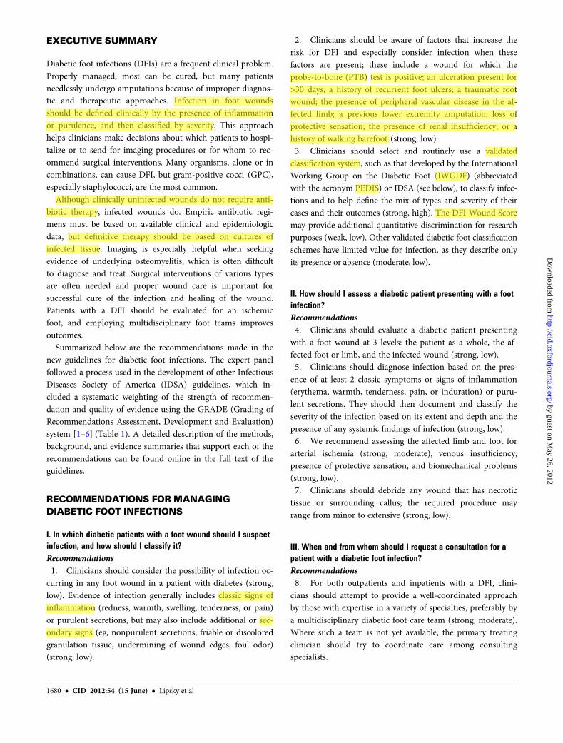
Summarized below are the recommendations made in thenew guidelines for diabetic foot infections. The expert panelfollowed a process used in the development of other InfectiousDiseases Society of America (IDSA) guidelines, which in-cluded a systematic weighting of the strength of recommen-dation and quality of evidence using the GRADE (Grading ofRecommendations Assessment, Development and Evaluation)system [1–6] (Table 1). A detailed description of the methods,background, and evidence summaries that support each of therecommendations can be found online in the full text of theguidelines.
RECOMMENDATIONS FOR MANAGINGDIABETIC FOOT INFECTIONS
I. In which diabetic patients with a foot wound should I suspectinfection, and how should I classify it?Recommendations1. Clinicians should consider the possibility of infection oc-
curring in any foot wound in a patient with diabetes (strong,low). Evidence of infection generally includes classic signs ofinflammation (redness, warmth, swelling, tenderness, or pain)or purulent secretions, but may also include additional or sec-ondary signs (eg, nonpurulent secretions, friable or discoloredgranulation tissue, undermining of wound edges, foul odor)(strong, low).
2. Clinicians should be aware of factors that increase therisk for DFI and especially consider infection when thesefactors are present; these include a wound for which theprobe-to-bone (PTB) test is positive; an ulceration present for>30 days; a history of recurrent foot ulcers; a traumatic footwound; the presence of peripheral vascular disease in the af-fected limb; a previous lower extremity amputation; loss ofprotective sensation; the presence of renal insufficiency; or ahistory of walking barefoot (strong, low).3. Clinicians should select and routinely use a validated
classification system, such as that developed by the InternationalWorking Group on the Diabetic Foot (IWGDF) (abbreviatedwith the acronym PEDIS) or IDSA (see below), to classify infec-tions and to help define the mix of types and severity of theircases and their outcomes (strong, high). The DFI Wound Scoremay provide additional quantitative discrimination for researchpurposes (weak, low). Other validated diabetic foot classificationschemes have limited value for infection, as they describe onlyits presence or absence (moderate, low).
II. How should I assess a diabetic patient presenting with a footinfection?Recommendations4. Clinicians should evaluate a diabetic patient presenting
with a foot wound at 3 levels: the patient as a whole, the af-fected foot or limb, and the infected wound (strong, low).5. Clinicians should diagnose infection based on the pres-
ence of at least 2 classic symptoms or signs of inflammation(erythema, warmth, tenderness, pain, or induration) or puru-lent secretions. They should then document and classify theseverity of the infection based on its extent and depth and thepresence of any systemic findings of infection (strong, low).6. We recommend assessing the affected limb and foot for
arterial ischemia (strong, moderate), venous insufficiency,presence of protective sensation, and biomechanical problems(strong, low).7. Clinicians should debride any wound that has necrotic
tissue or surrounding callus; the required procedure mayrange from minor to extensive (strong, low).
III. When and from whom should I request a consultation for apatient with a diabetic foot infection?Recommendations8. For both outpatients and inpatients with a DFI, clini-
cians should attempt to provide a well-coordinated approachby those with expertise in a variety of specialties, preferably bya multidisciplinary diabetic foot care team (strong, moderate).Where such a team is not yet available, the primary treatingclinician should try to coordinate care among consultingspecialists.
1680 • CID 2012:54 (15 June) • Lipsky et al
by guest on May 26, 2012
http://cid.oxfordjournals.org/D
ownloaded from
WIN SEVEN
Resaltado
WIN SEVEN
Resaltado
WIN SEVEN
Resaltado
WIN SEVEN
Resaltado
WIN SEVEN
Resaltado
WIN SEVEN
Resaltado
WIN SEVEN
Resaltado
WIN SEVEN
Resaltado
WIN SEVEN
Resaltado
WIN SEVEN
Resaltado
WIN SEVEN
Resaltado
9. Diabetic foot care teams can include (or should haveready access to) specialists in various fields; patients with aDFI may especially benefit from consultation with an infec-tious disease or clinical microbiology specialist and a surgeonwith experience and interest in managing DFIs (strong, low).
10. Clinicians without adequate training in wound debridementshould seek consultation from those more qualified for this task,especially when extensive procedures are required (strong, low).11. If there is clinical or imaging evidence of significant
ischemia in an infected limb, we recommend the clinician
Table 1. Strength of Recommendations and Quality of the Evidence
Strength ofRecommendation andQuality of Evidence
Clarity of Balance BetweenDesirable and Undesirable
EffectsMethodological Quality of Supporting
Evidence (Examples) Implications
Strong recommendation,high-quality evidence
Desirable effects clearlyoutweigh undesirableeffects, or vice versa
Consistent evidence fromwell-performed RCTs orexceptionally strong evidence fromunbiased observational studies
Recommendation can apply to mostpatients in most circumstances.Further research is unlikely tochange our confidence in theestimate of effect
Strong recommendation,moderate-qualityevidence
Desirable effects clearlyoutweigh undesirableeffects, or vice versa
Evidence from RCTs with importantlimitations (inconsistent results,methodological flaws, indirect, orimprecise) or exceptionally strongevidence from unbiasedobservational studies
Recommendation can apply to mostpatients in most circumstances.Further research (if performed) islikely to have an important impacton our confidence in the estimateof effect and may change theestimate
Strong recommendation,low-quality evidence
Desirable effects clearlyoutweigh undesirableeffects, or vice versa
Evidence for at least 1 criticaloutcome from observationalstudies, RCTs with serious flawsor indirect evidence
Recommendation may change whenhigher-quality evidence becomesavailable. Further research (ifperformed) is likely to have animportant impact on ourconfidence in the estimate ofeffect and is likely to change theestimate
Strong recommendation,very low-qualityevidence (very rarelyapplicable)
Desirable effects clearlyoutweigh undesirableeffects, or vice versa
Evidence for at least 1 criticaloutcome from unsystematicclinical observations or veryindirect evidence
Recommendation may change whenhigher-quality evidence becomesavailable; any estimate of effect forat least 1 critical outcome is veryuncertain
Weak recommendation,high-quality evidence
Desirable effects closelybalanced with undesirableeffects
Consistent evidence from well-performed RCTs or exceptionallystrong evidence from unbiasedobservational studies
The best action may differ dependingon circumstances or patients orsocietal values. Further research isunlikely to change our confidencein the estimate of effect
Weak recommendation,moderate-qualityevidence
Desirable effects closelybalanced with undesirableeffects
Evidence from RCTs with importantlimitations (inconsistent results,methodological flaws, indirect, orimprecise) or exceptionally strongevidence from unbiasedobservational studies
Alternative approaches likely to bebetter for some patients undersome circumstances. Furtherresearch (if performed) is likely tohave an important impact on ourconfidence in the estimate ofeffect and may change theestimate
Weak recommendation,low-quality evidence
Uncertainty in the estimatesof desirable effects, harms,and burden; desirableeffects, harms, and burdenmay be closely balanced
Evidence for at least 1 criticaloutcome from observationalstudies, RCTs with serious flaws,or indirect evidence
Other alternatives may be equallyreasonable. Further research isvery likely to have an importantimpact on our confidence in theestimate of effect and is likely tochange the estimate
Weak recommendation,very low-qualityevidence
Major uncertainty in theestimates of desirableeffects, harms, andburden; desirable effectsmay or may not bebalanced with undesirableeffects or may be closelybalanced
Evidence for at least 1 criticaloutcome from unsystematicclinical observations or veryindirect evidence
Other alternatives may be equallyreasonable. Any estimate of effect,for at least 1 critical outcome, isvery uncertain
Abbreviation: RCT, randomized controlled trial.
IDSA Guideline for Diabetic Foot Infections • CID 2012:54 (15 June) • 1681
by guest on May 26, 2012
http://cid.oxfordjournals.org/D
ownloaded from
consult a vascular surgeon for consideration of revasculariza-tion (strong, moderate).12. We recommend that clinicians unfamiliar with pressure
off-loading or special dressing techniques consult foot orwound care specialists when these are required (strong, low).13. Providers working in communities with inadequate
access to consultation from specialists might consider devisingsystems (eg, telemedicine) to ensure expert input on managingtheir patients (strong, low).
IV. Which patients with a diabetic foot infection should Ihospitalize, and what criteria should they meet before Idischarge them?Recommendations14. We recommend that all patients with a severe infection,
selected patients with a moderate infection with complicatingfeatures (eg, severe peripheral arterial disease [PAD] or lack ofhome support), and any patient unable to comply with therequired outpatient treatment regimen for psychological orsocial reasons be hospitalized initially. Patients who do notmeet any of these criteria, but are failing to improve with out-patient therapy, may also need to be hospitalized (strong, low).15. We recommend that prior to being discharged, a
patient with a DFI should be clinically stable; have had anyurgently needed surgery performed; have achieved acceptableglycemic control; be able to manage (on his/her own or withhelp) at the designated discharge location; and have a well-defined plan that includes an appropriate antibiotic regimento which he/she will adhere, an off-loading scheme (ifneeded), specific wound care instructions, and appropriateoutpatient follow-up (strong, low).
V. When and how should I obtain specimen(s) for culture from apatient with a diabetic foot wound?Recommendations16. For clinically uninfected wounds, we recommend not
collecting a specimen for culture (strong, low).17. For infected wounds, we recommend that clinicians
send appropriately obtained specimens for culture prior tostarting empiric antibiotic therapy, if possible. Cultures maybe unnecessary for a mild infection in a patient who has notrecently received antibiotic therapy (strong, low).18. We recommend sending a specimen for culture that is
from deep tissue, obtained by biopsy or curettage after thewound has been cleansed and debrided. We suggest avoidingswab specimens, especially of inadequately debrided wounds,as they provide less accurate results (strong, moderate).
VI. How should I initially select, and when should I modify, anantibiotic regimen for a diabetic foot infection? (See questionVIII for recommendations for antibiotic treatment ofosteomyelitis)Recommendations19. We recommend that clinically uninfected wounds not
be treated with antibiotic therapy (strong, low).20. We recommend prescribing antibiotic therapy
for all infected wounds, but caution that this is often insuffi-cient unless combined with appropriate wound care (strong,low).21. We recommend that clinicians select an empiric anti-
biotic regimen on the basis of the severity of the infection andthe likely etiologic agent(s) (strong, low).
a. For mild to moderate infections in patients who havenot recently received antibiotic treatment, we suggestthat therapy just targeting aerobic GPC is sufficient (weak,low).b. For most severe infections, we recommend startingbroad-spectrum empiric antibiotic therapy, pendingculture results and antibiotic susceptibility data (strong,low).c. Empiric therapy directed at Pseudomonas aeruginosais usually unnecessary except for patients with riskfactors for true infection with this organism (strong,low).d. Consider providing empiric therapy directed againstmethicillin-resistant Staphylococcus aureus (MRSA) in apatient with a prior history of MRSA infection; when thelocal prevalence of MRSA colonization or infection ishigh; or if the infection is clinically severe (weak, low).
22. We recommend that definitive therapy be based on theresults of an appropriately obtained culture and sensitivitytesting of a wound specimen as well as the patient’s clinicalresponse to the empiric regimen (strong, low).23. We suggest basing the route of therapy largely on infec-
tion severity. We prefer parenteral therapy for all severe, andsome moderate, DFIs, at least initially (weak, low), with aswitch to oral agents when the patient is systemically well andculture results are available. Clinicians can probably use highlybioavailable oral antibiotics alone in most mild, and in manymoderate, infections and topical therapy for selected mildsuperficial infections (strong, moderate).24. We suggest continuing antibiotic therapy until, but not
beyond, resolution of findings of infection, but not throughcomplete healing of the wound (weak, low). We suggest aninitial antibiotic course for a soft tissue infection of about 1–2weeks for mild infections and 2–3 weeks for moderate tosevere infections (weak, low).
1682 • CID 2012:54 (15 June) • Lipsky et al
by guest on May 26, 2012
http://cid.oxfordjournals.org/D
ownloaded from

VII. When should I consider imaging studies to evaluatea diabetic foot infection, and which should I select?Recommendations25. We recommend that all patients presenting with a new
DFI have plain radiographs of the affected foot to look forbony abnormalities (deformity, destruction) as well as forsoft tissue gas and radio-opaque foreign bodies (strong,moderate).26. We recommend using magnetic resonance imaging
(MRI) as the study of choice for patients who require further(ie, more sensitive or specific) imaging, particularly when softtissue abscess is suspected or the diagnosis of osteomyelitisremains uncertain (strong, moderate).27. When MRI is unavailable or contraindicated, clinicians
might consider the combination of a radionuclide bone scanand a labeled white blood cell scan as the best alternative(weak, low).
VIII. How should I diagnose and treat osteomyelitis of the foot ina patient with diabetes?Recommendations28. Clinicians should consider osteomyelitis as a potential
complication of any infected, deep, or large foot ulcer,especially one that is chronic or overlies a bony prominence(strong, moderate).29. We suggest doing a PTB test for any DFI with an open
wound. When properly conducted and interpreted, it can helpto diagnose (when the likelihood is high) or exclude (whenthe likelihood is low) diabetic foot osteomyelitis (DFO)(strong, moderate).30. We suggest obtaining plain radiographs of the foot, but
they have relatively low sensitivity and specificity for confirm-ing or excluding osteomyelitis (weak, moderate). Cliniciansmight consider using serial plain radiographs to diagnose ormonitor suspected DFO (weak, low).31. For a diagnostic imaging test for DFO, we recommend
using MRI (strong, moderate). However, MRI is not alwaysnecessary for diagnosing or managing DFO (strong, low).32. If MRI is unavailable or contraindicated, clinicians
might consider a leukocyte or antigranulocyte scan, preferablycombined with a bone scan (weak, moderate). We do not rec-ommend any other type of nuclear medicine investigations(weak, moderate).33. We suggest that the most definitive way to diagnose DFO
is by the combined findings on bone culture and histology(strong, moderate). When bone is debrided to treat osteomyelitis,we suggest sending a sample for culture and histology (strong,low).34. For patients not undergoing bone debridement, we
suggest that clinicians consider obtaining a diagnostic bonebiopsy when faced with specific circumstances, eg, diagnostic
uncertainty, inadequate culture information, failure ofresponse to empiric treatment (weak, low).35. Clinicians can consider using either primarily surgical or
primarily medical strategies for treating DFO in properly selectedpatients (weak, moderate). In noncomparative studies each ap-proach has successfully arrested infection in most patients.36. When a radical resection leaves no remaining infected
tissue, we suggest prescribing antibiotic therapy for only ashort duration (2–5 days) (weak, low). When there is persist-ent infected or necrotic bone, we suggest prolonged (≥4weeks) antibiotic treatment (weak, low).37. For specifically treating DFO, we do not currently
support using adjunctive treatments such as hyperbaricoxygen therapy, growth factors (including granulocyte colony-stimulating factor), maggots (larvae), or topical negativepressure therapy (eg, vacuum-assisted closure) (weak, low).
IX. In which patients with a diabetic foot infection shouldI consider surgical intervention, and what type of proceduremay be appropriate?Recommendations38. We suggest that nonsurgical clinicians consider request-
ing an assessment by a surgeon for patients with a moderateor severe DFI (weak, low).39. We recommend urgent surgical intervention for most
foot infections accompanied by gas in the deeper tissues, anabscess, or necrotizing fasciitis, and less urgent surgery forwounds with substantial nonviable tissue or extensive bone orjoint involvement (strong, low).40. We recommend involving a vascular surgeon early on
to consider revascularization whenever ischemia complicates aDFI, but especially in any patient with a critically ischemiclimb (strong, moderate).41. Although most qualified surgeons can perform an ur-
gently needed debridement or drainage, we recommend that inDFI cases requiring more complex or reconstructive procedures,the surgeon should have experience with these problems andadequate knowledge of the anatomy of the foot (strong, low).
X. What types of wound care techniques and dressings areappropriate for diabetic foot wounds?Recommendations42. Diabetic patients with a foot wound should receive ap-
propriate wound care, which usually consists of the following:a. Debridement, aimed at removing debris, eschar, andsurrounding callus (strong, moderate). Sharp (or surgi-cal) methods are generally best (strong, low), but mech-anical, autolytic, or larval debridement techniques maybe appropriate for some wounds (weak, low).b. Redistribution of pressure off the wound to the entire
weight-bearing surface of the foot (“off-loading”).
IDSA Guideline for Diabetic Foot Infections • CID 2012:54 (15 June) • 1683
by guest on May 26, 2012
http://cid.oxfordjournals.org/D
ownloaded from

While particularly important for plantar wounds, thisis also necessary to relieve pressure caused by dres-sings, footwear, or ambulation to any surface of thewound (strong, high).
c. Selection of dressings that allow for moist woundhealing and control excess exudation. The choice ofdressing should be based on the size, depth, and natureof the ulcer (eg, dry, exudative, purulent) (strong, low).
43. We do not advocate using topical antimicrobials fortreating most clinically uninfected wounds.44. No adjunctive therapy has been proven to improve res-
olution of infection, but for selected diabetic foot wounds thatare slow to heal, clinicians might consider using bioengineeredskin equivalents (weak, moderate), growth factors (weak, mod-erate), granulocyte colony-stimulating factors (weak, moder-ate), hyperbaric oxygen therapy (strong, moderate), ornegative pressure wound therapy (weak, low).
Notes
Acknowledgments. The panel members thank Drs Thomas File, MarkKosinski, and Brad Spellberg for their thoughtful reviews of earlier draftsof the guideline, and Dr James Horton (IDSA SGPC liaison), JenniferPadberg, and Vita Washington for overall guidance and coordination inall aspects of the development of this guideline.Financial support. Support for these guidelines was provided by the
Infectious Diseases Society of America.Potential conflicts of interest. The following list is a reflection of what
has been reported to the IDSA. In order to provide thorough transparency,
the IDSA requires full disclosure of all relationships, regardless of rele-vancy to the guideline topic. The reader of these guidelines should bemindful of this when the list of disclosures is reviewed. B. L. has served asa consultant to Merck, Pfizer, Cubist, Innocoll, TaiGen, KCI, and Dipex-ium. E. S. has served on the board of and consulted for Novartis. H. G. D.has served on the speakers’ bureau for Merck and Sanofi. J. P. has servedas a consultant to Pfizer and Ortho McNeil. M. P. has served as a consult-ant for Orthopedic Implants for Deputy Orthopedics and Small BoneInnovation. W. J. has served as a consultant for Merck, Pfizer, Cerexa, andDipexium and has served on the speakers’ bureaus of Merck and Pfizer. A.W. K. is on the boards of Pfizer and Merck and the speakers’ bureau forAstella, and consults for Novartis. All other authors report no potentialconflicts.All authors have submitted the ICMJE Form for Disclosure of Potential
Conflicts of Interest. Conflicts that the editors consider relevant to thecontent of the manuscript have been disclosed.
References
1. Guyatt GH, Oxman AD, Vist GE, et al. GRADE: an emerging consen-sus on rating quality of evidence and strength of recommendations.BMJ 2008; 336:924–6.
2. Guyatt GH, Oxman AD, Kunz R, et al. Going from evidence to rec-ommendations. BMJ 2008; 336:1049–51.
3. Jaeschke R, Guyatt GH, Dellinger P, et al. Use of GRADE grid to reachdecisions on clinical practice guidelines when consensus is elusive. BMJ2008; 337:a744.
4. Kish MA. Guide to development of practice guidelines. Clin Infect Dis2001; 32:851–4.
5. Schunemann HJ, Oxman AD, Brozek J, et al. Grading quality of evi-dence and strength of recommendations for diagnostic tests and strat-egies. BMJ 2008; 336:1106–10.
6. Guyatt GH, Oxman AD, Kunz R, et al. Incorporating considera-tions of resources use into grading recommendations. BMJ 2008; 336:1170–3.
1684 • CID 2012:54 (15 June) • Lipsky et al
by guest on May 26, 2012
http://cid.oxfordjournals.org/D
ownloaded from


![Pie Diabetico IDSA 2012[1]](https://static.fdocuments.us/doc/165x107/577cdb0b1a28ab9e78a739aa/pie-diabetico-idsa-20121.jpg)