Phys Rev Gordon Chen Chung Printedversion
36
COMPUTATIONAL METHODS OF STUDYING THE BINDING OF TOXINS FROM VENOMOUS ANIMALS TO BIOLOGICAL ION CHANNELS: THEORY AND APPLICATIONS Dan Gordon, Rong Chen, and Shin-Ho Chung Research School of Biology, The Australian National University, Australian Capital Territory, Australia L Gordon D, Chen R, Chung S-H. Computational Methods of Studying the Binding of Toxins From Venomous Animals to Biological Ion Channels: Theory and Applications. Physiol Rev 93: 767– 802, 2013; doi:10.1152/physrev.00035.2012.—The discov- ery of new drugs that selectively block or modulate ion channels has great potential to provide new treatments for a host of conditions. One promising avenue revolves around modifying or mimicking certain naturally occurring ion channel modulator toxins. This strategy appears to offer the prospect of designing drugs that are both potent and specific. The use of computational modeling is crucial to this endeavor, as it has the potential to provide lower cost alternatives for exploring the effects of new compounds on ion channels. In addition, computational modeling can provide structural information and theoretical understanding that is not easily deriv- able from experimental results. In this review, we look at the theory and computational methods that are applicable to the study of ion channel modulators. The first section provides an introduction to various theoretical concepts, including force-fields and the statistical mechanics of binding. We then look at various computational techniques available to the researcher, including molecular dynamics, Brownian dynamics, and molecular docking systems. The latter section of the review explores applications of these techniques, concentrating on pore blocker and gating modifier toxins of potassium and sodium channels. After first discussing the structural features of these channels, and their modes of block, we provide an in-depth review of past computational work that has been carried out. Finally, we discuss prospects for future developments in the field. I. INTRODUCTION 767 II. COMPUTATIONAL MODELS AND... 769 III. ENERGETICS AND BINDING THEORY... 777 IV. ION CHANNEL PHARMACOLOGY 785 V. ION CHANNEL STRUCTURES 785 VI. ION CHANNEL MODULATORS 785 VII. FUTURE PROSPECTS 794 I. INTRODUCTION Ion channels are ubiquitous in the human body. When a particular channel is over- or underexpressed, or contains a mutation which changes its conduction or gating character- istics, disease may result (12). There are many such chan- nelopathies, including type I diabetes, epilepsy, cystic fibro- sis, multiple sclerosis, long-QT syndrome, and migraines. Treatment of these diseases can be effected by introducing ion channel modulator drugs that regulate the function of the channels. For example, Ziconotide, the synthetic form of the -conotoxin MVIIA, which is a voltage-gated cal- cium channel blocker, has been approved to treat severe pain (216). These modulators may be agonists, which in- crease the conductance of the channels, or inhibitors, which reduce their conductance. Channel blockers are inhibitors that operate directly, by binding in the ion conducting pore. The block may be extracellular, as is the case for pore blocker toxins, or intracellular, for example, internal block of potassium channels by tetraethylammonium. Modula- tion may also be accomplished indirectly, by affecting the activation or inactivation gating of the channel. For exam- ple, the gating modifier hanatoxin binds to the voltage sen- sor of voltage-gated potassium channels and moves the ac- tivation curve of the channel to the right, thus requiring a greater depolarization to open the channel. Quinidine, on the other hand, is proposed to bind to the intracellular face of the Kv1.4 channel and allosterically promote the onset of C-type inactivation (243). Batrachotoxin has been proposed to bind in the pore of voltage-gated sodium channels (246) but does not block the flow of ions; instead, it locks the channel in a permanently open conformation. Thus there are a variety of modes by which channel modulators may function. Nature has devised a plethora of ion channel blockers and modulators, in the form of toxins that occur in the venoms of poisonous creatures such as scorpions, cone snails, sea anemones, spiders, and snakes. We have already mentioned two gating modifier toxins: hanatoxin and batrachotoxin. Many other toxins act by directly blocking the pore, usually Physiol Rev 93: 767– 802, 2013 doi:10.1152/physrev.00035.2012 767
Transcript of Phys Rev Gordon Chen Chung Printedversion

COMPUTATIONAL METHODS OF STUDYING THEBINDING OF TOXINS FROM VENOMOUS ANIMALSTO BIOLOGICAL ION CHANNELS: THEORY ANDAPPLICATIONSDan Gordon, Rong Chen, and Shin-Ho Chung
Research School of Biology, The Australian National University, Australian Capital Territory, Australia
LGordon D, Chen R, Chung S-H. Computational Methods of Studying the Binding ofToxins From Venomous Animals to Biological Ion Channels: Theory and Applications.Physiol Rev 93: 767–802, 2013; doi:10.1152/physrev.00035.2012.—The discov-ery of new drugs that selectively block or modulate ion channels has great potential toprovide new treatments for a host of conditions. One promising avenue revolves around
modifying or mimicking certain naturally occurring ion channel modulator toxins. This strategyappears to offer the prospect of designing drugs that are both potent and specific. The use ofcomputational modeling is crucial to this endeavor, as it has the potential to provide lower costalternatives for exploring the effects of new compounds on ion channels. In addition, computationalmodeling can provide structural information and theoretical understanding that is not easily deriv-able from experimental results. In this review, we look at the theory and computational methodsthat are applicable to the study of ion channel modulators. The first section provides an introductionto various theoretical concepts, including force-fields and the statistical mechanics of binding. Wethen look at various computational techniques available to the researcher, including moleculardynamics, Brownian dynamics, and molecular docking systems. The latter section of the reviewexplores applications of these techniques, concentrating on pore blocker and gating modifier toxinsof potassium and sodium channels. After first discussing the structural features of these channels,and their modes of block, we provide an in-depth review of past computational work that has beencarried out. Finally, we discuss prospects for future developments in the field.
I. INTRODUCTION 767II. COMPUTATIONAL MODELS AND... 769III. ENERGETICS AND BINDING THEORY... 777IV. ION CHANNEL PHARMACOLOGY 785V. ION CHANNEL STRUCTURES 785VI. ION CHANNEL MODULATORS 785VII. FUTURE PROSPECTS 794
I. INTRODUCTION
Ion channels are ubiquitous in the human body. When aparticular channel is over- or underexpressed, or contains amutation which changes its conduction or gating character-istics, disease may result (12). There are many such chan-nelopathies, including type I diabetes, epilepsy, cystic fibro-sis, multiple sclerosis, long-QT syndrome, and migraines.Treatment of these diseases can be effected by introducingion channel modulator drugs that regulate the function ofthe channels. For example, Ziconotide, the synthetic formof the �-conotoxin MVIIA, which is a voltage-gated cal-cium channel blocker, has been approved to treat severepain (216). These modulators may be agonists, which in-crease the conductance of the channels, or inhibitors, whichreduce their conductance. Channel blockers are inhibitors
that operate directly, by binding in the ion conducting pore.The block may be extracellular, as is the case for poreblocker toxins, or intracellular, for example, internal blockof potassium channels by tetraethylammonium. Modula-tion may also be accomplished indirectly, by affecting theactivation or inactivation gating of the channel. For exam-ple, the gating modifier hanatoxin binds to the voltage sen-sor of voltage-gated potassium channels and moves the ac-tivation curve of the channel to the right, thus requiring agreater depolarization to open the channel. Quinidine, onthe other hand, is proposed to bind to the intracellular faceof the Kv1.4 channel and allosterically promote the onset ofC-type inactivation (243). Batrachotoxin has been proposedto bind in the pore of voltage-gated sodium channels (246) butdoes not block the flow of ions; instead, it locks the channel ina permanently open conformation. Thus there are a variety ofmodes by which channel modulators may function.
Nature has devised a plethora of ion channel blockers andmodulators, in the form of toxins that occur in the venomsof poisonous creatures such as scorpions, cone snails, seaanemones, spiders, and snakes. We have already mentionedtwo gating modifier toxins: hanatoxin and batrachotoxin.Many other toxins act by directly blocking the pore, usually
Physiol Rev 93: 767–802, 2013doi:10.1152/physrev.00035.2012
767
andrea
Evidenziato
andrea
Evidenziato
andrea
Evidenziato
andrea
Evidenziato
by inserting a basic lysine or arginine side chain into theselectivity filter from the extracellular side. These toxinstend to be extremely potent. Often, they are relatively un-selective, affecting several members of a whole family of ionchannels. However, some are known to discriminate ex-tremely well between similar members of an ion channelfamily; for example, modified sea anemone ShK channelsbind to Kv1.3 voltage-gated potassium channels with atleast 100-fold selectivity over other Kv channels. This kindof selectivity, along with the general structural complexityof these toxins, gives hope that they may be used as a start-ing point to develop potent and selective drugs. Such toxinsform a particular focus of this review, although much of thetheoretical discussion in the earlier parts of the review ap-plies more generally.
A great deal of effort goes into the study and developmentof ion channel modulator drugs, due to the range of condi-tions which may be treated and the promising possibilitiesfor treatment. To be potent, such drugs should bindstrongly to their receptors. To avoid unwanted side effects,they should not bind to antitargets. Finally, when bound toreceptors, they should bring about the desired effect, forexample, by actually blocking or inhibiting current throughthe channel. Drug development is a costly and time-con-suming process. Typically, thousands of compounds areinitially screened. The most promising leads are then mod-ified, and various stages of further characterization and test-ing are carried out. State-of-the-art experimental technol-ogy automates many of the tasks involved in testing newcompounds (73), but despite the high cost and sophistica-tion of the techniques, only crude clues about the mecha-nisms of modulation or block may be gained.
These limitations have given rise to an intense interest in theuse of computational modeling as a complementary meansto investigate the binding modes, binding affinities, andmodulation mechanisms of channel modulators. Ideally,one would like to study the effects of channel modulatorson channel function, using a model assembly composed ofan ion channel embedded in a lipid bilayer, ions, and watermolecules. Computational modeling can in principle repro-duce many or all the experimental observables, such as thebinding affinity and specificity of a given modulator onvarious subfamilies of ion channels. Additionally, and per-haps more importantly, modeling has the potential to revealin exquisite detail the interactions, mechanisms, and struc-tural contacts involved in modulator binding. This is im-portant because such information is not easily deducedfrom electrophysiological experiments. Currently, no singleavailable computational method is able to achieve all theseidealized aims. There are, however, a number of computa-tional techniques that can perform some of these tasks withvarying degrees of efficiency. Each of these methodologieshas advantages and disadvantages. At various points in thedrug development cycle, it is necessary to make compro-
mises between computational speed and accuracy. For ex-ample, lead discovery typically requires a very fast, lessaccurate assessment of binding affinity, since thousands ofpotential lead compounds may need to be screened. Thetechniques discussed in this review can be ranked accordingto whether they are crude but fast, or slow but accurate.Different techniques will be appropriate at different pointsin the drug development cycle. The aim of devising a tech-nique that is both fast and accurate remains the holy grail ofthe field.
In this review, we will discuss three commonly used tech-niques: molecular dynamics, Brownian dynamics, anddocking. Molecular dynamics simulates all or many of theatoms in the system using a classical force field. Browniandynamics is similar, but always uses implicit water, andmakes extensive use of rigid and fixed molecules to achievegreater speed. Docking employs various algorithmic searchstrategies, and scoring functions to attempt to predict thebinding modes between a ligand and a receptor and to es-timate the binding affinity. A fourth approach, QSAR,makes use of multidimensional regression on various prop-erties of drugs and targets. Although we consider QSAR tobe somewhat outside the scope of this review, we mention itdue to its importance and potential usefulness as a tool fordrug discovery.
We will begin the review by detailing the computationaltechniques listed above: molecular dynamics, Brownian dy-namics, and docking. We will then go into more theoreticaldetail about the tasks that computational modeling andsimulation can help to solve. Binding affinity is a key deter-minant of modulator effectiveness. For example, if a chan-nel blocker always fully blocks the channel upon binding,then binding affinity will be directly related to the degree ofblock. We shall consider the theoretical basis of bindingaffinity and relate it to the free energy of binding for theblocker. Although calculating the free energy of binding isfar easier than directly calculating binding affinity fromsimulations, it is nonetheless a formidable computationaltask. Therefore, having laid the theoretical groundwork, wewill consider some of the techniques that have been devisedto calculate free energies of binding. We will next look atspecific computational studies of interactions between ionchannels and their modulators. Starting with an overview ofion channel diseases and pharmacology, and aspects of ionchannel structure, we will go on to review examples ofcomputational studies involving different mechanisms ofblock, namely, pore block in potassium and sodium chan-nels and gating modifiers in these channels. Finally, we willconclude the review with a discussion of future prospectsfor computational modeling. Due to the difficulty of theproblems faced, it is clear that much important theoreticaland computational work still lies ahead, and hence, wepredict important and revolutionary developments in thefuture.
COMPUTATIONAL METHODS FOR TOXIN BINDING TO ION CHANNELS
768 Physiol Rev • VOL 93 • APRIL 2013 • www.prv.org
A. Scope of This Review
The subject of ion channel modulators is broad, and theliterature is large. Computational studies of ion channelblock may employ a raft of techniques from physics, chem-istry, biology, bioinformatics, and computer science. Due tothe size and complexity of the subject, we have chosen toconcentrate on certain topics at the expense of others. Thereader should, however, be aware that there are other inter-esting computational techniques that may be applicable,from the realms of statistics, bioinformatics, and machine-learning. We touch on some of these techniques in passing:for example, QSAR and knowledge-based potentials relyon bioinformatics, and certain docking force fields, for ex-ample, AUTODOCK VINA, have only a loose connectionto physical principles, relying instead on machine learningapproaches to parametrization.
In the latter sections of the review, where we look at specificcomputational studies of ion channel block, we have notattempted to cover the full range of channels and modula-tors. Instead, we concentrate mainly on toxin-derived poreblockers and gating modifiers of potassium and sodiumchannels. Potassium and sodium channels were chosen dueto their importance and widespread occurrence in all man-ner of tissues, for example, they are primarily responsiblefor all nerve and muscle impulses, and because they areimplicated in a large number of channelopathies. Our focuson toxins is due to their prominence in current researcheffort and the hope they appear to offer for developing newdrugs that will selectively target individual types of chan-nels.
II. COMPUTATIONAL MODELS ANDMETHODS
A. Ion-Channel Specific Issues
Much of the theory of ion-channel modulators deals withthe generalities of protein-protein or protein-ligand bind-ing. However, ion channels do exhibit several peculiaritiesthat may need special consideration, and which the readershould keep in mind.
The pores of ion channels focus electric fields, due to thegeometry of the dielectric boundary (135). One effect of thisis to increase the interaction between resident ions in thechannel, and blockers that enter the pore. For example,Park and Miller (185) found that a modest but significantinteraction of around 3 kT1exists between the Lys27 resi-due in charybdotoxin and resident ions in the pore; this
would correspond to a factor of 20 in the binding affinity.Tetraethylammonium block in potassium channels is alsoknown to depend on ionic concentration (134). Anotherrelated effect of the dielectric boundary of ion channels isthat they exhibit a sharp voltage drop through the pore dueto the membrane potential, particularly across the selectiv-ity filter. The membrane potential can thus affect the bind-ing of blockers. The most notable examples of this are forsmall ionic molecules such as tetraethylammonium thatbind in the internal vestibules of ion channels. Positivelycharged tetraethylammonium was found to have a bindingaffinity to KcsA that increased with membrane depolariza-tion (108, 134), which is simply explained by the fact that anegative membrane potential creates a potential hill for themolecule as it moves upward into the pore, towards itsbinding site. Polypeptide toxins are also known to havevoltage-dependent affinities (72, 83). Because the electricfields found in ion channel pores are large, a careful com-putational treatment is needed if errors are to be avoided.
Another complication arises from the fact that ion channelsare membrane proteins. The presence of the lipid bilayermodifies the electrostatic environment. More importantly,certain toxins, such as the gating modifier hanatoxin, actfrom within the bilayer (221) in a complex manner, com-plicating computational modeling (17, 109, 153, 154, 179,251).
The pores of ion channels also provide a more fully enclosedreceptor environment than is usual in docking studies; thuscare needs to be taken in making sure that the conformationof the pore allows the blocker to correctly bind. In practice,this means that either a fully flexible treatment of the chan-nel may need to be employed, that a careful choice needs tobe made for the conformation of the channel model, or thatan ensemble of channel models might need to be employed(144, 161, 198, 199).
Ion channels undergo significant conformational changesthat are associated with (in)activation and gating. The bind-ing of many blocker molecules depends on the state or con-formation of the channel. For example, many small-mole-cule local anaesthetic blockers of sodium channels exhibitstate-dependent block (67). This may also manifest as ause-dependent block, where blocking efficiency is enhancedwhen channels cycle more frequently between closed, open,and inactivated states. Another example concerns the ap-parent state-dependent binding of small-molecule blockersin the inner cavity of Kv11.1/hERG channel (189). Statedependence of channel block presents an additional com-plication that may need to be taken into account whencarrying out computational work.
In the following section, we shall discuss the main compu-tational methods used in the study of ion channel modula-tors: molecular dynamics, Brownian dynamics, and dock-
1We use energy units of kT, where k is the Boltzmann constantand T is taken to be the temperature, 300 K unless otherwisestated: 1 kT � 0.60 kcal/mol.
GORDON, CHEN, AND CHUNG
769Physiol Rev • VOL 93 • APRIL 2013 • www.prv.org

ing. Each of these computational methods employs one ormore force fields in combination with a simulation or dock-ing protocol. Because the force fields are to some degreeinterchangeable between the computational methods, forexample, molecular mechanics force fields are deployed inboth molecular dynamics simulations as well as certaindocking systems, we shall begin the section with a look atforce fields, and then move on to consider the four compu-tational methods themselves.
B. Forces and Force Fields
In the study of biomolecules, force fields can be roughlyranked according to the degree of approximation andcoarse graining that is applied. Thus, at the lowest level, wehave quantum mechanical force field protocols, as used inab initio molecular dynamics. At increasing levels of ab-straction are the fully atomistic classical molecular mechan-ics force fields, united atom molecular mechanics forcefields, implicit solvent force fields, and various types ofcoarse grained force fields, such as the so-called “knowl-edge-based” PMFs. Since quantum calculations are tooslow to be practical for most questions directly relevant tomodulators and ion channels, we shall begin by looking atclassical molecular mechanics.
1. Atomistic molecular mechanics
In fully atomistic molecular mechanics, there are three typesof potentials to consider: the electrostatic potential describ-ing the Coulomb forces in the system, van der Waals termsdescribing the nonpolar interactions between pairs of at-oms, and covalent, or strain, potentials describing the ef-fects of covalent bonds between the atoms in the simulation.
Electrostatic Coulomb potentials act between pairs of at-oms and contribute to the internal and interaction energiesof the channel and the channel modulator. The potentialbetween two charges q1 and q2 separated by a distance r ina vacuum is
U�r� �1
4��0
q 1q2
r(1)
where �0 is the permittivity of free space. Coulomb poten-tials act at both long and short ranges and play a direct rolein binding. The long range part of the Coulomb potentialcan attract charged blockers into oppositely charged vesti-bules of ion channels, and the short range part causescharged groups to form salt bridges or hydrogen bonds,locking the blocker into place in the binding pocket.
van der Waals potentials are short range potentials thatdescribe both short range repulsive steric forces betweenatoms as well as longer range attractive dispersive interac-tions. They are usually modeled using the Lennard-Jones(6–12) potential
U�r� � ��� rmin
r �12
� 2� rmin
r �6� (2)
where r is the distance between two atoms, � is the depth ofthe attractive well, and rmin is the separation between twoatoms for minimum potential energy. van der Waals poten-tials play a role in binding by balancing attractive Coulombinteractions (thereby regulating hydrogen bonding) and inthe forces that arise when the surface of the modulatorcontacts the channel, such as the hydrophobic force.
Covalent, or strain, potentials describe the resistance of amolecule to deformations of the relative positions of theatoms relative to one another. The actual many-body forcefield is complicated and depends on the quantum chemistryof the molecule. Classical molecular mechanics force fieldsemploy a simplified treatment that considers forces betweenbonded pairs, triplets, and quadruplets of atoms, along withcross-term corrections. The precise implementation of theseterms will vary depending on which molecular dynamicsforce field is chosen. Here, as a typical example, we givedetails of the covalent potentials in the CHARMM forcefield (156). Covalent bonds are first defined for the mole-cule, as per the “ball and stick” models used in chemistry.Five kinds of covalent potentials are then assigned to thebond network.
1) Bond stretching potentials act between pairs of neighbor-ing atoms and take the form
Vbond � kb�b � b0�2 (3)
where kb is twice the harmonic spring constant, b is thedistance between the atoms, and b0 is the equilibrium bonddistance.
2) Angle bending potentials act between adjacent triplets ofatoms and take the form
Vangle � k��� � �0�2 (4)
where k� is again twice the spring constant, � is the anglesubtended by the adjacent bonds, and �0 is the equilibriumangle.
3) Dihedral potentials act between groups of four linearlyconnected atoms, and take the form
Vdihed � kn,��1 cos�n� � �� (5)
The dihedral angle � represents a torsion around the B-Cbond in the group A-B-C-D as the angle formed between theplanes defined by triangles A-B-C and B-C-D. kn,� gives thestrength of the potential, n gives its periodicity, and con-trols the angle at which the minimum potential occurs.
4) Improper dihedral potentials are defined for groups offour atoms consisting of a central atom C to which is con-nected three other atoms A, B, and D. They are used pri-
COMPUTATIONAL METHODS FOR TOXIN BINDING TO ION CHANNELS
770 Physiol Rev • VOL 93 • APRIL 2013 • www.prv.org
marily to maintain planarity for the group, and take theform
Vimprop � k��� � �0�2 (6)
where k� is twice the spring constant; � is the angle betweenthe plane containing atoms A, B, and C and that containingB, C, and D; and �0 is the equilibrium angle.
5) Urey-Bradley potentials represent an additional way ofmaintaining angles: they are simply a bond potential thatis defined between atoms A and C in a connected groupA-B-C.
Cross terms, such as the CHARMM CMAP backbone cor-rection (157), may also be defined to represent, for example,the interaction between adjacent dihedrals in the proteinbackbone.
2. Implicit solvent force fields
Water molecules account for a large proportion of atomsin biomolecular systems. By treating water molecules as acontinuum, we are able to gain computational speed atthe cost of accuracy. Implicit solvent force fields are usedin both implicit solvent molecular mechanics and othertechniques such as Brownian dynamics and various dock-ing protocols. Below, we will give some background tothe physical role played by water, and outline the tech-niques used for treating it implicitly as a mean field.Water has two important physical effects on biomolecu-lar systems. First, it strongly modifies the electrostaticforces experienced by the ions and biomolecules, andsecond, it gives rise to short-range nonpolar forces due tovan der Waals interactions and entropic effects. We shallconsider these two effects in turn.
The most important effect of water, when considered as amean-field, is that it approximates a linear dielectric me-dium with a high dielectric constant. This means that whentwo charges are surrounded by water, the Coulomb poten-tial, given in Equation 1, will be modified so that �0 ¡ �r�0,where �r is the relative dielectric constant of water, approx-imately equal to 80. Coulomb forces are therefore reducedby a factor of 80. Furthermore, when water surrounds a lowdielectric protein or lipid environment, a dielectric barrier iscreated between the water and the low dielectric region.Charges in the water are repelled from this barrier due toimage forces. Other more complicated interactions involv-ing two or more charges can also occur, especially in thepores of ion channels, where the narrow aqueous pore iscompletely surrounded by the protein. The physics of theseeffects is described by the Poisson-Boltzmann equation
� · ���r� � ��r�� � ��f�r� � �r�i
�i0exp��
�i0��r�kT � (7)
In this equation, �f is the solute charge distribution (i.e., thefixed charges in the ion channel and channel modulator), i
represents the different ion species in the solution (e.g., Na�
and Cl�), and �i0 are their number densities in uniform
solution that is far from the channel. The term in the expo-nential scales these densities by their Boltzmann factor.Note that the presence of � in this term makes the equationnonlinear. is a position-dependent scaling factor that rep-resents the accessibility of different regions to ions, for ex-ample, ions will not physically be able to penetrate theinterior of the channel protein, and thus would be zero inthis region. The equation may be linearized to more easilysolve it.
In the Poisson-Boltzmann equation, the mobile ions in thesystem are treated as a mean field. This approach has beenshown to have limitations when dealing with the narrowpores that are often present in ion channels (173). There-fore, it is often preferable to model the ions explicitly, bytreating them as part of pf in the above equation, and settingthe mean-field ion densities to zero. We then have Poisson’sequation
� · ���r� � ��r�� � ��f�r� (8)
which is a linear partial differential equation.
Since direct solutions of both the Poisson-Boltzmann andPoisson’s equation are time consuming, approximations areoften used. The simplest approximations use Coulomb po-tentials between charges, with a distance-dependent dielec-tric such as �(r) � 4r (111). The dielectric constant increaseslinearly with increasing r, which represents the fact that, forgreater r, there is likely to be more solvent between andaround the charges. However, details of the geometry arenot taken into account.
A more sophisticated method is the generalized Born ap-proach (15). Generalized Born electrostatics represents thefree energy of the polarization of the solvent as
F �1
8�� 1
�0�
1
� � i, j�1
N qiqj
rij2 aij
2exp��rij2 ⁄ 4aiaj�
(9)
where qi is the atomic partial charges of the ith atom, rij isthe distance between atoms i and j, and ai is a parameterknown as the generalized Born radius of atom i. There arevarious methods of computing the generalized Born radii,and their optimal determination lies at the heart of themethod. The precise form of Equation 9 is to some extentarbitrary, but is required to give the correct result for cer-tain limits. For large rij, the method gives the Coulomb forcefor two point charges in a dielectric medium. As the inter-atomic distance decreases, additional solvent screening andimage charge effects become manifest. For a single atom,with aij set to the atomic radius, the method gives the ana-lytical Born solvation energy. The correct expression is alsoobtained for the limit of a point dipole.
GORDON, CHEN, AND CHUNG
771Physiol Rev • VOL 93 • APRIL 2013 • www.prv.org

The electrostatics inside the pore of ion channels presentextra challenges, due to the presence of highly focused elec-tric fields, and other problems with the mean-field approx-imation due to the quantization of water and ions in narrowpores. This may be important to the study of channel-blocker interactions, including those where binding in-volves the insertion of a blocker lysine or arginine into thechannel pore. For example, tetrodotoxin is known to blockvoltage-gated sodium channels in a voltage-dependentmanner (72, 83), suggesting that the pore electrostatics playa large role in the block. Because of the electrostatic focus-ing effect, the pores of ion channels increase the strengthand range of charge-charge interactions in a position-de-pendent manner. Another way of looking at this is thatthere is less surrounding water to shield charges from oneanother. Furthermore, the membrane voltage drop occursalmost entirely over the length of the channel pore and isgreatest where the pore is narrow. It is possible that generalpurpose docking programs and the like, which may be op-timized for the docking of a ligand or protein onto a recep-tor located on the surface of another protein, may not per-form well under the highly enclosed conditions of ion chan-nel pores. However, very little work has been done toquantify these issues.
Water also plays an important role in effectively mediatingnonpolar interactions between the atoms in ion channels,channel modulators, and ions. In the absence of water, suchnonpolar interactions are described by van der Waalsforces, and can be adequately modeled using Lennard-Jonespotentials as described in the previous section. When wateris present, it interacts in a complex manner with the solutemolecules and strongly mediates the effect of the nonpolarforces. This is largely due to two factors. First, water exhib-its strongly attractive van der Waals interactions with itselfand other molecules. Therefore, when two solute atomsapproach each other closely, some water is displaced, andthere is an energetic cost to this displacement that needs tobe taken into account when determining the nonpolar in-teractions between the two molecules. Second, water is nota perfect linear dielectric: its molecules have a finite size andexhibit a strong hydrogen bonding structure. When water isdisplaced by the presence of solute atoms, the hydrogenbonding network is modified. Water tends not to form hy-drogen bonds with hydrophobic atoms. Adjacent to a hy-drophobic surface, the water will compensate for lost wa-ter-solute hydrogen bonds by forming extra water-waterhydrogen bonds, thereby creating a semi-rigid “cage” ofsurface tension around the atoms. The entropy of the wateris lowered, which translates to a raising of the free energy.To minimize this free energy penalty, groups of hydropho-bic atoms will therefore clump together so as to minimizetheir solvent exposed surface area. This is known as thehydrophobic effect and can be as important as hydrogenbonding in understanding the binding forces between chan-
nel modulators and ion channels. An example of water-mediated nonpolar interactions is shown in FIGURE 1.
It is difficult to accurately take into account the effects de-scribed above when implementing implicit solvent forcefields. The most common approach relies on linearly corre-lating the nonpolar hydration free energies of the solutewith its solvent-exposed surface area. In some treatments,the hydrophobicity is taken into account by defining differ-ent scaling constants for different atom types; typically onlya very small number of atom types are used. In more sophis-ticated treatments, a volume-dependent cavity term mayalso be included (81, 87, 86, 143). A surface area nonpolarhydration energy can be combined with the generalizedBorn approach to solvation, to give the often used general-ized Born/surface area (GBSA) model of solvation.
3. Coarse graining and knowledge-based potentials
Coarse graining, the practice of treating groups of atoms asa single entity, represents a further level of abstraction in thehierarchy of force fields. The least radical form of coarsegraining is to treat nonpolar hydrogens as being an implicitpart of their parent atom, the so-called “united atom” ap-proach seen in the CHARMM19 (202) and GROMOSforce fields (209). More radical versions of coarse grainingare also possible. The MARTINI force field (159), whichwas developed with the aim of modeling lipids and has been
-1.5
-1
-0.5
0
0.5
1
1.5
2
0 2 4 6 8 10 12
PM
F (k
T, T
= 3
00 K
)
Atom-atom distance (Å)
FIGURE 1. The potential of mean force between two neutral car-bon atoms in the presence of water, calculated using 10 ns ofmetadynamics simulation. The dotted line shows the bare Lennard-Jones potential for comparison. The Lennard-Jones parameters arethose of a carbonyl or guanadinium carbon atom, type C in theCHARMM27 force field. Since both atoms are neutral, the onlydirect forces between them are van der Waals forces. The presenceof water is seen to greatly modify these van der Waals interactions,due to the hydrophobic effect and the varying effect of van der Waalsforces between the atoms and water molecules as the atoms ap-proach each other.
COMPUTATIONAL METHODS FOR TOXIN BINDING TO ION CHANNELS
772 Physiol Rev • VOL 93 • APRIL 2013 • www.prv.org
extended to proteins (169), is one example that has beenused for ion-channel gating modifier studies (249). Eachinteraction center in the MARTINI force field representsapproximately four actual atoms.
The parametrization in MARTINI was undertaken usingsimilar principles to other all atom force fields such asGROMOS or CHARMM. In contrast, the so-called knowl-edge-based potentials (212, 227) are coarse-grained poten-tials that represent a very different, empirical approach tothe general problem of free energies in protein binding. Thebasic idea is to use data obtained from sources such as theprotein data bank to derive coarse-grained potentials, basedfor example on the distance between different types of res-idues. In the approach taken by Sippl (212), the distributionof various residue pairs as a function of distance is assumedto follow a Boltzmann distribution, and hence, knowledge-based potentials also tend to be called potentials of meanforce, although there has been a good deal of theoreticaldebate about whether they do indeed represent rigorouspotentials of mean force (18, 129, 172, 175, 231), andtherefore whether they describe genuine free energies. Mostrelevant to the study of ion channel modulators are thevarious specializations of knowledge-based potentials toprotein-ligand docking problems (e.g., see Refs. 90, 91,168, 174, 214), some of which take a consensus scoringapproach, with the knowledge-based potential score com-prising only part of the docking score.
C. Computational Techniques
1. Molecular dynamics
Molecular dynamics has now become one of the most im-portant computational tools for simulating biomolecularsystems. The availability of several user-friendly packagessuch as AMBER (31), CHARMM (26), GROMACS (106),and NAMD (190) has made the method accessible to anyresearcher. With the increasing speed of modern computers,it will become increasingly possible to study in real time theinteractions between polypeptide and ion channels at a mi-croscopic level and relate the mechanisms of blockade to itsunderlying molecular structure.
In molecular dynamics simulations, we follow the trajecto-ries of N particles interacting via a many-body potentialusing Newton’s equation of motion. The equation is numer-ically solved using algorithms such as the Verlet (241) orvelocity Verlet (224) algorithms. Such algorithms displaythe desirable properties of being time reversible and “sym-plectic.” In other words, the motion is derivable from someunknown Hamiltonian. This results in desirable stabilityproperties, for example, energy is approximately con-served.
In addition to fully atomistic molecular dynamics, implicitsolvent and coarse graining are sometimes used, as ex-
plained in the preceding section. To maintain computa-tional speed, implicit solvent simulations usually use thegeneralized Born model of solvation rather than Poissoncalculations. Various models may be used for the nonpolarhydration forces; surface area (SA)-based potentials arecommon.
Periodic boundary conditions are frequently employed inmolecular dynamics; this can be convenient in biophysicalsystems which contain a lipid bilayer (since an endless bi-layer can then be approximated by the periodic boundary)and water (since an infinite reservoir can be approximated).Because the Coulomb potential is a long-range pair poten-tial, the time needed to compute the forces would, withoutspecial treatment, scale as the square of the number of par-ticles. Fortunately, the electrostatics in periodic systems canbe very efficiently treated using a technique known as par-ticle mesh Ewald (60), which approximates the periodicpotential using fast Fourier transforms. Furthermore, mod-ifications to the propagation algorithm can be used to giveconstant pressure and temperature dynamics (19, 235).
A typical molecular dynamics simulation of a channel-blocker system would first contain the ion channel model.Frequently, this will be truncated to include only the pore-forming domain and might contain close to 10,000 atoms.This would be embedded in a lipid bilayer containing acouple of hundred lipid molecules, or around 20,000 at-oms. There might be a further 20,000 water molecules sur-rounding the system. The complete system might containclose to 100,000 atoms and have a size of around (100 Å),2
of which only �10% would relate directly to the channel/blocker system, the rest being made up of lipids and water.
Typical molecular dynamics simulations are too computa-tionally intensive to directly observe events such as ionchannel conduction and block and the binding dynamics ofmodulators. For the system described above, and using amodern supercomputer, it might be possible to performseveral dozen nanoseconds of continuous simulation in areasonable timeframe, whereas the dynamics of a channelmodulator interacting with a channel would evolve on time-scales of microseconds or perhaps much longer. Costly pur-pose-built hardware can increase the simulation timescaleto microseconds (116). Thus simulations are usually em-ployed to explore the stability and energetics of bound com-plexes, sometimes with the help of the free energy tech-niques described in section IIID, and to observe details ofdocked complexes such as atom-atom and contacts, hydro-
2A microstate specifies the positions and velocities of all atoms inthe system. A macrostate is a set, or ensemble, of microstates forwhich some experimentally observable property, for example, theblocker being bound to the channel, holds. Thermodynamic quanti-ties, such as entropy, are functions of macrostates, that is, they areactually functions of sets of microstates, rather than of individualmicrostates.
GORDON, CHEN, AND CHUNG
773Physiol Rev • VOL 93 • APRIL 2013 • www.prv.org

gen bonding, and so on. Another use for molecular mechan-ics is to refine docked poses that may have been produced bydocking programs or manually.
2. Brownian dynamics
Brownian dynamics simulations of ion channels use twokey assumptions to speed up the simulation: the ion channelis assumed to be a rigid or mostly rigid structure, and thesolvent is assumed to be implicit. The cell membrane isusually represented as an idealized dielectric slab. Implicitsolvent electrostatics is usually employed, and frictionaland random forces are introduced to model the Brownianmotion induced by the solvent and other atoms in the sys-tem (50). In most ion permeation studies, therefore, the onlymoveable objects are the ions, which are usually simulatedusing the Langevin equation or first-order Brownian mo-tion. In studies involving blocker molecules, additional mo-bile blockers also need to be simulated.
The electrostatics of the channel may be handled efficientlyby presolving Poisson’s equation for various configurationsof mobile charges (50). Other forces, for example, stericrepulsion, are handled in a variety of ways depending on theparticular model. Alternative ways of deriving the forcesexist, often based on prior molecular dynamics PMF calcu-lations, or other molecular dynamics sampling (21, 27, 74,92). Compared with molecular dynamics and docking, theuse of Brownian dynamics for ion channel blocker studies isin its infancy. A typical Brownian dynamics simulation cell(such as that used by Gordon et al., Ref. 97) is shown inFIGURE 2.
The simplest application of Brownian dynamics to the studyof ion-channel block relates to ionic block, since only mon-atomic ions need be considered (55, 56, 57, 242). In studiesinvolving molecular ion channel blockers, which are morerelevant to the development of treatments for diseases, therotational motion of the blocker molecules needs to betaken into account. It is possible to treat the blocker eitheras a single rigid body, a set of coupled rigid bodies, or as afully flexible object using a molecular mechanics force field.In the first two cases, a rigid-body motion algorithm willneed to be employed (16, 77, 95, 182). There exist very fewBrownian dynamics studies of channel-blocker interactions(59, 58, 84, 97). To date, these have employed fully rigidmodels for the channel and blocker and therefore sufferfrom many of the same problems as rigid-body docking.Recently, Brownian dynamics has been proposed as a dock-ing protocol (163).
While it is relatively easy to implement rigid body rotationalmotion algorithms (95), the treatment of force fields ismuch more challenging. The same problems that exist in allimplicit solvent systems, the accurate and fast representa-tion of the electrostatics and the treatment of nonpolarhydration forces, also apply to rigid body Brownian dynam-
ics. Unlike implicit solvent molecular dynamics, the as-sumption of relatively rigid channels and blocker moleculesintroduces the possibility to use lookup tables for the elec-tric field instead of resorting to approximations such as thegeneralized Born approximation, although the treatment ofelectrostatic self energies is difficult (98). As has been re-marked previously, the strong electric fields that runthrough the pores of ion channels may perhaps render theusual (e.g., GBSA) approaches to solvation inaccurate. Inthe future, it would be advantageous to extend Browniandynamics studies of blocker channel interactions by firsttaking into account some degree of flexibility in the blockerand perhaps the channel. A careful treatment of the electro-statics, nonpolar and solvation forces will also be needed tocorrectly model the energetics.
The use of rigid body blockers with fixed channels inBrownian dynamics presents a serious obstacle to achievingthe correct docking poses of the blocker. This is particularlyso in the case of large polypeptide blockers, which usuallyhave a very rigid backbone but bind with the aid of flexiblebasic side chains that form salt bridges to acidic residues onthe channel. The flexibility enables several sidechains tolocate closely at favorable sites on the channel, while also
ChannelproteinLipid slab
Ions
Water Cylindricalsimulationcell
ε = 80
Z
Boundblocker
ε = 2
FIGURE 2. A typical Brownian dynamics simulation cell. The chan-nel, shown in gray, is embedded in an idealized lipid slab. Watersurrounds the channel and lipid, and also permeates the pore of thechannel. A low dielectric constant (2 here) is assigned to the channeland lipid region and a high dielectric constant to the water-filledregion. A cylindrical simulation cell is defined as shown. Simulationbodies are the rigid-body blocker, shown in red, and the ions, shownin blue and orange.
COMPUTATIONAL METHODS FOR TOXIN BINDING TO ION CHANNELS
774 Physiol Rev • VOL 93 • APRIL 2013 • www.prv.org

avoiding unfavorable electrostatic interactions or stericclashes. It is also likely that channel flexibility will also playsome role in efficient binding. For example, the narrowselectivity filters of voltage-gated potassium channels mustmake subtle adjustments to accommodate the insertion ofblocker lysine residues. Another example is the possibilitythat the outer vestibules of channels may also undergochanges to accommodate the shape of the blocker molecule,increasing hydrophobic binding and eliminating stericclashes. Finally, a more problematic example concerns thepresence of highly mobile “turret” loops in the outer vesti-bule of many channels. These turrets may contain interac-tion sites for the blocker; their conformation can affectbinding. Note, however, that this latter example createsproblems not only for Brownian dynamics and docking, butalso for molecular dynamics, since the conformationalmovement of the turret regions can be very slow.
In principle, a number of steps may be taken to alleviate theproblems mentioned above. The most radical solutionwould be to introduce a large degree of flexibility with theaid of implicit solvent molecular mechanics. This approachwould blur the division between Brownian dynamics andmolecular dynamics. The use of lookup tables would bemade problematic in such an approach, and might need tobe replaced by the use of generalized Born electrostatics, forexample. Simulation speed would become a serious consid-eration in this approach.
The ability of selectivity filters to make subtle adjustments toaccommodate the insertion of side chains might be improvedby allowing single atoms, such as the carbonyl oxygens thatline the filter in Kv channels, to move in a harmonic potential.This was in fact tried by Chung and Corry (51) for modelingion permeation in the KcsA potassium channel.
Finally, the blockers and channels might be modeled usingmultiple coupled rigid bodies. For example, lysine and ar-ginine side chains in blocker molecules could be coupled tothe core of the blocker via various types of “hinge” con-straint. This latter approach is reminiscent of the treatmentof flexibility in many docking programs. The authors of thisreview have in fact implemented this approach and are cur-rently quantifying its effect.
3. Docking
Docking is a computational procedure that aims to producedocked “poses” representing complexes between the recep-tor and the docked ligand or protein. It is a rapidly growingand complex field. Compared with the other theoreticalmethods discussed in this review, there is a high degree ofarbitrariness and pragmatism in the various combinationsof theoretical and heuristic techniques that are employed torapidly dock candidates into receptors. In terms of ion-channel research, the primary aim is to find and rankdocked complexes of a channel modulator molecule with
the channel protein, with an emphasis on speed. To achievethis aim, docking programs need both a search algorithmthat explores the available configuration space of the chan-nel-ligand system to identify favorable complex geometries,and a scoring function that applies various considerationsto rank the complexes in line with their free energies ofbinding.
Original docking systems such as DOCK (133) employedonly rigid docking, where both the ligand and receptor arerigid bodies. In rigid docking, the search can operate in anapproximately exhaustive manner, by attempting to ex-plore all possible docked positions and orientations of theligand. To speed up the search, it is common to use precal-culated scoring grids. For each type of atom in the ligand,chemical and physical data (e.g., electrostatic energy, hy-drogen bonding, etc.) and geometric data (steric clashes,etc.) relating to that type of atom is calculated on a grid ofatom positions relative to the receptor. These grids are thenconsulted during the subsequent search, to rank the candi-date poses. The search itself may be performed by a numberof methods, including fast Fourier transform techniques, asemployed in ZDOCK (36), GRAMM (238), and DOT(158).
As was the case for rigid body Brownian dynamics, rigiddocking presents serious difficulties for the study of ion-channel modulators, as the modulator, especially the largepeptide toxins, or the channel, as in the case of potassiumchannels with their unordered loop turret regions, can ex-hibit substantial flexibility. Careful alignment of the keyfunctional groups is frequently needed to achieve strongbinding. A flexible docking approach is usually required.Most modern docking programs include some means oftreating flexible docking, and there are several ways inwhich this might be done.
One simple approach, which is really just an extension ofrigid-body docking, is “soft docking,” where the force fieldis modified to reduce the effects of steric clashes and slightmisalignments. This approach is very common in rigiddocking, and in the initial stages of many docking programsin general. For example, the GRAMM docking software(238) uses a smoothed energy function to consider dockingat a low spatial resolution; it has been used in protein-protein docking studies involving ion channels (186, 219).
Another approach, ensemble docking (110), considers anensemble of ligand or receptor conformations. The ensem-ble could be obtained from unbound simulations of theligand and/or receptor, or from multiple conformationsfound in databases such as the protein data bank. The as-sumption here, which has been shown to be reasonable, isthat unbound simulations of receptors and ligands will tendto approximately sample bound conformations (24, 144,198), and therefore using such ensembles can effectively
GORDON, CHEN, AND CHUNG
775Physiol Rev • VOL 93 • APRIL 2013 • www.prv.org

take account of induced conformational fit of the ligandand/or receptor. Ensemble docking can be performed in anad hoc manner, simply carrying out multiple rigid dockingruns using different conformations of the molecules (198,199), or using more efficient structured algorithms (53, 82).
A more direct but computationally intensive approach toflexibility directly explores the conformational space of theligand and possibly the receptor site. This is often done on arestricted basis by defining bonds around which rotationscan be made, such as rotations of side chains around theirC� bond. To accommodate the increased dimensionality ofthe problem, it will in most cases no longer be possible toperform a systematic search over the configurational space.Instead, techniques such as genetic algorithms [employed inAUTODOCK (170) and GOLD (239)], simulated anneal-ing [employed in HADDOCK (44, 70)], or Monte-Carlosampling [employed in GLIDE (204) and MCDOCK (150)]are required.
Lastly, there is the “anchor and grow” approach to ligandflexibility. Initially, a rigid “core” of the ligand is dockedinto the receptor site. The flexible parts are then incremen-tally added to the core in a manner that minimizes the freeenergy function and avoids steric clashes. Anchor and growis implemented in the latest versions of the DOCK software(136).
Protein-protein docking, which is relevant to polypeptidechannel modulator toxins, is considered to be far moredifficult than protein-ligand docking (8, 24, 99, 198), due tothe need for a large degree of flexibility, including backboneflexibility in many cases. Docking programs specialized toprotein-protein docking, or used in protein-protein dockingstudies, include BiGGER (184), DOT (158), HADDOCK(70, 62), GRAMM (238), and ZDOCK (36).
The docking protocols used in most of the docking pro-grams mentioned above can be complicated and may in-volve multiple stages of increasing refinement. For example,it is common to perform an initial crude docking stage, afterwhich candidates are ranked using more exact force fields.The most promising candidates may then be refined further,and so on.
Along with search and refinement algorithms, the scoringfunctions used by docking programs form a key determi-nant of their success or failure. There are myriad ap-proaches to scoring, and it is common for docking pro-grams to give the user a choice of scoring function, or toemploy several different scoring functions at differentstages of search and refinement. Scoring functions may beclassified according to whether they are force-field based,using molecular mechanics type physics based force fields,empirical, using scoring functions for complex forces suchas hydrogen bonding that are derived from regression anal-
ysis against known affinities, or knowledge based, wherethe scoring function is based on large-scale coarse-grainedstructural information gleaned from protein databases.
Entropy presents a particular complication to scoring. Us-ing rigid or partially rigid molecules means that entropiccontributions to the free energy that result from the internaldegrees of freedom of the molecule are not present. Further-more, most docking algorithms will also tend to ignore theentropic penalty that arises when a ligand that is free tomove and rotate in solution is confined by a binding site.Empirical or semiempirical scoring functions may in partimplicitly compensate for this fact through the optimizationof their parameters to a training set. Explicit entropic con-tributions, such as penalties arising when rotatable bondsare frozen by the formation of a contact, are also frequentlyemployed (75).
As an example of a typical approach to rapid scoring usedfor rigid docking, the scoring algorithm used by theZDOCK algorithm can apply three scoring components:shape complementarity, desolvation, and electrostatics.Shape complementarity penalizes steric clashes and rewardssurface-surface contacts. It is implemented by defining func-tions of the atomic coordinates on grids. Two variations forthe grid functions have been devised (36, 37). The scoringfunction is then defined as a sum involving terms bilinear inthe receptor and ligand grid functions. Electrostatics is car-ried out using scaled Coulomb potentials, and therefore, thecomplexities of the dielectric structure of the system areonly taken into account in a very approximate manner.Desolvation is treated in a similar simplified manner, usingempirically derived estimates of the free energy of formingpairwise atom-atom contacts. More recent versions ofZDOCK may employ a slower, but more accurate, statisti-cal potential (167).
An example of an empirically derived scoring function isChemScore (75), used by GOLD (239). This scoring func-tion defines only six atom types and considers simple pair-wise contact and longer range free energy terms for hydro-gen bonding, metal contacts with donors or acceptors, lipo-philic interactions, and entropic penalties for frozenrotatable bonds. The parameters are set based on regressionanalysis using a training set. The rationale behind this kindof force field is that precise functional forms are not impor-tant, as the regression will tend to compensate for the par-ticular features of the chosen functions, and will tend toaverage out the effects of insufficiently precise data, forexample, in the use of very general atom types. AutodockVina (234) is another example of an empirical potential,containing terms for steric repulsion, hydrophobic bondingterms, and hydrogen bonding terms. The force field param-eters are optimized to the training set using a stochasticglobal optimization algorithm.
COMPUTATIONAL METHODS FOR TOXIN BINDING TO ION CHANNELS
776 Physiol Rev • VOL 93 • APRIL 2013 • www.prv.org

Autodock4 (113) uses a semiempirical approach to scoring.Pairwise atom-atom potentials are defined, using van derWaals potentials based on the Amber force field, a hydrogenbonding term, screened Coulomb electrostatics with a dis-tance-dependent dielectric constant, and a desolvationterm. Each of these terms is further multiplied by an empir-ical weighting factor optimized based on a training set,which allows the scoring function to implicitly compensatefor factors, such as entropic penalties, not taken explicitlyinto account.
DOCK6 (136) allows a variety of scoring functions to beemployed, including a full force-field based scoring func-tion, Amber Score that more or less implements the fullAMBER molecular mechanics force field (30, 244) withgeneralized Born solvent-accessible electrostatics and des-olvation. This allows flexibility of the ligand and receptor tobe dealt with in a natural manner, and structure refinementto be carried out.
Knowledge-based potentials have also been fruitfully ap-plied to the problem of scoring. As discussed earlier in thisreview, knowledge-based potentials are based on the as-sumption that in a library of proteins or ligands, structuraldeterminants such as the observed distance between atomsof various types are distributed according to Boltzmannfactors, allowing potentials of mean force to be defined thataccurately reflect the free energy of the system. For example,the DrugScore scoring function (90) uses knowledge-baseddistance-dependent pair-potentials along with single-atompotentials describing the preference of atoms to be solvatedor buried.
A rather different approach, used in the HADDOCK pro-gram (43, 70, 62), uses experimental data about the dis-tance between functional groups (which might be takenfrom analysis of NMR studies, or from mutant cycle anal-ysis) to define distance restraints, which are then applied inthe force field.
There are dozens or more studies of ion-channel modula-tors that employ docking software; the following referencesprovide a sample of these: 1, 4, 6, 7, 20, 28, 43, 49, 66, 80,85, 89, 102, 131, 149, 155, 161, 181, 186, 192, 193, 196,197, 199, 201, 219, 233, 257, 260, 266.
4. Quantitative structure-activity relationships
Although quantitative structure-activity relationships (QSAR)are somewhat out of the scope of this review, we mention ithere due to its potential usefulness in drug discovery, espe-cially at the early lead screening stages where thousands ofcompounds may need to be considered. QSAR studies rep-resent a very high level of modeling that is often applied inthe drug design world. A number of attributes of differentcandidate drugs are defined. Attributes can be quantitiessuch as hydrophobicity, polarizability or refractivity, or
spatial structural information. The attributes are tabulatedalong with their activity (for example, in ion channel studiesthe activity could refer to the inhibitory constant, IC50).Then, essentially, a regression analysis is performed. Theresults can be used to predict the activities of as yet untestedcompounds. Numerous QSAR studies have been performedon ion channels (see, for example, Refs. 11, 176, 206, 226).
III. ENERGETICS AND BINDING THEORYOF MOLECULAR COMPLEXES
In the preceding sections, we introduced several types offorce fields and computational techniques that are used incomputational studies of ion channel modulators. In thefollowing section, we will take a more detailed look at someof the theory and applications used in the computationalstudies that employ these techniques. In particular, we willdiscuss the theory of binding affinity, a key metric by whichblockers and other modulators may be assessed, and com-putational methods by which it may be calculated.
Binding affinity measures the strength with which a channelmodulator is bound to an ion channel. Its inverse, the dis-sociation constant, predicts the propensity of complexes tobreak up. Statistical mechanics shows us that this key ex-perimentally measurable quantity is directly related to thefree energy of binding of the complex, the work needed tounbind the complex in a reversible manner. Because it iseasier to compute free energies than to directly performmeasurements of binding affinity, the free energy of a com-plex is perhaps the single most important theoretical quan-tity to consider when computationally assessing the effec-tiveness of a candidate channel modulator. Unfortunately,calculating the free energy of a biomolecular complex is notan easy matter. It depends in principle not just on a singlecomplexed state, but also on all possible configurations ofwater and other extraneous parts of the system, as well asall possible bound, unbound, and intermediate states of thechannel modulator.
We will begin our treatment of energetics and binding witha discussion of free energy. We will then define the dissoci-ation constant and show how the free energy and dissocia-tion constant are related. Finally, we will look at the com-putational methods used for calculating or estimating freeenergies of binding and, hence, the dissociation constant.The following sections are quite theoretical, and the readermay choose to skip ahead if desired.
A. Free Energy and Potentials of MeanForce
Suppose that we have a canonical (N, V, T) ensemble, withfixed particle number N, volume V, and temperature T. Themost important quantity, from the point of view of ligand
GORDON, CHEN, AND CHUNG
777Physiol Rev • VOL 93 • APRIL 2013 • www.prv.org

binding experiments, is the Helmholtz free energy3 of thesystem, F � E � TS, where E is the average total energy, Tis the temperature, and S is the entropy. Free energy has adirect relationship with the probability of the system beingin a given macrostate, whereas such a relation does not holdfor, say, the average potential energy of a macrostate.
Suppose we have a system, whose state space is �0, the set of allpossible microscopic configurations of particles. We are inter-ested in the probability of finding the system in a macrostate�b, which we will take to consist of the blocker bound to thechannel. We first calculate the free energy �b relative to �0
�F � Eb � E0 � T�Sb � S0� (10)
Here, Eb and E0 are the average total energy for the boundmacrostate and the whole state space, respectively, and Sb
and S0 are the corresponding entropies. We can write downthe probability of the blocker being bound as
P�b� � exp���F ⁄ kT� (11)
Note that this probability contains an energetic contri-bution, from E, as well as an entropic contribution, fromS. The entropy, S, of a macrostate is, broadly speaking, ameasure of the number of possible microstates that be-long to the macrostate. The higher the entropy, thehigher the probability of the system being in that macro-state.
The discussion given above refers only to macroscopicquantities such as entropy and total energy. Statistical me-chanics shows us how these concepts can be related to mi-croscopic states of the system. According to statistical me-chanics, the probability of the system being in the boundmacrostate �b is
P�b� �Zb
Z0(12)
where the (configurational) partition function Zs for a states (with s � b or 0) is
Zs � ��sexp��U ⁄ kT� (13)
where U is the potential energy of the system, with theintegral being taken over all microstates in either the boundor total macrostates. Since we are just concerned with prob-abilities, which always hinge on ratios of partition func-tions, we have omitted some constant factors in front of the
partition functions. The free energy of binding can then bedefined as
Fb � �kT ln �Zb
Z0� (14)
so that the binding probability is always exp(�Fb/kT). Thismakes the link between the definition of F in terms of energyand entropy, Equations 10 and 11, and the statistical me-chanical definition in terms of integrals over microstates.
The concept of free energy allows us to separate the degreesof freedom of the system that are directly relevant to thebinding process, such as the positions of the blocker andchannel, from those that are not directly relevant, such asthe configuration of all water molecules. This is possiblebecause the relevant degrees of freedom can be used todefine the macrostates while irrelevant degrees of freedomare ignored, as for example when center of mass distancesare used to define bound and unbound states independentlyof the positions of water molecules. Suppose we measurethe center of mass of the blocker relative to the channel.This quantity is a continuous (vector) parameter which se-lects subsets of the configuration space; in this case, thesesubsets are the macrostates where the blocker lies at a cer-tain position rCOM relative to the channel. The free energyw(rCOM) of each of these macrostates, as a function of thereaction coordinate, is termed the “potential of mean force”(PMF). The symbol w, rather than F, is typically used todenote that the free energy is expressed as the function of acontinuous set of reaction coordinates. The PMF is impor-tant because its gradient gives the average force along thereaction coordinate, just like a real potential. In the examplehere, the average force on the blocker is ��w.
The PMF in the example above is defined on a three-dimensional domain, but it is also common to defineone-dimensional PMFs. For an ion channel with approx-imate cylindrical symmetry running along the z-axis, it iscommon practice to take the projection of rCOM on the zaxis, or alternatively on an instantaneous axis of thechannel, as the reaction coordinate zCOM for a one-di-mensional PMF. The PMF for the blocker unbindingfrom the channel is then the free energy, as a function ofzCOM. When the blocker is a long way from the channel,it is free to wander to infinity in the x-y plane, and henceits free energy relative to the bound state will be negativeinfinity. Recall that we are working in a frameworkwhere there is a single channel and blocker, rather thanconcentrations of each. If the blocker is free to wanderinfinitely far from the channel, this will physically corre-spond to zero concentration of blockers, and binding willnever be observed. To have a well-defined one-dimen-sional PMF, therefore, some kind of restraining potentialis necessary. It is common to confine the blocker using acylindrical flat bottomed harmonic potential
3Often one works instead in a (N,P,T) ensemble, where the pres-sure is fixed and the volume varies. Although slightly more compli-cated, such ensembles are more experimentally relevant. For largesystems, the choice normally does not make much difference, henceour use of the simpler (N,V,T) ensemble. Note that most of thediscussion in this section remains relevant for an isobaric (N,P,T)ensemble, if energy E is replaced by enthalpy H and Helmholtz freeenergy F by Gibbs free energy G.
COMPUTATIONAL METHODS FOR TOXIN BINDING TO ION CHANNELS
778 Physiol Rev • VOL 93 • APRIL 2013 • www.prv.org

U�r� � �1
2k�r � R�2 : r � R
0 : r � R(15)
such that the blocker is free to move around within theinterior of a cylinder of radius R but is prevented frommoving out of this cylinder by a strong harmonic force.Adding the cylindrical restraining potential means that theone-dimensional PMF will be calibrated to the blocker con-centration set by the cylinder.
The process of actually obtaining potentials of mean forceand free energies will be explored in a later section, but it isclear from the discussion above, and in particular fromEquations 13 and 14, that free energies are a function not ofsingle microstates, but rather of large ensembles of micro-states, and for this reason their calculation can be verydifficult.
B. Dissociation Constants
In the discussion above, we have explained how the conceptof free energy is relevant to binding probability, and how itrelates to channel-blocker systems. Binding probability isusually observed experimentally by performing assayswhich measure the percentage of bound blockers in a solu-tion containing blockers and channels, or indirectly by mea-suring the actual block of current caused by the blockers. Insuch experiments, values such as the binding affinity or itsinverse, the dissociation constant, are most relevant.
Assume we have a solution containing concentrations ofunbound channels [C], unbound blockers [B], and channel-blocker complexes [CB], at equilibrium, we find that thequantity
Kd ��C��B��CB�
(16)
is a constant, independent of the concentrations. We callthis quantity the dissociation constant, as it measures thepropensity of [CB] to separate into components [C] and [B].If half the total channels are bound, then [C] � [CB] andhence Kd can be interpreted as the concentration of freeblockers [B] at which half the channels are bound. The factthat Kd is in fact constant is not immediately apparent fromthe equation above, but can be justified on microscopicgrounds, at least in the limit of low binding probability.
C. Statistical Mechanical Treatment ofBinding
Binding assay experiments measure dissociation constants.To relate the results of such experiments to theoretical cal-culations, we need a way to relate the dissociation constant
to the microscopic details of the channel-blocker interac-tion. Not surprisingly, the key quantity turns out to be thefree energy of channel-blocker binding. The reason this isthe case is that both the free energy of binding and thedissociation constant are directly related to the bindingprobability.
Suppose we have a large volume V containing c channelsand b blockers. Further suppose that the channels andblockers do not interact, except when a blocker enters asmall binding pocket located on the channel. Because thechannels are assumed not to interact with each other, theenergy of the system does not depend on their positions,only on the positions of the blockers relative to the chan-nels. Thus, although the problem can be formulated morerigorously to arrive at the same result, we can assume thatall channels occupy fixed positions. Taking the referenceenergy for an unbound blocker to be zero, each blocker thencontributes an amount equal to the volume V to the parti-tion function, Equation 13, as it ranges over the free spacebetween the channels. Furthermore, in forming the parti-tion function, the blockers also range over the bindingpockets of the channels, contributing an amount
I � �binding siteexp��w�x, y, z�
kT �dx dy dz (17)
to the partition function per interaction, where w is theblocker-channel PMF. We can now write the partition func-tion as a sum over the number of complexes i. The maxi-mum number of complexes that can be formed is the mini-mum of b and c. We need to multiply each term in the sumby a multiplicity factor that counts the number of ways offorming i complexes using c channels and b blockers: this is
multiplicity �c ! b!
i ! �c � i� !�b � i�!(18)
Putting all of this together, we can finally write down thepartition function
Zc,b � i�0
min�c.b�
Zi;c,b
� i�0
min�c.b� c ! b !
i !�c � i� !�b � i�!Vb�iIi
(19)
We can then find the average number of complexes as
� i� �1
Zc,b iZi;c,b
�cbI
V
Zc�1,b�1
Zc,b
(20)
This equation shows how the average number of complexesdepends on the properties of the sum Z. Here, we analyzeonly very dilute solutions, where the binding probabilitywill be small and thus we can throw away all terms but theleading terms Z0;c,q in the sums above. Doing so gives
GORDON, CHEN, AND CHUNG
779Physiol Rev • VOL 93 • APRIL 2013 • www.prv.org

� i � �cbI
V(21)
The binding probability is simply the average number ofcomplexes divided by the number of channels
Pbind �� i�
c
�cbI ⁄ V
c
� ��B� �CB��I
(22)
where we measure concentration in units of number den-sity. But we can also express the binding probability directlyusing the bound and unbound channel concentrations
Pbind ��CB�
�C� �CB�(23)
and thus, from Equations 22 and 23,
I ��CB�
��C� �CB����B� �CB��
�CB�
�C��B� �dilute, low binding prob.�
� Kd�1
(24)
The importance of this equation is that the dissociationconstant Kd is directly related to the standard free energy ofbinding, which is defined as �kT ln(C0I), where C0 is thestandard concentration of 1 M. The binding probabilityitself is proportional to I. The fact that I does not depend onthe channel and blocker concentrations shows that Kd re-ally is constant, at least in the limit of low binding proba-bility.
It remains to relate this equation to the typical geometryused in calculations involving ion channels (see FIG. 3). Irepresents the integral of the probability density over thebinding pocket (Eq. 17), which is somewhat arbitrary. How-ever, in typical cases involving binding in ion channels, thebinding pocket is a deep potential well, and the presence of theexponential in the integral ensures that by far the dominantcontribution comes from bound states even if the integral istaken over a region slightly larger than the actual bindingpocket. We only need to make sure that the region is largeenough to include the entire binding pocket. Due to the ap-proximate cylindrical symmetry of ion channels and for con-venience of calculation, normally I is calculated using a cylin-der of radius R whose axis runs along the axis of the channel,with one end of the cylinder z0 extending past the location ofthe binding pocket on the channel side, and the other end z1
extending far enough into the bulk region that the PMF is closeto its value in the bulk. The radius of the cylinder is chosen
based on two competing considerations. We wish to avoidexcluding any interactions contributing to the binding process(meaning R should be large), and we wish to avoid making oursample space too large in order that the simulation may con-verge (meaning R should be small).
As defined in Equation 17, I involves a three-dimensionalintegral over the binding pocket. This can be achieved usinga one-dimensional potential of mean force. Suppose thePMF w3(x, y, z) is defined on the cylindrical region dis-cussed above, where the z coordinate runs along the axis ofthe channel and the cylinder. Then the one-dimensionalPMF w1(z) is defined by
�R2exp��w1�z� ⁄ kT�� �circle R
exp��w3�x, y, z� ⁄ kT�dx dy (25)
Integrating �R2 exp[�w1(z)/kT] over z is equivalent to in-tegrating exp[�w3(x, y, z)/kT] over the whole cylinder. Thefactor �R2, which acts as an energy offset for w1, is insertedfor convenience so that, in the bulk (z � z1), w1 can bedefined to be at zero energy; its purpose is to set the conver-sion between number density and linear number density.We then have, from Equation 24,
Kd�1 � �R2�z0
z1exp��w1�z� ⁄ kT�dz (26)
z
Rcyl
zcom
FIGURE 3. Typical geometry for 1-dimensional PMFs. In this fig-ure, the channel is depicted in gray and the blocker in red. The z-axisruns along the axis of the pore. The blocker is confined within acylinder of radius Rcy1. The z coordinate of the blocker center ofmass, relative to the channel, defines the reaction coordinate zcom.
COMPUTATIONAL METHODS FOR TOXIN BINDING TO ION CHANNELS
780 Physiol Rev • VOL 93 • APRIL 2013 • www.prv.org
Finally, note that for ease of calculation we have been ex-pressing concentrations in number density: to convert Kd
from SI number density to the more normally used molesper liter, we need to insert a factor of 1,000 NA into theequation
Kd�1 � 1000�R2NA�z0
z1exp��w1�z� ⁄ kT�dz (27)
A similar, but slightly less general, analysis can be found inWoo and Roux (256).
D. Free Energy Methods
Since the free energy of binding is the ultimate determinantof binding affinity, it is perhaps the most important goal oftheoretical and computational studies to predict this quan-tity. Unfortunately, its prediction is extremely difficult, evenfor small blocker molecules. For large molecules such aspolypeptide toxins, binding free energies have been com-puted (39, 41, 42, 44, 45, 47, 48, 94), but such calculationsuse simulation times that are smaller than ideal, and hencetheir accuracy is still open to question.
In all free energy calculations, we wish to calculate thechange in free energy as the state of the system is changedfrom one macrostate to another. We take the first macro-state, state A, to be the state where the blocker is bound, andthe second, state B, to be the unbound state. The change infree energy can be expressed as
�F ��kT ln �ZA ⁄ Z� kT ln �ZB ⁄ Z���kT ln �ZA ⁄ ZB� (28)
where ZA and ZB are the partition functions for states A andB, respectively, and Z is the total partition function. Be-cause the partition functions involve integrals over micro-states, we can see that the free energy of binding involvesintegrals over all bound and unbound states of the system,and is therefore likely to be very computationally costly tocompute. Furthermore, it is not feasible to directly computethe partition functions ZA and ZB, meaning we cannot justsimulate the bound and unbound states and directly derivethe free energy difference. Instead, other means must beused to compute the free energy. The most rigorous meth-ods attempt to calculate the free energy according to Equa-tion 28 by calculating free energy differences along a con-tinuous path connecting the bound and unbound states.Other approximate methods exist, where only the end-points of the path (the bound and unbound states) are sam-pled, and other approximations of the physics, such as us-ing implicit solvent, or regression methods, are employed.The most approximate methods rely on coarse graining,where only a limited subset of the binding data is used toestimate the free energy, with parameters developed basedon extensive data from the protein data bank. Starting atthe most rigorous end of this hierarchy, we shall discuss themain methods for calculation of free energy of binding inturn.
1. Free energy perturbation and thermodynamicintegration
We can write the free energy �F as
�FA,B ��kT ln�ZA
ZB�
��kTln�ZA
Z1
Z1
Z2· · ·
Zn�1
Zn
Zn
ZB�
��kTln�ZA
Z1� � kTln�Z1
Z2� · · ·
�kTln�Zn�1
Zn� � kTln� Zn
ZB�
��FA,1 �F1,2 · · · �Fn�1,n �Fn,B
(29)
If we now assume that state A is close to state 1, state 1 isclose to state 2, and so on, this shows that the free energycan be written as a sum of small free energy differencesbetween a sequence of states connecting A and B in a nearlycontinuous manner. It is possible to define these variousstates as the equilibrium states of different potential energyfunctions; so, for example, the potential energy function forthe unbound state B would contain terms making it ener-getically unlikely for the system to be in the bound state A.
The first main idea behind the free energy perturbationtechnique is that the intermediate states 1, 2, . . . in theequation above need not represent physical states of thesystem. Thus, to make the transition from the boundblocker to the unbound blocker, we can employ twoblocker molecules, one bound and one in bulk. Initially, thepotential energy is defined so that the blocker in the bulkdoes not interact with the other molecules in the simulationat all: it is “turned off” and has zero potential energy. Witheach successive term in the sum above, the interactions be-tween the bound blocker and the rest of the system can begradually turned off while the interactions between the un-bound blocker and the rest of the system are graduallyturned on. In other words, the bound blocker is graduallymade to “disappear” while the unbound blocker is made to“appear.” The free energy differences at each step areadded, and the result is, in theory, the free energy of bind-ing.
The second idea behind these methods is that, if successiveterms in the sum above are sufficiently close, then the freeenergy difference can be calculated as, e.g.,
�F1,2 � �kT ln �exp����U2 � U1�� �1 (30)
where the average is taken by evaluating the potential en-ergy difference U2 - U1 over an ensemble generated using U1
alone. Note that the potential energy, rather than the totalenergy, can appear in the expression above for �F becausethe kinetic energy dependence will cancel out in the canon-ical ensemble when states 1 and 2 depend on position alone.
GORDON, CHEN, AND CHUNG
781Physiol Rev • VOL 93 • APRIL 2013 • www.prv.org

The technique just described is known as the free energyperturbation technique: the terms in Equation 29 representdiscrete jumps of the potential energy function. If we in-stead smoothly vary the potential energy function accord-ing to a parameter , and integrate du/d instead of sum-ming �U at each step, then we arrive at the related tech-nique of thermodynamic integration.
In practice, these techniques have practical difficulties whencalculating the absolute binding affinities of blockers. Forexample, the Lennard-Jones and electrostatic potentialsneed to be softened to overcome their infinitely large cores,and entropy effects that arise just before the states vanish,or have just started to appear, can also create problems.More seriously, the “disappearing” and “appearing”blocker paradigm means that the free energy is expressed asthe difference between two extremely large energies. Theerror in such a difference might be on the order of many kT.On a somewhat stronger footing is the use of either tech-nique to calculate the relative free energy difference be-tween two similar blockers: the bulk of the blocker remainsthe same, while one functional group is mutated into an-other (88).
2. Potential of mean force via umbrella sampling
The potential of mean force represents the free energy of asystem relative to a reaction coordinate, which, for exam-ple, can be the center of mass of a blocker relative to achannel. The free energy of binding can be calculated fromthe PMF by straightforward integration.
The most direct means to find free energy differences is tojust simulate the system and calculate the ratios of observedprobabilities. For example, the free energy of binding is
�Fbind � �kT ln �Pbound ⁄ Punbound� (31)
The problem with directly observing these probabilities isthat only the most likely probabilities will be adequatelysampled, making it hard to determine the entire potentialenergy function: only the bottoms of potential wells will beaccurately determined, and the system will furthermoretend to remain in whatever potential well it started in.
Umbrella sampling is a means of overcoming the diffi-culty of sampling rare events. Suppose we simulate thesystem using the actual physical potential U0 as well as anadditional biasing potential U1, where U1 is chosen sothat the system is forced into a region of configurationspace that would otherwise not be seen in simulations.Suppose we wish to measure the thermodynamic average�A(r)0 in the unbiased system. We simulate in the bi-ased system. The desired average is then �A exp(�U1)1/�exp(�U1)1 (the subscript 0 or 1 on the angle bracketsindicates whether the average is taken in the unbiased orbiased system), as we show below:
�A exp��U1� �1
�exp��U1� �1
�� A exp��U1�exp����U0 U1��d3Nr
� exp��U1�exp����U0 U1��d3Nr
�� A exp���U0�d3Nr
� exp���U0�d3Nr
� �A �0
(32)
We therefore have obtained a means of unbiasing averagescalculated using the biased potential.
Normally, U1 is chosen to be a harmonic potential for thereaction coordinate z that confines it to be near a fixed valuez0. Then the biased system will sample configurations closeto z0, even when these would not be sampled in the unbi-ased system. To obtain the potential of mean force, weperform multiple simulations using different values z0i forthe harmonic force center. Each simulation is known as anumbrella window. The mean force, as a function of posi-tion, is calculated in each window, and potentials are de-rived for each window using the unbiasing procedure givenabove. Because each potential contains an arbitrary con-stant offset, we then need to glue the potentials together.This is done by a statistical procedure, the weighted histo-gram analysis method (WHAM) (132) that minimises thediscrepancy between each umbrella window, in regionswhere the windows overlap.
Umbrella sampling is one of the most commonly usedmeans of obtaining potentials of mean force. It has beenused to obtain approximate PMFs for large polypeptidetoxins unbinding from ion channels. Such computationsare, however, still extremely computationally expensive,and convergence to the true PMF over practical computa-tional timescales is uncertain, due to the extreme difficultyof sampling over the full configurational space of the sys-tem.
3. History-dependent bias methods
Like umbrella sampling, methods that employ history-de-pendent bias potentials aim to force the system to sampleunlikely regions of the reaction coordinate, rather than sim-ply staying near the local minimum of a potential well.However, while umbrella sampling proceeds by runningmultiple simulations with fixed and predetermined bias po-tentials, history-dependent bias methods instead adaptivelymodify the bias potential of a single simulation such that thesystem is forced out of potential wells as the simulationprogresses. The advantage is that more complicated andabstract reaction coordinates could be easily employed,since, unlike umbrella sampling, there is no need to considerin advance the location and spacing of the umbrella win-dows or to generate initial configurations for each window.
COMPUTATIONAL METHODS FOR TOXIN BINDING TO ION CHANNELS
782 Physiol Rev • VOL 93 • APRIL 2013 • www.prv.org

This strategy was first applied to molecular dynamics sim-ulation in the local elevation method of Huber et al. (112).To force the system out of the most commonly visited mac-rostates, Gaussian hills are added at discrete locations closeto the current reaction coordinate. The height of these po-tentials depends on how often the current conformation hasbeen visited relative to other conformations. Conforma-tional flooding (101) is a similar idea that aims to speed therate of transitions between conformational states by“flooding” the local free energy minima with single Gauss-ian potentials whose parameters depend on the shape of thelocal minimum. Initially, these methods were proposed as ameans of performing a conformational search, but they canbe used as a basis by which to calculate PMFs. For example,local elevation umbrella sampling (105) uses the local ele-vation bias potential as an umbrella potential in a subse-quent PMF calculation. Below, we discuss two other com-monly used methods; other variations on this idea exist.
Metadynamics (114) is a popular application of history-dependent biasing approach. The system is initially simu-lated in an unconstrained state, and a dwell histogram isprogressively built up over the reaction coordinate. As thesystem evolves, biasing potentials, in the form of Gaussianpotential hills, are applied so as to flatten the potentiallandscape by filling in the potential wells. Eventually, thebiased system is driven to uniformly sample the entire con-figuration space, at which point the original PMF can beobtained as inverse of the bias potential.
The adaptive biasing force method (61) is another similarmethod that accumulates the average force system force,rather than occupation probability, in each bin; the biaspotential is thus the negative of this average force. Theaverage system force itself corresponds to the gradient ofthe free energy, and because force is continuous, the freeenergy can be pieced together using several simulations car-ried out in nonoverlapping windows.
We are not aware of such methods being used to derive freeenergies for blocker-channel systems. Such methods shouldbe roughly as effective as umbrella sampling for this pur-pose, but less sensitive to the choice of parameters (e.g.,umbrella spring constants and window spacing are not anissue). One problem is that, unlike umbrella samplingwhere many windows can easily be run in parallel, the sim-ulation needs to be run as a single serial simulation, unlessspecial steps are taken. For typical blocker channel systems,however, this latter requirement is not really a problem.
4. Potential of mean force via Jarzynski’s equality
This computational technique belongs to a different class tothe techniques discussed above, as it calculates the free en-ergy from nonequilibrium simulations. Jarzynski’s equalityis a remarkable equation which states that
�FA,B � �kT ln �exp[�� A,B(x0)] �A (33)
FA,B is the free energy difference between states A and B andA,B(x0) is the work performed along the reaction coordi-
nate that takes the system from state A to B along a micro-scopic trajectory that starts at the coordinates x0. There isonly one such trajectory for each point x0 because the equa-tions of motion are deterministic. Note, though: the Ham-iltonian is often taken to be time dependent. The anglebrackets �. . .A denote an averaging over the initial con-ditions x0 taken from the macrostate A.
Translating this into the problem of calculating the freeenergy of binding of a blocker-channel system, we can de-fine a time-dependent Hamiltonian in which the reactioncoordinate is pulled from the bound state A to the unboundstate B. Typically, this could be done by constraining thecenter of mass distance between the blocker/channel systemusing a time-dependent harmonic potential U(z,t) � �(1/2)k[z � z0(t)]2. The distance z0 could be made to increasemore with constant velocity from its initial value towards avalue where the blocker is in the bulk. The ensemble ofinitial conditions may be generated by taking frames from afree simulation of the bound blocker-channel system. Foreach initial point, the time-dependent pulling potential isapplied, and the average above is calculated to derive thefree energy between the end points of the path. Variouscomplications arise around the problem of sampling thelow probability trajectories, which are given a larger weightin the sum.
The use of this method as a practical means of treatingblocker-channel systems was in fact considered by Bastug etal. (13), who concluded that, for complex biomolecularsystems, umbrella sampling is more efficient.
5. Hybrid FEP/PMF method
The methods that we have discussed above were not spe-cially formulated to deal with the problems of ligand bind-ing and are perhaps more easily applicable to simple situa-tions such as the calculation of the free energy of a singleion. Because blockers are complex molecules, able to un-dergo conformational change and to orient themselvesalong any axis, the configurational space that must be sam-pled to obtain ligand binding PMFs is large, and hence thedegree of difficulty involved in accurately determining theirbinding free energy is much greater. Woo and Roux (256)have proposed a hybrid FEP/PMF method that is designedto overcome some of these difficulties.
The essence of their method is to define a pathway betweenthe bound and unbound states. The blocker is first con-strained to its bound orientation and conformation, and toa given axis, while in the binding site, is then pulled out ofthe binding site along a straight line path, and is finally
GORDON, CHEN, AND CHUNG
783Physiol Rev • VOL 93 • APRIL 2013 • www.prv.org

released from the conformational, orientational, and axialconstraints while in the bulk. The authors derive an expres-sion for the binding constant in terms of the free energiesassociated with the various constraints, as well as a termwhich involves the potential of mean force of the con-strained blocker being pulled out of the binding site, andwhich does not correspond to a free energy. The free energyterms are calculated using free energy perturbation, and thePMF is calculated using umbrella sampling.
6. Linear interaction energy method
The preceding methods have all attempted, more or less, tocalculate the exact value of the free energy from atomisticsimulations. The next two methods we shall consider stillmake use of atomistic simulations, but estimate the freeenergy based on regression, in the first case, and an approx-imation of the physics in the second.
The difficulty of the preceding methods is that they requireextensive simulations of all intermediate states between thebound state and the unbound state. The linear interactionenergy (LIE) method (9) was developed to overcome thiscomputational bottleneck. Two simulations are only per-formed, one on the bound state and one on the unboundstate. Both the van der Waals interaction energy (UVdW) andthe electrostatic interaction energy (Uelec) between the li-gand and its environment are averaged for each simulation,and the differences computed. The free energy is then esti-mated as
�Fbind � ���UVdW � ���Uelect � (34)
where � and � are phenomenological parameters, deter-mined by fitting to known data. The use of the van derWaals interaction energy occurs not because this is the maincontributor to the free energy (for example, hydrophobicforces may be more important) but rather because it is aneasily computed proxy for the solvent-exposed surface areaof the ligand, and therefore varies approximately linearlywith other surface area-dependent effects such as the hydro-phobic effect.
The linear interaction method appears to offer a reasonablehalfway house between the hugely computationally inten-sive methods discussed above and more approximate or adhoc methods. There is an issue around the transferability ofthe parameters � and � between different types of blockersystems; for example, while the standard values may beapplicable for small, neutral drug like molecules, a differentset of parameters may need to be employed for large,charged polypeptide toxins. Several ion channel blockerstudies exist for the case of small ligands (6, 155, 183, 199),but little or nothing has been done for polypeptide blockersor indeed any protein-protein complexes (10).
7. Implicit solvent methods
Moving to a further level of approximation, we shall nowconsider implicit solvent methods, in which the molecular dy-namics simulation is done away with altogether. Such simula-tions are typically seen in docking applications, where an ex-tremely rapid evaluation of the free energy (or a scoring func-tion that acts as a proxy to the free energy) is required.
As discussed earlier, there are two primary contributions tothe free energy: the electrostatic contribution and a nonpo-lar contribution that consists of van der Waals forces andhydration effects such as the hydrophobic force. The first ofthese is dealt with by solving the Poisson-Boltzmann equa-tion, often using an approximation such as the generalizedBorn approximation, and the second is normally dealt withusing a surface area-dependent potential. The main point tonote is that the free energy can be easily calculated from asingle configuration in both cases.
8. Molecular mechanics/Poisson-Boltzmann surfacearea method
The molecular mechanics/Poisson-Boltzmann surface area(MM/PBSA) method (54, 128, 215) shares some conceptualideas with the LIE method discussed above. In both meth-ods, atomistic simulations are performed on the bound andunbound systems, and then some approximation is used tocalculate the free energy difference between these twostates. However, while regression is used for LIE, MM/PBSA instead seeks to apply the sampled conformations ofthe molecular dynamics simulation to an implicit solventmodel (see above) in which the free energy can be moreeasily calculated. The advantage of this over LIE is sup-posed to be the fact that fitted parameters (which may notbe applicable over the full gamut of ligand/receptor types)are not used; on the other hand, pitfalls in applying contin-uum electrostatics may lead to other inaccuracies.
The free energy of the bound complex is first decomposedinto contributions, which are assumed to be additive.
�F � �FMM T�SMM �FPBSA T�SMM (35)
The bound and unbound systems are simulated using standard(usually explicit solvent) molecular dynamics. The term�FMM, which represents the molecular mechanics bonded en-ergies, is calculated by straightforward averaging over the tra-jectory. The term T�SMM is the entropic contribution to thebinding free energy and may be estimated using quasi-har-monic analysis or normal mode analysis. The term �FPBSA
represents the contribution to the free energy made by electro-static and nonpolar nonbonded terms. This term is difficult toderive directly from the molecular dynamics simulations. In-stead, representative structures taken from the simulation areevaluated using a continuum model. The electrostatic contri-bution is calculated by solving the Poisson-Boltzmann equa-
COMPUTATIONAL METHODS FOR TOXIN BINDING TO ION CHANNELS
784 Physiol Rev • VOL 93 • APRIL 2013 • www.prv.org

tion. The nonpolar contribution is given by the change in thesolvent exposed surface area of the ligand, scaled by a fittedconstant �; thus it is not completely true to say that thismethod is parameter free.
This concludes our general discussion of computationaltechniques and theory. In the rest of the review, we look atspecific examples of computational studies of ion channelblock and modulation.
IV. ION CHANNEL PHARMACOLOGY
Various ion channels, such as the voltage-gated K� (Kv) chan-nels (210, 258), the calcium-activated K� (KCa) channels (252,259), voltage-gated Na� (NaV) channels (68), Ca2� channels(213), and Cl� channels (240), have been shown to be in-volved in pathology or to act as drug targets. TABLE 1 listsseveral Kv channels whose malfunction may cause a range ofhuman diseases such as immune disorders and cardiac dis-eases. The pharmacological and pathological significance ofthe Kv channels has been reviewed comprehensively elsewhereby Shieh et al. (210) and Wulff et al. (258). Similar to K�
channels, NaV channels are also of high pharmacological im-portance. For example, several subtypes of NaV channels in-cluding NaV1.3, NaV1.7, NaV1.8, and NaV1.9 are potentialtargets of novel analgesics (68, 211). The NaV1.5 channel,expressed primarily in the heart, may be related to arrhyth-mias such as long QT syndrome and atrial fibrillation (2).Other less selective ion channels, such as transient receptorpotential (TRP) cation channels, have also been shown to beinvolved in the pathology of various diseases including pain,systemic diseases, aging, and cancer (178). Thus the patholog-ical roles of a variety of ion channels have been discovered.
V. ION CHANNEL STRUCTURES
The structure of the Kv1.2 channel (40, 151), as illustratedin FIGURE 4, shows that the channel is composed of fouridentical subunits, with each subunit containing a voltage-sensing domain, a pore domain, and a cytoplasmic domain.The voltage-sensing domain is comprised of four helices
S1-S4, which move in response to membrane depolarizationand open the channel, while the pore domain contains theS5-S6 helices that form the conduction pathway for ionsand the inactivation gate. This voltage-sensing domain doesnot exist in the bacterial K� channel KcsA (FIGURE 5). Oth-erwise, KcsA is very similar to the pore domain of Kv1.2.The crystal structure of the bacterial voltage-gated sodiumchannel NaVAb is displayed in FIGURE 6A. NaVAb is alsoformed by a pore domain and a voltage-sensing domain.The voltage-sensing domain of NaVAb formed by S1–S4helices is similar to that of Kv1.2, consistent with the pro-posal that the general architecture of the voltage-sensingdomain is conserved among different channels (35).
VI. ION CHANNEL MODULATORS
Numerous molecules, ranging from small ligands to largepolypeptide toxins, that occlude the ion-conducting path-way of ion channels (pore blocker), or interfere with themovement of the voltage-sensing domain of voltage-gatedion channels (gating modifier), have been identified. Thesemolecules are useful probes of the structure and function ofion channels, as well as having potential as drugs or drugscaffolds. Knowledge of the detailed mechanisms by which
A
B
FIGURE 4. The structure of Kv1.2 (PDB ID 3LUT; Ref. 40) viewedperpendicular to (A) and along (B) the channel axis. The three do-mains, voltage-sensing domain, pore domain, and cytoplasmic do-main, are highlighted in yellow, red, and blue, respectively. In A, theposition of membrane is indicated with horizontal bars.
Table 1. Pharmacological significance of Kv channels
Subtype Current Disease
Kv1.3 IKN AutoimmuneKv1.5 IKur AFKv4.3 IKto ArrhythmiaKv7.1 IKs AF, LQTSKv7.2 IKM EpilepsyKv10.1 IKdr CancerKv11.1 IKr Arrhythmia, cancer
AF, atrial fibrillation; LQTS, long QT syndrome.
GORDON, CHEN, AND CHUNG
785Physiol Rev • VOL 93 • APRIL 2013 • www.prv.org

these molecules interfere with channel gating should proveuseful for developing novel modulators with lower toxicityand higher efficacy, which may be of clinical use.
To understand the detailed interactions between a channeland its modulators, knowledge of the channel structure isessential. Fortunately, since the first crystal structure of theK� channel KcsA was published in 1998 by Mackinnon andco-workers (71), structures of various cationic channelshave been reported, such as the calcium-activated K� chan-nel MthK (117), the voltage-gated K� channel KvAP (118),the voltage-gated K� channel Kv1.2 (151), the Kv2.1-Kv1.2chimera channel (152), the inward-rectifier K� channelsKir2.2 (228) and Kir3.2 (255), the two-pore domain chan-nels K2P1.1 (166) and K2P4.1 (25), and the voltage-gatedNa� channel NaVAb (187). The available structures of di-verse channels have enabled experiments that probe thefunctional surface of channel modulators and their receptorsites. While the functional surface of the modulators and thelocation of their receptor sites on channels can be inferredwith fair confidence from experimental measurements, theexact binding modes such as the orientation of the toxinupon binding and the modulator-channel interacting resi-due pairs, which are important for the rational design ofnovel channel modulators, are only indirectly observablewith currently available experimental techniques. Although
the interatomic distances inferred from NMR experimentscan be used to construct ligand-channel complexes, as dem-onstrated by Yu et al. (263) and Lange et al. (137), thedistances represent ensemble averages and may be mislead-ing if distinct conformations exist. In addition, such exper-iments suffer from practical difficulties such as the slowtumbling of the large modulator-channel complex in solu-tion and high mobility of transmembrane helices of thechannel leading to broadening of proton signals, and thushave not found routine use. Theoretical approaches such asmolecular docking and molecular dynamics (MD) simula-tions, in which the interactions between channels and mod-ulators can be examined in atomic detail, have been used asan aid to experiment. In this section, we look at exampleswhere computational methods have been used to under-stand the mechanism of action by channel modulators. Wewill focus on potassium and sodium channels only, whosestructures are better understood than other ion channels.
A. Pore Blockers
1. Pore blockers of K� channels
A large number of polypeptide toxins that act on K�
channels by occluding the ion conduction pathway have
BOuter vestibule
Innervestibule
Inactivation gate
A
FIGURE 5. The structure of KcsA (PDB ID 1BL8; Ref.71) viewed from the periplasmic side along the channelaxis (A) and perpendicular to the channel axis (B). Thegreen spheres represent the K� in the selectivity filter.In A, the four channel subunits are highlighted with dif-ferent colors. In B, only two channel subunits are shownfor clarity.
A B
FIGURE 6. The structure of NaVAb viewed from theperiplasmic side along the channel axis (A) and perpen-dicular to the channel axis (B). In A, the four channelsubunits are shown in blue, orange, gray, and red,respectively. In B, the yellow sphere represents a so-dium ion.
COMPUTATIONAL METHODS FOR TOXIN BINDING TO ION CHANNELS
786 Physiol Rev • VOL 93 • APRIL 2013 • www.prv.org

been isolated from venomous creatures such as scorpi-ons, snakes, cone snails, and sea anemones. These toxins,usually containing 30 – 40 amino acids tightly packed bythree or four disulfide bonds, can block the K� currentacross K� channels with nanomolar or picomolar affin-ity. Several subtypes of K� channels, such as the voltage-gated K� channel Kv1.3, which is a target for the therapyof autoimmune diseases, have particularly high bindingaffinities to various polypeptide toxins. Kv1.3 and simi-lar channels exhibit one or several rings of acidic residueslining the wall of the extracellular vestibule. The posi-tively charged toxins, which are abundant in basic resi-dues, are drawn towards the outer vestibular wall of thechannel and the entrance of the selectivity filter, wherethey physically occlude the conducting pathway (137,138, 185, 263). For example, the NMR structure of thebound complex between charybdotoxin (ChTx) andKcsA (263) reveals that the toxin residue Lys27 wedgesinto the selectivity filter (FIGURE 7), in line with previousmutagenesis experiments (185). On the other hand,Kv1.5 and Kv4.3 channels carry several basic residues onthe outer vestibule, which are repelled by the positivelycharged toxins. Therefore, Kv1.5 and Kv4.3 are not sen-sitive to these toxins. For example, charybdotoxin(ChTx), dendrotoxin, and kaliotoxin have been shown toblock Kv1.5 with an IC50 of 100 nM, 1 �M, and 1�M, respectively, compared with the values of 2.6 nM,250 nM, and 0.65 nM for Kv1.3 (100).
Some of the most well studied toxins are ChTx, margatoxin(MgTx), agitoxin (AgTx), maurotoxin (MTx), �-kaliotoxin(KTx), and ShK which is isolated from a sea anemone. Thestructures of these toxins are very similar, as illustrated inFIGURE 8. One or two acidic residues are commonly foundat the opposite side of the key lysine residue, which occludesthe selectivity filter on binding. These acidic residues may beinvolved in orienting the toxin as it approaches the channel,such that the key lysine residue is pointing to the filter (44).
There also exist small K� channel blockers of nonpeptidicnature, such as PAP-1, 4-aminopyridine (4-AP), and tetraethy-ammonium (TEA). In general, the affinities of these smallblockers for K� channels are in the micromolar to millimolarrange, significantly lower than that of peptide toxins (104).
Computational studies of ion channel blockers typically in-volve two steps. First, binding modes of the toxin and channelare predicted, often with the help of existing knowledge fromexperiment. During this step, different methods, such asBrownian dynamics simulations, molecular docking, and mo-lecular dynamics simulations, with experimental knowledgebeing used to define restraints, may be used. Then, the pre-dicted binding modes are validated, and unrealistic or incor-rect binding modes ruled out. This can be done by calculatingexperimentally observable quantities such as interaction ener-gies and relative or absolute free energies of the binding modes,which can then be compared with experiment.
Without taking into account any existing knowledge fromexperiment, it may be possible to use Brownian dynamicssimulations to predict toxin-channel interactions. For ex-ample, Cui et al. (59, 58) investigated the binding of thescorpion toxin Lq2 to KcsA, and another scorpion toxinP05 to the calcium-activated K� channels of small conduc-tance (SKCa). Brownian dynamics calculations were per-formed to sample the binding modes by each toxin andchannel. Subsequently, triplet contact analysis was used toidentify the most frequent interacting residue pairs from theBrownian dynamics trajectories. It was found that threearginine residues of P05 form electrostatic interactions withtwo aspartate residues on the outer vestibular wall of SKCa
(58, 59). Different binding modes predicted were ranked bycalculating the potential energy of each bound toxin due tothe electrostatic field created by the channel protein (58,59). Using similar methodology, Yu et al. (262) examinedthe binding of six toxins, AgTx2, ChTx, KTx, MgTx, NTx,and Pi2, to Kv1.3. They used Brownian dynamics algo-rithms to survey the possible binding modes by each toxinand molecular dynamics simulations to measure the struc-tural flexibility of the proteins. It was shown that two basicresidues, one lysine and one arginine, of each toxin were infrequent contact with the outer vestibule of the channel,suggesting the important roles of these two residues in bind-ing (262). The potential energies of the toxins due to theelectrostatic field of the channel were computed as a mea-
FIGURE 7. The NMR structure of ChTx in complex with KcsA (PDBID 2A9H; Ref. 263). The side chain of the Lys27 residue of ChTxwhich occludes the selectivity filter is highlighted in blue. Two channelsubunits are shown in pink and light blue, respectively.
GORDON, CHEN, AND CHUNG
787Physiol Rev • VOL 93 • APRIL 2013 • www.prv.org

sure of the binding affinities. Moderate agreement betweenthe predictions and experiment (correlation coefficient r2 �0.6) was obtained (262), indicating that the binding modespredicted may be reasonable.
Given experimental knowledge of interaction pairs in thebound state, one can use biased molecular dynamics simu-lations to dock the toxin onto the outer vestibular wall ofthe channel. For example, in the work by Eriksson andRoux (76), harmonic restraints were applied to maintainthe distances between certain toxin-channel residue pairswithin certain ranges inferred from mutagenesis experi-ment. With the restraints applied, the toxin AgTx2, whenreleased in water �20 Å from the binding site, was gradu-ally drawn to the outer vestibule of the Shaker (Kv1.0)channel. Depending on the number of restraints applied,four different binding modes were obtained (76). Two ofthe four binding modes predicted appeared to be unstable inexplicit water in unrestrained molecular dynamics simula-tions, and thus were ruled out. The continuum solvationapproximation method was used to calculate the changes inbinding free energies due to point mutations of the toxinand channel (76). The binding free energies calculated forthe two binding modes agreed equally well with experi-ment, suggesting that there may be two possible bindingmodes or the method used was not accurate enough todiscriminate between the two binding modes (76). Never-theless, the two binding modes predicted can be validatedby specifically designed experiments (76). Recently, a simi-lar docking method was used by Chen and Chung (46) toexamine the binding modes between MTx and three closelyrelated channels, Kv1.1-Kv1.3. A distance restraint was ap-plied between the side-chain nitrogen atom of the Lys23residue of MTx and the carbonyl groups of a ring of fourglycine residues in the selectivity filter. The upper bound ofthe distance restraint was gradually reduced from 15 to 3 Å,
such that the Lys23 residue of MTx was gradually drawninto the channel selectivity filter (46). To verify the robust-ness of the biased molecular dynamics used as a dockingmethod, each simulation was repeated a second time withdifferent random initial velocities, and a third time with adifferent toxin orientation at the start of the simulation(46). In addition, the binding mode of MTx to Kv1.2 wasalso examined using a rigid-body docking method. It wasfound that the three simulations predict identical bindingmodes for all the three channels, and the docking calcula-tions of Kv1.2 was in good agreement with the MD simu-lations. Thus biased molecular dynamics may be used torapidly predict the binding modes between the toxin andchannel, if experimental data are available for at least onedistance restraint to be defined.
A third approach to predict toxin-channel binding modes isto combine molecular docking calculations and moleculardynamics simulations. For example, this method has beenused by Yi and co-workers (260, 261) and Jin et al. (119) forthe blockade of Kv1.2 by MTx and Kv1.3 by ShK, respec-tively. In all these studies, the docking calculations wereused to generate various toxin-channel complexes, whichwere subjected to cluster analysis. The clustering allowedthe distinct binding modes to be identified from the dockingcalculations. In the case of ShK-Kv1.3, two distinct bindingmodes were identified (119). Subsequently, molecular dy-namics simulations were used to relax the bound complexesin implicit solvent. The MM/PBSA method (128) was ap-plied to derive the change in binding free energy due topoint mutation for each binding mode. The predicted rela-tive free energies were compared with experiment, and thebinding mode most consistent with experiment was consid-ered as being correct. Alternatively, unrealistic bindingmodes predicted by docking calculations can be eliminatedwith available experimental data (42, 44, 45).
AgTx2 ChTx KTx
MgTx MTx ShK
FIGURE 8. The structures of the six poly-peptide toxins: AgTx (PDB ID 1AGT; Ref.130), ChTx (PDB ID 2CRD; Ref. 23), KTx(PDB ID 2UVS; Korukottu et al., unpub-lished data), MgTx (PDB ID 1MTX; Ref.120), MTx (PDB ID 1TXM; Ref. 22), andShK (PDB ID 1ROO; Ref. 236). The sidechain of one key lysine residue is highlightedin blue. The side chains of all acidic residuesare shown in red. �-Helix is shown in greenand �-sheet strand in yellow.
COMPUTATIONAL METHODS FOR TOXIN BINDING TO ION CHANNELS
788 Physiol Rev • VOL 93 • APRIL 2013 • www.prv.org
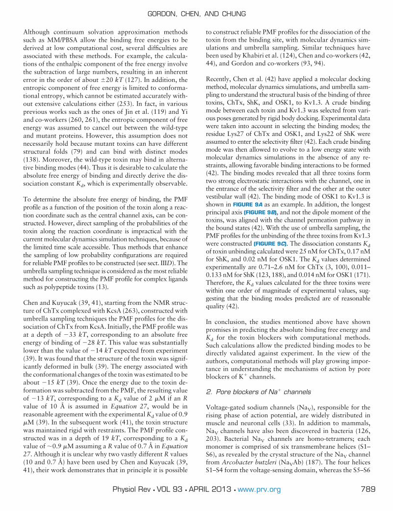
Although continuum solvation approximation methodssuch as MM/PBSA allow the binding free energies to bederived at low computational cost, several difficulties areassociated with these methods. For example, the calcula-tions of the enthalpic component of the free energy involvethe subtraction of large numbers, resulting in an inherenterror in the order of about 20 kT (127). In addition, theentropic component of free energy is limited to conforma-tional entropy, which cannot be estimated accurately with-out extensive calculations either (253). In fact, in variousprevious works such as the ones of Jin et al. (119) and Yiand co-workers (260, 261), the entropic component of freeenergy was assumed to cancel out between the wild-typeand mutant proteins. However, this assumption does notnecessarily hold because mutant toxins can have differentstructural folds (79) and can bind with distinct modes(138). Moreover, the wild-type toxin may bind in alterna-tive binding modes (44). Thus it is desirable to calculate theabsolute free energy of binding and directly derive the dis-sociation constant Kd, which is experimentally observable.
To determine the absolute free energy of binding, the PMFprofile as a function of the position of the toxin along a reac-tion coordinate such as the central channel axis, can be con-structed. However, direct sampling of the probabilities of thetoxin along the reaction coordinate is impractical with thecurrent molecular dynamics simulation techniques, because ofthe limited time scale accessible. Thus methods that enhancethe sampling of low probability configurations are requiredfor reliable PMF profiles to be constructed (see sect. IIID). Theumbrella sampling technique is considered as the most reliablemethod for constructing the PMF profile for complex ligandssuch as polypeptide toxins (13).
Chen and Kuyucak (39, 41), starting from the NMR struc-ture of ChTx complexed with KcsA (263), constructed withumbrella sampling techniques the PMF profiles for the dis-sociation of ChTx from KcsA. Initially, the PMF profile wasat a depth of �33 kT, corresponding to an absolute freeenergy of binding of �28 kT. This value was substantiallylower than the value of �14 kT expected from experiment(39). It was found that the structure of the toxin was signif-icantly deformed in bulk (39). The energy associated withthe conformational changes of the toxin was estimated to beabout �15 kT (39). Once the energy due to the toxin de-formation was subtracted from the PMF, the resulting valueof �13 kT, corresponding to a Kd value of 2 �M if an Rvalue of 10 Å is assumed in Equation 27, would be inreasonable agreement with the experimental Kd value of 0.9�M (39). In the subsequent work (41), the toxin structurewas maintained rigid with restraints. The PMF profile con-structed was in a depth of 19 kT, corresponding to a Kd
value of �0.9 �M assuming a R value of 0.7 Å in Equation27. Although it is unclear why two vastly different R values(10 and 0.7 Å) have been used by Chen and Kuyucak (39,41), their work demonstrates that in principle it is possible
to construct reliable PMF profiles for the dissociation of thetoxin from the binding site, with molecular dynamics sim-ulations and umbrella sampling. Similar techniques havebeen used by Khabiri et al. (124), Chen and co-workers (42,44), and Gordon and co-workers (93, 94).
Recently, Chen et al. (42) have applied a molecular dockingmethod, molecular dynamics simulations, and umbrella sam-pling to understand the structural basis of the binding of threetoxins, ChTx, ShK, and OSK1, to Kv1.3. A crude bindingmode between each toxin and Kv1.3 was selected from vari-ous poses generated by rigid body docking. Experimental datawere taken into account in selecting the binding modes; theresidue Lys27 of ChTx and OSK1, and Lys22 of ShK wereassumed to enter the selectivity filter (42). Each crude bindingmode was then allowed to evolve to a low energy state withmolecular dynamics simulations in the absence of any re-straints, allowing favorable binding interactions to be formed(42). The binding modes revealed that all three toxins formtwo strong electrostatic interactions with the channel, one inthe entrance of the selectivity filter and the other at the outervestibular wall (42). The binding mode of OSK1 to Kv1.3 isshown in FIGURE 9A as an example. In addition, the longestprincipal axis (FIGURE 9B), and not the dipole moment of thetoxins, was aligned with the channel permeation pathway inthe bound states (42). With the use of umbrella sampling, thePMF profiles for the unbinding of the three toxins from Kv1.3were constructed (FIGURE 9C). The dissociation constants Kd
of toxin unbinding calculated were 25 nM for ChTx, 0.17 nMfor ShK, and 0.02 nM for OSK1. The Kd values determinedexperimentally are 0.71–2.6 nM for ChTx (3, 100), 0.011–0.133 nM for ShK (123, 188), and 0.014 nM for OSK1 (171).Therefore, the Kd values calculated for the three toxins werewithin one order of magnitude of experimental values, sug-gesting that the binding modes predicted are of reasonablequality (42).
In conclusion, the studies mentioned above have shownpromises in predicting the absolute binding free energy andKd for the toxin blockers with computational methods.Such calculations allow the predicted binding modes to bedirectly validated against experiment. In the view of theauthors, computational methods will play growing impor-tance in understanding the mechanisms of action by poreblockers of K� channels.
2. Pore blockers of Na� channels
Voltage-gated sodium channels (NaV), responsible for therising phase of action potential, are widely distributed inmuscle and neuronal cells (33). In addition to mammals,NaV channels have also been discovered in bacteria (126,203). Bacterial NaV channels are homo-tetramers; eachmonomer is comprised of six transmembrane helices (S1–S6), as revealed by the crystal structure of the NaV channelfrom Arcobacter butzleri (NaVAb) (187). The four helicesS1–S4 form the voltage-sensing domain, whereas the S5–S6
GORDON, CHEN, AND CHUNG
789Physiol Rev • VOL 93 • APRIL 2013 • www.prv.org
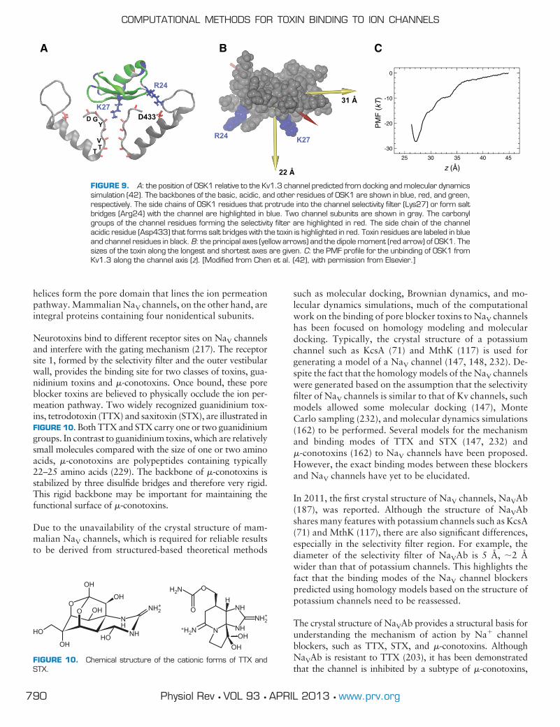
helices form the pore domain that lines the ion permeationpathway. Mammalian NaV channels, on the other hand, areintegral proteins containing four nonidentical subunits.
Neurotoxins bind to different receptor sites on NaV channelsand interfere with the gating mechanism (217). The receptorsite 1, formed by the selectivity filter and the outer vestibularwall, provides the binding site for two classes of toxins, gua-nidinium toxins and �-conotoxins. Once bound, these poreblocker toxins are believed to physically occlude the ion per-meation pathway. Two widely recognized guanidinium tox-ins, tetrodotoxin (TTX) and saxitoxin (STX), are illustrated inFIGURE 10. Both TTX and STX carry one or two guanidiniumgroups. In contrast to guanidinium toxins, which are relativelysmall molecules compared with the size of one or two aminoacids, �-conotoxins are polypeptides containing typically22–25 amino acids (229). The backbone of �-conotoxins isstabilized by three disulfide bridges and therefore very rigid.This rigid backbone may be important for maintaining thefunctional surface of �-conotoxins.
Due to the unavailability of the crystal structure of mam-malian NaV channels, which is required for reliable resultsto be derived from structured-based theoretical methods
such as molecular docking, Brownian dynamics, and mo-lecular dynamics simulations, much of the computationalwork on the binding of pore blocker toxins to NaV channelshas been focused on homology modeling and moleculardocking. Typically, the crystal structure of a potassiumchannel such as KcsA (71) and MthK (117) is used forgenerating a model of a NaV channel (147, 148, 232). De-spite the fact that the homology models of the NaV channelswere generated based on the assumption that the selectivityfilter of NaV channels is similar to that of Kv channels, suchmodels allowed some molecular docking (147), MonteCarlo sampling (232), and molecular dynamics simulations(162) to be performed. Several models for the mechanismand binding modes of TTX and STX (147, 232) and�-conotoxins (162) to NaV channels have been proposed.However, the exact binding modes between these blockersand NaV channels have yet to be elucidated.
In 2011, the first crystal structure of NaV channels, NaVAb(187), was reported. Although the structure of NaVAbshares many features with potassium channels such as KcsA(71) and MthK (117), there are also significant differences,especially in the selectivity filter region. For example, thediameter of the selectivity filter of NaVAb is 5 Å, �2 Åwider than that of potassium channels. This highlights thefact that the binding modes of the NaV channel blockerspredicted using homology models based on the structure ofpotassium channels need to be reassessed.
The crystal structure of NaVAb provides a structural basis forunderstanding the mechanism of action by Na� channelblockers, such as TTX, STX, and �-conotoxins. AlthoughNaVAb is resistant to TTX (203), it has been demonstratedthat the channel is inhibited by a subtype of �-conotoxins,
D GY
VTT
K27
R24
D433
K27R24
22 Å
31 Å
0
-10
-20
-30
25 30 35 40 45
PM
F (
kT)
z (Å)
A B C
FIGURE 9. A: the position of OSK1 relative to the Kv1.3 channel predicted from docking and molecular dynamicssimulation (42). The backbones of the basic, acidic, and other residues of OSK1 are shown in blue, red, and green,respectively. The side chains of OSK1 residues that protrude into the channel selectivity filter (Lys27) or form saltbridges (Arg24) with the channel are highlighted in blue. Two channel subunits are shown in gray. The carbonylgroups of the channel residues forming the selectivity filter are highlighted in red. The side chain of the channelacidic residue (Asp433) that forms salt bridges with the toxin is highlighted in red. Toxin residues are labeled in blueand channel residues in black. B: the principal axes (yellow arrows) and the dipole moment (red arrow) of OSK1. Thesizes of the toxin along the longest and shortest axes are given. C: the PMF profile for the unbinding of OSK1 fromKv1.3 along the channel axis (z). [Modified from Chen et al. (42), with permission from Elsevier.]
OH
OH
OH
OH OHOHHO
HO
HO
O
NH
NH
NH2+
H2N
O
O
NHNH2
+
NHN+H2N
FIGURE 10. Chemical structure of the cationic forms of TTX andSTX.
COMPUTATIONAL METHODS FOR TOXIN BINDING TO ION CHANNELS
790 Physiol Rev • VOL 93 • APRIL 2013 • www.prv.org

PIIIA (44). The selectivity filter includes a ring of glutamateresidues (E177), and a ring of glutamate residues (E189) islocated on the outer vestibule of the channel (see FIGURE 11A).With the use of molecular dynamics simulations, it was foundthat the positively charged toxin PIIIA, released in water 15 Åabove the binding site, spontaneously bound to the channelouter vestibular wall and occluded the ion permeation path-way with the side chain of either a lysine or an arginine residue(FIGURE 11B), indicating that PIIIA is capable of inhibitingNaVAb (44). The results also indicated that PIIIA may blockNaVAb with multiple binding modes, which was confirmed byfree energy calculations (44). The free energy calculationsshowed that different binding modes share similar free ener-gies of binding, suggesting that the binding modes are equallyprobable. This interpretation was also confirmed experimen-tally by Stevens et al. (218). Thus certain toxins may blockNaV channels with alternative binding modes, rather than thesingle binding mode generally assumed.
In addition to TTX, STX, and �-conotoxins, drug mole-cules like local anesthetics may also act as pore blockers ofNaV channels. The receptor site of local anesthetics hasbeen shown to be in the inner vestibule (177), rather thanthe outer vestibule for �-conotoxins. Recent docking calcu-lations suggest that local anesthetics may block the currentby creating a positive electrostatic field in the inner vesti-bule, which repels cations (148).
B. Gating Modifiers
1. Gating modifiers of K� channels
A number of polypeptide toxins that interfere with the gat-ing mechanism of voltage-gated K� (Kv) channels havebeen isolated from spider venoms. These tarantula toxinsbind to the voltage-sensing domain of Kv channels withinthe membrane and hinder the conformational changes thatoccur during channel opening (141, 165, 164, 220). Oncethe voltage-sensing domain is bound with toxins, a strongerdepolarization is required to activate the channel, thus shift-ing the activation-voltage curve to the right (220).
Tarantula toxins are typically of 25–40 amino acids inlength. The structure of tarantula toxins is rigid, as the
toxin backbones are cross-linked by three or four disulfidebonds forming the inhibitor cysteine knot motif, which isalso commonly found in pore-blockers (180). Some of themost well-characterized tarantula toxins are hanatoxin(HaTx) (221), Scodra griseipes toxin (SGTx1) (160), het-eropodatoxin (HpTx) (207), phrixotoxin (PaTx) (69), andthe voltage sensor toxin (VSTx) (205). The solution struc-ture of these toxins suggests that a hydrophobic surface isconserved across tarantula toxins (225, 121, 140), althoughthe size and shape of this surface varies. The critical role ofthis hydrophobic surface in toxin function has been dem-onstrated experimentally (165, 220, 245). The toxins mayuse this hydrophobic patch to interact with the voltage-sensing domain. Once bound to the voltage-sensing do-main, they may stabilize it in the closed conformation sothat the channel is harder to open. The functional surface oftarantula toxins on the voltage-sensing domain of Kv chan-nels has been examined extensively with mutagenesis tech-niques (64, 65, 145, 146, 191, 222, 223, 264). Theseshowed that the periplasmic half of the S3 helix forms partof the receptor site. However, the exact binding modes bytarantula toxins have not been resolved by experiments.
To date, most molecular dynamics simulations have beenconcerned with the energetics associated with partitioningtarantula toxins into membranes. For example, using ato-mistic molecular dynamics simulations, Bemporad et al.(17) examined the preferred location of VSTx1 within lipidmembranes. The simulations showed that VSTx1, whenplaced within the hydrophobic core or the aqueous phase,spontaneously moves to the surface region containing thelipid headgroups. When bound to the surface region, abouthalf of the toxin remains buried within the hydrophobiccore of the lipid bilayer, whereas the other half is interactingwith the head groups of lipids (17). Contrastingly, in thework of Nishizawa and Nishizawa (179), HaTx1 was ob-served to penetrate deeply to near the center of the bilayer,but not to form a stable complex with the bilayer whenbound to the surface region. In subsequent studies, the PMFprofiles of VSTx1 along the bilayer normal reveal that thepreferred binding location of VSTx1 is �20 Å from thecenter of a lipid bilayer (248, 250). Thus all the simulationsillustrated here are in support of the earlier proposal which
AE189
R185
E177 R2
E177
PIIIAB
FIGURE 11. A: structure of the pore domainof the NaVAb channel, viewed perpendicular tothe channel axis. Only two subunits are shownfor clarity. B: PIIIA blocks the ion conductionpathway of the NaVAb channel with the sidechain of the residue Arg2. Green ribbon repre-sents the toxin backbone, and the yellow sphererepresents a sodium ion. [From Chen and Chung(44), with permission from Elsevier.]
GORDON, CHEN, AND CHUNG
791Physiol Rev • VOL 93 • APRIL 2013 • www.prv.org

states that tarantula toxins bind to the voltage-sensing do-main within membranes. Molecular dynamics simulationshave also been used to understand the binding modes be-tween VSTx1 and the isolated voltage-sensing domain ofKVAP (249). VSTx1, which is in the water phase at thesimulation start, spontaneously binds to the voltage-sensingdomain of KVAP in multiple coarse-grained simulationseach on a time scale of 3 �s (249). The receptor site on thevoltage-sensing domain was found to be primarily formedby residues from the S1 and S4 helices (249).
2. Gating modifiers of Na� channels
Eukaryotic NaV channels are integral proteins consisting offour homologous subunits (I–IV); each subunit contains avoltage-sensing domain and a pore domain. A large numberof polypeptides isolated from venoms of scorpions, spiders,and sea anemones have been shown to interfere with thegating mechanism of NaV channels (32, 217). For example,scorpion �-toxins bind to the periplasmic side of the volt-age-sensing domain of the subunit IV and slow or inhibitchannel inactivation (96). Scorpion �-toxins, on the otherhand, bind to the voltage-sensing domain of the subunit II,trap the voltage-sensing domain in an outward position,resulting in the channel to open at less depolarized voltages(103). Scorpion �- and �-toxins are typically of 60–80residues in length and have rigid backbone cross-linked byfour disulfide bridges (195). Although �- and �-toxinshave distinct functional effects on NaV channels, theirshape and secondary structure are rather similar, as illus-
trated in FIGURE 12. As scorpion �- and �-toxins are be-lieved to be potential leads for developing novel insecticides(96, 103), numerous experiments have been carried out tounderstand their functional surface and binding modes toNaV channels. However, despite the fact that several mod-els have been constructed with molecular docking methodsfor the bindings of the �-toxin LqhII (247) and the �-toxinCss4 to NaV1.2 (34, 265), the binding modes of scorpion �-and �-toxins to NaV channels have not been fully resolvedby experiments. In addition to scorpion �-toxins, various-conotoxins isolated from marine cone snails have alsobeen shown to inhibit the fast inactivation of NaV channels(14, 78, 254). The receptor site of -conotoxins overlapswith that of scorpion �-toxins (142), suggesting that thetwo families of toxins may inhibit channel inactivation withsimilar mechanisms.
Using a molecular docking method and molecular dynamicssimulations, Chen and Chung (47) have examined in detailthe binding of two �-toxins, Css4 and Cn2, to the isolated IIvoltage-sensing domains of rat NaV1.2 and rat NaV1.6,respectively. Experimentally, Css4 has been shown to inter-fere with the gating mechanisms of both NaV1.2 andNaV1.6, whereas Cn2 selectively targets NaV1.6 (208), atnanomolar toxin concentrations. Yet, Css4 and Cn2 share asequence identity of 83%, being significantly different atonly three positions, 7, 8, and 64, which are in spatialproximity due to the disulfide bridge between Cys12-Cys65. As the difference in these three residues apparentlygives rise to the distinct sensitivity of Css4 and Cn2 to NaV
8
18
15 64
8
1815 64
8
18
15 64
8
18
15 64
AahII LqhαIT
Css4 Cn2
A
B
FIGURE 12. The secondary structure of two�-toxins, AahII (107) and Lqh�IT (237) (A), andtwo �-toxins, Css4 and Cn2 (194) (B). Thestructure of Css4 is modeled on Cn2. The twotoxins are 83% identical in sequence. Helicesare shown in purple, �-sheet strands in yellow,and others in gray. The side chains of residuesat positions 8, 18, and 64 are highlighted. Ba-sic residues are colored in blue, acidic in red,and others in green. [Modified from Chen andChung (48).]
COMPUTATIONAL METHODS FOR TOXIN BINDING TO ION CHANNELS
792 Physiol Rev • VOL 93 • APRIL 2013 • www.prv.org

channels, it is likely that they are involved in binding (208).Following the assumption that residues 7, 8, and 64 formpart of the functional surface, a docking method was usedto search for a plausible binding mode between Css4-NaV1.2 and Cn2-NaV1.6 (47). For each toxin-channel pair,a unique structure that is consistent with the assumptionwas selected out of 300 docking poses. Each selected struc-ture was then embedded in a lipid bilayer and a box ofexplicit water, and run for 20 ns with molecular dynamicssimulations without the presence of any restraints, allowingthe complex to evolve to a stable state in a membrane-likeenvironment. The toxin-channel interacting residue pairswere identified and validated by comparing with mutagen-esis experiments and previous models (47). The Kd valuesderived with umbrella sampling were 20 nM for Css4 and70 nM for Cn2, which were in reasonable agreement withthe experimental values of 1 and 40 nM, respectively (47).The simulations revealed that both Css4 and Cn2 wedgeinto the receptor site with a loop between positions 8 and 18(FIG. 13, A AND B), which is referred to as the Linker domain(47). In this loop, two charged residues, Lys13 and Glu15,are commonly found in anti-mammalian �-toxins. Thesetwo charged residues and a critical glutamate residue(Glu28) were observed to form hydrogen bonds and salt-bridges with the receptor site (FIGURE 13C), stabilizing thetoxin-channel complex. Thus the simulations suggest thatthe functional surface of anti-mammalian �-toxins is con-served, centered on the Linker domain (47). Interestingly,the crystal structure of Cn2 bound to its antibody alsoshows that the Linker domain plays a central role in thebinding (29).
The functional surface of �-toxins uncovered by Chen andChung (47) appears to overlap with the functional surfaceof �-toxins determined experimentally, consisting of theNC-domain around residues 7, 8, and 64, and the Coredomain around the residue 18. These two domains are in-terconnected by the Linker domain, which wedges into thereceptor site in �-toxins. Given the similarity between thestructure of �- and �-toxins, it seems that the functional
surface may be conserved not only in �-toxins, but alsobetween �- and �-toxins. Following this assumption, Chenand Chung used molecular dynamics simulations with dis-tance restraints as a docking method to examine the bindingof two �-toxins, the anti-mammalian toxin AahII, and theanti-insect toxin Lqh�IT, to the IV voltage-sensing domainof rat NaV1.2 (48). The distance restraints were determinedassuming that the Linker domain of the two �-toxins wedgeinto the receptor site in a similar orientation to that of�-toxins. With the distance restraints applied, the toxinswere drawn to the receptor site rapidly within a few nano-seconds. It was found that both AahII and Lqh�IT wereable to form several favorable electrostatic and hydropho-bic interactions with the receptor site, demonstrating thattoxin-channel complex is stable with the Linker domainwedging into the binding groove. The Kd values derivedwere 17 nM for AahII and 1 �M for Lqh�IT, which agreewell with experiment which shows that AahII is about threeorders of magnitude more potent than Lqh�IT for NaV1.2(122). Computational mutagenesis calculations were per-formed on two channel mutants (48), E1551R andR1626E. Experimentally, these two mutations did not haveany measurable effects on toxin binding affinities (247).However, both E1551 and R1626 were observed to formsalt bridges with the toxin in the models, which casts doubtson the consistency between the models and experiment. Thecomputational mutagenesis calculations eliminated thisdoubt. It was found that AahII was able to form an equiv-alent salt bridge when the one due to E1551 or R1626 wasbroken due to the mutation, such that each of the mutationsdid not change the binding affinity of AahII significantly.For example, the Kd value calculated for the E1551R mu-tant channel compared favorably with that of the wild type(48). This highlights the importance of interpreting mu-tagenesis data with caution. In support of the bindingmodes predicted for AahII-NaV1.2, the -conotoxin EVIAwas observed to bind to the VS domain of NaV1.2 sponta-neously in 50 ns, forming similar salt bridges as that ob-served in the AahII-NaV1.2 complex (48).
K18 K13
E15
S8
F44V793
E28R850
E15R853
A B CFIGURE 13. A: the position of Css4 bound tothe IIS1-S4 voltage-sensing domain of NaV1.2,relative to the lipid bilayer. The surface of thevoltage-sensing domain is shown in transparentsilver. Toxin backbone is in yellow. Horizontal linesindicate the average position of the phosphategroups of the lipids. The loop between positions 8and 18 of the toxin is highlighted in red. B: Css4bound to the voltage-sensing domain of NaV1.2.The side chains of two key residues of the toxin,Lys13 (blue) and Glu15 (red), are highlighted. TheS2 and S4 helices of NaV1.2 are highlighted inpink and lime, respectively. C: Css4 bound to thevoltage-sensing domain of NaV1.2 showing threeof the key contacts. Toxin backbone is shown inyellow. The complexes shown in B and C are ro-tated by �90° counterclockwise from that of A.[From Chen and Chung (47).]
GORDON, CHEN, AND CHUNG
793Physiol Rev • VOL 93 • APRIL 2013 • www.prv.org

In conclusion, the computational studies of Chen andChung (47, 48) demonstrated the key role of the Linkerdomain in the binding of �- and �-toxins to NaV channels.In all the models of �- and �-toxins bound to NaV channelspredicted (47, 48), at least one salt bridge between the toxinand the S4 helix of the VS domain was observed. It remainsto be elucidated whether or not such a salt bridge is vital forthe action of the toxins.
VII. FUTURE PROSPECTS
We have first outlined some theoretical principles underly-ing state-of-the-art computational tools currently beingused for studying biological macromolecules. We then sum-marized some of the salient features uncovered using com-putational methods of the actions of channel blockers, con-centrating on polypeptide toxins extracted from venomousanimals, on voltage-gated cationic channels. In the future,computational modeling of ligand-channel interactionsmay play a prominent role, providing physical explanationsof the mechanisms underlying modulations of channelproperties by large polypeptides and intermediate-sizeddrug molecules. For example, detailed knowledge of thebioactive conformations of polypeptide toxins to specificchannel subtypes may lead to a host of novel pharmaceuti-cal products.
In theory, one should be able to gain important knowledgeof ligand-channel interactions using computational tech-niques. For example, in silico studies should enable us toidentify the ligand binding site, reveal pairs of interactingligand-channel residues, and determine the binding affinity.Many efficient algorithms and computational tools havebeen devised over the past decades, designed to unveil themodes of interactions between macromolecules. Undoubt-edly, these tools will be further refined and improved, suchthat they will rapidly provide insights into how small orlarge molecules modulate the conductance properties of bi-ological ion channels.
To date, computational studies of blocker-channel interac-tions mainly rely on two-step procedures. First, with the useof molecular docking programs, Brownian dynamics, orbiased molecular dynamics simulations, a toxin is com-plexed to the external vestibule or the voltage-sensor of aspecific channel. If a docking program is used for this step,one or two docking poses among a large number of possibleposes generated by the program are selected based on priorexperimental knowledge. Many state of the art dockingprograms do not currently appear to be accurate enough toroutinely derive correctly docked bound states of polypep-tide toxins on ion channels without a degree of operatorinput and prior knowledge. This would be a problem for atruly high-throughput screening and ranking system, whichin any case we believe is some way away. When experimen-tal information is available, this can be incorporated into
the docking procedure using distance restraints (43). Biasedmolecular dynamics also works well for this purpose (46,48). Toxin pore blockers, which block the channel by in-serting a lysine or arginine residue into the pore, are aneasier case, because each toxin has only a small number ofsuch residues available to be inserted into the pore, andinserting a residue into the pore vastly constrains the num-ber of available poses. Thus, in these cases, the problemappears to simply be one of finding the correct protocol andtools rather than a deficiency in current algorithms.
Following the generation and selection of docked poses, thefree energy of binding and binding affinity, which are ex-perimentally observable quantities, may be calculated.Also, the bioactive surface of a specific toxin can be identi-fied by examining the residue pairs forming hydrogen bondsin the toxin-channel complex. Moreover, it is possible tocarry out theoretical site-directed mutagenesis to enhanceits sensitivity and selectivity (45, 123) or prune the moleculeto render it smaller and more durable (52, 63, 125). Inrecent studies, the Kd values derived using the umbrellasampling technique and explicit representation of water ap-pear to be within one to two orders of magnitude of thatmeasured experimentally in various toxin-channel systems(41, 42, 46, 94, 200). These approaches, however, cannotbe routinely used to search for lead candidates of channelblockers from large numbers of available compounds, be-cause of prohibitively high computational costs. Typically,it takes between 50,000 and 75,000 CPU hours of a modernsupercomputer to construct a profile of potential of meanforce of one toxin, using molecular dynamics umbrella sam-pling methods with simulation times that are barely suffi-cient. As discussed in the review, other, approximate, freeenergy methods are available, but it is harder to assess theiraccuracy in predicting the binding affinity of toxins to ionchannels, due to a lack of computational studies. The LIEmethod did not work well in predicting the relative bindingaffinities of MTx to Kv1.1, Kv1.2, and Kv1.3 in one study(46), possibly because the entropic component of the freeenergy which is ignored by the method cannot be cancelledout between the channels. The MM-PBSA method, on theother hand, appears to offer a plausible means of calculat-ing the changes in free energy due to a single mutation to thetoxin (119, 260, 261). But clearly more work needs to bedone to achieve a more generally applicable methodology.
It will therefore be desirable to devise a computational al-gorithm, either an improved version of molecular dockingprograms, a more sophisticated Brownian dynamics tech-nique incorporating the flexibility of ligands and channels,or a clever modification and specialization of standard mo-lecular dynamics, that can rapidly determine the bindingmode between the ligand and the channel and accuratelycalculate the free energy of the binding. Such an algorithmmight have the following features, all of which are found in
COMPUTATIONAL METHODS FOR TOXIN BINDING TO ION CHANNELS
794 Physiol Rev • VOL 93 • APRIL 2013 • www.prv.org

various existing programs and techniques but which willneed to be further synthesized and improved.
Implicit water, and possibly lipids, will probably be needed.Modeling all of the water molecules in the assembly takes usinto the realms of standard molecular dynamics, which istoo slow for our desired purpose. Note that not all the waterneeds to be implicit. Explicit water could in principle beemployed around the channel pore and blocker, with im-plicit water being used further away.
Large portions of the simulation assembly could be fixed,treated using rigid body approaches or otherwise treated ina simplified manner. The purpose is again to increase com-putational efficiency. It is probably too ambitious to take anapproach where all possibilities of large-scale conforma-tional change are taken into account. Instead, the confor-mations of large parts of the channel and blocker should betaken to be invariant, thus allowing fixing to take place.
The long-range electrostatics should be reasonable. This isparticularly important for ion channel blockers that bind inthe pore. Such blockers are usually highly charged and areattracted to the oppositely charged outer vestibule of thechannel by long-range electrostatic forces that contribute asignificant portion of the total binding affinity. The channelpore itself is a region where strong, highly focused electricfields are present, and thus getting the electrostatics right inthis region may be important.
Hydrogen bonding and salt bridges need to be correctlydealt with. These short range forces contribute a large per-centage of the binding affinity of typical ion channel mod-ulator toxins.
Nonpolar hydration forces, such as the hydrophobic force,need to also be reasonably well modeled, because theseforces are also important to binding.
Flexibility should be taken into account. We have spokenabove about fixing parts of the system. When protein toxinsbind to an ion channel, we would require that the residuesinvolved in key contacts be able to attain the necessarygeometric configuration. We would also require, where nec-essary, that the selectivity filter of the channel should beflexible enough to allow basic side chains to be inserted intothe filter, as is seen in many ion channel blocker toxins.
Implicit water, lipids, and the fixing of the interior of theprotein and lipid regions necessitate the use of some kind ofmacroscopic electrostatics treatment. We have discussedvarious approaches to macroscopic electrostatics in the ear-lier sections of this review. Presolving Poisson’s equation orthe Poisson-Boltzmann equation and then storing the re-sults in lookup tables is an extremely fast approach oftentaken by Brownian dynamics and docking programs. Ap-
proximations are necessary to deal with the interactionsbetween two or more molecules or ions in such an ap-proach, due to considerations of dimensionality of thelookup tables. Generalized Born electrostatics are able todeal with nonrigid bodies, and are also able to deal with allinteractions between simulation bodies, but are slower andpotentially less accurate. A combination of the two might beworth trying. If explicit water were to be employed onlyinside the channel and around the binding interface, then itmight be possible to get away with a very fast and simplifiedversion of macroscopic electrostatics.
The treatment of hydrogen bonding and salt bridges is alsodifficult. Hydrogen bonds exist as a delicate balance be-tween attractive Coulomb forces involving small, mobilehydrogen atoms, repulsive steric forces, attractive disper-sive forces due to the van der Waals potential, and themediating influence of the surrounding solvent. Atomisticmolecular mechanics often deal with this by explicitly mod-eling all elements of the above, using a carefully calibratedparametrization. Changing any of the elements, for exam-ple, fixing hydrogens or combining them with parent atomsor using an implicit solvent force field, will seriously disruptthis delicate balance. It seems likely that either of two ap-proaches might be taken. The first approach models thebinding interface and other important parts of the systemusing fully atomistic, explicit solvent molecular dynamicswhile retaining more approximate representations in moredistant regions. The second approach introduces specialempirical forces for hydrogen bonding and salt bridges. Thelatter treatment is common in docking programs.
The treatment of nonpolar forces faces problems similar tothat of hydrogen bonds. Implicit solvent force fields oftenrely on a surface area-based approach, where the nonpolarenergy of a group of one or more molecules scales with thesolvent accessible surface area. Scaling constants are pa-rametrized based on the calculated solvation free energies oflibraries of molecules. There is some danger that the param-eterizations may not lead to accurate binding forces due tothe simplified nature of the model. More sophisticatedtreatments exist, but at the time of writing it is hard to picka clear winner. Again, the approach of including explicitwater molecules around the binding region may help toameliorate the problems.
In general, state-of-the-art implicit solvent force fields seemto be able to predict the solvation energies of biomoleculesto within several kcal/mol (115, 139). Careful parameter-izations (38) seem to be able to derive potentials of meanforce for small molecular fragments that have a comparableerror scale. Ideally, we would like to be able to compare andrank the binding free energies of similar toxins, where anaccuracy of a few kilocalories per mole might still give use-ful information. Therefore, it appears that current algo-rithms are probably somewhere on the cusp of success ver-
GORDON, CHEN, AND CHUNG
795Physiol Rev • VOL 93 • APRIL 2013 • www.prv.org
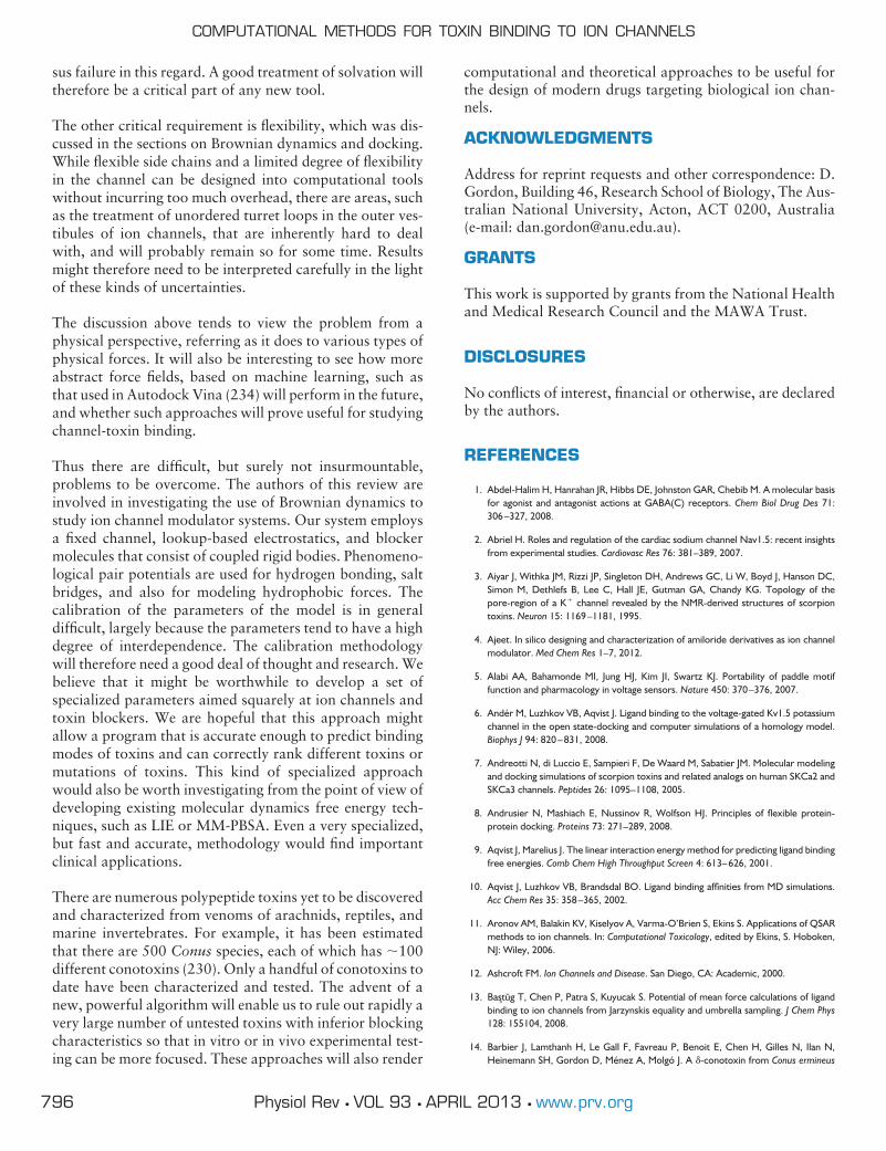
sus failure in this regard. A good treatment of solvation willtherefore be a critical part of any new tool.
The other critical requirement is flexibility, which was dis-cussed in the sections on Brownian dynamics and docking.While flexible side chains and a limited degree of flexibilityin the channel can be designed into computational toolswithout incurring too much overhead, there are areas, suchas the treatment of unordered turret loops in the outer ves-tibules of ion channels, that are inherently hard to dealwith, and will probably remain so for some time. Resultsmight therefore need to be interpreted carefully in the lightof these kinds of uncertainties.
The discussion above tends to view the problem from aphysical perspective, referring as it does to various types ofphysical forces. It will also be interesting to see how moreabstract force fields, based on machine learning, such asthat used in Autodock Vina (234) will perform in the future,and whether such approaches will prove useful for studyingchannel-toxin binding.
Thus there are difficult, but surely not insurmountable,problems to be overcome. The authors of this review areinvolved in investigating the use of Brownian dynamics tostudy ion channel modulator systems. Our system employsa fixed channel, lookup-based electrostatics, and blockermolecules that consist of coupled rigid bodies. Phenomeno-logical pair potentials are used for hydrogen bonding, saltbridges, and also for modeling hydrophobic forces. Thecalibration of the parameters of the model is in generaldifficult, largely because the parameters tend to have a highdegree of interdependence. The calibration methodologywill therefore need a good deal of thought and research. Webelieve that it might be worthwhile to develop a set ofspecialized parameters aimed squarely at ion channels andtoxin blockers. We are hopeful that this approach mightallow a program that is accurate enough to predict bindingmodes of toxins and can correctly rank different toxins ormutations of toxins. This kind of specialized approachwould also be worth investigating from the point of view ofdeveloping existing molecular dynamics free energy tech-niques, such as LIE or MM-PBSA. Even a very specialized,but fast and accurate, methodology would find importantclinical applications.
There are numerous polypeptide toxins yet to be discoveredand characterized from venoms of arachnids, reptiles, andmarine invertebrates. For example, it has been estimatedthat there are 500 Conus species, each of which has �100different conotoxins (230). Only a handful of conotoxins todate have been characterized and tested. The advent of anew, powerful algorithm will enable us to rule out rapidly avery large number of untested toxins with inferior blockingcharacteristics so that in vitro or in vivo experimental test-ing can be more focused. These approaches will also render
computational and theoretical approaches to be useful forthe design of modern drugs targeting biological ion chan-nels.
ACKNOWLEDGMENTS
Address for reprint requests and other correspondence: D.Gordon, Building 46, Research School of Biology, The Aus-tralian National University, Acton, ACT 0200, Australia(e-mail: [email protected]).
GRANTS
This work is supported by grants from the National Healthand Medical Research Council and the MAWA Trust.
DISCLOSURES
No conflicts of interest, financial or otherwise, are declaredby the authors.
REFERENCES
1. Abdel-Halim H, Hanrahan JR, Hibbs DE, Johnston GAR, Chebib M. A molecular basisfor agonist and antagonist actions at GABA(C) receptors. Chem Biol Drug Des 71:306–327, 2008.
2. Abriel H. Roles and regulation of the cardiac sodium channel Nav1.5: recent insightsfrom experimental studies. Cardiovasc Res 76: 381–389, 2007.
3. Aiyar J, Withka JM, Rizzi JP, Singleton DH, Andrews GC, Li W, Boyd J, Hanson DC,Simon M, Dethlefs B, Lee C, Hall JE, Gutman GA, Chandy KG. Topology of thepore-region of a K� channel revealed by the NMR-derived structures of scorpiontoxins. Neuron 15: 1169–1181, 1995.
4. Ajeet. In silico designing and characterization of amiloride derivatives as ion channelmodulator. Med Chem Res 1–7, 2012.
5. Alabi AA, Bahamonde MI, Jung HJ, Kim JI, Swartz KJ. Portability of paddle motiffunction and pharmacology in voltage sensors. Nature 450: 370–376, 2007.
6. Andér M, Luzhkov VB, Aqvist J. Ligand binding to the voltage-gated Kv1.5 potassiumchannel in the open state-docking and computer simulations of a homology model.Biophys J 94: 820–831, 2008.
7. Andreotti N, di Luccio E, Sampieri F, De Waard M, Sabatier JM. Molecular modelingand docking simulations of scorpion toxins and related analogs on human SKCa2 andSKCa3 channels. Peptides 26: 1095–1108, 2005.
8. Andrusier N, Mashiach E, Nussinov R, Wolfson HJ. Principles of flexible protein-protein docking. Proteins 73: 271–289, 2008.
9. Aqvist J, Marelius J. The linear interaction energy method for predicting ligand bindingfree energies. Comb Chem High Throughput Screen 4: 613–626, 2001.
10. Aqvist J, Luzhkov VB, Brandsdal BO. Ligand binding affinities from MD simulations.Acc Chem Res 35: 358–365, 2002.
11. Aronov AM, Balakin KV, Kiselyov A, Varma-O’Brien S, Ekins S. Applications of QSARmethods to ion channels. In: Computational Toxicology, edited by Ekins, S. Hoboken,NJ: Wiley, 2006.
12. Ashcroft FM. Ion Channels and Disease. San Diego, CA: Academic, 2000.
13. Bastug T, Chen P, Patra S, Kuyucak S. Potential of mean force calculations of ligandbinding to ion channels from Jarzynskis equality and umbrella sampling. J Chem Phys128: 155104, 2008.
14. Barbier J, Lamthanh H, Le Gall F, Favreau P, Benoit E, Chen H, Gilles N, Ilan N,Heinemann SH, Gordon D, Ménez A, Molgó J. A -conotoxin from Conus ermineus
COMPUTATIONAL METHODS FOR TOXIN BINDING TO ION CHANNELS
796 Physiol Rev • VOL 93 • APRIL 2013 • www.prv.org
venom inhibits inactivation in vertebrate neuronal Na� channels but not in skeletaland cardiac muscles. J Biol Chem 279: 4680–4685, 2004.
15. Bashford D, Case DA. Generalized Born models of macromolecular solvation effects.Annu Rev Phys Chem 51: 129–152, 2000.
16. Beard DA, Schlick T. Unbiased rotational moves for rigid-body Brownian dynamics.Biophys J 85: 2973–2976, 2003.
17. Bemporad D, Sands ZA, Wee CL, Grottesi A, Sansom MSP. Vstx1, a modifier of Kvchannel gating, localizes to the interfacial region of lipid bilayers. Biochemistry 45:11844–11855, 2006.
18. Ben-Naim A. Statistical potentials extracted from protein structures: Are these mean-ingful potentials? J Chem Phys 107: 3698, 1997.
19. Berendsen HJC. Simulating the Physical World. Cambridge, UK: Cambridge Univ.Press, 2007.
20. Berezhnoy D, Gibbs TT, Farb DH. Docking of 1,4-benzodiazepines in the �1/�2
GABAA receptor modulator site. Mol Pharmacol 76: 440–450, 2009.
21. Bernèche S, Roux B. A microscopic view of ion conduction through the K� channel.Proc Natl Acad Sci USA 100: 8644–8648, 2003.
22. Blanc E, Sabatier JM, Kharrat R, Meunier S, el Ayeb M, van Rietschoten J, Darbon H.Solution structure of maurotoxin, a scorpion toxin from Scorpio maurus, with highaffinity for voltage-gated potassium channels. Proteins 29: 321–333, 1997.
23. Bontems F, Gilquin BCR, Ménez A, Toma F. Analysis of side-chain organization on arefined model of charybdotoxin: structural and functional implications. Biochemistry31: 7756–7764, 1992.
24. Bonvin AMJJ. Flexible protein-protein docking. Curr Opin Struct Biol 16: 194–200,2006.
25. Brohawn SG, del Mámol J, Mackinnon R. Crystal structure of the human K2P TRAAK,a lipid- and mechano-sensitive K� ion channel. Science 335: 436–441, 2012.
26. Brooks BR, Brooks CL, III, Mackerell AD, Nilsson L, Petrella RJ, Roux B, Won Y,Archontis G, Bartels C, Boresch S, Caflisch A, Caves L, Cui Q, Dinner AR, Feig M,Fischer S, Gao J, Hodoscek M, Im W, Kuczera K, Lazaridis T, Ma J, Ovchinnikov V, PaciE, Pastor RW, Post CB, Pu JZ, Schaefer M, Tidor B, Venable RM, Woodcock HL, WuX, Yang W, York DM, Karplus M. CHARMM: The biomolecular simulation program.J Comp Chem 30: 1545–1615, 2009.
27. Burykin A, Schutz CN, Villa J, Warshel A. Simulations of ion current in realistic modelsof ion channels: The KcsA potassium channel. Proteins: Struct Funct Genet 47: 265–280, 2002.
28. Caballero NA, Meléndez FJ, Niño A, Muñoz-Caro C. Molecular docking study of thebinding of aminopyridines within the K� channel. J Mol Model 13: 579–586, 2007.
29. Canul-Tec JC, Riano-Umbarila L, Rudino-Pinera E, Becerril B, Possani LD, Torres-Larios A. Structural basis of neutralization of the major toxic component from thescorpion Centruroides noxius Hoffmann by a human-derived single-chain antibodyfragment. J Biol Chem 286: 20892–20900, 2011.
30. Case DA, Cheatham TE, III, Darden T, Gohlke H, Luo R, Merz KM, Onufriev A,Simmerling C, Wang B, Woods R. The Amber biomolecular simulation programs. JComput Chem 26: 1668–1688, 2005.
31. Case DA, Darden TA, Cheatham TE, III, Simmerling CL, Wang J, Duke RE, Luo R,Walker RC, Zhang W, Merz KM, Roberts B, Hayik S, Roitberg S, Seabra G, Swails J,Goetz AW, Kolossvary I, Wong KF, Paesani F, Vanicek J, Wolf RM, Liu J, Wu X, BrozellSR, Steinbrecher T, Gohlke H, Cai Q, Ye X, Wang J, Hsieh MJ, Cui G, Roe DR,Mathews DH, Seetin MG, Salomon-Ferrer R, Sagui C, Babin V, Luchko T, Gusarov S,Kovalenko A, Kollman PA. AMBER 12. San Francisco: Univ. of California, 2012.
32. Catterall WA, Cestele S, Yarov-Yarovoy V, Yu FH, Konoki K, Scheuer T. Voltage-gated ion channels and gating modifier toxins. Toxicon 49: 124–141, 2007.
33. Catterall WA, Goldin AL, Waxman SG. International Union of Pharmacology. XLVII.Nomenclature and structure-function relationships of voltage-gated sodium channels.Pharmacol Rev 57: 397–409, 2005.
34. Cestéle S, Yarov-Yarovoy V, Qu Y, Sampieri F, Scheuer T, Catterall WA. Structureand function of the voltage sensor of sodium channels probed by a �-scorpion toxin.J Biol Chem 281: 21332–21344, 2006.
35. Chakrapani S, Cuello LG, Cortes DM, Perozo E. Structural dynamics of an isolatedvoltage-sensor domain in a lipid bilayer. Structure 16: 398–409, 2008.
36. Chen R, Li L, Weng Z. ZDOCK: an initial-stage protein-docking algorithm. Proteins:Struct Funct Genet 52: 80–87, 2003.
37. Chen R, Weng Z. A novel shape complementarity scoring function for protein-proteindocking. Proteins: Struct Funct Genet 51: 39700408, 2003.
38. Chen J, Im W, Brooks CL III. Balancing solvation and intramolecular interactions:toward a consistent generalized Born force field. J Am Chem Soc 128: 3728–3736,2009.
39. Chen PC, Kuyucak S. Mechanism and energetics of charybdotoxin unbinding from apotassium channel from molecular dynamics simulations. Biophys J 96: 2577–2588,2009.
40. Chen X, Wang Q, Ni F, Ma J. Structure of the full-length Shaker potassium channelKv1.2 by normal-mode-based X-ray crystallographic refinement. Proc Natl Acad SciUSA 107: 11352–11357, 2010.
41. Chen PC, Kuyucak S. Accurate determination of the binding free energy for KcsA-charybdotoxin complex from the potential of mean force calculations with restraints.Biophys J 100: 2466–2474, 2011.
42. Chen R, Robinson A, Gordon D, Chung SH. Modeling the binding of three toxins tothe voltage-gated potassium channel (Kv1.3). Biophys J 101: 2652–2660, 2011.
43. Chen PC, Kuyucak S. Developing a comparative docking protocol for the predictionof peptide selectivity profiles: investigation of potassium channel toxins. Toxins 4:110–138, 2012.
44. Chen R, Chung SH. Binding modes of �-conotoxin to the bacterial sodium channel(NaVAb). Biophys J 102: 483–488, 2012.
45. Chen R, Chung SH. Engineering a potent and specific blocker of voltage-gated potas-sium channel Kv1.3, a target for autoimmune diseases. Biochemistry 51: 1976–1982,2012.
46. Chen R, Chung SH. Structural basis of the selective block of Kv1.2 by maurotoxin fromcomputer simulations. PLoS One 7: e47253, 2012.
47. Chen R, Chung SH. Conserved functional surface of anti-mammalian scorpion �-tox-ins. J Phys Chem B 116: 4796–4800, 2012.
48. Chen R, Chung SH. Binding modes and functional surface of anti-mammalian scorpion�-toxins to sodium channels. Biochemistry 51: 7775–7782, 2012.
49. Chouidhary G, Aliste MP, Tieleman DP, French RJ, Dudley SC Jr. Docking of mu-conotoxin GIIIA in the sodium channel outer vestibule. Channels 1: 344–352, 2007.
50. Chung SH, Allen TW, Kuyucak S. Conducting-state properties of the KcsA potassiumchannel from molecular and Brownian dynamics simulations. Biophys J 82: 628–645,2002.
51. Chung SH, Corry B. Conduction properties of KcsA measured using Brownian dy-namics with flexible carbonyl groups in the selectivity filter. Biophys J 93: 44–53, 2007.
52. Clark RJ, Akcan M, Kaas Q, Daly NL, Craik DJ. Cyclization of conotoxins to improvetheir biopharmaceutical properties. Toxicon 59: 446–455, 2012.
53. Claussen H, Buning C, Rarey M, Lengauer T. FlexE: efficient molecular docking con-sidering protein structure variations. J Mol Biol 308: 377–395, 2001.
54. Cornell W, Cieplak P, Bayly C, Gould I, Merz K, Ferguson D, Spellmeyer D, Fox T,Caldwell J, Kollman P. A second generation force field for the simulation of proteins,nucleic acids, and organic molecules. J Am Chem Soc 117: 5179–5197, 1995.
55. Corry B, Allen TW, Kuyucak S, Chung SH. A model of calcium channels. BiochimBiophys Acta 1509: 1–6, 2000.
56. Corry B, Allen TW, Kuyucak S, Chung SH. Electrostatic basis of valence selectivity incationic channels. Biophys J 80: 195–214, 2001.
57. Corry B, Vora T, Chung SH. Mechanisms of permeation and selectivity in calciumchannels. Biochim Biophys Acta 1711: 72–86, 2005.
58. Cui M, Shen J, Briggs JM, Fu W, Wu J, Zhang Y, Luo X, Chi Z, Ji R, Jiang H, Chen K.Brownian dynamics simulations of the recognition of the scorpion toxin P05 with the
GORDON, CHEN, AND CHUNG
797Physiol Rev • VOL 93 • APRIL 2013 • www.prv.org

small-conductance calcium-activated potassium channels. J Mol Biol 318: 417–428,2002.
59. Cui M, Shen J, Briggs JM, Luo X, Tan X, Jiang H, Chen K. Brownian dynamics simula-tions of interaction between scorpion toxin Lq2 and potassium ion channel. Biophys J80: 1659–1669, 2001.
60. Darden T, York D, Pedersen L. Particle mesh Ewald: An N-log(N) method for Ewaldsums in large systems. J Chem Phys 98: 10089–10092, 1993.
61. Darve E, Pohorille A. Calculating free energies using average force. J Chem Phys 115:9169–9183, 2001.
62. De Vries SJ, van Dijk ADJ, Krzeminski M, van Dijk M, Thureau A, Hsu V, Wassenaar T,Bonvin AMJJ. HADDOCK versus HADDOCK: new features and performance ofHADDOCK2.0 on the CAPRI targets. Proteins 69: 726–733, 2007.
63. Dekan Z, Vetter I, Daly NL, Craik DJ, Lewis RJ, Alewood PF. �-Conotoxin imiincorporating stable cystathionine bridges maintains full potency and identical three-dimensional structure. J Am Chem Soc 133: 15866–15869, 2011.
64. DeSimone CV, Lu Y, Bondarenko VE, Morales MJ. S3b amino acid substitutions andancillary subunits alter the affinity of Heteropoda venatoria toxin 2 for Kv4.3. MolPharmacol 76: 125–133, 2009.
65. DeSimone CV, Zarayskiy VV, Bondarenko VE, Morales MJ. Heteropoda toxin 2 in-teraction with Kv4.3, Kv4.1 reveals differences in gating modification. Mol Pharmacol80: 345–355, 2011.
66. Dey R, Chen L. In search of allosteric modulators of a7-nAChR by solvent densityguided virtual screening. J Biomol Struct Dyn 28: 695–715, 2011.
67. Dib-Hajj S, Priestly T. Voltage-gated sodium channels. In: Ion Channels: From Structureto Function, edited by Kew J, Davies C. Oxford, UK: Oxford Univ. Press, 2010.
68. Dib-Hajj SD, Cummins TR, Black JA, Waxman SG. Sodium channels in normal andpathological pain. Annu Rev Neurosci 33: 325–347, 2010.
69. Diochot S, Drici MD, Moinier D, Fink M, Lazdunski M. Effects of phrixotoxins on theKv4 family of potassium channels and implications for the role of Ito1 in cardiac elec-trogenesis. Br J Pharmacol 126: 251–263, 1999.
70. Dominguez C, Bolens R, Bonvin AMJJ. HADDOCK: a protein-protein docking ap-proach based on biochemical or biophysical information. J Am Chem Soc 125: 1731–1737, 2003.
71. Doyle DA, Cabral JM, Pfuetzner RA, Kuo A, Gulbis JM, Cohen SL, Chait BT, Mackin-non R. The structure of the potassium channel: molecular basis of K� conduction andselectivity. Science 280: 69–77, 1998.
72. Duch DS, Recio-Pinto E, Frenkel C, Urban BW. Human brain sodium channels inbilayers. Brain Res 464: 171–177, 1988.
73. Dunlop J, Bowlby M, Peri R, Vasilyev D, Arias R. High-throughput electro-physiology:an emerging paradigm for ion-channel screening and physiology. Nature Rev DrugDiscovery 7: 358–368, 2008.
74. Edwards S, Corry B, Kuyucak S, Chung SH. Continuum electrostatics fails to describeion permeation in the gramicidin channel. Biophys J 83: 1348–1360, 2002.
75. Eldridge MD, Murray CW, Auton TR, Paolini GV, Mee RP. Empirical scoring functions.I. The development of a fast empirical scoring function to estimate the binding affinityof ligands in receptor complexes. J Comp Aided Mol Des 11: 425–445, 1997.
76. Eriksson MA, Roux B. Modeling the structure of agitoxin in complex with the ShakerK� channel: a computational approach based on experimental distance restraintsextracted from thermodynamic mutant cycles. Biophys J 83: 2595–2609, 2002.
77. Ermak DL, McCammon AJ. Brownian dynamics with hydrodynamic interactions. JChem Phys 69: 1352–1360, 1978.
78. Fainzilber M, Kofman O, Zlotkin E, Gordon D. A new neurotoxin receptor site onsodium channels is identified by a conotoxin that affects sodium channel inactivation inmollusks and acts as an antagonist in rat brain. J Biol Chem 269: 2574–2580, 1994.
79. Fajloun Z, Mosbah A, Carlier E, Mansuelle P, Sandoz G, Fathallah M, di Luccio E,Devaux C, Rochat H, Darbon H, De Waard M, Sabatier JM. Maurotoxin versus Pi1/HsTx1 scorpion toxins. Toward new insights in the understanding of their distinctdisulfide bridge patterns. J Biol Chem 275: 39394–39402, 2000.
80. Farid R, Day T, Friesner RA, Pearlstein RA. New insights about HERG blockadeobtained from protein modeling, potential energy mapping, and docking studies.Bioorg Med Chem 14: 3160–3173, 2006.
81. Feig M, Brooks CL 3rd. Recent advances in the development and application ofimplicit solvent models in biomolecule simulations. Curr Opin Struct Biol 14: 217–224,2004.
82. Ferrari AM, Wei BQ, Costantino L, Shoichet BK. Soft docking and multiple receptorconformations in virtual screening. J Med Chem 47: 5076–5084, 2004.
83. Frenkel C, Wartenberg HC, Duch DS, Urban BW. Steady-state properties of sodiumchannels from healthy and tumorous human brain. Brain Res 59: 22–34, 1998.
84. Fu W, Cui M, Briggs JM, Huang X, Xiong B, Zhang Y, Luo X, Shen J, Ji R, Jiang H, ChenK. Brownian dynamics simulations of the recognition of the scorpion toxin maurotoxinwith the voltage-gated potassium ion channels. Biophys J 83: 2370–2385, 2002.
85. Gaiday A, Levandovskiy I, Byler K, Shubina T. Mechanism of influenza a m2 ion-channel inhibition: a docking and qsar study. Computat Sci 360–368, 2008.
86. Gallicchio E, Levy R. AGBNP: an analytic implicit solvent model suitable for moleculardynamics simulations and high-resolution modeling. J Comput Chem 25: 479–499,2004.
87. Gallicchio E, Zhang LY, Levy RM. The SGB/NP hydration free energy model based onthe surface generalized born solvent reaction field and novel nonpolar hydration freeenergy estimators. J Comput Chem 23: 517–529, 2002.
88. Gilson MK, Zhou HX. Calculation of protein-ligand binding affinities. Annu Rev BiophysBiomol Struct 36: 21–42, 2007.
89. Giorgetti A, Carloni P. Molecular modeling of ion channels: structural predictions.Curr Opin Chem Biol 7: 150–156, 2003.
90. Gohlke H, Hendlich M, Klebe G. Knowledge-based scoring function to predict pro-tein-ligand interactions. J Mol Biol 295: 337–356, 2000.
91. Gohlke H, Klebe G. Statistical potentials and scoring functions applied to protein-ligand binding. Curr Opin Struct Biol 11: 231–235, 2001.
92. Gordon D, Krishnamurthy V, Chung SH. Generalized Langevin models of moleculardynamics simulations, with applications to ion channels. J Chem Phys 131: 134102–1-134102–11, 2009.
93. Gordon D, Chen R, Ho J, Coote ML, Chung SH. Rigid body Brownian dynamics as atool for studying ion channel blockers. J Phys Chem B 116: 1933–1941, 2012.
94. Gordon D, Chung SH. Permeation and block of the Kv1.2 channel examined usingBrownian and molecular dynamics. Biophys J 101: 2671–2678, 2011.
95. Gordon D, Hoyles M, Chung SH. Algorithm for rigid-body Brownian dynamics. PhysRev E 80: 066703–1-066703–12, 2009.
96. Gordon D, Karbat I, Ilan N, Cohen L, Kahn R, Gilles N, Dong K, Stühmer W, Tytgat J,Gurevitz M. The differential preference of scorpion �-toxins for insect or mammaliansodium channels: implications for improved insect control. Toxicon 49: 452–472,2007.
97. Gordon D, Chen R, Ho J, Coote ML, Chung SH. Rigid body brownian dynamics as atool for studying ion channel blockers. J Phys Chem B 116: 1933–1941, 2012.
98. Gordon D, Krishnamurthy V, Chung SH. A generalised Langevin algorithm for study-ing permeation across biological ion channels. Mol Phys 108: 1353–1361, 2008.
99. Gray JJ. High-resolution protein-protein docking. Curr Opin Struct Biol 16: 183–193,2006.
100. Grissmer S, Nguyen AN, Aiyar J, Hanson DC, Mather RJ, Gutman GA, KarmilowiczMJ, Auperin DD, Chandy KG. Pharmacological characterization of five cloned voltage-gated K� channels, types Kv1.1, 12, 13, 15, and 31, stably expressed in mammaliancell lines. Mol Pharmacol 45: 1227–1234, 1994.
101. Grubmuller H. Predicting slow structural transitions in macromolecular systems: con-formational flooding. Phys Rev E 52: 2893–2906, 1995.
102. Guidoni L, Carloni P. Tetraethylammonium binding to the outer mouth of the KcsApotassium channel: implications for ion permeation. J Recept Signal Transduct Res 22:315–331, 2002.
COMPUTATIONAL METHODS FOR TOXIN BINDING TO ION CHANNELS
798 Physiol Rev • VOL 93 • APRIL 2013 • www.prv.org
andrea
Evidenziato
103. Gurevitz M, Karbat I, Cohen L, Ilan N, Kahn R, Turkov M, Stankiewicz M, Stühmer W,Dong K, Gordon D. The insecticidal potential of scorpion �-toxins. Toxicon 49: 473–489, 2007.
104. Gutman GA, Chandy KG, Grissmer S, Lazdunski M, McKinnon D, Pardo LA, Robert-son GA, Rudy B, Sanguinetti MC, Stuhmer W, Wang X. International Union of Phar-macology. LIII. Nomenclature and molecular relationships of voltage-gated potassiumchannels. Pharmacol Rev 57: 473–508, 2005.
105. Hansen HS, Hünenberger PH. Using the local elevation method to construct opti-mized umbrella sampling potentials: calculation of the relative free energies and in-terconversion barriers of glucopyranose ring conformers in water. J Comput Chem 31:1–23, 2010.
106. Hess B, Kutzner C, vand der Spoel D, Lindahl E. GROMACS 4: algorithms for highlyefficient, load-balanced molecular simulation. J Chem Theory Comput 4: 435–447,2008.
107. Housset D, Habersetzer-Rochat C, Astier JP, Fontecilla-Camps JC. Crystal structureof toxin II from the scorpion Androctonus australis Hector refined at 1.3 A resolution.J Mol Biol 238: 88–103, 1994.
108. Hoyles M, Krishnamurthy V, Sisik M, Chung SH. Brownian dynamics theory for pre-dicting internal and external blockages of tetraethylammonium (TEA) in the KcsApotassium channel. Biophys J 94: 366–378, 2008.
109. Huang PT, Shiau YS, Lou KL. The interaction of spider gating modifier peptides withvoltage-gated potassium channels. Toxicon 49: 285–292, 2007.
110. Huang SY, Zou X. Ensemble docking of multiple protein structures: consideringprotein structural variations in molecular docking. Proteins 66: 399–421, 2007.
111. Huang Z, Wong CF. Docking flexible peptide to flexible protein by molecular dynam-ics using two implicit solvent models: an evaluation in protein kinase and phosphatasesystems. J Phys Chem B 113: 14343–14354, 2007.
112. Huber T, Torda AE, van Gunsteren WF. Local elevation: a method for improving thesearching properties of molecular dynamics simulation. J Comput Aided Mol Des 8:695–708, 1994.
113. Huey R, Morris GM, Olson AJ, Goodsell DS. J Comput Chem 28: 1145–1152, 2007.
114. Iannuzzi M, Laio A, Parrinello M. Efficient exploration of reactive potential energysurfaces using Car-Parrinello molecular dynamics. Phys Rev Lett 90: 238302, 2003.
115. Im W, Lee S, Brooks CL III. Generalized Born model with a simple smoothing func-tion. J Comput Chem 24: 1691–1702, 2003.
116. Jensen MÖ, Borhani DW, Lindorff-Larsen K, Maragakis P, Jogini V, Eastwood MP,Dror RO, Shaw DE. Principles of conduction and hydrophobic gating in K� channels.Proc Natl Acad Sci USA 107: 5833–5838, 2010.
117. Jiang Y, Lee A, Chen J, Cadene M, Chait B, Mackinnon R. Crystal structure andmechanism of a calcium-gated potassium channel. Nature 417: 515–522, 2002.
118. Jiang Y, Lee A, Chen J, Ruta V, Cadene M, Chait BT, MacKinnon R. X-ray structure ofa voltage-dependent K� channel. Nature 423: 33–41, 2003.
119. Jin L, Wu Y. Molecular mechanism of the sea anemone toxin ShK recognizing theKv1.3 channel explored by docking and molecular dynamic simulations. J Chem InfModel 47: 1967–1972, 2007.
120. Johnson BA, Stevens SP, Williamson JM. Determination of the three-dimensionalstructure of margatoxin by 1H, 13C, 15N triple-resonance nuclear magnetic resonancespectroscopy. Biochemistry 33: 15061–15070, 1994.
121. Jung HJ, Lee JY, Kim SH, Eu YJ, Shin SY, Milescu M, Swartz KJ, Kim JI. Solutionstructure and lipid membrane partitioning of VSTx1, an inhibitor of the KvAP potas-sium channel. Biochemistry 44: 6015–6023, 2005.
122. Kahn R, Karbat I, Ilan N, Cohen L, Sokolov S, Catterall WA, Gordon D, Gurevitz M.Molecular requirements for recognition of brain voltage-gated sodium channels byscorpion �-toxins. J Biol Chem 284: 20684–20691, 2009.
123. Kalman K, Pennington MW, Lanigan MD, Nguyen A, Rauer H, Mahnir V, Paschetto K,Kem WR, Grissmer S, Gutman GA, Christian EP, Cahalan MD, Norton RS, ChandyKG. ShKDap22 a potent Kv1.3-specific immunosuppressive polypeptide. J Biol Chem273: 32697–32707, 1998.
124. Khabiri M, Nikouee A, Cwiklik L, Grissmer S, Ettrich R. Charybdotoxin unbindingfrom the mKv1.3 potassium channel: a combined computational and experimentalstudy. J Phys Chem B 115: 11490–11500, 2011.
125. Khoo KK, Wilson MJ, Smith BJ, Zhang MM, Gulyas J, Yoshikami D, Rivier JE, Bulaj G,Norton RS. Lactam-stabilized helical analogues of the analgesic �-conotoxin kiiia. JMed Chem 54: 7558–7566, 2011.
126. Koishi R, Xu H, Ren D, Navarro B, Spiller BW, Shi Q, Clapham DE. A superfamily ofvoltage-gated sodium channels in bacteria. J Biol Chem 279: 9532–9538, 2004.
127. Kollman PA. Free energy calculations: applications to chemical and biochemical phe-nomena. Chem Rev 93: 2395–2417, 1993.
128. Kollman PA, Massova I, Reyes C, Kuhn B, Huo S, Chong L, Lee M, Lee T, Duan Y,Wang W, Donini O, Cieplak P, Srinivasan J, Case DA, Cheatham TE. Calculatingstructures and free energies of complex molecules: combining molecular mechanicsand continuum models. Acc Chem Res 33: 889–897, 2000.
129. Koppensteiner WA, Sippl MJ. Knowledge-based potentials-back to the roots. Bio-chemistry 63: 247–252, 1998.
130. Krezel AM, Kasibhatla C, Hidalgo P, MacKinnon R, Wagner G. Solution structure ofthe potassium channel inhibitor agitoxin 2: caliper for probing channel geometry.Protein Sci 4: 1478–1489, 1995.
131. Krueger BA, Weil T, Schneider G. Comparative virtual screening and novelty detec-tion for NMDA-GlycineB antagonists. J Comput Aided Mol Des 23: 869–881, 2009.
132. Kumar S, Bouzida D, Swendsen RH, Kollman PA, Rosenberg JM. The weighted his-togram analysis method for free-energy calculations on biomolecules. I. The method.J Comput Chem 13: 1011–1021, 1992.
133. Kuntz ID, Blaney JM, Oatley SJ, Langridge R, Ferrin TE. A geometric approach tomacromolecule-ligand interactions. J Mol Biol 161: 269–288, 1982.
134. Kutluay E, Roux B, Heginbotham L. Rapid intracellular TEA block of the KcsA potas-sium channel. Biophys J 88: 1018–1029, 2005.
135. Kuyucak S, Andersen OS, Chung SH. Models of permeation in ion channels. Rep ProgPhys 64: 1427–1472, 2001.
136. Lang PT, Brozell SR, Mukherjee S, Pettersen EF, Meng EC, Thomas V, Rizzo RC, CaseDA, James TL, Kuntz ID. DOCK 6: combining techniques to model RNA-small mol-ecule complexes. RNA 15: 1219–1230, 2009.
137. Lange A, Giller K, Hornig S, Martin-Eauclaire MF, Pongs O, Becker S, Baldus M.Toxin-induced conformational changes in a potassium channel revealed by solid-stateNMR. Nature 440: 959–962, 2006.
138. Lanigan MD, Kalman K, Lefievre Y, Pennington M, Chandy KG, Norton RS. Mutatinga critical lysine in ShK toxin alters its binding configuration in the pore-vestibule regionof the voltage-gated potassium channel, Kv1.3. Biochemistry 41: 11963–11971, 2002.
139. Lee MS, Feig M, Salsbury, FR Jr, Brooks CL III. New analytic approximation to thestandard molecular volume definition and its application to generalized Born calcula-tions. J Comput Chem 24: 1348–1356, 2003.
140. Lee CW, Kim S, Roh SH, Endoh H, Kodera Y, Maeda T, Kohno T, Wang JM, Swartz KJ,Kim JI. Solution structure and functional characterization of SGTx1, a modifier ofKv2.1 channel gating. Biochemistry 43: 890–897, 2004.
141. Lee SY, MacKinnon R. A membrane-access mechanism of ion channel inhibition byvoltage sensor toxins from spider venom. Nature 430: 232–235, 2004.
142. Leipold E, Hansel A, Olivera BM, Terlau H, Heinemann SH. Molecular interaction of-conotoxins with voltage-gated sodium channels. FEBS Lett 3881–3884, 2005.
143. Levy RM, Zhang LY, Gallicchio E, Felts AK. On the nonpolar hydration free energy ofproteins: surface area and continuum solvent models for the solute-solvent interac-tion energy. J Am Chem Soc 125: 9523–9530, 2003.
144. Li X, Keskin O, Ma B, Nussinov R, Liang J. Protein-protein interactions: hot spots andstructurally conserved residues often locate in complemented pockets that pre-or-ganized in the unbound states: implications for docking. J Mol Biol 344: 781–795, 2004.
145. Li-Smerin Y, Swartz KJ. Localization and molecular determinants of the hanatoxinreceptors on the voltage-sensing domains of a K� channel. J Gen Physiol 115: 673–684, 2000.
GORDON, CHEN, AND CHUNG
799Physiol Rev • VOL 93 • APRIL 2013 • www.prv.org

146. Li-Smerin Y, Swartz KJ. Helical structure of the COOH terminus of S3 and its contri-bution to the gating modifier toxin receptor in voltage-gated ion channels. J Gen Physiol117: 205–217, 2001.
147. Lipkind GM, Fozzard HA. KcsA crystal structure as framework for a molecular modelof the Na� channel pore. Biochemistry 39: 8161–8170, 2000.
148. Lipkind GM, Fozzard HA. Molecular modeling of local anesthetic drug binding byvoltage-gated sodium channels. Mol Pharmacol 68: 1611–1622, 2005.
149. Liu HL, Lin JC. Molecular docking of the scorpion toxin Tc1 to the structural model ofthe voltage-gated potassium channel Kv1.1 from human Homo sapiens. J Biomol StructDyn 21: 639–650, 2004.
150. Liu M, Wang S. MCDOCK: a Monte Carlo simulation approach to the moleculardocking problem. J Comput Aided Mol Des 13: 435–451, 1999.
151. Long SB, Campbell EB, Mackinnon R. Crystal structure of a mammalian voltage-dependent Shaker family K� channel. Science 309: 897–903, 2005.
152. Long SB, Tao X, Campbell EB, MacKinnon R. Atomic structure of a voltage-depen-dent K� channel in a lipid membrane-like environment. Nature 450: 376–382, 2007.
153. Lou KL, Huang PT, Shiau YS, Liaw YC, Shiau YY, Liou HH. A possible molecularmechanism of hanatoxin binding-modified gating in voltage-gated K�-channels. J MolRecognit 16: 392–395, 2003.
154. Lou KL, Huang PT, Shiau YS, Shiau YY. Molecular determinants of the hanatoxinbinding in voltage-gated K�-channel drk1. J Mol Recognit 15: 175–179, 2002.
155. Luzhkov VB, Nilsson J, Arhem P, Åqvist J. Computational modelling of the open-stateKv 1.5 ion channel block by bupivacaine. Biochim Biophys Acta 1652: 35–51, 2003.
156. MacKerell, AD Jr, Bashford D, Bellott M, Dunbrack, RL Jr, Evanseck JD, Field MJ,Fischer S, Gao J, Guo H, Ha S, Joseph-McCarthy D, Kuchnir L, Kuczera K, Lau FTK,Mattos C, Michnick S, Ngo T, Nguyen DT, Prodhom B, Reiher WE, Roux B3rd,Schlenkrich M, Smith JC, Stote R, Straub J, Watanabe M, Wiorkiewicz-Kuczera J, YinD, Karplus M. All-atom empirical potential for molecular modelling and dynamicsstudies of proteins. J Phys Chem B 102: 3586–3616, 1998.
157. MacKerell AD Jr, Feig M, Brooks CL III. Extending the treatment of backbone ener-getics in protein force fields: limitations of gas-phase quantum mechanics in repro-ducing protein conformational distributions in molecular dynamics simulations. J Com-put Chem 25: 1400–1415, 2004.
158. Mandell JG, Roberts VA, Pique ME, Kotlovyi V, Mitchell JC, Nelson E, Tsigelny I, TenEyck LF. Protein docking using continuum electrostatics and geometric fit. Protein Eng14: 105–113, 2001.
159. Marrink SJ, Risselada HJ, Yefimov S, Tieleman DP, de Vries AH. The martini forcefield: coarse grained model for biomolecular simulations. J Phys Chem B 111: 7812–7824, 2007.
160. Marvin L, De E, Cosette P, Gagnon J, Molle G, Lange C. Isolation, amino acid sequenceand functional assays of SGTx1. The first toxin purified from the venom of the spiderscodra griseipes. Eur J Biochem 265: 572–579, 1999.
161. Masetti M, Cavalli A, Recanatini M. Modeling the hERG potassium channel in a phos-pholipid bilayer: molecular dynamics and drug docking studies. J Comput Chem 29:795–808, 2008.
162. McArthur JR, Singh G, O’Mara ML, McMaster D, Ostroumov V, Tieleman DP, FrenchRJ. Orientation of �-conotoxin PIIIA in a sodium channel vestibule, based on voltagedependence of its binding. Mol Pharmacol 80: 219–227, 2011.
163. Meng XY, Xu Y, Zhang HX, Mezei M, Cui M. Predicting protein interactions byBrownian dynamics simulations. J Biomed Biotech 2012: 121034/1–11, 2012.
164. Milescu M, Bosmans F, Lee S, Alabi AA, Kim JI, Swartz KJ. Interactions between lipidsand voltage sensor paddles detected with tarantula toxins. Nat Struct Mol Biol 16:1080–1085, 2009.
165. Milescu M, Vobecky J, Roh SH, Kim SH, Jung HJ, Kim JI, Swartz KJ. Tarantula toxinsinteract with voltage sensors within lipid membranes. J Gen Physiol 130: 497–511,2007.
166. Miller AN, Long SB. Crystal structure of the human two-pore domain potassiumchannel K2P1. Science 335: 432–436, 2012.
167. Mintseris J, Pierce B, Wiehe K, Anderson R, Chen R, Weng Z. Integrating statisticalpair potentials into protein complex prediction. Proteins Struct Funct Bioinfo 69: 511–520, 2007.
168. Mitchell J, Laskowski R, Alex A, Thornton J. BLEEPpotential of mean force describingprotein-ligand interactions. I. Generating potential. J Comput Chem 20: 1165–1176,1999.
169. Monticelli L, Kandasamy S, Periole X, Larson R, Tieleman D, Marrink S. The MARTINIcoarse-grained force field: extension to proteins. J Chem Theory Comput 4: 819–834,2008.
170. Morris GM, Huey R, Lindstrom W, Sanner MF, Belew RK, Goodsell DS, Olson AJ.AutoDock4 and AutodockTools4: automated docking with selective receptor flexi-bility. J Comput Chem 30: 2785–2791, 2009.
171. Mouhat S, Teodorescu G, Homerick D, Visan V, Wulff H, Wu Y, Grissmer S, DarbonH, De Waard M, Sabatier JM. Pharmacological profiling of Orthochirus scrobiculosustoxin 1 analogs with a trimmed N-terminal domain. Mol Pharmacol 69: 354–362,2006.
172. Moult J. Comparison of database potentials and molecular mechanics force fields. CurrOpin Struct Biol 7: 194–199, 1997.
173. Moy G, Corry B, Kuyucak S, Chung SH. Tests of continuum theories as models of ionchannels. I. Poisson-Boltzmann theory versus Brownian dynamics. Biophys J 78: 2349–2363, 2000.
174. Muegge I, Martin Y. A general and fast scoring function for protein-ligand interactions:a simplified potential approach. J Med Chem 42: 791–804, 1999.
175. Mulolinax JW, Noid WG. Recovering physical potentials from a model protein data-bank. Proc Natl Acad Sci USA 107: 19867–19872, 2010.
176. Muster W, Breidenbach A, Fischer H, Kirchner S, Müller L, Pähler A. Computationaltoxicology in drug development. Drug Discovery Today 13: 303–310, 2008.
177. Narahashi T, Frazier DT. Site of action and active form of local anesthetics. NeurosciRes 4: 65–99, 1971.
178. Nilius B, Owsianik G, Voets T, Peters JA. Transient receptor potential cation channelsin disease. Physiol Rev 87: 165–217, 2007.
179. Nishizawa M, Nishizawa K. Interaction between K� channel gate modifier hanatoxinand lipid bilayer membranes analyzed by molecular dynamics simulation. Eur BiophysJ 35: 373–381, 2006.
180. Norton RS, Pallaghy PK. The cystine knot structure of ion channel toxins and relatedpolypeptides. Toxicon 36: 1573–1583, 1998.
181. Obiol-Pardo C, Gomis-Tena J, Sanz F, Saiz J, Pastor M. A multiscale simulation systemfor the prediction of drug-induced cardiotoxicity. J Chem Inf Model 51: 483–492,2011.
182. Omelyan IP. Algorithm for numerical integration of the rigid-body equations of mo-tion. Phys Rev E 58: 1169–1172, 1998.
183. Osterberg F, Åqvist J. Exploring blocker binding to a homology model of the openhERG K� channel using docking and molecular dynamics methods. FEBS Lett 579:2939–2944, 2005.
184. Palma PN, Krippahl L, Wampler JE, Moura JJ. BiGGER: a new (soft) docking algorithmfor predicting protein interactions. Proteins 39: 372–384, 2000.
185. Park CS, Miller C. Interaction of charybdotoxin with permeant ions inside the pore ofa K� channel. Neuron 9: 307–313, 1992.
186. Patargias G, Zitzmann N, Dwek R, Fischer WB. Protein-protein interactions: model-ing the hepatitis C virus ion channel p7. J Med Chem 49: 648–655, 2006.
187. Payandeh J, Scheuer T, Zheng N, Catterall WA. The crystal structure of a voltage-gated sodium channel. Nature 475: 353–358, 2011.
188. Pennington MW, Byrnes ME, Zaydenberg I, Khaytin I, de Chastonay J, Krafte DS, HillR, Mahnir VM, Volberg WA, Gorczyca W, Kem WR. Chemical synthesis and charac-terization of ShK toxin: a potent potassium channel inhibitor from a sea anemone. IntJ Peptide Protein Res 46: 354–358, 1995.
189. Perry M, Sanguinetti M, Mitcheson J. Revealing the structural basis of action of hergpotassium channel activators and blockers. J Physiol 588: 3157–3167, 2010.
COMPUTATIONAL METHODS FOR TOXIN BINDING TO ION CHANNELS
800 Physiol Rev • VOL 93 • APRIL 2013 • www.prv.org

190. Phillips JC, Braun R, Wang W, Gumbart J, Tajkhorshid E, Villa E, Chipot C, Skeel RD,Kale L, Schulten K. Scalable molecular dynamics with NAMD. J Comp Chem 26:1781–1802, 2005.
191. Phillips LR, Milescu M, Li-Smerin YY, Mindell JA, Kim JI, Swartz KJ. Voltage-sensoractivation with a tarantula toxin as cargo. Nature 436: 857–860, 2005.
192. Pietra F. Binding of ciguatera toxins to the voltage-gated Kv1.5 potassium channel inthe open state Docking of gambierol and molecular dynamics simulations of a homol-ogy model. J Physical Organic Chem 21: 997–1001, 2008.
193. Pietra F. Docking and MD simulations of the interaction of the tarantula peptidepsalmotoxin-1 with ASIC1a channels using a homology model. J Chem Inf Model 49:972–977, 2009.
194. Pintar A, Possani LD, Delepierre M. Solution structure of toxin 2 from Centruroidesnoxius Hoffmann, a �-scorpion neurotoxin acting on sodium channels. J Mol Biol 287:359–367, 1999.
195. Possani LD, Becerril B, Delepierre M, Tytgat J. Scorpion toxins specific for Na�-channels. Eur J Biochem 264: 287–300, 1999.
196. Qadri YJ, Berdiev BK, Song Y, Lippton HL, Fuller CM, Benos DJ. Psalmotoxin-1docking to human acid-sensing ion channel-1. J Biol Chem 284: 17625–17633, 2009.
197. Qadri YJ, Song Y, Fuller CM, Benos DJ. Amiloride docking to acid-sensing ion chan-nel-1. J Biol Chem 285: 9627–9635, 2010.
198. Rajamani D, Thiel S, Vajda S, Camacho CJ. Anchor residues in protein-protein inter-actions. Proc Natl Acad Sci USA 101: 11287–11292, 2004.
199. Rajamani R, Tounge BA, Li J, Reynolds CH. A two-state homology model of the hERGK� channel: application to ligand binding. Bioorganic Med Chem Lett 15: 1737–1741,2005.
200. Rasid MD, Kuyucak S. Affinity and selectivity of ShK toxin for the Kv1 potassiumchannels from free energy simulations. J Phys Chem B 116: 4812–4822, 2012.
201. Recanatini M, Cavalli A, Masetti M. Modeling HERG and its interactions with drugs:recent advances in light of current potassium channel simulations. Chem Med Chem 3:523–535, 2008.
202. Reiher IWH. Theoretical Studies of Hydrogen Bonding (PhD thesis). Cambridge, MA:Harvard, 1985.
203. Ren D, Navarro B, Xu H, Yue L, Shi Q, Clapham DE. A prokaryotic voltage-gatedsodium channel. Science 294: 2372–2375, 2001.
204. Repasky MP, Shelley M, Friesner RA. Flexible ligand docking with glide. Curr ProtocolsBioinfo 18: 8.12.1–8.12.36, 2002.
205. Ruta V, Jiang Y, Lee A, Chen J, MacKinnon R. Functional analysis of an archaebacterialvoltage-dependent K� channel. Nature 422: 180–185, 2003.
206. Saini L, Gupta SP, Kumar Satluri VSA. A QSAR study on some series of sodium andpotassium channel blockers. Med Chem 5: 570–576, 2009.
207. Sanguinetti MC, Johnson JH, Hammerland LG, Kelbaugh PR, Volkmann RA, Sacco-mano NA, Mueller AL. Heteropodatoxins: peptides isolated from spider venom thatblock Kv4.2 potassium channels. Mol Pharmacol 51: 491–498, 1997.
208. Schiavon E, Sacco T, Cassulini RR, Gurrola G, Tempia F, Possani LD, Wanke E.Resurgent current and voltage sensor trapping enhanced activation by a �-scorpiontoxin solely in Nav1.6 channel Significance in mice Purkinje neurons. J Biol Chem 281:20326–20337, 2006.
209. Schmid N, Eichenberger AP, Choutko A, Riniker S, Winger M, Mark AE, van Gun-steren WF. Definition and testing of the GROMOS force-field versions 54A7 and54B7. Eur Biophys J 40: 843–856, 2011.
210. Shieh CC, Coghlan M, Sullivan JP, Gopalakrishnan M. Potassium channels: moleculardefects, diseases, and therapeutic opportunities. Pharmacol Rev 52: 557–594, 2000.
211. Silos-Santiago I. The role of tetrodotoxin-resistant sodium channels in pain states: arethey the next target for analgesic drugs? Curr Opin Investig Drugs 9: 83–89, 2008.
212. Sippl MJ. Calculation of conformational ensembles from potentials of mean force. Anapproach to the knowledge-based prediction of local structures in globular proteins.J Mol Biol 213: 859–883, 1990.
213. Sonkusare S, Palade PT, Marsh JD, Telemaque S, Pesic A, Rusch N. Vascular calciumchannels and high blood pressure: pathophysiology and therapeutic implications. VascPharmacol 44: 131–142, 2006.
214. Spitzmüller A, Velec HFG, Klebe G. MiniMuDS: a new optimizer using knowledge-based potentials improves scoring of docking solutions. J Chem Inf Model 51: 1423–1430, 2011.
215. Srinivasan J, Cheatham T, III, Cieplak P, Kollman P, David A. Continuum solventstudies of the stability of DNA, RNA, and phosphoramidate-DNA helices. J Am ChemSoc 120: 9401–9409, 1998.
216. Staats PS, Yearwood T, Charapata SG, Presley RW, Wallace MS, Byas-Smith M, FisherR, Bryce DA, Mangieri EA, Luther RR, Mayo M, McGuire D, Ellis D. Intrathecalziconotide in the treatment of refractory pain in patients with cancer or AIDS: arandomized controlled trial. JAMA 291: 63–71, 2004.
217. Stevens M, Peigneur S, Tytgat J. Neurotoxins and their binding areas on voltage-gatedsodium channels. Front Pharmacol 2: 71, 2011.
218. Stevens M, Peigneur S, Dyubankova N, Lescrinier E, Herdewijn P, Tytgat J. Design ofbioactive peptides from naturally occurring �-conotoxin structures. J Biol Chem 2012.
219. Subandiradoss C, Sethumadhavan R. Molecular docking simulation of short-chain fourdisulphide bridged scorpion toxins with structural model of human voltage-gatedpotassium ion channel Kv1.3. The Open Struct Biol J 3: 75–83, 2009.
220. Swartz KJ. Tarantula toxins interacting with voltage sensors in potassium channels.Toxicon 49: 213–230, 2007.
221. Swartz KJ, MacKinnon R. An inhibitor of the Kv2.1 potassium channel isolated fromthe venom of a Chilean tarantula. Neuron 15: 941–949, 1995.
222. Swartz KJ, MacKinnon R. Hanatoxin modifies the gating of a voltage-dependent K�
channel through multiple binding sites. Neuron 18: 665–673, 1997.
223. Swartz KJ, MacKinnon R. Mapping the receptor site for hanatoxin, a gating modifier ofvoltage-dependent K� channels. Neuron 18: 675–682, 1997.
224. Swope W, Andersen H, Berens P, Wilson K. A computer simulation method for thecalculation of equilibrium constants for the formation of physical clusters of mole-cules: application to small water clusters. J Chem Phys 76: 637, 1982.
225. Takahashi H, Kim JI, Min HJ, Sato K, Swartz KJ, Shimada I. Solution structure ofhanatoxin1, a gating modifier of voltage-dependent K� channels: common surfacefeatures of gating modifier toxins. J Mol Biol 297: 771–780, 2000.
226. Tan Y, Chen Y, You Q, Sun H, Li M. Predicting the potency of hERG K� channelinhibition by combining 3D-QSAR pharmacophore and 2D-QSAR models. J Mol Model18: 1023–1036, 2012.
227. Tanaka S, Scheraga HA. Medium- and long-range interaction parameters betweenamino acids for predicting three-dimensional structures of proteins. Macromolecules9: 945–950, 1976.
228. Tao X, Avalos JL, Chen JY, MacKinnon R. Crystal structure of the eukaryotic stronginward-rectifier K� channel Kir2.2 at 31 Å resolution. Science 326: 1668–1674, 2009.
229. Terlau H, Olivera BM. Conus venoms: a rich source of novel ion channel-targetedpeptides. Physiol Rev 84: 41–68, 2004.
230. Terlau H, Olivera BM. Conus venoms: a rich source of novel ion channel-targetedpeptides. Physiol Rev 84: 41–68, 2004.
231. Thomas P, Dill K. Statistical potentials extracted from protein structures: how accu-rate are they? J Mol Biol 257: 457–469, 1996.
232. Tikhonov DB, Zhorov BS. Modeling P-loops domain of sodium channel: homologywith potassium channels and interaction with ligands. Biophys J 88: 184–197, 2005.
233. Tran L, Choi SB, Al-Najjar BO, Yusuf M, Wahab HA, Le L. Discovery of potential M2channel inhibitors based on the amantadine scaffold via virtual screening and pharma-cophore modeling. Molecules 16: 10227–10255, 2011.
234. Trott O, Olson AJ. AutoDock Vina: improving the speed and accuracy of docking witha new scoring function, efficient optimization, and multithreading. J Comput Chem 31:455–461, 2010.
235. Tuckerman ME. Statistical Mechanics: Theory and Molecular Simulation. Oxford, UK:Oxford Univ. Press, 2010.
GORDON, CHEN, AND CHUNG
801Physiol Rev • VOL 93 • APRIL 2013 • www.prv.org
236. Tudor JE, Pallaghy PK, Pennington MW, Norton RS. Solution structure of ShK toxin, anovel potassium channel inhibitor from a sea anemone. Nat Struct Biol 3: 317–320, 1996.
237. Tugarinov V, Kustanovich I, Zilberberg N, Gurevitz M, Anglister J. Solution structuresof a highly insecticidal recombinant scorpion �-toxin and a mutant with increasedactivity. Biochemistry 36: 2414–2424, 1997.
238. Vakser IA. Evaluation of GRAMM low-resolution docking methodology on the hem-agglutinin-antibody complex. Proteins Suppl 1: 226–230, 1997.
239. Verdonk ML, Cole JC, Hartshorn MJ, Murray CW, Taylor RD. Improved protein-ligand docking using GOLD. Proteins 52: 609–623, 2003.
240. Verkman AS, Galietta LJV. Chloride channels as drug targets. Nat Rev Drug Discov 8:153–171, 2009.
241. Verlet L. Computer “experiments” on classical fluids. I. Thermodynamical propertiesof Lennard-Jones molecules. Phys Rev 159: 98–103, 1967.
242. Vora T, Corry B, Chung SH. A model of sodium channels. Biochim Biophys Acta 1668:106–116, 2004.
243. Wang S, Morales MJ, Qu YJ, Bett GCL, Strauss HC, Rasmusson RL. Kv1.4 channelblock by quinidine: evidence for a drug-induced allosteric effect. J Physiol 546: 387–401, 2003.
244. Wang J, Wolf RM, Caldwell JW, Kollman PA, Case DA. Development and testing of ageneral AMBER force field. J Comput Chem 25: 1157–1174, 2004.
245. Wang JM, Roh SH, Kim S, Lee CW, Kim JI, Swartz KJ. Molecular surface of tarantula toxinsinteracting with voltage sensors in Kv channels. Mol Pharmacol 69: 788–795, 2006.
246. Wang SY, Mitchell J, Tikhonov DB, Zhorov BS, Wang GK. How batrachotoxin mod-ifies the sodium channel permeation pathway: computer modeling and site-directedmutagenesis. J Gen Physiol 123: 455–467, 2004.
247. Wang JT, Yarov-Yarovoy V, Kahn R, Gordon D, Gurevitz M, Scheuer T, Catterall WA.Mapping the receptor site for �-scorpion toxins on a Na� channel voltage sensor. ProcNatl Acad Sci USA 108: 15426–15431, 2011.
248. Wee CL, Bemporad D, Sands ZA, Gavaghan D, Sansom MSP. SGTx1, a Kv channel gating-modifier toxin, binds to the interfacial region of lipid bilayers. Biophys J 92: L07–L09, 2007.
249. Wee CL, Gavaghan D, Sansom MSP. Interactions between a voltage sensor and atoxin via multiscale simulations. Biophys J 98: 1558–1565, 2010.
250. Wee CL, Ulmschneider MB, Sansom MSP. Membrane/toxin interaction energetics via serialmultiscale molecular dynamics simulations. J Chem Theory Comput 6: 966–976, 2010.
251. Wee CL, Gavaghan D, Sansom MS. Lipid bilayer deformation and the free energy ofinteraction of a Kv channel gating-modifier toxin. Biophys J 95: 3816–3826, 2008.
252. Wei AD, Gutman GA, Aldrich R, Chandy KG, Grissmer S, Wulff H. International Unionof Pharmacology. LII. Nomenclature and molecular relationships of calcium-activatedpotassium channels. Pharmacol Rev 57: 463–472, 2005.
253. Weis A, Katebzadeh K, Söderhjelm P, Nilsson I, Ryde U. Ligand affinities predictedwith the MM/PBSA method: dependence on the simulation method and the forcefield. J Med Chem 49: 6596–6606, 2006.
254. West PJ, Bulaj G, Yoshikami D. Effects of -conotoxins PVIA and SVIE on sodiumchannels in the amphibian sympathetic nervous system. J Neurophysiol 94: 3916–3924, 2005.
255. Whorton MR, MacKinnon R. Crystal structure of the mammalian GIRK2 K� channeland gating regulation by G proteins, PIP2, and sodium. Cell 147: 199–208, 2011.
256. Woo HJ, Roux B. Calculation of absolute protein-ligand binding constants with themolecular dynamics free energy perturbation method. Methods Mol Biol 443: 109–120, 2008.
257. Wu Y, Cao Z, Yi H, Jiang D, Mao X, Liu H, Li W. Simulation of the interaction betweenScyTx and small conductance calcium-activated potassium channel by docking andMM-PBSA. Biophys J 87: 105–112, 2004.
258. Wulff H, Castle NA, Pardo LA. Voltage-gated potassium channels as therapeutictargets. Nat Rev Drug Discov 8: 982–1001, 2009.
259. Wulff H, Miller MJ, Hansel W, Grissmer S, Cahalan MD, Chandy KG. Design of apotent and selective inhibitor of the intermediate-conductance Ca2�-activated K�
channel, IKCa1: a potential immunosuppressant. Proc Natl Acad Sci USA 97: 8151–8156, 2000.
260. Yi H, Qiu S, Cao Z, Wu Y, Li W. Molecular basis of inhibitory peptide maurotoxinrecognizing Kv1.2 channel explored by ZDOCK and molecular dynamic simulations.Proteins 70: 844–854, 2008.
261. Yi H, Qiu S, Wu Y, Li W, Wang B. Differential molecular information of maurotoxinpeptide recognizing IKCa and Kv1.2 channels explored by computational simulation.BMC Struct Biol 11: 3, 2011.
262. Yu K, Fu W, Liu H, Luo X, Chen KX, Ding JP, Shen J, Jiang H. Computational simula-tions of interactions of scorpion toxins with the voltage-gated potassium ion channel.Biophys J 86: 3542–3555, 2004.
263. Yu L, Sun C, Song D, Shen J, Xu N, Gunasekera A, Hajduk PJ, Olejniczak ET. Nuclearmagnetic resonance structural studies of a potassium channel-charybdotoxin com-plex. Biochemistry 44: 15834–15841, 2005.
264. Zarayskiy VV, Balasubramanian G, Bondarenko VE, Morales MJ. Heteropoda toxin 2is a gating modifier toxin specific for voltage-gated K� channels of the Kv4 family.Toxicon 45: 431–442, 2005.
265. Zhang JZ, Yarov-Yarovoy V, Scheuer T, Karbat I, Cohen L, Gordon D, Gurevitz M,Catterall WA. Structure-function map of the receptor site for �-scorpion toxins indomain II of voltage-gated sodium channels. J Biol Chem 286: 33641–33651, 2011.
266. Zhang R, Wang Z, Ling B, Liu Y, Liu C. Docking and molecular dynamics studies on theinteraction of four imidazoline derivatives with potassium ion channel (kir6.2). MolSimul 36: 166–174, 2010.
COMPUTATIONAL METHODS FOR TOXIN BINDING TO ION CHANNELS
802 Physiol Rev • VOL 93 • APRIL 2013 • www.prv.org