PHOSPHO-REGULATION AND METASTATIC POTENTIAL OF …
132
PHOSPHO-REGULATION AND METASTATIC POTENTIAL OF MURINE DOUBLE MINUTE 2 Christopher N. Batuello Submitted to the faculty of the University Graduate School in partial fulfillment of the requirements for the degree Doctor of Philosophy in the Department of Biochemistry and Molecular Biology, Indiana University August 2012
Transcript of PHOSPHO-REGULATION AND METASTATIC POTENTIAL OF …

PHOSPHO-REGULATION AND METASTATIC POTENTIAL OF
MURINE DOUBLE MINUTE 2
Christopher N. Batuello
Submitted to the faculty of the University Graduate School
in partial fulfillment of the requirements
for the degree
Doctor of Philosophy
in the Department of Biochemistry and Molecular Biology,
Indiana University
August 2012
ii
Accepted by the Faculty of Indiana University, in partial
fulfillment of the requirements for the degree of Doctor of Philosophy.
Lindsey D. Mayo, Ph.D., Chair
Joseph R. Dynlacht, Ph.D.
Doctoral Committee
Mark G. Goebl, Ph.D.
June 7, 2012
Karen E. Pollok, Ph.D.

iii
ACKNOWLEDGEMENTS
I would like to thank my entire family for all of their love and support. I
especially thank my wife, Emily, for supporting me in everything I do. I want to thank
my advisor, Dr. Lindsey Mayo, for mentoring and guiding me throughout my graduate
career. I would also like to thank my committee members, Dr. Joseph Dynlacht, Dr.
Mark Goebl, Dr. Karen Pollok, Dr. Hua Lu, and Dr. Ann Roman for all their assistance,
knowledge, and direction. Finally I need to thank all the members of the Mayo lab, Dr.
Jason Lehman, Dr. David Waning, and Jacob Eitel, for helping me throughout this
process.

iv
ABSTRACT
Christopher N. Batuello
Phospho-regulation and metastatic potential of Murine Double Minute 2
Murine double minute (Mdm2) is a highly modified and multi-faceted protein that
is overexpressed in numerous human malignancies. It engages in many cellular activities
and is essential for development since deletion of mdm2 is lethal in early stages of
embryonic development. The most studied function of Mdm2 is as a negative regulator of
the tumor suppressor protein p53. Mdm2 achieves this regulation by binding to p53 and
inhibiting p53 transcriptional activity. Mdm2 also functions as an E3 ubiquitin ligase that
signals p53 for destruction by the proteasome. Interestingly recent evidence has shown
that Mdm2 can also function as an E3 neddylating enzyme that can conjugate the
ubiquitin-like molecule, nedd8, to p53. This modification results in inhibition of p53
activity, while maintaining p53 protein levels. While the signaling events that regulate
Mdm2 E3 ubiquitin ligase activity have been extensively studied, what activates the
neddylating activity of Mdm2 has remained elusive. My investigations have centered on
understanding whether tyrosine kinase signaling could activate the neddylating activity of
Mdm2. I have shown that c-Src, a non-receptor protein tyrosine kinase that is involved in
a variety of cellular processes, phosphorylates Mdm2 on tyrosines 281 and 302. This
phosphorylation event increases the half-life and neddylating activity of Mdm2 resulting
in a neddylation dependent reduction of p53 transcriptional activity. Mdm2 also has
many p53-independent cellular functions that are beginning to be linked to its role as an
oncogene. There is an emerging role for Mdm2 in tumor metastasis. Metastasis is a

v
process involving tumor cells migrating from a primary site to a distal site and is a major
cause of morbidity and mortality in cancer patients. To date, the involvement of Mdm2 in
breast cancer metastasis has only been correlative, with no in vivo model to definitively
define a role for Mdm2. Here I have shown in vivo that Mdm2 enhances breast to lung
metastasis through the up regulation of multiple angiogenic factors, including HIF-1
and VEGF. Taken together my data provide novel insights into important p53-dependent
and independent functions of Mdm2 that represent potential new avenues for therapeutic
intervention.
Lindsey D. Mayo, Ph.D., Chair
vi
TABLE OF CONTENTS
List of Tables viii
List of Figures ix
List of Abbreviations xii
1. Background and Significance
1.1. Mdm2 1
1.2. c-Src 12
1.3. Cancer Metastasis 16
2. Materials & Methods
2.1. Cell culture 25
2.2. Transfection 25
2.3. Generation of shGFP and shMdm2 TMD-231 cell lines 25
2.4. Luciferase assay 26
2.5.GST pulldown assay 26
2.6. Cloning of Mdm2 mutants 27
2.6.1. Wild-Type Mdm2 27
2.6.2. Mdm2 90-383 27
2.6.3. Mdm2 4-268 27
2.6.4 Mdm2 102-491 27
2.7. His-ubiquitn and His-nedd8 pulldowns 29
2.8. Purification of recombinant proteins 29
2.9. In vitro kinase reactions 30
2.10. In vitro ubiquitination assays 30
2.11. Protein analysis, immunoprecipitation, and Western blotting 30

vii
2.12. Cell cycle analysis 31
2.13. Plating Efficiency 32
2.14. Cell Attachment 32
2.15. Matrigel Invasion Assay 32
2.16. Assessment of tumorigenicity in vivo 33
2.17. Tissue Preparation and Staining 33
2.18. Lung metastasis evaluation 33
3. c-Src phosphorylates and switches Mdm2 to a neddylating enzyme
3.1. Introduction 34
3.2. Results 35
3.3. Discussion 69
4. Mdm2 enhances breast cancer metastasis
4.1. Introduction 77
4.2. Results 79
4.3. Discussion 93
5. Summary and Perspectives 99
References 106
Curriculum Vitae

viii
LIST OF TABLES
Table 1: Mdm2 binding partners 13
Table 2: SH3 domain array 36

ix
LIST OF FIGURES
Figure 1: Schematic of Mdm2 domains 2
Figure 2: The Mdm2/p53 auto-regulatory feedback-loop 5
Figure 3: Organization of the human mdm2 gene 9
Figure 4: Known phosphorylation sites on Mdm2 10
Figure 5: Structure and activation of c-Src 15
Figure 6: The tumor metastatic process 17
Figure 7: HIF-1α/VHL pathway 21
Figure 8: Restriction sites used for cloning of Mdm2 truncation mutants 28
Figure 9: c-Src binds Mdm2 between aa90-268 37
Figure 10: Mdm2 and c-Src interact in vivo 39
Figure 11: Schematic of tyrosines in Mdm2 40
Figure 12: c-Src phosphorylates Mdm2 in vitro 41
Figure 13: c-Src phosphorylates Mdm2 at Y281 and Y302 42
Figure 14: Mdm2 is phosphorylated by c-Src in vivo 44
Figure 15: Overexpression of CA-Src increases Mdm2 protein levels 46
Figure 16: Increases in Mdm2 protein levels by c-Src is dependent on c-Src
phosphorylation sites Y281 and Y302 of Mdm2 47
Figure 17: Activation/inhibition of endogenous c-Src regulates endogenous
Mdm2 protein levels 49
Figure 18: c-Src does not induce Mdm2 transcription from the P2-promoter 50
Figure 19: c-Src increases Mdm2 protein half-life 51

x
Figure 20: Y281 and Y302 of Mdm2 are required for c-Src mediated increase
of Mdm2 half-life 53
Figure 21: Loss of Mdm2 ubiquitination by CA-Src 54
Figure 22: c-Src inhibits Mdm2 mediated ubiquitination of p53 55
Figure 23: Exogenous CA-Src elevates p53 protein levels 57
Figure 24: Activation/Inhibition of endogenous c-Src regulates endogenous
p53 protein levels 58
Figure 25: c-Src inhibits p53 transcriptional activity dependent on Mdm2
ligase activity 59
Figure 26: c-Src activates Mdm2 neddylation activity, dependent on
Y281 and Y302 61
Figure 27: MLN4924 decreases c-Src dependent modifications of p53 63
Figure 28: Inhibition of c-Src results in loss of endogenous neddylated p53 64
Figure 29: Specific in vitro E2 requirement to Mdm2 by c-Src
phosphorylation 65
Figure 30: Inhibition of neddylation reverses c-Src downregulation of p53
transcriptional activity 66
Figure 31: Inhibition of maspin promoter by c-Src is dependent on neddylation
and Mdm2 Y281/Y302 68
Figure 32: Inhibition of c-Src upregulates maspin transcription and
protein levels 70
Figure 33: Model of c-Src phosphorylation of Mdm2 and its downstream
effects 76
xi
Figure 34: Analysis of Mdm2 and p53 protein levels in shGFP and shMdm2
TMD-231 cells 81
Figure 35: Growth and cell cycle analysis of TMD-231shGFP and shMdm2
cells 82
Figure 36: In vitro invasion potential of shGFP and shMdm2 TMD-231 cells 83
Figure 37: In vivo tumor growth and final tumor weights 84
Figure 38: shMdm2 cells have diminished ability to bind fibronectin 86
Figure 39: Lung metastatic potential of shGFP and shMdm2 tumors 87
Figure 40: Loss of CD-31 staining in shMdm2 tumors 88
Figure 41: Mdm2 increases HIF-1 and VEGF protein levels in vitro 90
Figure 42: Mdm2 increases HIF-1α protein levels in tumors 91
Figure 43: Mdm2 augments protein levels of lung metastasis genes 92
Figure 44: c-Src phosphorylation of Mdm2 increases Mdm2/HIF-1 binding 101
Figure 45: Neddylation of HIF-1 by Mdm2 103
Figure 46: Inhibition of c-Src impairs Mdm2’s ability to enhance HIF-1/
transcriptional activity 104

xii
ABBREVIATIONS
AKT/PKB Protein Kinase B
ATM Ataxia telangiectasia mutated
bFGF Basic Fibroblast Growth Factor
CK2 Casein Kinase 2
DMEM Dulbecco's Modified Eagle's Medium
E1 Ubiquitin-Activating Enzyme
E2 Ubiquitin-Conjugating Enzyme
ECM Extra Cellular Matrix
EGF Epidermal Growth Factor
EGFR Epidermal Growth Factor Receptor
EMT Epithelial-Mesenchymal Transition
FAK Focal Adhesion Kinase
GM-CSF Granulocyte-Macrophage Colony-Stimulating Factor
GST Glutatione S-Transferase
H&E Hematoxylin and Eosin
HECT Homologous to E6-associated protein C-terminus
HER2 Human Epidermal Growth Factor Receptor 2
HER3 Human Epidermal Growth Factor Receptor 3
HIF-1 Hypoxia Inducible Factor 1-alpha
HR Hormone Receptor
HRE Hypoxic Response Element
IGF Insulin Growth Factor

xiii
IHC Immunohistochemistry
IL Interleukin
ILK Intergrin Linked Kinase
LB Lysogeny Broth
Mdm2 Murine Double Minute 2
MMP Matrix Metalloproteinases
mTOR Mammalian Target of Rapamycin
NAE Nedd8 Activating Enzyme
Nedd8 Neural Precursor Cell Expressed, Developmentally Down-Regulated Eight
NUB1 Nedd8 Ultimate Buster 1
OD Optical Denstiy
PE Plating Efficiency
PHD Prolyl Hydroxylases
PR Progesterone Receptor
PTHRP Parathyroid Hormone-Related Protein
RANK-L Receptor Activator of Nuclear Factor Kappa-B Ligand
Rb Retinoblastoma
RING Really Interesting New Gene
RLU Relative Luciferase Units
RSL Reactive Site Loop
SDS-PAGE Sodium Dodecyl Sulfate Polyacrylamide Gel Electrophoresis
SH2 Src homology 2 domain
SH3 Src homology 3 domain

xiv
SPARC Secreted Protein Acidc and Rich in Cysteine
STATs Signal Transducer and Activator of Transcription
TGF Tumor Growth Factor
TNF- Tumor Necrosis Factor
VEGF Vascular Endothelial Growth Factor
VEGF-R Vascular Endothelial Growth Factor Receptor
VHL von Hippel Lindau

1
1. Background and Significance
1.1 Mdm2
The murine double minute genes 1-3 (mdm) were identified in a transformed
murine BALB/C cell line, as genes that were amplified greater than 50 fold (1, 2). The
second gene product (Mdm2) was the only copy that was able to cause cellular
transformation, leading to its characterization as an oncogene (3). Its characterization as
an oncogene is further validated as its expression is elevated in high grade-human
cancers. This overexpression of Mdm2 correlates with poor prognosis for patients.
MDM2 is amplified in one-third of human sarcomas and detcted at high levels (40-90%)
by immunohistochemistry (IHC) in tumors of the brain, breast, ovary, cervix, lung, colon,
and prostate (4, 5).
Murine Mdm2 protein contains 489 amino acids, whereas the human form is 491
amino acids. Mdm2 has several conserved functional regions: the p53 binding domain in
the amino terminus, a region containing a nuclear localization sequence and nuclear
export signal; a central acidic domain that is essential for ubiquitination of p53 and itself;
a zinc finger which functions in Mdm2 interactions with ribosomal proteins; and a C-
terminal RING finger domain that is required for dimer formation and ligase activity
(Figure 1).
The C-terminal end of Mdm2 contains a RING (Really Interesting New Gene)
finger E3 ligase. The main purpose of this region is to mediate the attachment of
ubiquitin to substrates. While ubiquitination is normally associated with protein
degradation by the 26S proteasome (6), it is also involved in cellular trafficking,
endosomal sorting, polarization of neurons, and in development (7). The process of

2
Figure 1: Schematic of Mdm2 domains
Mdm2 contains multiple protein domains that regulate its function. The N-terminus
contains the p53 binding domain. The central region harbors the nuclear localization and
export sequences (NLS and NES), the acidic domain, and the zinc finger. The RING
finger E3 ligase is found on the C-terminus of Mdm2.

3
ubiquitination starts with the activation of ubiquitin by an ubiquitin-activating enzyme
(E1) through an ATP-dependent reaction. The activated ubiquitin is then transferred to
the catalytic cysteine of an ubiquitin-conjugating enzyme (E2). The E2 then interacts
with an E3 ligase to transfer ubiquitin to the lysine of a substrate. There are three types of
E3 ligases, HECT (homologous to E6-associated protein C-terminus), RING, and U-Box,
a modified RING motif without the full complement of Zn2+
-binding ligands. HECT
ligases have a direct role in transferring ubiquitin to substrates, whereas RING and U-
Box ligases act as adapter proteins that facilitate protein ubiquitination. Humans have an
estimated eight E1 enzymes (2 for ubiquitin) (8), around 40 E2s (28 for ubiquitin)(9), and
over 600 E3s (10). Ubiquitin modification of proteins can be either mono- or poly-
ubiquitination events and can take place through conjugation to any of the seven lysines
or the amino terminal methionine of the ubiquitin molecule. Interestingly, specific
linkage between ubiquitin may impart certain functions. Lysine 48 linkages are used as a
modification for targeting proteins to the proteasome, while lysine 63 is thought to induce
aggresome formation, lysosomal degradation, and protein-protein interactions (11).
The oncogenic activity of Mdm2 is attributed to its ability to bind and inhibit the
tumor suppressor protein p53. Mdm2 is able to accomplish this regulation through
functioning as an E3 ubiquitin ligase that signals p53 for destruction by the proteasome
(12-14). p53 plays an important role in the maintenance and integrity of the genome.
p53’s role in cellular stress response is to function as a transcription factor, through both
activation and repression, in a wide variety of cellular pathways including apoptosis, cell
cycle arrest, senescence, DNA repair, and cell metabolism (15). Due to involvement of
p53 in a multitude of cellular processes, it is highly regulated by many post-translational
4
modifications. p53 is modified on more than 36 amino acids as determined by various
biochemical and cell culture studies (16).
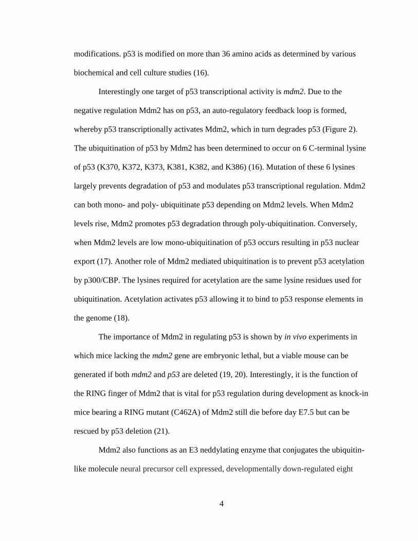
Interestingly one target of p53 transcriptional activity is mdm2. Due to the
negative regulation Mdm2 has on p53, an auto-regulatory feedback loop is formed,
whereby p53 transcriptionally activates Mdm2, which in turn degrades p53 (Figure 2).
The ubiquitination of p53 by Mdm2 has been determined to occur on 6 C-terminal lysine
of p53 (K370, K372, K373, K381, K382, and K386) (16). Mutation of these 6 lysines
largely prevents degradation of p53 and modulates p53 transcriptional regulation. Mdm2
can both mono- and poly- ubiquitinate p53 depending on Mdm2 levels. When Mdm2
levels rise, Mdm2 promotes p53 degradation through poly-ubiquitination. Conversely,
when Mdm2 levels are low mono-ubiquitination of p53 occurs resulting in p53 nuclear
export (17). Another role of Mdm2 mediated ubiquitination is to prevent p53 acetylation
by p300/CBP. The lysines required for acetylation are the same lysine residues used for
ubiquitination. Acetylation activates p53 allowing it to bind to p53 response elements in
the genome (18).
The importance of Mdm2 in regulating p53 is shown by in vivo experiments in
which mice lacking the mdm2 gene are embryonic lethal, but a viable mouse can be
generated if both mdm2 and p53 are deleted (19, 20). Interestingly, it is the function of
the RING finger of Mdm2 that is vital for p53 regulation during development as knock-in
mice bearing a RING mutant (C462A) of Mdm2 still die before day E7.5 but can be
rescued by p53 deletion (21).
Mdm2 also functions as an E3 neddylating enzyme that conjugates the ubiquitin-
like molecule neural precursor cell expressed, developmentally down-regulated eight

5
Figure 2: The Mdm2/p53 auto-regulatory feedback-loop
Under non-stressed conditions p53 transcriptionally activates Mdm2, which in turn
targets p53 for degradation. This feedback loop is a primary mechanism for regulating
p53 activity in normal cells.

6
(nedd8) to p53, which neutralizes p53 transcriptional activity (22). The nedd8 pathway
has been demonstrated to be crucial for viability of mice, C. elegans, and S. pombe (23-
25). In addition, the mammalian cell line TS-41, which has a temperature sensitive
mutation in the SMC gene (APP-BP1 in human) participates in multiple rounds
of DNA replication without entering a mitotic cycle (26). Interestingly, nedd8
conjugation has been shown to be upregulated in certain types of cancer and recently a
general inhibitor of the nedd8 pathway, MLN4924, has been developed (27). Inhibition
of neddylation in HCT 116 cells using MLN4924 leads to the same phenotype observed
in the TS-41 cell line. This phenotype of re-replication is known to induce DNA damage
and leads to apoptosis in human cancer cells and suppresses the growth of human tumors
in mouse xenografts.
The most well studied and first discovered substrate of nedd8 was Cdc53 in yeast
and Cul4a in humans. Both are member of the Cullin family and are neddylated at a
conserved lysine in their C-terminal domain. All yeast and mammalian cullins are now
known to be neddylated. The cullin family of proteins (Cul1, 2, 3, 4A, 4B, 5, 7) serves as
scaffolding proteins for multi-subunit ubiquitin E3 ligases. The cullins interact in their C-
terminal domain with a RING finger E3, either Rbx1 or Rbx2 and a substrate recognition
subunit is attached to the N-terminal region (28). This entire complex is needed for an
active ubiquitin ligase. The modification of cullins by nedd8 has been shown to increase
the ubiquitin activity of cullin ring ligases. This increase in ubiquitination activity is due
to a nedd8 dependent conformational change that increases Rbx binding of E2 and also
decreases the distance between E2 and the substrate (29). The cullins are responsible for

7
the ubiquitination of numerous proteins involved in cell cycle progression and cell
growth/survival (30).
While the cullins are the most studied, they are not the only proteins modified by
nedd8. Many other proteins have been shown to be neddylated. Relevant to this thesis is
the neddylation of p53 and Mdm2. There are two E3 ligases capable of conjugating a
nedd8 to p53, Mdm2 and FBXO11 (22, 31). Conjugation of a nedd8 molecule to p53
inhibits its transcriptional activity. This was shown as the TS-41 cells, which have a
temperature sensitive mutant of the Nedd8 Activating Enzyme (NAE), show higher levels
of p53 activity when grown at the restrictive temperature (39 C). Also a fusion protein
of p53 with a nedd8 attached to the C-terminal end resulted in lower activity as
determined by luciferase activity from the p21 promoter (31). The sites neddylated by
Mdm2 are lysines K370, K372, and K373 whereas FBXO11 neddylates K320 and K321
(22). The actual mechanism of p53 transcriptional inhibition by neddylation is still under
investigation. Unlike mono-ubiquitination of p53, which results in the movement of p53
from the nucleus to the cytoplasm, neddylation by itself has no effect on p53 cellular
compartmentalization (32). However expression of a nedd8 interacting protein, Nedd8
ultimate buster 1 (NUB1), has been shown to enhance cytoplasmic p53 through an
Mdm2/nedd8 dependent pathway (33). This mechanism cannot be fully responsible for
inhibition though, as neddylated p53 was still detectable in the nucleus, even with
overexpression of NUB1. Mdm2 itself has also been shown to be neddylated (22). This
neddylation modification results in an increase in Mdm2 protein half-life. This is
reversed when NEDP1 (a human nedd8-soecific protease) removes nedd8 from Mdm2.
8
Moreover, NEDP 1which can be induced by chemotherapeutics, and thus help to activate
p53 (34).
There are numerous splice variants of mdm2 mRNA and multiple isoforms of the
Mdm2 protein have been identified in tumors and normal tissues (35). Two of these
isoforms, p75 and p90, predominate in cells and migrate at 75 and 90 kDa on sodium
dodecyl sulfate polyacrylamide gels. The 90 kDa form of Mdm2 is transcribed from the
p53 responsive P2 promoter of Mdm2. This transcript results in a full length Mdm2
protein capable of regulating p53. Along with p53, the P2 promoter has been shown to be
responsive to AP-1, SP-1, and Smad3 transcription factors (11, 36, 37). The 75 kDa form
is produced from the constitutive P1 promoter independent of p53. This 75 kDa form
lacks the N-terminus of Mdm2 due to initiation of translation at two different internal
AUG codons (AA 62 and 102) rendering Mdm2 incapable of binding to p53. PTEN has
been shown to inhibit p75 Mdm2 transcription indirectly by modulating the activity of
unknown transcription factor(38). Meanwhile, the only transcription factor that has been
shown to induce expression from the P1 promoter is NF-κB (39). The biological
relevance for p75 Mdm2 is currently under investigation. Interestingly mdm2 transcripts
initiated from P1 undergo removal of exon 2 by splicing and retain exon 1; meanwhile p2
transcripts lack exon 1, and retain exon 2 (Figure 3).
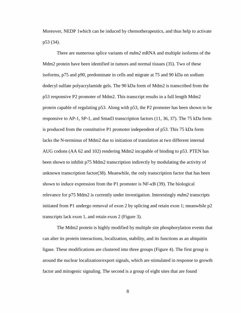
The Mdm2 protein is highly modified by multiple site phosphorylation events that
can alter its protein interactions, localization, stability, and its functions as an ubiquitin
ligase. These modifications are clustered into three groups (Figure 4). The first group is
around the nuclear localization/export signals, which are stimulated in response to growth
factor and mitogenic signaling. The second is a group of eight sites that are found

9
Figure 3: Organization of the human mdm2 gene
The mdm2 gene contains two unique promoters. P1 is a constitutive promoter, and P2 is
the p53-responsive promoter. The translation start codons are indicated for each
promoter. Initiation of translation from the P1 promoter results in an N-terminal truncated
Mdm2 protein.

10
Figure 4: Known phosphorylation sites on Mdm2
Phosphorylation sites are indicated by (P). Protein kinases (if known) and the effects of
phosphorylation events are indicated.

11
between amino acids 240-269. The final group is mostly clustered at the carboxyl-
terminus and becomes phosphorylated in response to DNA damage. The phosphorylation
sites around the bipartite NLS contain two Protein Kinase B (AKT/PKB) sites at 166 and
186. This phosphorylation event is triggered by insulin growth factor (IGF) and results in
the nuclear accumulation of Mdm2, which binds p53 and inhibits its transcriptional
activity (40). In the acidic domain, T216 of murine Mdm2 has been shown to be
phosphorylated by cyclinA/cdk. This modification results in stabilization of p53 and is
permissive of p19ARF binding (41, 42), a negative regulator of Mdm2. Casein Kinase 2
(CK2) phosphorylation of Mdm2 at S269 impairs Mdm2/Retinoblastoma (Rb) interaction
and allows the binding of Mdm2 to the basal transcription factor TFII250 (43). Also
under basal growth conditions, it has been suggested, that a group of serines located in
the acidic domain are phosphorylated. Through use of mutation analysis it has been
suggested that these phosphorylated serines promote Mdm2-mediated p53 turnover,
although the kinases responsible for these events are still undefined (Figure 4).
Many studies have focused on Mdm2 phosphorylation in response to genotoxic
stress. Ataxia telangiectasia mutated (ATM) has been shown to phosphorylate Mdm2 in
response to DNA damage at S395 and block nuclear export of p53 (44). Additional ATM
sites at S386, S425, and S428 all inhibit p53 degradation by preventing Mdm2 E3
oligomerization (45). Ataxia telangiectasia and Rad3-related protein (ATR)
phosphorylates Mdm2 at S407 and also prevents p53 nuclear export under certain stress
conditions (46). An additional phosphorylation site triggered by DNA damage at serine
17 is phosphorylated by DNA protein kinase (DNA-PK), an enzyme activated in

12
response to DNA double strand breaks. This phosphorylation event prevents Mdm2
interaction with p53 in vitro and activates p53 transcriptional activity (Figure 4) (47).
Interestingly there are only two known tyrosine kinases for Mdm2.The first, c-
Abl, phosphorylates Mdm2 at Y394 and results in an increase of apoptosis by enhanced
stability of p53. This increase in p53 stability is due to a c-Abl dependent promotion of
Mdm2-Mdmx heterodimers, which enhances the degradation of both Mdm2 and Mdmx
(48). c-Abl also phosphorylates Mdm2 at Y276 and results in enhanced p19ARF binding,
sequestering Mdm2 from p53 (49). The other tyrosine kinase is ErbB-4. Phosphorylation
of Mdm2 by ErbB-4 results in stabilization of p53 and increases Mdm2 ubiquitination
(50). However the tyrosine(s) in Mdm2 have yet to be identified (Figure 4).
While the most studied function of Mdm2 is attributed to its regulation of p53,
Mdm2 has been shown to engage in many p53 independent activities. Mdm2 has been
implicated in cell cycle control, differentiation, DNA repair, basal transcription, and other
processes. Some of Mdm2 p53-independent protein-protein interactions are detailed in
Table 1.
1.2 c-Src
v-Src was originally isolated as the transforming agent in the Rous sarcoma virus
(21). Its cellular counterpart c-Src is a member of the Src Family Kinase, a family of
membrane associated non-receptor tyrosine kinases including: c-Src, c-Yes, Fyn, Lyn,
Lck, Hck, Blk, Fgr, and Yrk. While most of the family members are expressed in the
hematopoietic origin, c-Src shows more ubiquitous expression and is involved in many

13
Mdm2 Binding
Partner Results of Mdm2 Binding
p73
Inhibits p73 transcriptional activity (does not
degrade)(51, 52)
p19(ARF) Blocks Mdm2/p53 binding(42)
Rb Inhibits Mdm2-E2F1 binding(53)
E2F1 Stabilizes E2F1 protein levels (54)
PCAF Ubiquitinates and degrades Mdm2(55)
JMY
Transcriptional activator that is degraded by Mdm2
(56)
Histone H2B Mdm2 mono-ubiquitinates H2B
L5 Inhibits Mdm2 ligase activity (57)
L11 Inhibits Mdm2 ligase activity (58)
L23 Inhibits Mdm2 ligase activity (59)
PML Sequesters Mdm2 to nucleolus (60)
Nbs1
Mdm2/Nbs1 binding inhibits onset of DNA
repair(61)
DNA Polymerase ε
Stimulates DNA Polymerase ε enzymatic
activity(62)
E-Cadherin Mdm2 ubiquitinates and degrades E-cadherin (63)
HIF-1α Enhances HIF-1α transcriptional activity(64)
NUMB Mdm2 ubiquitinates and degrades Numb(65)
SCF(beta-TRCP) Ubiquitinates and degrades Mdm2(66)
Table 1: Mdm2 binding partners

14
cellular processes including proliferation, survival, motility, invasiveness, and
angiogenesis (22).
c-Src contains an amino-terminal membrane localization sequence in the SH4
domain that requires myristylation for localization. c-Src also contains Src homology
domain 2 (SH2) and Src homology 3 domain (SH3) that mediate protein-protein
interactions, a tyrosine kinase domain, and a regulatory sequence at the carboxyl-terminal
end (Figure 4). SH3 domains generally bind to proline rich motifs with the minimal
consensus P-x-x-P (23), while SH2 domains bind phosphorylated tyrosines (24). c-Src
activity is regulated through phosphorylation and intra-molecular interactions.
Phosphorylation of c-Src by C-terminal Src Kinase (CSK) at Y530 results in inactivation
due to intermolecular binding between phosphorylated Y530 and the SH2 domain,
resulting in a “closed” conformation of the protein (25, 26) (Figure 5). The v-Src protein
has a C-terminal deletion resulting in loss of inhibitory phosphorylation on Y530.
Activation of c-Src can result from dephosphorylation of the Y530 by a protein
phosphatase. Protein tyrosine phosphatase (PTP)-alpha, PTP1, PTP1B, and SH2-
containing protein tyrosine phosphatase-1 (SHP-1) have all been implicated in c-Src
activation (27, 28). c-Src activity can be augmented by higher intermolecular binding of
the SH2 and SH3 between cellular ligands, such as platelet-derived growth factor
receptor (PDGFR) or focal adhesion kinase (1) (29, 30). This activation of c-Src results in
its open conformation with greater access to the SH2 and SH3 binding domains. For full
activation, c-Src must undergo auto-phosphorylation at Y419 in its catalytic domain.
c-Src is rarely mutated in human cancers. Only a small set of colon cancer
patients have been found to have an activating mutation similar to v-Src. Elevated levels

15
Figure 5: Structure and activation of c-Src
c-Src contains four homologous domains. The SH4 contains a myristoylation sequence
and supports plasma membrane binding. The SH3 domain is a proline rich sequence that
binds and interacts with other proteins. The SH2 domain recognizes phosphorylated
tyrosine residues and regulates binding to other proteins. The SH3 and SH2 domains also
function in intra-molecular interactions. The Y419 is located in the kinase domain and is
auto-phosphorylated during activation. c-Src activation is regulated by the
phosphorylation status of Y530. When phosphorylated, c-Src is in an inactive
conformation due to p-Y530 binding to the internal SH2 domain. When
dephosphorylated the conformation of c-Src opens allowing for activation.

16
of c-Src protein or an increase in activity are more commonly observed in human
cancers. This is seen in cancers of the lung, breast, colon, and skin (67-69). Even though
c-Src is involved in multiple pathways important for tumorigenesis and is seen at
elevated levels in human cancers, c-Src itself is not a strong transforming oncogene (70).
This has led to the hypothesis that c-Src can help facilitate the oncogenic roles of other
proteins rather than being a strong autonomous transforming agent. To aid in
tumorigenesis c-Src interacts with numerous tyrosine receptor kinases and other proteins
implemented in cancer progression including PDGFR, vascular endothelial growth factor
(VEGFR), epidermal growth factor receptor (EGFR), human epidermal growth factor
receptor 2 (HER2), human epidermal growth factor receptor 3 (HER3), signal transducer
and activator of transcription (STATs), heteromeric G proteins, cyclin D and E, and FAK
(71). This diverse list shows c-Src integration in numerous cellular processes and how
many different pathways c-Src may augment for cancer development.
1.3 Cancer Metastasis
Metastasis is a multi-staged process that cancerous cells undergo to relocate from
their primary location to a distant site. Metastasis usually correlates with a poor prognosis
and often results in death due to lack of treatment options. For metastasis to occur, tumor
cells must first invade into the surrounding tissue of the primary site. This is followed by
entrance into the circulation system, either through the lymph nodes or blood, where the
tumor cell must survive until it can find a distal site suitable for growth (Figure 6).

17
Figure 6: The tumor metastatic process
Metastasis of a tumor to a distal tissue involves a three-step process: First, tumor cells
must be able to leave the primary tumor site and invade the vasculature. Second, they
must survive in the bloodstream until they can arrest and extravasate into surrounding
tissue. Finally they must interact with the microenvironment to establish metastatic
colonization.

18
For a tumor cell to enter into the primary site around the tissue, it must lose
contact from its surrounding cells and to the extra-cellular matrix. The contacts between
cells are usually mediated by the cell attachment proteins of the cadherin family, which
bind cells together through their extracellular domains (72). Loss of E-cadherin is a
hallmark of epithelial-mesenchymal transition (EMT), which is defined by loss of
epithelial characteristics including apical-basal polarity, tight junctions, expression of E-
cadherin, and decreased cell mobility. The loss of these characteristics are combined with
the gain of mesenchymal phenotype markers such as increases in vimentin and myosin
expression, loss of polarity, and an increase in cellular mobility (73). Tumor cells are
bound to the extra cellular matrix (ECM) through heterodimers of one alpha and one beta
integrin protein. The alpha family consists of 18 members while the beta has 8. Each
heterodimer binds to a specific protein of the ECM (74). For a tumor cell to begin to
invade into surrounding tissues it must release its attachment to cells and ECM. This is
accomplished through the use of proteases that degrade the ECM and attachments. The
family of matrixmetalloproteinases (MMPs) is responsible for the breakdown of
extracellular matrix. MMPs are normally secreted in an inactive form and are activated
upon cleavage by extracellular enzymes (75).
Tumors first need to recruit a blood supply to aid in the metastatic process. The
formation of new blood vessels from pre-existing ones is termed angiogenesis.
Angiogenesis is required in order to support the growth and survival of tumors by
delivering oxygen and other nutrients and metabolites to the tumor cells (76).
Angiogenesis is simulated by pro-angiogenic factors that must counteract anti-angiogenic
factors. Thus, the angiogenic switch will commence when the pro-angiogenic factors

19
outnumber the inhibitors. Important to this thesis are the roles of the pro-angiogenic
molecule vascular endothelial growth factor (VEGF) and the anti-angiogenic molecule
Maspin. The VEGF and the VEGF receptor (VEGF-R) pathway is one of the most
studied pro-angiogenic factors. Multiple signaling networks are activated in response to
VEGF/VEGF-R signaling and result in endothelial cell survival, mitogenesis, migration,
and differentiation. VEGF also increases vascular permeability and initiates the
mobilization of endothelial progenitor cells from the bone marrow into the peripheral
circulation. The permeability induced by VEGF leads to access for the pro-angiogenic
factors to enter the vessels and facilitate angiogenesis. VEGF activates its angiogenic
switch through binding to one of three VEGF receptors. VEGF-R1 is responsible for
most physiologic and developmental neo-vascularization (77). VEGF-R2, meanwhile,
mediates the majority of the downstream effects of VEGF in angiogenesis, including
microvascular permeability, endothelial cell proliferation, invasion, migration, and
survival (78). VEGF-R3 is a receptor tyrosine kinase that is expressed mainly during
development in the embryonic vasculature but is only expressed in lymphatic endothelial
cells in adults (79). Due to its role in numerous cancers and angiogenesis, the
VEGF/VEGFR pathway has been targeted clinically with drugs that inhibit both VEGF
and VEGF-R.
Due to the prominent role VEGF plays in angiogenesis, understanding VEGF
regulation is very important. VEGF is regulated in numerous ways including by
oncogenes/tumor suppressor, transcription factors, and environmental factors. Important
for this thesis is the regulation of VEGF by decreases in oxygen concentration, termed
hypoxia, and the transcription factor hypoxia inducible factor 1-alpha (HIF-1α). In

20
response to hypoxic conditions, cells must adapt by changing metabolic, bioenergetic,
and redox demands for oxygen. To support these changes, cells must lower demands for
growth, conserve energy, and induce survival and pro-angiogenic factors. While many
pathways regulate the hypoxic response, including mammalian target of rapamycin
(mTOR) and the unfolded protein response, the main transcription factor to mediate a
transcriptional response to hypoxic conditions is HIF-1 α. The transcriptional response
driven by HIF-1α is dependent on its ability to bind canonical DNA sequences, called
hypoxia response elements (HRE), in the promoter of target genes. HIF-1α is a member
of the bHLH-PAS family of transcription factors. The active transcription factor consists
of a dimer combining a HIF-α and HIF-β subunit. The regulation of the HIF transcription
factor resides in the stability of the α-subunit, of which 3 are known. The α-subunit is
constitutively expressed but in the presence of oxygen the α-subunit is ubiquitinated and
degraded by the von Hippel Lindau (VHL) protein through the 26S proteasome. This
degradation takes place due to hydroxylation of HIF-1α under normal oxygen conditions.
These hydroxylation events, performed by specific prolyl hydroxylases (PHD), occur on
proline 402 and 564 in the oxygen dependent degradation domain of HIF-1α (80). These
proline modifications allow for the binding of VHL, which serves as the substrate
recognition component of an E3 ubiquitin ligase complex (Figure 7) (81). Under oxygen
deprivation, the proline hydroxylases are inactive resulting in a stabilization of the HIF-
1α subunit. Once stabilized HIF-1α subunits travel to the nucleus and dimerize with the
HIF-β subunits and transcribe at least 150 genes that regulate cell metabolism, survival,
motility, basement membrane integrity, angiogenesis, hematopoiesis, and other cellular
functions (Figure 6) (82).

21
Figure 7: HIF-1α/VHL pathway
Under normoxic conditions HIF-1α is hydroxylated on proline 402 and 564 by prolyl-
hydroxylases (PHD). This allows for the binding of VHL which ubiquitinates and
degrades HIF-1α. Under hypoxic conditions, PHD’s are not active resulting in stable
HIF-1α, which can interact with HIF-1β and activate hypoxic response genes.

22
One anti-angiogenic factor counteracting VEGF is the p53 induced tumor
suppressor Maspin. The maspin gene was discovered by subtractive hybridization by its
expression of mRNA in normal but not tumor-derived mammary epithelial cells (83). It
has been classified as a member of the serpin protease inhibitors family and further
defined as a member of the ov-serpin subfamily. This subfamily consists of thirteen
members, including plasminiogen activator inhibitor 1 and 2, which are involved in
angiogenesis, apoptosis, and embryogenesis. All ov-serpin proteins have serine protease
inhibitor function, except for maspin (84). This inhibition is accomplished through a
conformational change in the serpin reactive site loop (RSL) upon binding to the protease
resulting in a covalent bond between the RSL and the catalytic site of the protease.
Maspin however does not appear to be a protease inhibitor. Maspin RSL is not conserved
and is shorter than all other family members and does not undergo conformational
changes (85). Interestingly, maspin is the only ov-serpin whose deficiency is embryonic
lethal in mice (86).
The tumor suppressor functions of Maspin are attributed to its roles in cell
migration/invasion, angiogenesis, and apoptosis (87-89). For angiogenesis to occur,
vessel endothelial cells must be activated by factors (such as VEGF) that allow it to
escape from the vessel walls. Once free, these endothelial cells can participate in the
formation of new vasculature in tumors. Cell adhesion and migration regulate the release
of endothelial cells from the original vessel. Maspin regulates this process by increasing
endothelial adhesion to fibronectin, lamin, and collagen. This binding is increased
through upregulation of integrin-linked kinase (90) and FAK signal transduction
pathways that regulate attachment and focal adhesion disassembly (87). This prevents the
23
release and migration of endothelial cells from established vasculature. This is validated
in vivo as treatment with recombinant Maspin resulted in a loss of vessel formation, as
determined by CD31 staining, in tumors of nude mice. Also, Maspin effectively blocked
neovascularization mediated by basic fibroblast growth factor (bFGF) in rat corneas (91).
For its role in apoptosis, maspin mainly functions through the down-regulation of the
anti-apoptotic protein Bcl-2, although the exact mechanism is still unknown. This
downregulation of Bcl-2 allows Bax, an apoptotic inducing protein, to become active thus
increasing mitochondria membrane permeability (92, 93).
Once angiogenesis has taken place and the tumor cells are able to access the
vasculature, they must be able to survive in this new environment. This survival requires
evading immune cells and avoiding anoikis, cell death due to detachment from a
substratum. Beyond survival, tumor cells must at some point stop circulating (arrest) to
allow them to escape from the vessels. Arrest is mediated by tumor cells through
regulation of their own integrins and also by binding to coagulation factors (94, 95). It
has been proposed that vessel endothelial cell selectin molecules may also aid in tumor
cell arrest (96). Also, some cancer cells arrest due to size restriction of the capillaries.
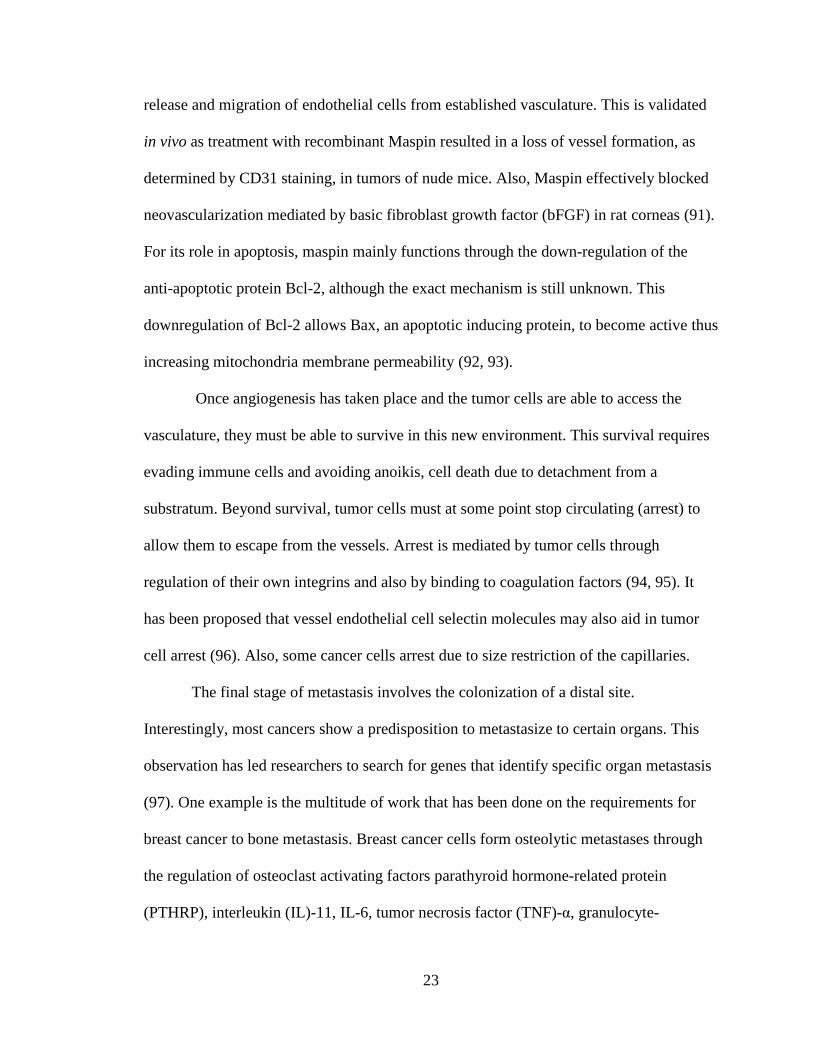
The final stage of metastasis involves the colonization of a distal site.
Interestingly, most cancers show a predisposition to metastasize to certain organs. This
observation has led researchers to search for genes that identify specific organ metastasis
(97). One example is the multitude of work that has been done on the requirements for
breast cancer to bone metastasis. Breast cancer cells form osteolytic metastases through
the regulation of osteoclast activating factors parathyroid hormone-related protein
(PTHRP), interleukin (IL)-11, IL-6, tumor necrosis factor (TNF)-α, granulocyte-

24
macrophage colony-stimulating factor (GM-CSF), and receptor activator of nuclear factor
kappa-B ligand (RANK-L). These factors are essential for the growth of osteolytic
lesions in the bone environment, but would offer no advantage to another metastatic site
or the tumor (98). Interestingly, c-Src has also been implicated in breast to bone
metastasis through its regulation of bone remodeling. c-Src knockout mice develop
osteopetrosis due to deficiency in bone remodeling (99). Mice injected with MDA-231
breast cancer cells overexpressing c-Src have more osteolytic bone metastases than
control mice, linking c-Src to bone colonization (100).

25
2. Materials & Methods
2.1 Cell Culture
All mammalian cells were cultured at 37 °C in a humidified incubator with 5%
CO2 in Dulbecco's modified Eagle's medium (DMEM) high glucose supplemented with
10% fetal bovine serum and penicillin-streptomycin solution. shGPF and shMdm2 TMD-
231 cells were kept under constant selection with puromycin (2 g/ml).
2.2 Transfection
Cells were transfected using the calcium phosphate method or Lipofectamine
(Invitrogen). For calcium phosphate method plasmid DNA (1-5 µg), water (130 µl), and
2M CaCl2 (17 µl) where mixed to a total volume of 150 l. This mixture was then added
dropwise to 150 l of 2X HBS (275 mM NaCl, 1.5 mM Na2HPO4, 50 mM HEPES, pH
7.0) while vortexing. Solution was incubated for 30 min at RT before adding dropwise to
cells. This (300 l) amount is used for an individual well of a 6 well plate. For 10 cm
dishes volumes are raised to 500 l each, for a total of 1 ml transfection reagent. DNA
concentration was also raised to 5 µg. Lipofectamine transfections were done according
to manufacturer’s protocol.
2.3 Generation of shGFP and shMdm2 TMD-231 cell lines
Retroviruses encoding shRNA to GFP and Mdm2 (pLKO.1 vector) were
packaged in 293T cells together with second-generation packaging constructs, pCMV-
dR8.74 and pMD2G. Supernatant media containing virus were collected at 36 to 48
hours, supplemented with 4 μg/mL polybrene, filtered through a 0.45-μm filter, and

26
added to cells overnight. Uninfected cells were removed by selection with puromycin (2
μg/mL).
2.4 Luciferase Assays
For the luciferase assays, H1299 and MCF7 cells were transfected with PG13-
Luc, Mdm2-P2-Luc, Maspin-Luc, Mutant Maspin-Luc (MT1) or HRE-Luc along with
Myc-LacZ for determination of β-galactosidase activity, using the calcium phosphate
method. Reporter activity was normalized to β-galactosidase activity. Data generated
were done in triplicate and standard deviation was calculated from the mean.
2.5 Glutathione S-Transferase-pull down assay
GST-SH3-Src and GST alone for control were incubated with glutathione-
sepharose beads that had been pre-washed in TEN100 Buffer (20 mM Tris, pH 7.4, 0.1
MmM EDTA and 100 mM NaCl) for1 hr at 4 C. After GST-proteins were immobilized
the complexes were spun down and washed four times for 15 min each with TEN100.
After washing, recombinant His-Mdm2 was incubated with immobilized GST-SH3-Src
or GST for 1 hr at 4 C. After complexes formed, they were spun down and were washed
four times in NTEN Buffer (.5%NP40, 1 mM EDTA, 20 mM Tris, pH 7.4, 1 M NaCl).
The bound proteins were eluted in SDS-Loading Buffer and western blot analysis was
performed.
27
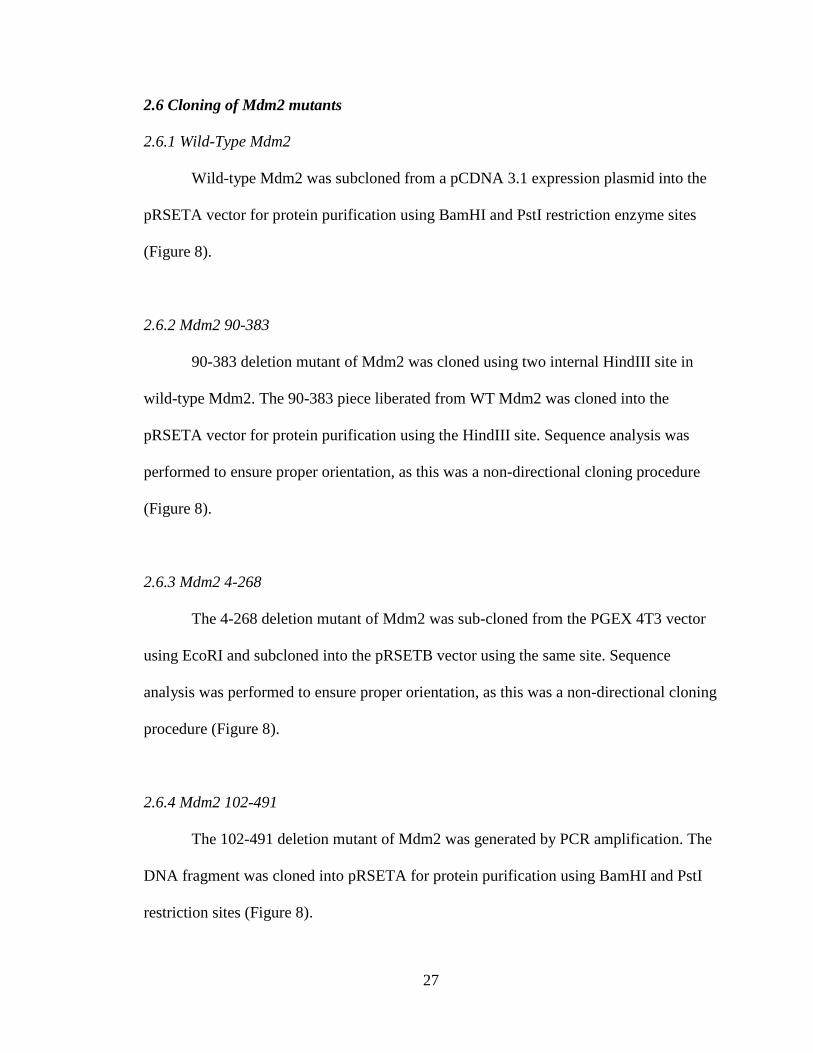
2.6 Cloning of Mdm2 mutants
2.6.1 Wild-Type Mdm2
Wild-type Mdm2 was subcloned from a pCDNA 3.1 expression plasmid into the
pRSETA vector for protein purification using BamHI and PstI restriction enzyme sites
(Figure 8).
2.6.2 Mdm2 90-383
90-383 deletion mutant of Mdm2 was cloned using two internal HindIII site in
wild-type Mdm2. The 90-383 piece liberated from WT Mdm2 was cloned into the
pRSETA vector for protein purification using the HindIII site. Sequence analysis was
performed to ensure proper orientation, as this was a non-directional cloning procedure
(Figure 8).
2.6.3 Mdm2 4-268
The 4-268 deletion mutant of Mdm2 was sub-cloned from the PGEX 4T3 vector
using EcoRI and subcloned into the pRSETB vector using the same site. Sequence
analysis was performed to ensure proper orientation, as this was a non-directional cloning
procedure (Figure 8).
2.6.4 Mdm2 102-491
The 102-491 deletion mutant of Mdm2 was generated by PCR amplification. The
DNA fragment was cloned into pRSETA for protein purification using BamHI and PstI
restriction sites (Figure 8).

28
Figure 8: Restriction sites used for cloning of Mdm2 truncation mutants
All mutants were cloned in the designated pRSET vector for protein purification. HA
represents a human influenza hemagglutinin tag.

29
2.7 His-ubiquitn and His-Nedd8 pulldowns
H1299 cells were transfected with His-ubiquitin or His-nedd8 and other plasmids
using calcium phosphate method. Forty-eight hr after transfection cells were lysed in 1 ml
of 6 M guanidinium–HCl, 0.1 M Na2HPO4/NaH2PO4, 0.01 M Tris–HCl pH 8.0 plus 5
mM imidazole and 10 mM -mercaptoethanol. After sonication, the lysates were mixed
with 30 l of Ni2+
-NTA-agarose beads (Qiagen) prewashed with lysis buffer and
incubated for 2 hr at room temperature. The beads were successively washed for 15 min
in each of the following: 6 M guanidinium–HCl, 0.1 M Na2HPO4/NaH2PO4, 0.01 M
Tris–HCl pH 8.0 plus 10 mM -mercaptoethanol; 8 M urea, 0.1 M Na2HPO4/NaH2PO4,
0.01 M Tris–HCl pH 8.0, 10 mM -mercaptoethanol; 8 M urea, 0.1 M
Na2HPO4/NaH2PO4, 0.01 M Tris–HCl pH 6.3, 10 mM -mercaptoethanol (Buffer A) plus
0.2% Triton X-100; buffer A and then buffer A plus 0.1% Triton X-100. After the last
wash with buffer A the beads were eluted with 200 mM imidazole in 5% SDS, 0.15 M
Tris–HCl pH 6.7, 30% glycerol, 0.72 M -mercaptoethanol. Elutes were subjected to
sodium dodecyl sulfate polyacrylamide gel electrophoresis (SDS–PAGE) and western
blotting.
2.8 Purification of recombinant proteins
For purification of His-tagged proteins a 50 ml culture of BL-21 Escherichia coli
was grown overnight at 37 C at 25,000 rpm. On the day of purification the culture was
reseeded in 200 ml of Lysogeny broth (LB) to a 0.1 optical density (OD) value. The
bacteria were allowed to grow to 0.6 OD before the induction with 100 mM of Isopropyl-
β-D-thio-galactoside (IPTG). Induction was performed at 24 C for four hr. After

30
induction cells were spun down at 6000 rpm for 10 min and resupsended in Buffer A (25
mM Hepes, 0.2% TX-100, 5 mM DTT, 1 M KCL) and sonicated 4 times for 30 sec on
ice. After centrifugation at 10,000 rpm for 25 min at 4 C, the supernatant was removed
and 40 l of pre-washed (Buffer A) Ni-NTA slurry (Qiagen) was added. Binding was
allowed to occur for 30 min at 4 C. Complexes were spun down at 1500 rpm for 5 min
and then washed six times for 15 min in Buffer B (Buffer A+10 mM imidazole). Proteins
were then for eluted in Buffer C (Buffer A+300 mM imidazole) and dialyzed into
Dialysis Buffer (50 mM HEPES pH 7.5, 100 mM NaCl, 10% glycerol, 1 mM DTT).
2.9 In vitro kinase reactions
Src and Abl kinase reactions were performed at 37 °C for 30 min in kinase buffer
(25 mM Tris, pH 7.4, 10 mM MgCl2, 1 mM MnCl2, 0.5 mM DTT, 10 μM ATP) using 0.1
μg of Src (Calbiochem) or Abl (Invitrogen).
2.10 In vitro ubiquitination reactions
For in vitro ubiquitination assays; 500 ng of p53 was incubated with 50 ng E1
(Boston Biochem), 200 ng Ubch5a (Boston Biochem), and 1 g ubiquitin (Boston
Biochem) in the presence of 500 ng phosphorylated or unphosphorylated Mdm2.
Reactions were performed for 2 hr at 37 C.
2.11 Protein analysis, immunoprecipitation, and Western blotting
Whole cell extracts lysates were prepared in Nonidet P-40 lysis buffer (25 mM
Tris, pH 8.0, 150 mM NaCl, 0.5 mM EDTA, 0.5 mM EGTA, 1% Nonidet P-40, 1 mM

31
sodium orthovanadate, 1 mM dithiothreitol (DTT)) and supplemented with protease
inhibitor mixture set III (Calbiochem) at 1:100 and incubated on ice for 30 min. Debris
was collected by centrifugation, and supernatant collected. Protein concentration was
determined using Bradford assay. For immunoprecipitation assays 500 µg of lysates were
incubated over night with antibody at 4 °C in 700 μl of PBS. 20 l of pre-washed Protein
A/G Plus Agaraose (Santa Cruz Biotechnology) was added to the mixture and incubated
an additional 4 hr at 4 °C, precipitates were washed three times in PBS, and samples were
resuspended in SDS loading buffer.
2.12 Cell cycle analysis
TMD-231 shGFP or shMdm2 cells were serum starved for 24 hr. After serum
starvation, cells were given 1% serum for 0, 8 and 24 hr. Cells were then harvested and
resuspended in 1.2 mL of PBS. 3 mL of 100% ethanol was then added dropwise to cells,
while vortexing. The cells were then fixed in this 70% ethanol solution for 30 min. After
fixing, cells were spun at 2200 rpm for 10 min and washed twice in 15 mL of PBS.
Pellets were resuspened in 4.5 mL of PBS and 0.5 mL RNase stock (1 mg Rnase/1 mL
H2O) and incubated for 30 min at 37º C. After incubation cells were washed twice in PBS
and resuspened in 2 mL propidium iodide (PI) stain solution (10 mg PI/100 mL PBS) at a
final concentration of 1x^6 cells/mL. The cells were analyzed on a flow cytometer.
ModFit analysis determined the percentage of cells in a specific stage of the cell cycle.

32
2.13 Plating Efficiency
TMD-231 shGFP and shMdm2 cells were counted and plated from a single cell
suspension for clonogenic survival. Cells grew for 14-21 days at 37°C. Cells were then
fixed with a 3:1 methanol/acetic acid mixture and stained with crystal violet. Colonies
with ≥50 cells were included in the survival analysis. Plating efficiency (PE) was
determined by number of colonies counted divided by number of cells plated.
2.14 Cell Attachment Assay
Six-well plates were coated with 5 μg/ml fibronectin at 37 °C for 1 hr, washed
twice with PBS, and then incubated with 1% bovine serum albumin at 37 °C for 1 hr.
Plates were then washed twice with PBS. 2x10^4
TMD-231 shGFP or shMdm2 cells were
plated with 2 ml of culture medium per well and incubated in the 37 °C, incubator for 30
min. The culture medium was removed and cells were washed twice with PBS,
trypsinized and counted on a Beckman Coulter Cell Analyzer.
2.15 Matrigel Invasion Assay
2.5x10^4
cells were seeded in into each well of matrigel chambers. 5% serum was
used as the chemoattractant. The chambers were incubated for 22 hr in a humidified
tissue culture incubator, at 37º C, 5% CO2. After incubation the media was removed and
the surface of the membrane was scrubbed using a cotton tip swab, to ensure removal of
non-invading cells. After scrubbing, the membranes were removed from the chambers
and stained in methanol for 2 min followed by 1% toludine blue for 2 min. The chambers
were counted at magnification of 20X. Multiple fields for each set were counted and the

33
experiment was done in triplicate. Percent invasion was calculated using the following
equation.
2.16 Assessment of tumorigenicity in vivo
The Indiana University In Vivo Therapeutics Core was responsible for all animal
work. 7.5x10^5
cells of either TMD-231 shGFP or ShMdm2 cells were injected into the
mammary fat pad of γ-null NOD-SCID mice (n=10 per group). All mice developed
tumors. Tumor growth was calculated by volumetric analysis using calipers. The tumors
were allowed to grow to an average volume of 600 mm.
2.17 Tissue preparation and staining
The Indiana University Pathology Core performed all tumor preparation and
staining for hematoxylin & eosin (H&E) and CD31.
2.18 Lung metastasis evaluation
The right and left lobe of the lungs from each mouse was stained with H&E and
mounted on a slide. The entire lung of each mouse was scored for metastases. A
metastatic legion was defined as ≥5 tumor cells.
34
3. c-Src phosphorylates and switches Mdm2 to a neddylating enzyme
3.1 Introduction
As previously described, c-Src is one of the 9 members of the Src-family kinases.
It is a cytoplasmic non-receptor tyrosine kinase that is a critical initiation site for multiple
signal transduction pathways and interactions.
One of the downstream effectors of c-Src is the survival factor Akt. Akt has been
shown to phosphorylate many substrates to promote cell survival including Mdm2 (40,
101). Mdm2 undergoes nuclear translocation in response to Akt phosphorylation at serine
166 and 186 (40). Once in the nucleus Mdm2 binds to the tumor suppressor p53 and
inhibits its transcriptional activity along with functioning as an E3 ubiquitin ligase to
signal nuclear export and proteasomal degradation. While normally kept at low levels,
p53 is stabilized through post-translational modifications to both itself and Mdm2 in
response to genotoxic stress (102). While the ubiquitin ligase activity of Mdm2 is
important for regulating p53 activity, other mechanisms are currently under investigation.
Interestingly Mdm2 can regulate p53 function by conjugation of nedd8. This
modification results in a transcriptionally inactive p53 but what regulates Mdm2
neddylating activity is still unknown (22).
In the tumor microenvironment there are numerous growth factors and cytokines
secreted from various invading cells and the stroma. This signaling leads to p53 levels
that are generally not lower but elevated. This suggests that the Mdm2-mediated
destabilization of p53 was not functional. Here we examined if Mdm2 neddylation
activity could be regulated in response to growth factor activated signaling cascades.
Here we show a novel Mdm2/c-Src interaction that results in the phosphorylation of

35
Mdm2 at Y281 and Y302. These phosphorylation sites are distinct from the previously
reported sites for c-Abl. This novel phosphorylation event increases the half-life of
Mdm2. Analysis of the enzymatic activity of Mdm2 shows that Mdm2 is unable to be
loaded with ubiquitin and thus did not conjugate ubiquitin to p53. In contrast, Src-
phorphorylated Mdm2 resulted in loading of nedd8 to itself and to p53. This switch of
Mdm2 enzymatic activity results in inactive but elevated levels of p53. This Src-mediated
activation of the neddylating activity of Mdm2 provides a mechanism for inactive-stable
p53 in cultured cells and tumor cells.
3.2 Results
Mdm2 is a substrate for the non-receptor kinase c-Abl under genotoxic stress
conditions (103). c-Abl contains an SH3 domain, which binds to partner proteins
containing an SH3 binding domain (P-x-x-P). Mdm2 contains two SH3 P-x-x-P binding
domains, so we investigated whether Mdm2 could bind to other tyrosine kinases that
have an SH3 domain. Recombinant Mdm2 was incubated on a SH3 domain array from
Panomics. Mdm2 bound to c-Abl, Abl2, c-Src and Hck-specific SH3 domains (Table 2).
Due to the prominent role of c-Src in tumorigenesis, we pursued this interaction
with Mdm2 further. To define specific Mdm2 domains required for SH3-Src binding, a
series of His-tagged recombinant Mdm2 truncation mutants were made (Figure 9A).
These Mdm2 mutants were incubated in a GST-pulldown assay with either GST or the
GST-SH3 domain of Src (SH3-Src). All Mdm2 mutants bound the SH3-Src domain, but
not by GST alone, revealing the portion of Mdm2 needed for the Src interaction is

36
Table 2: SH3 domain array
Recombinant Mdm2 was incubated with an SH3 domain array (Panomics) containing
SH3 domains of indicated proteins. Unbound Mdm2 was washed away and western
analysis performed for Mdm2. (+) indicates a positive interaction.

37
Figure 9: c-Src binds Mdm2 between aa90-268
(A) Schematic of recombinant Mdm2 truncation mutants. (B) Western blot analysis of
GST and Mdm2. GST-pulldown assay was done using recombinant GST or GST-SH3-
Src and recombinant Mdm2 proteins.

38
between residues 102-268 (Figure 9B). This is a region that contains an SH3 binding
domain. To further validate this interaction and determine if it would occur in vivo, an
immunoprecipitation of endogenous Src from MCF7 cells was performed and shows that
Mdm2 and c-Src are in a complex (Figure 10).
Since our data indicate that the non-receptor tyrosine kinase c-Src interacts with
Mdm2, we hypothesized that Mdm2 could be a substrate for tyrosine phosphorylation by
c-Src. Mdm2 is a highly phosphorylated protein and yet only two tyrosine kinases have
been identified to phosphorylate Mdm2 (49, 50, 103). The Mdm2 protein has 14 tyrosines
residues, 3 of which are already identified as c-Abl sites (Figure 11). To determine if c-
Src could phosphorylate Mdm2 an in vitro kinase reaction was performed using 32
P γ-
labeled ATP. Mdm2 was incubated alone, with c-Src or with c-Abl as a positive control.
As expected, Mdm2 was phosphorylated by c-Abl. Interestingly c-Src also
phosphorylated Mdm2, providing evidence that Mdm2 is a substrate for Src. (Figure 12).
To determine which tyrosine(s) is targeted for phosphorylation by c-Src another
in vitro kinase reaction was performed using the Mdm2 truncation mutants (Figure 9A).
The different mutants were incubated with c-Src and then immunoblot analysis was
performed using an anti-phosphotyrosine antibody (4G10). The blot was then stripped
and re-probed for Mdm2 (SMP14). Comparison of the blots revealed that the WT, 102-
491, and 90-383 were all effectively phosphorylated by c-Src (Figure 13A.). However,
the Mdm2 truncation mutant 4-268 was unable to be phosphorylated by c-Src as shown
by the outline squares (Figure 13A). Of note the 4-268 mutant of Mdm2 still contains a
SH3 binding domain and was able to bind to SH3-Src, thereby eliminating the possibility
that the loss of tyrosine phosphorylation was a result of Src not binding to Mdm2. Thus,

39
Figure 10: Mdm2 and c-Src interact in vivo
Western blot of immunoprecipitation of endogenous c-Src from MCF7 cell extracts was
followed by western blot analysis of c-Src and Mdm2.

40
Figure 11: Schematic of tyrosines in Mdm2 Mdm2 contains 14 tyrosines. There are three known c-Abl phosphorylation sites (P) in
Mdm2.

41
Figure 12: c-Src phosphorylates Mdm2 in vitro
Autoradiograph of 32
P incorporation in an in vitro kinase reaction of Mdm2 (p-Mdm2) by
c-Src. c-Abl phosphorylation of Mdm2 and c-Abl/c-Src autophosphorylation were used
as positive control.

42
Figure 13: c-Src phosphorylates Mdm2 at Y281 and Y302
(A) Western blot analysis of Mdm2 and phospho-tyrosine (pTyr) of in vitro
phosphorylation reaction on truncated Mdm2 proteins. Square outline highlights the
inability of truncation mutant 4-268 to be phosphorylated by c-Src. (B) Tyrosine to
phenylalanine mutants to each site Y281F and Y302F or in combination, Y281-302F,
were generated in the 90-383 Mdm2 background. In vitro kinase reaction was performed
with c-Src using Mdm2 point mutants followed by western blotting as performed in (A).
43
the in vitro kinase reaction narrowed down the potential c-Src phosphorylation site(s) to
amino acids 268-393, which incorporates Y276, Y281, and Y302. Since c-Src and c-Abl
are predicted to have different recognition sequences (104) and Y276 is already a known
c-Abl phosphorylation site, this left Y281 and Y302 as potential c-Src phosphorylation
sites. To establish which site is necessary for c-Src phosphorylation, site-directed
mutagenesis was performed. Y281F, Y302F, and anY281-302F double mutant were
created in the 90-383 truncated form of Mdm2. The mutants were used as substrates for
an in vitro kinase reaction with c-Src. As shown in Figure 13B, the Y281-302F mutant
was not phosphorylated by c-Src, while the single mutants were phosphorylated.
Since we mapped the phosphorylation sites on Mdm2 to Y281 and Y302, it was
necessary to determine if Mdm2 could be a substrate in vivo. This was tested by two
approaches. First, H1299 cells were transfected with HA-tagged Mdm2 (HA-Mdm2) with
either constitutively active c-Src (CA-Src) or a kinase dead Src (KD-Src). The CA-Src
has a point mutation, Y530F, that prevents the normal down regulation of kinase activity
and the KD-Src has a point mutation, K297R, which results in catalytically inactive c-
Src. An immunoprecipitation was then performed using the HA-tag followed by
immunoblot analysis using anti-phosphotyrosine antibody (4G10). Tyrosine
phosphorylation of Mdm2 was observed in the presence of CA-Src, but not KD-Src
(Figure 14A). Along with overexpression, it was crucial to determine if c-Src could
phosphorylate endogenous Mdm2. For this second approach pharmacologic inhibition of
c-Src was used. MCF7 cells were pretreated with the c-Src selective inhibitor PP1 for 16
hr followed by immunoprecipitation with Mdm2. Immunoblot analysis for
phosphorylated tyrosine revealed that c-Src inhibition resulted in a loss of endogenous

44
Figure 14: Mdm2 is phosphorylated by c-Src in vivo
(A) Immunoprecipitation of HA-tag from transient transfections of H1299 cells with HA-
Mdm2, KD-Src, or CA-Src. Immunoprecipitation was followed by western blot of p-Tyr
and HA. Western analyses of lysate show c-Src expression. (B) Western blot of Mdm2
and p-Tyr. Mdm2 was immunoprecipitated from MCF7 extracts after treatment with 10
μM of the c-Src inhibitor PP1 for 16 hr.
45
Mdm2 tyrosine phosphorylation (Figure 14B). Mdm2 levels were observed after the
immunoblot was re-probed for Mdm2. Thus, our in vitro and in vivo data shows that
Mdm2 is a substrate for c-Src.
Since we have previously shown that Mdm2 is rapidly degraded following DNA
damage induced c-Abl phosphorylation (48), we examined the possibility that c-Src
phosphorylation would also decrease the levels of Mdm2. H1299 cells were transfected
with Mdm2, Mdm2 + CA-Src, or Mdm2 + KD-Src. Conversely, western blot analysis
showed an induction of Mdm2 protein levels in the presence of CA-Src, but not in the
presence of KD-Src (Figure 15A). To examine if an increase in c-Src activity would
result in a dose-dependent increase in Mdm2 protein levels, H1299 cells were transfected
with Mdm2 and with increasing concentrations of CA-Src. As expected, Mdm2 protein
levels increased concomitantly as c-Src levels increased (Figure 15B). It is noteworthy
that the observed increases in Mdm2 protein levels are independent of p53 as H1299 cells
are devoid of p53. To verify that c-Src tyrosine phosphorylation of Mdm2 is required for
the increase in Mdm2 protein levels, the Y281-302F Src-phosphorylation mutant of
Mdm2 was used in transient transfection of H1299 cells. Overexpression of the Y281-
302F Mdm2 mutant, with or without CA-Src, in H1299 cells did not change Mdm2
protein levels (Figure 16).
To examine how c-Src regulates endogenous Mdm2 protein levels, we activated
c-Src through epidermal growth factor (EGF) stimulation of MCF7 cells. MCF7 cells
were serum-starved for 48 hr and then treated with 200 ng/mL of EGF for a time course
of 2 hr. EGF stimulation resulted in an increase of Mdm2 protein levels with a peak
achieved by 1 hr (Figure 17A). To ensure that this increase of Mdm2 by EGF

46
Figure 15: Overexpression of CA-Src increases Mdm2 protein levels
(A) Western blot of Mdm2, Src, and GAPDH from extracts of H1299 cells ectopically
expressing Mdm2, CA-Src, or KD-Src. (B) Western analysis of H1229 cells
overexpressing Mdm2 and increasing concentrations (0.5, 1, 5, 10μg) of CA-Src.

47
Figure 16: Increases in Mdm2 protein levels by c-Src is dependent on c-Src
phosphorylation sites Y281 and Y302 of Mdm2
H1299 cells ectopically expressing Mdm2 Y281-302F and CA-Src. Western blot analysis
was performed for Mdm2, c-Src, and GAPDH as indicated.

48
was due to c-Src activity, serum starved MCF7 cells were pre-treated with the c-Src
inhibitor, PP1 or DMSO control for 1 hr before addition of EGF. While EGF alone
resulted in an increase of Mdm2, exposure of cells to the c-Src inhibitor PP1 showed no
apparent increase in Mdm2 (Figure 17B). Since Mdm2 levels are increased due to c-Src
activation, then it would be anticipated that inhibition of c-Src should result in a decrease
in Mdm2 levels. This observation was validated, as treatment of MCF7 cells with
increasing PP1 concentrations resulted in a dose-dependent decrease in Mdm2 levels
(Figure 17C). Thus, the increase in Mdm2 protein level is due to activated c-Src and the
phosphorylation of Mdm2.
While Mdm2 is a substrate for c-Src we could not exclude the possibility that the
increased levels of Mdm2 was dependent on gene expression. To test if c-Src altered
mdm2 gene expression, we conducted reporter assays using the mdm2 P2 promoter
attached to luciferase. As anticipated, no difference was seen by overexpression of c-Src
in H1299 cells or with inhibition of endogenous c-Src using PP1 treatment in MCF7 cells
on the mdm2 promoter as determined by luciferase (Figure 18 A&B).
Since we eliminated the contribution of gene expression to increasing Mdm2
levels, we examined if c-Src would alter the half-life of Mdm2. To test the effect c-Src
has on Mdm2 protein half-life, H1299 cells were transfected with Mdm2 plus Myc-LacZ
for internal control and either CA-Src, KD-Src, or pUSE (Src vector control). After
transfection (24 hr) the cells were treated with cyclohexamide to stop protein synthesis.
The cells were then harvested at the indicated time points and subjected to
immunoblotting to determine Mdm2 levels (Figure 19A). The protein levels were

49
Figure 17: Activation/inhibition of endogenous c-Src regulates endogenous Mdm2
protein levels (A) MCF7 cells were serum-starved for 48 hr and then treated with 200 ng/mL of EGF
for times indicated. Endogenous Mdm2 and GAPDH were analyzed by western blot. (B)
MCF7 cells were serum starved for 48 hr and then pre-treated with either DMSO or PP1
(10μM) for 1 hr prior to addition of EGF (200ng/mL). EGF treatment was for 1 hr
followed by western analysis for Mdm2 and GAPDH. (C) MCF7 cells were treated with
increasing concentrations (1, 5, 10, 15, and 20 μM) of PP1 for 16 hr and western blot was
performed for Mdm2 and GAPDH.

50
Figure 18: c-Src does not induce Mdm2 transcription from the P2-promoter
(A) Luciferase assay of the mdm2-P2-Luc with Myc-LacZ in H1299 cells alone or
expressing CA-Src. (B) Luciferase activity was measured from of the mdm2-P2-Luc and
Myc-LacZ in MCF7 cells after treatment with 10μM of PP1 or DMSO for 16 hr. Y-axis
measurements are relative luciferase units (RLU), calculated from the ratio of
luciferase/β-gal activity. Error bars represent standard deviation.

51
Figure 19: c-Src increases Mdm2 protein half-life
(A) H1299 cells were transfected with Mdm2 and CA-Src, KD-Src or empty vector
(pUSE) in the presence of Myc-LacZ vector. Twenty-four hr post-transfection the cells
were treated with 50 μg/ml cyclohexamide (CHX) and harvested at different time points
as indicated. The cell lysates were immunoblotted with anti-Mdm2 and anti-Myc
antibodies as indicated. (B) The density of Mdm2 in each lane was quantified against the
level of Myc-LacZ and plotted in a graph.

52
quantitated and graphed as a ratio of Mdm2/LacZ by measuring the intensity of each
band. In the presence of KD-Src the half-life of Mdm2 was 25 min, which is consistent
with published reports of Mdm2 half-life. Interestingly, in the presence of CA-Src the
half-life of Mdm2 increased to 70 min (Figure 19B). However when the Mdm2 Y281-
302F mutant was co-expressed with CA-Src, the half-life remained at 25 min, the same as
WT Mdm2 in the presence of KD-Src (Figure 20 A&B). Thus, c-Src phosphorylation of
Mdm2 more than doubled the half-life of Mdm2.
Since Mdm2 possesses the ability to function as an ubiquitin ligase, we wondered
if c-Src phosphorylation might inhibit the ability of Mdm2 to be loaded with ubiquitin,
which would contribute to the increase in protein stability. A transient assay was
employed utilizing overexpressed and purified his-tagged ubiquitin protein conjugates.
We found that in the presence of CA-Src that Mdm2 ubiquitination is greatly reduced
(Figure 21). These experiments show that the increase in Mdm2 protein levels is due the
lack of ubiquitin being loaded to Mdm2, subsequently resulting in stabilization of Mdm2.
Since phosphorylation of Mdm2 by c-Src reduced loading of ubiquitin, we
questioned whether it could still serve as an E3 ubiquitin ligase towards p53. To this end,
an in vitro ubiquitination assay of p53 was performed using untreated or phosphorylated
Mdm2. Phosphorylation was done with c-Src or c-Abl. c-Abl is known to inhibit Mdm2
ubiquitination of p53, and thus served as a positive control. As shown in Figure 22,
phosphorylation with c-Src inhibited the ubiquitination of p53 to the same extent as c-
Abl. Since Mdm2 was unable to ubiquitinate p53 in vitro, we tested for p53 protein levels
in cells. H1299 cells were transfected with different combinations of p53, Mdm2, CA-
Src, and KD-Src. As expected, addition of Mdm2 resulted in a decrease of p53, but CA-

53
Figure 20: Y281 and Y302 of Mdm2 are required for c-Src mediated increase of
Mdm2 half-life (A) H1299 cells were transfected with Y281-302F of Mdm2 and CA-Src, or KD-Src in
the presence of Myc-LacZ vector. Twenty-four hr post-transfection the cells were treated
with 50 μg/ml cyclohexamide (CHX) and harvested at different time points as indicated.
The cell lysates were immunoblotted with anti-Mdm2 and anti-Myc antibodies as
indicated. (B) The density of Mdm2 in each lane was quantified against the level of Myc-
LacZ and plotted in a graph.

54
Figure 21: Loss of Mdm2 ubiquitination by CA-Src
(A) Western blot analysis of Mdm2 from a his-ubiquitin pull-down assay. His-ubiquitin
conjugates were purified from H1299 cells transfected with Mdm2, CA-Src and His-
ubiquitin (left). Western blotting from lysates shows expression of Mdm2, Src and
GAPDH.

55
Figure 22: c-Src inhibits Mdm2 mediated ubiquitination of p53
In vitro ubiquitination assay of recombinant p53 using Mdm2 or Mdm2 phosphorylated
by c-Src or c-Abl. Western blot was probed for p53. E1, E2, and ubiquitin were added as
indicated.

56
Src prevented Mdm2-mediated degradation of p53, while KD-Src had no such effect
(Figure 23A). To examine endogenous p53 levels, MCF 7 cells were transfected with
increasing amounts of CA-Src. As seen with the overexpression, endogenous levels of
p53 increased with c-Src expression and this increase of p53 correlated directly with the
increase of Mdm2 (Figure 23B). Furthermore, activation of endogenous c-Src by EGF
over a 1 hr time course resulted in a similar increase in p53 that mimics the increases of
c-Src and Mdm2 protein levels (Figure 24A). Additionally, inhibition of endogenous c-
Src with PP1 resulted in a dose dependent decrease in p53 levels (Figure 24B). These
results support the observation that c-Src phosphorylation of Mdm2 results in
stabilization of p53 levels due to Mdm2 inability to function as an ubiquitin ligase. Since
Mdm2 could not ubiquitinate p53, we needed to determine if this stability would coincide
with an increase in p53 activity. Using the PG13-Luc, which is an artificial p53 promoter
attached to luciferase, along with combinations of p53, Mdm2, CA-Src and KD-Src we
observed that p53 activity was decreased in the presence of Mdm2 as expected. However,
p53 activity was further decreased by an additional 60% in the presence of CA-Src and
reverted to the same level of Mdm2 alone in the presence of KD-Src. These data show
that Mdm2 inhibition of p53 is further enhanced in the presence of CA-Src (Figure 25A),
even though p53 protein levels are stabilized.
Mdm2 can inhibit p53 just though binding, yet the c-Src-phosphorylation sites
suggest that this inhibition is not dependent on increasing the p53-Mdm2 complex. To
test if this was evident, we conducted a luciferase assay using the ligase dead mutant
C464S of Mdm2. H1299 cells were again transfected with the PG-13-Luc along with
p53, C464S, and CA-Src. The luciferase assay showed that p53 activity was decreased in

57
Figure 23: Exogenous CA-Src elevates p53 protein levels
(A) Western blot analysis of lysates from H1299 cells overexpressing p53, Mdm2, CA-
Src, and KD-Src. (B) Western blot was performed to examine endogenous levels of
Mdm2, p53, GAPDH, and expression of Src from lysates of MCF7 cells ectopically
expressing CA-Src.

58
Figure 24: Activation/Inhibition of endogenous c-Src regulates endogenous p53
protein levels
(A) Western blot analysis was performed for Mdm2, p53, activated Src (p-Src Y419) and
GAPDH from MCF7 cells serum starved for 48 hr and incubated with EGF over a time
course of 1hr. (B) Western blot of p53 from MCF7 cell extracts treated for 16 hr with
increasing concentrations of PP1 (1, 5, 10, 15, 20 μM).

59
Figure 25: c-Src inhibits p53 transcriptional activity dependent on Mdm2 ligase
activity
(A) Luciferase assay of the PG13-sythetic promoter and Myc-LacZ in H1299 cells
transfected with p53, Mdm2, CA-Src, and KD-Src. (B) Luciferase assay using PG-13-
Luc and Myc-LacZ in H1299 cells transfected with p53, Mdm2 C464S, and CA-Src. *,#
represents statistical significance of p<0.05. Y-axis measurements are relative luciferase
units (RLU). The RLU was calculated from the ratio of luciferase/β-gal activity. Error
bars represent standard deviation of at least three experiments.

60
the presence of C464S Mdm2 but was not dramatically increased with CA-Src (Figure
25B). This experiment provides direct evidence that there is a dependence on the RING
domain of Mdm2 for c-Src to fully inactivate p53.
We have shown that p53 protein levels are stabilized but transcriptionally inactive
and that this inhibition is dependent on an intact Mdm2 RING finger and c-Src
phosphorylation. Because of these observations, we examined if c-Src phosphorylated
Mdm2 could still function as a neddylating enzyme. To test this, H1299 cells were
transfected with a His-nedd8 construct along with p53, Mdm2, and CA-Src or KD-Src. A
nickel pulldown assay was used to isolate neddylated proteins. Figure 25A illustrates that
in the presence of CA-Src, neddylated p53 dramatically increases. When the immunoblot
was stripped and re-probed with anti-Mdm2 antibodies, we were able to detect an
increase in Mdm2-nedd8 conjugates in the presence of CA-Src. Interestingly, nedd8
conjugated to p53 or Mdm2 was absent when KD-Src was present (Figure 26A). This
indicates that c-Src phosphorylation of Mdm2 is activating the neddylating activity of
Mdm2.
To provide further evidence that this switch of Mdm2 activity was dependent on
Src-phosphorylation Mdm2, a His-nedd8 pulldown assay using the phosphorylation
deficient mutant Y281-302F was performed. This result shows that p53 neddylation
increases dramatically in the presence of WT Mdm2 and CA-Src, but there was no
change in p53 neddylation in the presence of Y281-302F and CA-Src (Figure 26B).

61
Figure 26: c-Src activates Mdm2 neddylation activity, dependent on Y281 and Y302
(A) Western blot of p53 and Mdm2 from His-nedd8 pull-down assay of H1299 cells
expressing His-nedd8, p53, WT-Mdm2, CA-Src, and KD-Src. To verify expression,
lysates were subjected to western analysis as indicated. (B) His-nedd8 pulldown was
performed as in (A), but with the use of Mdm2 Y281-302F as indicated.

62
Since our transient transfection data indicated that Mdm2 can function as a
neddylating enzyme, we examined the effect of inhibiting neddylation by
pharmacological agents. H1299 cells transfected with p53, Mdm2, and CA-Src and
treated with the neddylation inhibitor, MLN4924, showed a loss of neddylated p53
(Figure 27). This result would be anticipated as c-Src directs Mdm2 to switch and
become a neddylating enzyme, and nedd8 would not be loaded to Mdm2. To integrate the
role of c-Src on endogenous neddylation of p53, the c-Src inhibitor PP1 was used. MCF7
cells were treated with PP1, and p53 was immunoprecipitated from cell extracts.
Immunoblot analysis was done using anti-nedd8. Figure 28 shows that inhibition of c-Src
using PP1 results in a decrease in endogenous neddylated p53. Since Mdm2 ligase
activity is specified by which E2 (ubiquitin or neddylation) it binds, we hypothesized that
c-Src phosphorylation of Mdm2 might inhibit or enhance interactions with a specific E2.
Recombinant GST-Ubc5 (ubiquitin E2) and GST-Ubc12 (neddylation E2) were
incubated with Mdm2 or phosphorylated Mdm2 and a GST pulldown assay performed.
As shown in Figure 29, c-Src phosphorylation of Mdm2 resulted in a substantial decrease
in the Ubc5/Mdm2 interaction compared to unphosphorylated Mdm2. Meanwhile the
Ubc12/Mdm2 interaction showed a slight increase after Mdm2 phosphorylation. This
experiment provides evidence that the mechanism of c-Src activation of Mdm2
neddylation activity may involve changes in specific E2 recruitment. Taken together,
these results show that c-Src phosphorylation of Mdm2 activates the neddylation activity
of Mdm2.

63
Figure 27: MLN4924 decreases c-Src dependent modifications of p53 (A) Western blot of p53 and GAPDH from H1299 extracts overexpressing p53, Mdm2,
and CA-Src. 24 hr post-transfection, cells were treated with either DMSO or 0.3 μM of
MLN4924 for 16 hr.

64
Figure 28: Inhibition of c-Src results in loss of endogenous neddylated p53
Western blot of p53 immunoprecipitated from MCF7 cells after treatment with PP1 or
DMSO for 16 hr. Western blot analysis for nedd8 was done to determine amount of
nedd8-conjugated p53.
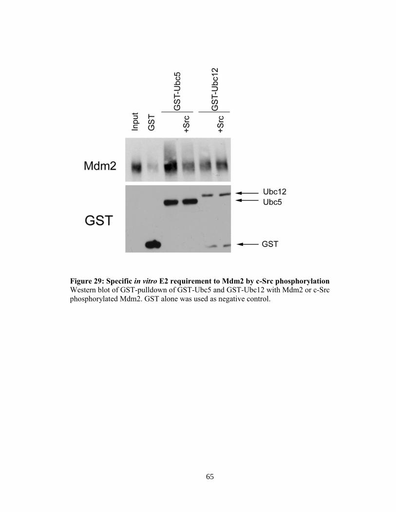
65
Figure 29: Specific in vitro E2 requirement to Mdm2 by c-Src phosphorylation
Western blot of GST-pulldown of GST-Ubc5 and GST-Ubc12 with Mdm2 or c-Src
phosphorylated Mdm2. GST alone was used as negative control.

66
Figure 30: Inhibition of neddylation reverses c-Src downregulation of p53
transcriptional activity
PG13-Luc activity was assayed using H1299 cells transfected with Myc-LacZ, p53,
Mdm2, and CA-Src. 24 hr post-transfection cells were treated with DMSO or 0.3 μM of
the NAE inhibitor MLN4924 for 16 hr. *, # represent statistical significance of p<0.05.
Y-axis measurements are relative luciferase units (RLU). The RLU was calculated from
the ratio of luciferase/β-gal activity. Error bars represent standard deviation.
67
Since c-Src phosphorylation of Mdm2 directs ligase activity towards neddylation, we
tested what effect inhibition of neddylation would have on p53 activity. H1299 cells were
transfected with the PG-13-Luc, p53, Mdm2 and CA-Src. Reduction in p53 activity by
Mdm2 was further increased in the presence of c-Src as expected. Treatment of H1299
cells with MLN4924 restores p53 activity to levels observed with Mdm2 alone, thereby
supporting an Src-Mdm2-nedd8 pathway that inactivates p53 (Figure 30). Since the
PG13-Luc is a synthetic construct we wanted to find a more relevant physiological
promoter. Since both c-Src and Mdm2 are intimately linked to regulation of angiogenic
processes, such as the positive regulation of VEGF (64, 106), we next determined what
effect c-Src phosphorylation of Mdm2 would have on an anti-angiogenic target. The
tumor suppressor maspin is a p53 target that plays an important role in regulating tumor
cell invasion and metastasis (107). We have previously shown that p53 is required for the
upregulation of Maspin protein levels under hypoxic conditions in glioblastoma cell lines
(108). To assess Src-phosphorylated Mdm2 effect on p53-induced maspin, a luciferase
assay utilizing the maspin promoter was performed. As shown in Figure 31A, H1299
cells transfected with the maspin promoter attached to the luciferase gene (maspin-Luc)
shows that p53 drives the expression of luciferase from the maspin promoter and that a
decrease is seen with Mdm2 overexpression. Just like with the PG13-Luc, expression of
CA-Src further inhibits p53 activity on the maspin promoter and the inhibition is
dependent on neddylation. To ensure that the decrease seen on the maspin promoter was
due to c-Src phosphorylation of Mdm2, another luciferase assay was done using the
Y281-302F Mdm2 and CA-Src. Here the maspin promoter luciferase decreased with the
addition of Mdm2 Y281-Y302F, but the addition of CA-Src did not result in an increase

68
Figure 31: Inhibition of maspin promoter by c-Src is dependent on neddylation and
Mdm2 Y281/Y302 (A) Maspin-Luc activity was assayed using H1299 cells transfected with Myc-LacZ, p53,
Mdm2, and CA-Src. Post-transfection (24 hr) cells were treated with DMSO or 0.3 μM of
the NAE inhibitor MLN4924 for 16 hr. (B) Luciferase assay of maspin-Luc in H1299
cells using p53, Mdm2 Y281-302F, and CA-Src.*, # represents statistical significance of
p<0.05. Y-axis measurements are relative luciferase units (RLU). The RLU was
calculated from the ratio of luciferase/β-gal activity. Error bars represent standard
deviation.

69
of inhibition as seen with WT Mdm2 (Figure 31B). This shows that the enhancement of
inhibition seen on the maspin promoter by c-Src is dependent on Mdm2 Y281 and Y302.
For further insight on how endogenous neddylation of p53 would affect maspin levels,
MCF7 cells were transfected with the maspin-Luc construct or a mutant maspin-Luc
(MT1) that is unresponsive to p53, to insure luciferase activity was dependent on p53. c-
Src was then inhibited for 16 hr using PP1 and a luciferase assay performed. Inactivation
of c-Src allowed for an increase of p53 activity on the maspin-Luc construct (Figure
32A). This increase of p53 activity was also seen at the protein level, as Maspin protein
was increased following inhibition of c-Src (Figure 32B). These findings provide insight
into the regulation of c-Src-induced neddylation of p53. This neddylation event is
responsible for the further reduction in p53 activity on both the PG-13-Luc and on its
ability to activate the tumor suppressor Maspin.
3.3 Discussion
Understanding how Mdm2 functions in tumor progression is of critical
importance since Mdm2 protein levels are detectable in 40-80% of high-grade human
malignances and 10% show mdm2 gene amplification (4). The current paradigm is that
Mdm2 upregulation would inhibit the p53 tumor suppressor, through destabilization,
resulting in tumor progression. However there are many human cancers, including breast,
gliomas, oral, renal, and cervical, that have elevated levels of Mdm2 and wild-type p53
(109-113). This correlation of high Mdm2 and p53 levels indicate that Mdm2 may have
other possible mechanisms for the inactivation of p53, besides destabilization. The Mdm2
protein has been shown to conjugate a nedd8 molecule to p53 that inactivates its

70
Figure 32: Inhibition of c-Src upregulates maspin transcription and protein levels
(A) Luciferase assay of MCF7 transfected with either maspin-Luc or mutant maspin-Luc
(MT1) and Myc-LacZ. After transfection (24 hr) cells were treated with DMSO or 10 μM
PP1 for 16 hr. (B) Western blot analysis of MCF7 cells treated with DMSO or 10μM
PP1for 16 hr.

71
transcriptional activity without affecting its protein stability, but the mechanism has
remained elusive (22).
The non-receptor tyrosine kinase c-Src regulates a number of normal and
pathological processes including proliferation, survival, motility, and angiogenesis (71).
c-Src functions poorly as an oncogenic factor on its own and activating mutations of c-
Src in human cancer are rare. This has led to the view that c-Src may facilitate and
promote the activity of other proteins, as opposed to being an autonomous dominant
transforming agent on its own. One c-Src interacting protein that has been extensively
studied is EGFR. This interaction results in phosphorylation of EGFR at Y845, and is c-
Src dependent (114). This pathway activates STAT5b (115) and mediates proliferation
and promotes cell survival through cyclooxygenase 2 (COXII) (116). FAK also interacts
with c-Src. FAK mediates cancer metastasis by regulating focal adhesion formation and
turnover. c-Src phosphorylation activates FAK kinase activity and generates docking
sites for Grb2 and other signaling molecules that regulate signaling pathways (117). In
this study we provide evidence that c-Src cooperates with Mdm2 to enhance oncogenic
activities.
Our data shows that Mdm2 interacts with and is phosphorylated by c-Src (Figures
9A, 10, 12, 14A&B). We demonstrate through in vitro GST pull-down assays and an
immunoprecipitation from MCF7 cells that the Mdm2/Src interaction takes place both in
vitro and in vivo. c-Src uses its SH3 protein domain for many of its protein-protein
interactions including those with paxillin and p85. Here the c-Src SH3 domain is
sufficient for binding to Mdm2. Also, through the use of truncation mutants of Mdm2 we
narrowed down the c-Src binding to amino acids 90-268 of Mdm2 (Figure 9B).

72
Interestingly, this truncation of Mdm2 encompasses the acidic domain, which is known to
contain many serine residues that are normally phosphorylated under non-stress
conditions (118). One hypothesis is that these modifications could enhance c-Src binding
of Mdm2. Hence any alteration to these modifications, such as dephosphorylation of by
PP2A (119), or any other protein interactions driven by cellular stresses could represent a
mechanism to regulate the c-Src-Mdm2 interaction. The phosphorylation of Mdm2 by c-
Src occurs on tyrosine 281 and 302. In vitro kinase reactions revealed that both Y281 and
Y302 have to be mutated to phenylalanine to inhibit c-Src mediated phosphorylation of
Mdm2 (Figure 13). Through the use of ectopically expressed c-Src or activation by EGF,
we show that c-Src phosphorylation of Mdm2 results in an increase in Mdm2 protein
levels (Figure 15 A&B, 17 A-C). These c-Src phosphorylation sites are within or near the
zinc finger domain of Mdm2. Recent work has shown that a mutation in the zinc finger
domain (C305F) has an enhanced half-life compared to wild-type (120). Interestingly, c-
Src phosphorylation of Mdm2 showed similar results (Figure 19 A&B) and mutation of
the two c-Src sites resulted in no change in half-life (Figure 20 A&B). Taken together
these results could hint that alteration of the Mdm2 zinc finger domain could have great
implication for protein stability.
The E3 ubiquitin ligase activity of Mdm2 has been extensively studied, especially
in regard to p53 degradation. We have previously shown that after DNA damage c-Abl
phosphorylation of Mdm2 at Y276 and Y394 enhances complex formation with Mdmx.
This complex leads to degradation of both Mdm2 and Mdmx and results in the
stabilization of p53 (48). Yet how growth factor activated tyrosine kinases affects Mdm2
ubiquitin ligase activity has not been determined. Utilizing an in vitro ubiquitin ligase

73
assay, c-Src inhibited Mdm2 mediated ubiquitination of p53 to the same extent as c-Abl
(Figure 22). This decrease in ubiquitination translated into p53 protein stability in H1299
cells ectopically expressing p53 (Figure 23 A). Treatment with EGF also revealed a
stabilization of p53 that correlated directly with increases in Mdm2 and activated c-Src
protein (Figure 24A). To compensate for this increase in p53 protein levels, c-Src
phosphorylation of Mdm2 further reduced p53 transcriptional activity in an Mdm2 RING
finger dependent manner (Figure 25 A&B). These results are in direct opposition of the
effect c-Abl phosphorylation signaling to Mdm2 has on p53 transcriptional activity. This
provides evidence that tyrosine phosphorylation of Mdm2 under different conditions,
may have different effects on p53 transcriptional activity, while maintaining protein
levels. Interestingly, this induction of protein levels coupled with concurrent inhibition of
transcriptional activity is not unheard of in the p53 family. It has been reported that p73
when bound to Mdm2 results in p73 stabilization, but leads to inhibition of its
transcriptional activity (51). Similarly, the c-Src family member Hck, has been shown to
phosphorylate p73 resulting in protein stability but decreases in its activity (121). These
findings support the notion that increases in p53 protein levels do not guarantee
transcriptional activation.
The decrease in p53 transcriptional activity by c-Src could be explained by the
increase of Mdm2 protein and half-life. Even without the ability to degrade p53 by
ubiquitin ligase activity, Mdm2 is still capable of inhibition by masking p53's N-terminal
transactivation domain (122). This is evident as the RING finger dead mutant of Mdm2,
C464S, is still efficient at inhibiting p53 transcriptional activity. Our data illustrate that
the RING domain of Mdm2 is required for a more dramatic reduction in p53 activity in

74
the presence of CA-Src (Figure 25B). The Mdm2 protein has dual ligase activity capable
of conjugating ubiquitin and nedd8. A previous report has shown that the histone acetyl
transferase Tip60 can inhibit Mdm2 mediated neddylation of p53, but not ubiquitination
(123). Also the original study showing p53 neddylation showed that CHO-TS-41 cells,
which have a temperature sensitive mutation in the APP-BP1 gene inhibiting neddylation,
still have ubiquitinated p53 even in the absence of neddylation (22). These studies taken
together reveal that Mdm2 ligase activities can be both separately and individually
regulated. However, signaling events regulating the activation of Mdm2 neddylation
activity have yet to be identified. Our data reveal that c-Src phosphorylation of Mdm2
results in increased neddylated p53 and Mdm2. This increase in neddylation of p53 is
dependent on the c-Src phosphorylation, as the Y281-302F Mdm2 mutant did not
increase neddylated p53 and inhibition of c-Src by PP1 resulted in less nedd8 bound to
p53 endogenously (Figure 26 A&B, 28). It appears that c-Src may regulate this switch by
determining which E2 is able to bind to Mdm2 (Figure 29). Furthermore, using the
inhibitor of neddylation, MLN4924, the inhibition on both the PG13-Luc promoter and
the maspin-Luc promoter by Mdm2 in the presence of Src was reversed (Figure 30, 31).
The effect on the maspin promoter is especially interesting due to the role of Maspin as a
tumor suppressor in breast cancer. Maspin is normally highly expressed in normal breast
epithelial cells, but is down regulated in invasive and metastatic breast carcinoma cells
(83). Maspin tumor suppressor activity was validated as stable transfection of maspin in
breast cancer cell lines led to an inhibition of tumor cell invasion in vitro and a decrease
in tumor metastasis in mice (124, 125). The ability of Maspin to inhibit tumor growth
was linked to its inhibition of tumor-induced angiogenesis. Maspin protein blocked

75
endothelial cell migration in response to growth signals and inhibited endothelial tube
formation in vitro (91). Also, Maspin expression correlated with decreased micro-vessel
staining in human breast cancer samples (126). This is important as both Mdm2 and c-Src
have been linked to angiogenesis and metastasis in breast cancer, and here we show that
inhibition of c-Src using PP1 results in greater activity of the maspin-Luc and greater
Maspin protein levels in the breast cancer cell line, MCF7 (Figure 32 A&B). Taken
together, these experiments prove that neddylation is a key component in c-Src ability to
inhibit the p53/maspin tumor suppressor network though Mdm2 (Figure 33).
The data presented here reveal a novel c-Src /Mdm2/p53/Maspin pathway. This
signaling cascade could be responsible for the high levels of Mdm2 seen in numerous
cancers that still maintain wild-type p53. We also show that this pathway has a direct
influence on the levels of the tumor suppressor maspin and might have implications in
tumor angiogenesis. Thus, future treatment options might benefit from inhibition of c-Src
and/or neddylation to engage the p53/Maspin anti-angiogenic pathway.

76
Figure 33: Model of c-Src phosphorylation of Mdm2 and its downstream effects
Phosphorylation of Mdm2 by c-Src at Y281 and Y302 switches Mdm2 ligase activity to a
neddylating enzyme, reducing p53 transcriptional activity on target genes, including
Maspin. Inhibitors of both c-Src and neddylation can be used to inhibit this pathway.

77
4. Mdm2 enhances breast cancer metastasis
4.1 Introduction
In the United States, women will have a 1 in 8 chance of developing breast cancer
in their lifetime (127). There are two main types of breast cancer. The first is ductal
carcinoma, which starts in the ducts that move milk from the breast to the nipple. The
other is lobular carcinoma and it starts in the lobules, which produce the milk. Breast
cancer is a heterogeneous disease but has clinically distinct subtypes based on the
expression of molecular markers. These markers include hormone receptor (HR),
progesterone receptor (PR), and Her2. Another subtype of breast cancer termed, triple
negative breast cancer, is diagnosed on the absence of all 3 of these markers, and carries
the basal like molecular profile (128). Metastasis, the process by which tumor cells
invade and colonize distal sites, is the leading cause of death, due to organ failure, for
breast cancer patients. Breast cancer has five primary sites of metastasis including lung,
bone, lymph nodes, liver and pleura. Metastatic tumors at these sites can be seen in over
50% of patients at autopsy, providing evidence of how frequently breast cancer is able to
undergo metastasis (129).
While there are many factors that regulate tumor metastasis, recent work has
implicated the Mdm2 network in this process. One study by Wang et al., illustrated how
Mdm2, through wild-type 53, is responsible for the ubiquitination and degradation of
Slug in lung cancer (130). Slug is a major transcriptional repressor of E-Cadherin, and
can trigger EMT. Interestingly, it was also shown that E-Cadherin itself can interact with
and undergo ubiquitination by Mdm2. Primary tumors from breast cancer patients had an
inverse correlation between Mdm2 and E-cadherin protein levels as seen by

78
immunohistochemistry. These high Mdm2/low E-cadherin levels were associated with
lymph node metastasis (63). We have reported that Mdm2 is upregulated in many late
stage breast cancers, due to a tumor growth factor (TGF)-β/Smad3 signaling event that
drives transcription of Mdm2. We demonstrated that approximately 65% of late-stage
carcinomas were positive for activated Smad3 and Mdm2, indicating a strong correlation
between TGF-β1-mediated induction of Mdm2 and late-stage tumor progression.
Furthermore 73% of the carcinomas that had known metastases stained for Mdm2 and
activated Smad3, possibly linking Mdm2 to a role in breast cancer metastasis (11).
Two in vivo studies have also tried to elucidate the role of Mdm2 in metastasis.
First, Zhang and Hill, showed that overexpression of Mdm2 in KHT cells resulted in
more lung metastasis after tail vein injection (131). Meanwhile, an orthotopic pancreatic
model using lentiviral knockdown of Mdm2, showed that tumor growth and metastasis
was decreased in tumors with lower levels of Mdm2 (132). However, both studies
concluded that the metastatic potential provided by Mdm2 was due to its regulation of
wild-type p53. While Mdm2 main function is attributed to the regulation of p53, it has
also been shown to promote tumorigenesis independent of p53. For example Mdm2
splice variants unable to bind p53 can still promote transformation and p53-null
transgenic mice overexpressing Mdm2 have a higher incidence of tumors compared to
littermate controls (133, 134). These experiments provide evidence that Mdm2 can
promote tumor growth in the absence of p53. But the question remains if Mdm2 can
facilitate metastasis in a p53-independent manner?
To answer this question we employed a known orthotopic mouse model of breast
cancer metastasis. We used the TMD-231 cell line, which are derived from MDA-MB-

79
231 cells that had been grown in the mammary fat pad of mice. The MDA-MB-231 is the
most widely used and characterized cell line for tissue specific metastasis of human
breast cancer. The MDA-MB-231 cell is derived from the pleural effusion of a late stage
breast cancer patient who died with multiple metastases (135). We chose these cells, as
they are known to metastasize to the lung and also contain mutant p53. The mutation is
R280K and is located in the DNA binding domain and inactivates p53 DNA binding
activity. Thus, this allows us to study Mdm2’s role in metastasis independent of p53
activity. To this end, we used a lentiviral construct harboring shRNA to knockdown
Mdm2 in the TMD-231 cell line. We show that knockdown of Mdm2 has no effect on
TMD-231 cellular growth in vitro or cell cycle progression, but knockdown of Mdm2 did
result in decreased invasive potential. Interestingly, knockdown of Mdm2 did slow tumor
growth in vivo, possible due to a decreased affinity for binding to fibronectin. Most
importantly, we found that knockdown of Mdm2 showed a dramatic loss in breast to lung
metastasis. This loss of metastatic potential was due to a reduction in HIF-1α and VEGF
protein levels leading to a loss of primary tumor vascularization. Here we provide
definitive evidence Mdm2 can enhance tumor metastasis, independent of p53.
4.2 Results
Mdm2 expression correlates with metastasis in both pancreatic and breast cancer
models (131, 132). However all studies to date have determined that the role of Mdm2 in
metastasis is due to the regulation of wild-type p53. No studies however, have
determined if Mdm2 plays a role in metastasis independent of p53. To determine if
Mdm2 has a p53-independent role in this process, we used the TMD-231 cell line, which
harbors mutant p53 (R280K), and generated a knockdown of Mdm2 using a lentiviral

80
vector harboring shRNA. Western blot analysis in Figure 34A shows that Mdm2 was
knockdowned in the shMdm2 compared to shGFP control cells. While Mdm2 is the main
E3 ubiquitin ligase for the wild-type p53, it has also been shown to ubiquitinate mutant
p53 (136). It has been established that mutant p53 can promote metastasis through its
gain of function properties, including interacting with p63 and p73, and inhibiting their
tumor suppressor functions (137). To ensure that any changes in metastatic potential in
cells with shMdm2 was not due to mutant p53 levels, western analysis for mutant p53
was performed. ShMdm2 had no detectable increases in mutant p53 levels compared to
shGFP cells ensuring that any changes in metastasis would not be due to levels of mutant
p53 (Figure 34B).
Mdm2 has been shown to promote positive effects on cell proliferation, through
its binding to Rb, and Cyclin A (54, 138). Thus, it was necessary to determine if
knockdown of Mdm2 would inhibit TMD-231 cell proliferation and cell cycle
progression. First, shGFP and shMdm2 TMD-231 cells were plated and grown for 96 hr.
Knockdown of Mdm2 did not show any inhibition of proliferation of TMD-231 cells up
to 96 hr, when normalized to the 24 hr time point (Figure 35A). For cell cycle analysis,
shGFP and shMdm2 TMD-231 cells were serum starved for 24 hr. After the addition of
serum no changes in cell cycle was seen at any time point (Figure 35B). Since no
differences in proliferation were observed, we hypothesized that knockdown of Mdm2 in
TMD-231 cells would lead to inhibition of their ability to migrate and invade, as has been
reported in other cell lines (63, 139). A Matrigel Invasion Assay was employed, using
serum as a chemoattractant, and showed that 70% of shGFP cells were able to migrate

81
Figure 34: Analysis of Mdm2 and p53 protein levels in shGFP and shMdm2 TMD-
231 cells (A) Western blot analysis of Mdm2 and GAPDH in shGFP and shMdm2 TMD-231 cells.
(B) Western blot analysis of p53 and GAPDH in shGFP and shMdm2 TMD-231 cells.

82
Figure 35: Growth and cell cycle analysis of TMD-231 shGFP and shMdm2 cells
(A) TMD-231 cells were grown for 96 hr with cell counts taken at 24 hr intervals. Cell
counts are normalized to 24 hr time point, to account for differences in plating
efficiencies. (B) Cell cycle analysis of TMD-231 cells that were serum starved for 24 hr
followed by addition of 1% serum for times indicated.

83
Figure 36: In vitro invasion potential of shGFP and shMdm2 TMD-231 cells
shGFP and shMdm2 cells were plated in 8 m Matrigel chambers and allowed to invade
for 22 hr using serum as a chemoattractant. Cells were counted in three random 25X
fields and experiments were done in triplicate. Percent invasion was determined as
defined in Materials and Methods. Error bars represent standard deviation.
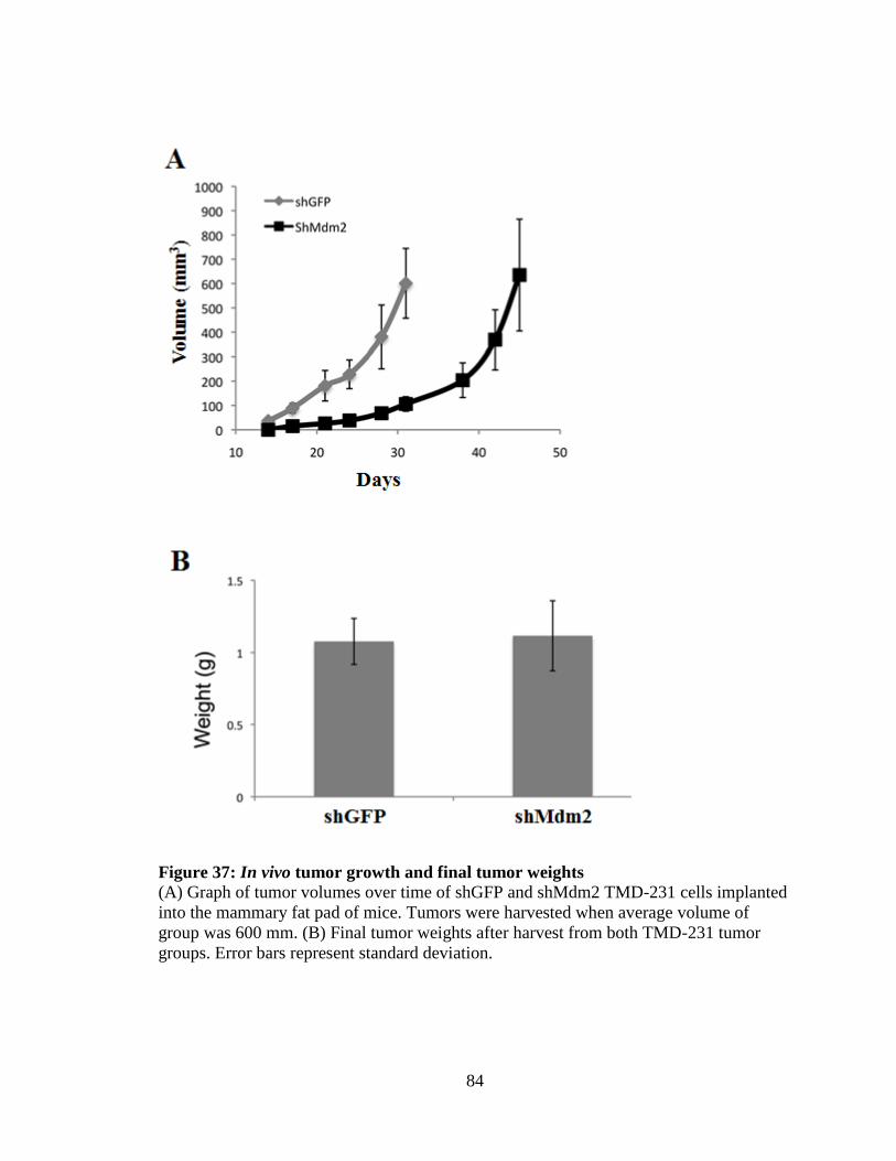
84
Figure 37: In vivo tumor growth and final tumor weights
(A) Graph of tumor volumes over time of shGFP and shMdm2 TMD-231 cells implanted
into the mammary fat pad of mice. Tumors were harvested when average volume of
group was 600 mm. (B) Final tumor weights after harvest from both TMD-231 tumor
groups. Error bars represent standard deviation.

85
through the chamber compared to only 42% of the shMdm2 cells (Figure 36). These
experiments indicate that knockdown of Mdm2 in TMD-231 cells does not affect their
proliferation or cell cycle progression, but does hinder the ability of the cells to migrate
and invade through Matrigel.
Since knockdown of Mdm2 reduced the ability of TMD-231 cells to migrate
though the Matrigel, we were interested to determine if Mdm2 would affect invasion and
metastasis in vivo. To this end, we implanted shGFP and shMdm2 TMD-231 cells into
the mammary fat pad of 6 week old γ-null NOD-SCID mice. To ensure that any
differences seen in metastasis were due to Mdm2 loss and not tumor size or weight, each
group was harvested when they reached an average final volume of 600mm. At the time
of harvest the average tumor weight from each group was identical (1.1g) (Figure 37B).
Interestingly the shMdm2 tumors grew at a slower rate, requiring an extra 2 weeks to
reach the final volume (Figure 37A). However we noted that the delayed in growth
appeared to occur in the initial time after implantation, thus we wondered if the shMdm2
cells may have a problem seeding in an in vivo environment. It has been shown that
Mdm2 can have effects on extracellular proteins involved with cell attachment (140,
141). Interestingly, shMdm2 cells ability to bind to plates coated with fibronectin was
decreased by 40% (Figure 38). This provides evidence that the shMdm2 cells may take
longer to adhere in the mammary fat pad and could explain the delayed growth in vivo.
The TMD-2331 cell line is a model of breast cancer metastasis to the lung (142).
To determine if Mdm2 had any role in this metastatic process we harvested the lungs
from both shGFP (n=10) and shMdm2 (N=10) and H&E was performed. All metastases
were counted in both the right and left lobe of the lung. The shGFP lungs had an average

86
Figure 38: shMdm2 cells have diminished ability to bind fibronectin
shGFP and shMdm2 cells were plated on cell culture dishes that were coated with 5μg/ml
fibronectin. Cells were incubated for 30 min and then removed and counted. Error bars
represent standard deviation.

87
Figure 39: Lung metastatic potential of shGFP and shMdm2 tumors
Lung metastatic legions from both tumor groups were counted as described in Materials
& Methods.

88
Figure 40: Loss of CD-31 staining in shMdm2 tumors
(A) Tumors from shGFP and shMdm2 were stained for CD-31 using
immunohistochemistry. (B) Bar graph representing quantification of CD-31 stating in
shGFP and shMdm2 tumors. Three separate 25x fields from 4 different tumors were
counted. Error bars represent standard deviation. * represents statistical significance of
p<0.05.

89
of 315 metastases per lung, with each lobe accounting for about half the total number.
However the shMdm2 had only an average of 130 metastases per lung, a statistically
significant decrease of 60% compared to the shGFP (Figure 39). While the Mdm2
knockdown affected the total number of metastatic foci, it did not affect the size of the
lesions, as both small and large metastases were noted in both the shGFP and shMdm2
groups. These results definitively show that Mdm2 is involved in breast cancer
metastases to the lung.
Angiogenesis, the formation of new blood vessels from pre-existing ones, is
essential in helping tumor cells metastasize. Interestingly Mdm2 has been shown to have
numerous angiogenic functions. We have previously shown that Mdm2 can bind to HIF-
1α resulting in the up regulation of the pro-angiogenic factor VEGF (64). Mdm2 can also
stabilize VEGF mRNA levels promoting angiogenesis (143). In light of this evidence, we
wondered if the tumors harboring knockdown of Mdm2 would be less vascularized. The
vessels of the primary tumors were stained with CD-31 and the numbers of blood vessels
were counted in multiple fields (Figure 40A). The shGFP showed an average
of 38 vessels per field while the shMdm2 tumors only had an average of 28, a significant
reduction in the amount of blood vessels by 25% (Figure 40B) from four tumors. To
verify that the decrease seen in vascularization was due to changes in HIF-1α and VEGF
protein levels, western blot analysis of both cell lines and tumors were performed. Figure
41A shows that HIF-1α protein levels are higher in shGFP cells compared to shMdm2
cells under both normoxic and hypoxic conditions. The same results were seen with
VEGF protein levels (Figure 41B). As expected, HIF-1α protein levels in the tumors
correlated exactly with the results from tissue culture. Four primary tumors from both

90
Figure 41: Mdm2 increases HIF-1 and VEGF protein levels in vitro
(A) TMD-231 cells were placed under normoxic or hypoxic conditions and western blot
analysis was performed for HIF-1 and GAPDH. (B) Western analysis of Mdm2 and
VEGF from shGFP and shMdm2 TMD-231 cells, under normoxic or hypoxic conditions.

91
Figure 42: Mdm2 increases HIF-1α protein levels in tumors
Tumors excised from the mammary fat pad of mice were lysed and western blotting
performed for HIF-1, Mdm2, and Tubulin.

92
Figure 43: Mdm2 augments protein levels of lung metastasis genes
Western blot analyses of MMP-2, SPARC, and GAPDH from shGFP and shMdm2
TMD-231 cells.

93
shGFP and shMdm2 showed that Mdm2 and HIF-1α are all lower in the shMdm2 tumors
(Figure 42). These results show that knockdown of Mdm2 results in lower levels of HIF-
1α and VEGF. This loss of pro-angiogenic factors lowers tumor vascularization, thus
leading to a decrease in lung metastasis.
Since Mdm2 facilitates many functions in the cell, we hypothesized that Mdm2
could be regulating other pro-metastatic proteins or pathways. Minn et al. recently
performed gene-array analysis and generated a list of gene candidates that could mediate
breast cancer to lung metastasis (144). From this list we found that both MMP-2 and
SPARC protein levels were down regulated in shMdm2 cells compared with shGFP,
hinting that Mdm2 could be promoting or regulating a pro-metastatic gene profile for
lung metastases (Figure 43). Further work is needed to determine the mechanism by
which Mdm2 is regulating these genes.
4.3 Discussion
The Mdm2 onco-protein is implicated in a variety of p53-independent cellular
processes including transcription regulation, translation control, DNA repair, and cell
cycle regulation, all in addition to its role as the primary regulator of p53. Understanding
how Mdm2 is regulating different tumorigenic properties is important treatment of
malignancies in the clinic. Mdm2 overexpression correlates with poor patient prognosis
and is seen in a variety of human cancers including 1/3 of sarcomas and cancers of the
brain, breast, ovary cervix, lung, colon, and prostate (4, 5). The role for Mdm2 in
promoting tumorigenesis is well established in both p53 dependent and independent

94
manner. Recent studies have also implicated Mdm2 in the process of tumor metastasis,
but it has not been determined if this role is dependent on p53 (63, 132).
To address this issue we generated a TMD-231 cell line with knockdown of
Mdm2 in the background of mutant p53 to use as a model for breast to lung metastasis.
This mutation in p53 occurs as a point mutation, R280K. This mutation resides within the
DNA binding domain of p53 and renders it incapable of functioning as a transcription
factor. This mutation allows us to determine if Mdm2 can promote metastasis
independent of wild-type p53 function. Mdm2 has been implemented in cell growth and
cell cycle progression not only by inhibiting p53 function, but also through p53-
independent mechanisms. Due to this role of Mdm2, we needed to ascertain if
knockdown of Mdm2 would affect TMD-231 cell growth and cell cycle progression.
Knockdown of Mdm2 in SW1990 pancreatic cells has been shown to reduce cellular
proliferation (132). In contrast, however the growth rates of p53-/- MEFS and p53-/-,
Mdm2 -/- MEFS are nearly identical (145). Furthermore, expression of Mdm2 splice
variants incapable of binding p53 can actually inhibit cellular proliferation in vitro (146).
The knockdown of Mdm2 in our TMD-231 cells did not affect cell growth, providing
evidence that Mdm2’s role for cell proliferation in cell culture may be cell type specific
(Figure 35A).
Mutant p53 can acquire gain of function attributes that aid in cell survival
advantages, such as increased proliferation, evasion of apoptosis, and chemoresistance.
These gain of function attributes are seen as the use of shRNA to mutant p53 in MDA-
MB-231 cells results in decreased cell proliferation, reduced invasion in boyden
chambers, and lower tumor weight in nude mice (147). Important to the scope of this

95
study is the gain of function ability of mutant p53 to enhance tumor metastasis. Mutant
p53 enhancement of metastatic potential is shown by the observation that p53 knockout
mice develop tumors at a high frequency, but have a low occurrence of metastasis or
invasive growth. In contrast, transgenic mice knocked-in with mutant p53 develop highly
metastatic tumors (148). Furthermore, mutant p53 was found to promote EMT by
facilitating the function of the key transcriptional regulators of this process, TWIST1 and
SLUG, and inhibit cell invasion via the inhibition of p63 (130, 149). Since we were
interested in the role of Mdm2 in metastasis in a p53-independent background, we had to
be sure that mutant p53 levels were not affected by Mdm2 knockdown. Mdm2 still
retains the ability to degrade mutant p53 (136), but our results indicated that knockdown
of Mdm2 had no bearing on mutant p53 protein levels (Figure 34 A&B). If Mdm2 was
regulating mutant p53 in TMD-231 cells, knockdown of Mdm2 should have resulted in
an increase of mutant p53 levels. Our result that mutant p53 levels are not altered is in
agreement with recent findings that suggest mutant p53 cannot interact with Mdm2 in
cancer cells. (147).
Comparing our in vitro cell proliferation data to in vivo tumor growth, we note
that knockdown of Mdm2 does slow in vivo tumor growth (Figure 37A). These differing
observations hint that Mdm2 may play a role in TMD-231 cells ability to adhere on
different growth substrates. Mdm2 has been implemented in regulation of cell adhesion
molecules such as E-cadherin and Syntaxin 6. Thus, loss of Mdm2 may interfere with the
ability of TMD-231 cells to seed in an in vivo environment. This hypothesis is attractive
as knockdown of Mdm2 significantly reduces the ability of cells to adhere to fibronectin-

96
coated plates (Figure 38). This reasoning would explain why the tumors after an initial
growth lag were able to eventually seed and grow to the same size as control tumors.
The ability of the shMdm2 tumors to grow to the same size as control tumors was
imperative, as we wanted to assure ourselves that the tumor size would have no bearing
on the ability of the tumors to metastasize. All tumors were harvested when the average
volume was 600 mm3. The average tumor weight for each group was 1.1g (Figure 37 B).
This allowed us to ensure that the difference seen in metastases was not due to a greater
tumor weight and volume in the shGFP group, and was solely due to differences in
Mdm2 expression. This scenario was observed, as knockdown of Mdm2 resulted in
drastic loss of metastases to the lung. A 60% reduction of lungs metastases was seen in
the shMdm2 group (Figure 39). Overall the size of the metastases between the two
groups was similar suggesting that Mdm2 did not have an effect on the ability of the
tumor cells to grow once they have seeded at a new site. Therefore, Mdm2 may also have
a role in the intravasation stage of metastasis. The ability of tumor cells to escape into the
blood stream is dependent on the capacity of the tumor to recruit new vasculature through
angiogenesis. Mdm2 has been described as having pro-angiogenic properties through its
regulation of HIF-1α and VEGF. We have shown that Mdm2 can bind HIF-1α and
enhance VEGF transcription as determined by luciferase activity assays (64). This
enhancement of HIF-1α transcriptional activity is from an increase in p300 recruitment
through an Mdm2 dependent process that inhibits hydroxylation of Asn803 in HIF-1α
both in vivo and in vitro (150). Interestingly, recent work has also shown an Mdm2
dependent increase of VEGF mRNA stability and translation through Mdm2 binding to
the AU-rich sequence within the 3' untranslated region of VEGF mRNA (143). Due to
97
Mdm2’s defined role in regulating these pro-angiogenic targets, it was logical to assume
that our cells and tumors would yield similar experimental results. As expected,
knockdown of Mdm2 resulted in the loss of HIF-1α and VEGF protein levels in both cell
culture and tumor lysates (Figure 41 & 42). This loss of angiogenic factors correlates
with a decrease in vascular endothelial cells in the tumor (Figure 40 A&B), as the
knockdown of Mdm2 prevented the formation of new vasculature in the tumor. These
results provide evidence that Mdm2 may enhance tumor metastasis through the
recruitment of angiogenic factors and allow vascularization of the primary tumor.
Metastasis is a multi-step process including tumor cell intravasation, survival in
circulation, extravasation into a distant organ, angiogenesis and uninhibited cellular
growth. Due to the role of Mdm2 in numerous cellular processes we could not eliminate
the possibility that Mdm2 could be functioning in other ways to promote lung metastasis
than just through regulating pro-angiogenic factors. Recent work by Minn and colleagues
using MDA-MB-231 cells and transcriptomic analysis identified a set of genes that
mediates breast cancer metastasis to the lungs (144). Their results showed a subset of
four genes (SPARC, IL13RA2, VCAM1 and MMP2) whose expression, as determined
by northern blot was generally seen in aggressive lung-metastatic populations. We show
that knockdown of Mdm2 results in lower levels of both MMP2 and SPARC (Figure 43).
Normally SPARC functions as a modulator of the extracellular matrix, but deregulation
has been linked to an invasive tumor cell phenotype and poor outcome in human
melanomas. Recent work has shown that SPARC augments tumor growth by stabilizing
Mdm2 levels through AKT phosphorylation at serine 166. This phosphorylation allows
Mdm2 to translocate to the nucleus and inactivate p53 in human melanomas (141). This

98
work, along with ours, could support a Mdm2/SPARC network to support tumor growth
and metastasis.
Here we show that Mdm2 is involved in tumor metastasis though multiple p53
independent functions including a role in tumor cell adherence, regulation of the pro-
angiogenic factors HIF-1α and VEGF, and perpetrating a lung metastatic gene signature
through increases in the protein levels of SPARC and MMP2. Understanding how Mdm2
regulates metastasis is important as metastatic disease is usually the terminal step in
cancer development, so understanding and prevention is critical. Here we present
evidence that Mdm2 can be a primary component of breast cancer to lung metastasis and
thus could be a viable clinical target.

99
5. Summary/Future Directions
Mdm2 serves many diverse functions in a variety of cellular processes and has a
well-established role in tumorigenesis. Here we have shown that Mdm2, through c-Src
mediated phosphorylation, can inhibit p53 activity through neddylation while maintaining
p53 protein levels. We have also confirmed that Mdm2 has a positive role in cancer
metastasis, thus adding more complex functions to Mdm2 regulation of tumorigenesis.
Since roles for Mdm2 in cancer are continuing to be identified, a sustained effort to find
Mdm2 inhibitors for clinical use is urgently needed. Most Mdm2 inhibitors developed
thus far have focused on inhibiting the p53-Mdm2 interaction in an attempt to re-activate
p53. The interaction between p53 and Mdm2 involves four residues in p53 (Phe 19, Leu
22, Trp 23, Leu 26) and a hydrophobic pocket in the N-terminal region of Mdm2. The
most studied inhibitor has been Nutlin-3a, which inhibits Mdm2-p53 binding by binding
to the Mdm2 hydrophobic pocket resulting in activation of p53 and leading to cell cycle
arrest, apoptosis, and growth inhibition of human tumor xenografts in nude mice (151). It
was first assumed that Nutlin would only function in wild-type p53 tumors, but recent
work has shown that Nutlin can inhibit angiogenesis through inhibition of the
Mdm2/HIF-1α interaction, independent of p53 (64, 150). Furthermore, it was recently
demonstrated that Nutlin-3a can upregulate apoptotic genes independent of p53, by
blocking the p73/Mdm2 interaction (152). These studies show that inhibition of Mdm2
can still have advantageous results even in absence of wild-type p53 and lends support to
the developmental ideology for inhibitors that target alternative domains outside of the
p53-binding region. Inhibition of the Mdm2 RING E3 ligase is an attractive target and
has been shown to activate p53, which is accomplished through inhibition of p53

100
ubiquitination and neddylation (153). Mdm2 specifies its ligase activity through the
recruitment of specific E2 enzymes, thus specific inhibitors for each interaction, could be
an invaluable tool depending on a specific tumor genetic makeup. For tumors with high
Mdm2/low p53 a Mdm2/Ubc5 inhibitor could be used to inhibit p53 ubiquitination.
While tumors with high Mdm2/high p53, would benefit from inhibition of Mdm2/Ubc12,
which would inhibit neddylation, thus personalizing cancer care by specifically targeting
Mdm2 function. This could be even more advantageous for mutant p53 tumors as Mdm2
has been shown to ubiquitinate and degrade mutant p53. Interestingly, work has not been
performed on neddylation of mutant p53 and the downstream signaling consequences. It
could be envisioned that neddylation of mutant p53 could enhance mutant p53 gain of
function status. Therefore, inhibition of neddylation, but not ubiquitination could be
clinically relevant in the treatment of certain cancers.
Angiogenesis facilitates tumor growth and metastasis. We show here that Mdm2
has a role in regulating cancer metastasis partly through controlling HIF-1/VEGF levels
and promoting vascularization of the tumor. Additionally, we also show that c-Src
phosphorylation of Mdm2 affects the angiogenic pathway through inhibiting the
expression of p53-mediated transcription of maspin.
This lead us to hypothesize if c-Src and Mdm2 together could be leading the
angiogenic switch, since both proteins have been implemented in the positive regulation
of HIF-1 and VEGF. Since we have shown that c-Src can phosphorylate Mdm2, we

101
Figure 44: c-Src phosphorylation of Mdm2 increases Mdm2/HIF-1 binding
In vitro GST-pulldown assay using GST-HIF-1 (1-535) and Mdm2. Mdm2 was left
unphosphorylated or was phosphorylated by c-Src before incubation with GST-HIF-1.
GST alone was used a negative control. Experiment was performed in duplicate.

102
wondered if this event might enhance the pro-angiogenic phenotype. Through the use of
in vitro GST-pulldown assays we have data showing that c-Src phosphorylated Mdm2
binds HIF-1 to a greater degree that unphosphorylated Mdm2, allowing for greater
transcriptional activity of HIF-1 (Figure 44). Recent work has shown that HIF-1 itself
is also neddylated, leading to an increase in the protein half-life (154). However the E3
ligase responsible for this neddylation has yet to be determined. Due to Mdm2 ability to
bind HIF-1 and function as a neddylating enzyme we wondered if this interaction could
result in neddylated HIF-1. Through the use of overexpression, and
immunoprecipitation of HA-nedd8, our data shows that HIF-1 is neddylated in the
presence Mdm2, but not in the presence of the ligase dead C464S (Figure 45). Thus c-Src
phosphorylation of Mdm2 results in an increased binding to HIF-1, which enables
Mdm2 to neddylate HIF-1. This neddylation event would result in an increase of HIF-
1 half-life, allowing for increased HIF-1 transcriptional activity. If this hypothesis is
correct, then inhibiting c-Src activity should result in the loss of Mdm2 ability to activate
HIF-1 transcriptional activity. This is shown as treatment with the c-Src inhibitor PP1
decreases the positive effect of Mdm2 on the HRE-Luc promoter as determined by a
luciferase assay (Figure 46). These experiments provide evidence that c-Src and Mdm2
may be collaborating for a pro-angiogenic phenotype, and support future endeavors into
determining its regulation.
In conclusion, we have shown novel regulation and activities of the oncogene
Mdm2. First we have provided evidence that c-Src phosphorylation of Mdm2 can switch
its E3 ligase activity from ubiquitin to neddylation resulting in stable, but

103
Figure 45: Neddylation of HIF-1 by Mdm2
H1299 cells, overexpressing HA-Nedd8 plus Mdm2 or C464S, were exposed to hypoxic
conditions for 16 hr, before the immunoprecipitation of HA-tag. Following
immunoprecipitation western blot analysis was performed for HIF-1. Bands represent
neddylated HIF-1.

104
Figure 46: Inhibition of c-Src impairs Mdm2’s ability to enhance HIF-1
transcriptional activity
H1299 cells overexpressing HRE-Luc, myc-LacZ, and Mdm2. Cells were treated with 10
M PP1 for 16 hr before determination of luciferase activity. Y-axis measurements are
relative luciferase units (RLU). The RLU was calculated from the ratio of luciferase/β-gal
activity. Error bars represent standard deviation.

105
transcriptionally inactive p53. We have also definitively shown that Mdm2 can enhance
breast cancer metastasis, independent of p53 status. Taken together, my data provide
novel insights into important p53-dependent and independent functions of Mdm2 and
provide potential novel avenues for therapeutic intervention.

106
References
1. Fakharzadeh SS, Trusko SP, George DL. Tumorigenic potential associated with
enhanced expression of a gene that is amplified in a mouse tumor cell line. EMBO J
1991; 10: 1565-9.
2. Finlay CA. The mdm-2 oncogene can overcome wild-type p53 suppression of
transformed cell growth. Mol Cell Biol 1993; 13: 301-6.
3. Cahilly-Snyder L, Yang-Feng T, Francke U, George DL. Molecular analysis and
chromosomal mapping of amplified genes isolated from a transformed mouse 3T3 cell
line. Somat Cell Mol Genet 1987; 13: 235-44.
4. Rayburn E, Zhang R, He J, Wang H. MDM2 and human malignancies:
expression, clinical pathology, prognostic markers, and implications for chemotherapy.
Curr Cancer Drug Targets 2005; 5: 27-41.
5. Momand J, Jung D, Wilczynski S, Niland J. The MDM2 gene amplification
database. Nucleic Acids Res 1998; 26: 3453-9.
6. Chau V, Tobias JW, Bachmair A, et al. A multiubiquitin chain is confined to
specific lysine in a targeted short-lived protein. Science 1989; 243: 1576-83.
7. Hicke L, Schubert HL, Hill CP. Ubiquitin-binding domains. Nat Rev Mol Cell
Biol 2005; 6: 610-21.
8. Schulman BA, Harper JW. Ubiquitin-like protein activation by E1 enzymes: the
apex for downstream signalling pathways. Nat Rev Mol Cell Biol 2009; 10: 319-31.
9. Wenzel DM, Stoll KE, Klevit RE. E2s: structurally economical and functionally
replete. Biochem J 2011; 433: 31-42.
10. Deshaies RJ, Joazeiro CA. RING domain E3 ubiquitin ligases. Annu Rev
Biochem 2009; 78: 399-434.
11. Araki S, Eitel JA, Batuello CN, et al. TGF-beta1-induced expression of human
Mdm2 correlates with late-stage metastatic breast cancer. J Clin Invest 2010; 120: 290-
302.
12. Haupt Y, Maya R, Kazaz A, Oren M. Mdm2 promotes the rapid degradation of
p53. Nature 1997; 387: 296-9.
13. Kubbutat MH, Jones SN, Vousden KH. Regulation of p53 stability by Mdm2.
Nature 1997; 387: 299-303.
14. Maki CG, Huibregtse JM, Howley PM. In vivo ubiquitination and proteasome-
mediated degradation of p53(1). Cancer Res 1996; 56: 2649-54.
15. Menendez D, Inga A, Resnick MA. The expanding universe of p53 targets. Nat
Rev Cancer 2009; 9: 724-37.
16. Kruse JP, Gu W. Modes of p53 regulation. Cell 2009; 137: 609-22.
17. Li M, Brooks CL, Wu-Baer F, Chen D, Baer R, Gu W. Mono- versus
polyubiquitination: differential control of p53 fate by Mdm2. Science 2003; 302: 1972-5.
18. Ito A, Lai CH, Zhao X, et al. p300/CBP-mediated p53 acetylation is commonly
induced by p53-activating agents and inhibited by MDM2. EMBO J 2001; 20: 1331-40.
19. Jones SN, Roe AE, Donehower LA, Bradley A. Rescue of embryonic lethality in
Mdm2-deficient mice by absence of p53. Nature 1995; 378: 206-8.
20. Montes de Oca Luna R, Wagner DS, Lozano G. Rescue of early embryonic
lethality in mdm2-deficient mice by deletion of p53. Nature 1995; 378: 203-6.

107
21. Itahana K, Mao H, Jin A, et al. Targeted inactivation of Mdm2 RING finger E3
ubiquitin ligase activity in the mouse reveals mechanistic insights into p53 regulation.
Cancer Cell 2007; 12: 355-66.
22. Xirodimas DP, Saville MK, Bourdon JC, Hay RT, Lane DP. Mdm2-mediated
NEDD8 conjugation of p53 inhibits its transcriptional activity. Cell 2004; 118: 83-97.
23. Lammer D, Mathias N, Laplaza JM, et al. Modification of yeast Cdc53p by the
ubiquitin-related protein rub1p affects function of the SCFCdc4 complex. Genes Dev
1998; 12: 914-26.
24. Kurz T, Pintard L, Willis JH, et al. Cytoskeletal regulation by the Nedd8
ubiquitin-like protein modification pathway. Science 2002; 295: 1294-8.
25. Tateishi K, Omata M, Tanaka K, Chiba T. The NEDD8 system is essential for cell
cycle progression and morphogenetic pathway in mice. J Cell Biol 2001; 155: 571-9.
26. Ohh M, Kim WY, Moslehi JJ, et al. An intact NEDD8 pathway is required for
Cullin-dependent ubiquitylation in mammalian cells. EMBO Rep 2002; 3: 177-82.
27. Soucy TA, Smith PG, Milhollen MA, et al. An inhibitor of NEDD8-activating
enzyme as a new approach to treat cancer. Nature 2009; 458: 732-6.
28. Kamura T, Conrad MN, Yan Q, Conaway RC, Conaway JW. The Rbx1 subunit of
SCF and VHL E3 ubiquitin ligase activates Rub1 modification of cullins Cdc53 and
Cul2. Genes Dev 1999; 13: 2928-33.
29. Duda DM, Borg LA, Scott DC, Hunt HW, Hammel M, Schulman BA. Structural
insights into NEDD8 activation of cullin-RING ligases: conformational control of
conjugation. Cell 2008; 134: 995-1006.
30. Pan ZQ, Kentsis A, Dias DC, Yamoah K, Wu K. Nedd8 on cullin: building an
expressway to protein destruction. Oncogene 2004; 23: 1985-97.
31. Abida WM, Nikolaev A, Zhao W, Zhang W, Gu W. FBXO11 promotes the
Neddylation of p53 and inhibits its transcriptional activity. J Biol Chem 2007; 282: 1797-
804.
32. Carter S, Vousden KH. p53-Ubl fusions as models of ubiquitination, sumoylation
and neddylation of p53. Cell Cycle 2008; 7: 2519-28.
33. Liu G, Xirodimas DP. NUB1 promotes cytoplasmic localization of p53 through
cooperation of the NEDD8 and ubiquitin pathways. Oncogene 2010; 29: 2252-61.
34. Watson IR, Li BK, Roche O, Blanch A, Ohh M, Irwin MS. Chemotherapy
induces NEDP1-mediated destabilization of MDM2. Oncogene 2010; 29: 297-304.
35. Bartel F, Taubert H, Harris LC. Alternative and aberrant splicing of MDM2
mRNA in human cancer. Cancer Cell 2002; 2: 9-15.
36. Bond GL, Hu W, Bond EE, et al. A single nucleotide polymorphism in the
MDM2 promoter attenuates the p53 tumor suppressor pathway and accelerates tumor
formation in humans. Cell 2004; 119: 591-602.
37. Phelps M, Darley M, Primrose JN, Blaydes JP. p53-independent activation of the
hdm2-P2 promoter through multiple transcription factor response elements results in
elevated hdm2 expression in estrogen receptor alpha-positive breast cancer cells. Cancer
Res 2003; 63: 2616-23.
38. Chang CJ, Freeman DJ, Wu H. PTEN regulates Mdm2 expression through the P1
promoter. J Biol Chem 2004; 279: 29841-8.

108
39. Busuttil V, Droin N, McCormick L, et al. NF-kappaB inhibits T-cell activation-
induced, p73-dependent cell death by induction of MDM2. Proc Natl Acad Sci U S A
2010; 107: 18061-6.
40. Mayo LD, Donner DB. A phosphatidylinositol 3-kinase/Akt pathway promotes
translocation of Mdm2 from the cytoplasm to the nucleus. Proc Natl Acad Sci U S A
2001; 98: 11598-603.
41. Kamijo T, Weber JD, Zambetti G, Zindy F, Roussel MF, Sherr CJ. Functional and
physical interactions of the ARF tumor suppressor with p53 and Mdm2. Proc Natl Acad
Sci U S A 1998; 95: 8292-7.
42. Pomerantz J, Schreiber-Agus N, Liegeois NJ, et al. The Ink4a tumor suppressor
gene product, p19Arf, interacts with MDM2 and neutralizes MDM2's inhibition of p53.
Cell 1998; 92: 713-23.
43. Allende-Vega N, McKenzie L, Meek D. Transcription factor TAFII250
phosphorylates the acidic domain of Mdm2 through recruitment of protein kinase CK2.
Mol Cell Biochem 2008; 316: 99-106.
44. Maya R, Balass M, Kim ST, et al. ATM-dependent phosphorylation of Mdm2 on
serine 395: role in p53 activation by DNA damage. Genes Dev 2001; 15: 1067-77.
45. Cheng Q, Chen L, Li Z, Lane WS, Chen J. ATM activates p53 by regulating
MDM2 oligomerization and E3 processivity. EMBO J 2009; 28: 3857-67.
46. Shinozaki T, Nota A, Taya Y, Okamoto K. Functional role of Mdm2
phosphorylation by ATR in attenuation of p53 nuclear export. Oncogene 2003; 22: 8870-
80.
47. Mayo LD, Turchi JJ, Berberich SJ. Mdm-2 phosphorylation by DNA-dependent
protein kinase prevents interaction with p53. Cancer Res 1997; 57: 5013-6.
48. Waning DL, Lehman JA, Batuello CN, Mayo LD. c-Abl phosphorylation of
Mdm2 facilitates Mdm2-Mdmx complex formation. J Biol Chem 2011; 286: 216-22.
49. Dias SS, Milne DM, Meek DW. c-Abl phosphorylates Hdm2 at tyrosine 276 in
response to DNA damage and regulates interaction with ARF. Oncogene 2006; 25: 6666-
71.
50. Arasada RR, Carpenter G. Secretase-dependent tyrosine phosphorylation of
Mdm2 by the ErbB-4 intracellular domain fragment. J Biol Chem 2005; 280: 30783-7.
51. Zeng X, Chen L, Jost CA, et al. MDM2 suppresses p73 function without
promoting p73 degradation. Mol Cell Biol 1999; 19: 3257-66.
52. Minsky N, Oren M. The RING domain of Mdm2 mediates histone ubiquitylation
and transcriptional repression. Mol Cell 2004; 16: 631-9.
53. Sdek P, Ying H, Zheng H, et al. The central acidic domain of MDM2 is critical in
inhibition of retinoblastoma-mediated suppression of E2F and cell growth. J Biol Chem
2004; 279: 53317-22.
54. Uchida C, Miwa S, Kitagawa K, et al. Enhanced Mdm2 activity inhibits pRB
function via ubiquitin-dependent degradation. EMBO J 2005; 24: 160-9.
55. Linares LK, Kiernan R, Triboulet R, et al. Intrinsic ubiquitination activity of
PCAF controls the stability of the oncoprotein Hdm2. Nat Cell Biol 2007; 9: 331-8.
56. Coutts AS, Boulahbel H, Graham A, La Thangue NB. Mdm2 targets the p53
transcription cofactor JMY for degradation. EMBO Rep 2007; 8: 84-90.
57. Dai MS, Lu H. Inhibition of MDM2-mediated p53 ubiquitination and degradation
by ribosomal protein L5. J Biol Chem 2004; 279: 44475-82.

109
58. Zhang Y, Wolf GW, Bhat K, et al. Ribosomal protein L11 negatively regulates
oncoprotein MDM2 and mediates a p53-dependent ribosomal-stress checkpoint pathway.
Mol Cell Biol 2003; 23: 8902-12.
59. Dai MS, Zeng SX, Jin Y, Sun XX, David L, Lu H. Ribosomal protein L23
activates p53 by inhibiting MDM2 function in response to ribosomal perturbation but not
to translation inhibition. Mol Cell Biol 2004; 24: 7654-68.
60. Wei X, Yu ZK, Ramalingam A, et al. Physical and functional interactions
between PML and MDM2. J Biol Chem 2003; 278: 29288-97.
61. Bouska A, Lushnikova T, Plaza S, Eischen CM. Mdm2 promotes genetic
instability and transformation independent of p53. Mol Cell Biol 2008; 28: 4862-74.
62. Asahara H, Li Y, Fuss J, et al. Stimulation of human DNA polymerase epsilon by
MDM2. Nucleic Acids Res 2003; 31: 2451-9.
63. Yang JY, Zong CS, Xia W, et al. MDM2 promotes cell motility and invasiveness
by regulating E-cadherin degradation. Mol Cell Biol 2006; 26: 7269-82.
64. LaRusch GA, Jackson MW, Dunbar JD, Warren RS, Donner DB, Mayo LD.
Nutlin3 blocks vascular endothelial growth factor induction by preventing the interaction
between hypoxia inducible factor 1alpha and Hdm2. Cancer Res 2007; 67: 450-4.
65. Sczaniecka M, Gladstone K, Pettersson S, McLaren L, Huart AS, Wallace M.
MDM2 protein-mediated ubiquitination of numb protein: identification of a second
physiological substrate of MDM2 that employs a dual-site docking mechanism. J Biol
Chem 2012; 287: 14052-68.
66. Inuzuka H, Tseng A, Gao D, et al. Phosphorylation by casein kinase I promotes
the turnover of the Mdm2 oncoprotein via the SCF(beta-TRCP) ubiquitin ligase. Cancer
Cell 2010; 18: 147-59.
67. Irby RB, Yeatman TJ. Role of Src expression and activation in human cancer.
Oncogene 2000; 19: 5636-42.
68. Finn RS. Targeting Src in breast cancer. Ann Oncol 2008; 19: 1379-86.
69. Giaccone G, Zucali PA. Src as a potential therapeutic target in non-small-cell lung
cancer. Ann Oncol 2008; 19: 1219-23.
70. Shalloway D, Coussens PM, Yaciuk P. Overexpression of the c-src protein does
not induce transformation of NIH 3T3 cells. Proc Natl Acad Sci U S A 1984; 81: 7071-5.
71. Yeatman TJ. A renaissance for SRC. Nat Rev Cancer 2004; 4: 470-80.
72. Cavallaro U. N-cadherin as an invasion promoter: a novel target for antitumor
therapy? Curr Opin Investig Drugs 2004; 5: 1274-8.
73. Savagner P. The epithelial-mesenchymal transition (EMT) phenomenon. Ann
Oncol 2010; 21 Suppl 7: vii89-92.
74. Guo W, Giancotti FG. Integrin signalling during tumour progression. Nat Rev
Mol Cell Biol 2004; 5: 816-26.
75. Overall CM, Kleifeld O. Tumour microenvironment - opinion: validating matrix
metalloproteinases as drug targets and anti-targets for cancer therapy. Nat Rev Cancer
2006; 6: 227-39.
76. Rak J, Yu JL. Oncogenes and tumor angiogenesis: the question of vascular
"supply" and vascular "demand". Semin Cancer Biol 2004; 14: 93-104.
77. Fong GH, Rossant J, Gertsenstein M, Breitman ML. Role of the Flt-1 receptor
tyrosine kinase in regulating the assembly of vascular endothelium. Nature 1995; 376:
66-70.

110
78. Millauer B, Wizigmann-Voos S, Schnurch H, et al. High affinity VEGF binding
and developmental expression suggest Flk-1 as a major regulator of vasculogenesis and
angiogenesis. Cell 1993; 72: 835-46.
79. Kaipainen A, Korhonen J, Mustonen T, et al. Expression of the fms-like tyrosine
kinase 4 gene becomes restricted to lymphatic endothelium during development. Proc
Natl Acad Sci U S A 1995; 92: 3566-70.
80. Schofield CJ, Ratcliffe PJ. Oxygen sensing by HIF hydroxylases. Nat Rev Mol
Cell Biol 2004; 5: 343-54.
81. Kaelin WG, Jr., Ratcliffe PJ. Oxygen sensing by metazoans: the central role of the
HIF hydroxylase pathway. Mol Cell 2008; 30: 393-402.
82. Brahimi-Horn MC, Pouyssegur J. The hypoxia-inducible factor and tumor
progression along the angiogenic pathway. Int Rev Cytol 2005; 242: 157-213.
83. Zou Z, Anisowicz A, Hendrix MJ, et al. Maspin, a serpin with tumor-suppressing
activity in human mammary epithelial cells. Science 1994; 263: 526-9.
84. Silverman GA, Bird PI, Carrell RW, et al. The serpins are an expanding
superfamily of structurally similar but functionally diverse proteins. Evolution,
mechanism of inhibition, novel functions, and a revised nomenclature. J Biol Chem 2001;
276: 33293-6.
85. Umekita Y, Hiipakka RA, Liao S. Rat and human maspins: structures, metastatic
suppressor activity and mutation in prostate cancer cells. Cancer Lett 1997; 113: 87-93.
86. Gao F, Shi HY, Daughty C, Cella N, Zhang M. Maspin plays an essential role in
early embryonic development. Development 2004; 131: 1479-89.
87. Qin L, Zhang M. Maspin regulates endothelial cell adhesion and migration
through an integrin signaling pathway. J Biol Chem 2010; 285: 32360-9.
88. Endsley MP, Hu Y, Deng Y, et al. Maspin, the molecular bridge between the
plasminogen activator system and beta1 integrin that facilitates cell adhesion. J Biol
Chem 2011; 286: 24599-607.
89. Zhang M, Maass N, Magit D, Sager R. Transactivation through Ets and Ap1
transcription sites determines the expression of the tumor-suppressing gene maspin. Cell
Growth Differ 1997; 8: 179-86.
90. Chen L, Gilkes DM, Pan Y, Lane WS, Chen J. ATM and Chk2-dependent
phosphorylation of MDMX contribute to p53 activation after DNA damage. EMBO J
2005; 24: 3411-22.
91. Zhang M, Volpert O, Shi YH, Bouck N. Maspin is an angiogenesis inhibitor. Nat
Med 2000; 6: 196-9.
92. Latha K, Zhang W, Cella N, Shi HY, Zhang M. Maspin mediates increased tumor
cell apoptosis upon induction of the mitochondrial permeability transition. Mol Cell Biol
2005; 25: 1737-48.
93. Zhang W, Shi HY, Zhang M. Maspin overexpression modulates tumor cell
apoptosis through the regulation of Bcl-2 family proteins. BMC Cancer 2005; 5: 50.
94. Wang H, Fu W, Im JH, et al. Tumor cell alpha3beta1 integrin and vascular
laminin-5 mediate pulmonary arrest and metastasis. J Cell Biol 2004; 164: 935-41.
95. Nash GF, Turner LF, Scully MF, Kakkar AK. Platelets and cancer. Lancet Oncol
2002; 3: 425-30.

111
96. Khatib AM, Auguste P, Fallavollita L, et al. Characterization of the host
proinflammatory response to tumor cells during the initial stages of liver metastasis. Am
J Pathol 2005; 167: 749-59.
97. Nguyen DX, Bos PD, Massague J. Metastasis: from dissemination to organ-
specific colonization. Nat Rev Cancer 2009; 9: 274-84.
98. Morgan H, Tumber A, Hill PA. Breast cancer cells induce osteoclast formation by
stimulating host IL-11 production and downregulating granulocyte/macrophage colony-
stimulating factor. Int J Cancer 2004; 109: 653-60.
99. Soriano P, Montgomery C, Geske R, Bradley A. Targeted disruption of the c-src
proto-oncogene leads to osteopetrosis in mice. Cell 1991; 64: 693-702.
100. Rucci N, Recchia I, Angelucci A, et al. Inhibition of protein kinase c-Src reduces
the incidence of breast cancer metastases and increases survival in mice: implications for
therapy. J Pharmacol Exp Ther 2006; 318: 161-72.
101. Manning BD, Cantley LC. AKT/PKB signaling: navigating downstream. Cell
2007; 129: 1261-74.
102. Waning DL, Lehman JA, Batuello CN, Mayo LD. Controlling the Mdm2-Mdmx-
p53 Circuit. Pharmaceuticals (Basel) 2010; 3: 1576-93.
103. Goldberg Z, Vogt Sionov R, Berger M, et al. Tyrosine phosphorylation of Mdm2
by c-Abl: implications for p53 regulation. EMBO J 2002; 21: 3715-27.
104. Schwartz D, Gygi SP. An iterative statistical approach to the identification of
protein phosphorylation motifs from large-scale data sets. Nat Biotechnol 2005; 23:
1391-8.
105. Cooper JA, Gould KL, Cartwright CA, Hunter T. Tyr527 is phosphorylated in
pp60c-src: implications for regulation. Science 1986; 231: 1431-4.
106. Mukhopadhyay D, Tsiokas L, Zhou XM, Foster D, Brugge JS, Sukhatme VP.
Hypoxic induction of human vascular endothelial growth factor expression through c-Src
activation. Nature 1995; 375: 577-81.
107. Zou Z, Gao C, Nagaich AK, et al. p53 regulates the expression of the tumor
suppressor gene maspin. J Biol Chem 2000; 275: 6051-4.
108. Eitel JA, Bijangi-Vishehsaraei K, Saadatzadeh MR, et al. PTEN and p53 are
required for hypoxia induced expression of maspin in glioblastoma cells. Cell Cycle
2009; 8: 896-901.
109. Takami K, Inui H, Nagayama K, et al. Low Grade Amplification of MDM2 Gene
in a Subset of Human Breast Cancers without p53 Alterations. Breast Cancer 1994; 1: 95-
102.
110. Korkolopoulou P, Christodoulou P, Kouzelis K, et al. MDM2 and p53 expression
in gliomas: a multivariate survival analysis including proliferation markers and epidermal
growth factor receptor. Br J Cancer 1997; 75: 1269-78.
111. Ralhan R, Sandhya A, Meera M, Bohdan W, Nootan SK. Induction of MDM2-P2
transcripts correlates with stabilized wild-type p53 in betel- and tobacco-related human
oral cancer. Am J Pathol 2000; 157: 587-96.
112. Noon AP, Polanski R, El-Fert AY, et al. Combined p53 and MDM2 biomarker
analysis shows a unique pattern of expression associated with poor prognosis in patients
with renal cell carcinoma undergoing radical nephrectomy. BJU Int 2012.

112
113. Skomedal H, Kristensen GB, Lie AK, Holm R. Aberrant expression of the cell
cycle associated proteins TP53, MDM2, p21, p27, cdk4, cyclin D1, RB, and EGFR in
cervical carcinomas. Gynecol Oncol 1999; 73: 223-8.
114. Biscardi JS, Ishizawar RC, Silva CM, Parsons SJ. Tyrosine kinase signalling in
breast cancer: epidermal growth factor receptor and c-Src interactions in breast cancer.
Breast Cancer Res 2000; 2: 203-10.
115. Kloth MT, Laughlin KK, Biscardi JS, Boerner JL, Parsons SJ, Silva CM.
STAT5b, a Mediator of Synergism between c-Src and the Epidermal Growth Factor
Receptor. J Biol Chem 2003; 278: 1671-9.
116. Boerner JL, Demory ML, Silva C, Parsons SJ. Phosphorylation of Y845 on the
epidermal growth factor receptor mediates binding to the mitochondrial protein
cytochrome c oxidase subunit II. Mol Cell Biol 2004; 24: 7059-71.
117. Schlaepfer DD, Mitra SK. Multiple connections link FAK to cell motility and
invasion. Curr Opin Genet Dev 2004; 14: 92-101.
118. Blattner C, Hay T, Meek DW, Lane DP. Hypophosphorylation of Mdm2
augments p53 stability. Mol Cell Biol 2002; 22: 6170-82.
119. Okamoto K, Li H, Jensen MR, et al. Cyclin G recruits PP2A to dephosphorylate
Mdm2. Mol Cell 2002; 9: 761-71.
120. Lindstrom MS, Jin A, Deisenroth C, White Wolf G, Zhang Y. Cancer-associated
mutations in the MDM2 zinc finger domain disrupt ribosomal protein interaction and
attenuate MDM2-induced p53 degradation. Mol Cell Biol 2007; 27: 1056-68.
121. Paliwal P, Radha V, Swarup G. Regulation of p73 by Hck through kinase-
dependent and independent mechanisms. BMC Mol Biol 2007; 8: 45.
122. Momand J, Zambetti GP, Olson DC, George D, Levine AJ. The mdm-2 oncogene
product forms a complex with the p53 protein and inhibits p53-mediated transactivation.
Cell 1992; 69: 1237-45.
123. Dohmesen C, Koeppel M, Dobbelstein M. Specific inhibition of Mdm2-mediated
neddylation by Tip60. Cell Cycle 2008; 7: 222-31.
124. Shi HY, Zhang W, Liang R, et al. Blocking tumor growth, invasion, and
metastasis by maspin in a syngeneic breast cancer model. Cancer Res 2001; 61: 6945-51.
125. Cher ML, Biliran HR, Jr., Bhagat S, et al. Maspin expression inhibits osteolysis,
tumor growth, and angiogenesis in a model of prostate cancer bone metastasis. Proc Natl
Acad Sci U S A 2003; 100: 7847-52.
126. Hojo T, Akiyama Y, Nagasaki K, et al. Association of maspin expression with the
malignancy grade and tumor vascularization in breast cancer tissues. Cancer Lett 2001;
171: 103-10.
127. Siegel R, Naishadham D, Jemal A. Cancer statistics, 2012. CA Cancer J Clin
2012; 62: 10-29.
128. Bertos NR, Park M. Breast cancer - one term, many entities? J Clin Invest 2011;
121: 3789-96.
129. Lee YT. Breast carcinoma: pattern of metastasis at autopsy. J Surg Oncol 1983;
23: 175-80.
130. Wang SP, Wang WL, Chang YL, et al. p53 controls cancer cell invasion by
inducing the MDM2-mediated degradation of Slug. Nat Cell Biol 2009; 11: 694-704.
131. Zhang L, Hill RP. Hypoxia enhances metastatic efficiency by up-regulating
Mdm2 in KHT cells and increasing resistance to apoptosis. Cancer Res 2004; 64: 4180-9.

113
132. Shi W, Meng Z, Chen Z, et al. RNA interference against MDM2 suppresses
tumor growth and metastasis in pancreatic carcinoma SW1990HM cells. Mol Cell
Biochem 2011; 1208-4
133. Fridman JS, Hernando E, Hemann MT, de Stanchina E, Cordon-Cardo C, Lowe
SW. Tumor promotion by Mdm2 splice variants unable to bind p53. Cancer Res 2003;
63: 5703-6.
134. Jones SN, Hancock AR, Vogel H, Donehower LA, Bradley A. Overexpression of
Mdm2 in mice reveals a p53-independent role for Mdm2 in tumorigenesis. Proc Natl
Acad Sci U S A 1998; 95: 15608-12.
135. Cailleau R, Young R, Olive M, Reeves WJ, Jr. Breast tumor cell lines from
pleural effusions. J Natl Cancer Inst 1974; 53: 661-74.
136. Lukashchuk N, Vousden KH. Ubiquitination and degradation of mutant p53. Mol
Cell Biol 2007; 27: 8284-95.
137. Muller PA, Vousden KH, Norman JC. p53 and its mutants in tumor cell migration
and invasion. J Cell Biol 2011; 192: 209-18.
138. Frum R, Ramamoorthy M, Mohanraj L, Deb S, Deb SP. MDM2 controls the
timely expression of cyclin A to regulate the cell cycle. Mol Cancer Res 2009; 7: 1253-
67.
139. Polanski R, Warburton HE, Ray-Sinha A, et al. MDM2 promotes cell motility and
invasiveness through a RING-finger independent mechanism. FEBS Lett 2010; 584:
4695-702.
140. Zhang Y, Shu L, Chen X. Syntaxin 6, a regulator of the protein trafficking
machinery and a target of the p53 family, is required for cell adhesion and survival. J Biol
Chem 2008; 283: 30689-98.
141. Fenouille N, Puissant A, Tichet M, et al. SPARC functions as an anti-stress factor
by inactivating p53 through Akt-mediated MDM2 phosphorylation to promote melanoma
cell survival. Oncogene 2011; 30: 4887-900.
142. Sheridan C, Kishimoto H, Fuchs RK, et al. CD44+/CD24- breast cancer cells
exhibit enhanced invasive properties: an early step necessary for metastasis. Breast
Cancer Res 2006; 8: R59.
143. Zhou S, Gu L, He J, Zhang H, Zhou M. MDM2 regulates vascular endothelial
growth factor mRNA stabilization in hypoxia. Mol Cell Biol 2011; 31: 4928-37.
144. Minn AJ, Gupta GP, Siegel PM, et al. Genes that mediate breast cancer metastasis
to lung. Nature 2005; 436: 518-24.
145. Jones SN, Sands AT, Hancock AR, et al. The tumorigenic potential and cell
growth characteristics of p53-deficient cells are equivalent in the presence or absence of
Mdm2. Proc Natl Acad Sci U S A 1996; 93: 14106-11.
146. Sigalas I, Calvert AH, Anderson JJ, Neal DE, Lunec J. Alternatively spliced
mdm2 transcripts with loss of p53 binding domain sequences: transforming ability and
frequent detection in human cancer. Nat Med 1996; 2: 912-7.
147. Li D, Marchenko ND, Schulz R, et al. Functional inactivation of endogenous
MDM2 and CHIP by HSP90 causes aberrant stabilization of mutant p53 in human cancer
cells. Mol Cancer Res 2011; 9: 577-88.
148. Lang GA, Iwakuma T, Suh YA, et al. Gain of function of a p53 hot spot mutation
in a mouse model of Li-Fraumeni syndrome. Cell 2004; 119: 861-72.

114
149. Adorno M, Cordenonsi M, Montagner M, et al. A Mutant-p53/Smad complex
opposes p63 to empower TGFbeta-induced metastasis. Cell 2009; 137: 87-98.
150. Lee YM, Lim JH, Chun YS, et al. Nutlin-3, an Hdm2 antagonist, inhibits tumor
adaptation to hypoxia by stimulating the FIH-mediated inactivation of HIF-1alpha.
Carcinogenesis 2009; 30: 1768-75.
151. Vassilev LT, Vu BT, Graves B, et al. In vivo activation of the p53 pathway by
small-molecule antagonists of MDM2. Science 2004; 303: 844-8.
152. Lau LM, Nugent JK, Zhao X, Irwin MS. HDM2 antagonist Nutlin-3 disrupts p73-
HDM2 binding and enhances p73 function. Oncogene 2008; 27: 997-1003.
153. Yang Y, Ludwig RL, Jensen JP, et al. Small molecule inhibitors of HDM2
ubiquitin ligase activity stabilize and activate p53 in cells. Cancer Cell 2005; 7: 547-59.
154. Ryu JH, Li SH, Park HS, Park JW, Lee B, Chun YS. Hypoxia-inducible factor
alpha subunit stabilization by NEDD8 conjugation is reactive oxygen species-dependent.
J Biol Chem 2011; 286: 6963-70.

Curriculum Vitae
Christopher N. Batuello
Education
Indiana University, Indianapolis, IN
Ph.D., Biochemistry and Molecular Biology, August 2012
Miami University, Oxford, OH
B.A., Microbiology, May 2005
Research and Professional Experience
Ph.D. Research, Indiana University, Department of Biochemistry and Molecular Biology,
Indianapolis, IN. August 2007-August 2012
Concurrent projects to understand the oncogenic functions of Mdm2
1) Signaling events involved in regulating Mdm2 E3 ligase activity
2) Characterization of Mdm2 as a positive regulator in breast cancer metastasis
Advisor: Lindsey Mayo, Ph.D.
Research Technician, Indiana University, Department of Radiation Oncology,
Indianapolis, IN. September 2005-August 2007
1) Identification of Mre11 as a target for heat-radiosensitization
2) Effects of gender and estrogen on radiation-induced cataractogenesis.
Advisor: Joseph Dynlacht, Ph.D.
Oral Research Presentations
Batuello, C.N., and Mayo, L.D. c-Src phosphorylates and switches Mdm2 ligase
activity. Indiana University Cancer Center Floor Meeting, Indianapolis, IN,
November 2011.
Batuello C.N., and Mayo, L.D. c-Src is a novel regulator of Mdm2. Indiana
University Cancer Center Floor Meeting, Indianapolis, IN, October 2010.
Posters
Batuello, C.N., Lehman, J.A., Waning, D.A., and Mayo, L.D. c-Src
phosphorylates and switches Mdm2 ligase activity. Cancer Research Day, Indiana
University School of Medicine, Indianapolis, IN, May 2012.
Batuello, C.N., Lehman, J.A., Waning, D.A., and Mayo, L.D. c-Src
phosphorylates and switches Mdm2 ligase activity. Biochemistry Day, Indiana
University School of Medicine, Indianapolis, IN, April 2012.

Posters (cont.)
Lehman, J.A., Waning, D.A., Batuello, C.N., and Mayo, L.D. A novel interaction
of the human tumor suppressor proteins, p73 and PTEN. Cancer Research Day,
Indiana University School of Medicine, Indianapolis, IN, May 2010.
Waning, D.L., Lehman, J.A., Batuello, C.N., and Mayo, L.D. c-Abl contributes to
Mdm2-Mdmx complex formation protecting p53 levels. American Association for
Cancer Research, 101st Annual Meeting, Washington, DC, 2010.
Batuello, C.N., Lehman, J.A., Waning, D.A., and Mayo, L.D. c-Src is a novel
regulator of Mdm2. Biochemistry Day, Indiana University School of Medicine,
Indianapolis, IN, April 2010.
Waning, D.L., Lehman, J.A., Batuello, C.N., Bijangi-Vishehsaraei K., and Mayo,
L.D. c-Abl phosphorylation leads to Mdm2-Mdmx heterodimer formation.
Ubiquitin Drug Discovery & Diagnostics 2009. Philadelphia, PA October 2009.
Lehman, J.A., Waning, D.A., Bijangi-Visheharaei, K., Batuello, C.N., and Mayo,
L.D. A novel auto-ubiquitination of the negative p53 regulator, Hdmx, is regulated
by c-Abl. Cancer Biology Chair and Program Directors Retreat (CABTRAC),
Clearwater Beach, FL, October 2009.
Henderson, M.A., Valluri, S., Rusek, A., DesRosiers, C., Lopez, J.T., Batuello,
C.N., Caperell-Grant, A., Mendonca, M.S., Pearson, C., Powers, E., Bigsby, R.M.,
and Dynlacht, J.R. Gender-related differences in radiation cataractogenesis. 18th
Annual NASA Space Radiation Investigators Workshop. July 2007
Henderson, M.A., Valluri, S., Rusek, A., DesRosiers, C., Lopez, J.T., Batuello,
C.N., Caperell-Grant, A., Mendonca, M.S., Pearson, C., Powers, E., Bigsby, R.M.,
and Dynlacht, J.R. Gender-related differences in radiation cataractogenesis. 13th
International Congress of Radiation Research. July 2007
Batuello, C.N., and Dynlacht, J.R. Mre11 as a target for heat radio-sensitization.
8th
Annual Midwest DNA Repair Conference, Indianapolis, IN, April 2006.
Honors/Awards
Best Poster Presentation-Honorable Mention, Cancer Research Day, Indiana
University School of Medicine, Indianapolis, IN, May, 2012.
Best Poster Presentation-Honorable Mention, Biochemistry Day, Indiana
University School of Medicine, Indianapolis, IN, April, 2012.

Training and Teaching Experience
Mentor, Student Research Program in Academic Medicine for first year medical
students. Indiana University School of Medicine, Indianapolis, IN. Summer 2011
Mentor, Summer Research Program for high school/undergraduate minority
scientists. Indiana University Cancer Center, Indianapolis, IN. Summer 2010
Publications
Batuello, C.N., Lehman, J.A., Waning, D.L., and Mayo, L.D. c-Src
phosphorylates and switches Mdm2 ligase activity. Submitted
Batuello, C.N., Waning, D.L., Pollok, K.E., and Mayo, L.D. Mdm2 enhances
breast cancer metastasis to the lung. In Preparation
Lehman, J.A., Waning, D.L., Batuello, C.N., Cipriano, R., Kadakia, M.P., and
Mayo, L.D. Induction of apoptotic genes by a p73-phosphatase and tensin
homolog (p73-PTEN) protein complex in response to genotoxic stress. J Biol
Chem. 2011 Oct 21;286(42):36631-40.
Dynlacht, J.R., Batuello, C.N., Lopez, J.T., Kim, K.K., Turchi, J.J. Identification
of Mre11 as a target for heat radiosensitization. Radiat Res. 2011 Sep;176(3):323-
32.
Waning, D.L., Lehman, J.A., Batuello, C.N., and Mayo, L.D. c-Abl
phosphorylation of Mdm2 facilitates Mdm2-Mdmx complex formation. J Biol
Chem. 2011 Jan 7;286 (1):216-22.
Waning, D.L., Lehman, J.A., Batuello, C.N., and Mayo, L.D. Controlling the
Mdm2-Mdmx-p53 Circuit. Pharmaceuticals (Basel). 2010 May 18;3(5):1576-
1593.
Araki, S., Eitel, J.A., Batuello, C.N., Bijangi-Vishehsaraei, K., Xie, X.J.,
Danielpour, D.,
Pollok, K.E., Boothman, D.A., and Mayo, L.D. TGF-beta1-induced expression of
human Mdm2 correlates with late-stage metastatic breast cancer. J Clin Invest.
2010 Jan;12 (1):290-302.
Lehman, J.A., Eitel, J.A., Batuello, C.N., and Mayo, L.D. Therapeutic
considerations for Mdm2: not just a one trick pony. Expert Opin Drug Discov.
2008 Nov;3(11):1309-1321.

Publications (cont.)
Henderson, M.A., Valluri, S., DesRosiers, C., Lopez, J.T., Batuello, C.N.,
Caperell-Grant, A., Mendonca, M.S., Powers, E.M., Bigsby, R.M., and Dynlacht,
J.R. Effect of gender on radiation-induced cataractogenesis. Radiat Res. 2009
Jul;172(1):129-33.
Batuello, C.N., Kelley, M.R., and Dynlacht, J.R. Role of Ape1 and base excision
repair in the radioresponse and heat-radiosensitization of HeLa Cells. Anticancer
Res. 2009 Apr;29(4):1319-25.