
Phase I Trial of the Murine Monoclonal Anti-GD2 Antibody...
7
(CANCER RESEARCH 52, 4342-4347, August 15, 1992] Phase I Trial of the Murine Monoclonal Anti-GD2 Antibody 14G2a in Metastatic Melanoma1 Mansoor N. Saleh,2 M. B. Khazaeli, Richard H. Wheeler, Edward Dropcho, Tiepu Liu, Marshall Urist, Donald M. Miller, Sharon Lawson, Pam Dixon, Charles H. Russell, and Albert F. LoBuglio Division of Hematology/Oncology and the Comprehensive Cancer Center [M. N. S., M. B. K., R. H. W., T. L., D. M. M., S. L., A. F. LJ and Departments of Nuclear Medicine [C. H. R.J, Surgical Oncology {M. U., P. D.J, and Neurology [E. D.], University of Alabama at Birmingham, Birmingham, Alabama 35294-3300 ABSTRACT In a phase I trial, 12 patients with (,,,, antigen-positive metastatic melanoma received the murine anii-Ã-.,,., monoclonal antibody 14G2a. The monoclonal antibody was administered in four doses over an 8-day period with total dose ranging from 10 to 120 mg. All patients receiving >10 mg of 14G2a experienced transient abdominal/pelvic pain during the antibody infusion. Five patients had a delayed extremity pain syn drome following the third and fourth antibody infusion. Four of the five patients developed neurological toxicity, including two patients with significant although reversible motor neuropathy. Two of the patients developed hyponatremia secondary to a syndrome of inappropriate an- tidiuretic hormone. All 12 patients developed high levels of human anti-14G2a antibody. The plasma half-life of 14G2a was 42 ±6(SD) h. One patient each had a partial response, mixed response, and stable disease, respectively. The very modest antitumor activity accompanied by dose-limiting neurological toxicity at total doses >80 mg may restrict the clinical utility of murine 14G2a. INTRODUCTION The therapeutic use of biological reagents in the treatment of malignant melanoma continues to be an area of ongoing inves tigation (1-3). A number of antigens that are highly expressed on melanoma cells have been identified (4-7). Several MoAbs3 directed against these antigens have been developed and have demonstrated potent antitumor activity in vitro (8-11). The disialoganglioside GD2 is preferentially expressed on tumors of neuroectodermal origin, including malignant melanoma, small cell carcinoma of the lung, and neuroblastoma (4, 10, 12). The GD2 antigen is expressed on microprocesses emanating from the tumor cell surface and is thought to play an important role in the attachment of tumor cells (to extracellular substratum) in tissue culture as well as in vivo (4, 13). In tissue culture, anti- GD2 antibodies cause detachment and aggregation of antigen- expressing tumor cells (4). The murine monoclonal anti-GD2 antibody 14.18 (IgG3) and its IgG2a switch variant 14G2a were developed by Mujoo et al. (IO, 14) and mediate antibody-de pendent cellular cytotoxicity as well as complement-dependent lysis of neuroblastoma and melanoma cell lines in vitro. Pre liminary studies with this MoAb have reported antitumor ac tivity in children with recurrent, therapy-resistant neuroblas toma (15). Cheung et al. (16) have reported objective antitumor responses in adult patients with neuroblastoma and metastatic melanoma using 3F8, an unrelated anti-GD2 MoAb. The disia loganglioside antigen GD3, a precursor of GD2 (17), has also Received 8/5/91; accepted 6/3/92. The costs of publication of this article were defrayed in part by the payment of page charges. This article must therefore be hereby marked advertisement in accord ance with 18 U.S.C. Section 1734 solely to indicate this fact. 1This work was supported by Grant NOI CM-97611 of the National Cancer Institute. 2 To whom requests for reprints should be addressed, at University of Alabama at Birmingham, Comprehensive Cancer Center, L. B. Wallace Tumor Institute— 263, Birmingham, AL 35294-3300. 3 The abbreviations used are : MoAb, monoclonal antibody; HAMA, human anti-mouse antibody; SIADH, syndrome of inappropriate antidiuretic hormone. been the target of therapeutic MoAbs. Investigators using the anti-GD3 MoAb R24 demonstrated objective responses in pa tients with malignant melanoma (18, 19). We report a phase I clinical trial of the murine anti-GD2 MoAb 14G2a in the treat ment of patients with metastatic melanoma. MATERIALS AND METHODS Patient Selection. Selection criteria permitted the inclusion of only those patients with GD2 antigen-positive metastatic melanoma. The presence of the target antigen was determined by immunoperoxidase technique on frozen sections of fresh biopsy specimen (10, 20). Patients had to have an Eastern Cooperative Oncology Group performance status of 0-2 and not have received more than one prior trial of che motherapy. Patients with symptomatic central nervous system métas tases were excluded. Therapy was administered at least 4 weeks after prior chemotherapy or radiotherapy and patients had to have recovered from any side effects of previous treatment. Laboratory selection crite ria included liver function tests no greater than 4-fold above normal, < 2 mg/dl serum creatinine, and < 30 mg/dl blood urea nitrogen. Patients had to have small or moderate tumor volume, defined as follows: small tumor burden, all lesions <5 cm in diameter; and moderate tumor burden at least one lesion >5 cm and none >10 cm. Treatment Regimen and Antibody Administration. The MoAb 14G2a was produced by Abbott Laboratories and provided by the Bi ologic Response Modifier Program of the National Cancer Institute (IND BB3159). The protocol was approved by the National Cancer Institute and by the Institutional Review Board of the University of Alabama at Birmingham. All patients were required to sign an informed consent at the time of enrollment. The strategy for MoAb administration was to establish a regimen which would produce continuous circulating levels of antibody for ap proximately 10 days. We thus administered antibody on days 1, 3, 5, and 8, similar to our previous schedule with murine MoAb 17-1A (21). Patients in group 1 received a total dose of 10 mg subdivided into 1-, 1-, and 4-, and 4-mg doses. Patients in group 2 received a total dose of 100 mg subdivided into 10, 10, 40, and 40 mg. Because of toxicity in group 2, patients in group 3 were given a reduced total dose of 80 mg subdi vided into four 20-mg doses. Following completion of this dose level, group 4 patients received a total dose of 120 mg subdivided into four 30-mg doses (Table 1). The antibody was provided in 10-ml vials containing 50 mg of anti body (5 mg/ml) and was stored at 4°C.At the time of treatment, the antibody was diluted in 200 ml of normal saline and infused over 60 min. Patients experiencing infusion-related toxicity with the 60-min infusion had the following dose infused over 4 h. Since this did not appear to influence the toxicity, all subsequent infusions were admin istered over 60 min regardless of antibody dose. All patients received a 0.5-mg i.v. test dose prior to each infusion followed by 30 min of careful monitoring for evidence of allergic reaction. If no evidence of an adverse reaction was observed, the therapeutic dose was infused and vital signs were determined at four 15-min intervals and then at four 30-min intervals or more frequently if required. To allow the study of MoAb localizing to tumor in vivo, patients 2-12 received 0.5 mgof 13ll-labeled 14G2a (5-10 mCi) together with the first therapeutic dose (day 1). The MoAb was radiolabeled using standard iodogen methodology followed by separation of free iodine and quality control testing as previously described (22). The immunoreactivity of the radiolabeled 4342 on May 19, 2018. © 1992 American Association for Cancer Research. cancerres.aacrjournals.org Downloaded from
Transcript of Phase I Trial of the Murine Monoclonal Anti-GD2 Antibody...

(CANCER RESEARCH 52, 4342-4347, August 15, 1992]
Phase I Trial of the Murine Monoclonal Anti-GD2 Antibody 14G2a in MetastaticMelanoma1
Mansoor N. Saleh,2 M. B. Khazaeli, Richard H. Wheeler, Edward Dropcho, Tiepu Liu, Marshall Urist,
Donald M. Miller, Sharon Lawson, Pam Dixon, Charles H. Russell, and Albert F. LoBuglio
Division of Hematology/Oncology and the Comprehensive Cancer Center [M. N. S., M. B. K., R. H. W., T. L., D. M. M., S. L., A. F. LJ and Departments ofNuclear Medicine [C. H. R.J, Surgical Oncology {M. U., P. D.J, and Neurology [E. D.], University of Alabama at Birmingham, Birmingham, Alabama 35294-3300
ABSTRACT
In a phase I trial, 12 patients with (,,,, antigen-positive metastaticmelanoma received the murine anii-Ã.,,.,monoclonal antibody 14G2a.The monoclonal antibody was administered in four doses over an 8-dayperiod with total dose ranging from 10 to 120 mg. All patients receiving>10 mg of 14G2a experienced transient abdominal/pelvic pain duringthe antibody infusion. Five patients had a delayed extremity pain syndrome following the third and fourth antibody infusion. Four of the fivepatients developed neurological toxicity, including two patients withsignificant although reversible motor neuropathy. Two of the patientsdeveloped hyponatremia secondary to a syndrome of inappropriate an-tidiuretic hormone. All 12 patients developed high levels of humananti-14G2a antibody. The plasma half-life of 14G2a was 42 ±6 (SD) h.One patient each had a partial response, mixed response, and stabledisease, respectively. The very modest antitumor activity accompaniedby dose-limiting neurological toxicity at total doses >80 mg may restrictthe clinical utility of murine 14G2a.
INTRODUCTION
The therapeutic use of biological reagents in the treatment ofmalignant melanoma continues to be an area of ongoing investigation (1-3). A number of antigens that are highly expressedon melanoma cells have been identified (4-7). Several MoAbs3
directed against these antigens have been developed and havedemonstrated potent antitumor activity in vitro (8-11). Thedisialoganglioside GD2 is preferentially expressed on tumors ofneuroectodermal origin, including malignant melanoma, smallcell carcinoma of the lung, and neuroblastoma (4, 10, 12). TheGD2 antigen is expressed on microprocesses emanating fromthe tumor cell surface and is thought to play an important rolein the attachment of tumor cells (to extracellular substratum) intissue culture as well as in vivo (4, 13). In tissue culture, anti-GD2 antibodies cause detachment and aggregation of antigen-expressing tumor cells (4). The murine monoclonal anti-GD2antibody 14.18 (IgG3) and its IgG2a switch variant 14G2a weredeveloped by Mujoo et al. (IO, 14) and mediate antibody-dependent cellular cytotoxicity as well as complement-dependentlysis of neuroblastoma and melanoma cell lines in vitro. Preliminary studies with this MoAb have reported antitumor activity in children with recurrent, therapy-resistant neuroblastoma (15). Cheung et al. (16) have reported objective antitumorresponses in adult patients with neuroblastoma and metastaticmelanoma using 3F8, an unrelated anti-GD2 MoAb. The disialoganglioside antigen GD3, a precursor of GD2 (17), has also
Received 8/5/91; accepted 6/3/92.The costs of publication of this article were defrayed in part by the payment of
page charges. This article must therefore be hereby marked advertisement in accordance with 18 U.S.C. Section 1734 solely to indicate this fact.
1This work was supported by Grant NOI CM-97611 of the National CancerInstitute.
2 To whom requests for reprints should be addressed, at University of Alabamaat Birmingham, Comprehensive Cancer Center, L. B. Wallace Tumor Institute—263, Birmingham, AL 35294-3300.
3 The abbreviations used are : MoAb, monoclonal antibody; HAMA, humananti-mouse antibody; SIADH, syndrome of inappropriate antidiuretic hormone.
been the target of therapeutic MoAbs. Investigators using theanti-GD3 MoAb R24 demonstrated objective responses in patients with malignant melanoma (18, 19). We report a phase Iclinical trial of the murine anti-GD2 MoAb 14G2a in the treatment of patients with metastatic melanoma.
MATERIALS AND METHODS
Patient Selection. Selection criteria permitted the inclusion of onlythose patients with GD2 antigen-positive metastatic melanoma. Thepresence of the target antigen was determined by immunoperoxidasetechnique on frozen sections of fresh biopsy specimen (10, 20). Patientshad to have an Eastern Cooperative Oncology Group performancestatus of 0-2 and not have received more than one prior trial of chemotherapy. Patients with symptomatic central nervous system métastases were excluded. Therapy was administered at least 4 weeks afterprior chemotherapy or radiotherapy and patients had to have recoveredfrom any side effects of previous treatment. Laboratory selection criteria included liver function tests no greater than 4-fold above normal, <2 mg/dl serum creatinine, and < 30 mg/dl blood urea nitrogen. Patientshad to have small or moderate tumor volume, defined as follows: smalltumor burden, all lesions <5 cm in diameter; and moderate tumorburden at least one lesion >5 cm and none >10 cm.
Treatment Regimen and Antibody Administration. The MoAb14G2a was produced by Abbott Laboratories and provided by the Biologic Response Modifier Program of the National Cancer Institute(IND BB3159). The protocol was approved by the National CancerInstitute and by the Institutional Review Board of the University ofAlabama at Birmingham. All patients were required to sign an informedconsent at the time of enrollment.
The strategy for MoAb administration was to establish a regimenwhich would produce continuous circulating levels of antibody for approximately 10 days. We thus administered antibody on days 1, 3, 5,and 8, similar to our previous schedule with murine MoAb 17-1A (21).Patients in group 1 received a total dose of 10 mg subdivided into 1-, 1-,and 4-, and 4-mg doses. Patients in group 2 received a total dose of 100mg subdivided into 10, 10, 40, and 40 mg. Because of toxicity in group2, patients in group 3 were given a reduced total dose of 80 mg subdivided into four 20-mg doses. Following completion of this dose level,group 4 patients received a total dose of 120 mg subdivided into four30-mg doses (Table 1).
The antibody was provided in 10-ml vials containing 50 mg of antibody (5 mg/ml) and was stored at 4°C.At the time of treatment, the
antibody was diluted in 200 ml of normal saline and infused over 60min. Patients experiencing infusion-related toxicity with the 60-mininfusion had the following dose infused over 4 h. Since this did notappear to influence the toxicity, all subsequent infusions were administered over 60 min regardless of antibody dose. All patients received a0.5-mg i.v. test dose prior to each infusion followed by 30 min of carefulmonitoring for evidence of allergic reaction. If no evidence of an adversereaction was observed, the therapeutic dose was infused and vital signswere determined at four 15-min intervals and then at four 30-minintervals or more frequently if required. To allow the study of MoAblocalizing to tumor in vivo, patients 2-12 received 0.5 mgof 13ll-labeled14G2a (5-10 mCi) together with the first therapeutic dose (day 1).The MoAb was radiolabeled using standard iodogen methodologyfollowed by separation of free iodine and quality control testing aspreviously described (22). The immunoreactivity of the radiolabeled
4342
on May 19, 2018. © 1992 American Association for Cancer Research. cancerres.aacrjournals.org Downloaded from
PHASE I TRIAL OF ANTI-GD2 ANTIBODY 14G2a
antibody was consistently >60°/o.Patient 1 did not receive the imaging
dose due to a technical problem during radiolabeling. The patients alsoreceived 10 drops of saturated solution of potassium iodide 1 day priorto the radiolabeled dose and for 14 days thereafter for thyroid protection.
Patient Follow-up and Response Criteria. Patients were hospitalizedin the General Clinical Research Center for the duration of therapy.Complete blood count, serum chemistry, and serum complement levelswere drawn prior to each infusion, 24 h following the end of therapy,and weekly thereafter for a total of 4 weeks. Serum samples for phar-
macokinetics were obtained at zero time and 1, 2, 4, 12, 24, 36, and 48h following the day 1 dose and at zero time, and 4, 8, and 24 h followingeach subsequent dose. Samples for human anti-mouse antibody response were drawn prior to therapy and weekly for 4 weeks andmonthly thereafter. All patients were reevaluated for evidence of objective response 4 weeks from the start of therapy. The criteria for response were (all tumor measurements consisted of the longest perpendicular diameters): (a) complete response, disappearance of all clinicalevidence of active tumor (objectively measurable and "évaluable"tumor
sites for a minimum of 4 weeks. The patient must be free of anysymptoms related to cancer; (b) partial response, 50% or greater decrease in the sum of the products of the perpendicular diameters of allobjectively measurable lesions; significant improvement in all évaluable(nonmeasurable) tumor sites. No simultaneous increase in the size ofany lesion or the appearance of new lesions may occur. The responsemust be maintained for at least 4 weeks, (c) stable disease, steady stateor response less than partial remission or progression for a minimum of6 weeks. There may be no appearance of new lesions and no worseningof the symptoms; (d) mixed response, complete regression of someindex lesions combined with <50% increase in size of others; and (e)progression, an increase of at least 50% in the size of any measuredlesions or the appearance of new lesion(s).
Radioimmune Imaging. Patients 2-12 underwent imaging with alarge-field-of-view scintillation gamma camera equipped with a high-energy parallel-hole collimator on days 3 and 5. Whole-body as well as
regions of interest were imaged on day 3. Areas of antibody uptake aswell as sites of known disease were again imaged on day 5.
Pharmacokinetics of MoAb 14G2a. The pharmacokinetics of themurine MoAb 14G2a was determined in patients 2-12 based upon thedisappearance of the radiolabeled tracer dose administered on day 1.The radioactivity of 1-ml serum samples collected at different timeintervals was measured using an LKB 1282 Compugamma CS counter.The cpm were converted to dpm and back-corrected to zero time forradiation decay. The radioactivity counts were then utilized to calculatethe pharmacokinetic parameters as previously described (23). Resultswere comparable to those obtained using an assay for circulating mouseantibody (data not shown).
Human Antibody to Murine 14G2a. Human antibody to murineMoAb 14G2a was assayed using the "double antigen assay" previously
described (23). Briefly, 100 nl of patient serum in triplicate were incubated with a 14G2a-coated polystyrene bead (Precision Plastic Ball,Chicago, IL) for 1 h on a laboratory oscillator at room temperature.Following incubation, the beads were washed with phosphate-bufferedsaline, pH 7.2, and incubated with 200 ng of '"I-labeled MoAb 14G2a
(approximately 200,000 cpm) for l h at room temperature. The beadswere again washed in phosphate-buffered saline and the bead-associatedradioactivity was determined on a Micromedic automatic gammacounter. The mean cpm bound per 100 ßlof serum were converted to ngof 12SI-labeIed MoAb 14G2a and results are expressed as ng 14G2abound/ml of patient serum. Serum from normal individuals bound 13 +5 ng/ml of 14G2a (n —¿�20). Prior to therapy, the patient's sera bound 13
±6 ng/ml (n = 11). This minimal binding represents nonspecific binding since it is not competitively inhibited by excess antigen (23).
Values >2 times the pretreatment level were considered positive. Thelinear range for the antibody assay is between 10 and 200 ng/ml bound.Serum samples yielding anti-14G2a values higher than 200 ng/ml underwent 10-fold serial dilution with normal human serum until valueswere in the linear range of the assay.
Data Analysis and Statistical Methods. The pharmacokinetic parameters for circulating antibody were estimated by fitting to a one- andtwo-compartment model. SIPHAR program (SIMED, Creteil, France)
and SAS NLIN procedure (24) were used for nonlinear estimation byleast squares method. Analyses of variance and covariance were used tocompare the significance of differences in pharmacokinetic parametersamong individual patients and dose groups (21, 24).
RESULTS
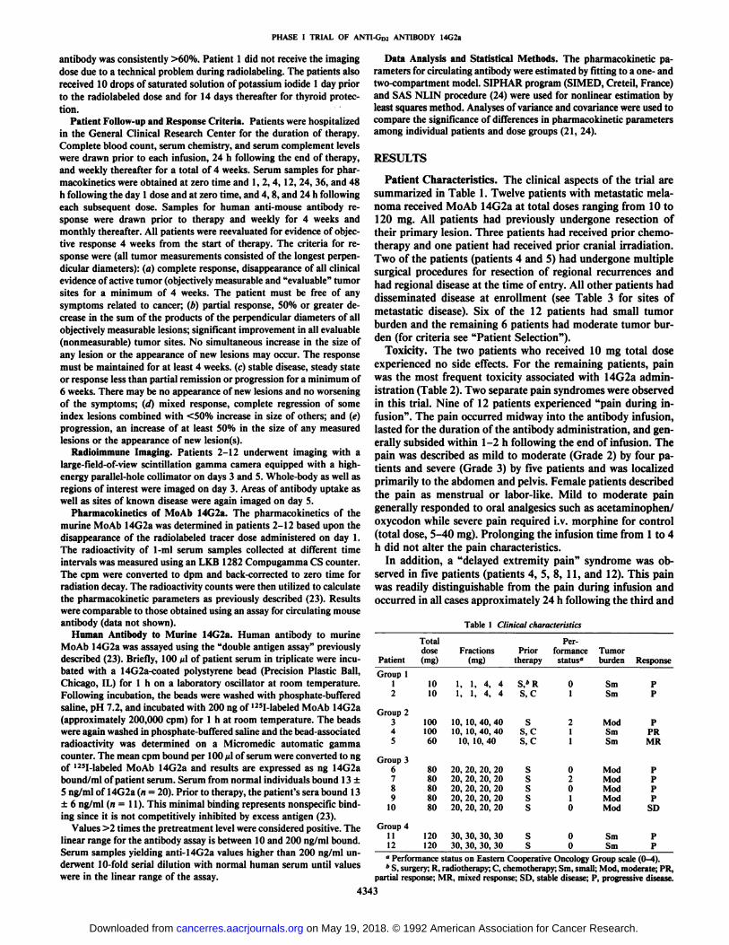
Patient Characteristics. The clinical aspects of the trial aresummarized in Table 1. Twelve patients with metastatic melanoma received MoAb 14G2a at total doses ranging from 10 to120 mg. All patients had previously undergone resection oftheir primary lesion. Three patients had received prior chemotherapy and one patient had received prior cranial irradiation.Two of the patients (patients 4 and 5) had undergone multiplesurgical procedures for resection of regional recurrences andhad regional disease at the time of entry. All other patients haddisseminated disease at enrollment (see Table 3 for sites ofmetastatic disease). Six of the 12 patients had small tumorburden and the remaining 6 patients had moderate tumor burden (for criteria see "Patient Selection").
Toxicity. The two patients who received 10 mg total doseexperienced no side effects. For the remaining patients, painwas the most frequent toxicity associated with 14G2a administration (Table 2). Two separate pain syndromes were observedin this trial. Nine of 12 patients experienced "pain during infusion". The pain occurred midway into the antibody infusion,
lasted for the duration of the antibody administration, and generally subsided within 1-2 h following the end of infusion. Thepain was described as mild to moderate (Grade 2) by four patients and severe (Grade 3) by five patients and was localizedprimarily to the abdomen and pelvis. Female patients describedthe pain as menstrual or labor-like. Mild to moderate paingenerally responded to oral analgesics such as acetaminophen/oxycodon while severe pain required i.v. morphine for control(total dose, 5-40 mg). Prolonging the infusion time from 1 to 4h did not alter the pain characteristics.
In addition, a "delayed extremity pain" syndrome was ob
served in five patients (patients 4, 5, 8, 11, and 12). This painwas readily distinguishable from the pain during infusion andoccurred in all cases approximately 24 h following the third and
Table 1 Clinical characteristics
PatientGroup
112Group
2345Group
3678910Group
41112Total
dose(mg)1010100100608080808080120120Fractions(mg)1,
1, 4,41,1, 4,410,
10, 40,4010,10, 40,4010,
10,4020,
20, 20,2020,20, 20,2020,20, 20,2020,20, 20,2020,20, 20,2030,
30, 30,3030,30, 30, 30Prior
therapyS/RS,
CSS,
CS,CsssssssPer
formancestatus"012110201000TumorburdenSmSmModSmSniModModModModModSmSmResponsePPPPRMRPPPPSDPP
" Performance status on Eastern Cooperative Oncology Group scale (0-4).* S, surgery; R, radiotherapy; C, chemotherapy; Sm, small; Mod, moderate; PR,
partial response; MR, mixed response; SD, stable disease; P, progressive disease.
4343
on May 19, 2018. © 1992 American Association for Cancer Research. cancerres.aacrjournals.org Downloaded from

PHASE l TRIAL OF ANTI-GD2 ANTIBODY 14G2a
Table 2 Toxicity associated with ¡4G2atherapy"
PatientGroup
112Group
2345Total
dose(mg)101010010060Pain
duringinfusion00233Delayed
extremitypain00033Neuro-toxicity0002(S)*4(M)3(C)Hypo-natremia00002Fever3
Group3678910Group
41112808080808012012002322330030023000002(S)4(M)4(C)00000004
" Toxicity reported by National Cancer Institute Consensus grading system(0-4).
* S, neurosensory; M, neuromuscular, C, neurocortical.
fourth infusions. The pain was described as severe and burningand affected both hands and both legs below the knee. The painrequired i.v. morphine for control in all patients except patient11 who responded to self-administered anti-inflammatoryagents and local heat application to the extremities. The paingenerally was resolved completely within 24-48 h except inpatient 4 who had recurrence of the extremity pain followingdischarge from the hospital.
Four of the five patients who experienced the delayed extremity pain also developed neurological toxicity (patients 4, 5, 11,and 12). Patient 4 noted recurrence of the severe distal leg andhand pain approximately 5 days following the last antibodydose. Two days later he was admitted to his local hospital withintractable leg pain accompanied by paresthesia and leg weakness. He was unable to walk without assistance because of theleg weakness and required a continuous infusion of morphinefor pain control. He underwent physical therapy and was discharged 10 days later with improvement in the weakness anddecrease in pain. Two weeks following his discharge, neurological examination revealed persistent decreased strength in theproximal thigh muscles, which was more pronounced on theright. Deep tendon reflexes were decreased in both lower extremities. Sensory examination revealed paresthesia in bothlower extremities with a stocking distribution. The patient alsocomplained of a cold sensation in both his hands and feet.Electromyography studies conducted at our institution revealedevidence of denervation in the proximal and distal muscles ofboth lower extremities, which was more pronounced on theright. Biopsy of the right sural nerve revealed demyelinationand a mononuclear infiltrate in the endoneural and perivascularspace. Immunohistological studies demonstrated GD2 on thepatient's nerve fibers. MoAb 14G2a was not detected on the
sural nerve tissue (the biopsy was performed approximately 4weeks after completion of MoAb therapy). The patient's clini
cal picture was compatible with the diagnosis of mononeuritismultiplex/polyradiculopathy. The patient refused steroid therapy. His neurological function gradually improved and returnedto base line over the ensuing 4 to 6 weeks. At last follow-up (20weeks from the end of therapy), the patient had regained full
sensory as well as motor function but continued to complain ofdysthermia in the hands and feet.
Patient 5 developed mental confusion and disorientation 24 hfollowing the third dose of 14G2a. The altered mental statuscoincided with onset of the delayed extremity pain. The patientwas incidentally noted to have a serum sodium of 124 mg/dl(with a normal electrolyte profile 3 days prior). Serum and urineosmolality studies were consistent with SIADH. The patient's
hyponatremia corrected promptly with fluid restriction. Shecontinued, however, to be confused and disoriented for approximately 1 week. Evaluation including computed tomographyscan of the head revealed no evidence of a specific abnormality.The patient did not receive the fourth dose of 14G2a.
Patient 11 complained of severe extremity pain and loss ofsensation in both lower extremities on the day following thefourth antibody infusion. The patient was at home and resortedto taking antiinflammatory agents and applying local heat tothe lower extremities with apparent relief of symptoms. He wasseen by his local physician the next day and was noted to havea completely normal neurological examination by that time.There were no further symptoms or signs of neurological damage.
Patient 12 received the same dose of antibody as patient 11.Six h following the last antibody infusion she complained ofinability to void. This was followed by progressive lower extremity weakness over the next 24 h to the point where she wasunable to raise her legs or stand without assistance. Neurological examination and nerve conduction studies were compatiblewith a neurogenic bladder and motor radiculopathy involvingthe L2-4 and SI nerve roots. Cerebral and spinal magneticresonance imaging scans revealed no evidence of intracranialmetastasis or cord compression. The patient also had a serumsodium of 113 mg/dl (with a normal electrolyte profile 2 daysprior). Serum and urine osmolality studies were compatiblewith SIADH. While hyponatremic, the patient had a grand malseizure. Treatment was started with 80 mg of prednisone dailyfor presumptive inflammatory myelopathy. Three weeks later,she had regained full bladder control and within 6 weeks wasable to ambulate with a walker. At last follow-up (16 weeksfrom the end of therapy), she was able to ambulate withoutassistance but had not attained full motor strength.
Six of the patients (patients 3, 4, 5, 8, 9, and 12) demonstrated a > 25% decrease in the total serum hemolytic complement between days 1 and 9. No other laboratory alteration wasobserved in the patient population. One patient (patient 4) developed transient fever up to 104°Cfollowing both the third and
fourth antibody infusions. None of the patients developed hypertension during antibody therapy.
Clinical Response. One patient each had a partial response,a mixed response, and stable disease at 4-week follow-up (Table1). Patient 4 had pathologically documented complete resolution of two of four s.c. index lesions, each measuring 1 x 1 cm(partial response). Patient 5 had approximately 50 small (5-7-mm) cutaneous melanotic lesions on the inner aspect of the leftankle as well as an inguinal lymph node measuring 2x1 cm.She had erythema and pruritus at the site of the small cutaneousdeposits during infusion therapy. Blanching of lesions was observed 1 week following therapy. Three lesions biopsied 1 weekfollowing therapy revealed necrotic tissue without residual melanoma. At 4-week follow-up, some of the lesions had regressedwhile others showed <50% increase in size. The index inguinallymph node measured 1 x 1 cm (mixed response). Patient 10had a pelvic mass as well as inguinal adenopathy originating
4344
on May 19, 2018. © 1992 American Association for Cancer Research. cancerres.aacrjournals.org Downloaded from

PHASE I TRIAL OF ANTI-GD¡ANTIBODY 14G2a
from melanoma of the vulva. Both sites showed no change insize at 4- and 8-week follow-up (stable disease). All other patients had disease progression.
Radioimmune Imaging. Eight of 11 patients (73%) hadMoAb localization to at least 1 tumor site by radioimmuneimaging (Table 3). Of a total of 34 lesions documented by eitherphysical examination or conventional diagnostic studies, 13(38%) were visualized upon imaging. With the exception of onesingle lesion that was observed only on day 5, all other lesionswere visualized equally well on days 3 and 5.
Pharmacokinetics. The plasma disappearance curves of ra-diolabeled 14G2a best fit a one-compartment model. Theplasma half-life ranged from 30 to 49 h with an overall meanT1,2 of 42 ±6 h (Table 4). High-performance liquid chroma-
tography analysis of randomly selected serum samples revealedthe radioactivity to be associated with the IgG fraction with noevidence of free iodine. The plasma half-life, volume of distribution, and clearance rate did not vary significantly amongpatients receiving different doses of unlabeled MoAb on day 1.
HAMA. Table 5 lists the antibody response to murine14G2a over a 4-week period. Prior to therapy, all patients except patient 3 had baseline levels of 14G2a binding similar tothose of healthy control donors. All 12 patients developed antibody to murine 14G2a. Patients frequently required a 1:100-to 1:1000-fold dilution of their serum to accurately quantitatethe magnitude of the antibody response. Antibody response wasdetectable by day 10 in all except three patients (patients 2, 11,
Table 3 Radioimmune imaging
Table 4 Pharmacokinetics of MoAb I4G2a
PatientGroup
12Group
2345Group
3678910Group
41112Total
dose(mg)10100100608080808080120120MetastaticsiteCutaneousLiverPelvic
massLiverPelvic
massCutaneousCutaneous
(multiple)InguinalLNLiverAdrenalOral
massLiverAxillary
LNLungPelvic
massSpleenChest
wallThigh(s.c.)SubmandibleLNInguinalLNPeriaorticLNLungRight
inguinalLNLeftinguinalLNPelvic
massAxillary
LNMediastinalLNAdrenalRight
lungLeftlungThigh(s.c.)LungSize
(cm)Nm»<2Nm5X24.51<12x
13x34x45x53x51.55
x104.5x4.5Ix1NmNm2x33
x2.52x21.5X22x2Nm2x25X31.5x212x261.5
x 1Imaging"——+++—++—+————+——+++—-——+———-—+-
Patient23456789101112Mean±SDDose(mg)°110101020202020203030T'/2(h)493049484033364047444842
±6yda*>(ml/kg)505841585236365944614850±9MRTC
(h)714371695847525867636961
±9CL"(ml/h/kg)0.711.330.580.840.900.760.701.010.650.960.690.83±0.21R299.097.298.499.499.199.198.998.599.098.997.698.5±0.8
" Dose of unlabeled 14G2a administered with the 0.5-mg radiolabeled dose onday 1.
* Vd„,volume of distribution at steady state; MRT, mean residence time; CL,clearance.
Table 5 Human antibody response to murine 14G2a
Days following firstdosePatientGroup
112Group
2345Group
3678910Group
41112Pre-14G2a15«151152412161041774191045181,5275713,0801,609755264721,3564711232800NT»2190NTNTNTNT956NT481591514,080111,80086,00025,60015,91011,5805,0101134,88029,0301,7959,850229,960123,700NTNT15,5706,3502,390518,3306,7509536,760296,660106,600NT17,980NT5,0409127622NT7472,091
" Radioimmune imaging.* Nm, nonmeasurable; LN, lymph node; —¿�,not visualized; +, unequivocal up
take of labeled MoAb.
" ng of '"I-labeled 14G2a bound/ml of serum.* NT, not tested.
and 12). Peak levels of human anti-14G2a antibody were evident by day 15 in ten patients and by day 22 in the remainingtwo patients (patients 2 and 9). Antibody to 14G2a persisted inall patients and could be detected as far out as day 122 (data notshown). The antibody response was not dose dependent in thatthe highest antibody response was noted in patient 2 (10-mgdose) and the lowest level of immune response occurred inpatient 8 (80-mg dose).
DISCUSSION
Administration of the anti-GD2 murine MoAb 14G2a in thistrial was associated with a spectrum of unusual and unanticipated side effects not observed with other antiganglioside Mo-Abs. Two distinct pain syndromes were observed in our trial.Nine often patients receiving >10 mg 14G2a developed a syndrome of severe abdominal/pelvic pain during each antibodyinfusion. Five patients also experienced a delayed extremitypain syndrome following the third and fourth infusion. Neuro-toxicity was noted in four of these five patients. Two patientsdeveloped hyponatremia secondary to SIADH. The maximumtolerated total dose was determined to be 80 mg since dose-limiting neurotoxicity occurred at 100-120 mg. The antitumorresponse was modest and one patient each had a partial response, mixed response, and stable disease.
4345
on May 19, 2018. © 1992 American Association for Cancer Research. cancerres.aacrjournals.org Downloaded from
PHASE I TRIAL OF ANTI-GD2 ANTIBODY 14G2a
The abdominal pain associated with 14G2a infusion is sim- clearance of the day 8 dose as compared to the day 1 infusion.ilar to that reported with the unrelated anti-GD2 MoAb 3F8(16). This side effect appears to be characteristic of anti-GD2antibodies since no comparable toxicity has been observed inpatients receiving the anti-GD3 MoAb R24 (18, 19). Chesttightness has been reported with R24 administered alone (19)or when combined with interleukin 2 (25). The GD2 (but not( M,.,) antigen has been found on peripheral nerve tissue (20) andsome investigators have speculated that the infusion-relatedpain syndrome may be due to binding of antibody to peripheralpain fibers (16). The location of the pain (abdominal as opposedto peripheral extremity) and the rather rapid cessation of painfollowing completion of antibody infusion (despite high levelsof circulating antibody), however, cannot be accounted for bysuch a scenario. We have demonstrated that 14G2a binds tosmooth muscle of the gastrointestinal tract, as well as the surrounding mesenteric blood vessels.4 Whether binding of anti
body to these sites may cause smooth muscle and vascularspasm and thereby contribute to the abdominal pain is unclear.Toxicity related to rate of antibody infusion has been observedwith other murine monoclonal antibodies (18, 26, 27). Thesesymptoms have been ascribed to binding of antibody to nontu-mor sites (26) or due to immune complex and/or aggregateformation (27). These symptoms frequently respond to slowingof the infusion rate (26, 27). Prolonging the infusion time of14G2a from 1 to 4 h in this trial did not alter the pain. Administration of 14G2a as a continuous infusion over 24 h has alsobeen associated with a similar pain syndrome (15, 28). It thusappears that the pain during infusion is not simply dependenton the infusion rate and etiology of this toxicity still remains tobe determined. Given the transient nature of this toxicity andadequate control obtained with i.v. morphine, pain during infusion was not dose limiting in this study. Hypertension was notobserved in this trial, although both 3F8 and high-dose R24therapy have been associated with blood pressure elevation(16, 29).
The delayed extremity pain syndrome observed with 14G2aoccurred approximately 24 h following the third and fourthinfusion. None of the three patients receiving the 10-mg dose,two of six patients receiving 60-80-mg doses, and three of fourpatients receiving >100 mg 14G2a experienced this side effect.Except for the higher dose of antibody administered, no specificclinical characteristic identified those patients predisposed tothis side effect. It is noteworthy that four of the five patientsexperiencing the extremity pain syndrome also developed neurological toxicity following their last infusion.
Neurological toxicity associated with 14G2a therapy consisted primarily of sensory and motor neuropathy. Altered mental status was noted in two patients, both of whom had concomitant hyponatremia secondary to SIADH. Patients 4 and 12developed an acute demyelinating process with severe motorneuropathy that reversed over the ensuing months. Both patients received >80 mg antibody. This finding was unanticipated since no such side effect was observed in our trial usingcomparable doses of the related human/mouse chimeric anti-GD2 MoAb ch 14.18 (30, 31). The precise etiology of the neu-rotoxicity associated with 14G2a and absence thereof with ch14.18 remains unclear. All patients exposed to 14G2a developed high levels of HAMA while ch 14.18 therapy has beenassociated with a weak immune response directed at the variable region (32). Patients 4 and 12 both had a much more rapid
4 Unpublished data.
No circulating 14G2a was detectable after the 48-h time point(day 10) (data not shown). Circulating HAMA was detected byday 11 (TableS). Whether the demyelinating neuropathy couldhave been caused by binding of 14G2a/anti-14G2a immunecomplexes to the peripheral nerve with resulting immune-mediated cytolysis remains speculative. The role of the day 8 doseand HAMA on the subsequent development of acute neurotox-icity remains unclear and cannot be conclusively ascertainedfrom our date. Neither of our patients had nerve biopsies at thetime of acute neurotoxicity looking for bound antibody and/orcomplement deposition. Interestingly, four of the five patientswho developed neurological side effects had a >25% reductionin total hemolytic complement during therapy.
The pharmacokinetics of 14G2a followed a one-compartment model similar to what has been observed with other murine reagents (21, 33). The immune response to 14G2a, however, exceeds that previously described for m-17 -1A (21) orm-B72.3 (33) by 2 to 3 logs using similar assay techniques. Ourresults demonstrated no correlation between the degree of immune response and the dose of antibody administered.
No major responses were observed at the doses of 14G2aused in this trial and the maximum tolerated dose was determined to be 80 mg (total dose). In previous trials of 3F8 andR24 higher doses could be administered and favorable clinicalresponses could be achieved (16, 18, 19). Ongoing studies withthe chimeric MoAb ch 14.18 have not been associated with anydose-limiting toxicity thus far (31) and we are proceeding withdose escalation using this reagent.
ACKNOWLEDGMENTS
The authors wish to acknowledge the help and expertise of SharonGarrison, Angela Kelley, and Connie Howton in preparing the manuscript.
REFERENCES1. Reisfeld, R. A., and Sell, S. Monoclonal Antibodies and Cancer Therapy, pp.
173-191. New York: Alan R. Liss, Inc., 1985.2. Goodman, G. E., Beaumier, P., Hellstróm, I., Fernyhough, B., and Hell
ström,K. E. Pilot trial of murine monoclonal antibodies in patients withadvanced melanoma. J. Clin. Oncol., 3: 340-352, 1985.
3. Scheinberg, D. A., and Houghton, A. N. Current status of antitumor therapywith monoclonal antibodies. Oncology (Basel), /: 31-40, 1987.
4. Cheresh, D. A., Harper, J. R., Schulz, G., and Reisfeld, R. A. Localization ofthe gangliosides GDI and (¡IMin adhesion plaques and on surface of humanmelanoma cells. Proc. Nati. Acad. Sci. USA, 81: 5767-5771, 1984.
5. Cheresh, D. A., Pierschbacher, M. D., Herzig, M. A., and Mujoo, K. Disia-logangliosides GD2 and GD3 are involved in the attachment of human melanoma and neuroblastoma cells to extracellular matrix proteins. J. Cell Biol.,¡02:688-696, 1986.
6. Hakomori, S-l., and Kannagi, R. Glycosphingolipids as tumor-associatedand differentiation markers. J. Nati. Cancer Inst., 71: 231-251, 1983.
7. Tsuchida, T., Saxton, R. E., Morton, D. L., and Irie, R. F. Gangliosides ofhuman melanoma. Cancer (Phila.), 63: 1166-1174, 1989.
8. Cheung, N. K., Saarinen, U. M., Neely, J. E., Landmeier, B., Donovan, D.,and Coccia, P. F. Monoclonal antibodies to a glycolipid antigen on humanneuroblastoma cells. Cancer Res., 45: 2642-2649, 1985.
9. Cheresh, D. A., Honsik, C. J., Staffileno, L. K., Jung, G., and Reisfeld, R. A.Disialoganglioside GD3 on human melanoma cells serves as a relevant targetantigen for monoclonal antibody-dependent cytotoxicity and immunother-apy. Proc. Nati. Acad. Sci. USA, 82: 5155-5159, 1985.
10. Mujoo, K., Cheresh, D. A., Yang, H. M., and Reisfeld, R. A. Disialoganglioside Go2 on human neuroblastoma cells: target antigen for monoclonalantibody-mediated cytolysis and suppression of tumor growth. Cancer Res.,47: 1098-1104, 1987.
11. Imai, K., Ng, A. K., and Ferrone, S. Characterization of monoclonal antibodies to melanoma-associated antigens. J. Nati. Cancer Inst., 66:489-496,1981.
12. Cheresh, D. A., Rosenberg, J., Mujoo, K., Hirschowitz, L., and Reisfeld, R.A. Biosynthesis and expression of the disialoganglioside GDI, a relevanttarget antigen on small cell lung carcinoma for monoclonal antibody-mediated cytolysis. Cancer Res., 46: 5112-5118, 1986.
4346
on May 19, 2018. © 1992 American Association for Cancer Research. cancerres.aacrjournals.org Downloaded from

PHASE I TRIAL OF ANTI-GD2 ANTIBODY 14G2a
13. Cheresh, D. A., Pierschbacher, M. D., Herzig, M. A., and Mujoo, K. Disia-logangliosides GDI and GDil are involved in the attachment of human melanoma and neuroblastoma cells to extracellular matrix proteins. J. Cell Biol.,¡02:688-696, 1986.
14. Mujoo, K., Kipps, T. J., Yahg, H. M., Cheresh, D. A., Wargalla, U., Sander,D. J., and Reisfeld, R. A. Functional properties and effects on growth suppression of human neuroblastoma tumors by isotype switch variants of monoclonal anti-ganglioside GD2 antibody 14.18. Cancer Res., 49: 2857-2861,1989.
15. Yu, A., Reisfeld, R., and Gillies, S. Immune response to monoclonal anti-GD2 antibody therapy. Proc. Am. Assoc. Cancer Res., 32: 263, 1991.
16. Cheung, N. K. V., Lazarus, H., Miraldi, F. D., Abramowsky, C. R., Kallick,S., Saarinen, U. M., Spitzer, T., Strandjord, S. E., Coccia, P. F., and Berger,N. A. Ganglioside Go2 specific monoclonal antibody 3F8: a phase I study inpatients with neuroblastoma and malignant melanoma. J. Clin. Oncol., 5:1430-1440, 1987.
17. Dippold, W. G., Dienes, H. P., Knuth, A., and Meyer zum Büschenfelde,K.H. Immunohistochemical localization of ganglioside G,,, in human malignant melanoma, epithelial tumors, and normal tissues. Cancer Res., 45:3699-3705, 1985.
18. Houghton, A. N., Mintzer, D., Cordon-Cardo, C., Welt, S., Fliegel, B.,Vadhan, S., Carswell, E., Melamed, M. R., Oettgen, H. F., and Old, L. J.Mouse monoclonal IgG3 antibody detecting GD2 ganglioside: a phase I trialin patients with malignant melanoma. Proc. Nati. Acad. Sci. USA, 82:1242-1246, 1985.
19. Vadhan-Raj, S., Cordon-Cardo, C., Carswell, E., Mintzer, D., Dantis, L.,Duteau, C., Templeich, M. A., Gettgen, H. F., Old, L. J., and Houghton, A.N. Phase I trial of a mouse monoclonal antibody against r,D, ganglioside inpatients with melanoma: induction of inflammatory responses at tumor sites.J. Clin. Oncol., 6: 1636-1648, 1988.
20. Schulz, G., Cheresh, D. A., Varki, M. N., Yu, A., Staffileno, L. K., andReisfeld, R. A. Detection of ganglioside GD2 in tumor tissue and sera ofneuroblastoma patients. Cancer Res., 44: 5914-5920, 1984.
21. Khazaeli, M. B., Saleh, M. N., Wheeler, R. H., Huster, W. J., Holden, H.,Carrano, R., and LoBuglio, A. F. Phase I trial of multiple large doses ofmurine monoclonal antibody CO 17-1A. II. Pharmacokinetics and immuneresponse. J. Nati. Cancer Inst., 80: 937-942, 1988.
22. Meredith, R. F., Khazaeli, M. B., Plott, W. E., Saleh, M. N., Liu, T. P., Allen,L. F., Russell, C. D., Orr, R. A., Colcher, D., Schlom, J., Shochat, D.,Wheeler, R. H., and LoBuglio, A. F. Phase I trial of iodine-131-chimericB72.3 (human IgG4) in metastatic colorectal cancer. J. NucÃ.Med., 33:23-29, 1992.
23. LoBuglio, A. F., Wheeler, R. H., Trang, J., Haynes, A., Rogers, K., Harvey,E. B., Sun, L., Ghrayeb, J., and Khazaeli, M. B. Mouse/human chimericmonoclonal antibody in man: kinetics and immune response. Proc. Nati.
Acad. Sci. USA, 86: 4220-4224, 1989.24. SAS Institute, Inc. SAS/STAT Users Guide, Version 6, Ed. 4, Vol. 2, pp.
1135-1156. Cary, NC: SAS Institute, Inc., 1989.25. Bajorin, D. F., Chapman, P. B., Wong, B., Coit, D. G., Kunicke, J., Dimag
gio, J., Cordon-Cardo, C., Urmacher, C., Dantes, L., and Templeton, M. A.Phase I evaluation of a combination of monoclonal antibody R24 and inter-leukin 2 in patients with metastatic melanoma. Cancer Res., 50:7490-7495,1990.
26. LoBuglio, A. F., Saleh, M. N., Lee, J., Khazaeli, M. B., Carrano, R., Holden,H., and Wheeler, R. H. Phase I trial of multiple large doses of murinemonoclonal antibody CO17-1A. I. Clinical aspects. J. Nati. Cancer Inst., 80:932-936, 1988.
27. Dillman, R., Shawler, D., Dillman, J., and Roystan, R. Therapy of chroniclymphocytic leukemia and cutaneous T-cell lymphoma with T101 monoclonal antibody. J. Clin. Oncol., 2: 881-891, 1984.
28. Murray, J. L., Cunningham, J. E., Brewer, H. M., Janus, M. H., Podoloff, D.A., Bhadkamaker, V. P., Kasi, L. P., Shah, R. S., Benjamin, R. S., Legha, S.S., Plager, C., Papadopoulos, N. E., Jaffee, N., Ater, J. L., Mujoo, K., Itoh,K., Ross, M., Bucana, C. D., and Rosenblum, M. G. Phase I trial of murineanti-ganglioside (GD2>monoclonal antibody (Mab) 14G2a in cancer patients.J. Immunother., 11: 135, 1992.
29. Bajorin, D. F., Chapman, P. B., Wong, C. I., Duteau, C., Cody, B., Dantis,L., Templeton, M. A., Urma-Cher, C., Oettgen, H. F., and Houghton, A. N.A phase I trial of high-dose R24 mouse monoclonal antibody in patients withmetastatic melanoma. Proc. Am. Assoc. Cancer Res., 32: 265, 1991.
30. Saleh, M. N., Khazaeli, M. B., Wheeler, R. H., Liu, T. P., Allen, L., Grizzle,W., Tilden, A. B., and LoBuglio, A. F. A phase I trial of anti-GD2 chimericmonoclonal antibody C14.18 in patients with metastatic melanoma: pliarmacokinetics and human immune response. Proc. Soc. Clin. Oncol. Ann.Meet., / 0:217, 1991.
31. Saleh, M. N., Khazaeli, M. B., Wheeler, R. H., Allen, L., Tilden, A. B.,Grizzle, W., Reisfeld, R. A., Yu, A. L., Gillies, S. D., and LoBuglio, A. F. Aphase I trial of the chimeric anti-GD2 monoclonal antibody ch 14.18 inpatients with malignant melanoma. Hum. Antibodies Hybridomas 3:19-24,1992.
32. Khazaeli, M. B., Saleh, M. N., Liu, T. P., Allen, L., Rogers, K., Wheeler, R.H., Reisfeld, R. A., and LoBuglio, A. F. Murine and chimeric antibody toGD2 ¡nmelanoma patients: pharmacokinetics and immune response. Proceedings of the 8th International Hammersmith Meeting, Advances in theApplications of Monoclonal Antibodies in Clinical Oncology, Porto Carras,Halkidiki, Greece, May 8-13, 1991, p. 19.
33. Khazaeli, M. B., Saleh, M. N., Liu, T. P., Kaladas, P. M., Oilman, S. C, andLoBuglio, A. F. Characterization of human immune response to murineB72.3 monoclonal antibody. Antibody Immunoconj. Radiopharm., 4: 212,1991.
4347
on May 19, 2018. © 1992 American Association for Cancer Research. cancerres.aacrjournals.org Downloaded from

1992;52:4342-4347. Cancer Res Mansoor N. Saleh, M. B. Khazaeli, Richard H. Wheeler, et al. 14G2a in Metastatic Melanoma
AntibodyD2Phase I Trial of the Murine Monoclonal Anti-G
Updated version
http://cancerres.aacrjournals.org/content/52/16/4342
Access the most recent version of this article at:
E-mail alerts related to this article or journal.Sign up to receive free email-alerts
Subscriptions
Reprints and
To order reprints of this article or to subscribe to the journal, contact the AACR Publications
Permissions
Rightslink site. Click on "Request Permissions" which will take you to the Copyright Clearance Center's (CCC)
.http://cancerres.aacrjournals.org/content/52/16/4342To request permission to re-use all or part of this article, use this link
on May 19, 2018. © 1992 American Association for Cancer Research. cancerres.aacrjournals.org Downloaded from


