PEDIATRIC ACQUIRED HEART DISEASESrwjms.rutgers.edu/departments_institutes/pedspweb/... · PEDIATRIC...
45
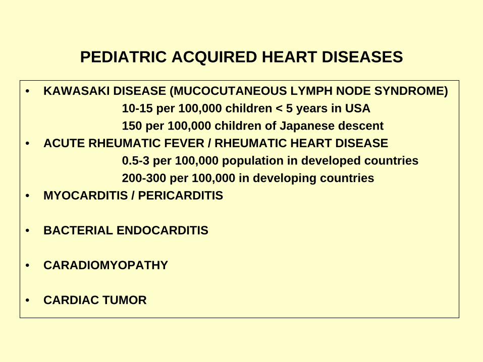
PEDIATRIC ACQUIRED HEART DISEASES • KAWASAKI DISEASE (MUCOCUTANEOUS LYMPH NODE SYNDROME) 10-15 per 100,000 children < 5 years in USA 150 per 100,000 children of Japanese descent • ACUTE RHEUMATIC FEVER / RHEUMATIC HEART DISEASE 0.5-3 per 100,000 population in developed countries 200-300 per 100,000 in developing countries • MYOCARDITIS / PERICARDITIS • BACTERIAL ENDOCARDITIS • CARADIOMYOPATHY • CARDIAC TUMOR
-
Upload
trinhduong -
Category
Documents
-
view
223 -
download
0
Transcript of PEDIATRIC ACQUIRED HEART DISEASESrwjms.rutgers.edu/departments_institutes/pedspweb/... · PEDIATRIC...
PEDIATRIC ACQUIRED HEART DISEASES
• KAWASAKI DISEASE (MUCOCUTANEOUS LYMPH NODE SYNDROME)10-15 per 100,000 children < 5 years in USA150 per 100,000 children of Japanese descent
• ACUTE RHEUMATIC FEVER / RHEUMATIC HEART DISEASE0.5-3 per 100,000 population in developed countries200-300 per 100,000 in developing countries
• MYOCARDITIS / PERICARDITIS
• BACTERIAL ENDOCARDITIS
• CARADIOMYOPATHY
• CARDIAC TUMOR
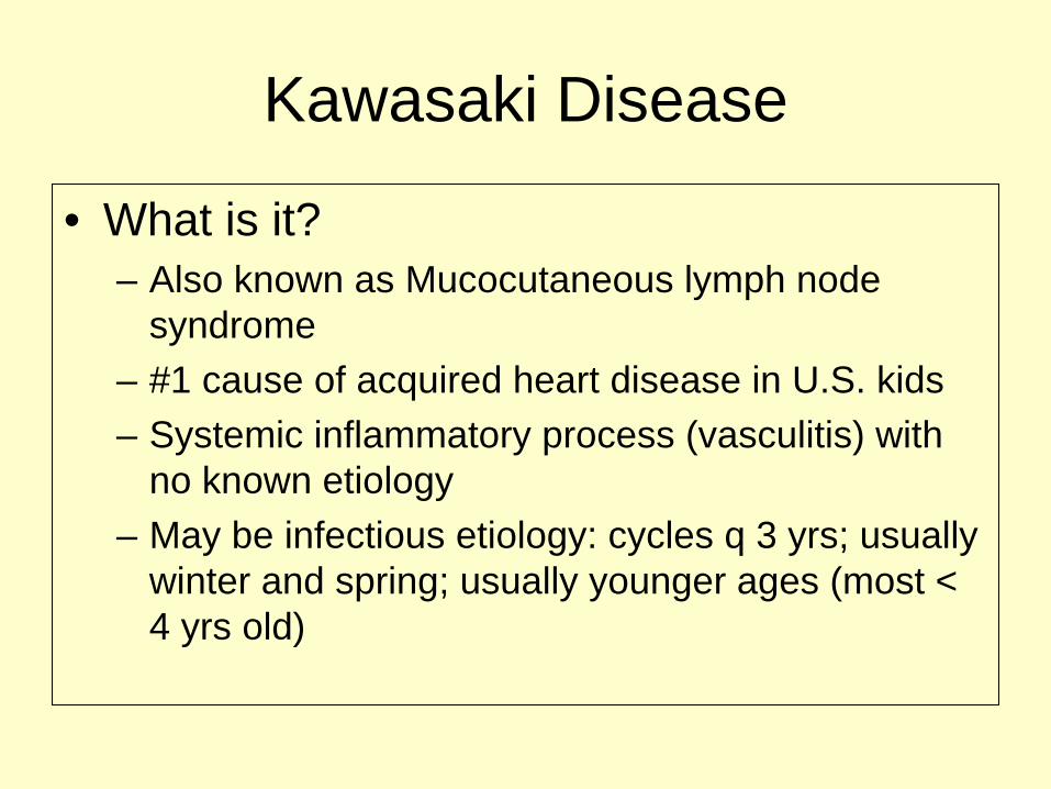
Kawasaki Disease
• What is it?– Also known as Mucocutaneous lymph node
syndrome– #1 cause of acquired heart disease in U.S. kids– Systemic inflammatory process (vasculitis) with
no known etiology– May be infectious etiology: cycles q 3 yrs; usually
winter and spring; usually younger ages (most < 4 yrs old)
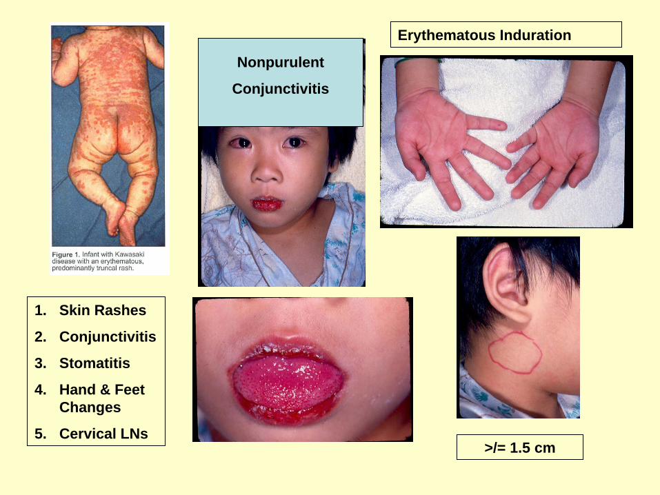
1. Skin Rashes
2. Conjunctivitis
3. Stomatitis
4. Hand & Feet Changes
5. Cervical LNs
Nonpurulent
Conjunctivitis
Erythematous Induration
>/= 1.5 cm
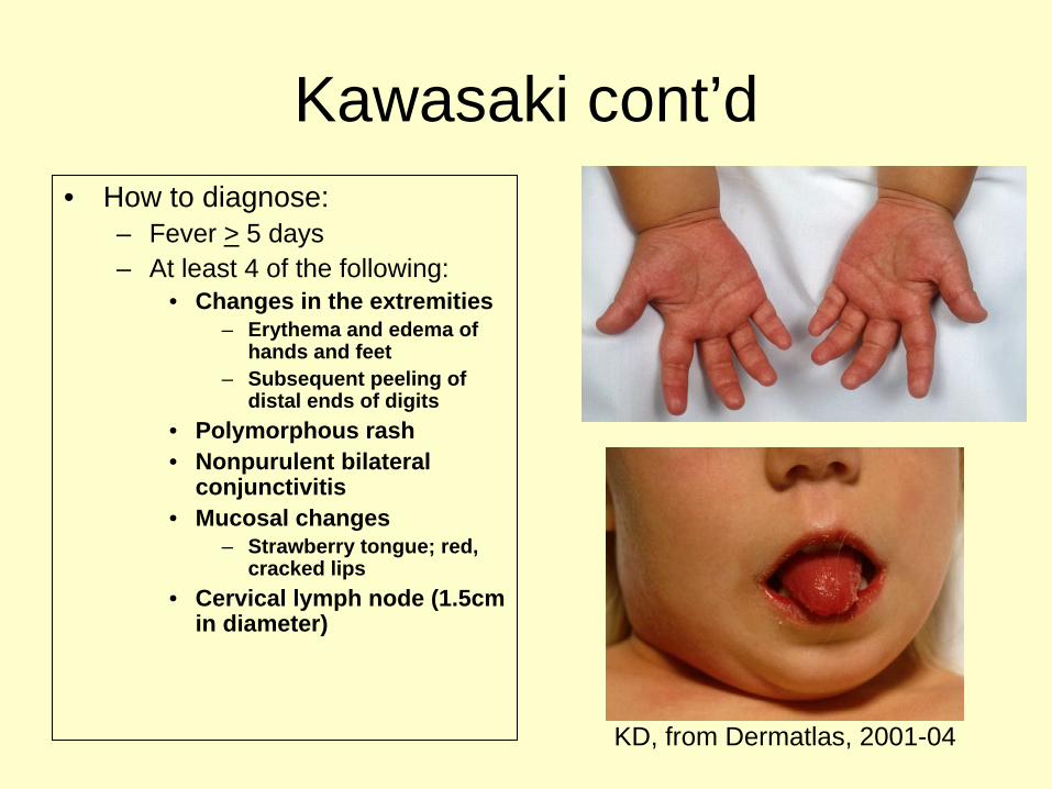
Kawasaki cont’d• How to diagnose:
– Fever > 5 days– At least 4 of the following:
• Changes in the extremities– Erythema and edema of
hands and feet– Subsequent peeling of
distal ends of digits• Polymorphous rash• Nonpurulent bilateral
conjunctivitis• Mucosal changes
– Strawberry tongue; red, cracked lips
• Cervical lymph node (1.5cm in diameter)
KD, from Dermatlas, 2001-04
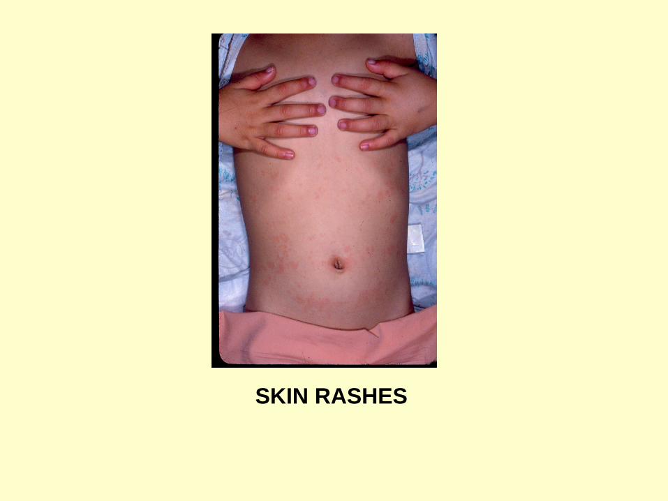
SKIN RASHES
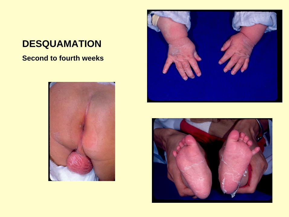
DESQUAMATIONSecond to fourth weeks


Copyright ©2004 American Academy of Pediatrics
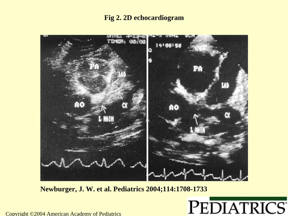
Newburger, J. W. et al. Pediatrics 2004;114:1708-1733
Fig 2. 2D echocardiogram
Copyright ©2004 American Academy of Pediatrics
Newburger, J. W. et al. Pediatrics 2004;114:1708-1733
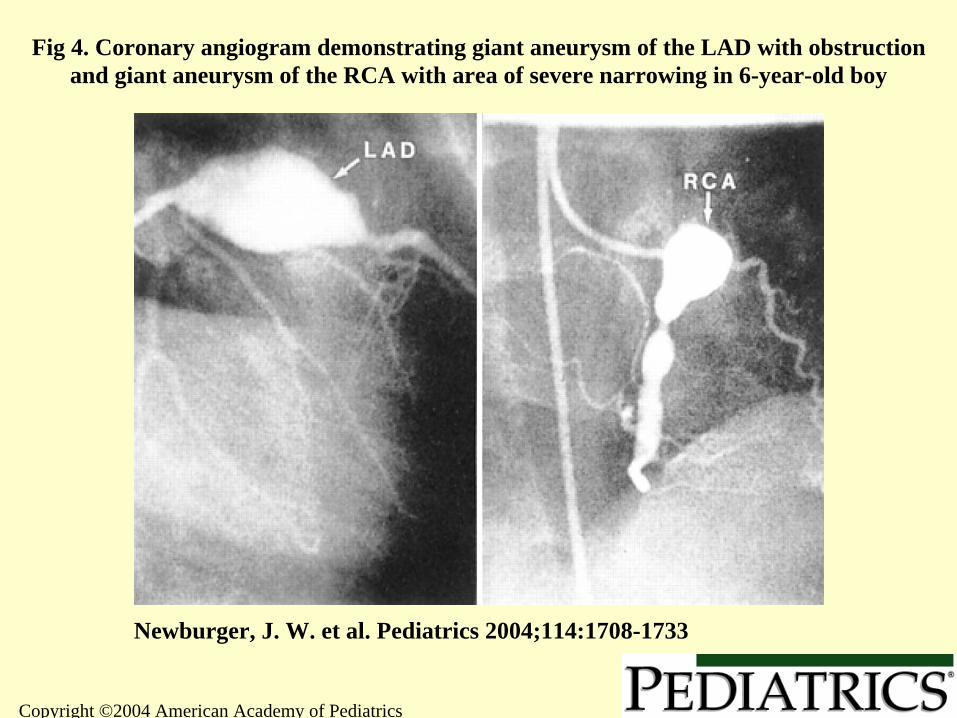
Fig 4. Coronary angiogram demonstrating giant aneurysm of the LAD with obstruction and giant aneurysm of the RCA with area of severe narrowing in 6-year-old boy
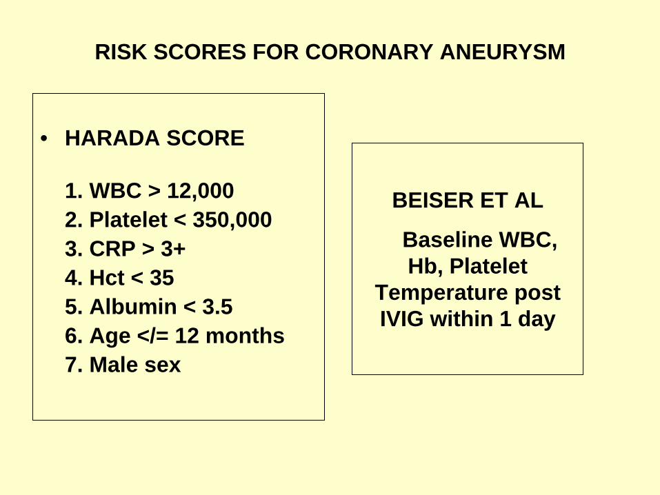
RISK SCORES FOR CORONARY ANEURYSM
• HARADA SCORE
1. WBC > 12,0002. Platelet < 350,0003. CRP > 3+4. Hct < 355. Albumin < 3.56. Age </= 12 months7. Male sex
BEISER ET AL
Baseline WBC, Hb, Platelet
Temperature post IVIG within 1 day
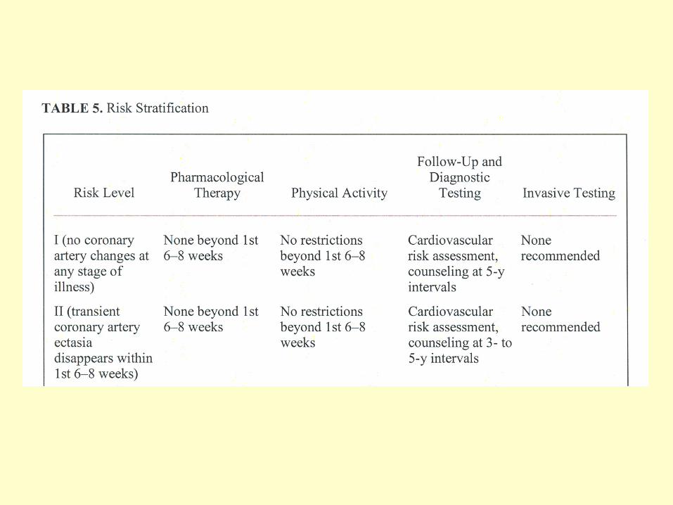
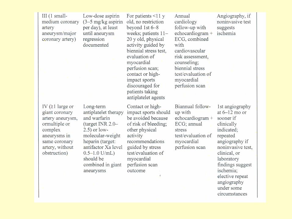
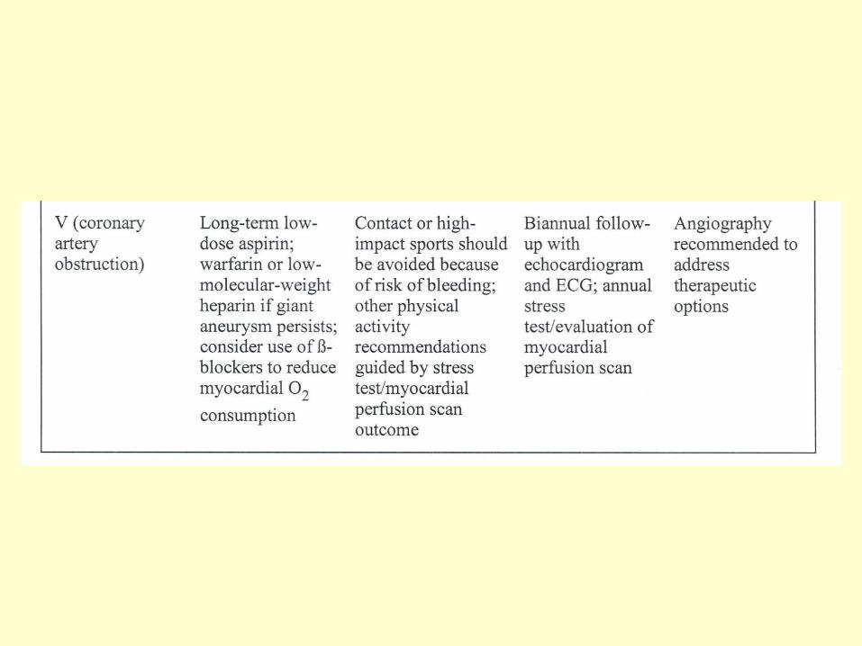

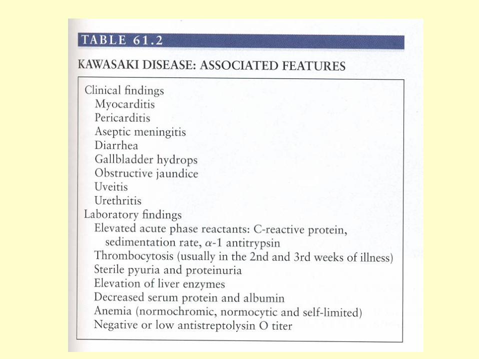
Kawasaki cont’d• Prognosis
– 1/2 to 2/3 of aneurysms will “resolve” by 1-2 yrs post disease onset
• Positive factors for regression:– Small (giant aneurysms (>8mm in diameter) have worst prognosis)– Fusiform (saccular and “beads on a string” have worse prognosis)– < 1 yr of age at time of disease onset– Aneurysm in a distal coronary segment
– Myocardial dysfunction resolves post treatment (unless ischemic damage)
• No correlation between severity of myocarditis and risk for coronary aneurysms
– Peak mortality: 15-45 days post fever onset• Myocardial infarction
– Recurrence rate: ~3% (Japan)
Acute Rheumatic Fever
• What is it?– A pathological immune mediated
inflammatory disorder of the heart, brain, joints, and skin after group A Strep throat infection
– More common in underdeveloped countries– How to diagnose:
• Evidence for group A Strep throat infection and 2 major, or 1 major and 2 minor, criteria
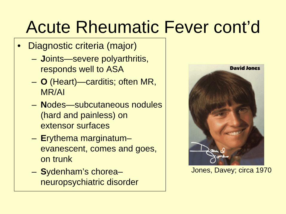
Acute Rheumatic Fever cont’d• Diagnostic criteria (major)
– Joints—severe polyarthritis, responds well to ASA
– O (Heart)—carditis; often MR, MR/AI
– Nodes—subcutaneous nodules (hard and painless) on extensor surfaces
– Erythema marginatum– evanescent, comes and goes, on trunk
– Sydenham’s chorea– neuropsychiatric disorder
Jones, Davey; circa 1970
Acute Rheumatic Fever cont’d
• Diagnostic criteria (minor)– Arthralgia– Fever– Elevated acute phase reactants (ESR, CRP)– Prolonged PR
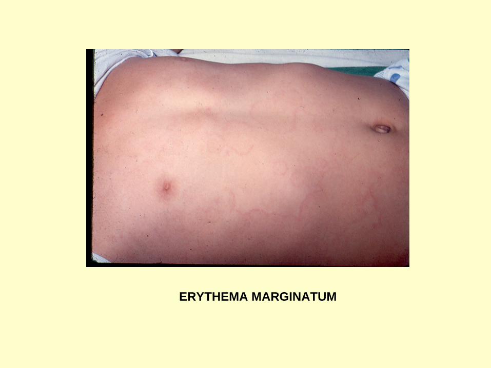
ERYTHEMA MARGINATUM
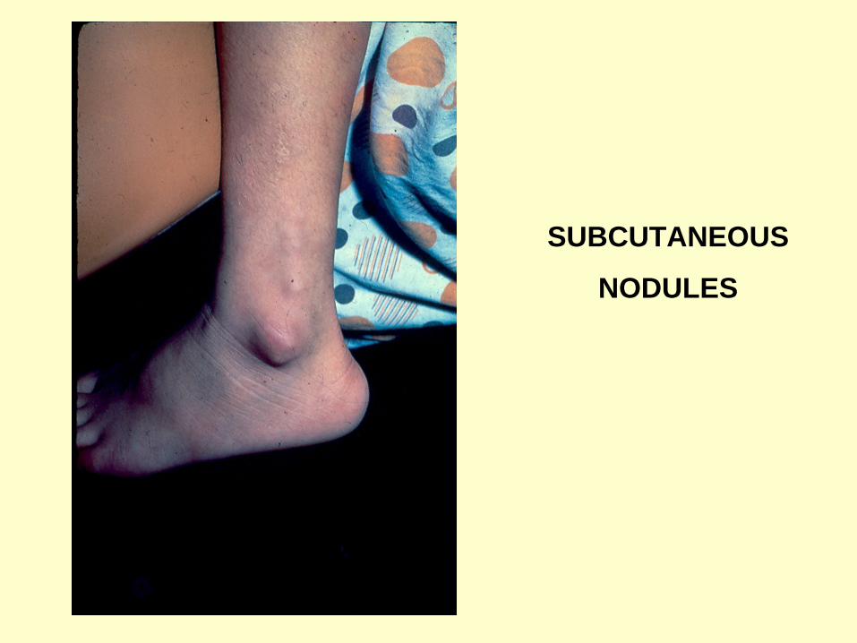
SUBCUTANEOUS
NODULES
Acute Rheumatic Fever cont’d
• Treatment:– Benzathine PCN—knocks out strep– ASA—relieves arthritis, helps mild to
moderate carditis– Prednisone—only for severe carditis– Support if CHF– ABX prophylaxis
Myocarditis• What is it?
– Inflammed myocardium• Etiology?
– Viral• Enteroviruses, esp. Coxsackie B• Adenoviruses• Others: Flu, HSV, Parvo, CMV, HCV, EBV, Mumps,
Rubella, Varicella, HIV, RSV…– Bacterial, Rickettsial, Fungal, Parasitic– Other Noninfectious Inflammatory Diseases
• SLE, Kawasaki, Rheumatic Fever…
Myocarditis cont’d
• Presentation:– Clinically, symptoms and signs similar to
dilated cardiomyopathy (CHF)– ECG: low voltages, arrhythmias (any type),
ST and T wave changes, + wide Q waves– CXR: cardiomegaly, pulmonary edema– Echo: dilated and poorly functioning
ventricles; often occurs with pericardial effusion
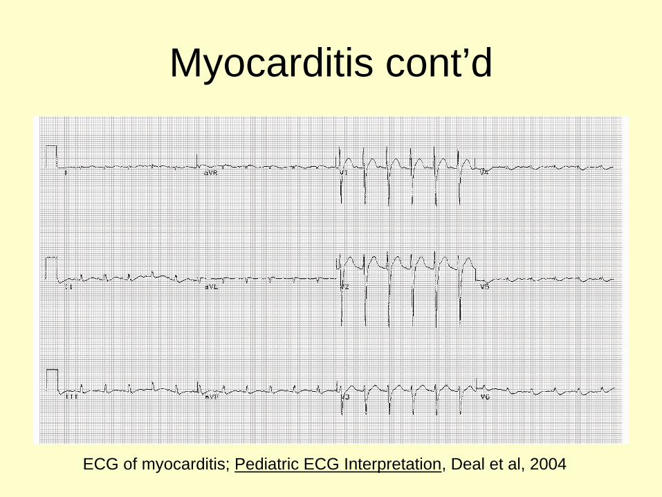
Myocarditis cont’d
ECG of myocarditis; Pediatric ECG Interpretation, Deal et al, 2004
Myocarditis cont’d• Treatment
– Support to relieve CHF and to maintain good cardiac output
• Don’t aggressively load Digoxin in acute inflammatory stage (hypersensitivity)
– Treat arrhythmias• Lidocaine or amiodarone for ventricular arrhythmias
(may require cardioversion if unstable)• Complete heart block: pace
– Treat etiology, if known– No proven benefit of immunosuppresives– If irreversible damage to myocardium, may need
surgical intervention
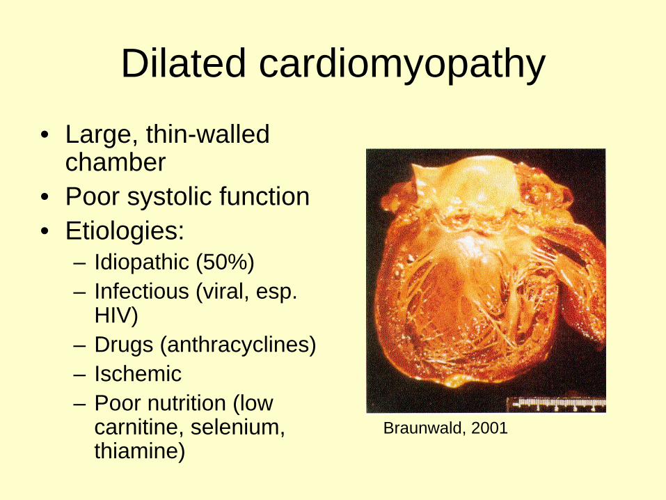
Dilated cardiomyopathy• Large, thin-walled
chamber• Poor systolic function• Etiologies:
– Idiopathic (50%)– Infectious (viral, esp.
HIV)– Drugs (anthracyclines)– Ischemic– Poor nutrition (low
carnitine, selenium, thiamine)
Braunwald, 2001
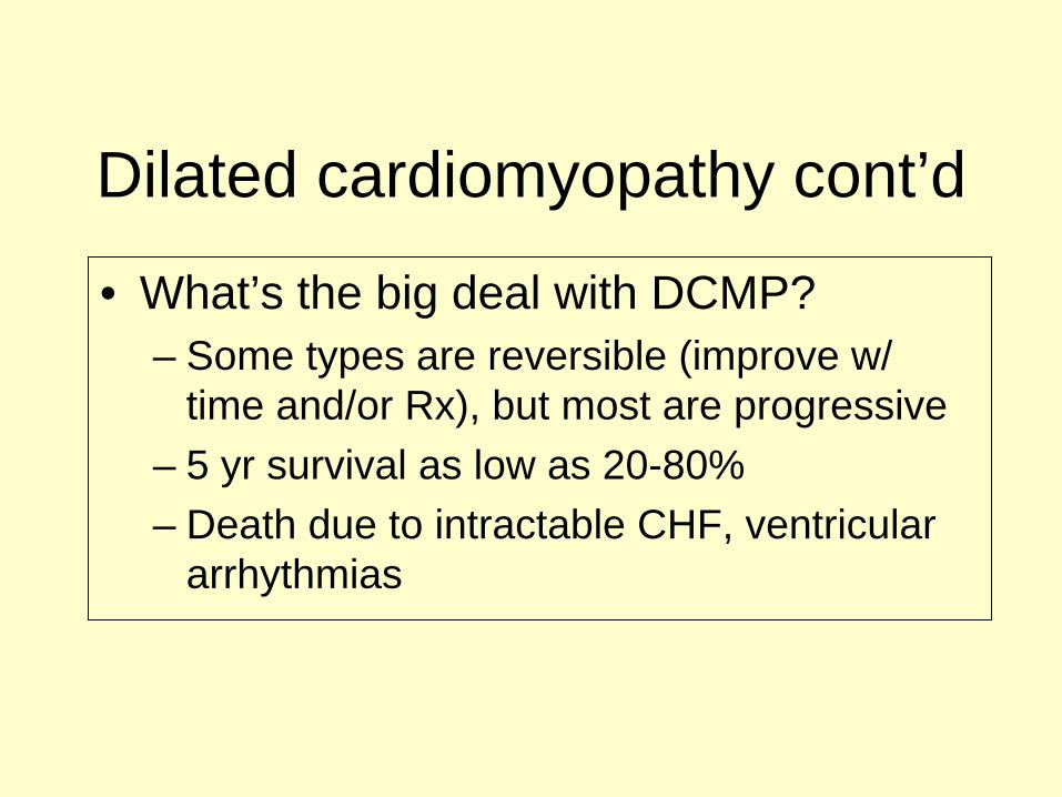
Dilated cardiomyopathy cont’d
• What’s the big deal with DCMP?– Some types are reversible (improve w/
time and/or Rx), but most are progressive– 5 yr survival as low as 20-80%– Death due to intractable CHF, ventricular
arrhythmias
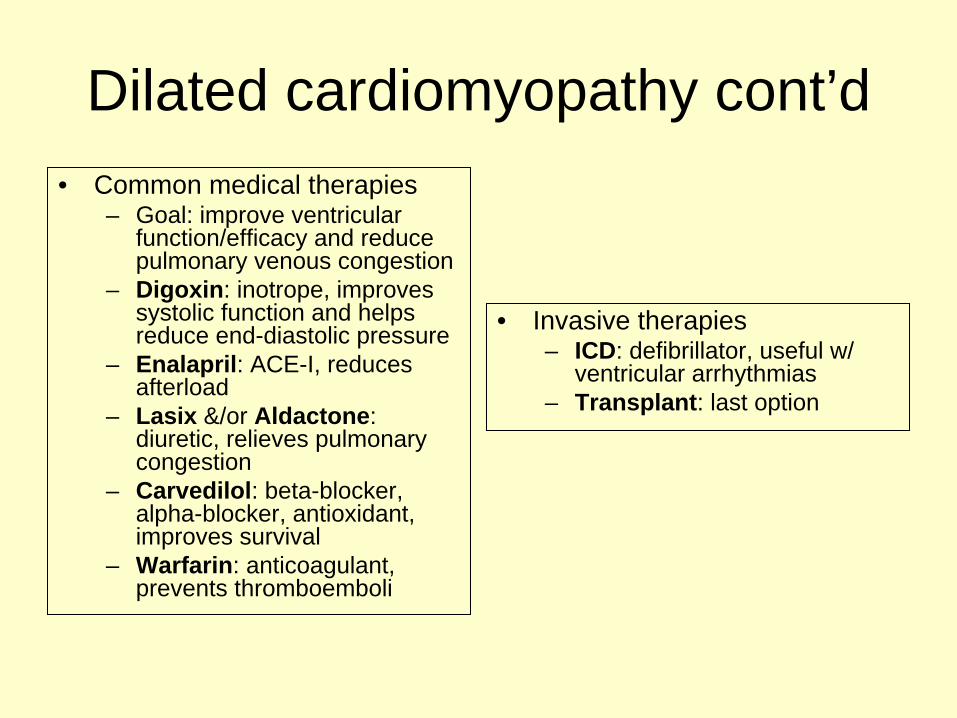
Dilated cardiomyopathy cont’d• Common medical therapies
– Goal: improve ventricular function/efficacy and reduce pulmonary venous congestion
– Digoxin: inotrope, improves systolic function and helps reduce end-diastolic pressure
– Enalapril: ACE-I, reduces afterload
– Lasix &/or Aldactone: diuretic, relieves pulmonary congestion
– Carvedilol: beta-blocker, alpha-blocker, antioxidant, improves survival
– Warfarin: anticoagulant, prevents thromboemboli
• Invasive therapies– ICD: defibrillator, useful w/
ventricular arrhythmias– Transplant: last option
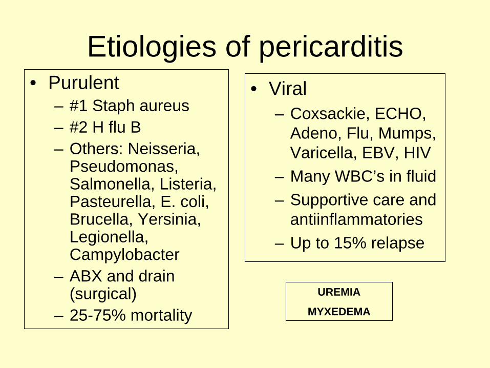
Etiologies of pericarditis• Purulent
– #1 Staph aureus– #2 H flu B– Others: Neisseria,
Pseudomonas, Salmonella, Listeria, Pasteurella, E. coli, Brucella, Yersinia, Legionella, Campylobacter
– ABX and drain (surgical)
– 25-75% mortality
• Viral– Coxsackie, ECHO,
Adeno, Flu, Mumps, Varicella, EBV, HIV
– Many WBC’s in fluid– Supportive care and
antiinflammatories– Up to 15% relapse
UREMIA
MYXEDEMA
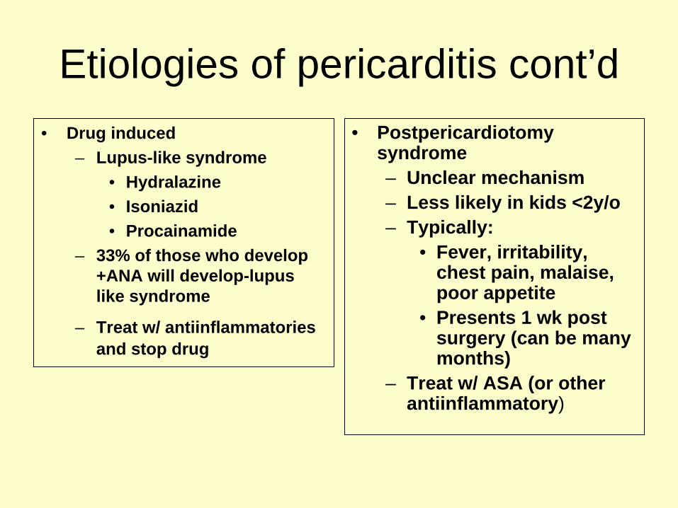
Etiologies of pericarditis cont’d• Drug induced
– Lupus-like syndrome• Hydralazine• Isoniazid• Procainamide
– 33% of those who develop +ANA will develop-lupus like syndrome
– Treat w/ antiinflammatories and stop drug
• Postpericardiotomy syndrome– Unclear mechanism– Less likely in kids <2y/o– Typically:
• Fever, irritability, chest pain, malaise, poor appetite
• Presents 1 wk post surgery (can be many months)
– Treat w/ ASA (or other antiinflammatory)
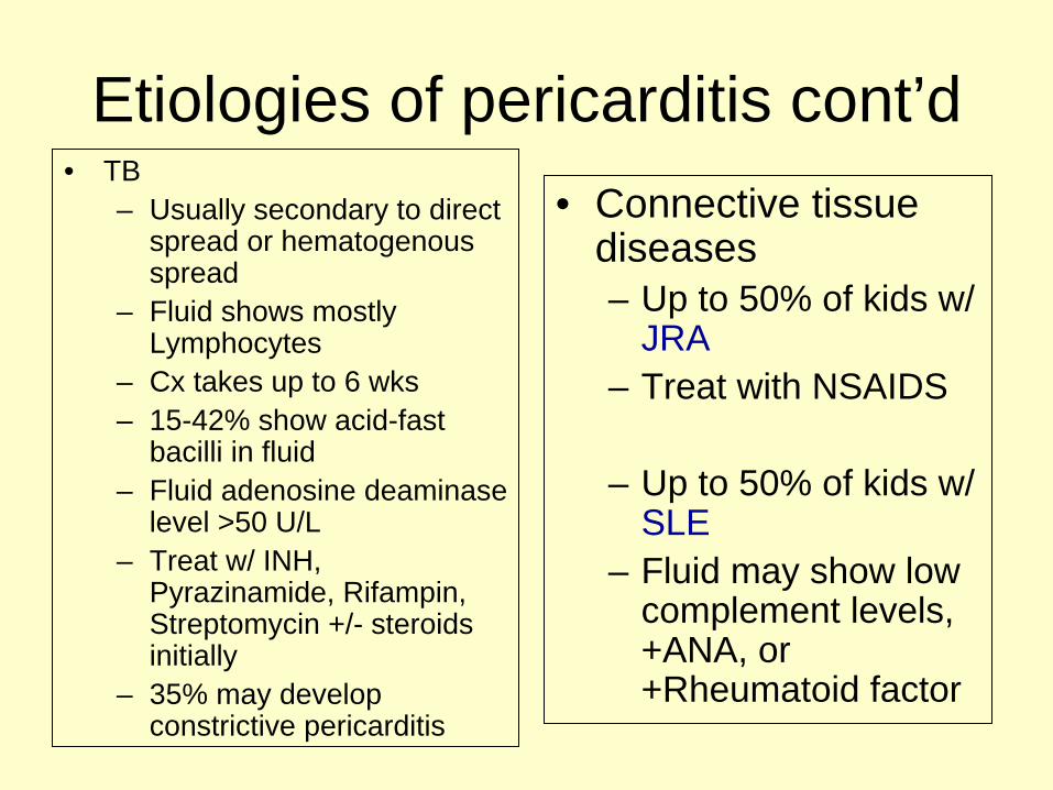
Etiologies of pericarditis cont’d• TB
– Usually secondary to direct spread or hematogenous spread
– Fluid shows mostly Lymphocytes
– Cx takes up to 6 wks– 15-42% show acid-fast
bacilli in fluid– Fluid adenosine deaminase
level >50 U/L– Treat w/ INH,
Pyrazinamide, Rifampin, Streptomycin +/- steroids initially
– 35% may develop constrictive pericarditis
• Connective tissue diseases– Up to 50% of kids w/
JRA– Treat with NSAIDS
– Up to 50% of kids w/ SLE
– Fluid may show low complement levels, +ANA, or +Rheumatoid factor
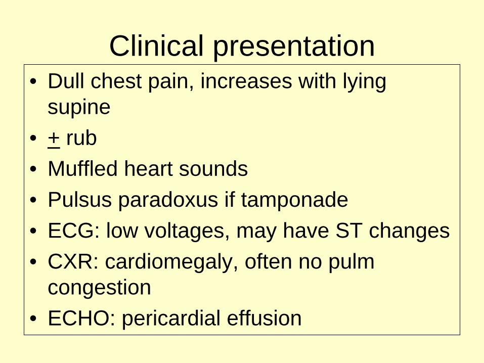
Clinical presentation• Dull chest pain, increases with lying
supine• + rub• Muffled heart sounds• Pulsus paradoxus if tamponade• ECG: low voltages, may have ST changes• CXR: cardiomegaly, often no pulm
congestion• ECHO: pericardial effusion

What is pulsus paradoxus?• SBP drop >10 mmHg with
inspiration• Mechanism in tamponade:
– Inspiration increases venous return to RA/RV
– Delayed venous return to LV and leftward displacement of IVS decreases LV filling (preload)
– Diminished SV– SBP drops
• Neurohormonal compensation for lower CO:– Increased sympathetic tone
and catechol release– Elevated HR, increased
contractility, vasoconstriction
Darsee & Braunwald, Heart Disease: a textbook of cardiovascular medicine.1980: 1535-1582

Pericarditis: typical ECG
ECG of pericarditis; Pediatric ECG Interpretation, Deal et al, 2004
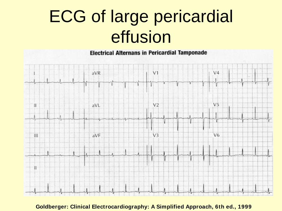
ECG of large pericardial effusion
Goldberger: Clinical Electrocardiography: A Simplified Approach, 6th ed., 1999
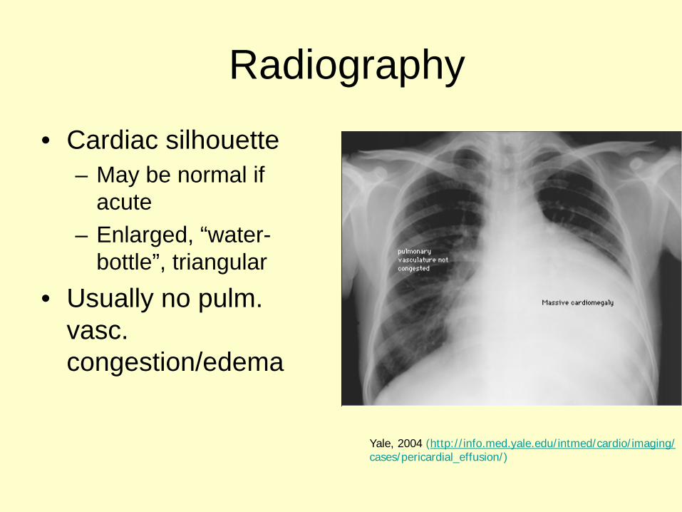
Radiography
• Cardiac silhouette – May be normal if
acute– Enlarged, “water-
bottle”, triangular• Usually no pulm.
vasc. congestion/edema
Yale, 2004 (http://info.med.yale.edu/intmed/cardio/imaging/cases/pericardial_effusion/)

Echocardiography• Presence and relative size of effusion• Chamber dimensions and wall dynamics (compression)
– Early diastole: RV free wall compression– Late diastole: RA compression– LA compression—very specific for tamponade– Swinging heart—large effusion
• May be limited by quality of imaging windows and location of fluid (if loculated)
Spodick, NEJM 2003
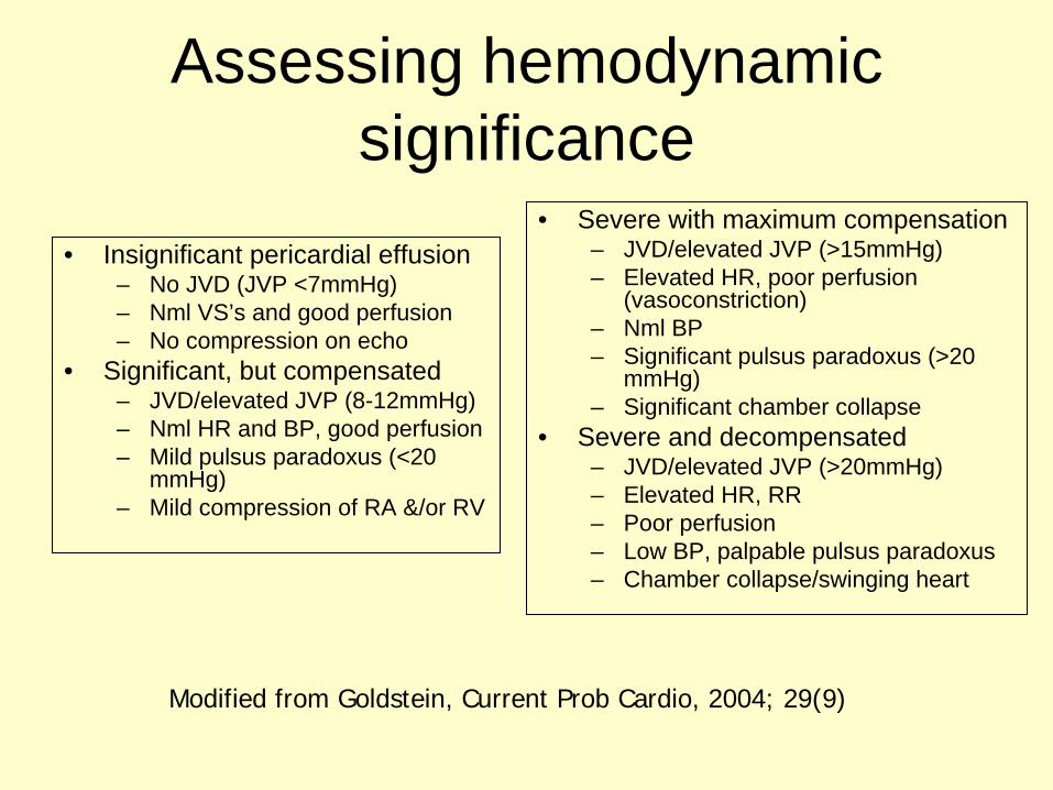
Assessing hemodynamic significance
• Insignificant pericardial effusion– No JVD (JVP <7mmHg)– Nml VS’s and good perfusion– No compression on echo
• Significant, but compensated– JVD/elevated JVP (8-12mmHg)– Nml HR and BP, good perfusion– Mild pulsus paradoxus (<20
mmHg)– Mild compression of RA &/or RV
• Severe with maximum compensation– JVD/elevated JVP (>15mmHg)– Elevated HR, poor perfusion
(vasoconstriction)– Nml BP– Significant pulsus paradoxus (>20
mmHg)– Significant chamber collapse
• Severe and decompensated– JVD/elevated JVP (>20mmHg)– Elevated HR, RR– Poor perfusion– Low BP, palpable pulsus paradoxus– Chamber collapse/swinging heart
Modified from Goldstein, Current Prob Cardio, 2004; 29(9)
Pericarditis cont’d
• Treatment– Treat underlying disorder (uremia, etc.)– If idiopathic or viral, ASA– If tamponade, pericardiocentesis– If purulent, surgical drainage/Antibiotics
Infective Endocarditis
• What is it?– Seeding of bacteria and inflamatory response within
the endocardial layer of the heart– Occurs when
• 1) pt is bacteremic and• 2) pt has intracardiac structural abnormality
• Clinical findings:– MURMUR– Fever– Emboli (skin, eye, nails, lungs, brain, kidney, etc.)
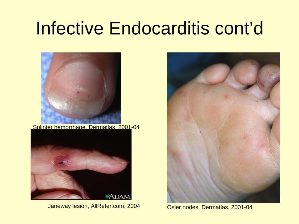
Infective Endocarditis cont’d
Janeway lesion, AllRefer.com, 2004
Splinter hemorrhage, Dermatlas, 2001-04
Osler nodes, Dermatlas, 2001-04
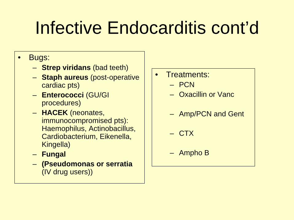
Infective Endocarditis cont’d• Bugs:
– Strep viridans (bad teeth)– Staph aureus (post-operative
cardiac pts)– Enterococci (GU/GI
procedures)– HACEK (neonates,
immunocompromised pts): Haemophilus, Actinobacillus, Cardiobacterium, Eikenella, Kingella)
– Fungal– (Pseudomonas or serratia
(IV drug users))
• Treatments:– PCN– Oxacillin or Vanc
– Amp/PCN and Gent
– CTX
– Ampho B