Pattern formation during de novo assembly of the...
10
DEVELOPMENT 3539 RESEARCH ARTICLE INTRODUCTION Regeneration of a patterned multicellular organism from isolated pieces of adult somatic tissues is a remarkable phenomenon that occurs both in plants and animals (Morgan, 1901). The small Cnidarian, Hydra, for example, can self-assemble a new correctly patterned body from re-aggregated cells derived from dissociated somatic cells of adult tissue (Gierer et al., 1972). Recently, the observation that several genes critical for proper embryonic development in higher animals are expressed during de novo Hydra head regeneration has lead to important insights into the molecular basis of animal self-organization (Hobmayer et al., 2000). However, animal model systems for studying de novo patterning, such as Hydra, are not well developed for molecular analysis or genetics compared to classical model organisms with established collections of mutants, transgenic lines and protocols (Lowenheim, 2003; Wittlieb et al., 2006). Assembly of a complete organism from fragments of adult somatic tissue is rare among animals, but many plants are capable of this type of regeneration. A half century ago Skoog and Miller demonstrated an in vitro system for regenerating flowering plants from fragments of adult somatic tissue (Skoog, 1950; Skoog and Miller, 1957). Remarkably, the identity of induced tissues in this in vitro system was shown to be driven by the ratio of two plant hormones: auxin and cytokinin. It was shown that transfer of tissue explants to medium with higher levels of auxin induced development of root regenerative tissues, whereas transfer of explants to medium with higher levels of cytokinin induced new shoot regenerative tissues, and inductive media containing both auxin and cytokinin induced a proliferation of cells termed callus. During post-embryonic development in flowering plants such as Arabidopsis thaliana, all above ground organs of the plant originate from stem cells within the apical tip of the shoot meristem. The origin of the primary shoot meristem during embryogenesis can be traced back to a small group of apical precursors (West and Harada, 1993). Throughout embryogenesis the apical lineage is marked by precisely regulated expression of many genes, which are required for proper patterning of the shoot meristem (Aida et al., 1997; Barton and Poethig, 1993; Laux et al., 1996; Long et al., 1996). For example, early patterning during embryogenesis is recognizable by expression of the auxin transporter, PIN-FORMED 1 (PIN1), required for the initiation and maintenance of auxin gradients within various tissues of the plant (Friml et al., 2003; Heisler et al., 2005). In the two-cell pro-embryo, PIN1 expression coincides with an initial differential activation of auxin response in the apical cell. Expression of the homeodomain transcription factor WUSCHEL (WUS) begins in the 16-cell stage embryo in two inner apical cells and maintains a tightly restricted pattern throughout embryogenesis (Mayer et al., 1998). The dynamic expression of the redundant transcription factors CUP-SHAPED COTYLEDON 1 and 2 (CUC1 and CUC2) and the homeodomain transcription factor, SHOOT MERISTEMLESS (STM), marks a small number of apical cells in the mid-globular stage embryo that are required for meristem initiation (Aida et al., 1997; Aida et al., 1999; Long and Barton, 1998). Although much is known about patterning of the shoot meristem during embryogenesis, there is little understanding of patterning that must occur during de novo induction of plant tissues in culture (Cary et al., 2002; Long and Barton, 1998). The cell proliferation observed during callus formation ensures that the ordered morphology of normal tissue is severely disrupted (Cary et al., 2002; White, 1939). Furthermore, new shoot meristems can be induced from root-derived explants, which differ in cell lineage, gene expression, and tissue structure from the shoot meristem (West and Harada, 1993), thus raising the question of how root cells react to changes in environment and initiate patterned shoot tissues. Pattern formation during de novo assembly of the Arabidopsis shoot meristem Sean P. Gordon 1 , Marcus G. Heisler 1 , G. Venugopala Reddy 1,2 , Carolyn Ohno 1 , Pradeep Das 1,3 and Elliot M. Meyerowitz 1, * Most multicellular organisms have a capacity to regenerate tissue after wounding. Few, however, have the ability to regenerate an entire new body from adult tissue. Induction of new shoot meristems from cultured root explants is a widely used, but poorly understood, process in which apical plant tissues are regenerated from adult somatic tissue through the de novo formation of shoot meristems. We characterize early patterning during de novo development of the Arabidopsis shoot meristem using fluorescent reporters of known gene and protein activities required for shoot meristem development and maintenance. We find that a small number of progenitor cells initiate development of new shoot meristems through stereotypical stages of reporter expression and activity of CUP-SHAPED COTYLEDON 2 (CUC2), WUSCHEL (WUS), PIN-FORMED 1 (PIN1), SHOOT-MERISTEMLESS (STM), FILAMENTOUS FLOWER (FIL, also known as AFO), REVOLUTA (REV), ARABIDOPSIS THALIANA MERISTEM L1 LAYER (ATML1) and CLAVATA 3 (CLV3). Furthermore, we demonstrate a functional requirement for WUS activity during de novo shoot meristem initiation. We propose that de novo shoot meristem induction is an easily accessible system for the study of patterning and self- organization in the well-studied model organism Arabidopsis. KEY WORDS: Auxin, Callus, Cytokinin, Regeneration, Self-organization, Shoot meristem, Arabidopsis thaliana Development 134, 3539-3548 (2007) doi:10.1242/dev.010298 1 Division of Biology, California Institute of Technology, Pasadena, CA, USA. 2 Department of Botany and Plant Sciences, University of California, Riverside, CA, USA. 3 Laboratorie RDP, Ecole Normale Superieur de Lyon, Lyon, France. *Author for correspondence (e-mail: [email protected]) Accepted 21 July 2007
Transcript of Pattern formation during de novo assembly of the...

DEVELO
PMENT
3539RESEARCH ARTICLE
INTRODUCTIONRegeneration of a patterned multicellular organism from isolatedpieces of adult somatic tissues is a remarkable phenomenon thatoccurs both in plants and animals (Morgan, 1901). The smallCnidarian, Hydra, for example, can self-assemble a new correctlypatterned body from re-aggregated cells derived from dissociatedsomatic cells of adult tissue (Gierer et al., 1972). Recently, theobservation that several genes critical for proper embryonicdevelopment in higher animals are expressed during de novo Hydrahead regeneration has lead to important insights into the molecularbasis of animal self-organization (Hobmayer et al., 2000). However,animal model systems for studying de novo patterning, such asHydra, are not well developed for molecular analysis or geneticscompared to classical model organisms with established collectionsof mutants, transgenic lines and protocols (Lowenheim, 2003;Wittlieb et al., 2006).
Assembly of a complete organism from fragments of adultsomatic tissue is rare among animals, but many plants are capable ofthis type of regeneration. A half century ago Skoog and Millerdemonstrated an in vitro system for regenerating flowering plantsfrom fragments of adult somatic tissue (Skoog, 1950; Skoog andMiller, 1957). Remarkably, the identity of induced tissues in this invitro system was shown to be driven by the ratio of two planthormones: auxin and cytokinin. It was shown that transfer of tissueexplants to medium with higher levels of auxin induced developmentof root regenerative tissues, whereas transfer of explants to mediumwith higher levels of cytokinin induced new shoot regenerativetissues, and inductive media containing both auxin and cytokinininduced a proliferation of cells termed callus.
During post-embryonic development in flowering plants such asArabidopsis thaliana, all above ground organs of the plant originatefrom stem cells within the apical tip of the shoot meristem. Theorigin of the primary shoot meristem during embryogenesis can betraced back to a small group of apical precursors (West and Harada,1993). Throughout embryogenesis the apical lineage is marked byprecisely regulated expression of many genes, which are requiredfor proper patterning of the shoot meristem (Aida et al., 1997;Barton and Poethig, 1993; Laux et al., 1996; Long et al., 1996). Forexample, early patterning during embryogenesis is recognizable byexpression of the auxin transporter, PIN-FORMED 1 (PIN1),required for the initiation and maintenance of auxin gradients withinvarious tissues of the plant (Friml et al., 2003; Heisler et al., 2005).In the two-cell pro-embryo, PIN1 expression coincides with aninitial differential activation of auxin response in the apical cell.Expression of the homeodomain transcription factor WUSCHEL(WUS) begins in the 16-cell stage embryo in two inner apical cellsand maintains a tightly restricted pattern throughout embryogenesis(Mayer et al., 1998). The dynamic expression of the redundanttranscription factors CUP-SHAPED COTYLEDON 1 and 2 (CUC1and CUC2) and the homeodomain transcription factor, SHOOTMERISTEMLESS (STM), marks a small number of apical cells in themid-globular stage embryo that are required for meristem initiation(Aida et al., 1997; Aida et al., 1999; Long and Barton, 1998).
Although much is known about patterning of the shootmeristem during embryogenesis, there is little understanding ofpatterning that must occur during de novo induction of planttissues in culture (Cary et al., 2002; Long and Barton, 1998). Thecell proliferation observed during callus formation ensures thatthe ordered morphology of normal tissue is severely disrupted(Cary et al., 2002; White, 1939). Furthermore, new shootmeristems can be induced from root-derived explants, whichdiffer in cell lineage, gene expression, and tissue structure fromthe shoot meristem (West and Harada, 1993), thus raising thequestion of how root cells react to changes in environment andinitiate patterned shoot tissues.
Pattern formation during de novo assembly of theArabidopsis shoot meristemSean P. Gordon1, Marcus G. Heisler1, G. Venugopala Reddy1,2, Carolyn Ohno1, Pradeep Das1,3 andElliot M. Meyerowitz1,*
Most multicellular organisms have a capacity to regenerate tissue after wounding. Few, however, have the ability to regenerate anentire new body from adult tissue. Induction of new shoot meristems from cultured root explants is a widely used, but poorlyunderstood, process in which apical plant tissues are regenerated from adult somatic tissue through the de novo formation of shootmeristems. We characterize early patterning during de novo development of the Arabidopsis shoot meristem using fluorescentreporters of known gene and protein activities required for shoot meristem development and maintenance. We find that a smallnumber of progenitor cells initiate development of new shoot meristems through stereotypical stages of reporter expression andactivity of CUP-SHAPED COTYLEDON 2 (CUC2), WUSCHEL (WUS), PIN-FORMED 1 (PIN1), SHOOT-MERISTEMLESS (STM),FILAMENTOUS FLOWER (FIL, also known as AFO), REVOLUTA (REV), ARABIDOPSIS THALIANA MERISTEM L1 LAYER (ATML1) andCLAVATA 3 (CLV3). Furthermore, we demonstrate a functional requirement for WUS activity during de novo shoot meristeminitiation. We propose that de novo shoot meristem induction is an easily accessible system for the study of patterning and self-organization in the well-studied model organism Arabidopsis.
KEY WORDS: Auxin, Callus, Cytokinin, Regeneration, Self-organization, Shoot meristem, Arabidopsis thaliana
Development 134, 3539-3548 (2007) doi:10.1242/dev.010298
1Division of Biology, California Institute of Technology, Pasadena, CA, USA.2Department of Botany and Plant Sciences, University of California, Riverside, CA,USA. 3Laboratorie RDP, Ecole Normale Superieur de Lyon, Lyon, France.
*Author for correspondence (e-mail: [email protected])
Accepted 21 July 2007

DEVELO
PMENT
3540
Live imaging of the Arabidopsis meristem has been recentlyapplied to the analysis of cell lineage and cell fate during activegrowth of the shoot meristem, to understand genetic control ofmeristem size, and to cell type specification leading to flowerprimordium initiation and patterning (Heisler et al., 2005; Reddy etal., 2004; Reddy and Meyerowitz, 2005). In this study we use a liveimaging approach to characterize stage-specific molecularpatterning events during de novo organization of the shoot meristemfrom callus (Fig. 1).
MATERIALS AND METHODSPlant materialsAll plants used in this study were in the Landsberg erecta (Ler) ecotypeexcept when stated otherwise. Plants and tissue cultures were grown at 22°Cunder continuous light. Transgenic plants were produced using theAgrobacterium-mediated floral dip method (Clough and Bent, 1998). Thestrong wus-1 mutant allele and the strong pin1-4 allele have been describedpreviously (Bennett et al., 1995; Mayer et al., 1998).
Construction of GFP reportersThe translational protein fusion constructs including the pPIN1::PIN1-GFP,pSTM::STM-VENUS, pREV::REV-VENUS, and pCUC2::CUC2-VENUSconstructs have been described previously (Heisler et al., 2005). Theupstream regulatory sequence reporters including the pDR5rev::3XVENUS-N7, pCUC2::3XVENUS-N7 and the pFIL::DsRED-N7 markers weredescribed previously (Heisler et al., 2005; Sieber et al., 2007). Thetranscriptional pCLV3::GFP-ER reporter was described previously in plantsbearing a construct consisting of a 35S promoter driving 29.1 plasmamembrane-localized yellow fluorescent protein (YFP) (Reddy andMeyerowitz, 2005). The pARR5::GFP reporter in the WS ecotype has beendescribed previously (Yanai et al., 2005) and was generously provided byJoseph Kieber (Department of Biology, University of North Carolina,Chapel Hill, USA).
The previously published pWUS::mGFP5-ER construct (Jonsson et al.,2005) contains 3 kb of upstream and 1.5 kb of downstream WUS genomicregulatory sequences separated by the mGFP-ER coding sequence in the T-DNA vector pPZP222 conferring gentamycin resistance in plants(Hajdukiewicz et al., 1994). The pWUS::DsDed-N7 construct, also inpPZP222, is composed of 4.4 kb of upstream and 1.5 kb of downstreamWUS genomic regulatory sequences separated by the DsRed coding regionfused to the N7 nuclear localization sequence. The pWUS::DsDed-N7construct was transformed into Ler harboring the pCLV3::GFP-ER reporter.The pWUS::DsDed-N7 reporter line gave a pattern of expression confinedto the rib zone of shoot meristems and floral meristems. A putative additivesignal or strong autofluorescence was detected in the older leaves of thepWUS::DsDed-N7 transformants, which was not found in pWUS::mGFP5-ER transformants. Spatial expression of the pWUS::DsRed-N7 marker wasverified by semi-quantitative RT-PCR to strictly correspond to areas of callussamples with WUS transcript (see Fig. S1A in the supplementary material),in contrast to random samples of callus.
The pRIBO::2XCFP-N7 construct in the T-DNA vector pPZP222 wascomposed of 2.6 kb of upstream regulatory sequence from the 60S ribosomalprotein L2 gene (At2g18020) fused to two tandem copies of eCFP(Clontech) followed by the N7 nuclear localization sequence (Cutler et al.,2000).
The pML1::GFP-ER construct in the T-DNA vector pPZP222 wascomposed of 3.4 kb of upstream regulatory sequence from the ML1 genecontaining a fragment demonstrated to drive L1-specific expression, fusedto mGFP-ER (Sessions et al., 1999).
The pPIN1::PIN1-CFP construct was created by substituting the CFPcoding sequence for the GFP coding sequence in the publishedpPIN1::PIN1-GFP construct. Plants bearing multiple transgenes and themutant alleles were combined by genetic crossing.
Regeneration conditionsRoot explants were harvested from 2-week-old seedlings grown in sterileculture on Murashige and Skoog basal salt mixture (MS) plates. Explantswere cultured on callus-inducing medium (CIM) consisting of modified
Gamborg’s B-5 medium (Sigma) containing 20 g/l glucose, 0.5 g/l MES(Sigma) and supplemented with 1� Gamborg’s vitamin solution (Sigma),500 �g/l of 2,4-D (Sigma) and 50 �g/l of kinetin (Sigma). Samples wereincubated on CIM tissue culture plates for 2 weeks. Callus samples were cutinto 2 cm length sections which were cultured on shoot-inducing medium(SIM) plates, consisting of MS medium containing 10 g/l sucrose, 0.5 g/lMES and supplemented with 1� Gamborg’s vitamin solution, 2 �g/mlzeatin (BioWorld, Dublin, OH, USA), 1 �g/ml d-biotin (Sigma), and0.4 �g/ml indole-3-butyric acid (IBA; Sigma).
For quantifying shoot meristem induction, samples were cultured in talltissue culture plates (USA Scientific) for a further 2 weeks, at which pointthe number of shoots per 2 cm callus explant was recorded. Shoots weredefined as described previously (Daimon et al., 2003). Each experimentcontained independent wild-type controls using the same batch of mediumand growth conditions.
Exogenous application of IAAIndole-3-acetic acid (IAA) lanolin paste from Carolina Biological SupplyCompany at a concentration of 500 ppm labeled with 1 �g/ml of propidiumiodide was applied directly to callus in the vicinity of developing shootmeristems.
Imaging conditionsCallus and regenerating shoots were imaged directly on respective media.For each marker line, at least 25 samples were imaged to confirm thatobserved patterns were representative of respective markers. Propidiumiodide for staining root cell outlines of root tissues was applied to samplesat a concentration of 10 �g/ml 10 minutes prior to imaging. The lipophilicdye FM4-64 (Molecular Probes) was used at a concentration of 10 �g/ml todemarcate cell membranes and specifically labeled regenerating shoottissues initiating from root-derived callus.
All imaging was done using a Zeiss 510 Meta laser scanning confocalmicroscope with either a 10� air objective, 20� air objective, or a 40�0.8 NA water dipping lens using the multi-tracking mode. Specific sets offilters used for the respective markers were similar to those alreadydescribed (Heisler et al., 2005; Reddy and Meyerowitz, 2005). Projectionsof confocal data were exported using Zeiss LSM software. Alternatively,volume renderings were made using Amira (Mercury ComputerSystems).
RESULTSAuxin/cytokinin response and gene expressionduring callus formationOur first goal was to determine a correlation between callus inductionand distribution of auxin response during callus formation from rootexplants on auxin-rich CIM. Auxin response was visualized using theauxin responsive DR5 element (Casimiro et al., 2001; Ulmasov et al.,1997) driving expression of tandem VENUS yellow fluorescentprotein localized to the cell nucleus, pDR5rev::3XVENUS-N7. In non-induced root explants, the DR5 reporter (green) marked root pericycle
RESEARCH ARTICLE Development 134 (19)
Fig. 1. Overview of the de novo shoot induction system. (A) Rootexplants were harvested from 2-week-old seedlings and (B) transferredto auxin-rich CIM, which induces cell proliferation, resulting in (C) callusformation. (D) Transfer of callus to cytokinin-rich SIM induces greeningand induction of shoot meristems from callus often in clusters (markedby two green leaves).

DEVELO
PMENT
cells, a subset of lateral root progenitors, and the distal tip of lateralroots including columellar root cap cells (Fig. 2A), as previouslyreported (Benkova et al., 2003). However, after 5 days incubation onCIM, proliferative growth was marked by the DR5 reporter and wasinitiated in the vicinity of lateral roots, root meristems and to a lesserextent, the root pericycle (Fig. 2B). DR5 response diminished overtime and was not observed within large callus outgrowths after 1 weekof culture (Fig. 2C). In addition, after 2-3 days induction on CIM, areporter for the auxin efflux carrier PIN-FORMED 1 (PIN1), wasinduced in callus outgrowths (green in Fig. S1B in the supplementarymaterial), but was later downregulated and was not detected after 10days of induction.
We next investigated the spatial distribution of cytokininresponse within root explants on CIM. The ARABIDOPSISRESPONSE REGULATOR 5 gene (ARR5) has been shown to betranscriptionally responsive to cytokinin, and its level ofexpression correlates with cytokinin content in various tissues(Aloni et al., 2004). We used transgenic plants bearing ARR5regulatory sequences driving GFP expression, pARR5::GFP, todynamically monitor cytokinin response. In the non-induced root,pARR5::GFP activity was observed in the root stele, rootmeristems and lateral root progenitor cells (Fig. 2D, green). After8 days of induction on CIM, signal from the ARR5 reporter wasdetected in the root explant vasculature and strongly markedproliferating callus cells (Fig. 2E), and after 2 weeks of inductionhad expanded throughout callus (Fig. 2F).
A recent study using an enhancer trap for CUC1 demonstratedthat CUC1 upregulation is associated with callus formation on CIM(Cary et al., 2002). We determined if transcription of the partiallyredundant gene CUC2 is also upregulated on CIM. Prior toinduction, a reporter consisting of CUC2 regulatory sequencesdriving tandem VENUS expression localized to the cell nucleus,pCUC2::3XVENUS-N7, was active in a subset of cells of the rootvascular cylinder and lateral root primordia founder cells (Fig. 2G,green). After 8 days of induction on CIM, the CUC2 reporter wasupregulated in small proliferating callus cells (Fig. 2H) and was laterobserved throughout the callus (Fig. 2I). By contrast, WUS, STM andCLV3 were not expressed in callus, consistent with previous RT-PCRdata (Cary et al., 2002), and no FILAMENTOUS FLOWER (FIL)and REVOLUTA (REV) reporter activity was observed.
CIM contains tenfold higher levels of the synthetic auxin 2,4-D,than of the cytokinin, kinetin. We next investigated which of thesehormones was responsible for callus induction and upregulation ofthe CUC2 reporter. Modified CIM, containing 2,4-D as the solehormone, induced callus and CUC2 reporter expression (Fig. 2J,green). On the same medium, the cytokinin responsive pARR5::GFPreporter was upregulated at sites of callus formation (Fig. 2K). Bycontrast, culture of explants on CIM containing only kinetin did notlead to callus proliferation and CUC2 reporter expression was faintand did not expand outside the vasculature of the primary root (Fig.2L). Expression of the auxin-responsive DR5 reporter also did notexpand on this medium (data not shown).
3541RESEARCH ARTICLEDe novo shoot meristem formation
Fig. 2. Hormone response and gene expressionduring callus induction. All samples were stained withpropidium iodide (red) to stain cell walls. (A) In wild-typeroots, the auxin-responsive reporter, pDR5rev::3XVENUS-N7 (green), was present in a subset of cells in the rootvasculature, lateral root progenitors and columellar rootcap cells. (B) Clusters of small cells marked bypDR5rev::3XVENUS-N7 reporter (green) proliferate toform callus, 5 days after induction on CIM. (C) After 8days of CIM induction, the pDR5rev::3XVENUS-N7reporter was weakly expressed in callus. (D) Pre-CIMcytokinin-responsive pARR5::GFP reporter expression(green) in the root stele, and lateral root progenitors.(E) pARR5::GFP reporter expression, 8 days and (F) 2weeks after CIM induction, was visible in proliferatingcallus cells. (G) Pre-CIM pCUC2::3XVENUS-N7 reporterexpression (green) in a subset of cells within the root steleand lateral root meristems. (H) pCUC2::3XVENUS-N7reporter expression 8 days and (I) 2 weeks after CIMinduction marked proliferating callus cells originating fromsites of lateral root formation, root meristems andpericycle. (J,K) Two weeks induction on CIM withoutcytokinin, resulted in cell proliferation and expression ofthe pCUC2::3XVENUS-N7 (J) and pARR5::GFP reporters(K). (L) Two weeks after induction on CIM without 2,4-D,callus was not induced and expression of thepCUC2::3XVENUS-N7 reporter was faint and confined tothe vasculature of the primary root. Scale bars: 50 �m(A,B,D,E,G,H); 100 �m (C,F,I-L). Arrowheads indicatelateral roots.
DEVELO
PMENT
3542
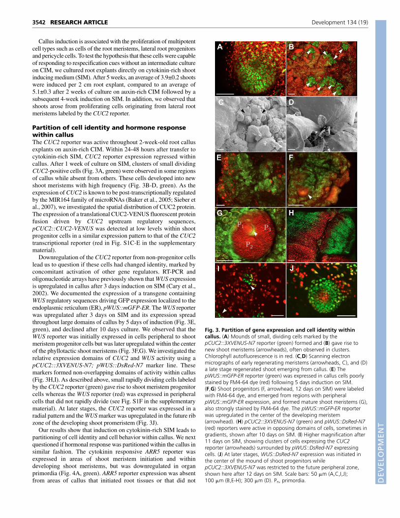
Callus induction is associated with the proliferation of multipotentcell types such as cells of the root meristems, lateral root progenitorsand pericycle cells. To test the hypothesis that these cells were capableof responding to respecification cues without an intermediate cultureon CIM, we cultured root explants directly on cytokinin-rich shootinducing medium (SIM). After 5 weeks, an average of 3.9±0.2 shootswere induced per 2 cm root explant, compared to an average of5.1±0.3 after 2 weeks of culture on auxin-rich CIM followed by asubsequent 4-week induction on SIM. In addition, we observed thatshoots arose from proliferating cells originating from lateral rootmeristems labeled by the CUC2 reporter.
Partition of cell identity and hormone responsewithin callusThe CUC2 reporter was active throughout 2-week-old root callusexplants on auxin-rich CIM. Within 24-48 hours after transfer tocytokinin-rich SIM, CUC2 reporter expression regressed withincallus. After 1 week of culture on SIM, clusters of small dividingCUC2-positive cells (Fig. 3A, green) were observed in some regionsof callus while absent from others. These cells developed into newshoot meristems with high frequency (Fig. 3B-D, green). As theexpression of CUC2 is known to be post-transcriptionally regulatedby the MIR164 family of microRNAs (Baker et al., 2005; Sieber etal., 2007), we investigated the spatial distribution of CUC2 protein.The expression of a translational CUC2-VENUS fluorescent proteinfusion driven by CUC2 upstream regulatory sequences,pCUC2::CUC2-VENUS was detected at low levels within shootprogenitor cells in a similar expression pattern to that of the CUC2transcriptional reporter (red in Fig. S1C-E in the supplementarymaterial).
Downregulation of the CUC2 reporter from non-progenitor cellslead us to question if these cells had changed identity, marked byconcomitant activation of other gene regulators. RT-PCR andoligonucleotide arrays have previously shown that WUS expressionis upregulated in callus after 3 days induction on SIM (Cary et al.,2002). We documented the expression of a transgene containingWUS regulatory sequences driving GFP expression localized to theendoplasmic reticulum (ER), pWUS::mGFP-ER. The WUS reporterwas upregulated after 3 days on SIM and its expression spreadthroughout large domains of callus by 5 days of induction (Fig. 3E,green), and declined after 10 days culture. We observed that theWUS reporter was initially expressed in cells peripheral to shootmeristem progenitor cells but was later upregulated within the centerof the phyllotactic shoot meristems (Fig. 3F,G). We investigated therelative expression domains of CUC2 and WUS activity using apCUC2::3XVENUS-N7; pWUS::DsRed-N7 marker line. Thesemarkers formed non-overlapping domains of activity within callus(Fig. 3H,I). As described above, small rapidly dividing cells labeledby the CUC2 reporter (green) gave rise to shoot meristem progenitorcells whereas the WUS reporter (red) was expressed in peripheralcells that did not rapidly divide (see Fig. S1F in the supplementarymaterial). At later stages, the CUC2 reporter was expressed in aradial pattern and the WUS marker was upregulated in the future ribzone of the developing shoot promeristem (Fig. 3J).
Our results show that induction on cytokinin-rich SIM leads topartitioning of cell identity and cell behavior within callus. We nextquestioned if hormonal response was partitioned within the callus insimilar fashion. The cytokinin responsive ARR5 reporter wasexpressed in areas of shoot meristem initiation and withindeveloping shoot meristems, but was downregulated in organprimordia (Fig. 4A, green). ARR5 reporter expression was absentfrom areas of callus that initiated root tissues or that did not
RESEARCH ARTICLE Development 134 (19)
Fig. 3. Partition of gene expression and cell identity withincallus. (A) Mounds of small, dividing cells marked by thepCUC2::3XVENUS-N7 reporter (green) formed and (B) gave rise tonew shoot meristems (arrowheads), often observed in clusters.Chlorophyll autofluorescence is in red. (C,D) Scanning electronmicrographs of early regenerating meristems (arrowheads, C), and (D)a late stage regenerated shoot emerging from callus. (E) ThepWUS::mGFP-ER reporter (green) was expressed in callus cells poorlystained by FM4-64 dye (red) following 5 days induction on SIM.(F,G) Shoot progenitors (F, arrowhead, 12 days on SIM) were labeledwith FM4-64 dye, and emerged from regions with peripheralpWUS::mGFP-ER expression, and formed mature shoot meristems (G),also strongly stained by FM4-64 dye. The pWUS::mGFP-ER reporterwas upregulated in the center of the developing meristem(arrowhead). (H) pCUC2::3XVENUS-N7 (green) and pWUS::DsRed-N7(red) reporters were active in opposing domains of cells, sometimes ingradients, shown after 10 days on SIM. (I) Higher magnification after11 days on SIM, showing clusters of cells expressing the CUC2reporter (arrowheads) surrounded by pWUS::DsRed-N7 expressingcells. (J) At later stages, WUS::DsRed-N7 expression was initiated inthe center of the mound of shoot progenitors whilepCUC2::3XVENUS-N7 was restricted to the future peripheral zone,shown here after 12 days on SIM. Scale bars: 50 �m (A,C,I,J);100 �m (B,E-H); 300 �m (D). Pn, primordia.
DEVELO
PMENT
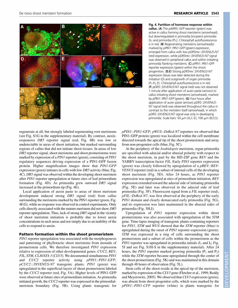
regenerate at all, but strongly labeled regenerating root meristems(see Fig. S1G in the supplementary material). By contrast, auxin-responsive DR5 reporter signal (red, Fig. 4B) was low orundetectable in areas of shoot initiation, but marked surroundingregions of callus that did not initiate shoot tissues. In areas of lowDR5 reporter signal, shoot meristems and shoot promeristems weremarked by expression of a PIN1 reporter (green), consisting of PIN1regulatory sequences driving expression of a PIN1-GFP fusionprotein. Higher magnification images show that PIN1-GFPexpression (green) initiates in cells with low DR5 activity (blue, Fig.4C). DR5 signal was observed within the developing shoot meristemafter PIN1 reporter upregulation at future sites of leaf primordiumformation (Fig. 4D). As primordia grew outward DR5 signalincreased at the primordium tip (Fig. 4E).
Local application of auxin paste to areas of shoot meristemdevelopment induced strong DR5 signal (red) from callussurrounding the meristems marked by the PIN1 reporter (green, Fig.4F,G), while no response was observed in control experiments. Onlycells directly associated with the mature meristem did not show DR5reporter upregulation. Thus, lack of strong DR5 signal in the vicinityof shoot meristem initiation is probably due to lower auxinconcentrations in such areas and not simply due to an inability of thecells to respond to auxin.
Pattern formation within the shoot promeristemPIN1 reporter upregulation was associated with the morphogenesisand patterning of phyllotactic shoot meristems from mounds ofpromeristem cells. We therefore investigated PIN1 expressionrelative to expression of other developmental genes: CUC2, REV,FIL, STM, CLAVATA 3 (CLV3). We documented simultaneous PIN1and CUC2 reporter activity using pPIN1::PIN1-GFP;pCUC2::3XVENUS-N7 transgenic plants. PIN1 (green) wasupregulated in the superficial layers of shoot promeristem labeledby the CUC2 reporter (red, Fig. 5A). Higher levels of PIN1-GFPwere observed at future sites of primordium initiation. As primordiainitiated growth, the CUC2 reporter was expressed in the primordial-meristem boundary (Fig. 5B). Using plants transgenic for
pPIN1::PIN1-GFP; pWUS::DsRed-N7 reporters we observed thatPIN1-GFP protein (green) was localized within the cell membranedirected towards the apical tip of the shoot promeristem and awayfrom non-progenitor cells (blue, Fig. 5C).
At the periphery of the Arabidopsis meristem, organ primordiaare specified with adaxial and/or abaxial polarity with respect tothe shoot meristem, in part by the HD-ZIP gene REV and theYABBY transcription factor FIL. Early PIN1 reporter expression(green) was closely followed by upregulation of a pREV::REV-VENUS reporter (red) in a subset of internal cells of the developingshoot meristem (Fig. 5D). After 24 hours, as PIN1 reporterexpression was upregulated at sites of primordium initiation, REVexpression extended toward the adaxial side of initiating primordia(Fig. 5E) and later was observed in the adaxial side of leafprimordia (Fig. 5F). Fluorescent signal from a FIL reporter (red),pFIL::DsRed-N7, was first observed at the periphery of the earlyPIN1 domain and clearly demarcated early primordia (Fig. 5G),and its expression was later maintained in the abaxial sides ofprimordia (Fig. 5H,I).
Upregulation of PIN1 reporter expression within shootpromeristems was also associated with upregulation of the STMgene. Time-lapse imaging of transgenic plants containing reportersfor PIN1, STM and WUS showed that the STM reporter (blue) isupregulated during the onset of PIN1 reporter expression (green).STM was expressed in a ring of cells surrounding the shootpromeristem and a subset of cells within the promeristem as thePIN1 reporter was upregulated in primordia initials (I1 and I2; Fig.5J and see Fig. S1H-S in the supplementary material). After 24hours, the PIN1 reporter marked growing primordia (P1 and P2)while the STM reporter became upregulated through the center ofthe shoot promeristem (Fig. 5K) and was maintained in this domainthrough 48 hours of observation (Fig. 5L).
Stem cells of the shoot reside at the apical tip of the meristem,marked by expression of the CLV3 gene (Fletcher et al., 1999; Reddyand Meyerowitz, 2005). We observed that CLV3 expression (green),was absent from shoot progenitor cells, which were marked by thepPIN1::PIN1-CFP reporter (white) in plants transgenic for
3543RESEARCH ARTICLEDe novo shoot meristem formation
Fig. 4. Partition of hormone response withincallus. (A) The pARR5::GFP reporter (green) wasactive in callus forming shoot meristems (arrowhead),but downregulated in primordia [incipient primordia(I)1 and primordia (P)1]. Chlorophyll autofluorescenceis in red. (B) Regenerating meristems (arrowheads)marked by pPIN1::PIN1-GFP (green) expression,emerged from callus with low pDR5rev::3XVENUS-N7(red) expression, while pDR5rev::3XVENUS-N7 signalwas observed in peripheral callus and within initiatingprimordia flanking meristems. (C) pPIN1::PIN1-GFPreporter expression (green) within the shootprogenitors. (D,E) Strong pDR5rev::3XVENUS-N7expression (blue) was later detected during theinitiation (D) and outgrowth of organ primordia(P1-P3; E). Chlorophyll autofluorescence is in red.(F) pDR5::3XVENUS-N7 signal (red) was not observed1 minute after application of auxin paste (arrows) tocallus initiating shoot meristems (arrowhead), markedby pPIN1::PIN1-GFP (green). (G) Four hours afterapplication of auxin paste (arrows) pDR5::3XVENUS-N7 signal (red) was observed throughout the callus incontrast to the meristem itself (arrowhead), in whichpDR5::3XVENUS-N7 signal was only in developingprimordia. Scale bars: 50 �m (A,C-E); 100 �m (B,F,G).

DEVELO
PMENT
3544
pCLV3::GFP-ER, pPIN1::PIN1-CFP and pWUS::DsRed-N7reporters (Fig. 5M). CLV3 reporter expression appeared duringupregulation of WUS reporter expression (red) within the center ofthe new meristem and the initiation of primordia (P1 and P2) fromthe meristem periphery (Fig. 5N). CLV3 reporter activity wasconfirmed in plants bearing a p35S::YFP 29-1 transgene (yellow),which express membrane-localized YFP within all cells of themature meristem (Fig. 5O).
L1 layer specification and development ofmeristem structureThe homeodomain transcription factor ARABIDOPSIS THALIANAMERISTEM L1 LAYER (ATML1) is redundantly required forspecification of the epidermal layer in Arabidopsis (Abe et al.,2003) and is restricted to the protodermal layer at the 16-cell stageonwards (Lu et al., 1996). We used a transgenic line containingpATML1::GFP-ER and pCUC2::3XVENUS-N7 reporters in orderto understand relative timing of L1 cell-type specification withregards to meristem organization. The ATML1 reporter wasrestricted to a subset of superficial cells within the shoot
promeristem marked by the CUC2 reporter (Fig. 6A,B). Bycontrast, the ATML1 reporter was often not L1 specific whenexpressed in callus (Fig. 6C). Primordium initiation began afterapproximately 72 hours of development and was associated withhomogenous expression of the ATML1 reporter within theprotoderm (Fig. 6E).
We further followed the shoot regeneration process in apPIN1::PIN1-GFP; pSTM::STM-VENUS; pRIBO::2XCFP-N7marker line. The pRIBO::2XCFP-N7 marker labeled all cells withina callus, enabling us to observe that shoot promeristems werecomposed of variable numbers of cells (Fig. 6G). Shootpromeristems composed of smaller numbers of cells developed intoshoot meristems with fewer initial leaf primordia compared to largerpromeristems (Fig. 6J).
Quantification of regeneration in wus-1 andpin1-4To determine if WUS and PIN1 are necessary for efficient initiationof new shoot meristems, we quantified the number of shoots formedfrom 2 cm callus explants in the strong wus-1 and pin1-4 mutants
RESEARCH ARTICLE Development 134 (19)
Fig. 5. Pattern formation within the shootpromeristem. (A) pPIN1::PIN1-GFP expression (green) wasupregulated in the superficial layer of shoot meristemprogenitor cells marked by pCUC2::3XVENUS-N7 reporterexpression (red), and in labeled primordium initials (I1 andI2). (B) After 24 hours, primordium initials grew intoprimordia (P1 and P2) and pCUC2::3XVENUS-N7 (red) wasexpressed in the meristem boundaries. (C) PIN1-GFPprotein was (green) polarized towards the apex of theshoot progenitors (arrows) and away from peripheral cellsmarked by the pWUS::DsRed-N7 reporter (blue). (D) EarlypREV::REV-VENUS expression (red) was observed in thecenter of the progenitors underneath the pPIN1::PIN1-GFP(green) domain. Chlorophyll autofluorescence is in blue.(E) 24 hours later in the same developing meristem,pREV::REV-VENUS expression (red) was expressed in theadaxial sides of initiating primordia (I1 and I2), and (F) wassimilarly expressed in primordia within later stage shootmeristems. (G-I) pFIL::DsRed-N7 expression (red) wasupregulated in areas flanking the early pPIN1::PIN1-GFP(green) domain (G) and was later upregulated on theabaxial side of early primordia (H) and older primordia (I).(J) pSTM::STM-VENUS (blue) was expressed in a ringsurrounding shoot progenitors and a subset of cells withinthe promeristem (11 days on SIM) while local pPIN1::PIN1-GFP reporter (green) upregulation marked sites ofprimordium initiation (I1 and I2). pWUS::DsRed-N7 reporter(red) was expressed in peripheral cells and upregulated inthe center of the developing meristem. (K,L) 24 hourslater in the same shoot progenitors, pSTM::STM-VENUS(blue) was upregulated within the meristem between thedeveloping primordia (P1 and P2; K) and was maintainedthrough 48 hours of imaging during which primordiagrew and two new primordia were initiated (I1 and I2; L).(M) pCLV3::mGFP5-ER expression (green) was absent fromshoot progenitors marked by pPIN1::PIN1-CFP expression(white) and peripheral cells marked by the pWUS::DsRed-N7 reporter (red). (N) pCLV3::mGFP5-ER expression (green)was detected after primordial outgrowth from theperiphery of the developing meristem. (O) pCLV3::mGFP5-ER expression (green) was also observed in later stageshoot meristems which expressed a p35S::YFP 29-1transgene (yellow). Scale bars: 50 �m (A,B,D-O); 5 �m (C).

DEVELO
PMENT
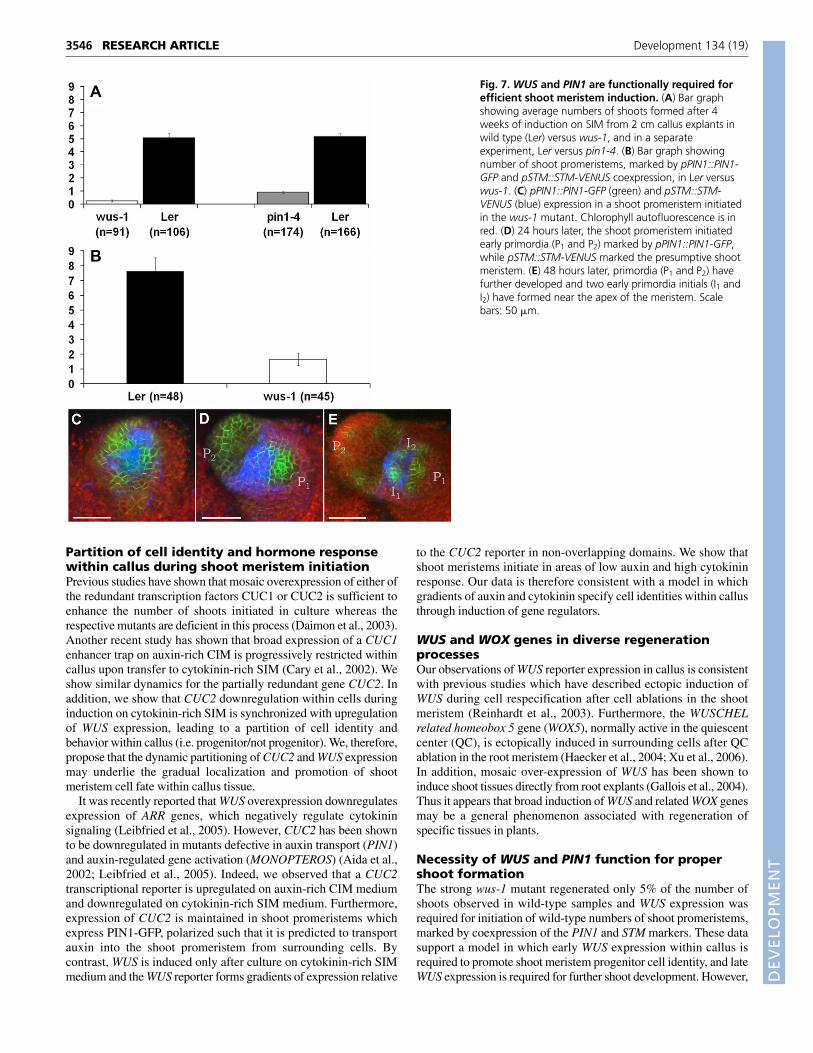
after 4 weeks of growth on SIM. The average number of shootsformed in the wus-1 mutant (n=91) decreased to 5% of wild-typenumber of shoots (n=106; 0.25±0.08 versus 5.06±0.04), whereas the
average number of shoots formed in the pin1-4 mutant (n=174)decreased to approximately 20% of wild type numbers (n=166;0.90±0.07 versus 5.16±0.24; Fig. 7A).
Quantification of an early versus late defect inwus-1The decrease in the number of shoot meristems observed in thewus-1 mutant could be due to an early defect, in which fewer shootpromeristems are initiated, or a late defect, in which shootpromeristems arrest at later stages of development prior toquantification. We differentiated between these two possibilities byexamining the number of early shoot promeristems, marked bypPIN1::PIN1-GFP and pSTM::STM-YFP co-expression, formed inwus-1. The number of early shoot promeristems was decreased inthe wus-1 mutant (n=45) to only 20% of wild-type promeristems(n=48; 7.63±0.92 versus 1.67±0.43; Fig. 7B). However, weobserved that in those shoot promeristems that do form, the PIN1and STM reporters are initially expressed in similar relative domainsto those of wild-type shoot meristem assembly (Fig. 7C-E).
DISCUSSIONCharacterization of hormone response and geneexpression during callus inductionPrior studies have shown that good auxin efflux substrates, suchas indole-3-acetic acid (IAA) or �-naphthalene acetic acid(NAA), induce lateral root growth in wild-type root explants, butcallus-like proliferation in mutants for pin auxin efflux carriers(Benkova et al., 2003). Furthermore, a reporter for CUC3expression was expanded in roots simultaneously treated withIAA and the auxin transport inhibitor NPA. 2,4-D is an auxinanalog that is poorly transported by the auxin efflux system(Delbarre et al., 1996). Our data shows that CIM containing 2,4-D as the sole added hormone in the growth medium is sufficientto induce callus formation, which involves proliferation ofmultipotent cell types including root pericycle cells, lateral rootprogenitors and cells of the root meristems. Combined, thesefindings suggest that callus induction is due to an inability of roottissue to regulate auxin distribution, leading to unrestrainedproliferation of multipotent cells of the root.
Recently, it has been shown that pericycle cells uniquely continuedivision through the elongation and differentiation zones of the rootafter exit from the root meristem (Dubrovsky et al., 2000). Later, asubset of these cells gives rise to lateral root primordia. The abilityof these cells to continue division may be linked with their enhancedresponse to environmental stimuli, such as the availability ofhormones. Consistent with this model, we observe that most cellsinitiating and proliferating as callus are marked by expression of theauxin-responsive DR5 and cytokinin-responsive ARR5 reporters.The enhanced capacity to divide in response to hormone inductionand the ability to give rise to multiple cell types may explain thepreferential proliferation of these cells on CIM and their plasticityduring induction of shoot tissues when transferred to a highcytokinin environment.
The different quantitative requirements for auxin and cytokinin inorder to induce various tissues in culture is probably in part due todifferent endogenous concentrations of these hormones withinexplants (Skoog, 1950). Root meristems are sites of endogenouscytokinin production (Aloni et al., 2005; Nordstrom et al., 2004).The upregulation of the cytokinin responsive ARR5 reporter withincallus forming on CIM containing 2,4-D but no exogenous cytokininsuggests that callus induced from root meristems may endogenouslyproduce cytokinin.
3545RESEARCH ARTICLEDe novo shoot meristem formation
Fig. 6. L1 layer specification and development of meristemstructure. (A) pML1::GFP5-ER reporter (green) was upregulated in asubset of superficial shoot meristem progenitors, marked by thepCUC2::3XVENUS-N7 (red) marker. (B,C) Expression of the pML1::GFP5-ER reporter (green) was L1 specific within the shoot progenitors (B) butnot L1 specific in cross-sections of callus (C). (D) 24 hours later,pML1::GFP5-ER expression was upregulated in the shoot progenitors.(E) 72 hours later, pML1::GFP5-ER expression was homogeneouslyexpressed within the L1 of the meristem as primordia (I1 and P1) wereinitiated. (F) 96 hours later, two early primordia (P1 and P2) wereevident. (G) pPIN1::PIN1-GFP (green) and pSTM::STM-VENUS (red) wereupregulated with similar timing in small patches of cells marked by theubiquitous pRIBO::2XCFP-N7 marker (blue). (H) 24 hours later,pPIN1::PIN1-GFP marked initiating primordia (I1, I2). (I) After 48 hours,primordia (P1 and P2) labeled by pPIN1::PIN1-GFP have grown outwardsand pSTM::STM-VENUS was expressed in the developing meristem.(J) After 72 hours of observation, meristems derived from smallnumbers of initial cells gave rise to fewer primordia than meristemswith more cells. Scale bars: 50 �m (A-J).
DEVELO
PMENT
3546
Partition of cell identity and hormone responsewithin callus during shoot meristem initiationPrevious studies have shown that mosaic overexpression of either ofthe redundant transcription factors CUC1 or CUC2 is sufficient toenhance the number of shoots initiated in culture whereas therespective mutants are deficient in this process (Daimon et al., 2003).Another recent study has shown that broad expression of a CUC1enhancer trap on auxin-rich CIM is progressively restricted withincallus upon transfer to cytokinin-rich SIM (Cary et al., 2002). Weshow similar dynamics for the partially redundant gene CUC2. Inaddition, we show that CUC2 downregulation within cells duringinduction on cytokinin-rich SIM is synchronized with upregulationof WUS expression, leading to a partition of cell identity andbehavior within callus (i.e. progenitor/not progenitor). We, therefore,propose that the dynamic partitioning of CUC2 and WUS expressionmay underlie the gradual localization and promotion of shootmeristem cell fate within callus tissue.
It was recently reported that WUS overexpression downregulatesexpression of ARR genes, which negatively regulate cytokininsignaling (Leibfried et al., 2005). However, CUC2 has been shownto be downregulated in mutants defective in auxin transport (PIN1)and auxin-regulated gene activation (MONOPTEROS) (Aida et al.,2002; Leibfried et al., 2005). Indeed, we observed that a CUC2transcriptional reporter is upregulated on auxin-rich CIM mediumand downregulated on cytokinin-rich SIM medium. Furthermore,expression of CUC2 is maintained in shoot promeristems whichexpress PIN1-GFP, polarized such that it is predicted to transportauxin into the shoot promeristem from surrounding cells. Bycontrast, WUS is induced only after culture on cytokinin-rich SIMmedium and the WUS reporter forms gradients of expression relative
to the CUC2 reporter in non-overlapping domains. We show thatshoot meristems initiate in areas of low auxin and high cytokininresponse. Our data is therefore consistent with a model in whichgradients of auxin and cytokinin specify cell identities within callusthrough induction of gene regulators.
WUS and WOX genes in diverse regenerationprocessesOur observations of WUS reporter expression in callus is consistentwith previous studies which have described ectopic induction ofWUS during cell respecification after cell ablations in the shootmeristem (Reinhardt et al., 2003). Furthermore, the WUSCHELrelated homeobox 5 gene (WOX5), normally active in the quiescentcenter (QC), is ectopically induced in surrounding cells after QCablation in the root meristem (Haecker et al., 2004; Xu et al., 2006).In addition, mosaic over-expression of WUS has been shown toinduce shoot tissues directly from root explants (Gallois et al., 2004).Thus it appears that broad induction of WUS and related WOX genesmay be a general phenomenon associated with regeneration ofspecific tissues in plants.
Necessity of WUS and PIN1 function for propershoot formationThe strong wus-1 mutant regenerated only 5% of the number ofshoots observed in wild-type samples and WUS expression wasrequired for initiation of wild-type numbers of shoot promeristems,marked by coexpression of the PIN1 and STM markers. These datasupport a model in which early WUS expression within callus isrequired to promote shoot meristem progenitor cell identity, and lateWUS expression is required for further shoot development. However,
RESEARCH ARTICLE Development 134 (19)
Fig. 7. WUS and PIN1 are functionally required forefficient shoot meristem induction. (A) Bar graphshowing average numbers of shoots formed after 4weeks of induction on SIM from 2 cm callus explants inwild type (Ler) versus wus-1, and in a separateexperiment, Ler versus pin1-4. (B) Bar graph showingnumber of shoot promeristems, marked by pPIN1::PIN1-GFP and pSTM::STM-VENUS coexpression, in Ler versuswus-1. (C) pPIN1::PIN1-GFP (green) and pSTM::STM-VENUS (blue) expression in a shoot promeristem initiatedin the wus-1 mutant. Chlorophyll autofluorescence is inred. (D) 24 hours later, the shoot promeristem initiatedearly primordia (P1 and P2) marked by pPIN1::PIN1-GFP,while pSTM::STM-VENUS marked the presumptive shootmeristem. (E) 48 hours later, primordia (P1 and P2) havefurther developed and two early primordia initials (I1 andI2) have formed near the apex of the meristem. Scalebars: 50 �m.

DEVELO
PMENT
once shoot promeristems are initiated, they are largely autonomousin their development and express PIN1 and STM in a pattern that isinitially similar to that of wild type. Other factors may compensatefor loss of WUS function to initiate shoot promeristem development,such as members of the WOX gene family or ENHANCER OFSHOOT REGENERATION (ESR1), which confers cytokinin-independent shoot regeneration (Banno et al., 2001). The pin1-4mutant was also deficient in shoot regeneration, though this was notas severe as in wus-1 mutant tissue. The pin1-4 deficiency produceda phenotype that was similar to that previously reported for stm-1mutant tissue (Barton and Poethig, 1993). PIN1 activity may bemore dispensable for shoot induction than WUS, because of itsgreater redundancy including other PIN proteins (Vieten et al.,2005), consistent with higher levels of NPA-blocking shootregeneration (Christianson and Warnick, 1984; Murashige, 1965)and redundancy of PIN family members during embryogenesis(Friml et al., 2003).
Model for de novo shoot regenerationOur observations demonstrate that cytokinin-rich SIM induces apartition of cell identity within callus marked by expression of theearly developmental regulators, CUC2 and WUS. CUC2 expressionmarks a small number of progenitor cells that proliferate to form arelatively homogeneous cell mass, which is then later patterned intoa new shoot meristem de novo. Patterning of the shoot promeristeminvolves local upregulation of genes expressed in the mature shootmeristem such as PIN1, STM, REV, FIL, ATML1 and CLV3 and theprogressive refinement of their expression to domains found duringlater development (Heisler et al., 2005). We, therefore, can break theshoot organization process into distinct events: callus induction,cytokinin-induced partition of cell identity within the callus, radialpatterning within shoot progenitors, and meristem morphogenesis(Fig. 8).
Classical tissue culture methods for studyingdevelopmental patterningOver a century ago, Haberlandt noted the possible utility of tissue andcell culture for understanding development. He pointed out that cellculture was particularly well suited to determine the potential ofindividual cells as well as their reciprocal influences on each other
(Haberlandt, 1902). Our study represents an early step towardsrealizing this potential. In vitro culture experiments support the ideathat cell identity in plants is largely governed by positional cuesmediated by specific hormones (Steward et al., 1964). We propose amodel in which partition of cell identity within a callus on SIM ismediated through non-homogeneous distributions of auxin andcytokinin, which are initially broadly distributed and therefore inducebroad CUC2 and WUS expression, respectively. The expression ofthese genes may further feed back on hormone synthesis, transportor perception, to enhance gradients of hormone signaling, which thenalters CUC2 and WUS expression. This feedback could lead to self-organizing patterns observed during de novo shoot meristeminitiation. If this is the case, the primary difference between shootmeristem initiation in planta and shoot meristem induction in cultureis the initial distribution of auxin and cytokinin. Auxin and cytokinindistribution is tightly controlled at all stages during development inplanta, whereas this distribution must be gradually reorganized fromdisrupted initial conditions during shoot induction in culture. In vivoimaging of this dynamic process during gene and hormoneperturbations should test the validity of this model.
We thank Joe Keiber for the ARR5::GFP seeds. We thank Kaoru Sugimoto forassistance with the supplemental RT-PCR data, Annick Dubois and ArnavazGarda for technical advice, and Elizabeth Haswell for critical comments on themanuscript. This work was funded by National Science Foundation grant IOS-0211670 to E.M.M.
Supplementary materialSupplementary material for this article is available athttp://dev.biologists.org/cgi/content/full/134/19/3539/DC1
ReferencesAbe, M., Katsumata, H., Komeda, Y. and Takahashi, T. (2003). Regulation of
shoot epidermal cell differentiation by a pair of homeodomain proteins inArabidopsis. Development 130, 635-643.
Aida, M., Ishida, T., Fukaki, H., Fujisawa, H. and Tasaka, M. (1997). Genesinvolved in organ separation in Arabidopsis: an analysis of the cup-shapedcotyledon mutant. Plant Cell 9, 841-857.
Aida, M., Ishida, T. and Tasaka, M. (1999). Shoot apical meristem and cotyledonformation during Arabidopsis embryogenesis: interaction among the CUP-SHAPED COTYLEDON and SHOOT MERISTEMLESS genes. Development 126,1563-1570.
Aida, M., Vernoux, T., Furutani, M., Traas, J. and Tasaka, M. (2002). Roles ofPIN-FORMED1 and MONOPTEROS in pattern formation of the apical region ofthe Arabidopsis embryo. Development 129, 3965-3974.
3547RESEARCH ARTICLEDe novo shoot meristem formation
Fig. 8. Schematic of de novo shoot meristemorganization from callus. Auxin-rich CIM (A) inducesproliferation of multipotent cells in the root leading tocallus formation (B). (C) Transfer to cytokinin-rich SIMinduces partition of cell identity and behavior withincallus marked by the CUC2 (yellow) and WUS (red)reporters. Arrowhead indicates shoot progenitors.(D) Clusters of CUC2-labeled shoot progenitors proliferateamong neighboring WUS expressing (red lines) non-progenitor cells in areas of high cytokinin and low auxinresponse. (E) 24-48 hours later, PIN1 and ML1 reporters(both green) are upregulated within the superficial layerof the shoot promeristem while STM (blue) is upregulatedin a ring of surrounding cells and within the promeristem.Within the membrane of shoot progenitors, PIN1 proteinis directed towards the apex of the promeristem (arrows),and thus is predicted to transport auxin into thepromeristem from surrounding cells. (F) 48-96 hours later,PIN1 becomes locally upregulated within the peripheralzone and marks sites of primordial initiation. PIN1 protein becomes locally polarized towards sites of primordia formation (arrows). FIL (magenta) isexpressed in the abaxial sides of newly initiated primordia. CLV3 expression (teal) is initiated within the central zone after WUS expression (red)initiates within the center of the meristem. pSTM::STM-VENUS is expressed within the meristem.
DEVELO
PMENT
3548
Aloni, R., Langhans, M., Aloni, E. and Ullrich, C. I. (2004). Role of cytokinin inthe regulation of root gravitropism. Planta 220, 177-182.
Aloni, R., Langhans, M., Aloni, E., Dreieicher, E. and Ullrich, C. I. (2005). Root-synthesized cytokinin in Arabidopsis is distributed in the shoot by thetranspiration stream. J. Exp. Bot. 56, 1535-1544.
Baker, C. C., Sieber, P., Wellmer, F. and Meyerowitz, E. M. (2005). The earlyextra petals1 mutant uncovers a role for microRNA miR164c in regulating petalnumber in Arabidopsis. Curr. Biol. 15, 303-315.
Banno, H., Ikeda, Y., Niu, Q. W. and Chua, N. H. (2001). Overexpression ofArabidopsis ESR1 induces initiation of shoot regeneration. Plant Cell 13, 2609-2618.
Barton, M. K. and Poethig, R. S. (1993). Formation of the shoot apical meristemin Arabidopsis thaliana: an analysis of development in the wild type and in theshoot meristemless mutant. Development 119, 823-831.
Benkova, E., Michniewicz, M., Sauer, M., Teichmann, T., Seifertova, D.,Jurgens, G. and Friml, J. (2003). Local, efflux-dependent auxin gradients as acommon module for plant organ formation. Cell 115, 591-602.
Bennett, S. R. M., Alvarez, J., Bossinger, G. and Smyth, D. R. (1995).Morphogenesis in pinoid mutants of Arabidopsis thaliana. Plant J. 8, 505-520.
Cary, A. J., Che, P. and Howell, S. H. (2002). Developmental events and shootapical meristem gene expression patterns during shoot development inArabidopsis thaliana. Plant J. 32, 867-877.
Casimiro, I., Marchant, A., Bhalerao, R. P., Beeckman, T., Dhooge, S.,Swarup, R., Graham, N., Inze, D., Sandberg, G., Casero, P. J. et al. (2001).Auxin transport promotes Arabidopsis lateral root initiation. Plant Cell 13, 843-852.
Christianson, M. L. and Warnick, D. A. (1984). Phenocritical times in the processof in vitro shoot organogenesis. Dev. Biol. 101, 382-390.
Clough, S. J. and Bent, A. F. (1998). Floral dip: a simplified method forAgrobacterium-mediated transformation of Arabidopsis thaliana. Plant J. 16,735-743.
Cutler, S. R., Ehrhardt, D. W., Griffitts, J. S. and Somerville, C. R. (2000).Random GFP:cDNA fusions enable visualization of subcellular structures in cellsof Arabidopsis at a high frequency. Proc. Natl. Acad. Sci. USA 97, 3718-3723.
Daimon, Y., Takabe, K. and Tasaka, M. (2003). The CUP-SHAPED COTYLEDONgenes promote adventitious shoot formation on calli. Plant Cell Physiol. 44, 113-121.
Delbarre, A., Muller, P., Imhoff, V. and Guern, J. (1996). Comparison ofmechanisms controlling uptake and accumulation of 2,4-dichlorophenoxy aceticacid, naphthalene-1-acetic acid, and indole-3-acetic acid in suspension-culturedtobacco cells. Planta 198, 532-541.
Dubrovsky, J. G., Doerner, P. W., Colon-Carmona, A. and Rost, T. L. (2000).Pericycle cell proliferation and lateral root initiation in Arabidopsis. Plant Physiol.124, 1648-1657.
Fletcher, J. C., Brand, U., Running, M. P., Simon, R. and Meyerowitz, E. M.(1999). Signaling of cell fate decisions by CLAVATA3 in Arabidopsis shootmeristems. Science 283, 1911-1914.
Friml, J., Vieten, A., Sauer, M., Weijers, D., Schwarz, H., Hamann, T.,Offringa, R. and Jurgens, G. (2003). Efflux-dependent auxin gradientsestablish the apical-basal axis of Arabidopsis. Nature 426, 147-153.
Gallois, J. L., Nora, F. R., Mizukami, Y. and Sablowski, R. (2004). WUSCHELinduces shoot stem cell activity and developmental plasticity in the rootmeristem. Genes Dev. 18, 375-380.
Gierer, A., Berking, S., Bode, H. R., David, C. N., Flick, K. M., Hansmann, G.,Schaller, H. and Trenkner, E. (1972). Regeneration of hydra from reaggregatedcells. Nat. New Biol. 239, 98-101.
Haberlandt, G. (1902). Kulturversuche mit isolierten Pflanzenzellen. Sitz. Akad.Wiss. Wien Math. Naturw. Kl. Abt. J. 111, 69-92.
Haecker, A., Gross-Hardt, R., Geiges, B., Sarkar, A., Breuninger, H.,Herrmann, M. and Laux, T. (2004). Expression dynamics of WOX genes markcell fate decisions during early embryonic patterning in Arabidopsis thaliana.Development 131, 657-668.
Hajdukiewicz, P., Svab, Z. and Maliga, P. (1994). The small, versatile pPZP familyof Agrobacterium binary vectors for plant transformation. Plant Mol. Biol. 25,989-994.
Heisler, M. G., Ohno, C., Das, P., Sieber, P., Reddy, G. V., Long, J. A. andMeyerowitz, E. M. (2005). Patterns of auxin transport and gene expressionduring primordium development revealed by live imaging of the Arabidopsisinflorescence meristem. Curr. Biol. 15, 1899-1911.
Hobmayer, B., Rentzsch, F., Kuhn, K., Happel, C. M., von Laue, C. C., Snyder,P., Rothbacher, U. and Holstein, T. W. (2000). WNT signalling molecules act inaxis formation in the diploblastic metazoan Hydra. Nature 407, 186-189.
Jonsson, H., Heisler, M., Reddy, G. V., Agrawal, V., Gor, V., Shapiro, B. E.,
Mjolsness, E. and Meyerowitz, E. M. (2005). Modeling the organization ofthe WUSCHEL expression domain in the shoot apical meristem. Bioinformatics21 Suppl. 1, i232-i240.
Laux, T., Mayer, K. F., Berger, J. and Jurgens, G. (1996). The WUSCHEL gene isrequired for shoot and floral meristem integrity in Arabidopsis. Development122, 87-96.
Leibfried, A., To, J. P., Busch, W., Stehling, S., Kehle, A., Demar, M., Kieber,J. J. and Lohmann, J. U. (2005). WUSCHEL controls meristem function bydirect regulation of cytokinin-inducible response regulators. Nature 438, 1172-1175.
Long, J. A. and Barton, M. K. (1998). The development of apical embryonicpattern in Arabidopsis. Development 125, 3027-3035.
Long, J. A., Moan, E. I., Medford, J. I. and Barton, M. K. (1996). A member ofthe KNOTTED class of homeodomain proteins encoded by the STM gene ofArabidopsis. Nature 379, 66-69.
Lowenheim, H. (2003). Regenerative medicine for diseases of the head and neck:principles of in vivo regeneration. DNA Cell Biol. 22, 571-592.
Lu, P., Porat, R., Nadeau, J. A. and O’Neill, S. D. (1996). Identification of ameristem L1 layer-specific gene in Arabidopsis that is expressed duringembryonic pattern formation and defines a new class of homeobox genes. PlantCell 8, 2155-2168.
Mayer, K. F., Schoof, H., Haecker, A., Lenhard, M., Jurgens, G. and Laux, T.(1998). Role of WUSCHEL in regulating stem cell fate in the Arabidopsis shootmeristem. Cell 95, 805-815.
Morgan, T. (1901). Regeneration. New York: Macmillan.Murashige, T. (1965). Effects of stem-elongation retardants and gibberellin on
callus growth and organ formation in tobacco tissue culture. Physiol. Plant 18,665-673.
Nordstrom, A., Tarkowski, P., Tarkowska, D., Norbaek, R., Astot, C., Dolezal,K. and Sandberg, G. (2004). Auxin regulation of cytokinin biosynthesis inArabidopsis thaliana: a factor of potential importance for auxin-cytokinin-regulated development. Proc. Natl. Acad. Sci. USA 101, 8039-8044.
Reddy, G. V. and Meyerowitz, E. M. (2005). Stem-cell homeostasis and growthdynamics can be uncoupled in the Arabidopsis shoot apex. Science 310, 663-667.
Reddy, G. V., Heisler, M. G., Ehrhardt, D. W. and Meyerowitz, E. M. (2004).Real-time lineage analysis reveals oriented cell divisions associated withmorphogenesis at the shoot apex of Arabidopsis thaliana. Development 131,4225-4237.
Reinhardt, D., Frenz, M., Mandel, T. and Kuhlemeier, C. (2003). Microsurgicaland laser ablation analysis of interactions between the zones and layers of thetomato shoot apical meristem. Development 130, 4073-4083.
Sessions, A., Weigel, D. and Yanofsky, M. F. (1999). The Arabidopsis thalianaMERISTEM LAYER 1 promoter specifies epidermal expression in meristems andyoung primordia. Plant J. 20, 259-263.
Sieber, P., Wellmer, F., Gheyselinck, J., Riechmann, J. L. and Meyerowitz, E.M. (2007). Redundancy and specialization among plant microRNAs: role of theMIR164 family in developmental robustness. Development 134, 1051-1060.
Skoog, F. (1950). Chemical control of growth and organ formation in planttissues. Annee Biol. 54, 545-562.
Skoog, F. and Miller, C. O. (1957). Chemical regulation of growth and organformation in plant tissues cultured in vitro. Symp. Soc. Exp. Biol. 54, 118-130.
Steward, F. C., Mapes, M. O., Kent, A. E. and Holsten, R. D. (1964). Growthand development of cultured plant cells. Science 143, 20-27.
Ulmasov, T., Murfett, J., Hagen, G. and Guilfoyle, T. J. (1997). Aux/IAAproteins repress expression of reporter genes containing natural and highlyactive synthetic auxin response elements. Plant Cell 9, 1963-1971.
Vieten, A., Vanneste, S., Wisniewska, J., Benkova, E., Benjamins, R.,Beeckman, T., Luschnig, C. and Friml, J. (2005). Functional redundancy of PINproteins is accompanied by auxin-dependent cross-regulation of PIN expression.Development 132, 4521-4531.
West, M. and Harada, J. J. (1993). Embryogenesis in higher plants: an overview.Plant Cell 5, 1361-1369.
White, P. R. (1939). Potentially unlimited growth of excised plant callus in anartificial nutrient. Am. J. Bot. 26, 59-64.
Wittlieb, J., Khalturin, K., Lohmann, J. U., Anton-Erxleben, F. and Bosch, T.C. (2006). Transgenic Hydra allow in vivo tracking of individual stem cells duringmorphogenesis. Proc. Natl. Acad. Sci. USA 103, 6208-6211.
Xu, J., Hofhuis, H., Heidstra, R., Sauer, M., Friml, J. and Scheres, B. (2006). Amolecular framework for plant regeneration. Science 311, 385-388.
Yanai, O., Shani, E., Dolezal, K., Tarkowski, P., Sablowski, R., Sandberg, G.,Samach, A. and Ori, N. (2005). Arabidopsis KNOXI proteins activate cytokininbiosynthesis. Curr. Biol. 15, 1566-1571.
RESEARCH ARTICLE Development 134 (19)


![Release of Apical Dominance in Potato Tuber Is Accompanied ... · Release of Apical Dominance in Potato Tuber Is Accompanied by Programmed Cell Death in the Apical Bud Meristem[C][W]](https://static.fdocuments.us/doc/165x107/5f7f32f3a4c7991f637f0da5/release-of-apical-dominance-in-potato-tuber-is-accompanied-release-of-apical.jpg)