The shoot apical meristem of oil palm (Elaeis - Annals of Botany
11
PART OF A SPECIAL ISSUE ON PALM BIOLOGY The shoot apical meristem of oil palm (Elaeis guineensis; Arecaceae): developmental progression and dynamics Stefan Jouannic 1, *, Marc Lartaud 2 , Jonathan Herve ´ 1, † , Myriam Collin 1 , Yves Orieux 1 , Jean-Luc Verdeil 2 and James W. Tregear 1 1 IRD, UMR DIAPC, IRD/CIRAD Palm Development Group, 911 avenue Agropolis, BP 64501, 34394 Montpellier Cedex 5, France and 2 CIRAD-BIOS, UMR DAP, avenue Agropolis, 34398 Montpellier Cedex 5, France † Present address: Laboratoire de Biologie Cellulaire, Institut Jean Pierre Bourgin, Institut National de la Recherche Agronomique, 78026 Versailles Cedex, France * For correspondence. E-mail [email protected] Received: 12 October 2010 Returned for revision: 22 November 2010 Accepted: 23 December 2010 Published electronically: 7 February 2011 † Background and Aims Oil palm, an unbranched perennial monocotyledon, possesses a single shoot apical mer- istem (SAM), which is responsible for the initiation of the entire above-groundstructure of the plant. To compare the palm SAM structure with those of other monocots and to study variations in its structure throughout the life of the plant, its organization was characterized from the embryonic stage to that of the reproductive plant. † Methods SAM structure was studied by a combination of stained histological sections, light and confocal microscopy, and serial section-based three-dimensional reconstructions. † Key Results The oil palm SAM is characterized by two developmental phases: a juvenile phase with a single tunica-corpus structure displaying a gradual increase in size; and a mature phase characterized by a stable size, a modified shape and an established histological zonation pattern. In mature plants, fluctuations in SAM shape and volume occur, mainly as a consequence of changes in the central zone, possibly in relation to leaf initiation. † Conclusions Development of the oil palm SAM is characterized by a juvenile to mature phase transition accompanied by establishment of a zonal pattern and modified shape. SAM zonation is dynamic during the plas- tochron period and displays distinct features compared with other monocots. Key words: Shoot apical meristem, oil palm, Elaeis guineensis, Arecaceae meristem, 3-D, development, evo-devo. INTRODUCTION The post-embryonic development of plants is accomplished through the organogenic activity of the shoot apical meristem (SAM), a group of dividing cells present at the shoot apex. Its activity is highly controlled in order to carry out two pro- cesses: its self-maintenance and the formation of lateral organs. Self-maintenance requires the preservation of the mer- istem body despite the continuous flow of cells through the meristem via cell division activity for the initiation of lateral organs. This depends on the differentiation of groups of cells and their gradual separation from the meristem proper. The vegetative SAM generates a stem, leaves and also axillary shoot meristems, thus contributing to the overall architecture of the plant during its lifetime. The SAM of gymnosperms and angiosperms is a hetero- geneous structure in which three zones can be distinguished based on parameters such as cell size, cell shape, cell position, staining characteristics, cell division pattern, mitotic index assessments and estimated cell cycle length (Foster, 1943; Evert, 2006; Kwiatkowska, 2008). The central zone (CZ) of low mitotic activity is located in the distal region of the SAM, and includes the initial or stem cells, from which are derived the cells of the other two zones. The rib meristem zone (RZ), which is located beneath the CZ and centrally located in the shoot apex, is characterized by higher mitotic activity and is involved in pith development. Finally, the per- ipheral zone (PZ), which displays the highest mitotic activity of the three SAM zones, surrounds both the CZ and RZ and is the zone of lateral organ initiation at the periphery of the SAM. The PZ has the smallest, most densely staining cells and generally lacks a preferred plane of cell division. The RZ has larger, more vacuolated, less densely staining cells with a preferred plane of cell division resulting in vertical files of cells. Superimposed on this pattern in angiosperms and some gymnosperm species is a tunica-corpus pattern in which the tunica layers have preferentially anticlinal divisions that make these layers distinct from the corpus, in which the cell mass divides into various planes. The CZ corresponds to the corpus and the portion of the tunica overlying the corpus. The SAM is usually a dome-shaped structure, the height, size and overall geometry of which can differ between species. The shape and size of the SAM may change during the plastochron period (i.e. the time between the initiation of two successive leaf primordia). As a conse- quence, the SAM is often characterized by a minimal area phase and a maximal area phase. The palm family (Arecaceae; order Arecales) are of particu- lar interest as they have distinctive features compared with other groups within the monocots. A key feature of palms is their arborescent development based entirely on primary growth. This is achieved through the activity of a single # The Author 2011. Published by Oxford University Press on behalf of the Annals of Botany Company. All rights reserved. For Permissions, please email: [email protected] Annals of Botany 108: 1477–1487, 2011 doi:10.1093/aob/mcr019, available online at www.aob.oxfordjournals.org Downloaded from https://academic.oup.com/aob/article/108/8/1477/159716 by guest on 24 December 2021
Transcript of The shoot apical meristem of oil palm (Elaeis - Annals of Botany

PART OF A SPECIAL ISSUE ON PALM BIOLOGY
The shoot apical meristem of oil palm (Elaeis guineensis; Arecaceae):developmental progression and dynamics
Stefan Jouannic1,*, Marc Lartaud2, Jonathan Herve1,†, Myriam Collin1, Yves Orieux1, Jean-Luc Verdeil2
and James W. Tregear1
1IRD, UMR DIAPC, IRD/CIRAD Palm Development Group, 911 avenue Agropolis, BP 64501, 34394 Montpellier Cedex 5,France and 2CIRAD-BIOS, UMR DAP, avenue Agropolis, 34398 Montpellier Cedex 5, France
†Present address: Laboratoire de Biologie Cellulaire, Institut Jean Pierre Bourgin, Institut National de la RechercheAgronomique, 78026 Versailles Cedex, France
* For correspondence. E-mail [email protected]
Received: 12 October 2010 Returned for revision: 22 November 2010 Accepted: 23 December 2010 Published electronically: 7 February 2011
† Background and Aims Oil palm, an unbranched perennial monocotyledon, possesses a single shoot apical mer-istem (SAM), which is responsible for the initiation of the entire above-ground structure of the plant. To comparethe palm SAM structure with those of other monocots and to study variations in its structure throughout the life ofthe plant, its organization was characterized from the embryonic stage to that of the reproductive plant.† Methods SAM structure was studied by a combination of stained histological sections, light and confocalmicroscopy, and serial section-based three-dimensional reconstructions.† Key Results The oil palm SAM is characterized by two developmental phases: a juvenile phase with a singletunica-corpus structure displaying a gradual increase in size; and a mature phase characterized by a stable size, amodified shape and an established histological zonation pattern. In mature plants, fluctuations in SAM shape andvolume occur, mainly as a consequence of changes in the central zone, possibly in relation to leaf initiation.† Conclusions Development of the oil palm SAM is characterized by a juvenile to mature phase transitionaccompanied by establishment of a zonal pattern and modified shape. SAM zonation is dynamic during the plas-tochron period and displays distinct features compared with other monocots.
Key words: Shoot apical meristem, oil palm, Elaeis guineensis, Arecaceae meristem, 3-D, development, evo-devo.
INTRODUCTION
The post-embryonic development of plants is accomplishedthrough the organogenic activity of the shoot apical meristem(SAM), a group of dividing cells present at the shoot apex. Itsactivity is highly controlled in order to carry out two pro-cesses: its self-maintenance and the formation of lateralorgans. Self-maintenance requires the preservation of the mer-istem body despite the continuous flow of cells through themeristem via cell division activity for the initiation of lateralorgans. This depends on the differentiation of groups of cellsand their gradual separation from the meristem proper. Thevegetative SAM generates a stem, leaves and also axillaryshoot meristems, thus contributing to the overall architectureof the plant during its lifetime.
The SAM of gymnosperms and angiosperms is a hetero-geneous structure in which three zones can be distinguishedbased on parameters such as cell size, cell shape, cell position,staining characteristics, cell division pattern, mitotic indexassessments and estimated cell cycle length (Foster, 1943;Evert, 2006; Kwiatkowska, 2008). The central zone (CZ) oflow mitotic activity is located in the distal region of theSAM, and includes the initial or stem cells, from which arederived the cells of the other two zones. The rib meristemzone (RZ), which is located beneath the CZ and centrallylocated in the shoot apex, is characterized by higher mitotic
activity and is involved in pith development. Finally, the per-ipheral zone (PZ), which displays the highest mitotic activityof the three SAM zones, surrounds both the CZ and RZ andis the zone of lateral organ initiation at the periphery of theSAM. The PZ has the smallest, most densely staining cellsand generally lacks a preferred plane of cell division. TheRZ has larger, more vacuolated, less densely staining cellswith a preferred plane of cell division resulting in verticalfiles of cells. Superimposed on this pattern in angiospermsand some gymnosperm species is a tunica-corpus pattern inwhich the tunica layers have preferentially anticlinal divisionsthat make these layers distinct from the corpus, in which thecell mass divides into various planes. The CZ corresponds tothe corpus and the portion of the tunica overlying thecorpus. The SAM is usually a dome-shaped structure, theheight, size and overall geometry of which can differbetween species. The shape and size of the SAM maychange during the plastochron period (i.e. the time betweenthe initiation of two successive leaf primordia). As a conse-quence, the SAM is often characterized by a minimal areaphase and a maximal area phase.
The palm family (Arecaceae; order Arecales) are of particu-lar interest as they have distinctive features compared withother groups within the monocots. A key feature of palms istheir arborescent development based entirely on primarygrowth. This is achieved through the activity of a single
# The Author 2011. Published by Oxford University Press on behalf of the Annals of Botany Company. All rights reserved.
For Permissions, please email: [email protected]
Annals of Botany 108: 1477–1487, 2011
doi:10.1093/aob/mcr019, available online at www.aob.oxfordjournals.org
Dow
nloaded from https://academ
ic.oup.com/aob/article/108/8/1477/159716 by guest on 24 D
ecember 2021

vegetative SAM, which initiates the entire above-ground struc-ture throughout the plant’s life, in conjunction with theprimary thickening activity, which is responsible to a largeextent for stem enlargement. Palms are perennial and mostoften single-stemmed plants, some of which are able to livefor more than 100 years. Different phases can be distinguishedduring the ontogeny of palms: the embryonic phase; the seed-ling phase; the growth establishment phase (i.e. the extendedperiod of early development with a gradual increase in stemdiameter and transition from juvenile to mature leaf shape);and finally the mature phase (i.e. apical growth with a fixedstem diameter), which itself can be subdivided into the vege-tative (non-flowering) and reproductive (flowering and fruit-ing) mature phases (Tomlinson, 1990). The reporteddifferences between the phases are related to changes in themorphology of leaves (from juvenile simple leaves to maturecompound leaves), the diameter of the stem and the productionof inflorescences. Various studies of SAM structure in palmshave been reported (Ball, 1941; Tomlinson, 1990). With theexception of Ball’s work in 1941 on species of the genusPhoenix, no detailed descriptions of palm SAM structure inrelation to the plant’s life cycle have been reported to date.
We describe here a detailed study of anatomical and mor-phological variations in the shoot apical meristem of theAfrican oil palm (Elaeis guineensis; subfamily Arecoideae)during the various developmental phases of the plant andwith respect to the plastochron period. Under favourable cli-matic conditions, the vegetative shoot meristem is continu-ously active, producing a new leaf in a spiral phyllotaxyapproximately every 2 weeks in mature palms. At the juvenilestage, the plastochron period may be as short as 9 d (Corleyand Gray, 1976). Only partial studies of oil palm developmenthave been reported in relation to embryo, leaf, root and repro-ductive development (Yampolsky, 1922; Beirnaert, 1935;Vallade, 1966a, b; Van Heel et al., 1987; Jourdan et al.,2000; Adam et al., 2005; Jouannic et al., 2007). This is thefirst study, through histological analysis and serial section-based three-dimensional (3-D) reconstructions, to describe indetail the variations in oil palm SAM structure which occurduring development. The results show that two clear phasescan be identified during ontogenesis of the SAM: a juvenilephase involving a gradual increase in SAM size without histo-logical zonation; and a mature phase characterized by a stablesize and histological zonation. Moreover, for mature plants, weprovide evidence to suggest that alterations in SAM shape andvolume occur during the plastochron period, mainly as a con-sequence of variations in CZ size that parallel the developmentof the leaf primordium.
MATERIALS AND METHODS
Plant materials
Vegetative shoot apex-containing samples were collected fromthe hearts of oil palm plants at different developmental stages:young seedlings (from early germination to 4-week-oldstages; five samples per stage) and 3-month-old palms (tensamples) originating from seed-derived plants (C1001 geno-type) grown in the greenhouse in Montpellier (France); and15-month-old and 10-year-old palms (five samples for each
stage) originating from the Coto plantation (ASD, Costa Rica).All palms were of the tenera variety, i.e. obtained from a crossbetween pisifera and dura palms (Hartley, 1988). Zygoticembryos were extracted from seeds collected at PobeExperimental Station (INRAB, Benin) 80 and 120 d after polli-nation (ten samples for each stage).
Fixation of samples
After dissection, sampled material was fixed for 1 h undervacuum and thereafter for 24 h at room temperature in fixationbuffer (2 % paraformaldehyde, 1 % glutaraldehyde, 1 % caf-feine, 0.1 mol L21 phosphate buffer, pH 7). Samples werethen dehydrated through a graded ethanol series (30, 50, 70,90 and 100 %, v/v; 1 h per step) and stored at 4 8C.
Histological studies, transmission light microscopy and confocalmicroscopy
For histological analysis, samples were transferred to a 50 %(v/v) ethanol/50 % (v/v) butanol solution for 1 d then to 100 %(v/v) butanol for 2 d. After transfer to a 50 % (v/v) butanol/50 % (v/v) resin (Technovit 7100, Heraeus Kulzer, Wehrheim,Germany) solution for 1 d, samples were embedded in 100 %resin according to the manufacturer’s instructions. Blockswere sectioned at 4 mm thickness using an HM 355 S microtome(Microm, Walldorf, Germany). For serial section analysis, indi-vidual sections were cut sequentially and collected on glasssides. They were then double stained with PAS stain (periodicacid/Schiff’s reagent – CI: 42500, Sigma-Aldrich, Lyon,France) for 10 min at room temperature in the dark) for thedetection of insoluble carbohydrate compounds (Clark, 1984)and naphthol blue-black (NBB – CI: 20470, Sigma-Aldrich;5 min at 50 8C) for the detection of proteins (Fisher, 1968) andpermanently mounted after dehydration with Permount and cov-erslips. Photographs were taken with a Leica DFC300 FXcamera in conjunction with a Leica DMRB microscope(Leica, Wetzlar, Germany) and images were processed usingPhotoshop (http://www.adobe.com; Adobe, France). For confo-cal microscopy, after fixation, samples were sectioned at 40 mmthickness using an HM650 vibratome (Microm) and thereafterwere dehydrated through a graded ethanol series and stored at4 8C. For confocal microscopy, the sectioned samples werestained using a 100 mg mL21 propidium iodide (PI) solutionin 0.1 mol L21 phosphate buffer, pH 7, for 10 min and wereimaged with a Leica TC5 SP confocal microscope (excitation488 nm, emission 590–765 nm). Images were processed usingthe Image J program (http://rsbweb.nih.gov/ij) and assembledusing Photoshop. The following measurements of SAM dimen-sion were performed: first, the width at the base of the meriste-matic dome above the youngest leaf primordium; andsecondly, the height from the apical tip of the dome to thelevel of attachment of the youngest leaf primordium on itsadaxial side.
3-D reconstructions and quantifications
3-D reconstructions were performed from three different10-year-old SAM serial sections stained with PAS and NBB.Images from the 4-mm-thick serial sections were taken using a
Jouannic et al. — Life cycle-related variations in oil palm SAM structure1478
Dow
nloaded from https://academ
ic.oup.com/aob/article/108/8/1477/159716 by guest on 24 D
ecember 2021

Leica DFC300 FX camera in conjunction with a Leica DMRBmicroscope (135, 141 and 92 images for the three series,respectively) and were aligned using ImageJ. The imagestacks obtained were optimized manually using a dedicatedprogram named Tomobuilder (http://www.eliis.fr/; Elite ImageSoftware, France). The resulting stacks of aligned imageswere used to manually define image by image the histologicalzonation of the shoot apex in conjunction with the ImageJprogram and the region of interest manager facilities. The result-ing segmentation stacks of the different zones were obtainedusing a specific macro in the ImageJ program. 3-D views wereobtained from these segmentation stacks using the Volocity pro-gramme (http://www.improvision.com; Improvision, UK) witha voxel (i.e. volumetric pixel) size unit of 1.28 mm for thewidth and the height, according to the image pixel resolution,and 4 mm for the depth, according to the thickness of the sec-tions. Quantifications (cell vs. nucleus surface area, cell circular-ity, grey level of pixels) were performed on the median sectionof one of the three apices (see supplementary Fig. S1; 30 cellsper zone were considered) using the ImageJ program in conjunc-tion with a dedicated macro. Volumes were determined usingthe Volocity program.
RESULTS
SAM organization during the embryonic phase
To investigate SAM organization during embryogenesis, twodevelopmental stages of the zygotic embryo were analysed:an immature stage (80 d after pollination, DAP) and amature stage before dormancy (120 DAP).
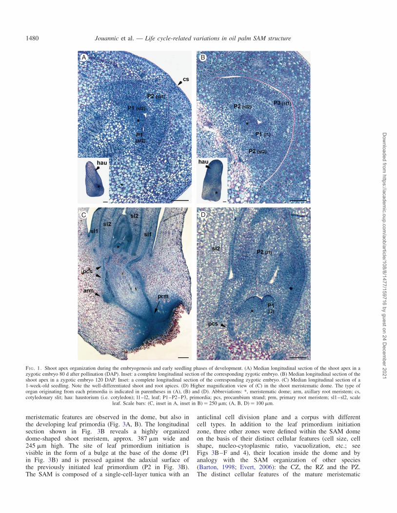
In pyramid-shaped embryos at the 80 DAP stage, the shootpole is located in the basal periphery of the embryo (seeinset to Fig. 1A). The SAM is a dome-shaped body about60–70 mm in width and 30–35 mm in height (Fig. 1A). TheSAM and the first scale leaf (embryonic leaf corresponding tothe sheath part only) are already established and the secondscale leaf is initiated at the periphery of the meristematicdome (Fig. 1A, see sl1 and sl2). The SAM dome has asingle-cell-layer tunica and a corpus of irregularly arrangedcells without visible histological zonation. Most of the cellsshare meristematic features (small isodiametric cells with ahigh nucleo-cytoplasmic ratio, a central nucleus, small or nodetectable vacuoles, deep blue NBB staining associated withhigh cytoplasmic protein content, mitotic figures, etc.).Mitotic figures are visible in the meristematic dome and in thesecond scale leaf primordium, indicating zones with highmitotic activity. At the mature stage (120 DAP), the secondscale leaf is developed and the first true leaf (or eophyll; i.e.bladed leaf) primordium is already initiated (Fig. 1B, see sl2and l1). The meristematic dome is slightly larger (82 mm wideand 48 mm high). At this stage, the embryo begins its dormantphase, with arrested organogenesis and an increase in storageprotein deposition, as illustrated by the dark blue-stained vacu-oles in the cells outside the shoot apex.
SAM organization during the seedling phase
The longitudinal section shown in Fig. 1C illustrates theorganization of the two apices 1 week after germination. At
this stage the plumule and radicle have not emerged. Theshoot apex is characterized by the two developed scaleleaves (sl1 and sl2 in Fig. 1C), the developing first eophyll pri-mordium and the primordium of the second eophyll (l1 and l2in Fig. 1D). As for the embryonic stages, the meristematicdome consists of cells with meristematic features with a pro-gressive vacuolization towards the base of the dome. The mer-istematic dome was larger than in the embryonic stages(152 mm wide and 113 mm high). A comparison of differentSAMs at the same developmental stage but at different timepoints of the plastochron period (based on the developmentalstage of the youngest leaf primordium) did not reveal anychanges, either in geometry (shape and size) or in the innerorganization of the meristematic dome (data not shown).This organization was also compared with that of 2-, 3- and4-week-old seedlings, but in each of these cases the shootorganization was similar without dramatic changes in thesize of the meristematic dome (data not shown).
SAM organization during the early establishment growth phase
The SAM structure in juvenile plants was analysed using3-month-old plants. The plants were 25 cm in heightwithout pinnate or bifid emerged leaves (see inset toFig. 2A). At this stage, plants were in their early establishmentgrowth phase and lacking inflorescence initiation. A closerview of the SAM showed that the meristematic dome hadthe same geometry and inner organization (i.e. tunica-corpuspattern without histological zonation) as in the youngerstages (i.e. embryos and plantlets; Fig. 2A) but with anincreased size (179 mm wide and 147 mm high). Similarly,cells in the dome and the youngest primordium shared thesame features (small and dense without a vacuole and witha high nucleo-cytoplasmic ratio; Fig. 2A). PI staining in con-junction with confocal microscopy clearly showed the pres-ence of a single-cell-layer tunica and no clear difference innuclear structure inside the meristematic dome (Fig. 2B). Ina transverse section of the upper region of the apex includingthe meristematic dome, PI staining of nuclei in conjunctionwith confocal microscopy illustrated the high nucleardensity of the meristematic dome and of the youngest leaf pri-mordium, as well as the angular arrangement of the develop-ing leaves (i.e. spiral phyllotaxis; Fig. 2C).
Shoot apex organization during the mature reproductive phase
Shoot apices from 10-year-old plants were analysed. Theplants used were about 5 m high with approx. 25 expandedleaves (see inset to Fig. 3A). At this stage, under tropicalhumid climatic conditions, the palms are mature with regularinflorescence production, an established stem width andregular leaf production, at an average of one or two leavesper month, indicating regular activity of the SAM.
Meristematic dome and histological zonation. The SAM andyoung leaf primordia are located in a depression surroundingthe stem tissue (Fig. 3A). This depression results fromprimary thickening activity characterized by extensive cellfiles resulting from polarized cell divisions at the base ofthe expanding leaf primordia (data not shown). Cells with
Jouannic et al. — Life cycle-related variations in oil palm SAM structure 1479
Dow
nloaded from https://academ
ic.oup.com/aob/article/108/8/1477/159716 by guest on 24 D
ecember 2021
meristematic features are observed in the dome, but also inthe developing leaf primordia (Fig. 3A, B). The longitudinalsection shown in Fig. 3B reveals a highly organizeddome-shaped shoot meristem, approx. 387 mm wide and245 mm high. The site of leaf primordium initiation isvisible in the form of a bulge at the base of the dome (P1in Fig. 3B) and is pressed against the adaxial surface ofthe previously initiated leaf primordium (P2 in Fig. 3B).The SAM is composed of a single-cell-layer tunica with an
anticlinal cell division plane and a corpus with differentcell types. In addition to the leaf primordium initiationzone, three other zones were defined within the SAM domeon the basis of their distinct cellular features (cell size, cellshape, nucleo-cytoplasmic ratio, vacuolization, etc.; seeFigs 3B–F and 4), their location inside the dome and byanalogy with the SAM organization of other species(Barton, 1998; Evert, 2006): the CZ, the RZ and the PZ.The distinct cellular features of the mature meristematic
A B
C D
FI G. 1. Shoot apex organization during the embryogenesis and early seedling phases of development. (A) Median longitudinal section of the shoot apex in azygotic embryo 80 d after pollination (DAP). Inset: a complete longitudinal section of the corresponding zygotic embryo. (B) Median longitudinal section of theshoot apex in a zygotic embryo 120 DAP. Inset: a complete longitudinal section of the corresponding zygotic embryo. (C) Median longitudinal section of a1-week-old seedling. Note the well-differentiated shoot and root apices. (D) Higher magnification view of (C) in the shoot meristematic dome. The type oforgan originating from each primordia is indicated in parentheses in (A), (B) and (D). Abbreviations: *, meristematic dome; arm, axillary root meristem; cs,cotyledonary slit; hau: haustorium (i.e. cotyledon); l1–l2, leaf; P1–P2–P3, primordia; pcs, procambium strand; prm, primary root meristem; sl1–sl2, scale
leaf. Scale bars: (C, inset in A, inset in B) ¼ 250 mm; (A, B, D) ¼ 100 mm.
Jouannic et al. — Life cycle-related variations in oil palm SAM structure1480
Dow
nloaded from https://academ
ic.oup.com/aob/article/108/8/1477/159716 by guest on 24 D
ecember 2021

dome were observed in both the corpus and thesingle-cell-layered tunica. A number of different parameterswere quantified for the cells of each zone in both the
tunica and the corpus (Table 1; see Supplementary DataFig. S1 for details, available online).
At the top of the dome, the CZ was characterized by largecells, central nuclei, low mitotic activity (i.e. rare mitoticfigures) and small but clearly observable vacuoles (Fig. 3B, C).The cells in this zone displayed an average surface area ofapprox. 214 mm2, a nucleo-cytoplasmic surface area ratio of0.33 and nearly circular nuclei (Table 1). They were alsocharacterized by a generally deep pink coloration of theircell walls with PAS staining compared with other cells ofthe meristematic dome, indicating that they were enriched inpolysaccharides and/or they possess a larger cell wall com-pared with other cells of the dome region (Fig. 3C).
The other two zones of the meristematic dome were locatedbeneath the CZ. One, defined as the RZ, was located in thecentral part of the meristematic dome. It was characterizedby the presence of large polyhedral cells with an averagesurface of 209 mm2 and a low nucleo-cytoplasmic ratio associ-ated with a large vacuole pushing the cytoplasm and nuclei tothe periphery of the cells (Fig. 3B, D; Table 1). In the upper-most region of the RZ, periclinal cell divisions were seen todominate, leading to vertical files of cells. The third zonewas defined as the PZ and corresponded to the peripheralregions of the dome beneath the CZ (Fig. 3B, E). The cellsin this region were smaller, with an average surface area of135 mm2, a thinner cell-wall structure, small vacuoles and ahigher nucleo-cytoplasmic surface area ratio compared withthe cells of the CZ and the RZ (Table 1). These cells also con-tained nuclei with deeper NBB blue staining compared withthe other zones (see grey levels in Table 1), suggestinghigher protein content. Mitotic figures were observable inthis zone. The most striking feature was the existence ofdistal–proximal files of cells from the CZ to the leaf primor-dium initiation site resulting from an orderly arrangement ofcell division in this region.
At the basal periphery of the dome, in connection with thePZ, is the site of leaf primordium development (Fig. 3B, F). Atthe stage shown, it consisted of cells with features intermediatebetween those of the CZ and PZ with an average cell surfacearea of approx. 156 mm2 (Table 1). The main specific featuresobserved for cells of this zone were first the presence ofnumerous tiny vacuoles and a dense deep blue NBB-stainedcytoplasm (see grey level in Table 1) and secondly a largenumber of mitotic figures. The enlargement of this zone isassociated with anticlinal divisions of the protoderm andboth anticlinal and periclinal divisions in the cell layersbeneath the protoderm.
Analysis of a plant during its late establishment growthphase (15-month-old plant approx. 1.50 m high) revealedthat this SAM geometry and its histological zonation arealready established (see Supplementary Data Fig. S2, availableonline), with a similar size and a less deep depression of theshoot apex compared with 10-year-old plants.
Variations of mature SAM structure. Series of longitudinal sec-tions were obtained for three different shoot meristems. Theyrevealed structural variations of the meristematic dome,which may be related to plastochronic changes associatedwith the developmental stage of the leaf primordium P1(Fig. 4). The histological zones illustrated were defined as
A
B
C
FI G. 2. Shoot apex organization during the early establishment growth phase.(A) Shoot apex median longitudinal section stained with PAS-NBB froma 3-month-old plant. Inset: the corresponding plant used for the shoot apexpreparation. (B, C) Confocal microscopic views of longitudinal (B) andtransverse (C) sections of shoot apices stained with propidium iodide from3-month-old plants. Abbreviations: *, meristematic dome; L1, tunica celllayer; P1–P2–P3, leaf primordia. Scale bars: (A, C) ¼ 100 mm; (B) ¼ 50 mm;
(inset in A) ¼ 10 cm.
Jouannic et al. — Life cycle-related variations in oil palm SAM structure 1481
Dow
nloaded from https://academ
ic.oup.com/aob/article/108/8/1477/159716 by guest on 24 D
ecember 2021

mentioned above: the CZ at the top of the meristematic domeand the RZ and PZ beneath the CZ (Fig. 4). The SAM shownin Fig. 4A (designated Mer1) is the same as that detailed inFig. 3. Analysis of the two other SAMs (Mer2 and Mer3)revealed older developmental stages of the leaf primordium
P1 that were distinct from each other (Fig. 4B, C). In thelatter cases, the meristematic domes were approx. 195 mm inheight for both with a basal width of about 342 and 390 mmfor Mer2 and Mer3, respectively. In comparison, Mer1 had amore elongated conical shape with a higher height/width
A B
C D
E F
FI G. 3. Shoot apex organization in a mature reproductive oil palm. (A) Shoot apex median longitudinal section from a 10-year-old plant. Inset: the correspond-ing plant used for shoot apex preparation. (B) Higher magnification view of (A) in the meristematic dome region. (C–F) Higher magnification views of thedifferent histological zones of the meristematic dome shown in (B). (C) The central zone at the top of the meristematic dome – note the large cells withthick cell wall and central uncondensed nuclei. (D) The rib meristem zone and the parenchyma beneath the meristematic dome – note the progressive vacuoliza-tion and cell size enhancement. (E) The peripheral zone in the left part of the longitudinal section – note the anticlinal files of cells with a thinner cell wall andprogressive vacuolization. (F) P1 leaf primordium at the early stage of protrusion – note the dense cytoplasm and nuclei, and limited vacuolization. The insets in(C–F) indicate the corresponding region of the SAM. Abbreviations: *, meristematic dome; P1–P2–P3, leaf primordia. Scale bars: (A) ¼ 250 mm; (B) ¼
100 mm; (C–F) ¼ 25 mm.
Jouannic et al. — Life cycle-related variations in oil palm SAM structure1482
Dow
nloaded from https://academ
ic.oup.com/aob/article/108/8/1477/159716 by guest on 24 D
ecember 2021
ratio (245 mm high, 387 mm wide). Moreover, this shape vari-ation was observed to be associated with an altered surfacearea of the different histological zones in the median sections(Fig. 4). A higher CZ surface area and a lower PZ surface areawere observed in Mer1 and a lower CZ surface area and higherPZ surface area in Mer3. On the basis of our analyses of thelongitudinal sections, it can be inferred that the overall shapeof the mature meristematic dome varies in relation to the fluc-tuating pattern of histological zonation.
To view these variations more precisely, 3-D reconstruc-tions were undertaken using the corresponding serial sections(Fig. 5). Four different regions were delimited for the recon-structions based on histological features as defined pre-viously: one region corresponding to the CZ, two regionscorresponding to the leaf primordia P1 and P2, respectively,and one region corresponding to the PZ and RZ. Based onthe 3-D reconstructions obtained, the volumes of the meris-tematic dome and CZ were calculated (Table 2). In asimilar way to the surface area differences observed in thelongitudinal sections, variations in the volume of the meris-tematic dome were noted between the three samples. Itshould be borne in mind that the volume of the dome ofMer3 was an underestimate, as part of the meristematicdome was missing in the series of sections of Mer3 usedfor the reconstruction (see Fig. 5C). The most important ten-dency observed was the fluctuation in CZ volume: in Mer1,the CZ volume represented 27 % of the total volume of thedome as compared with Mer2 in which the CZ correspondedto 20 % of the dome’s volume. The percentage CZ volumecalculated for Mer3 should be even lower, as the meriste-matic dome was not complete in the corresponding 3-Dreconstruction. Overall, the 3-D reconstructions clearly illus-trated the variations in dome shape between the threesamples, with a more elongated conical shape for Mer1 com-pared with the other two SAMs analysed.
Although we cannot completely exclude the possibility thatour results reflect inter-plant variability in mature SAM struc-ture, we hypothesize that the observed SAM size/shape differ-ences and related variations in histological zone proportionsare linked to the developmental progression of the youngestleaf primordium during the plastochron period, as seen inthe 3-D reconstructions.
DISCUSSION
Ontogenic variation of SAM organisation in oil palm
Our study revealed variations in SAM structure in terms of bothvolume and histological zonation during oil palm development(Fig. 6). During the growth and increase in diameter and heightof the oil palm shoot from the embryonic to the mature state, theshoot apical dome increases in size from about 60 mm width inthe embryo to 400 mm in the reproductive phase of the plant,corresponding to an approx. seven-fold increase in volume.The shoot apical dome size increases to a large extent and in agradual fashion between the embryonic and early establishmentphases and is nearly stable during the late establishment phase(from 15-month-old to 10-year-old plants). The second keyfeature is the alteration, during the life cycle of the plant, ofthe overall shape of the SAM, which is linked to changes inthe spatial organization of leaf primordium initiation (i.e. theposition of the organogenic region in relation to the apicalregion). The last main feature is the appearance of zonationduring the early establishment phase of development in parallelwith the stabilization of SAM size. Before histological zonationbecomes apparent, SAM organization can be summarized asbeing a single-cell-layered tunica and a corpus of irregularlyarranged cells sharing the same histological features. In asimilar way to oil palm, histological zonation also appearsduring the early establishment phase in Phoenix canariensisand seems to be associated with the stabilization of SAM size(Ball, 1941).
In this light, the development of the palm SAM can bedivided into two main phases (Fig. 6): a juvenile phase witha tunica-corpus organization and a mature phase with a histo-logical zonation superimposed on the tunica-corpus organiz-ation, with a transition operating between 3 and 10 monthsafter germination, during the establishment phase of palmdevelopment, which is characterized by a gradual extensionof stem diameter and a transition from the juvenile to themature leaf shape. Thus, in addition to the observable morpho-logical features (increase in height and stem diameter; juvenilevs. mature leaf shape), the transition from the juvenile to theadult phase in oil palm is also associated with a modificationof SAM structure, with the mature phase of the SAM occurringbefore the mature phase of the plant.
TABLE 1. Cell features in the different histological zones of the SAM
CZ PZ RZ P1
Cell surface area (mm2) 213.54+61.68 135.15+28.06 209.25+56.79 155.76+34.04Cell circularity 0.81+0.07 0.81+0.05 0.82+0.07 0.78+0.05Cell grey level 131.27+8.34 151.56+12.78 158.10+12.99 111.87+9.73Nuclei surface area (mm2) 50.43+8.93 38.56+8.36 38.09+7.39 44.50+9.17Nuclei circularity 0.94+0.01 0.94+0.04 0.92+0.02 0.91+0.04Nuclei grey level 85.11+11.77 73.59+13.00 90.13+24.10 65.40+11.77N/C ratio 0.33+0.08 0.41+0.09 0.24+0.07 0.41+0.10
Circularity, surface areas and grey levels were calculated from the longitudinal cross-section of the shoot apex from a 10-year-old plant, using the ImageJprogram in conjunction with the images in Fig. S1 (Supplementary Data, available online). Values are mean+ s.d. (n ¼ 30 cells per zone). A circularity valueof 1 corresponds to a perfect circle. As the value approaches 0, it indicates an increasingly elongated polygon. Grey levels correspond to means of grey levelsin cell or nuclei (0, black; 255, white).
Abbreviations: CZ, central zone; PZ, peripheral zone; RZ, rib meristem-like zone; P1, emerging leaf primordium; N/C ratio, nucleo-cytoplasmic surfaceratio.
Jouannic et al. — Life cycle-related variations in oil palm SAM structure 1483
Dow
nloaded from https://academ
ic.oup.com/aob/article/108/8/1477/159716 by guest on 24 D
ecember 2021

The stage of plant development at which zonation is estab-lished in the SAM can vary among species (Evert, 2006). Thelate establishment of zonation in the palm SAM is notobserved in species of the Poales, such as rice and maize.
One hypothesis is that this might reflect a differencebetween perennial and annual monocotyledonous species,with a longer juvenile phase in perennial plants.
A
B
C
FI G. 4. Structural variation in the mature shoot apical meristem. (A–C)Median longitudinal sections of shoot apices from three different10-year-old plants showing the P1 leaf primordium at different developmentalstages. Histological zonation is indicated as follows: the central zone in yellow(CZ); the peripheral zone in green (PZ); the rib meristem-like zone in orange
(RZ); the P1 leaf primordia in blue (P1). Scale bars ¼ 100 mm.
A
B
C
FI G. 5. 3-D reconstructions of mature shoot apical meristems. (A–C)Reconstructions of mature shoot apical meristems from sections of the threeshoot apices from 10-year-old plants shown in Fig. 4. Histological zonationis indicated as follows: the central zone in yellow, the peripheral and the ribmeristem-like zones in green, and the leaf primordia P1 and P2 in blue andred, respectively. The top right views correspond to the 3-D reconstructions
without the leaf primordium P2. Scale bars ¼ 100 mm.
Jouannic et al. — Life cycle-related variations in oil palm SAM structure1484
Dow
nloaded from https://academ
ic.oup.com/aob/article/108/8/1477/159716 by guest on 24 D
ecember 2021

Mature oil palm SAM structure in comparison with other monocotspecies
In comparison with the large size of the plant at its maturestage, the dome-shaped SAM of oil palm is small with a widthranging from 350 to 400 mm. Nevertheless, various studies ofSAM morphology in monocots have suggested that the size ofthe apex is correlated with mature stem diameter, with apexwidth ranging from 95 mm for Acorus species (plants of her-baceous habit in the Acorales) to 1300 mm for Xanthorrhoeaspecies (tree-like plants in the order Asparagales) (Martinand Tucker, 1985). The histological organization of themature SAM has been detailed or reported in at least eightother palm species from different genera and subfamilies:Phoenix, Washingtonia, Trachycarpus and Corypha from thesubfamily Coryphoideae (Ball, 1941; Tomlinson, 1990),Juania from the subfamily Ceroxyloideae (Tomlinson, 1990),and rattan genera Calamus and Korthalsia (Calamoideae sub-family) (Tomlinson, 1990). The SAM width measured for theother palm species studied ranged from 200 to 550 mm,
making them not dissimilar to mature oil palm, with the excep-tion of the rattan species characterized by a smaller SAM (lessthan 100 mm wide). Histological zonation of the SAM wasobservable for these species with some differences comparedwith oil palm. The main difference that can be notedbetween oil palm and the other palm species is the unclear dis-tinction between the PZ and the RZ, with orderly organizedcell divisions observed in the entire region beneath the CZin the species of the Coryphoideae studied (Ball, 1941). Thelatter are also characterized by a SAM located in a depressionat the apex (i.e. a concave apex) resulting from high primarythickening activity in these large-stemmed species. In contrast,in climbing palms of the rattan genera Calamus andKorthalsia, the shoot apex organization differs in that thereis a convex apex, with the presence of an orderly organizedRZ in the SAM, in contrast to the PZ. This organization issimilar to that observed in monocot species such as maize.Together, these observations illustrate a relationship betweenthe apex structure (concave vs. convex) and the primarythickening activity (i.e. stem enlargement). Importantly, stemenlargement could also be associated with cell division pat-terns of the corpus (PZ and MZ), even if the SAM does notcontribute significantly to the primary thickening activity.
The other difference observed between the different palmspecies concerns the tunica, which is single-layered in oilpalm and in the Phoenix, Juania and rattan species, andtwo-cell-layered in Washingtonia, Trachycarpus and Corypha.This indicates that the number of cell layers in the tunica doesnot correlate with the phylogenetic relationships of the palmspecies studied, nor with SAM size. In a similar way, thenumber of cell layers in the tunica has been found to be variablein other monocot orders and shows no phylogenetic pattern, asillustrated by the Poales (Brown et al., 1957).
Thus, mature palm SAMs are highly organized with a histo-logical zonation pattern defined by cells with different featuresreflecting different cell activities/identities but which candiffer to some extent between species, especially with regard
TABLE 2. Deduced volumes of the mature SAMs on the basis of3-D reconstructions
Histological zone Voxel count Volume (mm3) % ZC vs. Dome
Mer1 Dome 1064 898 6978 915ZC 294 462 1929 786 27.65
Mer2 Dome 939 926 6159 899ZC 196 129 1285 351 20.87
Mer3 Dome 765 490 5016 715ZC 153 457 1005 696 20.05
Abbreviations: CZ, central zone; Dome, whole meristematic dome. Voxelcount is the number of voxels (i.e. volumetric pixels) in a defined 3-D zone.Volumes were calculated according to the voxel size of the reconstructions(1.28 mm for the width and height according to the image resolution, 4 mmfor the depth according to the thickness of the sections). Mer1, mer2 andmer3 correspond to SAM reconstructions shown in Fig. 4A, B and C,respectively.
1 2
Juvenile phase Mature phase
3 4 5
FI G. 6. Ontogenic and plastochronic variations in the oil palm SAM. Outlines of median longitudinal sections of the SAM and leaf primordium P1 at differentdevelopmental stages: embryo (1); germinating plantlet (2); 3-month-old plant (3); 10-month-old plant (4); and 10-year-old-plant (5). The three different matureSAMs observed from 10-year-old plants are represented in (5). The tunica is indicated by the two outlines. The central zone and peripheral and rib meristem-like
zones are indicated in light and dark grey, respectively, in (4) and (5). Scale bar ¼ 100 mm.
Jouannic et al. — Life cycle-related variations in oil palm SAM structure 1485
Dow
nloaded from https://academ
ic.oup.com/aob/article/108/8/1477/159716 by guest on 24 D
ecember 2021

to the distinction between the PZ and the RZ. The most notablefeature of histological zonation seen in palms compared withother monocot species is the presence of a PZ with highlyorganized cell division patterns, which separate spatially theCZ and the initiation sites of the leaf primordium. This zoneis similar to that observed in some gymnosperm species dis-playing a so-called ‘transition zone’ with a highly organizedcell division pattern (Evert, 2006). To our knowledge, thiskind of organization has not been reported for any othermonocot species.
SAM changes during the plastochron period
As a consequence of the limited accessibility of the oil palmSAM structure (embedded in leaf primordia and developingleaves), scanning electron microscopy (Kwiatkowska andDumais, 2003), confocal microscopy (Laufs et al., 1998;Reddy et al., 2004) or optical coherence microscopy (Reeveset al., 2002) and tomography techniques such as X-ray com-puted tomography (Kaminuma et al., 2008) and optical projec-tion tomography (Sharpe et al., 2002; Lee et al., 2006) werenot feasible strategies to obtain a global 3-D reconstructionof the oil palm SAM, even with the younger developmentalstages. An alternative approach using serial sections of thewheat and the bulrush millet SAMs has, however, been usedpreviously to obtain 3-D reconstructions based on manualdrawings of the histological structures. This method producedgood representations of the 3-D structure but was limited toone angle of view as defined by the drawing (Metcalf et al.,1975; Williams and Langer, 1975; Williams et al., 1975).For this study, we obtained computer-assisted 3-D reconstruc-tions from series of microtome-derived sections, this approachhaving the advantage that histological stains can be viewedreadily, a large number of images can be aligned easily anddifferent structures can be traced and placed together in asingle 3-D view that can be animated (Fiala, 2005). Finally,the volume of the different structures can be calculated. Toour knowledge, this paper provides the first documentedexample of such an approach being applied to the SAM of aperennial monocotyledonous species.
Our study suggests that the overall shape, volume and histo-logical zonation pattern of the meristematic dome are dynamicand oscillate in relation to the organogenic activity of theSAM. The maximal volume of the SAM (for which therewas a characteristic elongated conical shape) was observed atthe very early stage of leaf primordium protrusion (leaf but-tress stage), CZ volume also being maximal at this stage.During later stages of leaf primordium development, thevolume of the meristematic dome decreases concomitantlywith changes in its shape (towards a less elongated conicalform) and a decrease in the CZ proportion within the dome(from 27 to 20 %). Later, the development of the leaf primor-dium continues in association with an increase in dome widthand a decrease in the CZ proportion within the dome. Theseobservations corroborate with those made on other speciesshowing plastochronic changes in which maximal volume pre-cedes initiation of the leaf primordium and minimal volumecoincides with the early stages of leaf primordium (Evert,2006).
Although the dynamics of SAM structure during the plasto-chron period have yet to be detailed for other palms, we canspeculate that the patterns seen here will also apply to otherspecies in the family. Global size and geometry changesduring the plastochron period have been reported for anumber of different monocot and eudicot species; the samecannot be said, however, for the phenomenon of major vari-ations in inner cell type proportions described here (Smithand Rogan, 1979; Martin and Tucker, 1985). Indeed,obvious changes in CZ size during the plastochron periodhave not to our knowledge been reported to date for anyother monocot species. In species such as maize or rice, inwhich the SAM is elevated considerably above the leaf primor-dium initiation site, plastochronic changes in size are notobserved (Evert, 2006). This situation contrasts with that ofthe oil palm SAM, which displays plastochronic changes butwhich also possesses a SAM which is elevated with respectto the organogenic region. These size variations which charac-terize the mature palm SAM may be in some way related to therelatively large volume of the SAM compared with the otherspecies studied. In this context, it is interesting to note thatno variations in oil palm SAM size were observed during theplastochron period in the juvenile phase. This suggests a linkbetween SAM plastochronic variations on the one hand andhistological zonation and/or SAM size on the other. Tosupport these views, it would be interesting to study thedynamics of the SAM in other species possessing a largeSAM.
Conclusions
Eudicot and monocot species differ significantly in the func-tioning of their SAMs in relation to the mechanisms involvedin leaf primordium founder cell recruitment, which in turnaffects leaf shape (Poethig and Szymkowiak, 1995;Nardmann et al., 2004; Evert, 2006). Moreover, this diversityin SAM structure and function exists within the monocot groupitself. Because of their perennial character and their specificfeatures such as compound leaf development and primarygrowth-based stem thickening, palms are distinct from othermonocots and of great interest in an evo-devo context. Asshown here, oil palm SAM ontogenesis is characterized by ajuvenile phase and a mature phase, with histological zonationspecific to the mature phase. A distinct feature of mature oilpalm SAM zonation is the presence of a large PZ withorderly cell files, which may act as a transition zone betweenthe CZ and the sites of leaf primordium initiation, as observedin some gymnosperm species. Plastochronic changes seem tooccur in the mature oil palm SAM with variations in CZvolume fluctuating in parallel with the early development ofthe leaf primordium. We showed recently that compoundleaf formation in oil palm seems to be associated with aKNOX-dependent pathway as in other compound-leavedplant species, even though the mechanism of leaflet formationdiffers from the canonical blastozone fractionation mechanism(Jouannic et al., 2007). In the same way, in an evo-devocontext, it will be interesting to characterize the expression pat-terns in palms of orthologues to genes that regulate SAM func-tion in other species, so as to understand better the evolution ofthis process in angiosperms.
Jouannic et al. — Life cycle-related variations in oil palm SAM structure1486
Dow
nloaded from https://academ
ic.oup.com/aob/article/108/8/1477/159716 by guest on 24 D
ecember 2021

SUPPLEMENTARY DATA
Supplementary data are available online at www.aob.oxford-journals.org and consist of the following figures. Figure S1:illustration of the views used for quantitative analysis. FigureS2: shoot apex organization during the late establishmentgrowth phase of oil palm.
ACKNOWLEDGEMENTS
We gratefully acknowledge the generous support of colleaguesat INRAB (Pobe, Benin) and ASD (Coto, Costa Rica) for pro-viding plant material. We thank Axel Labeyrie, Bruno Nouy(PalmElit, France) and David Cros (CIRAD, France) for logis-tical help. We are grateful to Fabienne Morcillo and ThierryBeule (CIRAD, UMR DIAPC, France) for collecting zygoticembryo samples. Finally, we would like to thank PatrickLaufs, Barry Tomlinson and Darleen A. DeMason for criticalreading of the manuscript. This work was supported by insti-tutional funding from IRD and CIRAD.
LITERATURE CITED
Adam H, Jouannic S, Escoute J, Verdeil JL, Duval Y, Tregear JW. 2005.Reproductive developmental complexity in the African oil palm (Elaeisguineensis). American Journal of Botany 92: 1836–1852.
Ball E. 1941. The development of the shoot apex and the primary thickeningmeristem in Phoenix canariensis Chaub., with comparisons toWashingtonia filifere Wats. and Trachycarpus excelsa Wendl. AmericanJournal of Botany 92: 1836–1852.
Barton MK. 1998. Cell type specification and self renewal in the vegetativeshoot apical meristem. Current Opinion in Plant Biology 1: 37–42.
Beirnaert A. 1935. Introduction a la biologie florale du palmier a huile (Elaeisguineensis Jacquin). Publications de l’Institut National pour l’EtudeAgronomique du Congo Belge (I.N.E.A.C.), Brussels, Belgium.Botanical Reviews 48: 1–69.
Brown WV, Heimsch C, Emery WHP. 1957. The organisation of the grassshoot apex and systematics. American Journal of Botany 44: 590–595.
Clark G. 1984. Staining procedures. 4th edn. Baltimore: Williams & Wilkins.Corley RHV, Gray BS. 1976. Growth and morphology. In Corley RHV,
Hardon JJ, Wood BJ, eds. Developments in crop, vol. 1, Oil palmresearch. Amsterdam: Elsevier, 7–21.
Evert RF. 2006. Esau’s plant anatomy: meristems, cells, and tissues of theplant body – their structure, function and development, 3rd edn.New York: Wiley-Interscience
Fiala RC. 2005. Reconstruct: a free editor for serial section microscopy.Journal of Microscopy 218: 52–61.
Fisher DB. 1968. Protein staining of ribboned epon sections for lightmicroscopy. Histochemie 16: 92–96.
Foster AS. 1943. Zonal structure and growth of the shoot apex in Microcycascalocoma (Miq.) A. DC. American Journal of Botany 30: 56–73.
Hartley CWS. 1988. The oil palm, 3rd edn. Longman Agriculture Series.London: Longman.
Jourdan C, Michaux-Ferriere N, Perbal G. 2000. Root system architectureand gravitropism in the oil palm. Annals of Botany 85: 861–868.
Jouannic S, Collin M, Vidal B, Verdeil JL, Tregear JW. 2007. A class IKNOX gene from the palm species Elaeis guineensis (Arecaceae) is
associated with meristem function and a distinct mode of leaf dissection.New Phytologist 174: 551–568.
Kaminuma E, Yoshizumi T, Wada T, Matsui M, Toyoda T. 2008.Quantitative analysis of heterogeneous spatial distribution of Arabidopsisleaf trichomes using micro X-ray computed tomography. Plant Journal56: 470–482.
Kwiatkowska D. 2008. Flowering and apical meristem growth dynamics.Journal of Experimental Botany 59: 187–201.
Kwiatkowska D, Dumais J. 2003. Growth and morphogenesis at the vegeta-tive shoot apex of Anagallis arvensis L. Journal of Experimental Botany54: 1585–1595.
Laufs P, Grandjean O, Jonak C, Kieu K, Traas J. 1998. Cellular parametersof the shoot apical meristem in Arabidopsis. Plant Cell 10: 1375–1390.
Lee K, Avondo J, Morrison H, Blot L, Stark M. 2006. Visualizing plantdevelopment and gene expression in three dimensions using optical pro-jection tomography. Plant Cell 18: 2145–2156.
Martin BF, Tucker SC. 1985. Developmental studies in Smilax (Liliaceae). I:Organography and the shoot apex. American Journal of Botany 72:66–74.
Metcalf RA, Fernandez A, Williams RF. 1975. The genesis of form inbulrush millet (Pennisetum americanum (L.) K. Schurn.). AustralianJournal of Botany 23: 761–773.
Nardmann J, Ji J, Werr W, Scanlon MJ. 2004. The maize duplicate genesnarrow sheath1 and narrow sheath2 encode a conserved homeobox genefunction in a lateral domain of shoot apical meristems. Development 131:2827–2839.
Poethig RS, Szymkowiak EJ. 1995. Clonal analysis of leaf development inmaize. Maydica 40: 67–76.
Reddy GV, Heisler MG, Ehrhardt DW, Meyerowitz EM. 2004. Real-timelineage analysis reveals oriented cell divisions associated with morpho-genesis at the shoot apex of Arabidopsis thaliana. Development 131:4225–4237.
Reeves A, Parsons RL, Hettinger JW, Medford JI. 2002. In vivo three-dimensional imaging of plants with optical coherence microscopy.Journal of Microscopy 208: 177–189.
Sharpe J, Ahlgren U, Perry P et al. 2002. Optical projection tomography as atool for 3-D microscopy and gene expression studies. Science 296:541–545.
Smith DL, Rogan PG. 1979. Growth of the shoot apex of Agropyron repens(L.) Beauv. during successive plastochrons. Annals of Botany 44: 27–34.
Tomlinson PB. 1990. The structural biology of palms. Oxford: OxfordScience Publications.
Vallade J. 1966a. Aspect morphologique et cytologique de l’embryon quies-cent d’Elaeis guineensis Jacq. Compte Rendu de l’Academie des Sciences262: 856–859.
Vallade J. 1966b. L’evolution de l’embryon quiescent d’Elaeis guineensisJacq. au cours de la germination. Compte Rendu de l’Academie desSciences 262: 856–859.
Van Heel WA, Breure CJ, Menendez T. 1987. The early development ofinflorescences and flowers of oil palm (Elaeis guineensis Jacq.) seenthrough the scanning electron microscope. Blumea 32: 67–78.
Williams RF, Langer RHM. 1975. Growth and development of the wheattiller. II The dynamics of tiller growth. Australian Journal of Botany23: 745–759.
Williams RF, Shavman BC, Langer RHM. 1975. Growth and developmentof the wheat tiller. I Growth and form of the tiller bud. Australian Journalof Botany 23: 715–743.
Yampolsky C. 1922. A contribution to the study of the oil palm Elaeisguineensis Jacq. Bulletin du Jardin Botanique Buitenzorg, serie III, volV, 107–174.
Jouannic et al. — Life cycle-related variations in oil palm SAM structure 1487
Dow
nloaded from https://academ
ic.oup.com/aob/article/108/8/1477/159716 by guest on 24 D
ecember 2021


![In vitro plant production through apical meristem culture ... · regeneration from callus cultures [7]. Meristem-tip culture is an important technique for the production of disease](https://static.fdocuments.us/doc/165x107/5ea00a71a584c3433161b086/in-vitro-plant-production-through-apical-meristem-culture-regeneration-from.jpg)