Patient Safety Framework for Mitigating Wrong-level Spine ...
24
Patient Safety Framework for Mitigating Wrong-level Spine Surgery RECOMMENDATIONS OF THE SPINE SURGERY SAFETY TASK FORCE Created: 2018
Transcript of Patient Safety Framework for Mitigating Wrong-level Spine ...
Patient Safety Framework for Mitigating
Wrong-level Spine Surgery
RECOMMENDATIONS OF THE SPINE SURGERY SAFETY TASK FORCE
Created: 2018
ABOUT THIS DOCUMENT
The recommendations for Patient Safety Framework for Mitigating Wrong-level Spine Surgery were developed under the auspices of the Academic Medical Center Patient Safety Organization (AMC PSO) Spine Surgery Safety Task Force. These consensus recommendations are for informational purposes only and should not be construed or relied upon as a standard of care. The AMC PSO recommends institutions review these guidelines and accept, modify or reject these recommendations based on their own resources and patient populations. Additionally, institutions should continue to review and modify these recommendations as the field continues to evolve.
©2018 Academic Medical Center Patient Safety Organization (AMC PSO), a component patient safety organization within The Risk Management Foundation of the Harvard Medical Institutions Incorporated
AMC PSO | SPINE SURGERY
1© 2018 AMC PSO
EXECUTIVE SUMMARY
In 2017, at the request of its membership, the Academic
Medical Center Patient Safety Organization (AMC PSO)
convened the Spine Surgery Safety Task Force to arrive at
a set of consensus-supported guidelines for patient safety
considerations for spinal surgery.
As technology advances and care evolves, 0pportunities exist to align and standardize practice to reflect this evolution.
The Task Force began with a review of the latest scientific evidence, guidance, and opinion statements from relevant professional societies, as well as input from frontline providers in Neurosurgical and Orthopedic Spine Surgery. Further insights were gathered from convening AMC PSO member subject matter experts across various surgical specialties, hospital operations, Anesthesiology, Nursing, Radiology, Risk Management, and Patient Safety.
The AMC PSO sought to build a set of consensus-based and literature-supported recommendations that reflect patient safety concerns during spinal procedures.
What follows is a document that represents the aim, mission, and consensus opinion of the Task Force. It offers guidance for clinicians in their efforts to provide the safest possible care to patients.
AMC PSO | SPINE SURGERY
2© 2018 AMC PSO
DEFINITIONS
Cognitive Bias An error in reasoning, evaluating, remembering, or other cognitive process, often occurring as a result of holding onto one’s preferences and beliefs regardless of contrary information.
Cognitive Tunneling (aka Cognitive Capture) Describes a process called “inattentional blindness”4 resulting in a mental state in which the brain fixates on detail that is front of mind, and as a result, it does not “see” the rest of the environment or other relevant data.
Confirmation Bias The tendency to interpret new evidence as confirmation of one’s existing beliefs or theories.
Image Marker Radiopaque instrumentation that is placed proximate to the operative level(s) and stays in place until imaging, confirmation of level, and placement of a Reference Mark has been performed.
Reference MarkAn intraoperative mark that serves to identify, confirm, and definitively mark a level from which to count to the operative level(s) and generally remains for the duration of a case unless obliterated in the course of the procedure.
Skin Site Marking A marking of the surgical site by the surgeon in the preoperative holding area. The skin mark is placed as proximate to the incision site as anatomically possible using a single-use, indelible ink skin marker to indicate both laterality and level.
AMC PSO | SPINE SURGERY
3© 2018 AMC PSO
TABLE OF CONTENTS
Executive Summary . . . . . . . . . . . . . . . . . . . . . . . . . . . . . . . . . . . . . . . . . . . . . . . . . . . . . . . . . . . . . . . . . . . . . . . 1
Definitions . . . . . . . . . . . . . . . . . . . . . . . . . . . . . . . . . . . . . . . . . . . . . . . . . . . . . . . . . . . . . . . . . . . . . . . . . . . . . . . . . . . . 2
Case Study . . . . . . . . . . . . . . . . . . . . . . . . . . . . . . . . . . . . . . . . . . . . . . . . . . . . . . . . . . . . . . . . . . . . . . . . . . . . . . . . . . . . . 4
Introduction . . . . . . . . . . . . . . . . . . . . . . . . . . . . . . . . . . . . . . . . . . . . . . . . . . . . . . . . . . . . . . . . . . . . . . . . . . . . . . . . . . . 5
Patient-related Factors . . . . . . . . . . . . . . . . . . . . . . . . . . . . . . . . . . . . . . . . . . . . . . . . . . . . . . . . . . . . . . . . . 6
Image Quality . . . . . . . . . . . . . . . . . . . . . . . . . . . . . . . . . . . . . . . . . . . . . . . . . . . . . . . . . . . . . . . . . . . . . . . . . . . . . . . . . 7Technology . . . . . . . . . . . . . . . . . . . . . . . . . . . . . . . . . . . . . . . . . . . . . . . . . . . . . . . . . . . . . . . . . . . . . . . . . . . . . . . . 7Technique . . . . . . . . . . . . . . . . . . . . . . . . . . . . . . . . . . . . . . . . . . . . . . . . . . . . . . . . . . . . . . . . . . . . . . . . . . . . . . . . . 9Training . . . . . . . . . . . . . . . . . . . . . . . . . . . . . . . . . . . . . . . . . . . . . . . . . . . . . . . . . . . . . . . . . . . . . . . . . . . . . . . . . . . . 10
Surgical . . . . . . . . . . . . . . . . . . . . . . . . . . . . . . . . . . . . . . . . . . . . . . . . . . . . . . . . . . . . . . . . . . . . . . . . . . . . . . . . . . . . . . . . 11Standardization of Practice . . . . . . . . . . . . . . . . . . . . . . . . . . . . . . . . . . . . . . . . . . . . . . . . . . . . . . . 11Documentation . . . . . . . . . . . . . . . . . . . . . . . . . . . . . . . . . . . . . . . . . . . . . . . . . . . . . . . . . . . . . . . . . . . . . . . . 16
Team-based Care & Human Factors . . . . . . . . . . . . . . . . . . . . . . . . . . . . . . . . . . . . . . . . . . . . . . . 17Communication . . . . . . . . . . . . . . . . . . . . . . . . . . . . . . . . . . . . . . . . . . . . . . . . . . . . . . . . . . . . . . . . . . . . . . . . 17Bias . . . . . . . . . . . . . . . . . . . . . . . . . . . . . . . . . . . . . . . . . . . . . . . . . . . . . . . . . . . . . . . . . . . . . . . . . . . . . . . . . . . . . . . . . 19
Production Pressure . . . . . . . . . . . . . . . . . . . . . . . . . . . . . . . . . . . . . . . . . . . . . . . . . . . . . . . . . . . . . . . . . . . . 20
CASE EXAMPLE
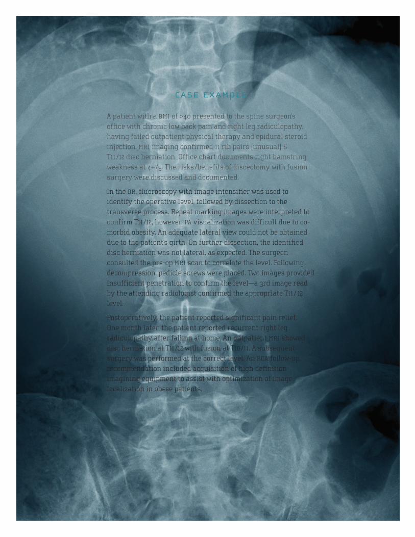
A patient with a BMI of >40 presented to the spine surgeon’s office with chronic low back pain and right leg radiculopathy, having failed outpatient physical therapy and epidural steroid injection. MRI imaging confirmed 11 rib pairs (unusual) & T11 / 12 disc herniation. Office chart documents right hamstring weakness at 4+ / 5. The risks / benefits of discectomy with fusion surgery were discussed and documented.
In the OR, fluoroscopy with image intensifier was used to identify the operative level, followed by dissection to the transverse process. Repeat marking images were interpreted to confirm T11 / 12, however, PA visualization was difficult due to co-morbid obesity. An adequate lateral view could not be obtained due to the patient’s girth. On further dissection, the identified disc herniation was not lateral, as expected. The surgeon consulted the pre-op MRI scan to correlate the level. Following decompression, pedicle screws were placed. Two images provided insufficient penetration to confirm the level—a 3rd image read by the attending radiologist confirmed the appropriate T11 / 12 level.
Postoperatively, the patient reported significant pain relief. One month later, the patient reported recurrent right leg radiculopathy after falling at home. An outpatient MRI showed disc herniation at T11 / 12 with fusion at T10 / 11. A subsequent surgery was performed at the correct level. An RCA follow-up recommendation included acquisition of high definition imagining equipment to assist with optimization of image localization in obese patients.
AMC PSO | SPINE SURGERY
5© 2018 AMC PSO
INTRODUCTION
The incidence of wrong-level spine surgery is difficult to determine, but is estimated to be one
in 3,110 spine procedures.1 Available data sources are limited. Physician surveys have provided
insights into the magnitude of the problem, including the emotional impact of wrong-site
surgery, related adverse event disclosure conversations, and litigation on providers. Statewide
adverse event and / or “Never Event” (as defined by the National Quality Forum) databases
track rates as a percentage of total adverse events; insurance and malpractice reports express
wrong-level spine surgery rates as a percentage of total claims. A limitation of these sources of
information is that the denominator of spine surgeries is unquantified.
Literature review and professional society member surveys2 identify a range of intraoperative practices with respect to the identification of the operative level, including marking of the spinous process, lamina, and pedicle.Both fluoroscopy and X-ray are routinely employed for confirmation. The North American Spine Society (NASS) “Sign, Mark, and X-ray” program specifies only that ‘a bony landmark’ be identified with a radiopaque marker.3 While there is general agreement that imaging should be obtained with a metallic marker located on a bony landmark, surgical marking practice and methods vary considerably.
An analysis of closed claims and submitted root cause analysis data spanning 2006–2016 was conducted by the AMC PSO to identify themes and commonalities in wrong-level spine procedures.
Contributing factors gleaned from the review included:
• poor team communication, including providers being absent from the immediate pre-procedure timeout
• reliance on preliminary skin localization images in the absence of intraoperative marking images
• spot fluoroscopy images with an insufficient field width to allow for the assessment of relevant anatomy or anatomical variation, including unrecognized transitional vertebrae
• suboptimal intraoperative images associated with obesity
• misinterpretation of rotated (non-orthogonal) images by both surgeons and radiologists
• inadequate documentation and transcription errors in the pre-hospital and hospital record
• marking films that were referenced in documentation, but not archived to the permanent record and, thus, unavailable for subsequent review
At the request of its membership, aggregated and contextually non-identifiable learnings from this review formed the basis for an initial AMC PSO safe table convening discussion of patient safety considerations relevant to wrong-level spine surgery. This initial convening was followed by a series of multidisciplinary, subject matter expert informed safe tables conducted under the auspices of the AMC PSO that sought to develop consensus-based risk mitigation strategies for wrong-level spine surgery.
AMC PSO | SPINE SURGERY
6© 2018 AMC PSO
Patient-related Factors
RISKS
• High BMI / Osteoporosis
- Spine may be difficult to visualize
- Lower C-spine may be obscured by the shoulders
- Intraoperative inferior shoulder traction is often necessary for C-spine visualization, resulting in patient positioning and stabilization considerations
- Intraoperative images are often inferior to fully optimized preoperative images due to the nature of the study and positioning
• Transitional vertebrae / deformity
- Associated with wrong-level determination and imaging report variability in the interpretation and assignment of the recorded level
- Preoperative comparative images are not available in the operating room
RISK MITIGATION STRATEGIES
{ Heightened awareness of clinical risk factors
• Assignment of the level may vary between imaging studies
{ High BMI /Osteoporosis
• Adequate visualization of the base of skull and the cervical spine are integral to localization and marking
• Optimization: obtain the best possible intraoperative image
- Availability of equipment to depress shoulders
- Digital imaging is superior to C-arm fluoroscopy
{ Transitional Vertebrae/Deformity
• Direct correlation in the operating room of preoperative and intraoperative images is recommended
• A second, independent confirmation of the level by the First Assist in the operating room is recommended
- If possible, consider closed-loop confirmation of the level by a Radiologist at the time of the intraoperative marking film
1
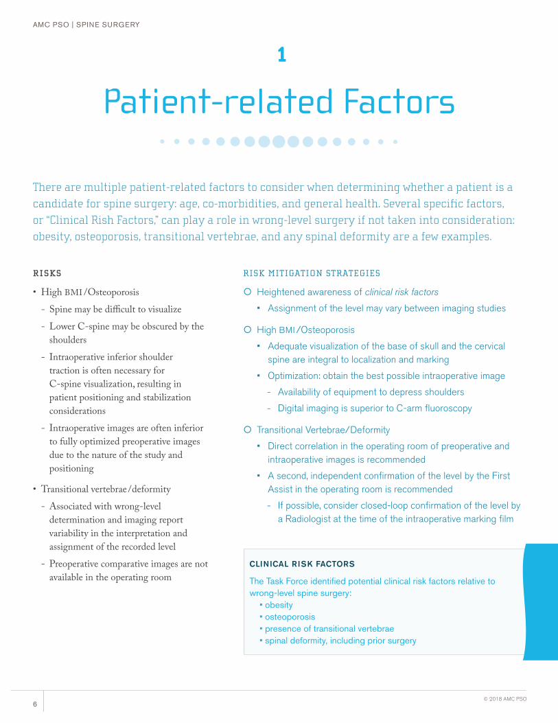
There are multiple patient-related factors to consider when determining whether a patient is a
candidate for spine surgery: age, co-morbidities, and general health. Several specific factors,
or “Clinical Rish Factors,” can play a role in wrong-level surgery if not taken into consideration:
obesity, osteoporosis, transitional vertebrae, and any spinal deformity are a few examples.
CLINICAL RISK FACTORS
The Task Force identified potential clinical risk factors relative to wrong-level spine surgery:
• obesity• osteoporosis• presence of transitional vertebrae• spinal deformity, including prior surgery
AMC PSO | SPINE SURGERY
7© 2018 AMC PSO
2
Image Quality
Access to optimal imaging across the continuum of preoperative assessment through the
intraoperative period is of key importance to ensuring correct level spine surgery. Several
factors contribute to achieving high-quality imaging, including technology, technique, and the
training and engagement of key personnel. Team members’ familiarity and comfort with the
equipment, the environment, the procedure—and each other—are essential elements of being
able to deliver safe, high quality care .
RISKS
• Lack of familiarity of the technologist with the equipment
• Lack of access to intraoperative images postoperatively
- Unarchived images are not available for adverse event review
• Variability in image quality between imaging modalities and technology
RISK MITIGATION STRATEGIES
{ Training
• Annual vs. periodic training and competency evaluations for technologists
• Involve Radiologists in the training of technologists
• Include technologists in team training sessions
{ Establish a process for archiving key images
{ Digital portable imaging provides superior image quality
{ High-resolution and wide-field digital detectors
• Provide improved image resolution in high BMI patients
• May improve image optimization and reduce risks related to shoulder traction and spine stabilization in cervical procedures
TECHNOLOGY Fluoroscopy / Portable Imaging
AMC PSO | SPINE SURGERY
8© 2018 AMC PSO
RISKS
• Lack of availability of “outside hospital” and preoperative images in PACS and / or in the operating room
• PACS monitors in the OR are set to logout automatically after a prescribed interval in order to protect Personal Health Information (PHI). In the OR, this may lead to the unintended consequence of rendering comparison images unavailable for review by the scrubbed surgeon.
RISK MITIGATION STRATEGIES
{ Develop a process to load and archive “outside hospital” preoperative images into the PACS system
• If preoperative images cannot be preloaded into PACS, ensure that hard copies of images are available in the operating room for comparison with intraoperative images
{ Work with Compliance and Information Systems Security to ensure there is access to images in the operating room at critical steps of the procedure
TECHNOLOGY Picture Archiving and Communication System (PACS)
[Image Quality]
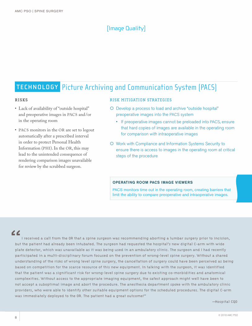
OPERATING ROOM PACS IMAGE VIEWERS
PACS monitors time out in the operating room, creating barriers that limit the ability to compare preoperative and intraoperative images.
“I received a call from the OR that a spine surgeon was recommending aborting a lumbar surgery prior to incision,
but the patient had already been intubated. The surgeon had requested the hospital’s new digital C-arm with wide
plate detector, which was unavailable as it was being used in an ambulatory clinic. The surgeon and I had recently
participated in a multi-disciplinary forum focused on the prevention of wrong-level spine surgery. Without a shared
understanding of the risks of wrong level spine surgery, the cancellation of surgery could have been perceived as being
based on competition for the scarce resource of this new equipment. In talking with the surgeon, it was identified
that the patient was a significant risk for wrong-level spine surgery due to existing co-morbidities and anatomical
complexities. Without access to the appropriate imaging equipment, the safest approach might well have been to
not accept a suboptimal image and abort the procedure. The anesthesia department spoke with the ambulatory clinic
providers, who were able to identify other suitable equipment options for the scheduled procedures. The digital C-arm
was immediately deployed to the OR. The patient had a great outcome!”
—Hospital CQO
AMC PSO | SPINE SURGERY
9© 2018 AMC PSO
AMC PSO | SPINE SURGERY
RISKS
• Suboptimal images hamper visualization of key landmarks, particularly in the cervical and lumbar spine
• Obliqued (non-orthogonal) films are inadequate
• Technologist engagement
- Lack of engagement and empowerment of the technologist as a member of the surgical team can affect patient safety
RISK MITIGATION STRATEGIES
{ Image set up
• Ensure that imaging is centered on the operative level
• Establish protocols to ensure standardization of the distance
• Obtain true lateral / orthogonal spine films
• Width of the spot fluoroscopy field should be wide enough to assess the relevant anatomy and adjacent landmarks (such as the base of skull and odontoid vs. sacrum)
• Operative level should be centered on the fluoro spot
• Be aware of high-risk patient factors/conditions: osteoporsis, high BMI, transitional vertebrae, and spinal deformity
{ Do not accept suboptimal images!
{ Active engagement of the technologist by the surgeon
• Empower and afford the time for the technologist to have a discussion of the critical elements with the surgeon: “What are your landmarks, and what are you looking for?”
TECHNIQUE Suboptimal Images
Clinicians should not accept suboptimal images. The process of setting up an image in order to obtain a fully optimized image adds value and supports clinical decision making. Obliqued films pose challenges with interpretation. Efforts should be made to obtain lateral/orthogonal spine films.
BEST PRACTICE
Empower and engage the technologist. The radiology technologist is engaged in active dialogue with the surgical team and routinely queries the surgeon upon entering the room: “What are your landmarks and what are you looking for?”
[Image Quality]
AMC PSO | SPINE SURGERY
10© 2018 AMC PSO
AMC PSO | SPINE SURGERY
RISKS
• Personal performance factors: deficits of knowledge, skill, and ability introduce organizational risk
• Reliance on staff who cross-cover multiple services and modalities (particularly on weekends, holidays, and in community hospitals)—poses challenges
RISK MITIGATION STRATEGIES
{ Assess opportunities to establish a core team of spine surgery technologists (high-volume centers) vs. focused training for technologists assigned to the operating room
• Equipment familiarity: fluoroscopy
• Procedure and anatomy familiarity: spine surgery / musculoskeletal
{ Team training: hierarchical issues
• Empower technologists to speak up and call out images they identify as suboptimal
TRAINING Technologist Training
[Image Quality]
RISKS
• Lack of quality assurance/quality improvement focus on intraoperative imaging contributes to suboptimal images and wrong-level spine surgery
RISK MITIGATION STRATEGIES
{ Establish a multidisciplinary peer review process for intraoperative imaging
• Engage radiologists, technologists, and surgeons in film review
• Standardization of the distance, voltage, and protocols
• Education and periodic assessment of competencies
TECHNIQUE Quality Assurance / Quality Improvement
AMC PSO | SPINE SURGERY
11© 2018 AMC PSO
Surgical3
Spine surgery poses unique challenges that carry a risk of wrong-level surgery. Aside from
patient-related factors and imaging, having timely access to critical patient information as well
as a standard approach are important in mitigating this risk.
RISKS
• Lack of availability of the consent, plan of care/office notes, and preoperative images in the holding area
• Overreliance on a preoperative H&P not completed by the surgeon (e.g., PAT Nurse Practitioner, PCP) for key components of the planned procedure may introduce risk on the day of surgery
• Potential for variation in assignment of level between different studies and on outside hospital image reports, especially with transitional vertebrae
RISK MITIGATION STRATEGIES
{ Establish a process to ensure availability for review of the surgeon’s plan of care and relevant clinic /office notes
• Plan of care should specify detail related to the planned procedure, including site, level, and laterality and, as such, serves as a key written handoff communication tool
{ Consent should specify procedure, laterality, and level
• Recommendation: completion of the consent in the surgeon’s office
{ Preoperative images should be available in the operating room
STANDARDIZATION OF PRACTICE Transitions of Care Documentation: Clinic to Holding Area
BEST PRACTICE
When possible, the informed consent process occurs and is documented in the surgeon’s office setting.
PRE-OP IMAGING STUDIES
Access—in the holding area and OR—to previously obtained, high-quality, fully-optimized preoperative imaging studies plays a crucial role in the assessment and accurate evaluation of spinal deformities, alignment, and landmarks.
AMC PSO | SPINE SURGERY
12© 2018 AMC PSO
RISKS
• Site marking not visible / site marking washes off with surgical prep: use of non-indelible pens contributes to the need for repeat skin marking in the operating room, delaying cases, and introducing risk
• Prone positioning
• Anterior cervical approaches may be contralateral to the target pathology
• A poor signal of laterality and level in spine surgery due to the midline nature of the spine and the imprecise localization of the skin marking relative to the surgical anatomy
RISK MITIGATION STRATEGIES
{ Site markings serve to further orient and engage the surgical team and patient in the preoperative preparation of the patient
{ Use indelible pens
{ If removed by the surgical prep, re-mark with full exposure of the torso in the operating room
{ Marking must be visible after the drapes are placed
{ Recommendations:
• Site marking should designate both the laterality and the level of pathology
• For lumbar, posterior cervical, and posterior thoracic approaches, place the site marking ipsilateral to pathology
- Of note: the incision and related site marking for anterior cervical approaches may be contralateral to the target pathology
STANDARDIZATION OF PRACTICE Site Markings
[Surgical]
REGULATION STANDARDS
Although a regulatory requirement,5 site marking, in and of itself, is not adequate in preventing wrong-level spine surgery.
The skin mark is placed as proximate to the incision site as anatomically possible using a single-use, indelible ink skin marker. Although variation in practice is acknowledged, surgical site marking, in general, has traditionally consisted of the surgeon’s initials, which may also serve as a signal to the team that the surgeon has personally evaluated the patient in the holding area.
AMC PSO | SPINE SURGERY
13© 2018 AMC PSO
RISKS
• Failure to include key landmarks
• Patient factors
- Transitional vertebrae
- Deformity
- Prior spine surgery
RISK MITIGATION STRATEGIES
{ Obtain marking images after dissection and placement of a retractor
{ Recommendation: Images must include prominent landmarks:
• Base of skull for cervical spine procedures—odontoid process
• Sacrum for lumbar procedures
• Thoracic spine procedures:
- Visualization of the skull base vs. ribs vs. sacrum
- Pre-existing hardware / implant may be helpful in localization
{ Consider an independent confirmation of the level (Radiologist, colleague) with:
• Transitional vertebrae
• Deformity
• Prior spine surgery
{ Attention to confirmation and cognitive biases that may affect site verification (see Section 4)
STANDARDIZATION OF PRACTICE Intraoperative Marking Images
RISKS
• Inadequate for the determination of level
RISK MITIGATION STRATEGIES
{ Encouraged. However, the needle placed through the skin for the preliminary localization image should not be relied upon for a definitive determination of the operative level
{ Utility of preliminary localization images includes:
• Improves precision in placement and size of the incision rather than reliance on an estimation based on external landmarks
• Validates the image quality and permits needed adjustments (patient positioning, technical, fluoro vs. digital portable imaging) before surgery has commenced
STANDARDIZATION OF PRACTICE Preliminary Localization Images
[Surgical]
AMC PSO | SPINE SURGERY
14© 2018 AMC PSO
RISKS
• Cognitive drift can be a factor in translating the identification of the determined level on the marking image to the placement of the Reference Mark
RISK MITIGATION STRATEGIES
{ From marking image to placement of the Reference Mark:
• Recommendation: The image marker should remain in place until the level is confirmed on imaging and the Reference Mark is placed
• Active call out of the Reference Mark creates an opportunity for a cognitive pause and verification
STANDARDIZATION OF PRACTICE Spine Marking Strategies
[Surgical]
REFERENCE MARK
The purpose of the Reference Mark is to identify, confirm, and definitively mark a level from which to count to the operative level(s).
AMC PSO | SPINE SURGERY
15© 2018 AMC PSO
RISKS
• A restricted field limits visualization of key landmarks
RISK MITIGATION STRATEGIES
{ Drape from the sacrum
{ In the operating room, consider digital wide-plate detectors that permit visualization of the sacrum
STANDARDIZATION OF PRACTICE Thoracic Procedures: Surgical Field
[Surgical]
RISKS
• Determination of the thoracic level can be challenging and is reliant on the accurate identification of related structures
• Thoracotomy approach limits confirmatory imaging options
RISK MITIGATION STRATEGIES
{ Outpatient preoperative imaging must include prominent landmarks for orientation, such as:
• Inclusion of the skull base
• Documentation of the ribs
• Inclusion of the sacrum
{ Identification of the operative level relative to the Reference Mark—suggested localization techniques:
• Counting from the first rib downward
• Counting from the sacrum or inferior-most rib upward
• Thoracotomy approach: manual localization by palpation of the ribs
• Be consistent in the direction used to count
• Pre-existing hardware/implant may be helpful in localization
• Method of preoperative localization and intraoperative localization should be consistent and should be documented in preoperative surgical notes
STANDARDIZATION OF PRACTICE Thoracic Procedures: Determination of Level
AMC PSO | SPINE SURGERY
16© 2018 AMC PSO
DOCUMENTATION
RISKS
• Inadequate understanding of the risks, benefits, and non-surgical alternatives
• Inadequate documentation of the informed consent process
RISK MITIGATION STRATEGIES
{ Informed consent should document a review of the risks of surgery and alternatives to surgery
{ Activation of the patient: utilization of shared medical decision-making tools to engage the patient in the plan of care
{ Consent should specify procedure, laterality, and level
• Recommendation: completion and documentation of the consent process in the surgeon’s office
DOCUMENTATION Physician-Patient Preoperative Communication
RISKS
• Medical decision-making and localization technique are not supported by documentation in the event of an adverse outcome
RISK MITIGATION STRATEGIES
{ A description of localization technique should be dictated into the operative record
DOCUMENTATION Operative Note
RISKS
• Images are not available to support clinical decision making in the event of an adverse outcome
• Inadequate documentation of key elements of a procedure in the event of an adverse outcome
RISK MITIGATION STRATEGIES
{ Recommendation: Marking films, including key spot fluoroscopy images, must be archived to the permanent patient record
• The surgeon designates, through an active call-out process, which images will be archived
DOCUMENTATION Intraoperative Imaging
[Surgical]
AMC PSO | SPINE SURGERY
17© 2018 AMC PSO
Team-based Care & Human Factors
Effective communication among all members of a surgical team is imperative to ensuring
patient safety in the operating room. This means engaging all relevant stakeholders before,
during, and after a procedure.
4
RISKS
• Lost opportunities to fully engage and leverage expertise of all the team members
• Technologists often arrive after a patient is draped, limiting their ability to actively participate in the full clinical assessment of the patient
RISK MITIGATION STRATEGIES
{ Active engagement by the surgeon promotes a culture of safety and respect, while serving to level hierarchical barriers for effective team communication
{ Active call out of the level and participation in obtaining and interpreting images
• Example: surgeon: “I need your help getting the right image—this is what I am looking for.”
COMMUNICATION Engagement of the Radiology Technologist
RISKS
• Loss of situational awareness by the team introduces unnecessary risk
RISK MITIGATION STRATEGIES
{ Opportunities:
• Active call out /confirm the consented level
• Ensure availability of the preoperative office notes /plan of care in the operating room
• Awareness and acknowledgement by the team of the presence of any anatomic variation—especially transitional vertebrae and deformity
• Selection of marking images to be archived
• Situational awareness around placement of the Reference Mark
COMMUNICATION Briefings, Hard Stops, and Active Call Outs—Heightened Situational Awareness
AMC PSO | SPINE SURGERY
18© 2018 AMC PSO
RISKS
• Increased risk of litigation:
- Ineffective communication may convey a lack of empathy
- Patient/family perception that information is being withheld
• Providers lack training in initial disclosure conversations
• Scripting of templated patient notification letters may be perceived by patients and families as cold and uncaring
RISK MITIGATION STRATEGIES
{ The informed consent process and use of shared medical decision-making aids in establishing trust with the patient and family
{ Initial disclosure: Engage Risk Management and Patient Safety to assist with disclosure process.
{ Engage Patient Relations early to establish a longitudinal relationship with the patient and family
{ Consider collaborating with the Patient Family Advisory Council (PFAC) to review the content of patient notification letters (e.g., sender: “Patient Relations” or “Patient Safety,” not “Risk Manager”)
COMMUNICATION Communication with Patients After an Adverse Event
[Team-based Care & Human Factors]
RISKS
• Cognitive tunneling with incomplete information and inadequate images
• Cognitive drift can occur when attention is diverted from the surgical field prior to placement of the Reference Mark
RISK MITIGATION STRATEGIES
{ Heightened awareness of high-risk Clinical risk factors, especially transitional vertebrae and deformity
{ Consider a hard stop when placing the Reference Mark
{ Fully engage surgical team members through briefings, active call outs, and hard stops
{ If in doubt, obtain an objective second set of eyes on images (colleague, Radiologist)
BIAS Cognitive Bias
Cognitive tunneling is recognized as a contributing factor in wrong-level spine surgery. Suboptimal images and anatomical variations, such as transitional vertebrae, increase this risk. There is a risk of cognitive drift when shifting focus between images and anatomical structures.
AMC PSO | SPINE SURGERY
19© 2018 AMC PSO
RISKS
• Hierarchical issues
• Team loses objectivity
RISK MITIGATION STRATEGIES
{ Empower team members—consider team training
{ Consider an independent second set of eyes in high-risk patients to mitigate risk related to confirmation bias
{ A culture of safety that supports high reliability processes and challenges rules decreases the level of hierarchy and increases safety
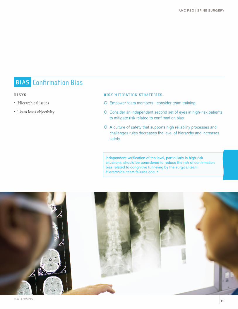
BIAS Confirmation Bias
Independent verification of the level, particularly in high-risk situations, should be considered to reduce the risk of confirmation bias related to congnitive tunneling by the surgical team. Hierarchical team failures occur.
AMC PSO | SPINE SURGERY
20© 2018 AMC PSO
Production Pressure
In the operating room, the pressure to move patients through quickly and efficiently is
high. This pressure creates a dynamic tension with patient safety considerations, making
high reliability processes a cornerstone of providing safe, high quality care. Due to unique
considerations in spinal surgery, a deliberate approach is required to facilitate reliable
confirmation of the correct level.
It takes time and interdisciplinary collaboration to obtain a high quality intraoperative image. Optimization may necessitate repeat imaging. Investment in the training of technologists is integral to mitigating risk in wrong-level spine surgery.
The ability to obtain real-time interpretation of intraoperative spine X-rays is limited by many factors, including access to digital imaging, availability of sub-specialty radiologists, and the proximity to the OR of diagnostic radiology.
If in doubt in high risk scenarios: STOP. Get another set of eyes on the images. If engaging a colleague or trainee, be aware of the potential for cognitive bias and confirmation bias.
RISKS
• Safety may be compromised by attempts to optimize production
• Inadequacy of staff training
• Inadequacy of staffing levels
RISK MITIGATION STRATEGIES
{ It takes time and interdisciplinary collaboration to obtain a high quality intraoperative image
• It may require several attempts
• Never accept suboptimal images
{ Consider dedicated operating room / musculoskeletal radiology technologists in high-volume centers
{ Develop focused training / competencies for radiology technologists
{ Include radiology technologists in team training
{ For high-risk cases, establish a process to obtain criteria-driven, real-time interpretation of images or a second objective, independent review with:
• Images that can’t be further optimized
• Transitional vertebrae
• Deformity
• Anytime the surgeon or a team member is uncomfortable
5
©2018 Academic Medical Center Patient Safety Organization (AMC PSO), a component patient safety organization within The Risk Management Foundation of the Harvard Medical Institutions Incorporated
© 2018 The Risk Management Foundation of the Harvard Medical Institutions, Incorporated. All rights reserved. This material may not be reproduced, displayed, modified or distributed without the express prior written permission of the copyright holder.
For permissions and secure methods of communication to the AMC PSO, please contact: [email protected], 617.450.5586
photography: iStock Neck Ache/Ingram Publishing, iStock Anteroposterior (AP) X-ray image of lumbosacral (LS)spine/Sutthaburawonk, Sharpie/Alison Anderson, Getty Images Caiaimage/Sam Edwards
References 1. Simons D, Chabris C. Gorillas in our midst: sustained inattentional blindness for dynamic events. Perception.
1999;28:1059–74.2. Mody M, Nourbakhsh A, Stahl D, Gibbs M, Alfawareh M, Garges K. The prevalence of wrong level surgery
among spine surgeons. Spine. 2008;33(2):194–98.3. Sebet G, Broms M. Preventative measures to minimize the risk of wrong level spine surgery. First Do No Harm.
Massachusetts Board of Registration in Medicine. September 2012.4. Bartol S, Brooks A, Danisa O, et al. Sign, Mark, and X-ray: Prevention of Wrong-Site Spinal Surgery. North
American Spine Society. 2014.5. Wrong Site Surgery Resources. Universal Protocol. www.jointcommission.org/standards_information/up.aspx.
Published 2018.
SPINE SURGERY SAFETY TASK FORCE
Della Abedi-Tari Massachusetts General Hospital
Christopher Bono Brigham and Women’s Hospital
Lawrence Borges Massachusetts General Hospital
Kerry Carrara Beth Israel Deaconess Hospital-Milton
Debra Clements Beth Israel Deaconess Hospital-Milton
Terence Doorly North Shore Medical Center
Pat Folcarelli Beth Israel Deaconess Medical Center
Daniel Hoh Univeristy of Florida Health Shands Hospital
James Holsapple Boston Medical Center
Carol KeohaneCRICO
Louis Jenis Newton Wellesley Hospital
AMC PSO SUPPORT AND CONTRIBUTORSKathy DwyerJerin RajLuke SatoBessie Szum
PROJECT SUPPORT: CRICO
Alison Anderson Missy Padoll
Russell Nauta Mount Auburn Hospital
Catherine O’Malley Massachusetts General Hospital
Efstathios Papavassiliou Beth Israel Deaconess Medical Center/Hospital-Milton
Ralph Reichle Mount Auburn Hospital
Debra Ricciardelli Massachusetts General Hospital
Daniel Rosenthal Massachusetts General Hospital
Laura Rossi Massachusetts General Hospital
Pierre Sasson Mount Auburn Hospital
Brooke Swearingen Massachusetts General Hospital
Chadi Tannoury Boston Medical Center
William Tomford Massachusetts General Hospital
Scott Tromanheuser New England Baptist Hospital
Andrew White Beth Israel Deaconess Medical Center
Ashley YeatsAcademic Medical Center Patient Safety Organization
Katherine ZigmontAcademic Medical Center Patient Safety Organization
1325 Boylston Street • Boston, MA 02215617.450.5500 • www.rmf.harvard.edu
About the AMC PSOIn 2005, the Patient Safety and Quality Improvement Act (PSQIA) was enacted to create a culture of safety by providing federal privilege and confidentiality protections for information that is assembled and reported to a PSO, or developed by a PSO, for the conduct of patient safety activities.
The act promotes the sharing of best practices and knowledge to continuously improve the quality of patient care. Before the PSQIA, legal protections for quality activities were limited in scope and existed only at the state level.
The PSQIA encourages voluntary reporting. Identification of common, systemic errors can be achieved more effectively through the aggregation of information reported from providers across the health care delivery system.
In 2010, the Risk Management Foundation of the Harvard Medical Institutions, Inc. formed a component entity, the Academic Medical Center Patient Safety Organization (AMC PSO) to function as a national convener of clinicians and health care organizations to collect, aggregate, and analyze data in a secure environment in an effort to identify and reduce the risks and hazards associated with patient care.
Our objectives:
• Create a bridge between themes driving
malpractice activity and factors seen in
real-time data with a particular focus
on high-severity / high-significant events
seen in root cause analysis (RCA)
• Convene member organizations in
response to real-time events and bring
context to patient safety issues by
providing a secure venue for discussion
• Translate learnings gleaned from our
convening sessions and data analyses
into focused clinical interventions that
can improve quality, reduce costs, and
decrease liability
• Reach beyond data reporting and
generate actionable responses that can
inform the development of best practice
recommendations
• Inform institutional patient safety efforts
by pinpointing the areas of highest
risk and vulnerability to help guide
organizational patient safety initiatives