Pathway for the management of Antepartum Haemorrhage
9
Obstetric Pathways WAHT-TP-094 Page 1 of 9 This information should be used in conjunction with the Obstetric Pathways – WAHT-TP-094. Use the version on the internet to ensure the most up to date information is being used. Pathway for the management of Antepartum Haemorrhage Key Document code: WAHT-TP- 094 Key Documents Owner/Lead: Dr Anna Fabre-Gray Consultant in Obstetrics and Fetal Medicine Approved by: Maternity Governance Meeting Date of Approval: 19 th March 2021 Date of review: This is the most current document and should be used until a revised version is in place: 19 th March 2024 Key Amendment Date Amendment Approved by March 2020 New Guideline This guideline is to be used in conjunction with the following guidelines; ‘management of placenta praevia’ and ‘management of major obstetric haemorrhage’ Definitions Antepartum haemorrhage (APH) is bleeding from the genital tract after 24 weeks of gestation up to and including delivery of the fetus. Volume lost is often difficult to quantify, therefore it is paramount to be guided by clinical signs and that fetal compromise or demise serves as an important indicator of volume loss. Spotting – streaking/staining or blood spotting on underwear or pad Minor haemorrhage - <50ml that has settled Major haemorrhage – 50-1000ml w/o signs of shock Massive haemorrhage - >1000ml or any volume with evidence of shock Causes Placenta praevia Placental abruption Unclassified APH Vasa praevia Local causes e.g. bleeding from cervix / vagina Bleeding from previous caesarean scar/ uterine rupture Trauma (consider domestic violence as a cause)
Transcript of Pathway for the management of Antepartum Haemorrhage

Obstetric Pathways WAHT-TP-094
Page 1 of 9 This information should be used in conjunction with the Obstetric Pathways – WAHT-TP-094.
Use the version on the internet to ensure the most up to date information is being used.
Pathway for the management of Antepartum Haemorrhage
Key Document code: WAHT-TP- 094
Key Documents Owner/Lead: Dr Anna Fabre-Gray Consultant in Obstetrics and Fetal Medicine
Approved by: Maternity Governance Meeting
Date of Approval: 19th March 2021
Date of review: This is the most current document and should be used until a revised version is in place:
19th March 2024
Key Amendment Date Amendment Approved by
March 2020 New Guideline
This guideline is to be used in conjunction with the following guidelines; ‘management of placenta
praevia’ and ‘management of major obstetric haemorrhage’
Definitions
Antepartum haemorrhage (APH) is bleeding from the genital tract after 24 weeks of gestation up to and
including delivery of the fetus. Volume lost is often difficult to quantify, therefore it is paramount to
be guided by clinical signs and that fetal compromise or demise serves as an important indicator
of volume loss.
Spotting – streaking/staining or blood spotting on underwear or pad
Minor haemorrhage - <50ml that has settled
Major haemorrhage – 50-1000ml w/o signs of shock
Massive haemorrhage - >1000ml or any volume with evidence of shock
Causes
Placenta praevia
Placental abruption
Unclassified APH
Vasa praevia
Local causes e.g. bleeding from cervix / vagina
Bleeding from previous caesarean scar/ uterine rupture
Trauma (consider domestic violence as a cause)

Obstetric Pathways WAHT-TP-094
Page 2 of 9 This information should be used in conjunction with the Obstetric Pathways – WAHT-TP-094.
Use the version on the internet to ensure the most up to date information is being used.
Risk Factors APH has heterogeneous pathology and cannot be predicted, however modifiable risk-factors include;
Smoking (encourage cessation)
Cocaine and amphetamine misuse (encourage cessation)
Use of artificial reproductive techniques
Clinicians should note that APH (regardless of the cause) is associated with increased perinatal
morbidity and mortality and as such those presenting with APH should be considered a high-risk
pregnancy and transferred to consultant-led care
Management
History - including any risk factors and smear history
Maternal observations
Abdominal palpation
Speculum examination
Digital examination – only perform if certain there is no placenta praevia
Consider – USS for placental localisation (if not known or evidence to suggest it may be low lying)
Consider Kleihauer if RhD –ve
Assess fetal wellbeing – CTG/USS
Women presenting with spotting, who are no longer bleeding and where placenta praevia has been
excluded may go home, following a reassuring initial clinical assessment, in the absence of
additional risk factors.
All women with an initial APH, heavier than spotting or with ongoing bleeding should remain in hospital
until the bleeding has stopped (usually 24hours after the bleeding has stopped).
Paired steroids, for fetal lung maturation should be considered from 24+0-34+6 if preterm delivery is
likely. For women presenting with spotting, where lower genital tract bleeding is the likely source
and imminent delivery is unlikely, steroids are unlikely to be of benefit, but should still be
considered.
Tocolysis should only be used to allow for steroid administration if the woman is stable and there is no
fetal compromise. This decision should be made by the senior decision maker.
Following a single episode of spotting from a cervical ectropion antenatal care need not be altered,
however following an APH from any other source, the pregnancy should be reclassified as ‘high
risk’, the women booked under consultant led care with serial growth scans performed.
RhD –ve women should be given anti D Ig following an APH regardless of whether prophylaxis has
been given or not. In those with recurrent APH, anti D Ig should be given every 6 weeks as a
minimum.

Obstetric Pathways WAHT-TP-094
Page 3 of 9 This information should be used in conjunction with the Obstetric Pathways – WAHT-TP-094.
Use the version on the internet to ensure the most up to date information is being used.
Timing of Delivery
Women with an APH and evidence of maternal/fetal compromise require immediate delivery.
Optimum timing of delivery for women presenting with an unexplained APH without evidence of
maternal/fetal compromise is not known. Therefore, timing of delivery in such cases should be
individualised and made by a senior obstetrician.
Fetal monitoring in labour
Women with a history of APH require continuous, external fetal monitoring in labour. For those with a
single minor APH/episode of spotting and no subsequent concerns or risk factors, intermittent
auscultation is appropriate
Placenta Praevia and antepartum haemorrhage (APH)
Management will depend upon gestation, amount of bleeding, site/ type of placenta and
haemodynamic status of the woman.
Vaginal examination should be avoided in all known cases of placenta praevia and in
cases where placenta praevia has not been excluded.
Massive haemorrhage should be dealt with in accordance with the protocol for massive
obstetric haemorrhage.
Tocolysis for women presenting with symptomatic placenta praevia or a low-lying placenta
may be considered for 48 hours to facilitate administration of antenatal corticosteroids.
If delivery is indicated based on maternal or fetal concerns, tocolysis should not be used in an
attempt to prolong gestation.
Women with minor placenta praevia who present with APH should be admitted and
monitored. If bleeding settles and there are no further episodes of bleeding PV over 48
hours they may be managed as an outpatient with careful counselling. It should be made
clear to any woman being managed at home that she should attend hospital immediately
if she experiences any bleeding, any contractions or any pain.
Women less than 34 weeks with major praevia who have previously bled should
initially be managed as an inpatient for at least 48 hours after the bleeding settles.
They may then be managed as an outpatient after careful counselling and review by a consultant.
Women from 34 weeks of gestation with major placenta praevia who have
previously bled should be reviewed and counselled on an individual basis. There
should be a low threshold for offering admission from 34/40 .
Decisions regarding blood availability during inpatient antenatal care should be based on
clinical factors relating to individual cases, as well as local blood bank services. Women
with atypical antibodies form a particular high-risk group and discussions in these cases
should involve the local haematologist and blood bank.
Where hospital admission has been decided, an assessment of risk factors for venous
thromboembolism in pregnancy should be performed as outlined in the Royal College

Obstetric Pathways WAHT-TP-094
Page 4 of 9 This information should be used in conjunction with the Obstetric Pathways – WAHT-TP-094.
Use the version on the internet to ensure the most up to date information is being used.
Obstetricians and Gynaecologists Green‐top Guideline No. 37a. This will
need to balance the risk of developing a venous thromboembolism against the risk of
bleeding from a placenta praevia or low lying placenta.
Mode of Delivery for pateints with APH caused by placenta praevia should be by caesarean
section.
The timing of delivery
Late preterm (34+0 to 36+6 weeks of gestation) delivery should be considered for women
presenting with placenta praevia or a low-lying placenta and a history of vaginal bleeding or
other associated risk factors for preterm delivery.
Delivery timing should be tailored according to antenatal symptoms and, for women
presenting with uncomplicated placenta praevia, delivery should be considered between 36+0
and 37+0 weeks of gestation.
Emergency caesarean section will be influenced by individual circumstances.
Placenta Abruption
Definition
Partial or complete premature separation of a normally situated placenta before the delivery of the
baby.
Risk factors
previous abruption
pre-eclampsia
IUGR
Malpresentation
Polyhydramnios
advanced maternal age
Multiparity
low BMI
IVF
Intrauterine infection
PROM
Trauma
Smoking and drug use (cocaine and amphetamines)
Maternal thrombophilias
The bleeding may be concealed, revealed or both.
Patient’s hemodynamic status does not always correspond to apparent blood loss. Beware of
large concealed abruption where revealed APH may be minimal.
All women presenting with APH should have their pulse and blood pressure checked.
Obstetric Pathways WAHT-TP-094
Page 5 of 9 This information should be used in conjunction with the Obstetric Pathways – WAHT-TP-094.
Use the version on the internet to ensure the most up to date information is being used.
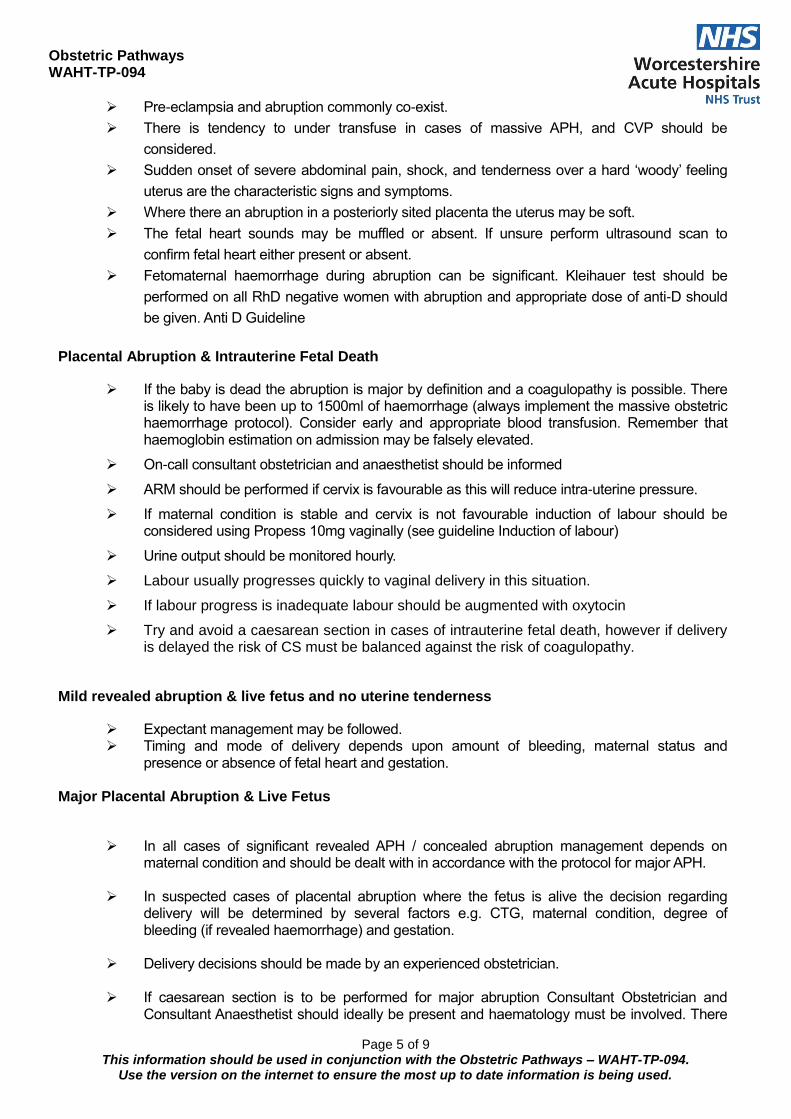
Pre-eclampsia and abruption commonly co-exist.
There is tendency to under transfuse in cases of massive APH, and CVP should be
considered.
Sudden onset of severe abdominal pain, shock, and tenderness over a hard ‘woody’ feeling
uterus are the characteristic signs and symptoms.
Where there an abruption in a posteriorly sited placenta the uterus may be soft.
The fetal heart sounds may be muffled or absent. If unsure perform ultrasound scan to
confirm fetal heart either present or absent.
Fetomaternal haemorrhage during abruption can be significant. Kleihauer test should be
performed on all RhD negative women with abruption and appropriate dose of anti-D should
be given. Anti D Guideline
Placental Abruption & Intrauterine Fetal Death
If the baby is dead the abruption is major by definition and a coagulopathy is possible. There is likely to have been up to 1500ml of haemorrhage (always implement the massive obstetric haemorrhage protocol). Consider early and appropriate blood transfusion. Remember that haemoglobin estimation on admission may be falsely elevated.
On-call consultant obstetrician and anaesthetist should be informed
ARM should be performed if cervix is favourable as this will reduce intra-uterine pressure.
If maternal condition is stable and cervix is not favourable induction of labour should be considered using Propess 10mg vaginally (see guideline Induction of labour)
Urine output should be monitored hourly.
Labour usually progresses quickly to vaginal delivery in this situation.
If labour progress is inadequate labour should be augmented with oxytocin
Try and avoid a caesarean section in cases of intrauterine fetal death, however if delivery is delayed the risk of CS must be balanced against the risk of coagulopathy.
Mild revealed abruption & live fetus and no uterine tenderness
Expectant management may be followed. Timing and mode of delivery depends upon amount of bleeding, maternal status and
presence or absence of fetal heart and gestation.
Major Placental Abruption & Live Fetus
In all cases of significant revealed APH / concealed abruption management depends on
maternal condition and should be dealt with in accordance with the protocol for major APH.
In suspected cases of placental abruption where the fetus is alive the decision regarding delivery will be determined by several factors e.g. CTG, maternal condition, degree of bleeding (if revealed haemorrhage) and gestation.
Delivery decisions should be made by an experienced obstetrician.
If caesarean section is to be performed for major abruption Consultant Obstetrician and
Consultant Anaesthetist should ideally be present and haematology must be involved. There

Obstetric Pathways WAHT-TP-094
Page 6 of 9 This information should be used in conjunction with the Obstetric Pathways – WAHT-TP-094.
Use the version on the internet to ensure the most up to date information is being used.
may be cases of major abruption where urgent caesarean section may be required and senior most obstetrician available should proceed with the delivery while awaiting arrival of on-call consultant.
Unclassified APH
In almost 50% of cases cause of APH cannot be established. Some of these are minor degrees of placenta praevia and placental abruption and others may be disruption of sinuses or small vessels (marginal bleed)
Unexplained APH is known to be associated with increased maternal and perinatal morbidity
and mortality, there is also an associated with DV.
Usually associated with mild APH and in some cases recurrent episodes of minimal bleeding PV.
These cases may be managed as an outpatient after initial assessment for 24 hours.
Fetal growth should be monitored by serial growth scans as there is a higher risk of IUGR and perinatal loss.
In cases of recurrent unclassified APH optimum timing of delivery is not clear, therefore each decision should be individualised and consultant led. Induction of labour should be considered at or near term even if fetal growth is satisfactory.
Kleihauer test should be performed on all RhD negative women with abruption and
appropriate dose of anti-D should be given. Anti D Guideline
Local Causes
Bleeding can occasionally occur from the cervix either due to cervical ectopy, polyps or cervical carcinoma (please check smear history).
The bleeding however is not usually massive, but may be present as multiple, small APH’s
Bleeding from the vagina is usually secondary to infection or trauma. Candidiasis is a common, treatable cause.
Vulval bleeding can result from the rupture of vulval varicosities. Immediate treatment is by applying pressure to the vulva and resuscitation. Surgery may well be necessary to stop the bleeding.
Bleeding may occur from perineal laceration caused by descending fetal parts during active second stage. If there is significant bleeding while awaiting delivery of the baby it should be expedited e.g. by giving episiotomy or instrumental delivery as maternal shock can occur from significant APH whatever the cause.
Management of Major APH
1. Prompt recognition
Consultant obstetrician / anaesthetist
Registrar obstetrician / anaesthetist
Midwives and delivery suite coordinator
Porters should be informed to be on standby for urgent blood collection from the lab
Haematologist and blood transfusion technicians
HCAs
Scribe
Obstetric Pathways WAHT-TP-094
Page 7 of 9 This information should be used in conjunction with the Obstetric Pathways – WAHT-TP-094.
Use the version on the internet to ensure the most up to date information is being used.
2. Activate 2222 call and say ‘Major Obstetric Haemorrhage’ and give location Switchboard will contact the lab, the anaesthetist on call, the porters and consultant haematologist. The major haemorrhage pack will be initiated (appendix 1).
Follow ABCD rule
A - Airway
Assess and maintain patency.
O2 via face mask (Hudson).
Attach pulse oximeter to patient.
B - Breathing
Assess
Protect airway
Monitor respiratory rate
C - Circulation
Restoration of circulating volume should be the first priority - insert 2 large bore IV cannulae.
Send bloods for FBC, clotting studies, PET bloods and X–match 4 units blood. If the baby is dead the abruption is major by definition and a coagulopathy is possible. There is likely to have been up to 1500ml of haemorrhage. It is important to give up to 2 units of blood as soon as possible. Do not be fooled by haemoglobin estimation on admission as it may well be falsely elevated.
Clinical vigilance & ongoing assessment of patient. Continuous pulse/BP/ECG/Oximeter monitoring
Regularly assess volume loss
Consider CVP/arterial line.
Catheterise and monitor urine output hourly.
Replace volume loss and urgent access to blood
Warm IV fluids and infuse with a pressure bag. Initially infuse up to 2 litres of Hartmann’s solution followed by colloid e.g. Volplex 500mls. Administer blood as soon as it is possible.
If blood loss appears life threatening consider giving O Rh negative red cells. Preferably however use group specific fully X matched blood.
When requesting blood be clear in your request to the haematologist and porters about the urgency and state what you need, when you need it and enquire when it will be ready for collection alert
D - Diagnose the cause of APH and manage accordingly as above with regards to fetal monitoring
and delivery.
Inform paediatricians and request attendance of experienced paediatrician for delivery.
NB: APH predisposes to PPH (see PPH guideline)
After Care
All women following major APH require intensive monitoring for at least first 24 hours.
In some cases transfer to high dependency unit or ITU may be required
Remember accurate documentation at all times.

Obstetric Pathways WAHT-TP-094
Page 8 of 9 This information should be used in conjunction with the Obstetric Pathways – WAHT-TP-094.
Use the version on the internet to ensure the most up to date information is being used.
Debrief/discuss with patients & colleagues following delivery.
Datix should be completed for all major APH cases.
It is important to remember that thrombo-embolic disease (TED) is still one of the commonest causes of maternal death. TED stockings should be the bare minimum in these cases. Consider pneumatic calf compression devices and continue them post-operatively until it is safe to give heparin (e.g. Enoxaparin
References:
Royal College of Obstetricians and Gynaecologists, Green-top Guideline no.63, Antepartum Haemorrhage

Obstetric Pathways WAHT-TP-094
Page 9 of 9 This information should be used in conjunction with the Obstetric Pathways – WAHT-TP-094.
Use the version on the internet to ensure the most up to date information is being used.