PATHOPHYSIOLOGY OF CONGENITAL AND VALVULAR HEART...
42
PATHOPHYSIOLOGY OF CONGENITAL AND VALVULAR HEART DISEASES
Transcript of PATHOPHYSIOLOGY OF CONGENITAL AND VALVULAR HEART...

PATHOPHYSIOLOGY OF CONGENITAL AND VALVULAR HEART DISEASES
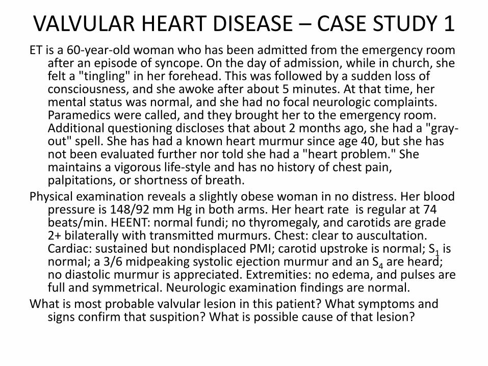
VALVULAR HEART DISEASE – CASE STUDY 1 ET is a 60-year-old woman who has been admitted from the emergency room
after an episode of syncope. On the day of admission, while in church, she felt a "tingling" in her forehead. This was followed by a sudden loss of consciousness, and she awoke after about 5 minutes. At that time, her mental status was normal, and she had no focal neurologic complaints. Paramedics were called, and they brought her to the emergency room. Additional questioning discloses that about 2 months ago, she had a "gray-out" spell. She has had a known heart murmur since age 40, but she has not been evaluated further nor told she had a "heart problem." She maintains a vigorous life-style and has no history of chest pain, palpitations, or shortness of breath.
Physical examination reveals a slightly obese woman in no distress. Her blood pressure is 148/92 mm Hg in both arms. Her heart rate is regular at 74 beats/min. HEENT: normal fundi; no thyromegaly, and carotids are grade 2+ bilaterally with transmitted murmurs. Chest: clear to auscultation. Cardiac: sustained but nondisplaced PMI; carotid upstroke is normal; S1 is normal; a 3/6 midpeaking systolic ejection murmur and an S4 are heard; no diastolic murmur is appreciated. Extremities: no edema, and pulses are full and symmetrical. Neurologic examination findings are normal.
What is most probable valvular lesion in this patient? What symptoms and signs confirm that suspition? What is possible cause of that lesion?

A previously healthy but inactive 42-year-old man is seen in the ER after his first episode of syncope, which occurred while he was playing full-court basketball for the first time in 10 years. On questioning, he describes a 2-month history of exertional chest pain. He has not seen a physician during his adult life. Physical examination reveals the following findings. His supine blood pressure is 100/80 mm Hg without any significant orthostatic change. There is no jugular venous distention, but there are slowly rising, small-amplitude, and somewhat sustained arterial pulses. His lungs are clear. A sustained and slightly laterally displaced apex beat is noted, as well as a soft S1, and single S2, prominent S4, and a grade 3/6 harsh, late-peaking, crescendo-decrescendo systolic murmur heard best at the cardiac base and radiating to the carotids with a high-frequency component at the cardiac apex. No clubbing, cyanosis, or edema is noted.
1. What is the most likely valvular lesion in this patient? 2. What is the most likely underlying cause of that lesion in this age group? 3. What causes left ventricular concentric hypertrophy in the course of that
lesion?
VALVULAR HEART DISEASE – CASE STUDY 2

A 50-year-old woman who had an "innocent" murmur diagnosed in childhood presents with dyspnea on exertion, orthopnea, and paroxysmal nocturnal dyspnea of several months' duration. On questioning she describes a 1-year history of fatigue and exhaustion that has limited her daily activities. She has not seen a physician in years.
On physical examination, her blood pressure is found to be 110/70 mm Hg. Her jugular venous pressure is 8 cm H2O and she exhibits 1+ pulses with normal arterial upstrokes and bibasilar rales. There is a laterally displaced apex but with a palpable S3, a soft S1 a widely split S2, a loud S3, and a grade 3/6 blowing, high-pitched, systolic murmur heard best at the apex and radiating to the axilla and left infrascapular area. There is trace edema but no clubbing or cyanosis.
1. What is the valvular lesion in this patient? 2. What are possible underlying causes of that lesion? 3. What are possible complications of that lesion?
VALVULAR HEART DISEASE – CASE STUDY 3

Heart murmurs
• result of turbulent blood flow
• functional murmur (physiologic/innocent/benign) primarily due to physiologic conditions outside the heart
• structural murmurs (pathologic murmurs)
– narrowing or leaking of valves
– presence of abnormal passages

Mechanisms of murmur formation
MECHANISM EXAMPLE
increased flow through a normal structure
aortic systolic murmur in anemia
stenosis obstruction to flow turbulence murmur
mitral stenosis and aortic stenosis
flow into a dilated chamber vortex formation when the blood flows from a narrower to a larger chamber
aortic valve is of normal size and the aorta is dilated relative constriction
membrane, which vibrates as the fluid flows by
papillary muscle rupture
flow of blood from a high pressure chamber to a lower pressure chamber
ventricular septal defects

Structural heart murmurs
• categorization by timing
– systolic
– diastolic
– continuous

MITRAL STENOSIS (MS)

Causes of mitral stenosis (MS)
• rheumatic endocarditis (most common)
• tumors
• bacterial growth
• calcification
• thrombi
• combination of MS with a congenital atrial septal defect (Lutembacher’s syndrome)

Rheumatic fever
• systemic disease affecting the peri-arteriolar connective tissue
• can occur after an untreated Group A Beta hemolytic streptococcal pharyngeal infection
• antibody cross-reactivity - Type II hypersensitivity reaction - molecular mimicry
• B cells-derived plasma cells produce antibodies against the cell wall of Streptococcus (M protein)
• The antibodies may also react against the perivascular connective tissue rheumatic fever
Valvular changes: • leaflet thickening • commissural fusion • shortening and thickening of the
tendineous cords the main opening shrinks

Pathomechanism of MS
stenosis
increased flow resistance
diminished blood flow across the valve from left atrium to left ventricle during diastole
reduced cardiac output

Compensatory mechanisms in MS
• Peripheral oxygen extraction: arteriovenous oxygen difference (AVDO2) can increase
• Diastolic filling time per unit of time can be increased by reducing the heart rate stroke volume is raised more than proportionately increased cardiac output
• increase in left atrial pressure (PLA) increase of the pressure gradient between atrium and ventricle (PLA – PLV) diastolic flow rate (Qd) is raised despite the stenosis

Negative effects of compensation
high pressure in LA
the left atrium hypertropies and
dilates (P mitrale in the ECG)
damage of the LA atrial fibrillation
lack of proper contraction of the
fibrillating atria formation of thrombi
increased risk of arterial emboli with infarction (especially
of the brain)
ventricular rate is also increased
(tachyarrhythmia)
the diastolic duration is
reduced
shortened diastolic filling
time
pressure in LA rises
pressure increase in the pulmonary
veins dyspnea
varicosis of bronchial veins
hemoptysis from ruptured
veins
pulmonary edema
pulmonary hypertension
increased stress on the right
heart
right heart failure

MS symptoms
The total opening area (OA) at the mitral valve ring is normally 4–6 cm2.
Main symptoms of MS • Dyspnea • Fatigue • Hemoptysis Severity of symptoms depend on OA • OA <2.5 cm2 symptoms develop
on strenuous physical activity • OA < 1.5 cm2 during ordinary
daily activities • OA < 1 cm2 at rest • OA < 0.3 cm2 incompatible with
life

Cardiac auscultation in MS
• The first heart sound (I or S1) is loud and delayed (up to 90 ms, normally 60ms)
• The second heart sound (II or S2) is followed by the so-called mitral opening snap (MOS), which can best be heard over the cardiac apex.
Murmurs: • mid-diastolic murmur (MDM) • presystolic crescendo murmur
(PSM) caused by the rapid inflow (poststenotic turbulence) during systole of atria

MS pathophysiology - summary

MITRAL REGURGITATION

Mitral regurgitation – MR (mitral insufficiency)
• the mitral valve has lost its function as a valve • during systole some of the blood in the left ventricle flows back
(“regurgitates”) into the left atrium Causes • mitral valve prolapse (Barlow’s syndrome) the chordae are too
long the leaflets bulge into the left atrium, where they open • rheumatic or bacterial endocarditis the leaflets and chordae
shrink, thicken, and become more rigid impaired valve closure • coronary heart disease rupture of a papillary muscle / poor
contraction • Marfan’s syndrome (genetic, generalized disease of the connective
tissue) lengthened chordae / dilated annulus

MR pathophysiology
• Mitral opening area in systole • Systolic pressure in the LV
• Compliance (distensibility ) of the LA
• Duration of systole
part of the stroke volume is pumped
back into the LA
regurgitant volume may amount to as
much as 80% of the SV
the regurgitant volume/ time is dependent on
Effect increased volume load on the left heart

Two versions of MR
Acute MR
• Low atrial compliance
• High pressure in the LA
• Increased pressure in pulmonary veins and capillaries
• Pulmonary oedema
Chronic MR
• High atrial compliance
• Pressure in the LA and in pulmonary veins is only moderately raised
• High amount of blood is pumped back to LA during LV contration
• Stroke volume in decreased fatigue
• LV dilation and LV failure

Auscultation of the heart
• Systolic murmur
• Rapid filling wave third heart sound (III or S3)
• Contraction of dilated atrium high amount of blood pumped into LV fourth heart sound (IV or S4)

AORTIC STENOSIS

Causes of aortic stenosis (AS)
• subvalvar and supravalvar stenosis
• congenital stenosis malformations of the valve (age at manifestation < 15 years)
• congenital bicuspid malformation of the valve (manifestation up to 65 years of age)
• rheumatic–inflammatory stenosis of an originally normal tricuspid valve
• degenerative changes along with calcification (manifestation in elderly

Pathophysiology of AS
Aortic stenosis Increased systolic pressure in LV (up
to 300 mmHg) LV hypertrophy
Stiffening of LV walls (decreased
compliance)
Increased diastolic pressure in LV
LA hypertrophy
LA pumps 30-40% of LV stroke volume
(normal: 20%)
Dramatic decrease of cardiac output in
case of atrial fibrillation

Symptoms of AS
• Congestive heart failure very high afterload in LV increased pressure in LA
increased pressure in pulmonary veins and capillaries pulmonary congestion
• Angina pectoris – insufficient oxygen supply of myocardium – Increased LV muscle mass (hypertrophy) – Increased systolic pressure in LV increased tension of LV walls – Increased diastolic pressure in LV coronary flow decreased
• Syncope – LV cannot increase cardiac output on exertion – Effort leads to widening of arteries in skeletal muscles – Cerebral blood flow decreses

Auscultation of heart - AS
• Systolic murmur (SM) over the aortic valve
• Fourth heart sound (IV or S4)– LV filling gallop
• Click of aortic valve opening

AS - summary

AORTIC REGURGITATION
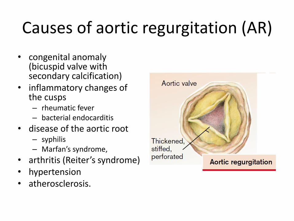
Causes of aortic regurgitation (AR)
• congenital anomaly (bicuspid valve with secondary calcification)
• inflammatory changes of the cusps – rheumatic fever – bacterial endocarditis
• disease of the aortic root – syphilis – Marfan’s syndrome,
• arthritis (Reiter’s syndrome) • hypertension • atherosclerosis.

Pathomechanism of AR
• Reverse flow of blood from aorta to LV during diastole
• To achieve an adequate effective stroke volume the total stroke volume must be increased by the amount of the regurgitant volume, which is possible only by raising the enddiastolic volume
• Regurgitation volume depends on – opening area in the aortic valve
during diastole – pressure difference between
aorta and LV during diastole – duration of diastole.
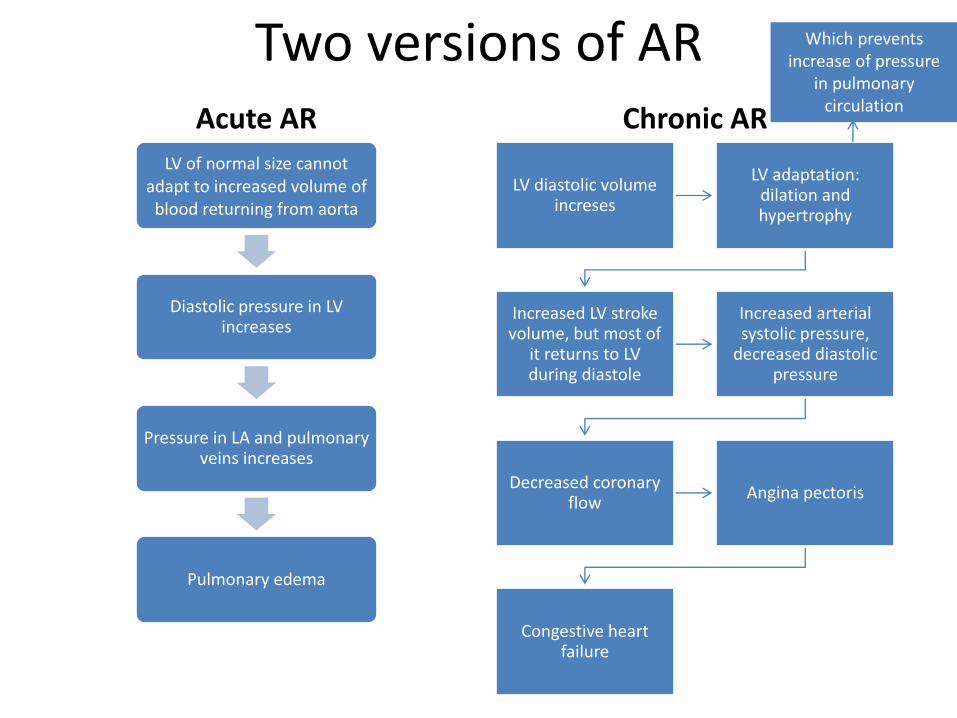
Two versions of AR Acute AR
LV of normal size cannot adapt to increased volume of
blood returning from aorta
Diastolic pressure in LV increases
Pressure in LA and pulmonary veins increases
Pulmonary edema
Chronic AR
LV diastolic volume increses
LV adaptation: dilation and hypertrophy
Increased LV stroke volume, but most of
it returns to LV during diastole
Increased arterial systolic pressure,
decreased diastolic pressure
Decreased coronary flow
Angina pectoris
Congestive heart failure
Which prevents increase of pressure
in pulmonary circulation

Symptoms and signs of AR
• Dyspnea on exertion • Symptoms of hyperdynamic
circulation: – Arterial hypertension with high
arterial pressure amplitude – Capillary pulsation under the finger
nails (Quincke’s sign) – pulse-synchronous head nodding
(Musset’s sign)
• At auscultation – an early diastolic decrescendo
murmur (EDM) can be heard over the base of the heart and mid-diastolic murmur over apex, produced by the regurgitation
– click and a systolic murmur due to the forced large-volume ejection

AR – from compansation to decompensation
Complications of AR:
• Myocardial ischemia
• Congestive heart failure
• Complications of arterial hypertension

CONGENITAL HEART DISEASES

Fetal circulation
Features of fetal circulation • low resistance in the
systemic circulation (placenta)
• high pressure in the pulmonary circulation
• high resistance in the pulmonary circulation (lungs unexpanded and hypoxic vasoconstriction)
• right-to-left shunt through the foramen ovale (FO) and ductus arteriosus Botalli (DA)
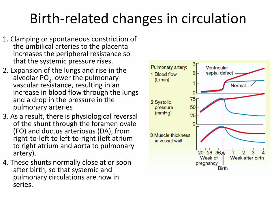
Birth-related changes in circulation 1. Clamping or spontaneous constriction of
the umbilical arteries to the placenta increases the peripheral resistance so that the systemic pressure rises.
2. Expansion of the lungs and rise in the alveolar PO2 lower the pulmonary vascular resistance, resulting in an increase in blood flow through the lungs and a drop in the pressure in the pulmonary arteries
3. As a result, there is physiological reversal of the shunt through the foramen ovale (FO) and ductus arteriosus (DA), from right-to-left to left-to-right (left atrium to right atrium and aorta to pulmonary artery).
4. These shunts normally close at or soon after birth, so that systemic and pulmonary circulations are now in series.

Circulatory shunts Causes: • patency of the duct (patent or persisting
DA [PDA]) • Patency of the FO (PFO) • defects in the atrial or ventricular
septum (ASD or VSD) • arteriovenous fistulae Size and direction of the shunt depend on: • the cross-sectional area of the shunt
opening • the pressure difference between the
connected vessels or chambers If the shunt between functionally similar
vascular spaces (e.g., aorta and pulmonary artery; atrium and atrium, ventricle and ventricle) is across a large cross-sectional area, pressures in the two vessels or chambers become (nearly) equalized. In this case the direction and volume of the shunt is determined by:
• Outflow resistance from the shunt-connected vessels or chambers
• their compliance

Causes of congenital heart diseases
• Genetic factors – Down’s syndrome – Turner’s syndrome – 18th pair trisomia
• Teratogenic drugs – Talidomid – Warfarin – Phenytoin
• Viral infections – rubeola • Maternal diseases
– Diabetes – Alcoholism – Systemic lupus erythematosus

Congenital heart diseases - categorization
With respect to presence of the shunt
• With the shunt
• Without the shunt
With respect to presence of cyanosis
• Cyanotic
• With temporary cyanosis
• Non-cyanotic

CYANOSIS
Cyanosis as an effect of the shunt between pulmonary and systemic circulation appears in case of:
• Increased pulmonary vessels resistance
• Increased of systemic venous return
• Eisenmenger’s syndrome (appearence of cyanosis) confirms the swich of the shunt from left-right to right-left
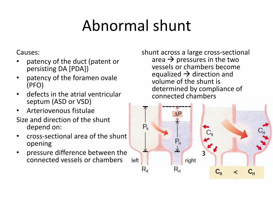
Abnormal shunt
Causes: • patency of the duct (patent or
persisting DA [PDA]) • patency of the foramen ovale
(PFO) • defects in the atrial ventricular
septum (ASD or VSD) • Arteriovenous fistulae Size and direction of the shunt
depend on: • cross-sectional area of the shunt
opening • pressure difference between the
connected vessels or chambers
shunt across a large cross-sectional area pressures in the two vessels or chambers become equalized direction and volume of the shunt is determined by compliance of connected chambers

Atrial septal defect (ASD)
Initially right ventricle (RV) is
more distensible than the left ventricle (LV)
less RV resistance to filling during
diastole
left-to-right shunt
volume load causes RV
hypertrophy
decreased RV compliance
Increased right atrium pressure
Shunt reversal