PATHOLOGY OF BRAIN TUMORS - Neurosurgery...
154
PATHOLOGY OF BRAIN TUMORS Presented By : Dr Amit Thapa
Transcript of PATHOLOGY OF BRAIN TUMORS - Neurosurgery...
PATHOLOGY OF BRAIN TUMORS
Presented By : Dr Amit Thapa

CONTENTSEPIDEMIOLOGYCLASSIFICATIONPATHOPHYSIOLOGY IN BRIEFPATHOLOGY OF INDIVIDUAL TUMOR GROUPSDIAGNOSTIC APPROACH
RADIOLOGICALTUMOR MARKERSCYTOLOGY INTRAOPERATIVE
FROZEN SECTIONFluorescent imaging (Chemical probe)
HISTOLOGYMODERN METHODS
IMMUNOHISTOCHEMICALMOLECULAR
PROGNOSTIC MARKERSPathology of brain tumors
- Dr Amit Thapa

Epidemiologyincidence
Primary cerebral malignancy-4 to 10/Lac general population
1.6% of all primary tumors2.3% of all cancer related deaths
Francis Ali-Osman, 2005
2nd most common cancer in children20% of all cancers in children <15 yrs
Pathology of brain tumors- Dr Amit Thapa

Epidemiological incidence of individual tumor
Classification Incidence / 100,000 population/yr
Metastatic 6
Astrocytoma 1.5
Glioblastoma 3
Meningioma 3
Primary CNS lymphoma
Immunocompetent 0.3
Overall 0.8-6.8
Medulloblastoma 0.5
Germ cell tumor 0.2
Pinealoma/ pineoblastoma 0.1Parkin, 1997

Epidemiologycomparative incidence
ALL INTRACRANIAL TUMORS
NEUROEPITHELIAL TUMORS
34%
METASTASIS21%
OTHERS16%
VASCULAR MALFORMATIONS
3%
PITUITARY TUMORS
8%
SCHWANNOMA6%
MENINGIOMA12%
Pathology of brain tumors- Dr Amit Thapa

EpidemiologyRelative incidence at AIIMS (2002-2007)
Pathology of brain tumors- Dr Amit Thapa
Astrocytoma98521%
Oligodendroglioma2385%
Ependymoma1704%
Mixed glioneuronal tumor721%
Embryonal type1643%
Pineal tumors190%
Others241851%
Meningioma57712%
Lymphoma581%
Germ cell Tumor230%
Hemangiopericytoma461%
Hemangioblastoma611%
Melanoma5
0%
n= 5076 patients
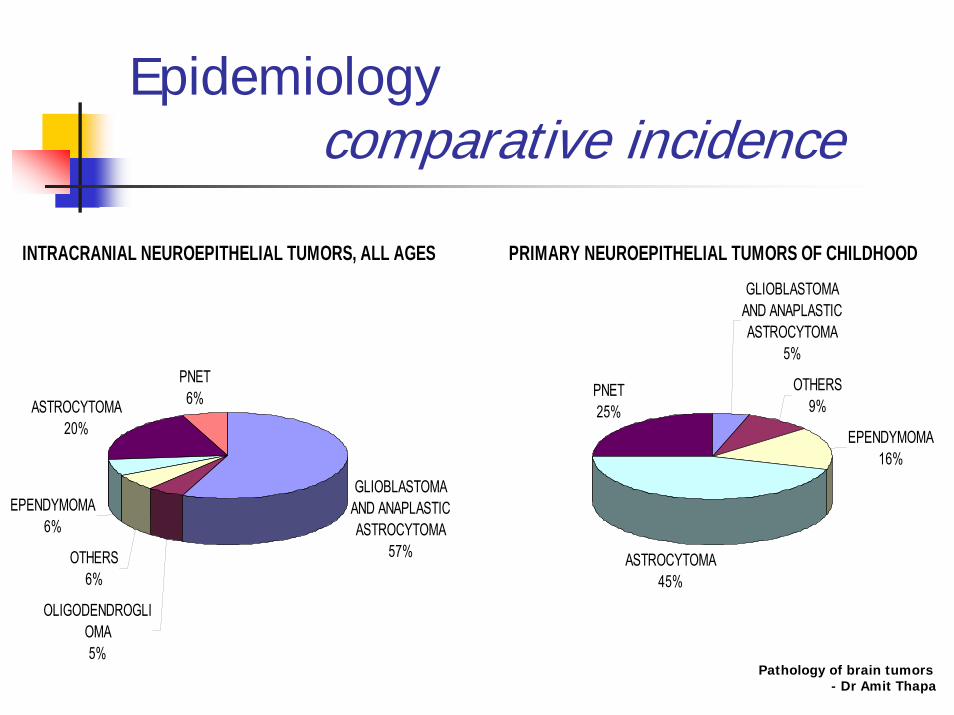
Epidemiologycomparative incidence
INTRACRANIAL NEUROEPITHELIAL TUMORS, ALL AGES
GLIOBLASTOMA AND ANAPLASTIC ASTROCYTOMA
57%
OLIGODENDROGLIOMA5%
OTHERS6%
EPENDYMOMA6%
ASTROCYTOMA20%
PNET6%
Pathology of brain tumors- Dr Amit Thapa
PRIMARY NEUROEPITHELIAL TUMORS OF CHILDHOODGLIOBLASTOMA
AND ANAPLASTIC ASTROCYTOMA
5%
OTHERS9%
EPENDYMOMA16%
ASTROCYTOMA45%
PNET25%

Epidemiological profile…age wise distribution
Pathology of brain tumors- Dr Amit Thapa
0
10
20
30
40
50
60
70
80
90
100
Relative incidence
0 10 20 30 40 50 60 70Age (years)
Posterior fossaMedulloblastomaEpendymomaPilocytic astrocytomaOther sitesCranopharyngiomaChorioid plexus tumorus
Cerebellar hemisphereDiffuse astrocytomaAnaplastic astroyctomaOligodendrogliomaEpendymomaOther sitesMeningioma
Cerebellar hemisphereGlioblastomaAnaplastic astroyctomaAnaplasticOligodendrogliomaMetastatic carcinomaLymphomaOther sitesMeningiomaSchwannomaPituitary adenoma
Pathology of brain tumors- Dr Amit Thapa
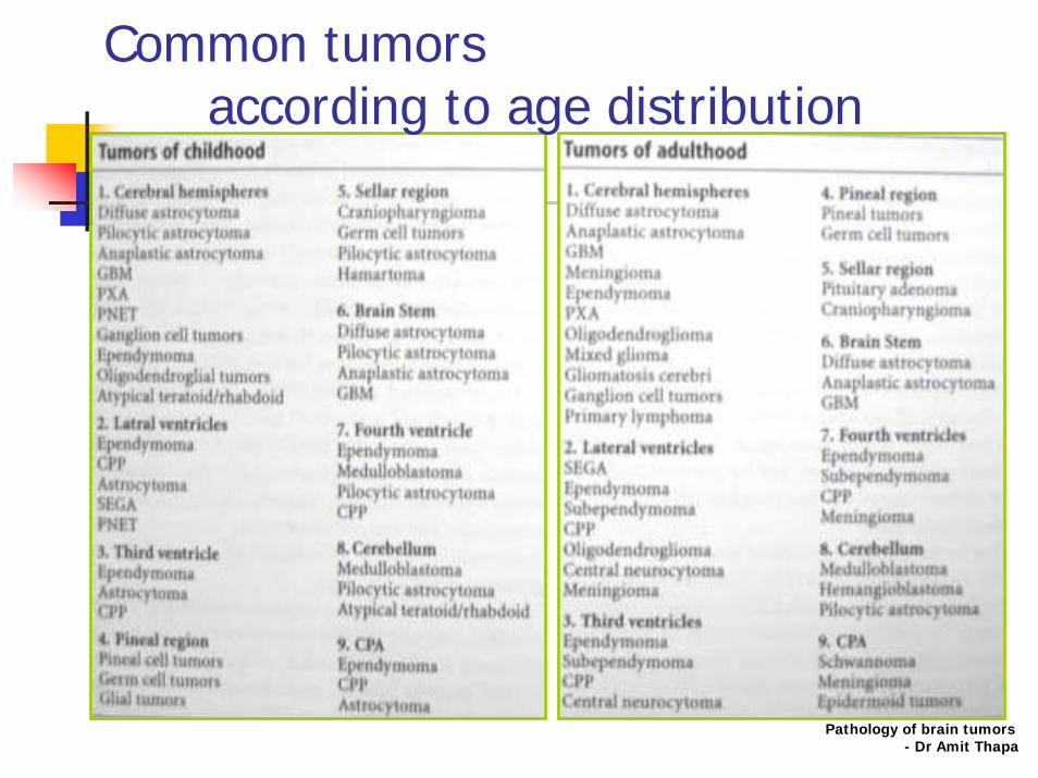
Common tumors according to age distribution

EpidemiologyGender
•Males are more likely to be diagnosed with brain tumors than females-( 1.5:1 )
•Meningiomas and pituitary adenomas are slightly more common in women than in men.
Pathology of brain tumors- Dr Amit Thapa
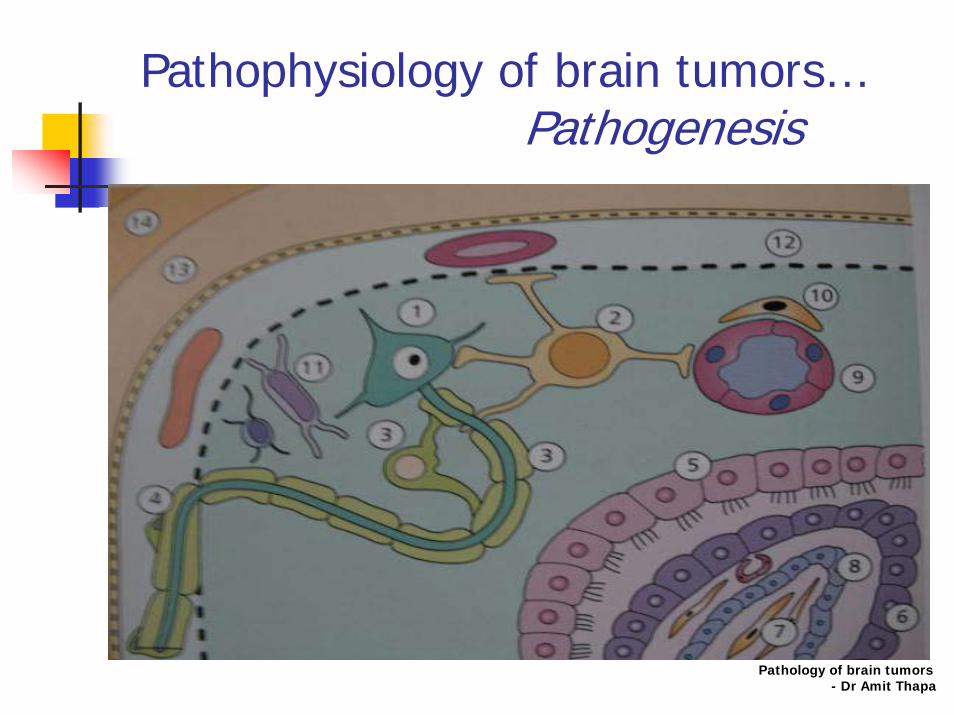
Pathophysiology of brain tumors…Pathogenesis
Pathology of brain tumors- Dr Amit Thapa

Pathophysiology of brain tumors…Pathogenesis
Cells of origin for most brain tumors – debatable
Molecular enquiries-
most likely cells of origin are multipotential stem cells
reside in both the developing and adult brain.Am J Pathol 2001; 159: 779-86Genes Dev 2001; 15: 1311-33
Pathology of brain tumors- Dr Amit Thapa

Pathophysiology of brain tumors…ONCOGENES AND CNS
ONCOGENES
TUMOR SUPRESSOR GENES
Pathology of brain tumors- Dr Amit Thapa
GROWTH FACTORSsis, FGFs, CSFs, EGF, TGFα
RECEPTORSTYROSINE KINASE- erbB, fms, kit, ros, met, trk, neuGROWTH HORMONE- mpl, epoANGIOTENSIN- masSTEROID HORMONE- erbA
Second messenger signals
Transcription factorsfos, erb A, jun, myc, rel, myb, ets
Active transcription complex
TRANSDUCERS
ras, src, raf, mos, abl Genes
CYTOPLASM
NUCLEUS

Pathophysiology of brain tumors…Classification- anatomical
Pathology of brain tumors- Dr Amit Thapa
MULTISTEP
CARCINOGENESIS
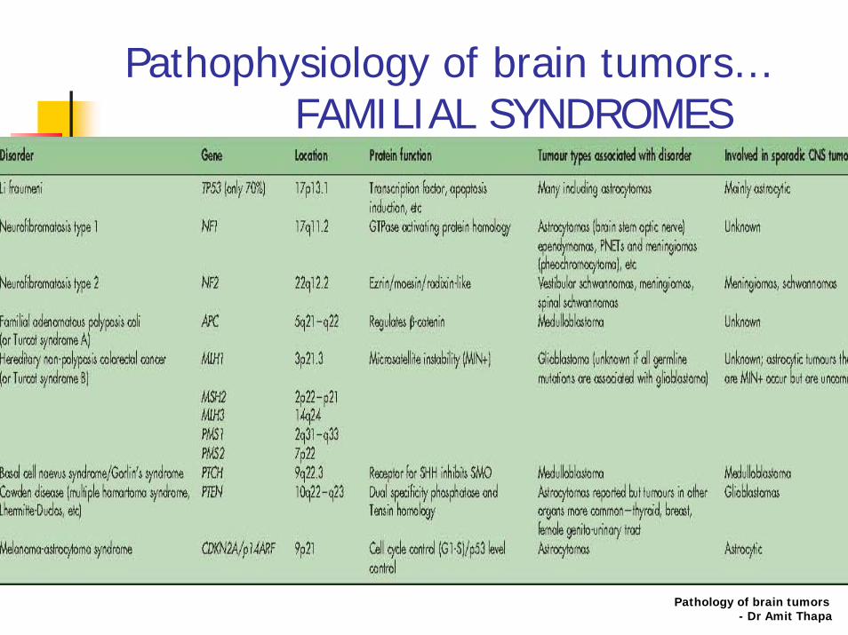
Pathophysiology of brain tumors…FAMILIAL SYNDROMES
Pathology of brain tumors- Dr Amit Thapa
Pathophysiology of brain tumors…ETIOGENESIS
VIRUSES
• RNA virus- oncorna family
Rous sarcoma virus, ASV, MSV, SSV
• DNA virus- Papovaviruses, Adenoviruses
(Bovine papilloma virus, Human JC virus, SV40)
NO CONCLUSIVE PROOF OF VIRAL INDUCTION OF HUMAN BRAIN TUMORS
Pathology of brain tumors- Dr Amit Thapa
Pathophysiology of brain tumors…ETIOGENESIS
Pathology of brain tumors- Dr Amit Thapa
RADIATION- Fibrosarcoma, meningiomas, GBM (?)
• True incidence unknown
• Criteria
1. Tumor must occur within ports of radiation therapy
2. Adequate latent period must have elapsed
3. No other predisposing factors- NF, MEN
4. Definitive tumor diagnosis
5. Rarely occur spontaneously in control
Pathophysiology of brain tumors…ETIOGENESIS
CHEMICAL AGENTS
• Methylcholanthrene pellets- 1939
• Polycyclic hydrocarbons (PCHs)-
gliomas (7-14 months),
depending upon location
• Alkylating agents- most commonly used agent
gliomas (oligodendrogliomas)
Pathology of brain tumors- Dr Amit Thapa

Pathophysiology of brain tumors…IMMUNOLOGY OF BRAIN TUMORS
Pathology of brain tumors- Dr Amit Thapa
• Tumor associated-• transplantation antigen, tumor specific antigen, viral antigen, fetal antigen
• Recognition Proliferation Effector
• Cellular immunity-
relative suppressor dominance,
balance between helper & suppressor
• Humoral immunity
• Is brain an immunologically privileged site ?
• Immunologic response in brain tumor• Host suppression
• Cytokines, MHC antigen
• Organ and organ related antigens
• Cellular infiltration
• Mechanism of suppression and blocking

Classification-cell origin and derivative
Pathology of brain tumors- Dr Amit Thapa
Classificationof brain tumors
•Bailey and Cushing- 1926, first attempt to classifyBailey P, Cushing HA. A classification of the tumors of the glioma group on a histogenetic basis with a correlated study of prognosis. Philadelphia: JP Lippincott, 1926.
•Zulch and an international team (1979)
1st WHO classification of tumors of the CNS
Pathology of brain tumors- Dr Amit Thapa

Classification
Primary tumors of the brain Tumors of Neuroepithelial tissueTumors of MeningesTumors of the sellar regionGerm cell tumorChoroid plexus tumors Tumors of nerves and/or nerve sheathCysts and tumor like lesionsOther primary tumors, including skull base Hematopoietic neoplasms
Metastatic brain tumors and carcinomatous meningitis
Pathology of brain tumors- Dr Amit Thapa
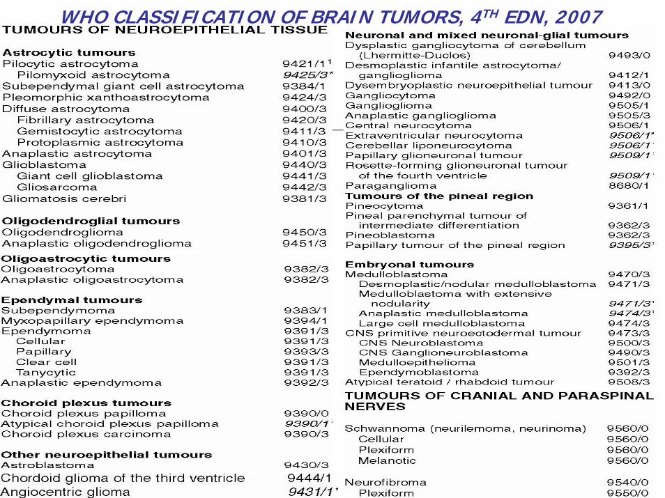
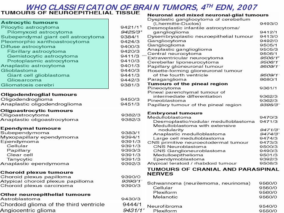
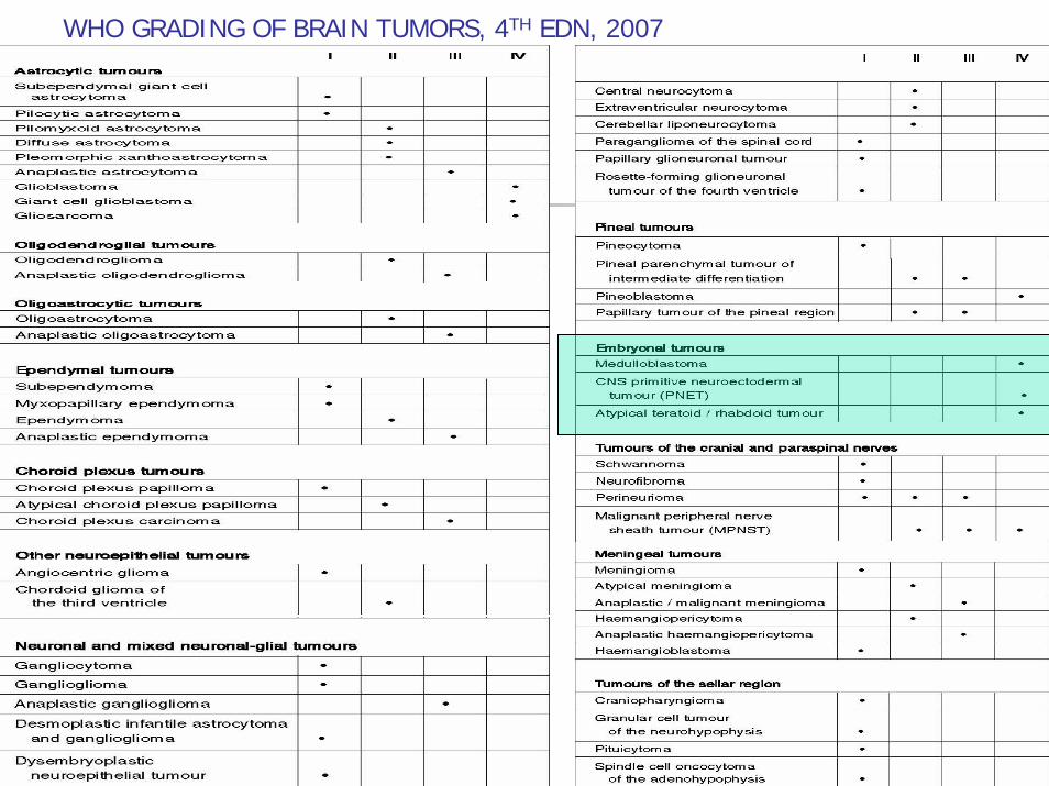
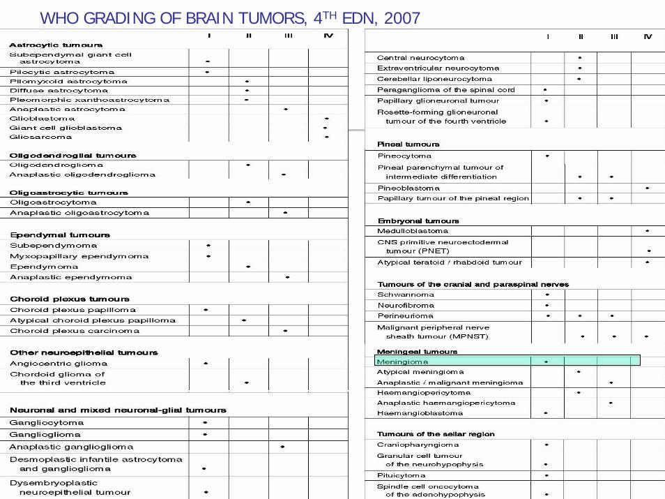
WHO CLASSIFICATION OF BRAIN TUMORS, 4TH EDN, 2007
Pathology of brain tumors- Dr Amit Thapa

WHO CLASSIFICATION OF BRAIN TUMORS, 4TH EDN, 2007 CONTD…

GRADING
Pathology of brain tumors- Dr Amit Thapa
Histopathological grading•Predict biological behavior of a neoplasm
•Clinical setting- influence choice of therapy
•Broder’s four tiered grading- general pathology
•Kernohan and Sayre- 1952,
graded gliomas into 1 to 4
degree of their dedifferentiation
•St Anne/Mayo or Daumass- Duport system- 4 grades
nuclear atypia, mitoses, endothelial proliferation, necrosis

GRADING
WHO classification of tumors of the nervous system
• includes a grading scheme - ‘malignancy scale’across a wide variety of neoplasms
• rather than a strict histological grading system
• widely used, but not a requirement for the application of the WHO classification
Pathology of brain tumors- Dr Amit Thapa

WHO GradingGrade I • low proliferative potential possibility of cure
(surgical resection alone)
Grade II + cytological atypia • low-level proliferative activity• generally infiltrative in nature• often recur• tend to progress to higher grades of
malignancy
Survive >5 yrs
Grade III • + nuclear atypia/ anaplasia
• + brisk mitotic activity
adjuvant radiation +/- chemotherapy
Survive 2-3 yrs
Grade IV • + microvascular proliferation
• + /- necrosis• cytologically
malignant,• mitotically active,• necrosis-prone
neoplasms
• rapid pre- and postoperative disease evolution
• fatal outcome. • In some-• Widespread infiltration of surrounding
tissue • craniospinal dissemination
adjuvant radiation +/- chemotherapy
Depends upon therapy,Survive <1 yr
Pathology of brain tumors- Dr Amit Thapa

WHO Grading
Pathology of brain tumors- Dr Amit Thapa
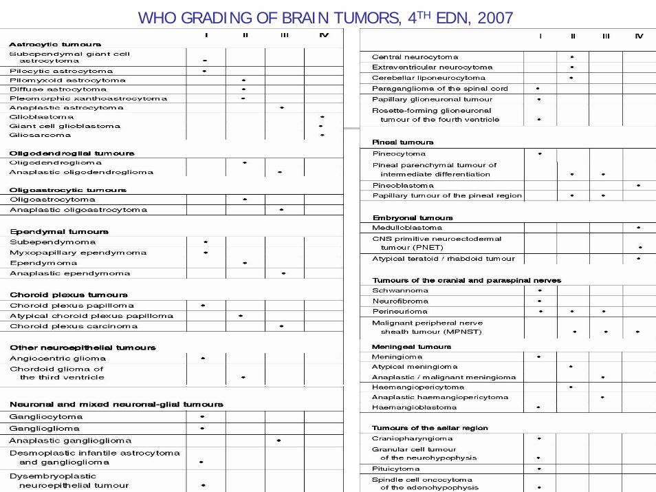
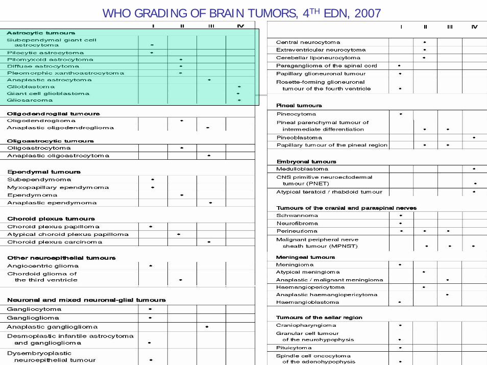
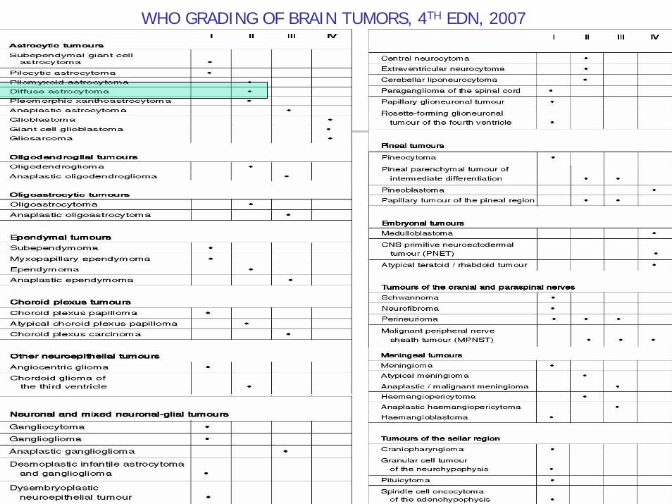
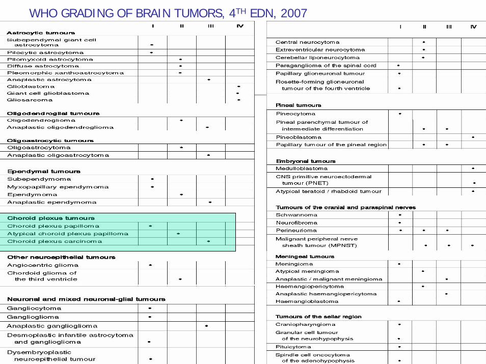
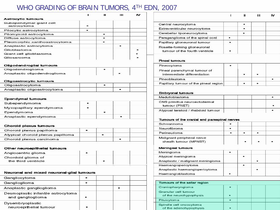
WHO GRADING OF BRAIN TUMORS, 4TH EDN, 2007
Pathology of brain tumors- Dr Amit Thapa
Pathophysiology of brain tumors…Classification- anatomical
Pathology of brain tumors- Dr Amit Thapa
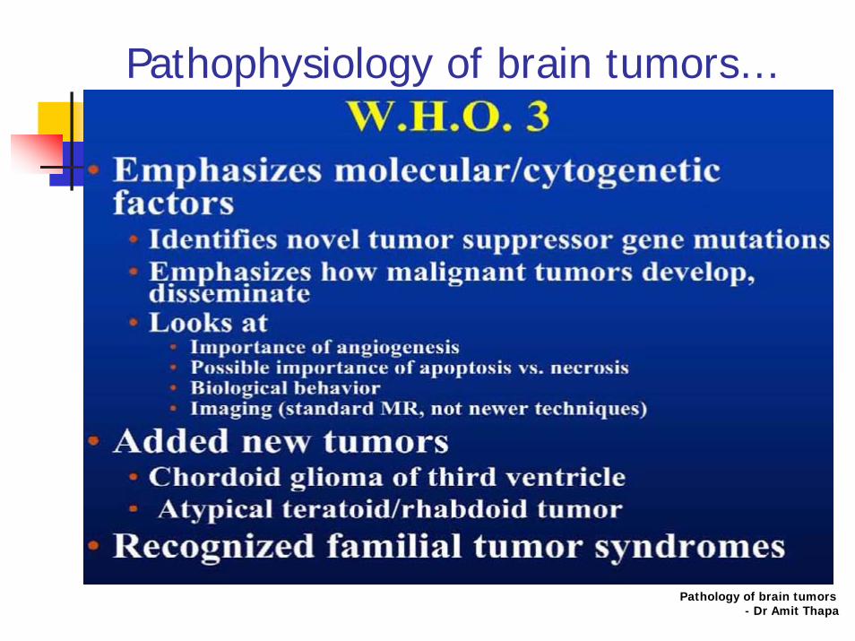
Pathophysiology of brain tumors…What’s new in WHO , 4th edition, 2007
Pathology of brain tumors- Dr Amit Thapa
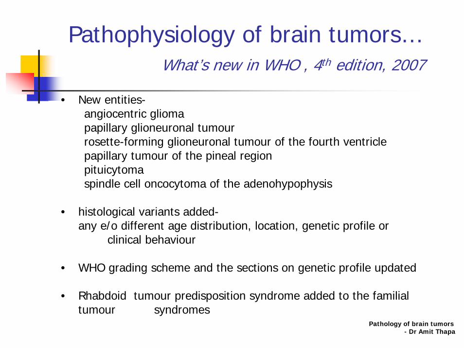
• New entities-angiocentric gliomapapillary glioneuronal tumourrosette-forming glioneuronal tumour of the fourth ventriclepapillary tumour of the pineal region pituicytoma spindle cell oncocytoma of the adenohypophysis
• histological variants added-any e/o different age distribution, location, genetic profile or
clinical behaviour
• WHO grading scheme and the sections on genetic profile updated
• Rhabdoid tumour predisposition syndrome added to the familial tumour syndromes
Pathophysiology of brain tumors…Classification-
The international classification of diseases for oncology (ICD-O)
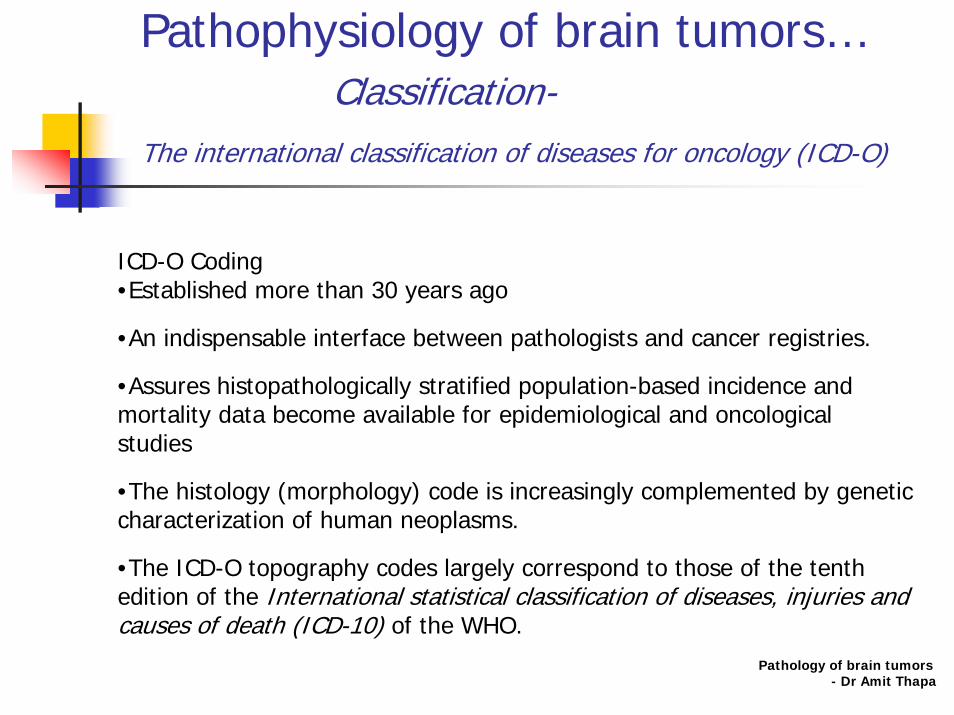
ICD-O Coding•Established more than 30 years ago
•An indispensable interface between pathologists and cancer registries.
•Assures histopathologically stratified population-based incidence and mortality data become available for epidemiological and oncological studies
•The histology (morphology) code is increasingly complemented by genetic characterization of human neoplasms.
•The ICD-O topography codes largely correspond to those of the tenth edition of the International statistical classification of diseases, injuries and causes of death (ICD-10) of the WHO.
Pathology of brain tumors- Dr Amit Thapa

Clinical presentations
Pathology of brain tumors- Dr Amit Thapa
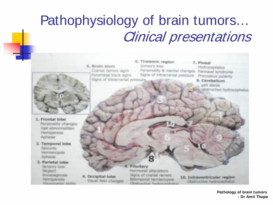
1. Due to direct tissue destruction, 2. local brain infiltration or 3. secondary effect of increased ICP (Cushing’s triad)
Depends upon location-positive ( headache/ seizure), negative symptoms (loss of function)
Headache-35% as first symptoms. 70% in growing tumor. Associated with vomiting/ nausea, papilledema, focal cerebral signs
Facial pain- tumors at base of skull or nasopharynx
Seizure-30% as first symptom. 98% in oligodendroglioma and 18% in mets
Pathophysiology of brain tumors…Clinical presentations
Pathology of brain tumors- Dr Amit Thapa

Pathology of specific tumors
Pathology of brain tumors- Dr Amit Thapa

Metastatic (20 malignant) tumor
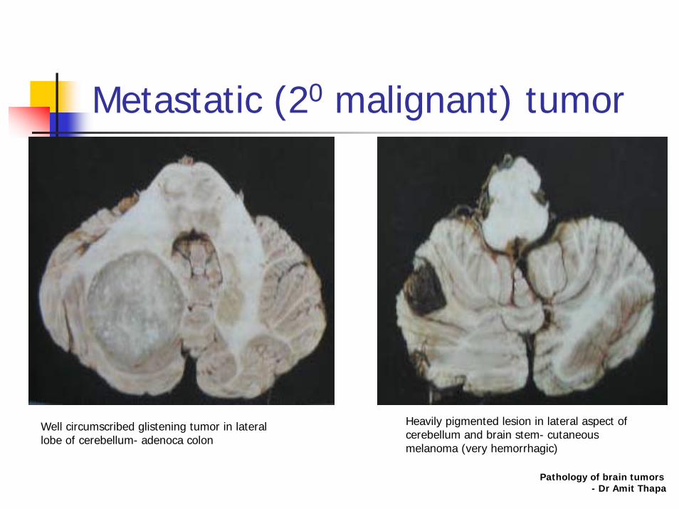
• 3 times more common than primary brain tumor• Often lodge- gray- white junction of cerebral, cerebellar hemisphere• Commonly from lung, breast, kidney• 2 major forms:
1. Single/ multiple well circumscribed deposits (commonest)
2. Carcinomatous meningitis
Leptomeningeal (breast, lung)dural metastasis (non CNS lymphoma)
• Route- hematogenous/ direct/ CSF • Abundant hemorrhage- melanoma, RCC, Chorioca• Multiplicity common• Retain primary characteristics
Pathology of brain tumors- Dr Amit Thapa
Metastatic (20 malignant) tumor
Heavily pigmented lesion in lateral aspect of cerebellum and brain stem- cutaneous melanoma (very hemorrhagic)
Well circumscribed glistening tumor in lateral lobe of cerebellum- adenoca colon
Pathology of brain tumors- Dr Amit Thapa

Metastatic (20 malignant) tumor
Adenocarcinomatous mets-Retain nuclei with prominent nucleoli, vacuolated cytoplasm, glands with mucinous secretionLung/ ovary/ breast/ colon
Papillary ca kidney/ colon/ thyroid-exhibit papillary structure. Prominent nucleoli and irregular crowding of nuclei
Pathology of brain tumors- Dr Amit Thapa
WHO CLASSIFICATION OF BRAIN TUMORS, 4TH EDN, 2007
Pathology of brain tumors- Dr Amit Thapa

Astrocytoma
Classification by cell typeOrdinary-
FibrillaryGemistocyticprotoplasmic
Special- favorable prognosisPilocyticMicrocystic cerebellarSubependymal giant cell
Pathology of brain tumors- Dr Amit Thapa
WHO GRADING OF BRAIN TUMORS, 4TH EDN, 2007
Pathology of brain tumors- Dr Amit Thapa

Pilocytic Astrocytoma
Most common brain tumor in childrenCerebellum> adj 3rd ventricle> brainstemCircumscribed cystic mass with mural noduleGenetics: sporadic/ syndromicSlow growingHistology:
Classic biphasic patternCompacted bipolar cells with rosenthal fibresLoose textured mulitpolar cells
Leptomeningeal seeding Pathology of brain tumors- Dr Amit Thapa

Pilocytic Astrocytoma
Combination of mildly cellular and loose areas with microcyst
Rosenthal fibres
Pathology of brain tumors- Dr Amit Thapa

WHO GRADING OF BRAIN TUMORS, 4TH EDN, 2007
Pathology of brain tumors- Dr Amit Thapa
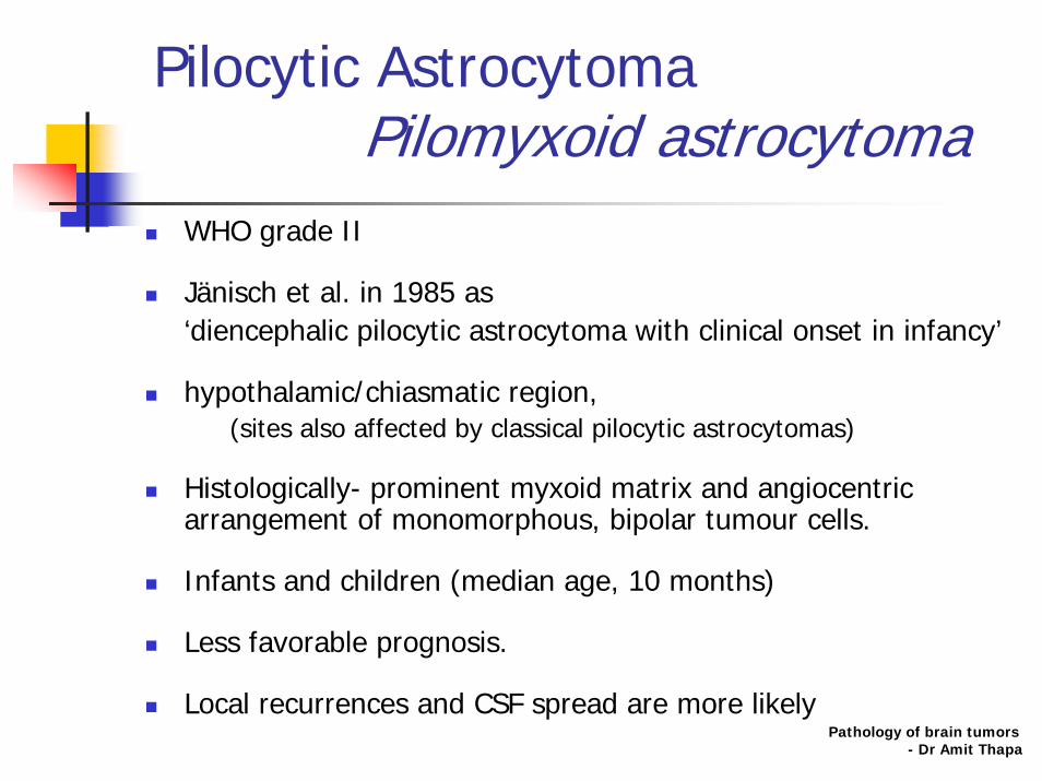
Pilocytic AstrocytomaPilomyxoid astrocytoma
WHO grade II
Jänisch et al. in 1985 as ‘diencephalic pilocytic astrocytoma with clinical onset in infancy’
hypothalamic/chiasmatic region, (sites also affected by classical pilocytic astrocytomas)
Histologically- prominent myxoid matrix and angiocentric arrangement of monomorphous, bipolar tumour cells.
Infants and children (median age, 10 months)
Less favorable prognosis.
Local recurrences and CSF spread are more likelyPathology of brain tumors
- Dr Amit Thapa
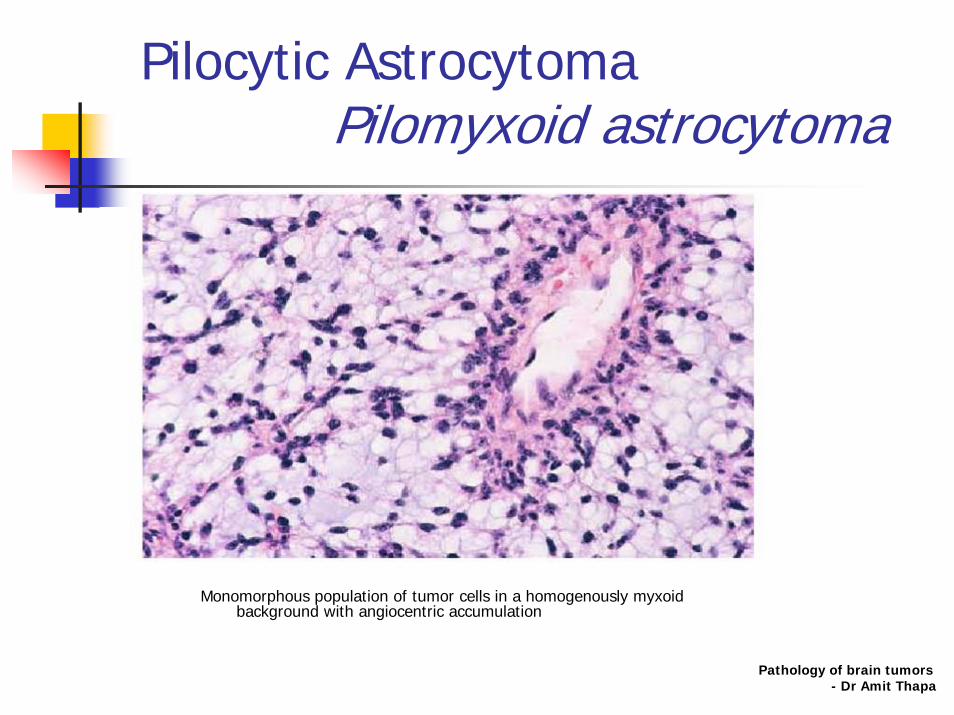
Pilocytic AstrocytomaPilomyxoid astrocytoma
Monomorphous population of tumor cells in a homogenously myxoid background with angiocentric accumulation
Pathology of brain tumors- Dr Amit Thapa

WHO GRADING OF BRAIN TUMORS, 4TH EDN, 2007
Pathology of brain tumors- Dr Amit Thapa

Subependymal giant cell astrocytoma
Invariably with Tuberous sclerosis, 8-18yrs
Near foramen of monroHydrocephalus
Histology: spindle to epithelioid large cells with abundant glassy eosinophilic cytoplasm, in perivascular pseudorosettes
Pathology of brain tumors- Dr Amit Thapa

WHO GRADING OF BRAIN TUMORS, 4TH EDN, 2007
Pathology of brain tumors- Dr Amit Thapa

Pleomorphic xanthoastrocytoma
Exclusive young adultsRare but important cause: TLESupratentorial intracortical cystic mass with mural nodule abutting meninges with dural tail
Pathology of brain tumors- Dr Amit Thapa
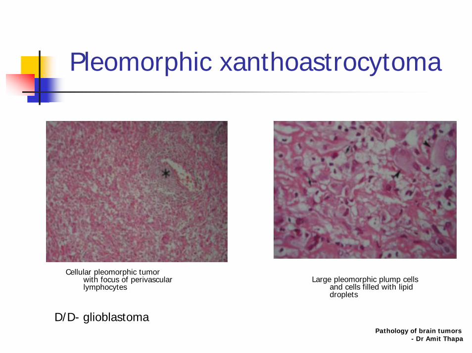
Pleomorphic xanthoastrocytoma
Cellular pleomorphic tumor with focus of perivascular lymphocytes
Large pleomorphic plump cells and cells filled with lipid droplets
D/D- glioblastomaPathology of brain tumors
- Dr Amit Thapa
WHO GRADING OF BRAIN TUMORS, 4TH EDN, 2007
Pathology of brain tumors- Dr Amit Thapa
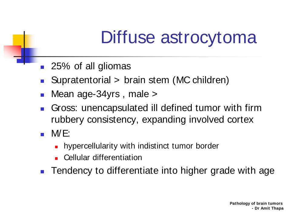
Diffuse astrocytoma25% of all gliomasSupratentorial > brain stem (MC children)Mean age-34yrs , male >Gross: unencapsulated ill defined tumor with firm rubbery consistency, expanding involved cortexM/E:
hypercellularity with indistinct tumor borderCellular differentiation
Tendency to differentiate into higher grade with age
Pathology of brain tumors- Dr Amit Thapa

Diffuse astrocytomaGrading system
Kernohan St A/M Current
WHO current
Ringertz UCSF current Bailey and Cushing 1926, 1930
1 2 II Astrocytoma MoAA Astrocytoma
2 3 III Anaplastic Astrocytoma
HAA Astroblastoma
3 4 IV GBM GBM Spongioblastoma multiforme
4
Pathology of brain tumors- Dr Amit Thapa

Diffuse astrocytomasubtypes
protoplasmic astrocytoma
homogenous, translucent, gelatinous appearance
Composed- neoplastic astrocytes (small, round- oval nuclei, which are moderately rich in chromatin) surrounded by scanty cytoplasm with few processes. Microcytic and mucoid degenerations are common
GFAP - sparse.
Microcystic degeneration
Pathology of brain tumors- Dr Amit Thapa

Diffuse astrocytomasubtypes
Fibrillary astrocytomagross- firm rubbery, cut surfaces: whitish-gray.
Composed: small stellate, elongated astrocytes fibrillary processes- fine in loose meshwork & bundles, leaving the pre-existing tissue relatively preserved. GFAP - variable.
Microcystic degenerations +/-
Gemistocytic astrocytoma soft & homogenous.
Composed: large, plump neoplastic astrocytes with abundant glassy eosinophilic cytoplasms and peripherally displaced nuclei
GFAP expression commonPathology of brain tumors
- Dr Amit Thapa
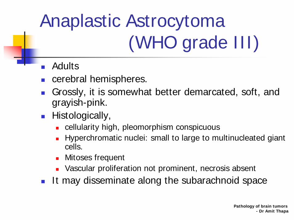
Anaplastic Astrocytoma(WHO grade III)
Adultscerebral hemispheres. Grossly, it is somewhat better demarcated, soft, and grayish-pink. Histologically,
cellularity high, pleomorphism conspicuousHyperchromatic nuclei: small to large to multinucleated giant cells. Mitoses frequentVascular proliferation not prominent, necrosis absent
It may disseminate along the subarachnoid space
Pathology of brain tumors- Dr Amit Thapa

Anaplastic Astrocytoma(WHO grade III)
Hypercellularity urges one to see for mitotic figures to establish anaplasia by Dumas- Duport grading (H&H)
Significant nuclear pleomorphism and frequent mitotic figures but no necrosis
Pathology of brain tumors- Dr Amit Thapa

Anaplastic Astrocytoma(WHO grade III)
Pathology of brain tumors- Dr Amit Thapa
Nuclei showing typical smudgy or clumped chromatin pattern with nuclear pleomorphisk and elongated nuclear profile (H&H)
Touch smear- prominent fibrillar processes and moderate nuclear pleomorphism. Cytoplasm trailing away from a nucleus resulting in a unipolar appearance

Anaplastic Astrocytoma(WHO grade III)
Lack of satellitosis more common as they infiltrate the cortex
GFAP- diffuse smudgy appearance rather than the fibrillary appearance in higher grade where every cell is not reactive
Pathology of brain tumors- Dr Amit Thapa

Anaplastic Astrocytoma(WHO grade III)
MIB-I reactive against Ki-67 antigen Abundant cytoplasmic intermediate filaments (both within fibrillary process and around nucleus)
Pathology of brain tumors- Dr Amit Thapa
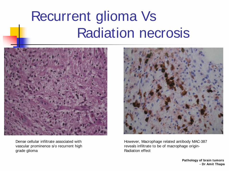
Recurrent glioma Vs Radiation necrosis
Dense cellular infiltrate associated with vascular prominence s/o recurrent high grade glioma
However, Macrophage related antibody MAC-387 reveals infiltrate to be of macrophage origin-Radiation effect
Pathology of brain tumors- Dr Amit Thapa
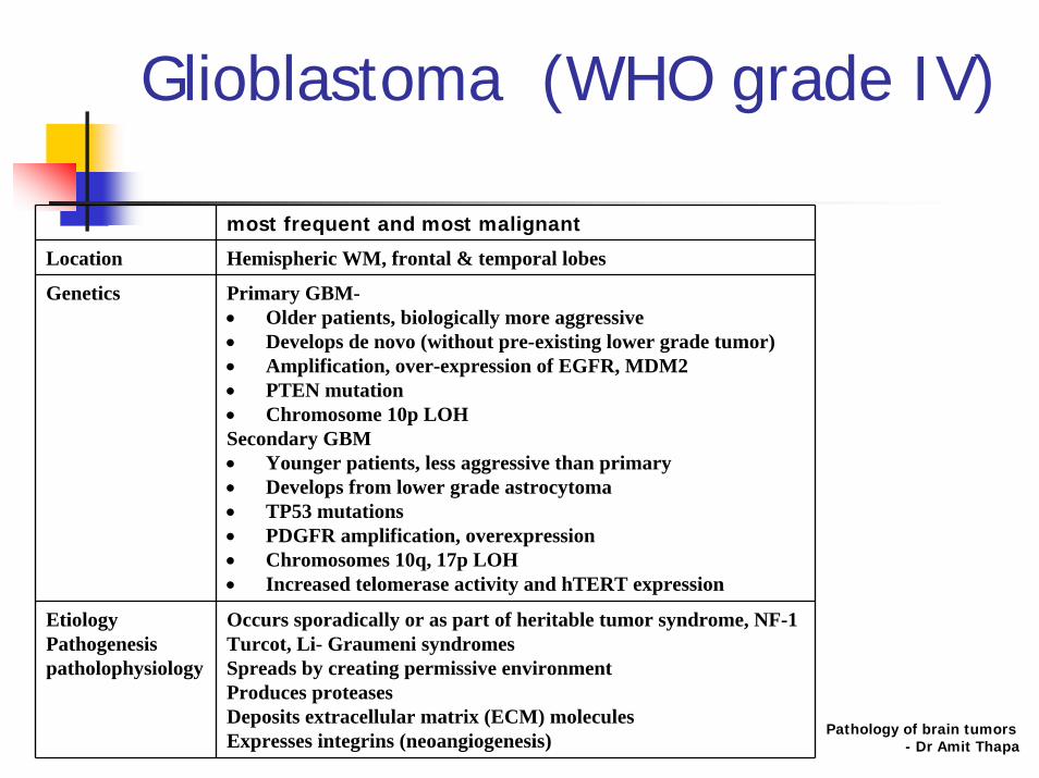
Glioblastoma (WHO grade IV)
most frequent and most malignant
Location Hemispheric WM, frontal & temporal lobes
Genetics Primary GBM-• Older patients, biologically more aggressive• Develops de novo (without pre-existing lower grade tumor)• Amplification, over-expression of EGFR, MDM2• PTEN mutation• Chromosome 10p LOHSecondary GBM• Younger patients, less aggressive than primary• Develops from lower grade astrocytoma• TP53 mutations• PDGFR amplification, overexpression• Chromosomes 10q, 17p LOH• Increased telomerase activity and hTERT expression
EtiologyPathogenesispatholophysiology
Occurs sporadically or as part of heritable tumor syndrome, NF-1Turcot, Li- Graumeni syndromesSpreads by creating permissive environmentProduces proteasesDeposits extracellular matrix (ECM) moleculesExpresses integrins (neoangiogenesis)
Pathology of brain tumors- Dr Amit Thapa

Glioblastoma (WHO grade IV)
Pathology of brain tumors- Dr Amit Thapa
Gross pathology •Reddish gray ‘rind’ of tumor surrounds necrotic core•Infiltrating mass with poorly delineated margins•Often expands invaded structures•May appear discrete but tumor always infiltrates
uncommon- cysts, hemorrhage
Microscopic features
•Increased cellularity•Marked mitotic activity•Distinct nuclear atypia•High nuclear cytoplasmic ratio•Coarse nuclear chromatin•necrosis or microvacular proliferation•Histologic variant- Gemistocytic
Immuno-pathology
•MIB-1 : 5-10%•GFAP + (multifocally reactive)
Presentation •Bimodal – small peak around 5yrs, Peak: 40- 50yrs•M:F= 1.8:1•Seizures, focal neurological feficits•May have headache or raised ICP
Natural history •Progession to secondary GBM common•Commonly arises as recurrence after resection of Grade II tumor•Spreads along WM tracts•Other sites- ependymoma, leptomeninges, CSF

Glioblastoma (WHO grade IV)
Smear preparation- nuclear pleomorphism, fibrillar processes with glial cells, proliferative vessel with very prominent endothelial cells
Pathology of brain tumors- Dr Amit ThapaIrregular foci of hemorrhage indicative
of glioblastomatous degeneration
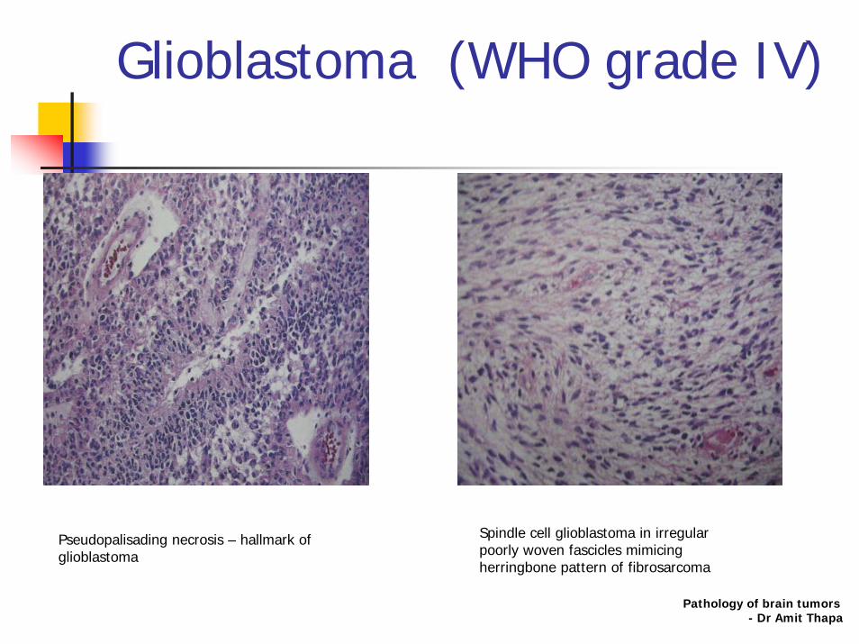
Glioblastoma (WHO grade IV)
Spindle cell glioblastoma in irregular poorly woven fascicles mimicing herringbone pattern of fibrosarcoma
Pseudopalisading necrosis – hallmark of glioblastoma
Pathology of brain tumors- Dr Amit Thapa

Glioblastoma (WHO grade IV)
Pathology of brain tumors- Dr Amit Thapa
GFAP stain identifies nests of obvious glial cells amidst exuberantly proliferative non reactive cellular component. Sarcomatoid appearance in infact a metaplastic appearance
Occasionally exhibit exuberant stromal myxoid change with glomeruloid vascular proliferation

Glioblastoma (WHO grade IV)
Superficial glioblastoma infiltrates into brain parenchyma, this differentiates it from malignant meningioma
Pseudopalisading necrosis – hallmark of glioblastoma
Pathology of brain tumors- Dr Amit Thapa

WHO GRADING OF BRAIN TUMORS, 4TH EDN, 2007
Pathology of brain tumors- Dr Amit Thapa

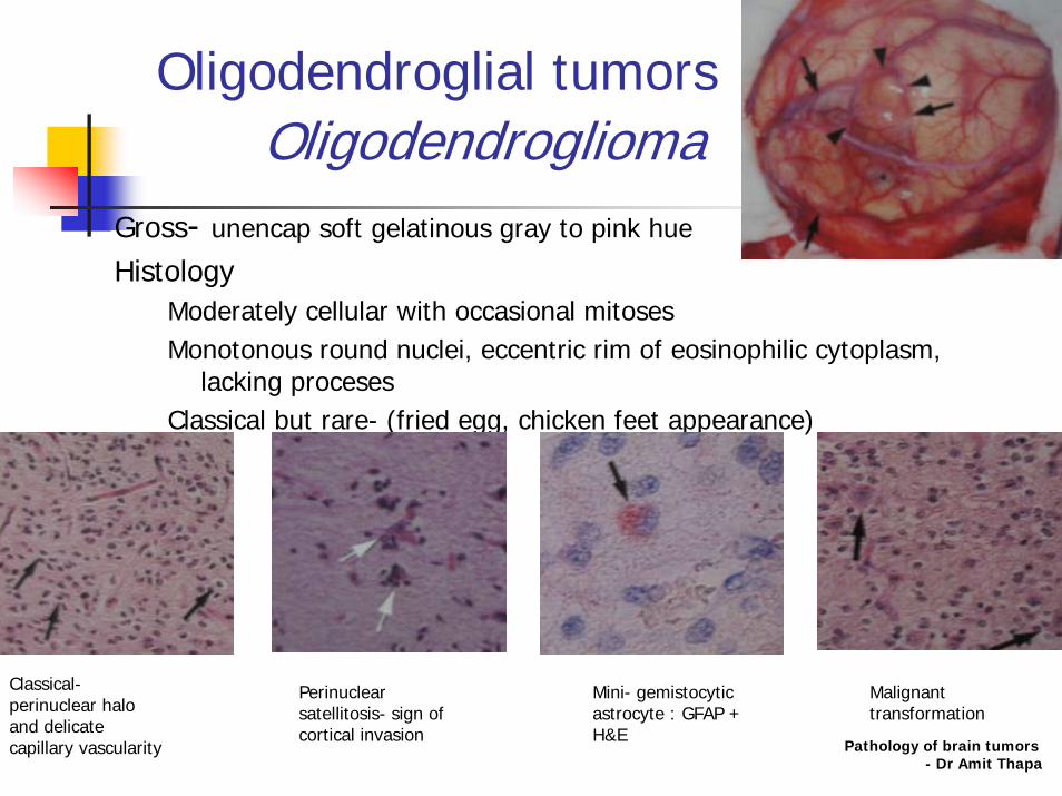
Oligodendroglial tumorsOligodendroglioma
Partially calcified well differentiated slowly growing but infiltrating cortical mass in middle age adult
Calcification: 90% CTFrontal > TPO lobeSeizure: 50-80%20-50% aggressive (anaplastic)
high cell densitypleomorphism + anaplastic nucleiNumerous mitosesMicrovascular proliferationNecrosis+/-
Pathology of brain tumors- Dr Amit Thapa
Oligodendroglial tumorsOligodendroglioma
Gross- unencap soft gelatinous gray to pink hue
HistologyModerately cellular with occasional mitosesMonotonous round nuclei, eccentric rim of eosinophilic cytoplasm,
lacking procesesClassical but rare- (fried egg, chicken feet appearance)
Pathology of brain tumors- Dr Amit Thapa
Classical-perinuclear halo and delicate capillary vascularity
Perinuclear satellitosis- sign of cortical invasion
Mini- gemistocytic astrocyte : GFAP + H&E
Malignant transformation

Oligodendroglial tumors
GradingSmith (AFIP) system
pleomorphismnecrosisN/C ratioendothelial proliferationCell density
Grade Median survival (month)
A 94B 51C 45D 17
Pathology of brain tumors- Dr Amit Thapa

Oligodendrogliomavariants
Microgemistocytic oligodendroglioma displays small cells with round eosinophilic cytoplasm & eccentric nucleus GFAP +
Anaplastic oligodendroglioma (grade 3) Increased cellularity, nuclear pleomorphism, mitotic activityVascular proliferation, hemorrhages, & micronecroses.Leptomeningeal spread & subarachnoid dissemination.
Oligoastrocytoma well-differentiated neoplastic astrocytes (>25%) and oligodendrocyteseither diffusely intermingled or separatedOrigin: GFOC
Pathology of brain tumors- Dr Amit Thapa

WHO GRADING OF BRAIN TUMORS, 4TH EDN, 2007
Pathology of brain tumors- Dr Amit Thapa

Ependymal tumorsEpendymoma
Ependymal lining of ventricular wall, projects into the ventricular lumen or invades the parenchymaPredominant children and adolescents.Fourth ventricleAccounting for 6% to 12% of intracranial childhoodDrop mets: 11%
Pathology of brain tumors- Dr Amit Thapa

Ependymal tumorsEpendymoma
VariantsNon anaplastic (low grade)
Clear cellCellulartanycyticPapillary- classic lesion, 30% metastatise, dark small nuclei. 2 cytoplasmic patterns
Differentiation along glial lines forms perivascular pseudorosettesCuboidal cells form ependymal tubules around a central bv (true rosettes)
Myxopapillary ependymoma- filum terminale. Papillary with microcystic vacuoles and mucosubstanceSubependymoma
Anaplastic : pleomorphism, multinucleation, giant cells, mitoticfigures, vascular changes, necrosis (ependymoblastoma)
Pathology of brain tumors- Dr Amit Thapa

Ependymal tumorsEpendymoma
Ependymoma medulloblastoma
Mass in 4th ventricle Floor Roof (fastigium), 4th
ventricle drapes around tumor (banana sign)
Calcifications Common <10%
T1WI Inhomogenous Homogenous
T2WI High intensity exophytic component
Mildly hyperintense
Pathology of brain tumors- Dr Amit Thapa

Ependymal tumorsEpendymoma
Circumscribed cherry sized firm, lobulated tumor filling lumen of fourth ventricle
Perivascular rosette
H&E papillary pattern
Pathology of brain tumors- Dr Amit Thapa

Ependymal tumorsSubependymoma
Subependymal glial cellsAnterior lateral ventricles or posterior fourth ventricle
Nests of tumor cells in a fibrillary matrix
Pathology of brain tumors- Dr Amit Thapa
WHO GRADING OF BRAIN TUMORS, 4TH EDN, 2007
Pathology of brain tumors- Dr Amit Thapa

Choroid Plexus tumors
0.4- 1% all intracranial tumors70% patients are <2yrsAdults: infratentorialChildren: lateral ventricleClinical: raised ICP, Seizures, SAH
Pathology of brain tumors- Dr Amit Thapa

Choroid Plexus tumorsChoroid Plexus Papilloma
intraventricular papillary neoplasms
derived from choroid plexus epithelium
benign in nature, cured by surgery
Gross: circumscribed moderately firm, cut surface: cauliflower- like appearance.
Histology: tumor resembles a normal choroid plexus, but is more cellular,with cuboidal and columnar epithelial cells resting on a fine fibrovascular stroma.
Hemorrhages and calcifications: commonPathology of brain tumors
- Dr Amit Thapa
Choroid Plexus tumorsAtypical Choroid Plexus Papilloma
WHO grade II
Intraventricular papillary neoplasms (from choroid plexus epithelium)
intermediate features
distinguished from the choroid plexus papilloma by increased mitotic activity
Curative surgery is still possible
but the probability of recurrence significantly higher Pathology of brain tumors- Dr Amit Thapa
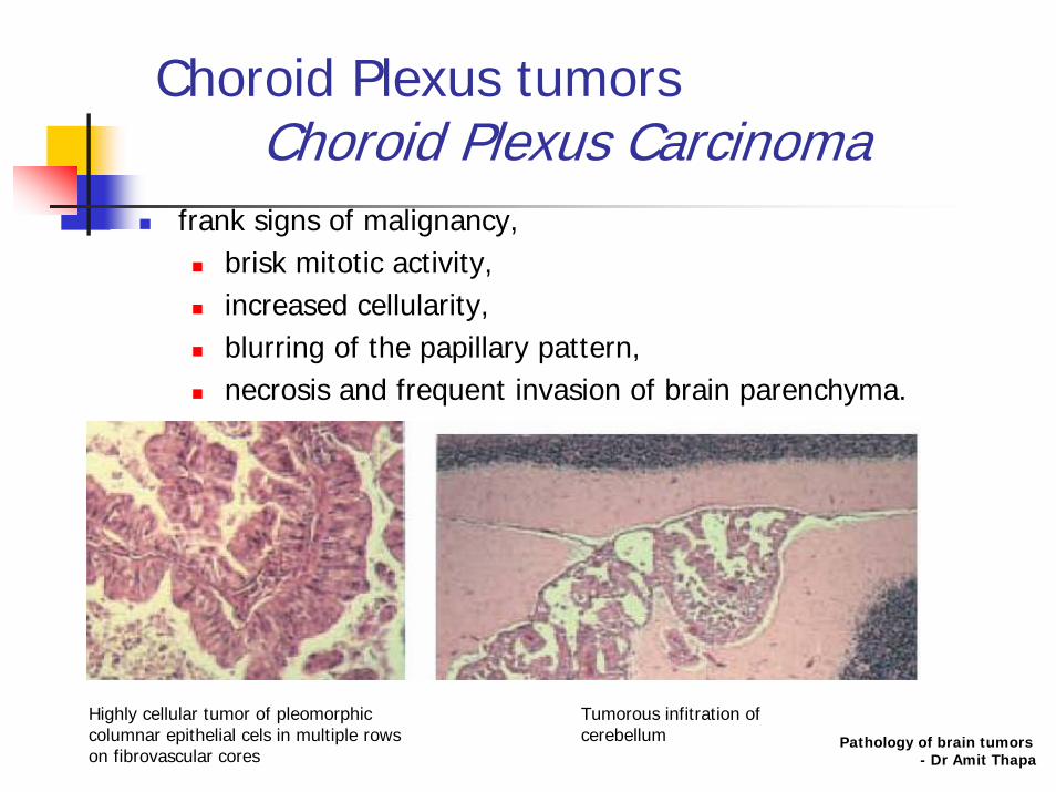
Choroid Plexus tumorsChoroid Plexus Carcinoma
frank signs of malignancy, brisk mitotic activity, increased cellularity, blurring of the papillary pattern, necrosis and frequent invasion of brain parenchyma.
Highly cellular tumor of pleomorphic columnar epithelial cels in multiple rows on fibrovascular cores
Tumorous infitration of cerebellum Pathology of brain tumors
- Dr Amit Thapa

Other Neuroepithelial tumorsAngiocentric glioma
WHO grade 1Predominantly children and young adults (17yrs)Refractory epilepsy- leading symptomTotal – 28 caseslocated superficially- fronto-parietal, temporal, hippocampal region. FLAIR - well delineated, hyperintense, non-enhancing cortical lesions, often with a stalk-like extension to the subjacent ventricle Stable or slowly growing Histopathology-
monomorphous bipolar cells, an angiocentric growth pattern
Immunoreactivity- EMA, GFAP, S-100 protein and vimentin, Not for neuronal antigens.
D/D- ependymal variant- Frequent extension of angiocentric glioma to the ventricular wall, M/E ependymal differentiation
Pathology of brain tumors- Dr Amit Thapa

Other Neuroepithelial tumorsAngiocentric glioma
Elongated tumor cells with concentric perivascular arrangement
Perivascular tumor cells strongly express GFAP
Pathology of brain tumors- Dr Amit Thapa
Neuronal and mixed neuronal-glial tumors
Pathology of brain tumors- Dr Amit Thapa
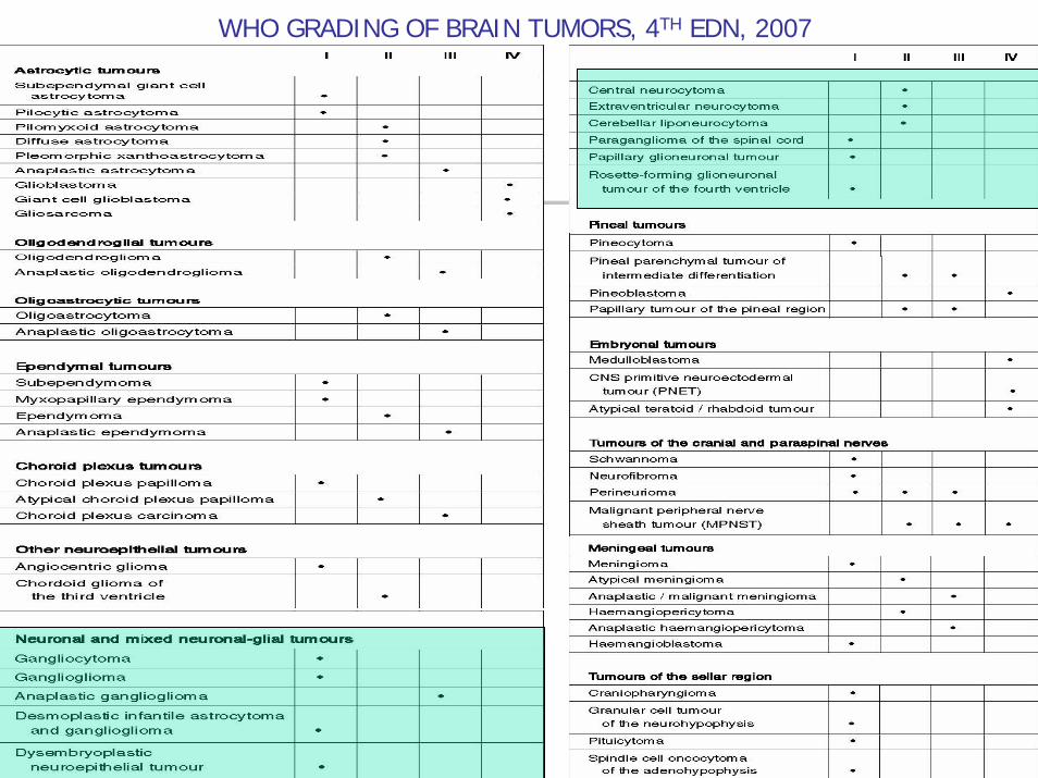
WHO GRADING OF BRAIN TUMORS, 4TH EDN, 2007
Pathology of brain tumors- Dr Amit Thapa

Neuronal and mixed neuronal-glial tumorsGangliocytoma & Ganglioglioma
Temporal and frontal lobes.
Gross: gray, firm, and often cystic.
Gangliocytomas - atypical neoplastic neurons within fibrillary matrix
Gangliogliomas- mixture of neoplastic neurons & glial cells, mostly astrocytes.
Immunoreact for synaptophysin and neurofilament proteins.
Calcifications, eosinophilic globules, and perivascular lymphocytic infiltrations common.
Mitoses are rare, necrosis is absent.Pathology of brain tumors
- Dr Amit Thapa

Neuronal and mixed neuronal-glial tumorsCentral neurocytoma
Lateral or third ventricle at the Foramen Monro
well-demarcated soft tumor
Uniformly small neurocytes
Several architectural patterns resembling oligodendroglial and ependymal tumors
Calcifications- common, hemorrhages may occur.
Pathology of brain tumors- Dr Amit Thapa
Neuronal and mixed neuronal-glial tumorsDysembryoplastic neuroepithelial tumor (DNET)
Often temporal lobe, less cerebellum & pons.
mucinous or gelatinous appearance.
Neoplastic neurons, astrocytes, and oligodendrocytes in a nodular pattern.
Pools of mucin, calcifications, abnormal blood vessels
Pathology of brain tumors- Dr Amit Thapa

Neuronal and mixed neuronal-glial tumorsExtraventricular neurocytoma
WHO grade IINeuronal tumour with pathological features distinct from cerebral neuroblastoma,Young adultsPreferential location- lateral ventricles in region of the foramen of Monro Favourable prognosisCentral neurocytomas- uniform round cells Additional features - Fibrillary areas mimicking neuropil, & low proliferation rateImmunohistochemical and ultrastructural e/o neuronal differentiation
Pathology of brain tumors- Dr Amit Thapa

Neuronal and mixed neuronal-glial tumorsPapillary glioneuronal tumor (PGNT)
WHO grade IKomori et al.- 1998wide age range (mean 27 years)Location- temporal lobe. CT & MRI- contrast-enhancing, well delineated mass, occasionally showing a cyst-mural nodule pattern. Histologically-
single or pseudostratified layer of flat to cuboidal GFAP-positive astrocytes surrounding hyalinized vascular pseudopapillae synaptophysin-positive interpapillary sheets of neurocytes, large neurons and intermediate size “ganglioid” cells.
Pathology of brain tumors- Dr Amit Thapa
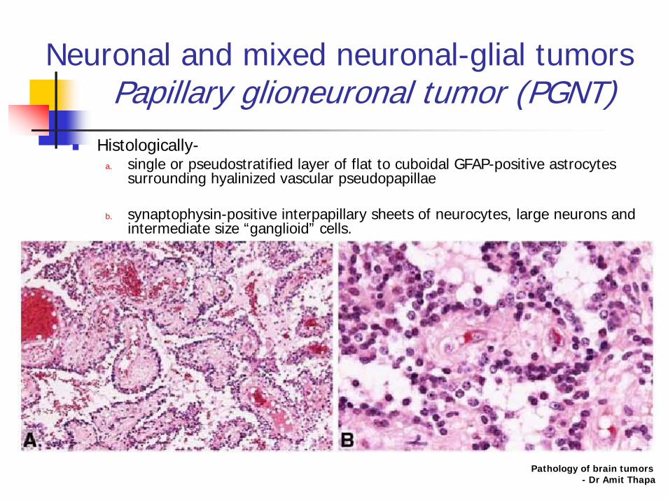
Neuronal and mixed neuronal-glial tumorsPapillary glioneuronal tumor (PGNT)
Histologically-a. single or pseudostratified layer of flat to cuboidal GFAP-positive astrocytes
surrounding hyalinized vascular pseudopapillae
b. synaptophysin-positive interpapillary sheets of neurocytes, large neurons and intermediate size “ganglioid” cells.
Pathology of brain tumors- Dr Amit Thapa
Neuronal and mixed neuronal-glial tumorsRosette forming glioneuronal tumor of the fourth ventricle
WHO grade IInitially described as dysembryoplastic neuroepithelial tumour (DNT) of the cerebellumKomori et al. in 2002 , total of 17 cases Rare slowly growing tumour of the fourth ventriclular regionYoung adults (mean age 33 years)Ostructive hydrocephalus, ataxia- most common clinical manifestation. Typically midline, involves the cerebellum and wall or floor of the fourth ventricle. T2WI- well delineated, hyperintense tumour. Histopathologically- a biphasic neurocytic and glial architecture
Neuronal component consists of neurocytes that form neurocytic rosettes with eosinophilic, synaptophysin-positive cores and/or perivascular pseudorosettes.Glial component dominates and typically exhibits features of pilocytic astrocytoma.
Benign clinical behaviour with the possibility of surgical cure
Pathology of brain tumors- Dr Amit Thapa

Neuronal and mixed neuronal-glial tumorsRosette forming glioneuronal tumor of the fourth ventricle
Pseudorosette with ring like arrangement of neurocytic tumor cell nuclei around an eosinophilic neuropil core
Neuropil core show strong immunoreactivity to synaptophysin
Pathology of brain tumors- Dr Amit Thapa

Neuronal and mixed neuronal-glial tumorsParaganglioma
Chemodectoma or glomus tumorsSlow growing (<2cm in 5 yrs)Histologically benign, <10% LN or distant metsMost secretory granules (Epinephrine/ NE)Site: carotid bifurcation, superior vagal ganglion, auricular branch of vagus, inferior vagal (nodose) ganglionGJ from glomus body in area of jugular bulb, and track along vesselsMay have finger like extensions
Pathology of brain tumors- Dr Amit Thapa

Tumors of the pineal region
Pineal region: bounded dorsally by splenium of corpus callosum and tela choroidea, ventrally by quadrigeminal plate and midbrain tectum, rostrally by posterior aspect of 3rd ventricle and caudally by cerebellar vermis
3-8% of paediatric brain tumors, <1% adults
Substrate Tumor
Pineal glandular tissue Pineocytomas, pineoblastomas
Glial cells Astrocytomas, oligodendroglioma, cyst
Arachnoid cells Meningiomas, cyst
Ependymal lining Ependymomas
Sympathetic nerves Chemodectomas
Rests of germ cells Choriocarcinoma, germinoma, embryonal ca, endodermal sinus tumor, teratoma
No BBB Hematogenous mets
Pathology of brain tumors- Dr Amit Thapa

WHO GRADING OF BRAIN TUMORS, 4TH EDN, 2007
Pathology of brain tumors- Dr Amit Thapa

Tumors of the pineal regionPineocytoma
Well differentiated CSF metsradiosensitive
Pathology of brain tumors- Dr Amit Thapa
Tumors of the pineal regionPineoblastoma
Malignant tumor – a PNETMetastasize through CSFRadiosensitive
Pathology of brain tumors- Dr Amit Thapa

Tumors of the pineal regionPapillary tumor of the pineal region
(PTPR)
WHO grade II/IIIchildren and adults (mean age 32 years)Relatively large (2.5–4 cm), and well-circumscribed,MRI- low T1 and increased T2 signal , contrast enhancement.2003, Jouvet et al.- total of 38 cases Histologically, papillary architecture and epithelial cytologyimmunoreactivity for cytokeratin and, focally, GFAP. Macroscopically indistinguishable from pineocytomaUltrastructural features s/o ependymal differentiation and a possible origin from specialized ependymal cells of the subcommissural organBiological behaviour- variable Pathology of brain tumors
- Dr Amit Thapa

Tumors of the pineal regionPapillary tumor of the pineal region
(PTPR)
Pathology of brain tumors- Dr Amit Thapa
WHO GRADING OF BRAIN TUMORS, 4TH EDN, 2007
Pathology of brain tumors- Dr Amit Thapa

Embryonal tumorsMedulloblastoma
Most common malignant paediatric Ca1st decade of lifeMale: Female= 2:1Cerebellar vermis, apex of 4th ventricle roof (fastigium)Cl: early hydrocephalus, cerebellar signsSolid midline contrast enhancing Highly radiosensitive and moderately chemosensitiveRecurrence: 10-35%, extraneural mets: 5%Poorly demarcated, pinkish-gray and soft.Histology-
densely cellular & small cells with round, oval, or carrot- shaped hyperchromatic nuclei surrounded by scanty cytoplasm (blue cell tumor).
Pathology of brain tumors- Dr Amit Thapa

Embryonal tumorsMedulloblastoma
Medulloblasts may differentiate into neurons and glial cells. Neuronal differentiation – NSE+ & synaptophysin+Glial differentiation- GFAP-positive Disseminate via CSF pathway- small nodules & diffuse infiltrates in the ventricular wall and subarachnoid space
Pale island in nodular variant (HE)Highly cellular tumor – anaplastic cells with small round to oval hyperchromatic nuclei surrounded by scanty cytoplasm (HE).
Pathology of brain tumors- Dr Amit Thapa
Embryonal tumorsMedulloblastoma
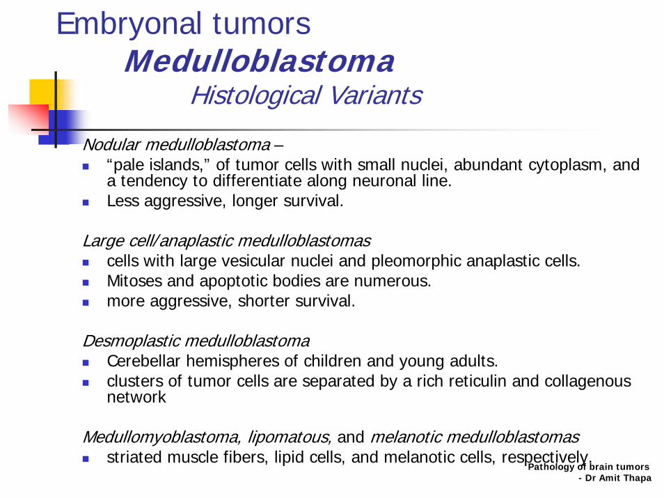
Histological Variants
Nodular medulloblastoma –“pale islands,” of tumor cells with small nuclei, abundant cytoplasm, and a tendency to differentiate along neuronal line.Less aggressive, longer survival.
Large cell/anaplastic medulloblastomascells with large vesicular nuclei and pleomorphic anaplastic cells. Mitoses and apoptotic bodies are numerous.more aggressive, shorter survival.
Desmoplastic medulloblastoma Cerebellar hemispheres of children and young adults. clusters of tumor cells are separated by a rich reticulin and collagenous network
Medullomyoblastoma, lipomatous, and melanotic medulloblastomas striated muscle fibers, lipid cells, and melanotic cells, respectively.
Pathology of brain tumors- Dr Amit Thapa
Embryonal tumorsMedulloblastoma
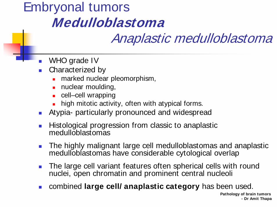
Anaplastic medulloblastomaWHO grade IVCharacterized by
marked nuclear pleomorphism,nuclear moulding, cell–cell wrappinghigh mitotic activity, often with atypical forms.
Atypia- particularly pronounced and widespread
Histological progression from classic to anaplastic medulloblastomas
The highly malignant large cell medulloblastomas and anaplastic medulloblastomas have considerable cytological overlap
The large cell variant features often spherical cells with roundnuclei, open chromatin and prominent central nucleoli
combined large cell/anaplastic category has been used. Pathology of brain tumors
- Dr Amit Thapa

Embryonal tumorsCNS primitive neuroectodermal tumor
Wide variety with common pathologic features
Originate from primitive neuroectodermal cells
May disseminate through CSF
Pathology of brain tumors- Dr Amit Thapa
Embryonal tumorsCNS primitive neuroectodermal tumor
Ependymoblastoma
Highly cellular embryonal form of ependymal tumorAge <5rsPrognosis poor with median survival 12-20 months100% mortality at 3 yrs
Pathology of brain tumors- Dr Amit Thapa

WHO GRADING OF BRAIN TUMORS, 4TH EDN, 2007
Pathology of brain tumors- Dr Amit Thapa

Tumors of Cranial Nerves and Paraspinal NvSchwannoma
Misnomer- acoustic neuromaArise form superior vestibular division of CN VIIILoss of suppressor gene on 22q (NF)Cl: hearing loss, tinnitus, dysequilibriumHistology:
Antoni A – narrow elongated bipolar cellsAntoni B- loose reticulated
Pathology of brain tumors- Dr Amit Thapa

Tumors of Cranial Nerves and Paraspinal NvSchwannoma
Cherry sized encapsulated tumor in R CP angle Spindle shaped bipolar cells forming
fascicles and palisading with alternating zones of nuclei and processes
Verocay body, Anntony B
Pathology of brain tumors- Dr Amit Thapa
WHO GRADING OF BRAIN TUMORS, 4TH EDN, 2007
Pathology of brain tumors- Dr Amit Thapa

Tumors of the meningesTumor of the meningothelial cells
MeningiomaSlow growing extra-axial Arising from arachnoid not duraFalx> convexity> sphenoid boneHead injury and therapeutic radiation – predispose meningioma. Solitary or multiple - NF2Hyperostosis of adjacent boneFrequently calcifiedGrossly- extra-axial, encapsulated,round, oval, or lobulated; firm or moderately soft. Blood supply- meningeal branches of ECA Cut surfaces- pinkish-gray, granular, or gritty. Histology:
Classical- psammoma bodiesEMA+, Vimetin+, inconsistently for S-100 protein
Pathology of brain tumors- Dr Amit Thapa
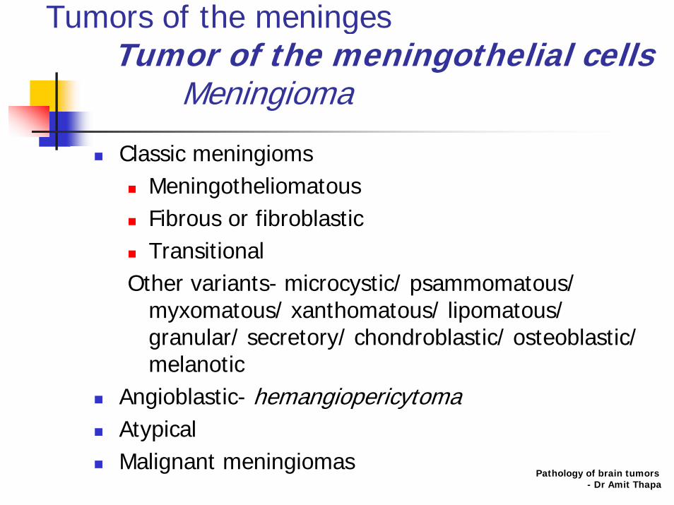
Tumors of the meningesTumor of the meningothelial cells
Meningioma
Classic meningiomsMeningotheliomatousFibrous or fibroblasticTransitional
Other variants- microcystic/ psammomatous/ myxomatous/ xanthomatous/ lipomatous/ granular/ secretory/ chondroblastic/ osteoblastic/ melanotic
Angioblastic- hemangiopericytomaAtypicalMalignant meningiomas
Pathology of brain tumors- Dr Amit Thapa
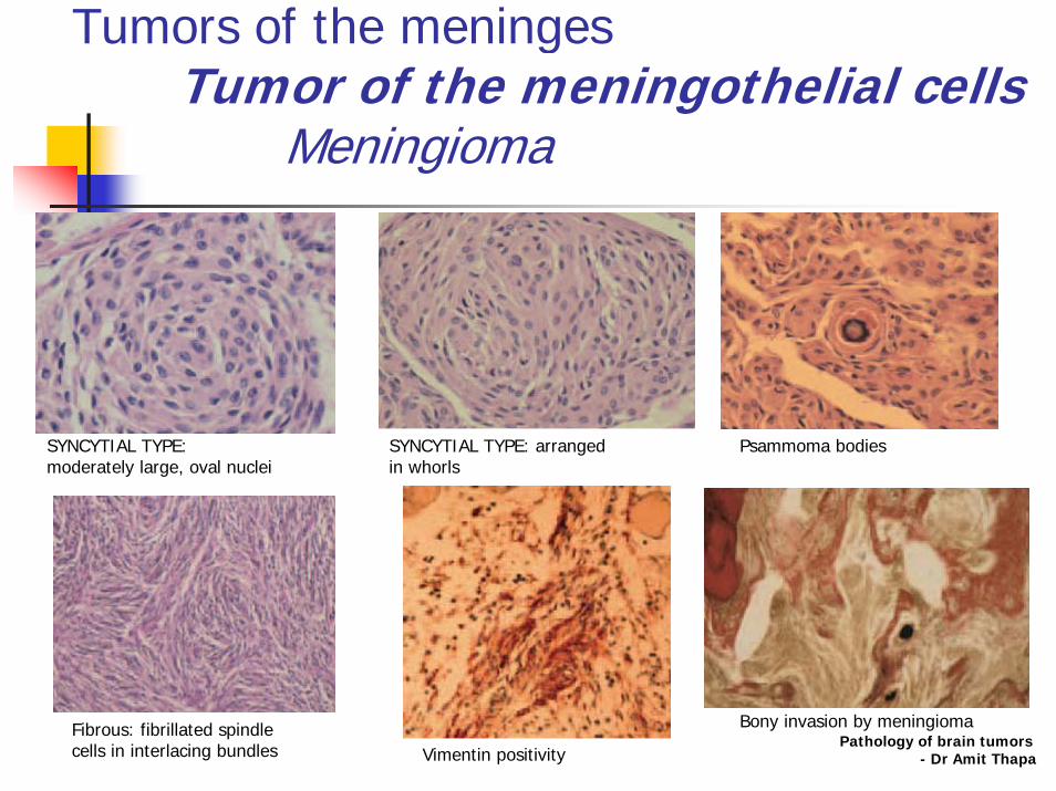
Tumors of the meningesTumor of the meningothelial cells
Meningioma
SYNCYTIAL TYPE: arranged in whorls
Psammoma bodiesSYNCYTIAL TYPE: moderately large, oval nuclei
Bony invasion by meningiomaFibrous: fibrillated spindle cells in interlacing bundles Pathology of brain tumors
- Dr Amit ThapaVimentin positivity

Tumors of the meningesTumor of the meningothelial
cells Meningioma
Pathology of brain tumors- Dr Amit Thapa
GRADE DEGREE OF REMOVAL
I Macroscopically complete removal with excision of dural attachment and abnormal bone
II Macroscopically complete with endothermy coagulation of dural attachment
III Macroscopically complete without resection or coagulation of dural attachment/ extradural extensions
IV Partial removal leaving tumor in situ
V Simple decompression +/- biopsy
Simpson etal, 1957
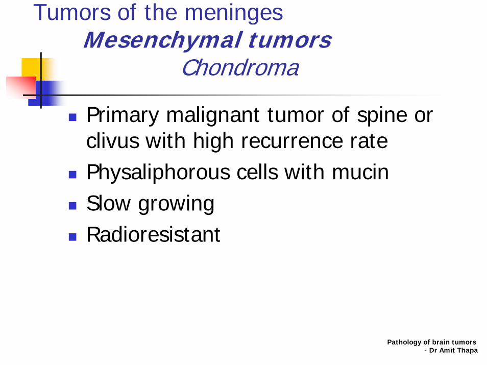
Tumors of the meningesMesenchymal tumors
Chondroma
Primary malignant tumor of spine or clivus with high recurrence ratePhysaliphorous cells with mucinSlow growing Radioresistant
Pathology of brain tumors- Dr Amit Thapa

Tumors of the meningesother related neoplasms
HaemangioblastomaBenign WHO grade11% all intracranial, 7% posterior fossa (adults)80% solitary, occassionally with VHLAdults: 30-65yrsLocation: cerebellum (83-86%)
Spinal cord (3-13%)Medulla ( 2-5%)Cerebrum (1.5%)
Cl: occipital headacheLab: polycythemia (erythropoietin)-20%Prognosis: 5-20yr survival following Sx
Pathology of brain tumors- Dr Amit Thapa

Tumors of the meningesother related neoplasms
Haemangioblastoma
Char: network of capillary like channels separated by trabeculae or islands of stromal cells with lipid droplets
Cystic hemangioblastoma (60%) with Small mural nodule
Pathology of brain tumors- Dr Amit Thapa
Tumors of the meningesother related neoplasms
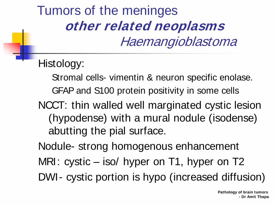
HaemangioblastomaHistology:
Stromal cells- vimentin & neuron specific enolase.GFAP and S100 protein positivity in some cells
NCCT: thin walled well marginated cystic lesion (hypodense) with a mural nodule (isodense) abutting the pial surface.
Nodule- strong homogenous enhancementMRI: cystic – iso/ hyper on T1, hyper on T2DWI- cystic portion is hypo (increased diffusion)
Pathology of brain tumors- Dr Amit Thapa
Tumors of the meningesother related neoplasms
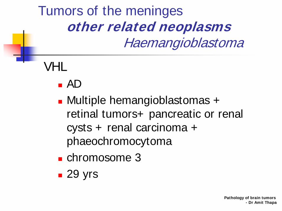
Haemangioblastoma
VHLADMultiple hemangioblastomas + retinal tumors+ pancreatic or renal cysts + renal carcinoma + phaeochromocytomachromosome 329 yrs
Pathology of brain tumors- Dr Amit Thapa
Germ cell tumors
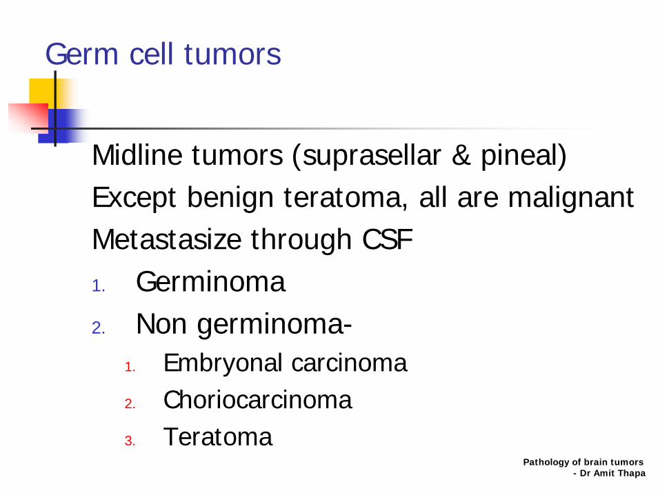
Midline tumors (suprasellar & pineal)Except benign teratoma, all are malignantMetastasize through CSF1. Germinoma2. Non germinoma-
1. Embryonal carcinoma2. Choriocarcinoma 3. Teratoma
Pathology of brain tumors- Dr Amit Thapa

Tumors of the sellar regionPituitary adenomas
10% of all intracranial tumors, common 3rd & 4th decades
Arise from adenohypophysis;
neurohypophysis – rare (glioma, granular cell tumor)
ClassificationSize- Microadenoma <1cm diameterEndocrine function- 2/3 secretoryAnatomical- Modified Hardy systemHistological- chromophobe/ acidophil/ basophilElectron microscopic appearance Pathology of brain tumors
- Dr Amit Thapa
WHO GRADING OF BRAIN TUMORS, 4TH EDN, 2007
Pathology of brain tumors- Dr Amit Thapa
Tumors of the sellar regionPituitary adenomas
Cl: visual disturbanceEndocrine abnormalitiesPituitary apoplexy- 1 to 2%
Gross: discrete grayish yellow, soft mass <1 cm dia
Histology: small round or oval nuclei with stippled chromatinAggressive: mitoses, pleomorphism
Hormone IHC identify specific hormonePathology of brain tumors
- Dr Amit Thapa
Tumors of the sellar regionPituitary adenomas
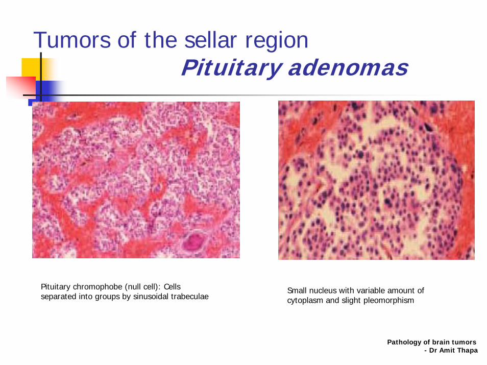
Pituitary chromophobe (null cell): Cells separated into groups by sinusoidal trabeculae
Small nucleus with variable amount of cytoplasm and slight pleomorphism
Pathology of brain tumors- Dr Amit Thapa

Tumors of the sellar region
Pathology of brain tumors- Dr Amit Thapa

Tumors of the sellar regionCraniopharyngioma
Pathology of brain tumors- Dr Amit Thapa
• More often children
• slowly growing
• originates from remnant epithelial cells of craniopharyngeal duct
• Mixed signal intensity with enhancing solid component; calcification
• Grossly: cystic with thick machine oil like contents
•Histlogy: multistratified squamous epithelial cells.
Two types:• The adamantinomatous type- cells form strands and cords
calcifications, amorphous masses of keratin (wet keratin)cholesterol clefts characteristic
• The papillary type- cells rest on a fibrovascular stoma. lacks calcifications and cholesterol crystalsGlial reaction and Rosenthal fibers round the tumor

Tumors of the sellar regionCraniopharyngioma
CECT showing large cystic mass within third ventricle
Large cyst with small tumorous mural nodules
Pathology of brain tumors- Dr Amit Thapa

Tumors of the sellar regionCraniopharyngioma
Mural nodule contains squamous epithelial cells arranged in small islands
Cyst wall loosely attached to ventricular wall- contains astrocytic fibers and Rosenthal fibers
Pathology of brain tumors- Dr Amit Thapa

Tumors of the sellar regionPituicytoma
WHO grade IRare, solid, low grade, spindle cell, glial neoplasm of adults Originates in the neurohypophysis or infundibulum < 30 cases reportedVisual disturbance, headache, hypopituitarismWell-circumscribed, solid masses, can measure up to several centimetres.Histologically- compact architecture consisting of elongate, bipolar spindle cells arranged in interlacing fascicles or assuming a storiform pattern.Mitotic figures are absent or rare.Positive- vimentin, S-100 protein ,variable- GFAP. slow growthpossibility of curative surgery
Pathology of brain tumors- Dr Amit Thapa
Tumors of the sellar regionSpindle cell oncocytoma
of adenohypophysis
Pathology of brain tumors- Dr Amit Thapa
• WHO grade II
• 2002, Roncaroli et al. – till today 10 cases
• Oncocytic, non-endocrine neoplasm of the anterior pituitary
• Adults (mean age 56 years)
• Macroscopically be indistinguishable from a non-functioning pituitary adenoma and follow a benign clinical course
• The eosinophilic, variably oncocytic cytoplasm contains numerous mitochondria
• Immunoreactive for the anti-mitochondrial antibody 113-I, S-100 protein and EMA, but is negative for pituitary hormones

Tumors of the sellar regionSpindle cell oncocytoma
of adenohypophysis
Spindle and somewhat epithelioid cells with abundance of variably granular cytoplasm, different degree of nuclear atypia and focal inflammatory reaction
Generalised staining for S-100 protein
Pathology of brain tumors- Dr Amit Thapa

Diagnostic approach Radiology
• XRay Skull• CT Scan- plain and Contrast enhanced• MRI brain and spinal cord• Angiography• PET• SPECT• MRS• Myelography
Pathology of brain tumors- Dr Amit Thapa
Diagnostic techniques in pathologyTUMOR MARKERS
Pathology of brain tumors- Dr Amit Thapa
Oncofetal proteins
Placental proteins
Ectopic hormones
Enzymatic markers
Polyamines
Desmosterol
Beta-2-Microglobulin
Immunochemically Defined markers

Diagnostic techniquesImmunohistochemistryPanel- LCA, GFAP, Cytokeratin, EMAIf non reactive- vimentin, S100, synaptophysin
Needle biopsy Gliosis – confined to tumor/ brain interfaceInflammation and necrosis- within tumor
Strong cytoplasmic reactivity to EMA s/o mets papillary adenoca and R/o Choroid plexus neoplasm as single layer of epithelium upon papillary frond raise suspicion
Touch smear-clumps of tumor cells adherent to one other metastatic epithelial neoplasm
Pathology of brain tumors- Dr Amit Thapa
Diagnostic techniques in pathologyintra-operative diagnosis
When to ask
• Definitive neurosurgical management will be influenced
• When an unexpected lesion is encountered during surgery, or when the appearances of lesion visualized during surgery suggest an alternative diagnosis
• The main aim to obtain a tissue based diagnosis.Pathology of brain tumors
- Dr Amit Thapa

Diagnostic techniques in pathologyFROZEN SECTION
• In stereotactic biopsy- adequacy of the specimen
•Diagnosis and classification of a tumor - overall less reliable than diagnosis made on paraffin sections
Pathology of brain tumors- Dr Amit Thapa
Diagnostic techniques in pathologyFluorescent imaging (Chemical probe)
•Cytoreductive surgery
•Intravenous injection of fluorescein Na (0.2 cc/kg body weight)
•the yellow-stained tumor is visible to the naked eye
•in an eloquent area- resect at the surface of the yellow-stained tumoror debul within the yellow-colored lesion until the resection surface becomes pale yellow.
•in non-eloquent regions- suction of peritumoral white matter
•no special equipment •wide applicability in resection of malignant gliomas
No Shinkei Geka. 2007;35(6):557-62
Pathology of brain tumors- Dr Amit Thapa

Diagnostic techniques in pathologyBIOPSY
Paraffin sections better-
Greater amount of tissue
Better cytology
More time to assess the section
Pathology of brain tumors- Dr Amit Thapa

Diagnostic techniques in pathologyApproach to histological diagnosis of tumor biopsy
Pathology of brain tumors- Dr Amit Thapa

Diagnostic techniques in pathology
Pathology of brain tumors- Dr Amit Thapa
Useful information to be provided by surgeons
• Relationship of the lesion to adjacent structures• changes in the character and nature of the tissue • calcifications (which can be missed by MRI)• Any changes in the tissue specimen occurring due to surgery• Preoperative embolisation• Precise location of different portion of biopsy• Record whether certain key areas were sampled• Any apparent multiple lesions• Extent of resection• If lobectomy or larger resection- identify the resection margins• Relationship to blood vessels, other associated lesions, reactive
changes
Diagnostic techniques in pathologyBIOPSY
Pathology of brain tumors- Dr Amit Thapa
Tissue handling and sampling
• Specimen not to be fixed-•Allows tissue to be sampled •Stored for a number of techniques •Allow use of greater range of tissue fixatives
•Optimal fixation in glutaraldehyde for EM.•Usually fixed in 10% formalin, buffered at neutral pH•Then within 6-24hrs, sampling for paraffin section
processing done depending upon size and volume of the specimens

Pathology of brain tumors- Dr Amit Thapa
Protocol for handling a lobectomy specimen
• receive fresh and orient prior to dissection.• describe carefully - tumor on the external surface, relationship to
specialized structures (e.g. the hippocampus) and the resection margins.
• sample - microbiology, virology or molecular genetic studies.• then fix overnight for further dissection or dissected fresh • after fixation, section serially in the coronal plane at 5mm intervals• the cut surfaces - inspected and photograph. • the entire specimen should be blocked out on non adjacent faces• all tissues should be processed for histology• use of special stains and immuncytochemistry as appropriate.
Diagnostic techniques in pathologyBIOPSY

Diagnostic techniques in pathologyTinctorial stains used in CNS tumors
Haematoxylin and eosin General histological features
Toluidine blue Rapid staining of smears for intraop diagnosis
Reticulin Reticulin framework around blood vessels in gliomas and lymphomas; soft tissue tumors
Van gieson Dural infiltration in meningioma
Periodic acid Schiff Glycogen (diastase sensitive)Mucins ( intra and extracellular)
Alcian blue Mucins ( intra and extracellular)
Mucicarmine Mucins ( intra and extracellular)
Singh, Masson- Fontanna Melanin
Luxol fast blue Myelin
Solochrome cyanin Myelin
Pathology of brain tumors- Dr Amit Thapa
Diagnostic techniques in pathologyImmunocytochemistry
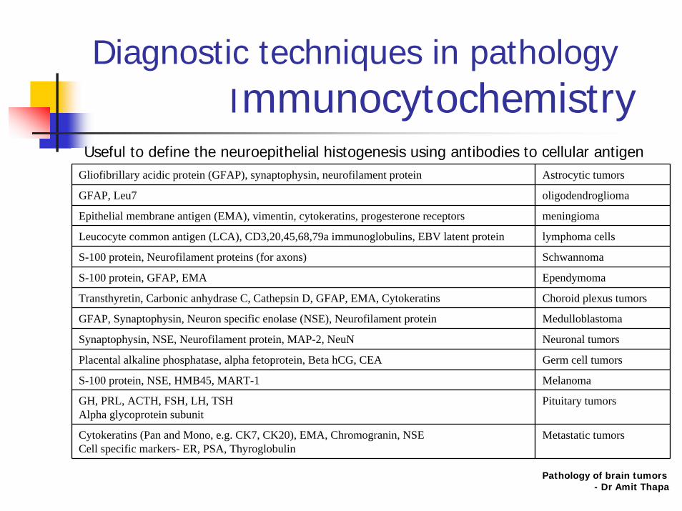
Gliofibrillary acidic protein (GFAP), synaptophysin, neurofilament protein Astrocytic tumors
GFAP, Leu7 oligodendroglioma
Epithelial membrane antigen (EMA), vimentin, cytokeratins, progesterone receptors meningioma
Leucocyte common antigen (LCA), CD3,20,45,68,79a immunoglobulins, EBV latent protein lymphoma cells
S-100 protein, Neurofilament proteins (for axons) Schwannoma
S-100 protein, GFAP, EMA Ependymoma
Transthyretin, Carbonic anhydrase C, Cathepsin D, GFAP, EMA, Cytokeratins Choroid plexus tumors
GFAP, Synaptophysin, Neuron specific enolase (NSE), Neurofilament protein Medulloblastoma
Synaptophysin, NSE, Neurofilament protein, MAP-2, NeuN Neuronal tumors
Placental alkaline phosphatase, alpha fetoprotein, Beta hCG, CEA Germ cell tumors
S-100 protein, NSE, HMB45, MART-1 Melanoma
GH, PRL, ACTH, FSH, LH, TSHAlpha glycoprotein subunit
Pituitary tumors
Cytokeratins (Pan and Mono, e.g. CK7, CK20), EMA, Chromogranin, NSECell specific markers- ER, PSA, Thyroglobulin
Metastatic tumors
Useful to define the neuroepithelial histogenesis using antibodies to cellular antigen
Pathology of brain tumors- Dr Amit Thapa

Diagnostic techniques in pathologyPrognostic indicators
Pathology of brain tumors- Dr Amit Thapa
• Nuclear hyperchromasia & nuclear: cytoplasmic • Large densely staining nuclei• Raised N: C ratio• Enlarged nuclei showing hyperchromasia and pleomorphism• Mitotic and proliferation indices
• Necrosis• Blood vessels, blood- brain barrier and edema• Invasion, spread and metastasis• Cytoplasmic features of tumour cells• Expression of proteins detectable by immunocytochemistry • Organoid arrangements of the cells • High P glycoprotein levels• Amplification of the c-myc oncogene, • Elevated levels of c-myc mRNA• Ki-67/ MIB-1 labelling indices
Good prognosis• high TrkC mRNA expression

Diagnostic techniques in pathologyProliferative potentials
Pathology of brain tumors- Dr Amit Thapa
Histologically similar tumors may have different proliferative potentials J Neurooncol 1989; 7: 137-143
• Mitotic figure counts- M phase fraction
• 3H Thymidine- S phase fraction
• Bromodeoxyuridine and Iododeoxyuridine (BUdR, IUdR)
• AgNORs
• PCNA/ Cyclin
• DNA polymerase Alpha
• Ki-67/ MIB 1
Prognostic variables
For each tumour entity, combinations of parameters
WHO grade Clinical findings- age/ neurologic performance statusTumour locationRadiological features - contrast enhancementExtent of surgical resectionProliferation indicesGenetic alterations
Pathology of brain tumors- Dr Amit Thapa

RECENT ADVANCESModern techniques
Molecular techniques
DNA analysis: structural changes in genes and chromosomes
Southern blot
PCR
FISH
SSCPPathology of brain tumors
- Dr Amit Thapa
RECENT ADVANCESModern techniques
LOH ANALYSIS:
Chromosomal loss- reflects inactivation of tumor suppressor genes
(Comparative genomic hybridization) CGH:
A screening technique – detect large genomic gains or losses
Pathology of brain tumors- Dr Amit Thapa
RECENT ADVANCESModern techniques
RNA analysis: changes in levels of mRNA expression
Northern blot:
In-situ hybridization (ISH):
Protein analysis: changes in levels of protein expression, structural and functional protein changes
Western blot:
Immunohistochemistry:
Pathology of brain tumors- Dr Amit Thapa

THANK YOU
Pathology of brain tumors- Dr Amit Thapa