Epilepsy surgery preoperative work-up -...
72
Epilepsy surgery preoperative work-up
Transcript of Epilepsy surgery preoperative work-up -...
Epilepsy surgery preoperative work-up
When to consider epilepsy surgery ?
Persistent Seizures despite adequate pharmacological treatment
Drug resistant epilepsy may be defined as failure of adequate trials of two tolerated and appropriately chosen and used AED schedules (whether as monotherapies or in combination) to achieve sustained seizure freedom)
Definition of drug resistant epilepsy: Consensus proposal by the ad hoc Task Force of the ILAE Commission on Therapeutic Strategies :Patrick Kwan, yAlexis Arzimanoglou, zAnne T. Berg, xMartin J. Brodie, {W. Allen Hauser, #2Gary Mathern, **Solomon L. Moshe´ , yyEmilio Perucca, zzSamuel Wiebe, and xx2Jacqueline French:Epilepsia, **(*):1–9, 2009
Significant impairment of quality of life
The goal of the presurgical evaluation
To generate a “surgical diagram” depicting Epileptogenic region (ER) Location Extent Critical cortex ( CC) Surgical resection = ER - CC The most common location of seizure onset in adults is the temporal
lobe, especially the medial temporal lobe (hippocampus).This is also the seizure location most amenable to surgical cure.

90 % of adult patients with seizure disorder have partial or localization related epilepsy.
80 % of the patients of partial seizure experience temporal lobe seizures
Nearly 90 % of the partial seizures of temporal lobe origin emanate from the medial temporal lobe
The majority of extratemporal epilepsy patients have seizures of frontal lobe origin
(Chabolla DR ,Cascino GD .The treatment of epilepsy ;Principles and
Practice,1996:264-279)
Presurgical tests
No single test adequately defines the epileptogenic region
Mandatory noninvasive – “universal access”
Clinical semiology and Exam
Scalp EEG
Imaging MRI
Optional Noninvasive – “Limited Access “
Video EEG
Magnetoencephalography (MEG),
EEG-fMRI,
Nuclear imaging (PET,SPECT )
Neuropsychological testing
Wada (intracarotid amobarbital) test
Invasive EEG monitoring
Clinical semiology and Exam
TLE may have an epigastric aura, followed by a quiet period of unresponsiveness with staring, lipsmacking (oral automatisms).
Picking at sheets or clothes (manual automatisms), contralateral dystonic posturing, and postictal confusion and lethargy.
If from the dominant hemisphere, there is usually delayed recovery of language, often with transient aphasia on testing.
A typical frontal lobe seizure will occur from sleep with no warning, may show restlessness and/or prominent bilateral limb movements (such as bicycling or asymmetric tonic posturing), and will end quickly with immediate recovery; this may recur several times in one night.
Occipital lobe seizures often have a visual aura, and may progress (due to electrical spread) into a temporal lobe or frontal lobe type of seizure.
Parietal lobe seizures are the least common, may have a sensory aura, and tend to mimic frontal lobe seizures.
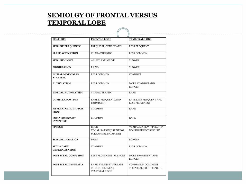
SEMIOLGY OF FRONTAL VERSUS TEMPORAL LOBE
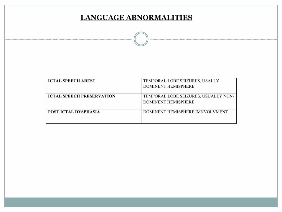
LANGUAGE ABNORMALITIES
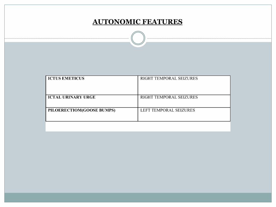
AUTONOMIC FEATURES

MOTOR ABNORMALITIES

EXAMINATION OF PATIENT DURING THE SEIZURE OF SEMIOLOGIC CHARACTERISTICS
TEMPORAL LOBE SEIZURES
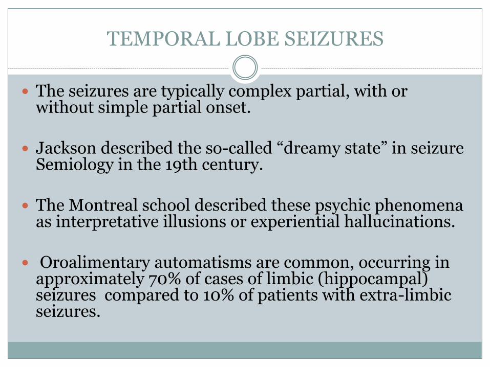
The seizures are typically complex partial, with or without simple partial onset.
Jackson described the so-called “dreamy state” in seizure Semiology in the 19th century.
The Montreal school described these psychic phenomena
as interpretative illusions or experiential hallucinations. Oroalimentary automatisms are common, occurring in
approximately 70% of cases of limbic (hippocampal) seizures compared to 10% of patients with extra-limbic seizures.

Unilateral dystonic posturing of an arm is classical
of contralateral temporal lobe epilepsy.
Ipsilateral upper arm automatism.
Postictal coughing.
Olfactory auras - poorly recognizable, always
unpleasant, foul smell - “uncal” or “uncinate” fits.
Fear is another limbic aura considered to be
amygdaloid in origin.
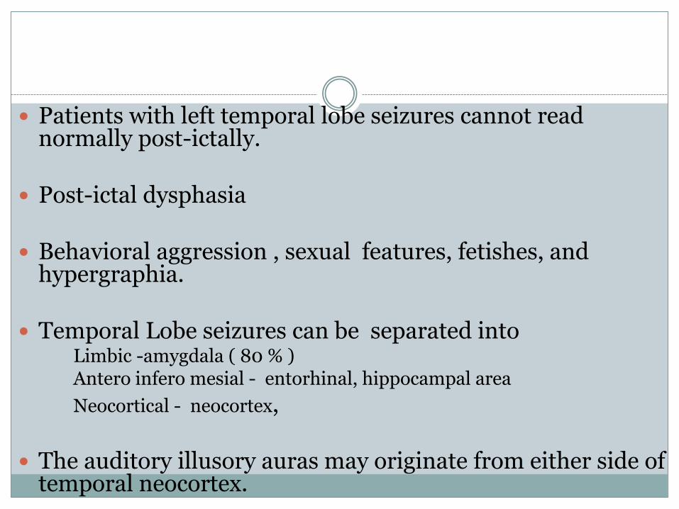
Patients with left temporal lobe seizures cannot read normally post-ictally.
Post-ictal dysphasia
Behavioral aggression , sexual features, fetishes, and
hypergraphia. Temporal Lobe seizures can be separated into Limbic -amygdala ( 80 % ) Antero infero mesial - entorhinal, hippocampal area
Neocortical - neocortex, The auditory illusory auras may originate from either side of
temporal neocortex.
FRONTAL LOBE SEIZURES
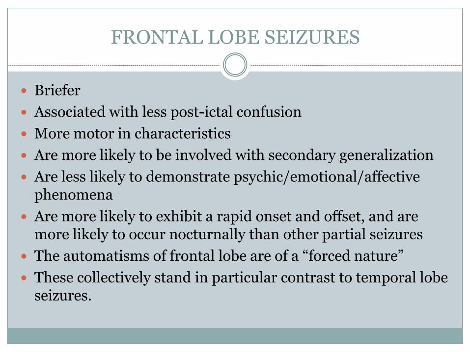
Briefer
Associated with less post-ictal confusion
More motor in characteristics
Are more likely to be involved with secondary generalization
Are less likely to demonstrate psychic/emotional/affective phenomena
Are more likely to exhibit a rapid onset and offset, and are more likely to occur nocturnally than other partial seizures
The automatisms of frontal lobe are of a “forced nature”
These collectively stand in particular contrast to temporal lobe seizures.
Fronto-polar:
Origin are often from scars following head injuries.
They have the greatest likelihood of simply being characterized by what appear clinically to be primary generalized seizures,
they may have some of the general characteristics of FLE, especially contralateral head deviation.
Orbito-frontal
May have semiology that mimics temporal lobe seizures in which case the origin is usually attributed to the posterior part of the orbital cortex.
Once again, however, it may involve, in various combinations, other frontal lobe semiology, especially contralateral head and eye deviation.

Premotor:
Head and eye deviation with varying bilateral tonic posturing is the commonest seizure.
When the deviation occurs at the onset of the seizure when consciousness is intact it has significant localizing value to the contra lateral frontal cortex.
Those seizures arising in the mid part of the frontal lobe have a prominent bilateral tonic posturing. Bipedal automatisms may take the form of symmetric bicycling or kicking movements.
Prominent leg movements favor involvement of the supplementary motor area.
The so-called “fencing posture” is classically associated with
the contralateral FL, particularly the SMA. This complex posture is characterized by abduction, external
rotation, and partial flexion of the contralateral arm at the shoulder, contralateral deviation of the head and eyes such that they “look at” the contralateral arm, extended ipsilateral arm downwards and backwards, and with the feet apart so as to support the partially contralaterally rotated trunk. Occasionally, the upper limb is also flexed at the elbow with the hand raised to the face that has turned forcefully towards it.
There is occasionally guttural, ill-understood speech.
Dominant opercular
Usually begins with an alteration in speech.
The alteration may be typical dysphasic speech, a form of non-specific guttural speech, or an arrest of speech.
Post-ictal dysphasia.
Rolandic
Rolandic seizures combine both motor and sensory components.
Sensory features can be positive (e.g. pins and needles,
pain, pricking, tingling) or negative (numbness). Elementary paraesthesiae are reported to be the most
characteristic of seizures arising in the post-central gyrus.
In pure sensory seizures there is nearly always
dysfunction of the part involved, which may be awkwardness, typical sensory ataxia, or paralysis.

Spread (intracortically) within the Rolandic cortex, e.g., sensory and/or motor, over contiguous parts of the associated homunculus, clinically is reflected in a “march” from one place on the body to another.
Unlike the tonic activity associated with seizures arising outside of the Rolandic cortex, which often has unreliable localizing and lateralizing value, Rolandic motor activity is clonic and is unambiguously localizing to the contralateral motor cortex in the area of the homunculus from which it arises, and usually is associated with transient post-ictal weakness in the involved part.

EEG
Developed in 1930s.
Awake & Sleep
Ictal & Interictal
Eye opening & Closure, Hyperventilation, Photic stimulation.
Scalp EEG
Lateralization (left vs. right hemisphere) and localization.
Additional electrodes that are particularly sensitive to medial temporal discharges.
The presence of interictal (between seizures) epileptiform discharges.
EDs: sharp waves or spikes.
Lack of EDs does NOT rule out epilepsy (up to 10%).
Spike waves : negative transients with steep ascending and descending limbs and a duration of 20 to 70 ms.
Sharp waves have broader potential (70 – 200 ms).
TIRDA
PLEDs

Interictal s/o increased epileptogenic potential.
Ictal Eds s/o classification of seizure, brain region of seizure activity.
Sensitivity and specificity depends upon,
Localization of epileptic brain tissue.
Duration of recording.
Use of supplementary electrodes.
Frequency of seizures
Timing in relation to last seizure.
Medial temporal lobe epilepsy patients usually have EDs from the anterior-mid temporal lobe (electrodes F7/F8, T3/T4, as well as anterior temporal, sphenoidal, and inferior temporal electrodes if used).
Bilateral temporal lobe EDs are not unusual(25-30%)
Diagnostic yield increases to 90 % in TLE by obtaining sleep study (Vs 50 % in awake)
(Chabolla DR ,Cascino GD .The treatment of epilepsy ;Principles and
Practice, 1996:264-279)

Video EEG
The combined use of video and EEG recording improves the sensitivity and specificity over EEG recording alone.
In many cases of medically refractory seizures, video-EEG recording of the patient's symptoms should be performed as a first study in the proper classification of the seizure disorder.
Inpatient monitoring is often necessary when AEDs are withdrawn to enhance the chance of recording a seizure during monitoring.

Ambulatory
Analog Video
Digital video
Telemetered EEG

A typical duration of inpatient monitoring is 3 to 5 days.
Video-EEG monitoring has been demonstrated to- # to accurately differentiate between epileptic and non-
epileptic seizures,
# to distinguish between primarily generalized and partial onset seizures,
# to determine seizure onset localization and lateralization
Clinical applications
Diagnostic classification
Surgical localization
To modify AEDs
To identify nonepileptic spells

Imaging CT
Sturge weber syndrome – Tram track calcification.
Calcification associated with neurocysticercosis.
Tuberous sclerosis sub ependymal tubers.
Abandoned in most epilepsy centers.
95 % Vs 32 % sensitivity (MRI Vs CT).
Probability of positive CT in negative MR 0 % while 80 % positive MR in CT negative cases.
(Bronen RA,Fullbright RF,Spencer SS etal;Refractory epilepsy
:comparison of MR imaging with CT and Histopathology,Radiology 201;97-106,1996.)

Imaging MRI
The primary role to locate and define lesions.
Overall sensitivity 86% compared with surgical findings.
In TLE sensitivity is 75 % to 100 % for hippocampal sclerosis and more than 90 % for focal abnormalities.
Specificity 70- 85 % and 90 % respectively.
(Bronen RA,Fullbright RF,Spencer SS etal;Refractory epilepsy :comparison of MR imaging with CT and Histopathology,Radiology 201;97-106,1996.)

MRI also can depict relation of epileptogenic foci and functional important tissue.
Candidature for surgery
Need for invasive EEG, planning electrodes.
Prognostication
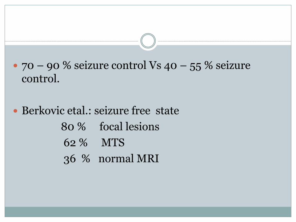
70 – 90 % seizure control Vs 40 – 55 % seizure control.
Berkovic etal.: seizure free state
80 % focal lesions
62 % MTS
36 % normal MRI

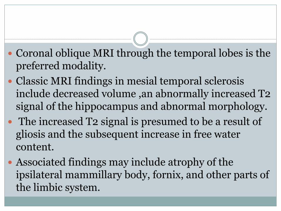
Coronal oblique MRI through the temporal lobes is the preferred modality.
Classic MRI findings in mesial temporal sclerosis include decreased volume ,an abnormally increased T2 signal of the hippocampus and abnormal morphology.
The increased T2 signal is presumed to be a result of gliosis and the subsequent increase in free water content.
Associated findings may include atrophy of the ipsilateral mammillary body, fornix, and other parts of the limbic system.

Because fluid-attenuated inversion recovery (FLAIR) imaging nulls the CSF signal, abnormal signal intensity in the hippocampus is relatively more apparent.
A pitfall of coronal FLAIR imaging is the slight hyperintensity of all limbic structures relative to the neocortex.
Thin-section volumetric T1-weighted imaging is occasionally used to calculate hippocampal volume; however, because it does not depict abnormal signal intensity, it is less useful than FLAIR and T2-weighted spin-echo imaging for visual detection of mesial temporal sclerosis.

T2-weighted spin-echo imaging is somewhat better than FLAIR imaging for demonstrating the internal architecture of the hippocampus; however, the degree of signal abnormality is somewhat more obvious on FLAIR imaging.
Special oblique coronal thin sections perpendicular to the plane of the hippocampus have high sensitivity and specificity for mesial temporal sclerosis.
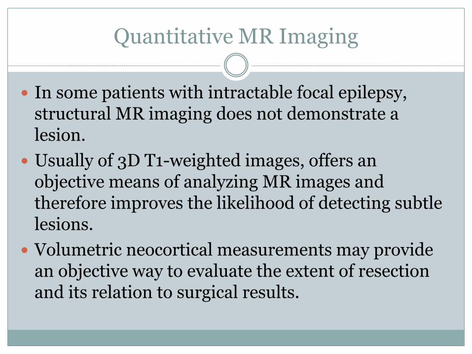
Quantitative MR Imaging
In some patients with intractable focal epilepsy, structural MR imaging does not demonstrate a lesion.
Usually of 3D T1-weighted images, offers an objective means of analyzing MR images and therefore improves the likelihood of detecting subtle lesions.
Volumetric neocortical measurements may provide an objective way to evaluate the extent of resection and its relation to surgical results.
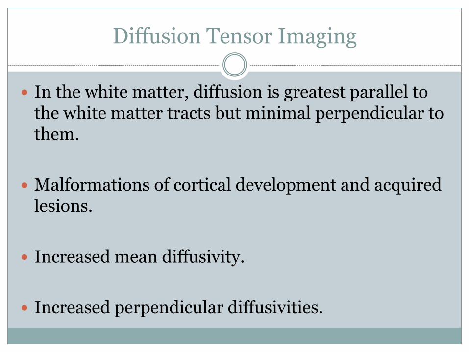
Diffusion Tensor Imaging
In the white matter, diffusion is greatest parallel to the white matter tracts but minimal perpendicular to them.
Malformations of cortical development and acquired lesions.
Increased mean diffusivity.
Increased perpendicular diffusivities.
Magnetic Resonance Spectroscopy
Magnetic resonance spectroscopy is a powerful adjunct to MR imaging.
Reduced NAA and increased Cho in TLE. The MR spectroscopy findings were consistent with the
histopathological characteristics of reduced neuron cell counts or neuronal dysfunction and increased glial cell numbers.
Extratemporal lobe epilepsy, the ability of MR spectroscopy to
lateralize the epilepsy is less(50%). Maton BM, Kuzniecky RI: Proton MRS: N-acetyl aspartate, creatine, and choline. Adv Neurol 83:253–259, 2000
Functional MR Imaging
Used for mapping language, memory, and sensorimotor location for presurgical planning.
This imaging modality is based on the observation that increased neuronal activity is associated with an increase in CBF and therefore an increase in the oxyhemoglobin/ deoxyhemoglobin ratio.
Magnetoencephalography (MEG)
The MEG modality measures extracranial magnetic fields perpendicular to the direction of intracellular currents in apical dendrites.
One of the advantages of MEG as opposed to EEG studies is that magnetic fields are minimally affected by conductivities of intervening structures and tissues between brain and scalp.
Presurgical mapping of eloquent cortex.
Nuclear imaging
PET:
Positron emission tomography (PET) utilizes an injection of radio-labeled glucose (18FDG) to measure brain metabolism. Interictal PET usually shows hypometabolism in the seizure focus, especially in TLE.
Ictal PET is not practical due to the extremely short half life of the radiotracers used.
PET is most useful in MRI-negative TLE, though it may be helpful in extra-temporal epilepsy as well.

FDG PET identifies sites of interictal metabolism in 70 % patients with TLE and 60 % frontal lobe onset seizure.
Less than 5 % false positivity.
(Davinsky O,Pacia S : Epilepsy surgery. Neurology clinics 11:951-971,1993)
C -11 flumazenil PET effective in hippocampal sclerosis & shows pathological foci in a more circumscribed fashion.
(Szelies B ,Pawlik G etal: MRI guided flumazenil and FDG –PET in temporal lobe epilepsy ,Neuroimage 3 : 109-118,1996.)

SPECT
Ictal SPECT ideal method for localization and lateralization of seizure onset,
Shows hypermetabolism,.
89 % sensitivity for localization (Vs PET 63 % )
SPECT also performed better in the setting of negative MRI.
False lateralization rare. (Ho SS, Berkovic SF etal: Comparison of Ictal SPECT and interictal PET in the
presurgical evaluation of temporal lobe epilepsy, Annals of neurology 347:738-745.)
Post Ictal SPECT Comparable to interictal PET (70 %).
Inter ictal scan – Hypoperfusion large area in the hemisphere of onset.
Interictal scan has less sensitivity for seizure, 50 %.
Ictal /Interictal quantitative difference analysis provides for the best and most reliable seizure focus localization
Subtraction ictal- interictal SPECT co-registered to MRI (SISCOM):
A newer methodology that has greater accuracy than either ictal or
interictal SPECT scanning. This requires obtaining scans(separated by at least 48 hrs to
accommodate radionucleotide wash out) during an interictal period and within seconds of seizure onset.
These scans are then subtracted from one another with the use of
specialized computer software. This leaves a better indication of the cortical area of ictal onset. This subtracted scan can then be co-registered onto the patient's
MRI to provide support for the location of the focus.
Invasive EEG monitoring
INDICATIONS: Seizures are lateralized but not localized. Seizures are localized but not lateralized. Seizures are neither localized nor lateralized (eg, stereotyped complex
partial seizures with diffuse ictal changes). Seizure localization is discordant with other data. Relationship of seizure onset to lesion must be determined (eg,
dual pathology or multiple intracranial lesions). .
Temporal lobe seizures -Doubtful side
-Normal MRI
Bilateral pathology
- Discordant non-invasive testing Extratemporal seizures -Definition of extent of epileptogenic area
-Extra-operative cortical mapping
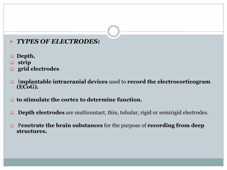
TYPES OF ELECTRODES:
Depth, strip grid electrodes Implantable intracranial devices used to record the electrocorticogram
(ECoG). to stimulate the cortex to determine function. Depth electrodes are multicontact, thin, tubular, rigid or semirigid electrodes. Penetrate the brain substances for the purpose of recording from deep
structures.

Intracranial strip electrodes are a linear array of 2-16 disk electrodes embedded in a strip of silastic (Wyler, 1984), or they can be tubular in structure, (Dubeau, 2000). Grid electrodes are parallel rows of similar numbers of electrodes that can be configured in standard or custom designs.
Grid and strip electrodes are designed to be in direct contact with brain neocortex. Electrodes are placed in the subdural space. Occasionally be used in the epidural space.
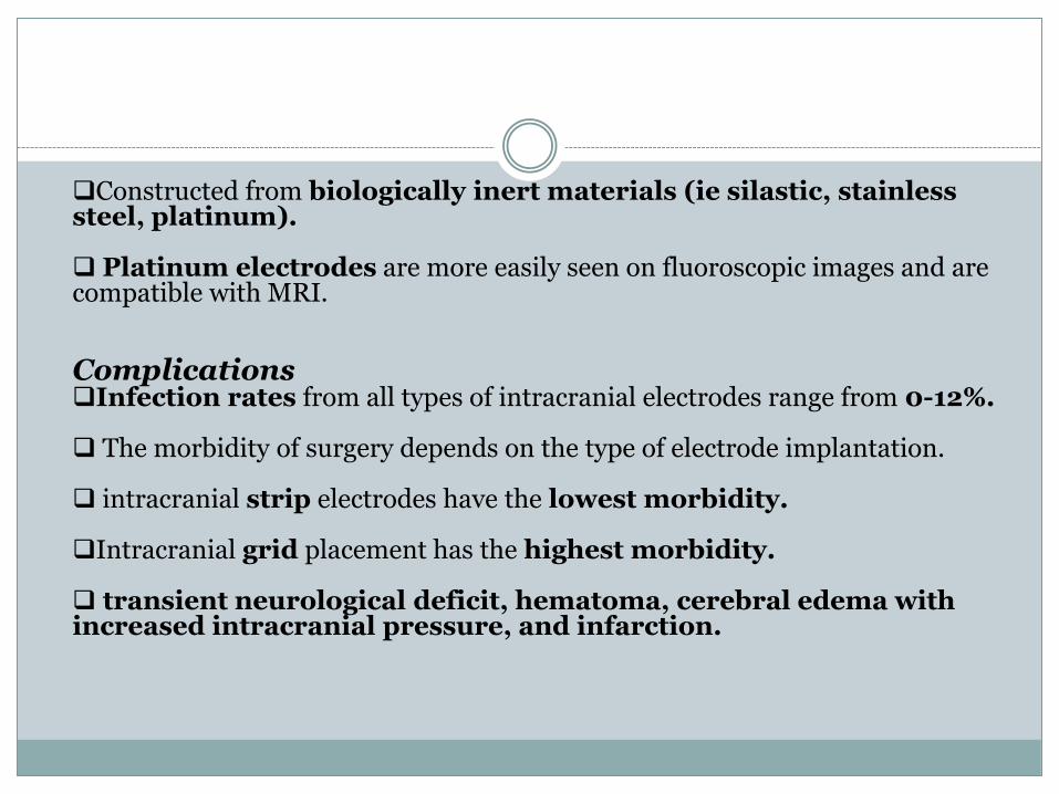
Constructed from biologically inert materials (ie silastic, stainless steel, platinum). Platinum electrodes are more easily seen on fluoroscopic images and are compatible with MRI.
Complications Infection rates from all types of intracranial electrodes range from 0-12%. The morbidity of surgery depends on the type of electrode implantation. intracranial strip electrodes have the lowest morbidity. Intracranial grid placement has the highest morbidity.
transient neurological deficit, hematoma, cerebral edema with increased intracranial pressure, and infarction.

Intracarotid amobarbital (Wada) test Developed by Jun Wada. To preoperatively determine which hemisphere contains language function, this remains its primary use. Also used to test memory function within each hemisphere when considering AMTR. Accomplished by individually cannulating each internal carotid artery. After contrast arteriography verifies that blood flows to the corresponding hemisphere and not to the brainstem or contralateral side,======== a dose of sodium amobarbital (sufficient to impede hemispheric function) is injected.

If speech persists in the face of this hemiparesis, language function is assumed to not be represented within that hemisphere.
The deficiencies of this evaluation for memory
function directly relate to the multiple problems. Injection of a drug into the internal carotid artery does not
assure drug effect in the basal temporal area in general or the hippocampal region specifically.
This is due to variations in the direct blood supply to
the hippocampus.

MR Positive
Concordant EEG PET ,SPECT Not discordant
Surgery Invasive EEG
Discordant EEG

MR Normal
Concordant EEG,PET WADA ,SPECT Not discordant
Surgery Invasive EEG
Scalp EEG Temp
location
Rest unconfirmed
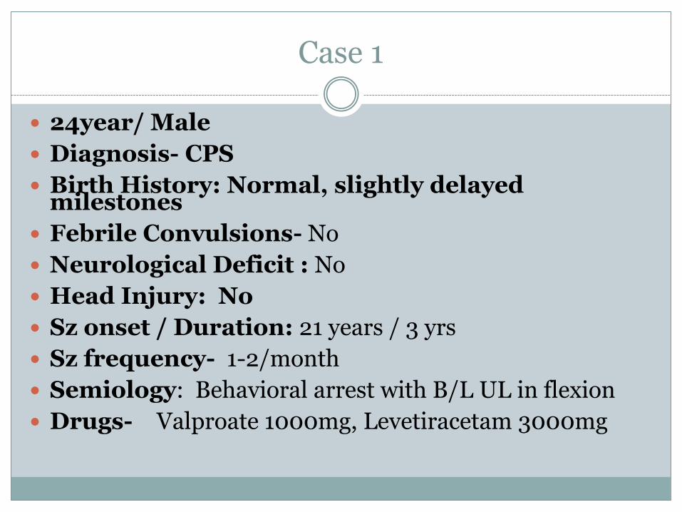
Case 1
24year/ Male
Diagnosis- CPS
Birth History: Normal, slightly delayed milestones
Febrile Convulsions- No
Neurological Deficit : No
Head Injury: No
Sz onset / Duration: 21 years / 3 yrs
Sz frequency- 1-2/month
Semiology: Behavioral arrest with B/L UL in flexion
Drugs- Valproate 1000mg, Levetiracetam 3000mg
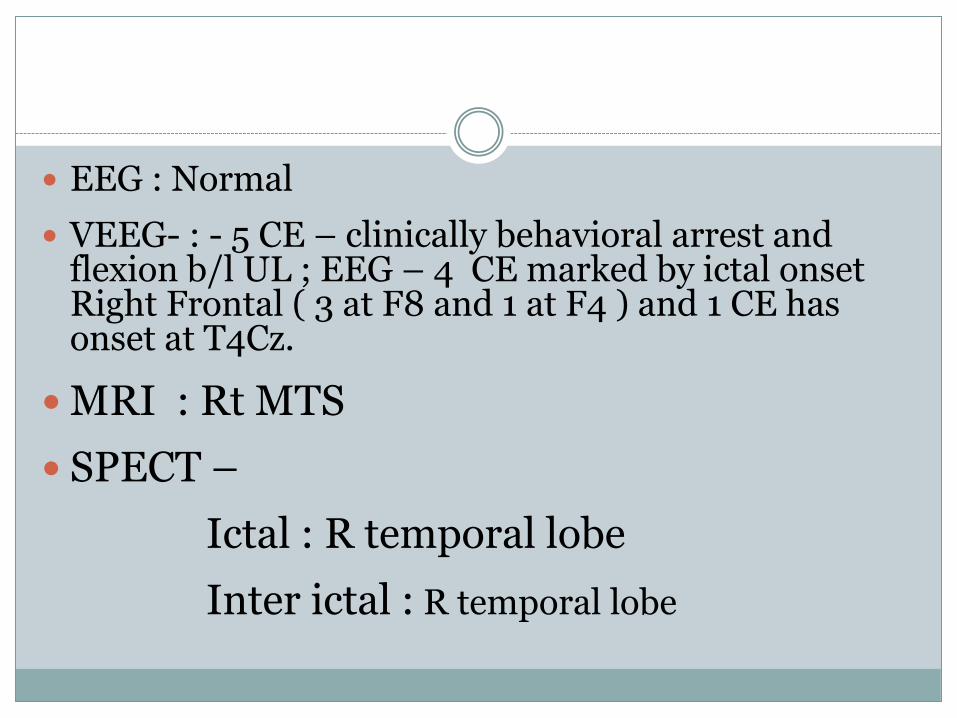
EEG : Normal
VEEG- : - 5 CE – clinically behavioral arrest and flexion b/l UL ; EEG – 4 CE marked by ictal onset Right Frontal ( 3 at F8 and 1 at F4 ) and 1 CE has onset at T4Cz.
MRI : Rt MTS
SPECT –
Ictal : R temporal lobe
Inter ictal : R temporal lobe

Case 2
6 year/male
Diagnosis- CPS
Birth History: Normal
Febrile Convulsions- No
Semiology: Automatic activity, aggressive behavior, vocalizations, bizarre movements of limbs
Sz onset/ Duration /frequency- 4 years/ 2 yrs/ 12-15/day
Drugs- Ox CBZ 600, Levetiracetam 1000, Valproate 800

VEEG-LTV-32/06- 5 CE, all were non localizing CPS. Ictal EEG s/o LRE from right centrotemporal in three and unclear in two. Interictal EEG s/o bilateral fronto temporal discharges R>L
MRI Brain :– Focal Cortical Dysplasia Rt inferior frontal and insular gyri
SPECT: R frontal lobe
SISCOS-likely localization of foci in right inferior frontal cortex. the focus in right ant - temporal region appears to be due to spread from right frontal cortex.
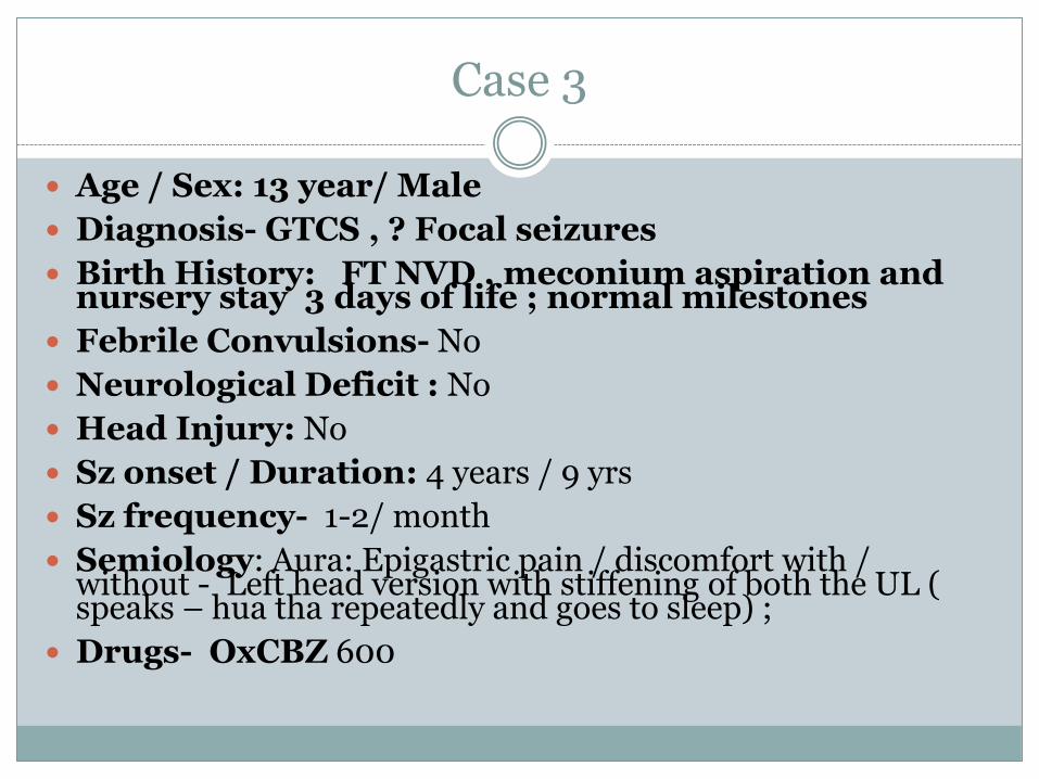
Case 3
Age / Sex: 13 year/ Male
Diagnosis- GTCS , ? Focal seizures
Birth History: FT NVD , meconium aspiration and nursery stay 3 days of life ; normal milestones
Febrile Convulsions- No
Neurological Deficit : No
Head Injury: No
Sz onset / Duration: 4 years / 9 yrs
Sz frequency- 1-2/ month
Semiology: Aura: Epigastric pain / discomfort with / without - Left head version with stiffening of both the UL ( speaks – hua tha repeatedly and goes to sleep) ;
Drugs- OxCBZ 600
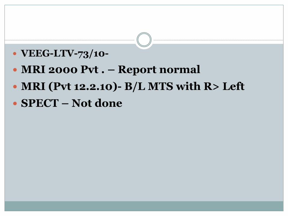
VEEG-LTV-73/10-
MRI 2000 Pvt . – Report normal
MRI (Pvt 12.2.10)- B/L MTS with R> Left
SPECT – Not done

Age / Sex: 17year/ Male
Diagnosis- Left CPS seizures
Birth History : FT LSCS for contracted pelvis ? Cry; normal milestones
Febrile Convulsions- Yes ; 1sz at 1 ½ yrs of age , clinching of teeth with high grade fever
Neurological Deficit : Left Hemiparesis , hypothyroidism +
Head Injury: H/O blunt trauma to head at the age of 3 yrs ; no LOC no CT brain.
Sz onset / Duration: 8 years / 9 yrs
Sz frequency- 10-15/day
Semiology: Abduction of Rt UL and clonic mvt Rt UL and Left LL and UL ; face deviated to left , eye uprolling
Drugs- Valproate 1500mg, Clobazam 20mg.
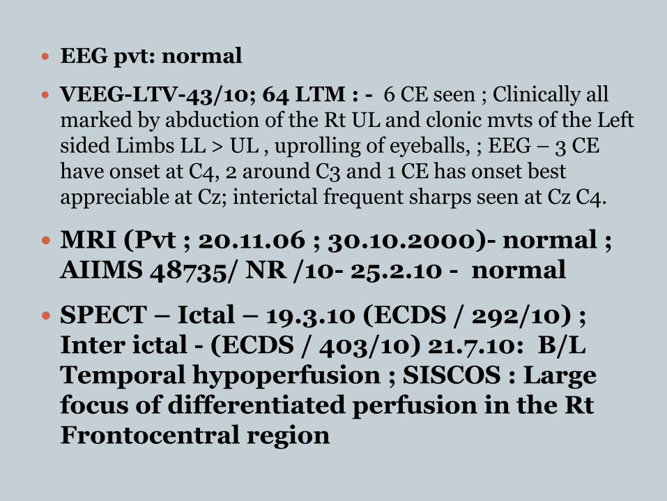
EEG pvt: normal
VEEG-LTV-43/10; 64 LTM : - 6 CE seen ; Clinically all marked by abduction of the Rt UL and clonic mvts of the Left sided Limbs LL > UL , uprolling of eyeballs, ; EEG – 3 CE have onset at C4, 2 around C3 and 1 CE has onset best appreciable at Cz; interictal frequent sharps seen at Cz C4.
MRI (Pvt ; 20.11.06 ; 30.10.2000)- normal ; AIIMS 48735/ NR /10- 25.2.10 - normal
SPECT – Ictal – 19.3.10 (ECDS / 292/10) ; Inter ictal - (ECDS / 403/10) 21.7.10: B/L Temporal hypoperfusion ; SISCOS : Large focus of differentiated perfusion in the Rt Frontocentral region

19 year/ male
Diagnosis- intractable Left CPS
Birth History: FTNVD at hospital, Normal mile stones
Febrile Convulsions- No
O/E: no focal neurological deficit
Sz Onset / Duration: 4 years / 15 yrs
Sz frequency- 3-4 per month
Drugs- PHB 120, Cloba- 10mg, VLP- 1000
Semiology: stiffening of Left UL with deviation of head to Left and then generalization.
EEG: jun 99 (pvt): LRE Right temporal region
VEEG-LTV- 45-10- s/o 4 CE characterized by Ictal onset F8 T4 in 3 and F8 F4 in one.
MRI: pvt-2009- - Large inferior right frontal lesion
? DNET ??Cortical dysplasia
CT head: 2009: right frontal hypodense enhancing mass
SPECT: not done