Pathogenesis of Extremity Deformity in Leprosy - ILSLila.ilsl.br/pdfs/v40n4a04.pdf ·...
14
INTE.RNATIONAI. JOURNAL OF LE.I' ROSY Volume 40, Number 4 Printed in the U.S .A. Pathogenesis of Extremity Deformity in Leprosy A Pathologic Study on Large Sections of Amputated Extremities In Relation to Radiological Appearance 1,2 O. K. Skinsnes, I. Sakurai and T. I. Aquina 3 Crippling deformities are one of the most serious complications of leprosy, and have been extensively studied chiefly by clinical and radiologic technics. There have been a few correlative analyses of radiologic ap- pearances and histopathologic findings relating to bone changes (2, 15. 25), but the emphasis has usually been on either radiological appearance (20. 22. 23, 24, 26) or on histologic analysis (11 . 17, 18). The present study primarily relates to pathologic analysis but pre- and/or post- amputation X rays were available for most of the specimens. Large sections of whole, amputated hands and feet made possible the histopathologic examination of not only the involved bones but also of the neigh- boring soft tissues and, therefore, of the relationship of the disease processes in all the tissues. The purpose of this study was to analyze the various factors in the path- ogenesis of bone changes in leprosy by correlating morphologic appearances in relation to radiologic findings utilizing the classifications of bone changes based on roentgenologic appearance as proposed by Paterson in 1961 (26) and Lechat in 1962 (22) . 1 Received for publication 20 January 1973. 2 This work was initiated in the Department of Pathology, University of Chicago and completed at the ALM Leprosy Atelier, Department of Pa- thology, University of Hawaii, Honolulu, Hawaii 96816. Preliminary results of this study were re- ported at the VIII Congresso Internacional Leprologia, Rio de Janeiro, September 1963. ThiS work was supported by NIH grants R22 AI 10034, and AI 04353_ 3 O. K. Skinsnes, M.D., Ph.D., Professor of Pa- thOlogy, Department of Pathology, University of Hawaii; I. Sakurai, M.D., Sc.D., formerly Assistant Researcher at the Department of Pathology, Uni - verSity of Hawaii, presently at the Department of Pathology, Nihon University School of Medicine, Ohyaguchi-kami-machi 30, Itabashi - ku, Tokyo, Japan; T. I. Aquino, M.D., Ph.D., formerly Re- search Assistant at the Department of Pathology, University of Chicago, presently at the St. John's Mercy Medical Hospital, 615 South New Ballas Road, St. Louis, Mo. 63141. MATERIALS AND METHODS Fifty-three amputation specimens from 39 leprosy patients and three control speci- mens from nonleprous individuals of the same racial and socioeconomic background were studied. All patients were Chinese living either in Hong Kong or in Taiwan. Their ages ranged from 16 to 51 years in the 19 cases for which clinical information was available. Seventeen were males and two were females, and information was not available for twenty cases. Of the patients, 8 were clinically lepromatous, 14 tubercu- loid, and 5 intermediate ( dimorphous), and in 12 cases the clinical classification was not available. The duration of the disease ranged from 8 to 20 years in the 19 cases with reasonably adequate clinical data. There were 8 feet, 22 fore-feet, 18 toes and 5 fingers available. The technic used for the large sections was, with slight variations, that used at the Armed Forces Institute of Pathology, Washington, D.C. ( 1 ). Each specimen was fixed in 10% formalin. After examining X-ray films, they were cut through the lesions, and decal- cified by electrolysis, processed in graded alcohols and then embedded in paraffin in a vacuum oven. Paraffin-embedded tissues were sectioned at 5 to 25 micron thick- nesses depending on their size by means of a J ung L-1 sliding microtome. Each speci- men was stained with hematoxylin and eosin, and/or selected special stains chosen from a repetoire of Masson's trichrome, saffron trichrome, Triff ( 30), Weigert-Van Gieson's stain and phosphotungstic acid he- matoxylin. Pre- and/or post-amputation X rays were also examined in 44 specimens from 30 cases. Acid-fast staining was not generally satisfactory due apparently to electrolytic decalcification and age of some specimens. RESULTS The results are summarized in Table 1. 375
Transcript of Pathogenesis of Extremity Deformity in Leprosy - ILSLila.ilsl.br/pdfs/v40n4a04.pdf ·...

INTE.RNATIONAI. JOURNAL OF LE.I' ROSY Volume 40, Number 4 Printed in the U.S .A.
Pathogenesis of Extremity Deformity in Leprosy
A Pathologic Study on Large Sections of Amputated Extremities In Relation to Radiological Appearance 1,2
O. K. Skinsnes, I. Sakurai and T. I. Aquina3
Crippling deformities are one of the most serious complications of leprosy, and have been extensively studied chiefly by clinical and radiologic technics. There have been a few correlative analyses of radiologic appearances and histopathologic findings relating to bone changes (2, 15. 25), but the emphasis has usually been on either radiological appearance (20. 22. 23, 24, 26)
or on histologic analysis (11 . 17, 18).
The present study primarily relates to pathologic analysis but pre- and/or postamputation X rays were available for most of the specimens. Large sections of whole, amputated hands and feet made possible the histopathologic examination of not only the involved bones but also of the neighboring soft tissues and, therefore, of the relationship of the disease processes in all the tissues. The purpose of this study was to analyze the various factors in the pathogenesis of bone changes in leprosy by correlating morphologic appearances in relation to radiologic findings utilizing the classifications of bone changes based on roentgenologic appearance as proposed by Paterson in 1961 (26) and Lechat in 1962 (22) .
1 Received for publication 20 January 1973. 2 This work was initiated in the Department of
Pathology, University of Chicago and completed at the ALM Leprosy Atelier, Department of Pathology, University of Hawaii , Honolulu, Hawaii 96816. Preliminary results of this study were reported at the VIII Congresso Internacional ~e Leprologia, Rio de Janeiro, September 1963. ThiS work was supported by NIH grants R22 AI 10034, and AI 04353_
3 O. K. Skinsnes, M.D., Ph.D., Professor of PathOlogy, Department of Pathology, University of Hawaii; I. Sakurai, M.D., Sc.D., formerly Assistant Researcher at the Department of Pathology, UniverSity of Hawaii, presently at the Department of Pathology, Nihon University School of Medicine, Ohyaguchi-kami-machi 30, Itabashi -ku, Tokyo, Japan; T. I. Aquino, M.D., Ph.D., formerly Research Assistant at the Department of Pathology, University of Chicago, presently at the St. John's Mercy Medical Hospital, 615 South New Ballas Road, St. Louis, Mo. 63141.
MATERIALS AND METHODS Fifty-three amputation specimens from
39 leprosy patients and three control specimens from nonleprous individuals of the same racial and socioeconomic background were studied. All patients were Chinese living either in Hong Kong or in Taiwan. Their ages ranged from 16 to 51 years in the 19 cases for which clinical information was available. Seventeen were males and two were females, and information was not available for twenty cases. Of the patients, 8 were clinically lepromatous, 14 tuberculoid, and 5 intermediate ( dimorphous), and in 12 cases the clinical classification was not available. The duration of the disease ranged from 8 to 20 years in the 19 cases with reasonably adequate clinical data. There were 8 feet, 22 fore-feet, 18 toes and 5 fingers available. The technic used for the large sections was, with slight variations, that used at the Armed Forces Institute of Pathology, Washington, D.C. ( 1 ). Each specimen was fixed in 10% formalin. After examining X-ray films, they were cut through the lesions, and decalcified by electrolysis, processed in graded alcohols and then embedded in paraffin in a vacuum oven. Paraffin-embedded tissues were sectioned at 5 to 25 micron thicknesses depending on their size by means of a J ung L-1 sliding microtome. Each specimen was stained with hematoxylin and eosin, and/or selected special stains chosen from a repetoire of Masson's trichrome, saffron trichrome, Triff (30), Weigert-Van Gieson's stain and phosphotungstic acid hematoxylin. Pre- and/or post-amputation X rays were also examined in 44 specimens from 30 cases. Acid-fast staining was not generally satisfactory due apparently to electrolytic decalcification and age of some specimens.
RESULTS The results are summarized in Table 1.
375

376 International Journal of Leprosy 1972
TABLE 1. Frequency analysis of bone changes.
.~ '" ~ ~ .;;; ~ ...... .9
..... 0 ~ ~
0 ..... u ·rJ >- ~
C1S .0 .~ .~ bO >,
.~~ :§. 5 .S .....
~ Q) 'Ql 5 'Ql '" C1S ...... ..... .9 ..... 0
"@", .S '" 0 .~ t'd ...... >, ~ Q)
'" ~ .9 Q) ........ 0 ..c:: ~ ;; .9 ..c:: 'M ~ ..... .~ '"00 C1S .... '" Q) 0. C1S '"0 .... Q) ;.a o.·E ~
Q) 0 '" ~ '" .... 0 .... 0 Q)
'" ~ Si..c:: 0.", 0 g § ~ .... bO ~ 0. '" 0 Q) 0 Q .... ..... ..... '"
Q) ...... .... t:: :0 Q:O '2 0 "£5
Q C1S >'Q) '" > ~ .5 0 !SC1S C1S '2 '6) 0. C1S ~ ~ ~ ~
~ .... Q)
al o .... .... 3 0 0 Q) '"0 Q) Q !Sal Q) 0 ..... bO Q) 0 ..... .... 0 .... .... > 0 ~
Q) 0. Q .... '" 0 ..... ........... ~ '" 0..0 0:9 0. .... 0 0. >, Q) .....
S ..c:: O >, '" '" Q) 0 is j .s
Q) Q)
U rJ) E-< ~~ u~ p.. 0 0'"0 U ~ ....l Z ~
4 RF + + + 2 3 5 RF + + + + + + + + 3 5 6 L'F + + + 3 3 7 3Fi L / + + + + 5 I 8 RF L + + + + + + 2 3 9 RF L + + 4
10 LF L + + + + + 2 II LF L + + + + + + + 5 2 13 LF T + + + + 3 3 15 RF + + + 3 16 RF L + + + + + I 5 17 LF T + + + 2 2 18 RF T + + + + + 3 5
LF 19 5RT T + + + + I 3 20 2Fi D / +. + + 5 2 21 RF T + + + + + 5 22 RF T + + + + + 1 4 23 LF T + + + + 4 5 24 RF T + + + 3 26 lLT T + + + + 4 27 lRT T + + 1 28 4LT T + + + 3 1 29 2T D + + 1 30 RF T + + + + 2 1
LF 31 2T D + 3 2 32 lRT L + + 1 33 RF L + + + + 3 34 RF D + + + + 4 35 RF T + + + 3 3 36 lRT D 4- + I 38 lLT T + + + 2 49 LF + + + + + 1 50 LF + + + + + 4 51 RF + + 1 52 LF + + + + + + I 3 54 F + + + 4 56 F + + + 3 2 57 F + + + + + + I 2 58 F + + + + 2 Frequency· 20/3716/3729/3713/37 12/37 13/37 15/37 10/37 5/37 6/37 4/37
(54%) (43%) (78%) (35%) (32%) (35%) (41%) (27%) 03%) (16$) (II~
T = Tuberculoid; L = Lepromatous; D - RF = Right foot ; LF = Left foot. Dimorphous. RT = Right toe (s) ; L T = Left toe (s) .
• Cases 7 and 20 are excluded in frequency Fi = Finger (s) . analysis.

40, 4 Pathogenesis of Extremity Deformity 377
The degree of morphologic change of blood vessels, mainly arteries, and of nerves was graded on a subjective scale of 1 (slight) to 5 ( severe). The bone changes listed in Table 1 are mainly based on the classification proposed by Lechat (22) with some modifications. The terms "metatarsophalangeal osteoarthritis" and "distal atrophy" are used in the same connotation employed by Lechat. Included under "osteomyelitis" are only the lesions that disclosed typical evidence of present or previous suppurative inflammation in the bone marrow spaces. "Nonspecific chronic periosteitis and osteitis" are terms used to designate the changes characterized by noninfectious, inflammatory cell infiltration mainly of lymphocytes and/ or fibrosis in the periosteum close to the osseous cortex, or in the nutrient foramens of the cortex, or in the spongy bones, which are frequently associated with increased osteoblastic and/or osteoclastic activity ("bone turnover"). Changes similar to those categorized in "chronic peri osteitis and osteitis" were often seen in the distal heads of the metatarsal bones, but these were not grouped in "chronic periosteitis
and osteitis," but were classified as "metatarsophalangeal osteoarthritis," because they are regarded as partial findings of the latter category. Fibrous nodule in the bones was usually presented as pseudocyst formation on X rays.
In the percentage analysis of the relative frequency of each change, cases 7 and 20 were excluded, as they were the specimens from the upper extremities. They were, however, noted as being good examples of leprous specific osteitis resulting in bone absorption.
According to the classification proposed by Lechat (22), bone changes in leprosy can be divided into two major groups: (1) specific leprous osteitis and (2) nonspecific bone changes. From a pathologic point of view, leprous granulomatous periosteitis, osteitis and osteomyelitis belong in the first category. Lechat classified cyst-formation and honeycomb appearance on X rays as the group of specific changes. In our study, fibrous nodule in the bones also shows cyst-formation on X rays. They may result either from chronic nonspecific osteitis or
FIG. 1. Chronic nonspecific periosteitis and osteitis (case 49, H & E stain, original mag. X 35). Fibrotic tissue invades bone marrow space through cortical defect. Cortex is thinned and atrophied by osteoclastic activity.

378 International Journal of Leprosy 1972
FIG. 2. Metatarsophalangeal osteoarthritis (case 23, Weigert-Van Gieson's stain). An X-ray film shows advanced bone destruction in metatarsal heads, and corresponding specimens from 1st right (A) and 2nd (B) toes reveal concentric atrophy of metatarsal heads with thickening and tapering of cortex and narrowing of bone marrow spaces.
osteoarthritis, or from inactivated healed lepromas.
In frequency analysis of each type of bone change, chronic nonspecific periosteitis and osteitis were most frequently found (78%), Most cases were associated with ostearthritic lesions in metatarsal heads. In some instances chronic periosteitis and osteitis were seen in the shaft areas of the bones far from the joints. In such cases, the lesions were frequently found in the plantar surface
of the affected bones, and thought to be possibly caused by pressure effect without causing an ulceration (Fig. 1).
Metatarsophalangeal osteoarthritis was found frequently (54%). It is noteworthy that the bone changes appeared earlier in the metatarsal heads in our sequences of preamputation X rays and were more severe, as judged by histopathologic sections, than the changes seen in the proximal ends of the phalanges. In cases 22, 23, 30, 33, and 34 the metatarsal heads were severely damaged, while the phalangeal proximal basis showed only minor changes (Fig. 2). In the damaged distal ends of the metatarsal bones, concentric atrophy which is seen in many of the cases with this type of change, consists of thinning and tapering of the distal metatarsal shaft with markedly thickened and sclerosed cortex and narrowed medullary spaces. In some instances, chronic inflammation was seen in the joint membrane.
Ulceration frequently followed by secondary infection and pyogenic osteomyelitis is thought to be one of the important factors in bone destruction in leprosy. Sixteen of 37 cases (43%) had ulceration with associated infection of soft tissues, usually in the plantar surfaces of feet, presumably caused by a long-standing pressure effect and circulatory disturbances in distal parts of the extremities due to neural involvement by leprosy as well as to acute injury. Many of the cases with ulceration were associated with pyogenic osteomyelitis (Fig. 3) with bone destruction and reactive new bone formation around the lesions. Virtually total bone resorption of the foot (Fig. 4) was seen without the influence of pyogenic osteomyelitis.
In most cases there was some degree of decreased radiologic density of the affected bones; on histology this corresponded to a decreased number and thinning of the trabeculi in the medulla, and thinning of the cortex. Not only on X rays but also on corresponding histology sections,there was some degree of evidence of osteoporosis in 12 cases. Case 30 showed especially severe porotic changes in histopathologic section.
Ankylosis or osteochondritis in the jOints other than the metatarsophalangeals was
•

40,4 Pathogenesis of Extremity Deformity 319
FIG. 3. Pyogenic osteomyelitis following ulceration and secondary infection in calcaneus (case 4, Masson trichrome stain, section through the 5th toe, right foot).
found in 13 cases (35%). Cases with ankylosis in the interphalangeal joints presented a perfect continuity between two phalanges on histology sections, probably representing post-osteochondritis of the joints.
Contractures of joints were seen in 15 cases (41%). These were frequently associ-
ated with dislocation of the affected jOints. Contracture usually resulted from sCqrring in soft tissue around the affected joints, and in about half of the cases was associated with ulceration and was also frequently associated with chronic periosteitis.
Distal atrophy of phalanges was seen in
380 InternatioTUll Journal of Leprosy 1972
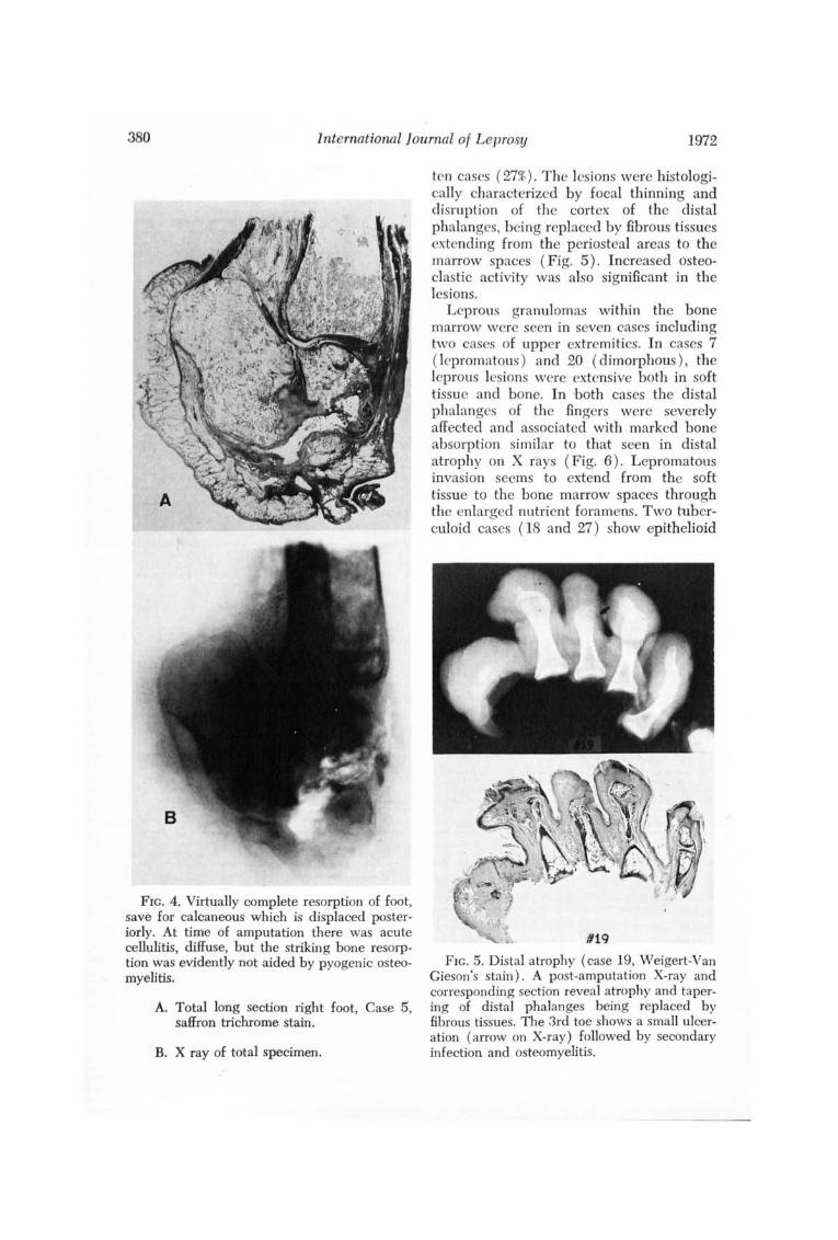
FIG. 4. Virtually complete resorption of foot, save for calcaneous which is displaced posteriorly. At time of amputation there was acute cellulitis, diffuse, but the striking bone resorption was evidently not aided by pyogenic osteomyelitis.
A. Total long section right foot, Case 5, saffron trichrome stain.
B. X ray of total specimen.
tcn cases (27%). The lcsions werc histologically characterized by focal thinning and disruption of the cortex of the distal phalanges, being replaced by fibrous tissues extending from the periosteal areas to the marrow spaces (Fig. 5). Increased osteoclastic activity was also significant in the lesions.
Leprous granulomas within the bone marrow wcre seen in seven cases including two cases of upper extremities. In cases 7 (lepromatous) and 20 (dimorphous), the leprous lesions were extensive both in soft tissue and bone. In both cases the distal phalanges of the fingers were severely affected and associated with marked bone absorption similar to that seen in distal atrophy on X rays (Fig. 6). Lepromatous invasion seems to extend from the soft tissue to the bone marrow spaces through the enlarged nutrient foramens. Two tuberculoid cases (18 and 27) show epithelioid
FIG. 5. Distal atrophy (case 19, Weigert-Van Gieson's stain). A post-amputation X-ray and corresponding section reveal atrophy and tapering of distal phalanges being replaced by fibrous tissues. The 3rd toe shows a small ulceration (arrow on X-ray) followed by secondary infection and osteomyelitis.

40,4 Pathogenesis of Extremity Deformity 381
cell granulomas with Langhans type giant cells in the bones. Leprous peri osteitis was seen in six cases including two cases of upper extremities and was usuaBy associated with lepromas in the bone marrow spaces. Some cases show cortical bone absorption due to leprous periosteal invasion.
Fibrous nodules presenting pseudoeystformation on X rays was found in six cases. The cases were usuaBy associated with osteoarthritis of metatarsophalangeal joints as well as other joint surfaces. Fibrous tissue pcnetrated through cortical defects or dilated nutrient canals near the margin of articular cartilage on the plantar surface of the bones in cases with metatarsophalangcal osteoarthritis (Fig. 7).
The nerve changes ranged from peripheral fibrosis to fibrous replacement of entire bundles. Perineural, chronic nonspecific inflammatory infiltrates were found in most of the specimens. Cases 22 and 23 showed
FIG. 6. Leprous granuloma in bone and leprous peri osteitis (case 7, 4th finger, H & E stain). Distal phalanges are involved by leprous infiltration both in peri ostium and bone marrow space. An X-ray shows atrophy of distal phalanges which simulates that seen in distal atrophy.
# 21
FIG. 7. Fibrous nodules in bone (case 21, 1st toe, Weigert-Van Gieson's stain). Fibrous nodules destruct and replace bones in metatarsal head and proximal phalanx. An X-ray reveals pseudocysts in corresponding locations.
tuberculoid granulomas repiacing medium sized trunks.
Almost every case showed some degree of alterations in the medium sized and small arteries and arterioles. The alterations consisted of medial hypertrophy, and in advanced cases, accompanying intimal thickening. Vascular sclerosis with lumenal narrowing was extreme in many instances ( cases 5, 16, 18, 21, 23) (Fig. 8), which are all associated with metatarsophalangeal osteoarthritis, and chronic peri osteitis and osteitis. Table 2 presents correlation between the degree of vascular changes and each type of osseous alteration. The condition of the arteries was not directly related to the age of the patients, being of severe degree in a 16 year old adolescent (case 26) and of mild degree in a 45 year old man (case 30). Severe arterial changes, graded 4 to 5, were found in 11 cases among 37 individuals (30%). Cases 10 and 21 had acute or subacute nonspecific vascu-

382 International Journal of Leprosy 1972
FIG. 8. Fibrotic change in peripheral nerve (A) (case 30, H & E stain, original mag. X 100), and arterial alterations (B) (case 23, Weigert-Van Gieson's stain, original mag. X 35) with concentric hypertrophy of media and hyalinous fibrosis of intima.
litis with infiltration of polymorphonuclear leucocytes and/or plasma cells in the walls of arteries, and case 11 showed specific leprous granulomatous arteritis.
DISCUSSION
On the basis of the radiologic studies of Paterson ( 26) and Lechat ( 22), bone changes in leprosy can be classified into two major groups: (1) specific leprous osteitis and (2) nonspecific osteitis including pyogenic infection.
It has long been evident that direct leprous involvement of bones is one of the factors in bone absorption. In 1934, Cass and Rishi (11) found acid-fast bacilli in the bone marrow of 17 of 21 cases having mixed cutaneous and neural leprosy, and Hayashi (15) indicated that leprous inflammation extended into bone through the Haversian canals and that both cortical absorption and periosteal thickening could be demonstrated by X-ray studies. Kozuma ( 21) and Job (17) also pointed out that direct leprous invasion could result in bone atrophy and destruction. On X rays leprous involvcment of the Haversian canals may present as enlarged nutrient foramens, as pseudocysts and even as a honeycomb appearance (25. 26). Pseudocyst may, however, also result from fibrous nodules in the bones as shown in cases 5, 21, 34, 56, 57 and 58 in our series. Erickson and Johansen (7) reported that pseudocysts sometimes healed under sulfone therapy, and Job (17) noted that leprous lesions in bones may heal by fibrosis. However, it seems possible that in many cases these fibrous bone lesions may result from pressure effects, since they tend to be located close
TABLE 2. Relationship between vascular alteration and each bone change.
Severity of vascular change Bone changes 1 2 3 4 5
Metatarsophalangeal osteoarthritis 2/8 4/8 5/10 4/6 5/5n
Ulceration and infection, soft tissue 3/ 8 4/8 6/10 3/6 0/5 Chronic periosteitis and osteitis 5/8 6/8 8/10 4/6 5/5 Pyogenic osteomyelitis 2/8 3/8 5/10 3/6 1/5 Osteoporosis 2/8 2/8 3/10 1/6 3/5 Osteochrondritis deformans or ankylosis 3/ 8 4/8 3/10 1/6 3/5 Con tracture of joint 1/8 2/8 6/10 3/6 3/5 Distal atrophy 2/ 8 2/8 2/10 2/6 2/5 Leprosy granuloma in bone 2/8 1/8 0/10 0/6 2/5 Fibrous nodule in bone 0/8 3/8 0/10 1/6 2/5 Leprous periosteitis 0/8 1/8 11 10 1/6 1/5
a For example: 5/5 indicates that all five cases with degree five of vascular change have metatar-sophalangeal osteoarthritis.

40, 4 Pathogenesis of Extremity Deformity 383
to articular cartilages and related to osteoaJthritis. Cortical crosion or destruction by specific leprous periosteitis as sccn in cases 7, 11, 13, 16, 20 and 22 occurs both in the lepromatous and tuberculoid forms.
One of the readily understandable and important factors in bone absorption is osteitis and osteomyelitis following ulceration and secondary infection as shown in Figure 3 (casc 4). Ulceration of thc skin and soft tissue is prcsumably relatcd to a longstanding pressure or repeated trauma and circulatory disturbances in distal parts of the extremities of anesthetic patients. Even if there is no ulceration of the skin, the metatarsophalangeal joints and the calcaneus are the most effected pressure points in such patients. Prolonged pressure or repeated trauma may lead to ischemia accompanied or followed by chronic aseptic inRammation and resultant fibrosis in the metatarsal heads and plantar surfaces of the bones and peri ostium. Chronic vasculitis and chronic repeated pressure or traumatic effect on soft tissues of the extremities are also thought to lead to such arterial alterations as was noted in many instances in the present study. Such arterial
sclerosis in the involved extremiti es was considercd important also by Murakami (25). Harada and Takashima (J.I) studied leprous vasculitis which was similar histopathologically to that in periarteritis nodosa but it seemed to be far milder in degree and chronic in course, and charactcrizcd QY hyaline degeneration in thc vascular walls instead of fibrinoid necrosis, and by absence of association with complete occlusion, thrombosis or aneurysm which were frequently associated with p eriateritis nodosa. It is evident that vasculitis tends to occur during therapy especially as a manifestation of erythema nodosU1n leprosum.
In addition to vasculitis, reactional phases also harbor other factors which may be important in the development of extremity deformity. Edema in soft tissues, panniculitis or necrotizing erythema nodosum leprosum may result in sclerodern1ie lesions (27), and contracture of various joints (10), and the importance of prevention of deformity in such reactive phases by adequate physiotherapeutic management has been stressed by Furness et al (10). Another well-known phenomenon in reactive
Disruption of Vascular Reflex Arc
Syrin90myrlio; Combin, d cord
d'9 rnrr ot io n ( SpInO I cord )
, /~rmmol funL'flonol YOH:ulor tJ~d
Sudrc/('s otrophy ( p /uus )
L'prosy; Diala tts
FIG. 9. Comparative locales of vascular reflex arc disruption in various bone resorption entities. Based on Johnson's suggestions (19).

384 International Journal of Leprosy 1972
phases which play a role in the development of extremity deformity ;is arthritis. Ramu and Balakrishnan (27) studied 18 cases with recurrent attacks of arthritis in lepromatous leprosy and pointed out that in its early acute phase lepra reaction resembles acute rheumatic fever, and in its
PERIPHERAL NERVE AFFECT
(autonomic, sensory & motor)
Interruption of vascular reflex arc:
Hyperemic bone resorption
"Fluctuant stress" vascular sclerosis
Pressure atrophy & trauma 2° to sensory loss & deformity 2° to loss of muscle support
BONE CHANGES
recurrent states ;it simulates rheumatoid arthritis with its resulting deformities in clinical and biochemical characteristics. Innami ( 16) proposed the concept that the pathogenesis of spina ventosa leprosa has a close relation to autoimmune mechanisms, as suggested hy studies of immune cross-
SPECIFIC LEPROUS INFLAMMATION
(Hematogenous & direct extension, predomina ntly lepromatous end of spectrum. Effect & extent of tuberculoid lesions unknown)
Inflammatory morphology reflective of leprosy type; granuloma, leproma, fibrous nodule. No sequestration.
Enlarged nutrient canals
Pseudocysts
"Honeycomb" appearance
Inflammatory vasculitis & eventual sclerosis.
Osteoporosis & bone resorption, - concentric atrophy -
Specific leprous osteitis . periosteitis & osteomyelitis
Suppurative periosteitis, osteitis & osteomyelitis with sequestration
Metatarso-phalangeal osteoarthritis
Osteochondritis deformans
Joint contracture
! IMMUNOLOGIC REACTIONS
(Predominantly lepromatous & lower borderline ~ff~tti spectrum. TUberculoid bone reaction & effects not documented.)
Rheumatoid arthritis-like changes with joint contracture
ENL vasculitis with eventual vascular sclerosis & i~~~ relative ischemia.
\ SECONDARY INFLUENCES
(Externally originating)
Pyogenic periosteitis, osteitis & osteomyelitis 2° to ulceration & trauma sequential to anesthesia.
Trauma 2° to sensory loss
FIG~ 10. Factors initiating and sustaining progressive bone deformity.

40,4 Pathogenesis of Extremity Deformity 385
reactions between M. leprae, BCG, and the human phalanx.
M~ller-Christensen et al ( 24 ) studied changes in the anterior nasal spine and alveolar process of the maxilla in seven cases and reported no sensitivity changes found in any patient which might indicate that atrophy was due to neurotrophic disturbance of bone, and they held that neurological examination did not give support to the neurotrophic theory of pathogenesis of changes in those bones. Michman and Sagher (23) also noted that no disturbance of sensitivity was found in any of 44 cases studied with respect to changes in the nasal spine and maxillary bone in leprosy. Waller (W), in a study of seven cranial specimens, attributed the changes in maxillary bones and the loss of the anterior spine to changes in the overlying soft tissues.
Circulatory abnormalities, however, have been entertained for a long time as one of the possible factors in the pathogenesis of bone absorption in leprosy.
Fite (9) noted that involvement of blood vessels in leprosy has been known at least since the work of J oelsohn in 1893. Fite (9) himself contributed a detailed study, being, however, largely concerned with leprous infection of the blood vessels rather than the chronic effect of denervation on the vessels. In amputation specimens, such as utilized in the present study, the findings of necessity deal with longstanding changes in which it is virtually impossible to determine the initial cause of the vascular thickening. By analogy, organs such as the spleen and uterus, which undergo repeated physiologic stress and change in size, often show marked hyalin thickening of arterioles. It's possible that extremity arterioles respond similarly when stressed by chronic circulatory change due to loss of neurovascular reflex control. The vascular changes are likely, then, to be of multiple pathogenic origins. Functional disturbances, secondary to vascular innervations, have been considered important, as stressed by Lechat (22) . Barnetson demonstrated that the vasodilatation reflex, as demonstrated by immersion of the other limb in warm water, was slow or absent in
leprosy patients (3) and stressed that disturbance of reflex vasomotor response following leprous interstitial neuritis was an important factor in pathogenesis of neurotrophic atrophy (2. 4). Chatterjees also pOinted out that variations of extremity temperature were more prominent in leprosy pat~ents than in normal individuals (6). Faget and Mayoral (8) performed a radiologic arteriographic study of the extremities of leprosy patients, which revealed narrowing of the arteries. Paterson ( 26) demonstrated by angiography of the digital arteries, vascular end-loops and nutrient vessels to the phalanges of 12 leprosy patients that there was diminution in the vascular end-loops even in cases without bone absorption, and that narrowing of the caliber of digital arteries was present in the cases with osteoporosis and deformed by bone absorption. In seven cases there was a considerable increase in the circulation time of the fingers. Basu et al (1\) also reported vascular changes manifested as circulatory stasis in the digits as demonstrated by an angiographic analysis of 20 cases of nonlepromatous leprosy. Statistically there is a close relationship between anesthesia and distal absorption as noted by Lechat (22). Changes in Charcot's joint from the study of a single leprous specimen were described by Johnson (19), who noted that in the acute phase the bone rcvealed three zones in the metaphysis; normal bone, porotic bone with osteoclastic resorption and teleangiectasis, and new bone formation with dilated sinusoids. Such bone changes may be initiated by circulatory changes resulting from interruption of the neurovascular reflex arc at any level by a variety of affiictions including leprosy as outlined in Figure 9. The same mechanism operative at the same reflex level in neurovascular reflex is thought to be a factor in bone absorption in diabetic neuropathy ( 20). Johnson (H)) noted that hyperemia occurring as telangiectatic dilatation of sinusoids in the midst of normal fatty marrow is associated with osteoclastic bone resorption and such hyperemia is active. In contrast, hyperemia with edema and serous atrophy of fat is associated with osteoblastic bone formation and is passive.

386 International Journal of Leprosy 1972
In active hyperemia blood flows rapidly, lymphatic flow is reduced, and there is minimum loss of oxygen, sugar, proteins and all nutriments to the tissues in passage between supplying arterioles and draining venules. Active hyperemia, with its high oxygen tension and metabolic activity supports and induces osteoclastic activity. In passive hyperemia blood flow is slow, lymphatic drainage is increased and there is maximum escape of oxygen, sugar, protein and nutriments to the tissues during this same traverse. Thus, passive hyperemia with its high tissue-fluid protein supports and induces osteoblastic activity. The distinction between the two types of hyperemia and their effects on bone depends upon differences in flow rate. Thus, Coutelier (6a) in a recent study utilizing microradiology and fluorescence microscopy of bone sections found that either bone destruction or bone formation may occur as isolated phenomena in leprosy. In most instances, however, the two phenomena were intermingled. The eventual result is thus a balance of these factors. These concepts are supported by the studies of Gorham and West (13) who determined experimentally in mice that a very vascular tumor was associated with bone resorption, whereas a spindle-cell sarcoma blocking the arterial ·circulation was accompanied by marked osteogenesis. Likewise, tissue culture studies by Goldhaber (12) indicate that a high concentration of oxygen (60% to 95%) stimulates osteoclastic resorption of bone and potentiates the in vitro action of parathyroid hormone and vitamin D on bone. Supplementing these findings Shaw and Bassett (28) determined that a somewhat lower oxygen concentration ( 35%) favors bone formation in culture. They also noted that oxygen deprivation blocks osteogenesis, diminishes collagen formation and favors chondrogenesis.
The pathogenesis of . nonspecific bone resorption with consequent deformity in leprosy is thus a complex process (Figure lO) for which a pathophysiologic mechanism can now be postulated. The vascular changes and circulatory alterations are not, however, static but the result of alterations in response to varying factors such as the position of the
extremities and resultant effects of gravity, ambient temperature, inflammation and many other factors . Added to these are yet other mechanisms such as disuse and pressure atrophy. The sclerosis described must be slowly developing and have its modifying effect on the circulation. In balance, during the time course of the patient's affliction, the effect is that of an overbalance of osteolysis and resorption as compared with osteoblastic bone formation. The recognition that vascular denervation through interruption of the vascular reflex arc, and slowly progressive, associated, vascular sclerosis are permanent disabilities, suggests that bone resorption may be ongoing in the presence of disease arrest and may continue after cure of the disease. This recognition reiterates the desirability of achieving early therapeutic success in arresting and curing leprosy while neural involvement is yet limited.
/ SUMMARY
/ A pathologic study of 53 separate amputation specimens from 39 leprosy patients together with three normal controls was made by means of large sections paraffin histopathologic preparations correlated with pre- and post-amputation X-ray visualizations of changes in the same speci~ mens. Utilizing the classifications proposed by Paterson and Lechat, the percentage analysis of each type of bone change found were: metatarsophalangeal osteoarthritis 54%, ulceration and infection 43%, chronic nonspecific periosteitis and osteitis 78%, pyogenic osteomyelitis 35%, osteoporosis 32%, osteochrondritis deformans or ankylosis 35%, contracture of joint 41%, distal atrophy 27%, leprous leproma or granuloma in bone 13%, fibrous nodule in bone 16%, and leprous peri osteitis 11%.
Attention is called to the vascular alterations, which were found to some degree in every case. Eleven cases had severe (graded 4 to 5) vascular changes. Cases with severe vascular changes tended to have a _ higher degree of metatarsophalangeal osteoarthritis, and chronic nonspecific periosteitis and osteitis. Various probable pathogenic mechanisms in extremity deformity and bone adsorption in leprosy are dis-

40,4 Pathogenesis of Extremity Deformity 387
cussed, emphasizing the disturbance of neurovascular reflex and resulting vascular sclerosis.
/ RESUMEN / Se hizo un estudio anatomo-patologico de 53
muestras individuales de amputacion obtenidas de 39 pacientes con lepra y de tres controles normales, por medio de preparaciones histologicas de cortes grandes en parafina, relacionandolas con los cambios observados en estas mismas muestras por medio de visualizacion por rayos X, efectuadas pre y post-amputacion. Vltizando las clasificaciones propuestas por Paterson y Lechat, el analisis porcentual de los tipos de aIteraciones oseas encontradas fue: osteoartritis metatarsofalangica 54 %, u1ceracion e infeccion 43 %, periosteitis y osteitis crOnIca no especffica 78 %, osteomielitis piogenica 35%, contractura articular 41 %, atrofia distal 27 %, leproma 0 granuloma leproso en el hueso 13 %, nodulo fibroso en el hueso 16% y periosteitis leprosa 11 %.
Se desea lIamar la atencion hacia las alteraciones vasculares, que se encontraron en cierto grado en todos los casos. Once casos mostraron alteraciones vasculares severas (graduacion 4 y 5). Los casos con aIteraciones vasculares severas tendian a presentar mayor grado de osteoartritis metatarsofalangica y periosteitis y osteitis cronica no especifica. Se sugieren varios probables mecanismos patogenicos en deformidades extremas y adsorciones de huesos en lepra, con especial enfasis en las alteraciones de los reflejos neurovasculares y la esclerosis vascular resultante.
MSUM:£ Vne etude pathologique de 53 echantillons
proven ant d'amputation chez 39 malades de la lepre, et chez trois temoins normaux, a ete menee au moyen de preparations histopathologiques enrobees dans des coupes de paraffine de grandes dimensions. Les resultats ont ete mis en correlation avec les aspects radiographiques des modifications des memes echantiJIons, constates avant et apres amputation. En utilisant les classifications proposees par Paterson et par Lechat, la repartition des differents types de modifications osseuses qui ont ete trouvees est la suivante: osteoarthrite metatarso-phalangienne 54 pour cent, uli::eration et infection 43 pour cent, periostite non specifique chronique et osteite 78 pour cent, osteomyelite pyogene 35 pour cent, osteoporose 32 pour cent, osteochondrite detormante ou ankylose 35 pour cent, contracture de I'articulation 41 pour cent, atrophie distale 27 pour cent, leprome ou granulome lepreux dans I'os
13 pour cent, nodule fibreux dans 1'0s 16 pour cent, et periostite lepreuse 11 pour cent.
L'attention a ete attiree sur les alterations vasculaires, relevees it des degres differents dans chaque cas. Onze cas presentaient des modifications vasculaires graves, de degre 4 ou 5. Les cas presentant des modifications vasculaires graves tendaient egalement it presenter un degre plus eleve d'osteoarthrite metatarsophalaflgienne, de periostite non specifique chronique et d'osteite. Les divers mecanismes pathogeniques probables des difformites des extremites et de la resorption osseuse dans la lepre sont discutes. On souligne les troubles des reflexes neurovasculaires, et la sclerose vasculaire qui en resulte.
Acknowledgements. Dr. D. J. Harman, formerly of Hay Ling Chau Leprosarium, Hong Kong, and presently of the Leprosy Study Centre, London, provided many of the specimens for this study. Additional specimens were subsequently provided by Dr. A. Crace Warren, Medical .Superintendent, Hay Ling Chau Leprosarium.
REFERENCES 1. AMBROGI, L. P. and BALLOU, E. F. (eds.).
Manual of HistologiC and Special Staining Technics, 2nd ed., Armed Forces Institute of Pathology, New York, McGrawHill, 1960.
2. BARNETsoN, J. Osseous changes in neural leprosy. Correlation between histopathological and radiological findings. Acta Radiol. 34 (1950) 35-64.
3. BARNETSON, J. Skin temperature studies in neural leprosy. Trans. Roy. Soc. Trop. Med. Hyg. 43 (1950) 539-544.
4. BARNETSON, J. Pathogenesis of bone changes in neural leprosy. Internat. J. Leprosy 19 (1951) 297-307.
5. BASU, S. P., and CHOSH, S., MUKERJEE, N. and Roy, K. P. Angiography in leprosy. Indian J. Radiol. 14 (1960) 180-190.
6. CHAITERJEES, S. N. Mechanism of blister formation in leprosy patients. In tern at. J. Leprosy 27 (1959) 305-320.
6a. COUTELIER, L. The bone lesions in leprosy. A study based on microradiography and fluorescence microscopy. Internat. J. Leprosy 39 (1971) 231-243.
7. ERICKSON, P. T. and JOHANSEN, F. A. Bone changes in leprosy under sulfone therapy. Internat. J. Leprosy 15 (1948) 147-156.
8. FACET, C. H. and MAYORAL, A. Bone changes in leprosy: A clinical and roent-

388 I nternational Journal of Leprosy 1972
genologic study of 505 cases. Radiology 42 (1944) 1-13.
9. FITE, G. L. The yascular lesions of leprosy. Internat. J. Leprosy 9 (194 1) 193-202.
10. FURNESS, M. A., KARAT, A. B. A. and KARAT, S. Deformity in the reactive phase in leprosy. Leprosy Rev. 39 (1968) 135-141.
11. GASS, H. H. and RISHI, D. P. Examination of the bone marrow for M. leprae. Leprosy in India 6 (1934) 8. Reprinted in Leprosy Rev. 5 (1934) 144.
12. GOLDHABER, P. Some chemical factors influencing bone resorption in tissue culture. In: Mechanisms of Hard Tissue Destruction . Amer. Ass. Adv. Sci., Publication 75, 1963, pp 609-636.
13. GORHAM, L. W. and WEST, W. T. Circulatory changes in osteolytic and osteoblastic reactions. Arch. Path. 78 ( 1964) 673-680.
14. HARADA, Y. aml TAKASHIMA, S. Studies on the lepromatous leprosy, especially on the lepromatous vasculitis. La Lepro 24 (1955) 297-320 and 375-383.
15. HAYASHI, Y. Leprous changes in ulna and tibia. Tokyo Iji-shinshi 2867 (1934) 503-507.
16. INNAMI, S. Immune cross-reaction among M. leprae, BeG and human phalanx (a cause of spina ventosa leprosa). La Lepro 37 (1968) 331-339.
17. JOB, C. K. Pathology of leprous osteomyelitis. Internat. J. Leprosy 31 (1963) 26-33.
18. JOB, C. K. Pathology of deformity in leprosy. Physiotherapy 54 (1968) 310-316. Abstract in Internat. J. Leprosy 37 (1969) 93.
19. JOI-INSON, L. C. Circulation and Bone (Charcot's Disease and Trophic Change). H. M. Frost, ed. , Henry Ford Hospital International Symposium Bone Biodynam-
ics, Boston: Little, Brown & Co., 1964, pp 603-606.
20. KARAT, S., KARAT, A. B. A. and FOSTER, R. Radiological changes in bones of the limbs in leprosy. Leprosy Rev. 39 (1968) 147-169.
21. KOZUMA, A. A study of bone marrow in leprosy. J. Kyushu Hemat. Soc. 9 (1959) 32-48.
22. LECHAT, M. F. Bone lesions in leprosy. Internat. J. Leprosy 30 (1962) 125-137.
23. MICHMAN, J. and SACHER, F. Changes in the anterior nasal spine and the alvolar process of the maxillary bones in leprosy. Internat. J. Leprosy 25 (1957) 217-222.
24. MPLLER-CHRISTENSEN, B., BAKKE, S. N., MELSOM, R. S. and WAALER, E. Changes in the anterior nasal spine and the alveolar process of the maxillary bone in leprosy. Internat. J. Leprosy 20 (1952) 335-340.
25. MURAKAMI, Y. Radiological and histopathological studies on bone changes of feet in leprosy patients. J. Kumamoto Med. Ass. 41 (1967) 437-474.
26. PATERSON, D. E. Bone changes in leprosy. Their incidence, progress, prevention and arrest. Internat. J. Leprosy 29 (1961) 393-422.
27. RAMU, G. and BALAKRISHNAN, S. Arthritis in lepromatous leprosy. Clinical features and biochemical findings. Leprosy in India 15 ( 1968) 62-69.
28. SHAW, J. L. and BASSET, C. A. L. Improved method for evaluating osteogenesis in vitro. Anat. Rec. 149 (1964) 57-66.
29. W AALER, E. Changes in the maxillary bone in leprosy. Paper presented at the Madrid Congress. Abstract in Internat. J. Leprosy 21 (1953) 617.
30. WHEELER, E. A., HAMILTON, E. G. and HARMAN, D. J. An improved technique for histopathological diagnosis and classification of leprosy. Leprosy Rev. 36 (1965) 37-39.