Part 2: General Embryology
76
1 Practical & Self-Assessment Book (Anatomy) For First Year Medical Student (AME 102) Part 1: Introduction to Basics of Anatomy (P: 1-35) Part 2: General Embryology (P: 36-75) By PROF. DR. HODA EL AASAR PROFESSOR & HEAD OF ANATOMY DEPARTMENT Faculty of Medicine -MTI PROFESSOR OF ANATOMY - CAIRO UNIVERSITY
Transcript of Part 2: General Embryology

1
Practical & Self-Assessment Book (Anatomy) For First Year Medical Student
(AME 102)
Part 1: Introduction to Basics of Anatomy
(P: 1-35)
Part 2: General Embryology
(P: 36-75)
By
PROF. DR. HODA EL AASARPROFESSOR & HEAD OF ANATOMY DEPARTMENT
Faculty of Medicine -MTI PROFESSOR OF ANATOMY - CAIRO UNIVERSITY

2
I. Basic Anatomy
A. Skeletal system
1. Cartilage
1. Name the type of cartilage marked with dotted arrow.
-----------------------------------------------------------------------
2. Name the marked cartilage with the dotted arrow
and mention its type.
Name of the cartilage:
-------------------------------------------------------
Type of cartilage:

3
3. Name the marked cartilage with the dotted arrow and mention its type:
Name of the cartilage:
a. -------------------------------------------------------
Type of cartilage:
b. --------------------------------------------------------
3. Name the marked cartilage with
the dotted arrow and mention its type
a. Name of the cartilage:
-------------------------------------------------------
b. Type of cartilage:
--------------------------------------------------------

4
2. Bones
Types of bones
1. Mention name, site and type of the bone:
Name: --------------------------------------------------------
Site : ---------------------------------------------------------
Type: ----------------------------------------------------------

5
2. Mention name, site and type of the bone:
Name: --------------------------------------------------------
Site : --------------------------------------------------------
Type: ----------------------------------------------------------

6
3. Mention name, site and type of the bone:
Name: --------------------------------------------------------
Site : --------------------------------------------------------
Type: ----------------------------------------------------------
4. Mention name and type of the marked bones
Name: --------------------------------------------------------
Type: ----------------------------------------------------------

7
5. Mention name and type of the marked bones
Name: --------------------------------------------------------
Type: -------------------------------------------------------
6. Mention name and type of ossification of the marked bone:
Name: --------------------------------------------------------
Type: -------------------------------------------------------

8
7. Mention name and type of ossification of the bone:
Name: --------------------------------------------------------
Type: -------------------------------------------------------
8. Mention name and type of ossification
of the marked bones:
Name: --------------------------------------------------------
Type: -------------------------------------------------------

9
B. Articular system
1. Mention name and type of the marked joint: 2.
Name: --------------------------------------------------------
Type: ---------------------------------------------------------
2. Mention name and type of the marked joint:
Name: --------------------------------------------------------
Type: ---------------------------------------------------------

10
3. Mention name and type of the marked joint:
Name: --------------------------------------------------------
Type: ---------------------------------------------------------
4. Mention name and type of the marked joint:
Name: --------------------------------------------------------
Type: ---------------------------------------------------------
11
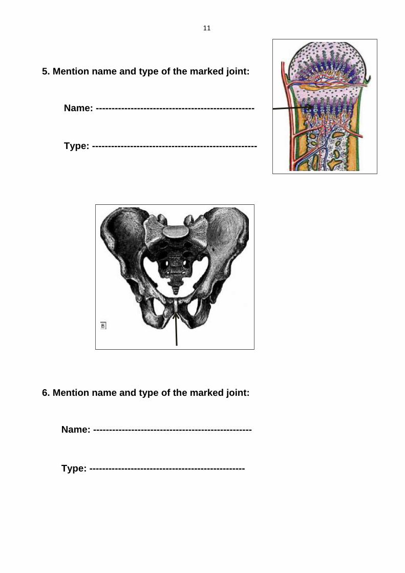
5. Mention name and type of the marked joint:
Name: --------------------------------------------------
Type: ----------------------------------------------------
6. Mention name and type of the marked joint:
Name: --------------------------------------------------
Type: -------------------------------------------------

12
7. Mention name and type of the marked joint:
Name: --------------------------------------------------
Type: ---------------------------------------------------
8. Mention name and type of the marked joint:
Name: --------------------------------------------------
Type: ---------------------------------------------------

13
9. Mention name of the bone and type of joint related to
Its upper end :
Name of bone: --------------------------------------------------
Type of joint: ---------------------------------------------------
10. Mention name and type of the marked joint:
Name: --------------------------------------------------
Type: ---------------------------------------------------
14
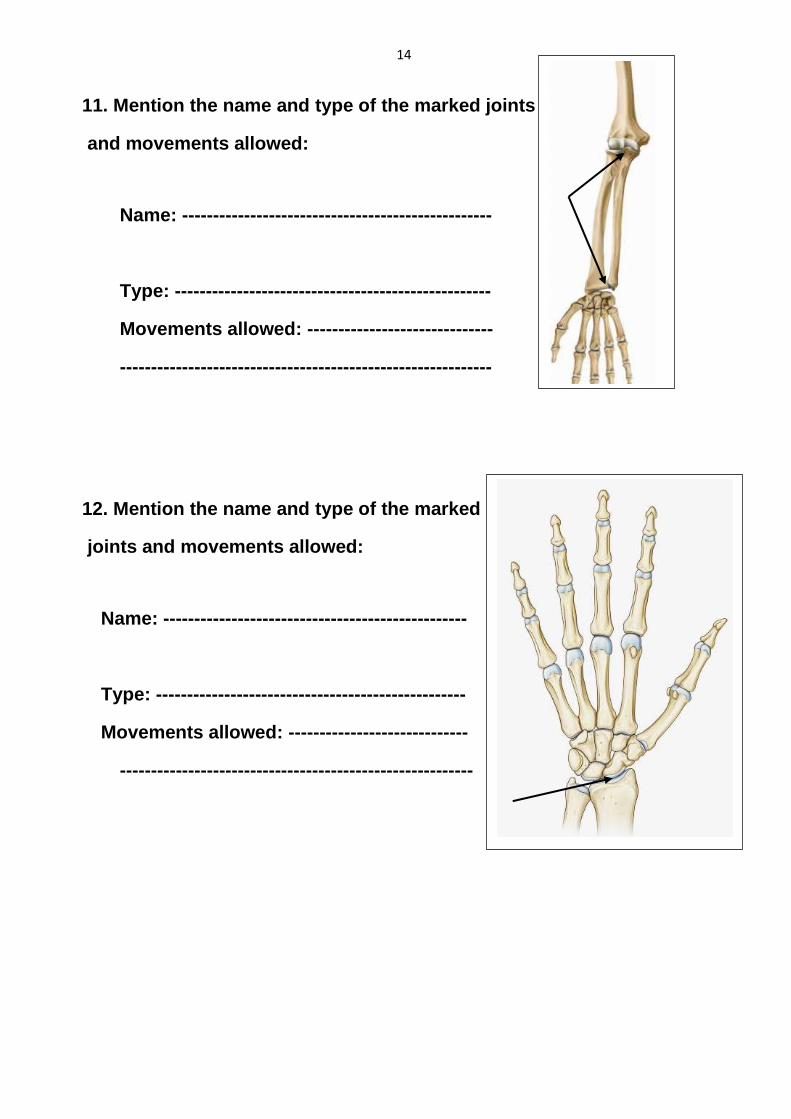
11. Mention the name and type of the marked joints
and movements allowed:
Name: --------------------------------------------------
Type: ---------------------------------------------------
Movements allowed: ------------------------------
------------------------------------------------------------
12. Mention the name and type of the marked
joints and movements allowed:
Name: -------------------------------------------------
Type: --------------------------------------------------
Movements allowed: -----------------------------
---------------------------------------------------------

15
13. Mention the name and type of the
Marked joints and movements allowed.
Name: ------------------------------------------
Type: --------------------------------------------
Movements allowed: ------------------------
-------------------------------------------------------
14. Give the type and movements allowed at the marked joints
Type: -----------------------------------------------
Movements: --------------------------------------

16
15. Name the site of the bone and the type of the joint related to
the marked area
Site of bone : ------------------------------------
Type of related joint: -------------------------
16. Give the name of the marked joint and the movements
allowed.
Name of joint: ------------------------------------
Movements allowed: --------------------------
--------------------------------------------------------
-------------------------------------------------------

17
17. Name the bone and the type of the joint related to its lower
end.
Name of the bone: -------------------------------------
Joint related to its lower end: ----------------------
18. Name the type of the marked bone and the joint related to it.
Name type of bone: -----------------------------
Joint related: -------------------------------------

18
19. Give the name of the marked bone and the type and
movements allowed at the joint related to its upper end.
Name of bone: -----------------------------------
Type of joint: --------------------------------------
Movements allowed: -----------------------------
------------------------------------------------------------
20. Give the name and type of the marked joints and the
movements allowed.
Name and type of joints: -------------------------
--------------------------------------------------------------
------------------------------------------------------------
Movements allowed: -------------------------------
-------------------------------------------------------------
-------------------------------------------------------------

19
21. Give the name and type of the marked joint and the
movements allowed.
Name of joint: -----------------------------------------
Type of joint: -------------------------------------------
Movements allowed: ---------------------------------
---------------------------------------------------------------
22. Give the name and type of the marked joints and the
movements allowed.
Name of joint: -----------------------------------------
Type of joint: -------------------------------------------
Movements allowed: ---------------------------------
---------------------------------------------------------------

20
Self-Assessment Questions
I. Short assay questions:
1. Give types of cartilage with examples:
2. Classify bones according to their position in the body:
3. Classify bones according to the methods of their ossification
with examples:

21
4. Mention the types of bones according to their shape with
examples:
5. Give types of arterial supply of the long bones:
6. What are the functions of the bones:

22
7. Explain how the bones increase in length and width.
8. Determine the location of the growing end in long bones.
9. What are the criteria of the fibrous joints?

23
10. Give the types of fibrous joints with examples.
11. What are the criteria of the primary cartilaginous joints?
12. Mention the characteristics of the secondary cartilaginous
joints and their location.

24
13. What are the characteristics of the synovial joints?
14. Give the criteria of the plane joints with example.
15. Describe each type of the uniaxial joints with examples.

25
16. Give the criteria of each type of the biaxial joints and the
directions of their axes.
17. Give examples for the multiaxial joints and the movements
allowed

26
II. M.C.Q's
1. The following statement describes the anatomical position:
a. The eyes are looking backwards.
b. The upper limbs are hanging by the sides.
c. The palms are facing backwards.
d. The thumbs are directed medially.
2. Anatomical position is:
a. A term used referring to the body facing forwards and sitting down.
b. The position in which the body is lying down with feet parallel.
c. The position in which the body is erect, facing front with feet parallel
and arms hanging at the sides with the palms facing forwards.
d. The position in which the body is erect, facing forward with feet parallel
and arms hanging at the sides with palms facing backwards.
3. The plane which divides the body into 2 equal halves right and left is:
a. Median plane
b. Coronal plane
c. Paramedian plane
d. Horizontal plane
4. Which of the following is true about the coronal plane?
a. It is a horizontal plane.
b. It divides the body into anterior and posterior halves.
c. It cuts the body into upper and lower halves.
d. It is a vertical plane that divides the body into 2 equal right and left
halves.
5. The anatomical term that means “away from the median plane” is:
a. Lateral.
b. Medial.
c. Distal.
d. Proximal.
6. The anatomical term that means “nearer to the root of the limb” is:
a. Lateral.
b. Medial.
c. Distal.
d. Proximal.

27
7. The superficial layer of the skin is composed of which type of
tissues:
a. Simple columnar epithelium.
b. Stratified squamous epithelium.
c. Dense fibrous tissue.
d. Stratified columnar epithelium.
8. The type of tissue that makes up the bulk of the dermis is:
a. Collagen.
b. Melanin.
c. Keratin.
d. Fibroplastin.
9. One of the functions of superficial fascia is:
a. Presence of skin muscles.
b. Formation of aponeurosis.
c. Formation of broad sheets.
d. Formation of interosseous membranes.
10. One of the functions of deep fascia is that it:
a. Facilitates the movement of the skin.
b. Prevents heat loss from the body.
c. Contains many types of glands.
d. Forms the interosseous membranes.
11. The intermuscular septa and interosseous membranes:
a. Surround the muscles of the upper and lower limbs.
b. Separate different groups of muscles which have different actions.
c. Are transverse thickened bands of deep fascia present at wrist joint.
d. Form tough sheaths around big blood vessels.
12. The retinacula:
a. Surround the muscles of the upper and lower limbs.
b. Separate different groups of muscles which have different actions.
c. Are transverse thickened bands of deep fascia present at wrist joint.
d. Form tough sheaths around big blood vessels.
13. Yellow elastic fibrocartilage is present in:
a. Developing bone of the fetus.
b. Auricle of the ear.
c. Articular cartilage of joints.
d. Symphysis pubis.

28
14. White fibrocartilage is present in:
a. Epiglottis.
b. Auricle of the ear.
c. Articular cartilage of joints.
d. Intervertebral disc.
15. Hyaline cartilage is present in:
a. Epiglottis.
b. Auricle of the ear.
c. Articular cartilage of joints.
d. Intervertebral disc.
16. Appendicular skeleton is formed of:
a. Skull and mandible.
b. Bones of the upper and lower limbs.
c. Ribs and sternum.
d. Vertebral column.
17. Intra-membranous ossification occurs in:
a. Clavicle.
b. Vertebrae.
c. Ribs.
d. Base of the skull.
18. Intra-cartilaginous ossification occurs in:
a. Roof of the skull.
b. Clavicle.
c. Skull cap.
d. Vertebrae.
19. The expanded upper and lower ends of the long bone are called:
a. Epiphysis.
b. Epiphyseal plate.
c. Diaphysis.
d. Metaphysis.
20. The compact bone with a central medullary cavity is called:
a. Epiphysis.
b. Epiphyseal plate.
c. Diaphysis.
d. Metaphysis.

29
21. The part which is responsible for the growth of long bones in length
is called:
a. Epiphysis.
b. Epiphyseal plate.
c. Diaphysis.
d. Metaphysis.
22. The scapula is a:
a. Short bone.
b. Long bone.
c. Flat bone.
d. Sesamoid bone.
23. The ribs are:
a. Short bone.
b. Long bone.
c. Flat bone.
d. Seasamoid bone.
24. The Paranasal sinuses are:
a. Short bone.
b. Long bone.
c. Flat bone.
d. Pneumatic bones.
25. The patella is a:
a. Short bone.
b. Long bone.
c. Flat bone.
d. Seasamoid bone.
26. What is the type of joint that connects the roots of the teeth to their
sockets?
a. Gomphosis.
b. Sutures.
c. Cartilaginous.
d. Synovial.

30
27. What is the type of joint that connects the bones of the skull?
a. Gomphosis.
b. Sutures.
c. Cartilaginous.
d. Synovial.
28. What is the type of joint present at the ends of the growing long
bones?
a. Fibrous.
b. Primary cartilaginous.
c. Secondary cartilaginous.
d. Synovial.
29. What is the type of joint of the intervertebral discs?
a. Fibrous.
b. Primary cartilaginous.
c. Secondary cartilaginous.
d. Synovial.
30. One of the following joints is uniaxial:
a. Shoulder.
b. Wrist.
c. Elbow.
d. Knee.
31. One of the following is biaxial joint:
a. Shoulder.
b. Hip.
c. Elbow.
d. Knee.
32. One of the following is a polyaxial joint:
a. Hip.
b. Knee.
c. Wrist.
d. Superior radioulnar.

31
33. The shoulder joint is:
a. Hinge.
b. Ellipsoid.
c. Bicondylar.
d. Ball and socket.
34. The elbow joint is:
a. Hinge.
b. Ellipsoid.
a. Bicondylar.
b. Ball and socket.
35. The wrist joint is:
a. Hinge.
b. Ellipsoid.
c. Bicondylar.
d. Ball and socket.
36. The hip joint is:
a. Hinge.
b. Ellipsoid.
c. Bicondylar.
d. Ball and socket.
37. The skeletal muscles are:
a. Involuntary.
b. Present in the heart.
c. Present in the wall of blood vessels.
a. Attached to the bones.
38. The smooth muscles are:
a. Voluntary.
b. Present in the heart.
c. Present in the wall of blood vessels.
d. Attached to the bones.
39. An example of strap-like muscle is:
a. Rectus abdominis.
b. Sartorius.
c. Deltoid.
d. Supinator.

32
40. An example of strap-like muscle with tendinous intersections is:
a. Sartorius.
b. Deltoid.
c. Rectus abdominis.
d. Tibialis anterior.
41. An example of multipennate muscle is:
a. Deltoid.
b. Tibialis anterior.
c. Rectus anterior.
d. Supinator.
42. An example of bipennate muscle is:
a. Rectus abdominis.
b. Rectus femoris.
c. Sartorius.
d. Deltoid.
43. The right atrium receives blood from:
a. Superior and inferior vena cava.
b. Pulmonary artery.
c. Aorta.
d. Pulmonary veins.
44. Regarding the systemic circulation, which of the following statements
is true?
a. The blood passes from the right ventricle to the right atrium.
b. Oxygenated blood is collected by the veins into the right atrium.
c. Deoxygenated blood passes through the aorta to all tissues of the body.
d. Oxygenated blood passes through the aorta to all tissues of the body.
45. Regarding the pulmonary circulation, which of the following
statements is true?
a. The blood passes from the right ventricle to the right atrium.
b. Oxygenated blood is collected by the veins into the right atrium.
c. Oxygenated blood returns from the lung to the left atrium.
d. Deoxygenated blood passes through the aorta to all tissues of the body.
33
46. Regarding the portal circulation, which of the following statements is
true?
a. The venous blood from the digestive system enters the liver through the
portal vein.
b. The venous blood leaves the liver through the portal vein.
c. The venous blood from the digestive system enters the liver through the
hepatic vein.
d. The venous blood enters the liver through the inferior vena cava.
47. The thoracic duct drains:
a. The right side of the head and neck.
b. The right lower limb.
c. The right upper limb.
d. The right half of the thorax.
48. The right lymphatic duct drains:
a. The whole body below the diaphragm.
b. The right lower limb.
c. The right upper limb.
d. The right half of the abdomen.
49. The brain stem consists of:
a. 2 cerebral hemispheres.
b. Midbrain, pons, cerebellum.
c. Midbrain, medulla oblongata.
d. Mid brain, pons & medulla oblongata.
50. The fourth ventricle lies between:
a. Cerebellum anteriorly and pons and medulla posteriorly.
b. Midbrain and medulla anteriorly and cerebellum posteriorly.
c. Pons and medulla anteriorly and cerebellum posteriorly.
d. Midbrain anteriorly and cerebellum posteriorly.
51. The segments of the spinal cord are:
a. 8 cervical, 10 thoracic, 5 lumbar, 5 sacral and one coccygeal.
b. 10 cervical, 12 thoracic, 5 lumbar, 5 sacral and one coccygeal.
c. 8 cervical, 12 thoracic, 5 lumbar, one sacral and one coccygeal.
d. 8 cervical, 12 thoracic, 5 lumbar, 5 sacral and one coccygeal.

34
MODEL ANSWERS
1- b 15- c 29- c 43- a
2- c 16- b 30- c 44- d
3- a 17- a 31- d 45- c
4- b 18- d 32- a 46- a
5- a 19- a 33- d 47- b
6- d 20- d 34- a 48- c
7- b 21- b 35- b 49- d
8- a 22- c 36- d 50- c
9- a 23- c 37- d 51- d
10- d 24- d 38- c
11- b 25- d 39- b
12- c 26- a 40- c
13- b 27- b 41- a
14- d 28- b 42- b

35
III. Fill in the blanks
1. The position which is used as a reference in describing human body
is called ………………
2. The plane which divides the body into 2 equal halves right and left is
called ………………
3. The vertical plane which is perpendicular on the median plane is
called …………
4. Near to the front of the body is ……………
5. Near to the back of the body is ……………
6. Near to the median plane is ……………
7. Away from the median plane is ……………
8. Approximation of 2 ventral aspects is ……………
9. When the 2 ventral aspects move away from each other it is called
…………
10. Movement towards the median plane is ……………
11. Movement away from the median plane is ……………
12. Lateral rotation of forearm is ……………
13. Medial rotation of forearm is ……………
14. When the sole of foot is directed inwards it is called ……………
15. When the sole of foot is directed outwards it is called ……………
16. An example of a muscle present in the superficial fascia of the neck
is …………
17. An example of a muscle present in the superficial fascia of the upper
limb is ……………
18. The muscle which is present in the superficial fascia of the scrotum
is …………
19. The deep fascia forms sheaths around big vessels and nerves e.g.
……………… Sheath in the lower limb and …………… sheath in the
head and neck
20. Deep fascia forms …………… for insertion of muscles of both sides in
the middle line

36
21. An example of a muscle of which its tendon passes under a fibrous
pulley is ……………
22. Epiphysis of bone is covered with ……………
23. An example of pneumatic bone is ……………
24. A bone embedded in a tendon is called …………… bone
25. The largest sesamoid bone in the body is ……………
26. An example of uniaxial joint is ……………
27. An example of biaxial joint is ……………
28. An example of polyaxial joint is ……………
29. An example of plane synovial joints is ……………

37
Placenta and Birth Defects
1. Name the marked surface (A) of the placenta and the structure
(B).
(A): --------------------------
(B): -----------------------------
2. Name the marked structures and determine the surface of the
placenta shown in the figure.
The structures are:-------------
The surface is: --------------------
A
B

38
3. Identify the marked structures (A) and (B)
(A):-----------------------------
(B):------------------------------
4. Identify the marked structures (A) and (B)
(A) -----------------------------
(B) ------------------------------
A
B
A
B

39
5. Name the placental abnormality and identify the marked
structure.
Placental abnormality: ------------------------------------------
The marked structure: ----------------------------------------------
6. Name the placental abnormality and identify the marked
structure.
Placental abnormality: -----------------------
The marked structure: -----------------------

40
7. Identify the placental abnormality and name the marked
structures.
Abnormality:------------------------
Structures: ----------------------------
8. Identify the placental abnormality and describe it.
Abnormality:-------------------------------
Definition: --------------------------------------------------------------------
------------------------------------------------------------------------------------

41
9. Name the abnormality as shown in the figure and describe it.
Abnormality:-------------------------------
Definition: --------------------------------------------------------------------
------------------------------------------------------------------------------------
10. Identify the placental abnormality and name the marked
structures (A) and (B).
Abnormality: -------------------------
Structure A: ----------------------------
Structure B:-----------------------------
B
A

42
11. Name the abnormality as shown in the figure and describe it.
Abnormality: -------------------------------
Definition: --------------------------------------------------------------------
------------------------------------------------------------------------------------
12. Identify the placental abnormality and name the marked
surface.
Abnormality: ------------------------
Structures: ----------------------------
43
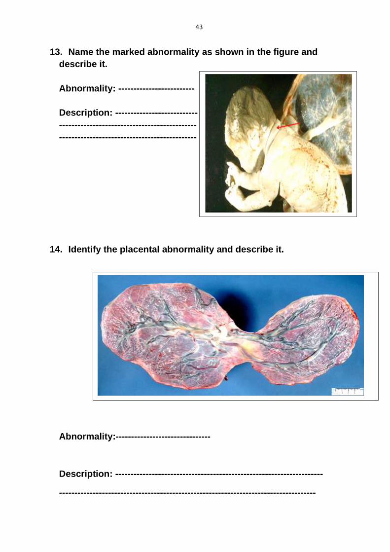
13. Name the marked abnormality as shown in the figure and
describe it.
Abnormality: -------------------------
Description: ---------------------------
---------------------------------------------
---------------------------------------------
14. Identify the placental abnormality and describe it.
Abnormality:-------------------------------
Description: --------------------------------------------------------------------
------------------------------------------------------------------------------------

44
15. Identify the placental abnormality and describe it.
Abnormality:----------------------------
Description: -------------------------------------------------------------------------
16. Identify the placental abnormality and name the marked
structure.
Abnormality:------------------------
Structure: ----------------------------

45
17. Name the abnormal placental infiltration: A, B and C.
(A): --------------------------------------
(B): --------------------------------------
(C): --------------------------------------
18. Name the abnormal placental implantation as shown in the
figure.
Name: -------------------------------------------
-----------------------------------------------------
A
B
C

46
19. Name the birth defect and mention the cause.
Name: ---------------------------------------------
Cause: ---------------------------------------------
20. Name the birth defect and mention the cause.
Name: ---------------------------------------------
Cause: ---------------------------------------------

47
21. Name the birth defect and mention the cause.
Name: ---------------------------------------------
Cause: ---------------------------------------------
22. Name the birth defect and mention the cause.
Name: ---------------------------------------------
Cause: ---------------------------------------------

48
Self-Assessment Questions
I. Short assay questions:
1. Give the shape, weight , diameter and thickness of the the
full term placenta.
2. Give the components of the chorionic plate.
3. What are the structures of the chorionic villi of the
placenta.
49
4. What are the placental septa. Give its structure.
5. Describe the fetal part of the placental circulation.
6. Describe the maternal part of the placental circulation.
50
7. Give the early placental barrier.
8. Determine the late placental barrier.
9. Give the functions of the placental barrier.
51
10. Give the functions of the placenta.
11. Describe the different types of placenta previa.
12. Mention the placental abnormalities according to the shape
and its attachment to umbilical cord.

52
13. Give the function of the amniotic fluid at early and late
pregnancy.
14. Give the abnormalities of the amniotic fluid.

53
II. M.C.Q’s
Choose the proper answer:
1. Regarding formation of sperms.
a. Starts at birth and stopped at old age.
b. Starts at birth and stopped at puberty.
c. Starts at puberty and continues till old age.
d. Starts at old age and continues till death.
2. Regarding oogenesis.
a. Starts at puberty and continues till age.
b. Starts at birth and continues till menopause.
c. Starts at birth and continues till puberty.
d. Starts in the intra-uterine life, then arrested to be continued at puberty.
3. One of the following parts of sperm is responsible for production of
energy
a. Mitochondrial sheath.
b. Nucleus.
c. Acrosomal cap.
d. Tail.
4. Movement of the sperm depends on
a. Acrosomal cap and tail.
b. Mitochondrial sheath and tail.
c. Head and mitochondrial sheath.
d. Head and acrosomal cap.
5. Sperm receptors are present on
a. Zona pellucida.
b. Corona radiata.
c. Cell membrane.
d. Nuclear membrane.
6. The outer cover of the mature ovum:
a. Zona pellucida.
b. Cell membrane.
c. Corona radiata.
d. Nuclear membrane.

54
7. Fertilization occurs in the:
a. Fallopian tube.
b. Surface of ovary.
c. Uterine cavity.
d. Pelvic cavity.
8. Sperm capacitation is:
a. Increase the amount of acrosomal enzymes.
b. Increase movement of the tail.
c. Decease amount of the cytoplasm.
d. Removal of glycoprotein coat from the head of the sperm.
9. Phase II of fertilization is:
a. Penetration of corona radiate.
b. Capacitation of sperms.
c. Penetration of cell membrane of ovum.
d. Penetration of zona pellucida.
10. Phase III of fertilization is:
a. Penetration of corona radiate.
b. Capacitation of sperms.
c. Penetration of cell membrane of ovum.
d. Penetration of zona pellucida.
11. Zona reaction
a. Is the sperm penetration of zona pellucida.
b. Occurs by release of lysosomal enzymes from cortical granules of the
ovum.
c. Occurs after capacitation of the sperms.
d. Is the degeneration of zona pellucida.
12. Cortical and zona reactions occur
a. After passage of sperms through corona radiate.
b. During sperm penetration of zona pellucida.
c. After entrance of the sperm to the cytoplasm of the ovum.
d. After formation of the zygote.
13. One of the following is a result of fertilization:
a. Determination of sex.
b. Cortical and zona reaction.
c. Formation of male and female pronuclei.
d. Zygote nucleus contains haploid number of chromosomes.

55
14. Regarding morula:
a. It is formed in uterine tube within 3 days after formation of zygote.
b. It is formed after degeneration of zona pellucida.
c. It contains a cavity.
d. It is the stage that starts implantation.
15. One of the following is true about blastocyst:
a. It has no cavity.
b. It starts implantation by its abembryonic pole.
c. It is formed 3 days after formation of the zygote.
d. Its inner cell mass is called embryoblast.
16. Regarding implantation:
a. It starts at the 9th day of pregnancy.
b. Chorionic vesicle is the stage that starts implantation.
c. It occurs at the upper part of the posterior wall of uterine cavity.
d. Implantation cavity is formed through the action of proteolytic enzymes
produced from embryonic disc.
17. Placenta previa:
a. Is the implantation of blastocyst in the lower segment of uterine cavity.
b. Is the implantation of blastocyst in the uterine tube.
c. Is the implantation of blastocyst at the surface of ovary.
d. Is the implantation of blastocyst in the pelvic cavity.
18. Decidua basalis:
a. It is the post-implantation endometrium at the lower segment of uterine
cavity.
b. It is the covering of blastocyst after implantation.
c. It is the part of endometrium between the implanted blastocyst and
myometrium.
d. It is the endometrium that lines uterine cavity.
19. Amniotic cavity starts formation at the:
a. 7th day of pregnancy.
b. 8th day of pregnancy.
c. 9th day of pregnancy.
d. 10th day of pregnancy.
56
20. One of the following is an event of the 8th day of pregnancy:
a. Formation of hypoblast.
b. Formation of primary yolk sac.
c. Formation of extraembryonic mesoderm.
d. Formation of primary chorionic villi.
21. One of the following is an event of the 9th day of pregnancy:
a. Formation of hypoblast.
b. Formation of primary yolk sac.
c. Formation of extraembryonic mesoderm.
d. Formation of primary chorionic villi.
22. Chorionic vesicle is formed at the:
a. 10th day of pregnancy.
b. 11th day of pregnancy.
c. 8th day of pregnancy.
d. 13 day of the pregnancy.
23. Splanchnic extraembryonic mesoderm:
a. Covers amniotic cavity.
b. Covers yolk sac cavity.
c. Lines Cytotrophoblast.
d. Lines syncytiotrophoblast.
24. Somatic extraembryonic mesoderm:
a. Is the connecting stalk.
b. Covers yolk sac cavity.
c. Lines Cytotrophoblast.
d. Lines syncytiotrophoblast.
25. The middle layer of chorion is:
a. Cytotrophoblast.
b. Syncytiotrophoblast.
c. Somatic extraembryonic mesoderm.
d. Splanchnic extraembryonic mesoderm.
26. Primary chorionic villi:
a. Is composed of cytotrophoblast and syncytiotrophoblast.
b. It contains fetal blood vessels.
c. It starts formation at the middle of the 3rd week of pregnancy.
d. It forms the fetal part of placenta

57
27. Secondary chorionic villi:
a. Is composed of cytotrophoblast and syncytiotrophoblast.
b. It contains fetal blood vessels.
c. It is formed at the middle of the 3rd week of pregnancy.
d. It forms the fetal part of placenta.
28. Cytotrophoblastic shell is formed from cytotrophoblast of:
a. Primary chorionic villi.
b. Secondary chorionic villi.
c. Stem tertiary chorionic villi.
d. Floating or absorbing tertiary chorionic villi.
29. Chorionic plate:
a. Is the chorion leave.
b. Is the chorion frondosum.
c. Is the decidua basalis.
d. Is the decidua capsularis.
30. Gastrulation is:
a- The formation of trilaminar embryonic disc.
b- The formation of bilaminar embryonic disc.
c- The formation of chorion and chorionic villi.
d- The start of the process of folding.
31. The source of the three germ layers is:
a. Hypoblast.
b. Epiblast.
c. Extraembryonic mesoderm.
d. Cytotrophoblast.
32. Notochord is developed from:
a. The epiblast cells at the primitive streak.
b. The hypoblast cells.
c. Intraembryonic mesoderm.
d. Epiblast cells at the wall of primitive pit.
33. Regarding the notochord:
a. It persists as the annulus fibrosus part of intervertebral disc.
b. It lies between cloacal and buccopharyngeal membrane.
c. It limits the head fold during folding of the embryonic disc.
d. It is formed during the second week of pregnancy.

58
34. Neurenteric canal is the communication between:
a. Amniotic and chorionic cavities.
b. Amniotic and yolk sac cavities.
c. Amniotic and uterine cavities.
d. Yolk sac and chorionic cavities.
35. Intraembryonic mesoderm is formed between ectoderm and
endoderm at:
a. Buccopharyngeal membrane.
b. Cloacal membrane.
c. On both sides of notochord and neural tube.
d. Median region between primitive pit and buccopharyngeal membrane.
36. Neurenteric canal is formed due to degeneration of:
a. Floor of notochordal canal and underlying endoderm.
b. Roof of notochordal canal and median endoderm.
c. Roof of notochordal canal with median ectoderm.
d. Buccopharyngeal membrane.
37. Neural plate is a thickened median region of:
a. Endoderm.
b. Mesoderm.
c. Ectoderm.
d. Notochord.
38. Neural tube gives rise to:
a. Peripheral nerves.
b. Sensory, sympathetic and parasympathetic ganglia.
c. Suprarenal medulla.
d. Central nervous system.
39. Sensory, sympathetic and parasympathetic ganglia are developed
from:
a. Neural crest.
b. Neural tube.
c. Surface ectoderm.
d. Intraembryonic mesoderm.

59
40. Epidermis of skin is derived from:
a. Neural crest.
b. Neural tube.
c. Surface ectoderm.
d. Intraembryonic mesoderm.
41. Segmentation of somites starts at:
a. Occipital region.
b. Cervical region.
c. Thoracic region.
d. Lumbar region.
42. The age of an embryo with 16 pairs of somites is:
a. 23 days.
b. 24 days.
c. 25 days.
d. 26 days.
43. How many somites the embryo has at the occipital region?
a. Four
b. Six
c. Three
d. Five
44. Regarding sclerotome of somite.
a. It is dorsolateral part of the somite.
b. It forms vertebral column.
c. It forms skeletal muscles behind vertebral column.
d. It forms three meninges around spinal cord.
45. Intraembryonic coelom gives rise to:
a. Serous sacs of the body.
b. Respiratory tract.
c. Digestive tract.
d. Heart.
46. Extraembryonic mesoderm communicates with:
a. Paraxial mesoderm.
b. Lateral plate mesoderm.
c. Intermediate mesoderm.
d. Neural plate.

60
47. Extraembryonic coelom communicates with:
a. Pericardium.
b. Pleura.
c. Peritoneal canals.
d. Digestive tract.
48. Smooth muscles of gut are developed from:
a. Splanchnic intraembryonic mesoderm.
b. Somatic intraembryonic mesoderm.
c. Myotomes of paraxial mesoderm.
d. Intermediate mesoderm.
49. Parenchyma of the liver is developed from:
a. Endoderm.
b. Ectoderm.
c. Neural crest.
d. Intraembryonic mesoderm.
50. One of the following is a derivative of intra-embryonic mesoderm:
a. Lining epithelium of intestine.
b. Urogenital system.
c. Central nervous system.
d. Connecting stalk.
51. Folding of the embryonic disc occurs at which week of pregnancy?
a. Second
b. Third
c. Fourth
d. Fifth
52. Tail fold of the embryo is limited by :
a. Notochord.
b. Primitive streak.
c. Yolk sac.
d. Amniotic cavity.
53. After folding, the most cranial structure in the head fold is:
a. Buccopharyngeal membrane.
b. Cloacal membrane.
c. Septum transversum.
d. Pericardium.

61
54. Before folding, the most cranial structure in the head fold is:
a. Buccopharyngeal membrane.
b. Cloacal membrane.
c. Septum transversum.
d. Pericardium.
55. Before folding, the most caudal structure in the tail fold is:
a. Cloacal membrane.
b. Primitive streak.
c. Primitive node.
d. Connecting stalk.
56. After folding, the most caudal structure at tail fold is:
a. Primitive streak.
b. Cloacal membrane.
c. Connecting stalk.
d. Buccopharyngeal membrane.
57. Definitive yolk sac is connected with midgut through:
a. Connecting stalk.
b. Vitelline duct.
c. Allantois.
d. Hindgut.
58. Regarding the stomodeum:
a. It is lined with ectoderm.
b. It is separated from foregut by cloacal membrane.
c. It separates between Forebrain bulge and primitive umbilical ring.
d. It is formed before folding of the embryonic disc.
59. Regarding the placental (decidual) septa, choose the correct
statement:
a. They are composed of chorion frondosum.
b. They are extensions from decidual plate.
c. They are fixed to the chorionic plate.
d. They contain maternal blood vessels.
60. Placental barrier is the separation between:
a. Fetal and maternal surfaces of placenta.
b. Chorionic and decidual plates of placenta.
c. Cytotrophoblastic shell and intervillous spaces.
d. Fetal and maternal blood inside placenta.

62
61. Late placental barrier is composed of:
a. Syncytiotrophoblast and Cytotrophoblast.
b. Syncytiotrophoblast and endothelium of fetal blood vessels.
c. Cytotrophoblast and extraembryonic mesoderm.
d. Cytotrophoblast and endothelium of blood vessels.
62. One of the following is correct regarding the functions of placenta:
a. It allows the passage of maternal antibodies.
b. It prevents passage of gases.
c. It doesn’t produce progesterone hormone.
d. It prevents the passage of all viruses.
63. Marginal attachment of umbilical cord to the placenta is known as:
a. Battle-door placenta.
b. Velamentous placenta.
c. Placenta previa.
d. Placenta accrete.
64. Thin and wide placenta is known as:
a. Placenta membranacea.
b. Placenta increta.
c. Velamentous placenta.
d. Accessory placenta.
65. Early amniotic cavity separates between:
a. Amnioblast and Cytotrophoblast.
b. Amnioblast and epiblast.
c. Amnioblast and hypoblast.
d. Amnioblast and yolk sac.
66. Expansion of amniotic cavity will lead to:
a. Formation of secondary yolk sac.
b. Formation of connecting stalk.
c. Formation of amniochorionic membrane.
d. Formation of neural tube.
67. The earliest source of amniotic fluid is:
a. Epiblast.
b. Diffusion from fetal blood.
c. Fetal urinary system.
d. Amnioblast.
63
68. One of the following conditions may lead to oligohydramnios:
a. Renal agenesis.
b. Maternal diabetes.
c. Fetal esophageal atresia.
d. Fetal anencephaly.
69. Polyhydramnios is the increase of the volume of amniotic fluid to be
more than ……….. Liters:
a. One
b. Two
c. Three
d. Four
70. Regarding the primitive umbilical ring:
a. It is surrounded with amniochorionic membrane.
b. It contains definitive yolk sac.
c. It contains vitelline duct.
d. It contains loops of intestine.
71. Physiological hernia occurs when a loop of intestine is present in:
a. Abdominal cavity.
b. Primitive umbilical ring.
c. Primitive umbilical cord.
d. Definitive umbilical cord.
72. Definitive umbilical cord contains:
a. Two umbilical arteries.
b. Two umbilical veins.
c. Vitelline duct.
d. Loop of intestine.
73. Abnormally long umbilical cord may lead to:
a. Formation of false knots.
b. Early separation of placenta during delivery.
c. Strangulation of the baby during delivery.
d. Adhesion between the fetus with the wall of uterus.

64
74. Heuser’s membrane lines the:
a. Definitive yolk sac.
b. Primary yolk sac.
c. Secondary yolk sac.
d. Vitelline duct.
75. Allantois is a dorsal extension from:
a. Secondary yolk sac.
b. Amniotic cavity.
c. Chorionic cavity.
d. Primary yolk sac.
76. Yolk sac shares in the formation of :
a. Foregut, midgut and hindgut.
b. Kidneys.
c. Forebrain, midbrain and hind brain.
d. Spleen.
77. Fetal period starts at the beginning of:
a. 2nd month.
b. 3rd month.
c. 4th month.
d. 5th month.
78. The weight of fetus at full term ranges between:
a. 900 – 1300 grams.
b. 1400 – 2100 grams.
c. 3000 – 3400 grams.
d. 4600 – 5300 grams.
79. At full term, the head of the fetus represent about:
a. 1/5 of the CH length.
b. 1/6 of the CH length.
c. 1/3 of the CH length.
d. 1/4 0f the CH length.
80. Fetal movements are clearly recognized starting from:
a. The 5th month.
b. The 6th month.
c. The 7th month.
d. The 8th month.
65
81. Duration of pregnancy is about:
a. 246 days.
b. 256 days.
c. 266 days.
d. 236 days.
82. Regarding the dizygotic twin:
a. Are non-identical in shape.
b. Always of same sex.
c. Has a common amnion.
d. Has a common chorion.
83. Regarding monozygotic twin:
a. Is the commonest type.
b. Twins are always of same sex.
c. Are non-identical in shape.
d. Are developed by fertilization of two ova.
84. The risky gestational period ranges between:
a. 3rd to 8th week.
b. 3rd to 4th month.
c. 3rd to 8th month.
d. 3rd to 4th week.
85. Down syndrome is due to:
a. Triosomy 15
b. Triosomy 17
c. Triosomy 13
d. Triosomy 21
86. Cri-du-chat syndrome is due to:
a. Partial deletion of short arm of chromosome 5.
b. Partial deletion of long arm of chromosome 5.
c. Partial deletion of short arm of chromosome 15.
d. Partial deletion of long arm of chromosome 15.
87. Regarding Turner syndrome:
a. Occurs only in males.
b. Somatic cells contains 47 chromosomes.
c. Has XO sex chromosomes.
d. Has normal genital system.

66
88. Regarding Klinefelter syndrome:
a. Occurs only in females.
b. Somatic cells contains 47 chromosomes.
c. Has XO sex chromosomes.
d. Has normal genital system.

67
Key Answer
1- c 19- b 37- c 55- d 73- C
2- d 20- a 38- d 56- b 74- b
3- a 21- b 39- a 57- b 75- a
4- b 22- d 40- c 58- a 76- a
5- a 23- b 41- a 59- b 77- b
6- c 24- c 42- c 60- d 78- c
7- a 25- a 43- a 61- b 79- d
8- d 26- a 44- b 62- a 80- a
9- d 27- c 45- a 63- a 81- c
10- c 28- c 46- b 64- a 82- a
11- b 29- b 47- c 65- b 83- b
12 - c 30- a 48- a 66- c 84- a
13- a 31- b 49- a 67- d 85- d
14- a 32- d 50- b 68- a 86- a
15- d 33- c 51- c 69- b 87- c
16- c 34- b 52- b 70- c 88- b
17- a 35- c 53- a 71- c
18- c 36- a 54- c 72- a

68
III. Fill in the blanks
1. In monosomy, the zygote contains ……….. chromosomes while in
trisomy it contains ………. chromosomes.
2. The zona pellucida disappears at the ….. day after fertilization and
implantation begins at the ………. day. Implantation is completed by
the …………. day.
3. The bilaminar germ disc is formed of 2 layers of cells called ………..
and ……….
4. By the end of the 2nd week of pregnancy, the trophoblast
differentiates into …… and ………….., while the lateral plate
mesoderm split into 2 layers ………… and ……….
5. The primary yolk sac is roofed by the ……….. and its floor is formed
by …….......
6. The amniotic cavity is roofed by the ………… and its floor is formed
by ………..…
7. Each somite differentiates into a dorsolateral part called ………….
and a ventromedial part called …………
8. The trilaminar germ disc forms at the beginning of the …… week and
is constituted of 3 layers named ……., …………… and ……………
9. Chorionic villi develop in 3 stages: ……………, ………….and ………….
10. The embryonic period extends from ………….. to ……….. weeks of
development and is called the period of …………….
11. The fetal period extends from ………… to ……… weeks of
development.
12. The neural crest cells give rise to …………., …………., ………….. and
…………….
13. The somite period of embryonic development extends from the
……….….. till the ………… days.
14. After folding, the part of the secondary yolk sac enclosed within the
embryo is called ……… and the remaining part is called …………
15. The part of the gut within the head fold is called …………., the part
within the tail fold is called ………

69
16. After implantation, the decidua becomes divided into 3 parts: decidua
…………, decidua ……….. and decidua ………….
17. At birth, the average CH length of the fetus is…..…cm, the head
constitutes……. of this length. Its average weight is …….kg.
18. The full term placenta is a disc of about …… cm in diameter and
…….. gm in weight.
19. The fetal surface of the placenta is covered by ……… and about its
center is attached ………..
20. The early placental barrier is formed of …… layers which are
…………………………………………………………..
21. The late placental barrier is formed of ……… layers which are
………………………………………………………….
22. Among the hormones secreted by the placenta …………….,
……………. and ……………..
23. Placenta previa leads to ……………… during late pregnancy and/or
……. of the baby during labor.
24. The umbilical cord is about …… cm in length. It is surrounded by
…………… and contains ……….. artery(ies) and ………..vein(s).
25. Monozygotic twins result from fertilization of ……… ovum (ova) by
…… sperm(s) while dizygotic twins result from fertilization of ………
ovum (ova) by …… sperm(s).
26. The incidence of monozygotic twins is ………. /1000 births and that of
dizygotic twins is ……… /1000 births.
27. The most sensitive period to teratogenic factors during pregnancy is
……….…. to ……. weeks because it is the period of …………..
28. Among the teratogenic infectious agents are …………., ………… and
………..
29. Among teratogenic radiations are ……….., ………… and ………..

70
Key Answer
1. 45, 47.
2. 5th, 7th, 11th.
3. Epiblast, hypoblast.
4. Cytotrophoblast, syncytiotrophoblast, somatopleuric,
splanchnopleuric,
5. Hypoblast, Heuser's membrane.
6. Amnion (amnioblasts), epiblast.
7. Dermomyotome, sclerotome.
8. 3rd, ectoderm, mesoderm, endoderm.
9. 1ary, 2ary, 3ary.
10. 4th, 8th, organogenesis.
11. 9th, 40th.
12. Any 4 derivatives e.g. forebrain, midbrain, hindbrain ………….. etc.
13. 20th, 30th.
14. Gut, definitive yolk sac.
15. Foregut, hindgut.
16. Basalis, parietalis, capsularis.
17. 50, 1/4, 3.5.
18. 15-25, 500-600.
19. Amnion, the umbilical cord.
20. 4, syncytiotrophoblast, cytotrophoblast, extraembryonic mesoderm,
endothelium of fetal capillaries.
21. 2, syncytiotrophoblast, endothelium of fetal capillaries.
22. Estrogen, progesterone, chorionic gonadotropin, placental lactogen
(any 3).
23. Bleeding, asphyxia.
24. 50, amnion, two, one.
25. One, one two, two.
26. 3-4, 7-11.
27. 3rd, 8th, embryogenesis.
28. Measles, mumps, hepatitis, syphilis, toxoplasmosis ….etc (any 3).
29. X-rays, gamma rays, atomic radiations.
71
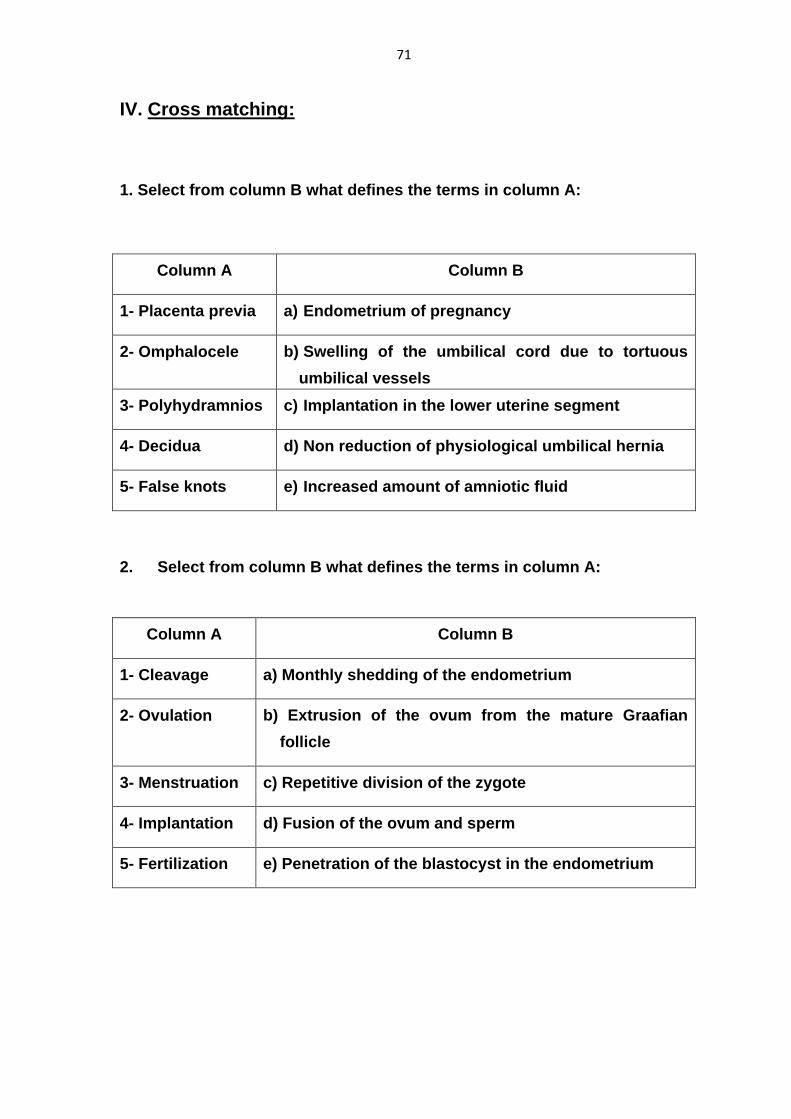
IV. Cross matching:
1. Select from column B what defines the terms in column A:
Column A Column B
1- Placenta previa a) Endometrium of pregnancy
2- Omphalocele b) Swelling of the umbilical cord due to tortuous
umbilical vessels
3- Polyhydramnios c) Implantation in the lower uterine segment
4- Decidua d) Non reduction of physiological umbilical hernia
5- False knots e) Increased amount of amniotic fluid
2. Select from column B what defines the terms in column A:
Column A Column B
1- Cleavage a) Monthly shedding of the endometrium
2- Ovulation b) Extrusion of the ovum from the mature Graafian
follicle
3- Menstruation c) Repetitive division of the zygote
4- Implantation d) Fusion of the ovum and sperm
5- Fertilization e) Penetration of the blastocyst in the endometrium

72
3. Select from the column B what defines the terms in column A:
Column A Column B
1- Notochord a) A layer of cells which will give rise to all to
organs of the embryo
2- Septum transversum b) A cord of extraembryonic mesoderm that
makes the precursor of the umbilical cord
3- Connecting stalk c) The primitive axis of the body
4- Epiblast d) U-shaped cavity inside the lateral plate
mesoderm
5- Intraembryonic
coelom
e) A mass of mesoderm at the cephalic end of the
trilaminar germ disc
4. Select from column B what suits the terms in column A:
Column A Column B
1- Sclerotome a) Cells lying on the dorsolateral aspect of the
neural tube
2- Decidua basalis b) Forms the fetal component of the placenta
3- Allantois c) Forms the maternal component of the placenta
4- Neural crest d) A Diverticulum in the hindgut that protrudes in
the connecting stalk
5- Chorion frondosum e) Part of the somite that migrates to form the
vertebral column
73
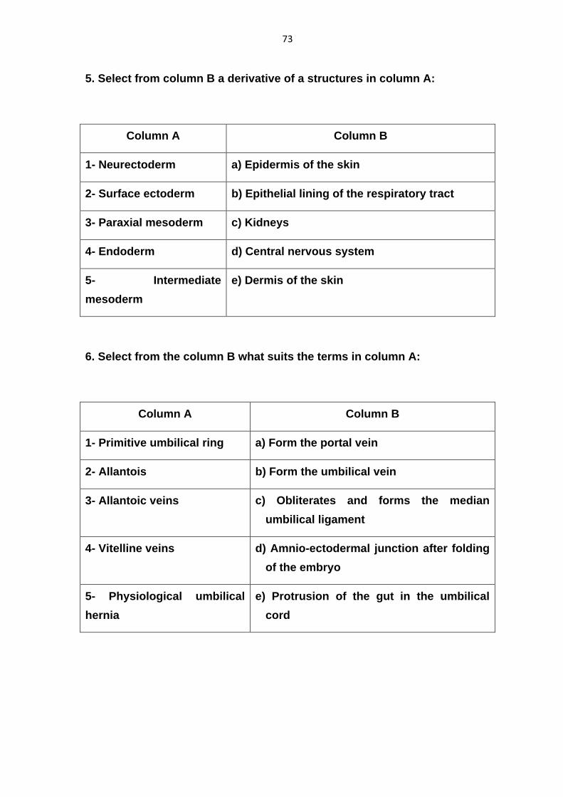
5. Select from column B a derivative of a structures in column A:
Column A Column B
1- Neurectoderm a) Epidermis of the skin
2- Surface ectoderm b) Epithelial lining of the respiratory tract
3- Paraxial mesoderm c) Kidneys
4- Endoderm d) Central nervous system
5- Intermediate
mesoderm
e) Dermis of the skin
6. Select from the column B what suits the terms in column A:
Column A Column B
1- Primitive umbilical ring a) Form the portal vein
2- Allantois b) Form the umbilical vein
3- Allantoic veins c) Obliterates and forms the median
umbilical ligament
4- Vitelline veins d) Amnio-ectodermal junction after folding
of the embryo
5- Physiological umbilical
hernia
e) Protrusion of the gut in the umbilical
cord
74
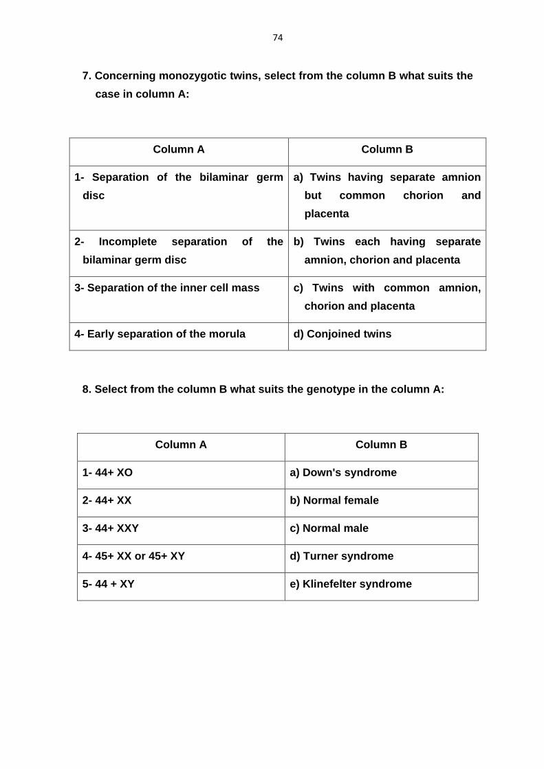
7. Concerning monozygotic twins, select from the column B what suits the
case in column A:
Column A Column B
1- Separation of the bilaminar germ
disc
a) Twins having separate amnion
but common chorion and
placenta
2- Incomplete separation of the
bilaminar germ disc
b) Twins each having separate
amnion, chorion and placenta
3- Separation of the inner cell mass c) Twins with common amnion,
chorion and placenta
4- Early separation of the morula d) Conjoined twins
8. Select from the column B what suits the genotype in the column A:
Column A Column B
1- 44+ XO a) Down's syndrome
2- 44+ XX b) Normal female
3- 44+ XXY c) Normal male
4- 45+ XX or 45+ XY d) Turner syndrome
5- 44 + XY e) Klinefelter syndrome
75
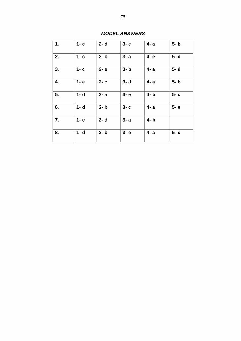
MODEL ANSWERS
1. 1- c 2- d 3- e 4- a 5- b
2. 1- c 2- b 3- a 4- e 5- d
3. 1- c 2- e 3- b 4- a 5- d
4. 1- e 2- c 3- d 4- a 5- b
5. 1- d 2- a 3- e 4- b 5- c
6. 1- d 2- b 3- c 4- a 5- e
7. 1- c 2- d 3- a 4- b
8. 1- d 2- b 3- e 4- a 5- c

76
First semester: Basic Anatomy & General Embryology
Student’s name:
Group :
Date :
Number :
Supervisor :
Mark :