PAR2 Knock-Out C57Bl6 Mice as a Model for …fb.cuni.cz/file/5640/FB2012A0013.pdf · Folia...
6
Folia Biologica (Praha) 58, 81-86 (2012) Short Communication PAR2 Knock-Out C57Bl6 Mice as a Model for Evaluating Metastases of Cancer Cells: Pilot in Vivo Study of the Metastatic Potential of B16 Melanoma in Knock-Out (PAR2-/-) Animals (PAR-2 / trypsin / melanoma / metastasis / knock-out) R. MATěJ 1,2 , M. ZADINOVÁ 3 , P. POUČKOVá 3 , J. KUKAL 4 , T. OLEJÁR 1,3 1 Department of Pathology and Molecular Medicine, Thomayer Teaching Hospital, Prague, Czech Republic 2 Department of Pathology, Third Faculty of Medicine, Charles University in Prague, Czech Republic 3 Institute of Biophysics and Informatics, First Faculty of Medicine, Charles University in Prague, Czech Republic 4 Department of Software Engineering in Economy, Faculty of Nuclear Science and Physical Engineering, Czech Technical University, Prague, Czech Republic Abstract. Proteinase-activated receptor 2 (PAR-2) is a ubiquitous surface molecule. It belongs to the fam- ily of G protein-coupled receptors activated by site- specific proteolysis by trypsin. Altered function of PAR-2 has been described in different malignant tu- mours, both in vivo and in vitro. In the present study, we investigated differences of metastatic spread of B16 melanoma in knock-out animals compared with C57Bl6 mice. Knock-out mice B6.Cg-F2rl1 tm1Mslb /J (PAR2-/-) and C57Bl6 controls were subcutaneously inoculated with the B16 melanoma tissue cell line. Fourteen days after inoculation, all primary tumours were removed and histopathologically analysed. After one month, animals in both group started to die. Autopsy showed metastatic spread of the mela- noma to various organs in both groups. Our experi- ment confirmed growth and metastatic spread in both groups of mice. Excised tumours differed in vol- ume and weight; average weight (0.62 g in PAR2-/- and 0.4 g in control animals). Metastatic spread was observed in both groups and reached 80 % in PAR2-/- Received August 7, 2011. Accepted November 16, 2011. This study was partly supported by IGA MZ NS/10423-3 and Grant No. 309/09/P204 of the Grant Agency of the Czech Re- public. Corresponding author: Radoslav Matěj, Department of Pathology and Molecular Medicine, Thomayer Teaching Hospital, Vídeňská 800, 140 59 Prague 4 – Krč, Czech Republic. Phone: (+420) 261 083 741; Fax.: (+420) 261 083 742; e-mail: radoslav.matej@ ftn.cz Abbreviations: FCS – foetal calf serum, HE – haematoxylin-eo- sin, PAR – proteinase-activated receptor, PAR2-/- – knock-out mice B6.Cg-F2rl1 tm1Mslb /J, WT – wild type. and 50 % in control animals. While in control mice only lung metastases were observed, local tumour recurrence, renal and lung metastases were observed in PAR2-/- mice. The absence of functional PAR-2 could be an important factor influencing the growth and spread of melanoma in vivo, probably associated with tumour cell migration, invasiveness and metas- tasis formation. Introduction The role of various proteinases and their inhibitors in cancer development and malignant behaviour and the relationship of these enzymes to the prognosis of cancer is a constant topic of discussion in professional litera- ture. The role of tissue matrix metalloproteinases, cath- epsins and other enzymes produced by cancer cells has been extensively investigated. However, there are many other enzymes produced by malignant cells and/or tu- mour-associated cells such as fibroblasts and immuno- competent cells. One of these enzymes, trypsin, is a pro- teinase that is produced by many tissues including exocrine pancreatic and intestinal Paneth cells; addi- tionally, enhanced production of trypsin has been re- ported in malignant cells. Trypsin and similar enzymes act on the cell surface via specific receptors – protein- ase-activated receptors (PARs). Proteinase-activated receptor 2 (PAR-2) is a ubiqui- tous surface molecule participating in many biological processes. It belongs to the family of G protein-coupled receptors activated by tethered ligand sequences within the amino-terminal part of the molecule that is made ac- cessible by site-specific proteolysis. PAR-2 activation, after site-specific proteolysis of the N-terminus by trypsin and presentation of the tethered ligand sequence
Transcript of PAR2 Knock-Out C57Bl6 Mice as a Model for …fb.cuni.cz/file/5640/FB2012A0013.pdf · Folia...
Folia Biologica (Praha) 58, 81-86 (2012)
Short Communication
PAR2 Knock-Out C57Bl6 Mice as a Model for Evaluating Metastases of Cancer Cells: Pilot in Vivo Study of the Metastatic Potential of B16 Melanoma in Knock-Out (PAR2-/-) Animals(PAR-2/trypsin/melanoma/metastasis/knock-out)
R.MATěJ1,2, M. ZADINOVá3,P.POUČKOVá3, J. KUKAL4, T. OLEJáR1,3
1Department of Pathology and Molecular Medicine, Thomayer Teaching Hospital, Prague, Czech Republic2Department of Pathology, Third Faculty of Medicine, Charles University in Prague, Czech Republic
3Institute of Biophysics and Informatics, First Faculty of Medicine, Charles University in Prague, Czech Republic 4Department of Software Engineering in Economy, Faculty of Nuclear Science and Physical Engineering, Czech Technical University, Prague, Czech Republic
Abstract. Proteinase-activated receptor 2 (PAR-2) is a ubiquitous surface molecule. It belongs to the fam-ily of G protein-coupled receptors activated by site-specific proteolysis by trypsin. Altered function of PAR-2 has been described in different malignant tu-mours, both in vivo and in vitro. In the present study, we investigated differences of metastatic spread of B16 melanoma in knock-out animals compared with C57Bl6 mice. Knock-out mice B6.Cg-F2rl1tm1Mslb/J (PAR2-/-) and C57Bl6 controls were subcutaneously inoculated with the B16 melanoma tissue cell line. Fourteen days after inoculation, all primary tumours were removed and histopathologically analysed. After one month, animals in both group started to die. Autopsy showed metastatic spread of the mela-noma to various organs in both groups. Our experi-ment confirmed growth and metastatic spread in both groups of mice. Excised tumours differed in vol-ume and weight; average weight (0.62 g in PAR2-/- and 0.4 g in control animals). Metastatic spread was observed in both groups and reached 80 % in PAR2-/-
ReceivedAugust7,2011.AcceptedNovember16,2011.
This studywas partly supported by IGAMZNS/10423-3 andGrantNo. 309/09/P204of theGrantAgencyof theCzechRe-public.
Correspondingauthor:RadoslavMatěj,DepartmentofPathologyandMolecularMedicine,ThomayerTeachingHospital,Vídeňská800, 140 59 Prague 4 – Krč, Czech Republic. Phone: (+420)261083741;Fax.:(+420)261083742;e-mail:[email protected]
Abbreviations:FCS–foetalcalfserum,HE–haematoxylin-eo-sin, PAR – proteinase-activated receptor, PAR2-/- – knock-outmiceB6.Cg-F2rl1tm1Mslb/J,WT–wildtype.
and 50 % in control animals. While in control mice only lung metastases were observed, local tumour recurrence, renal and lung metastases were observed in PAR2-/- mice. The absence of functional PAR-2 could be an important factor influencing the growth and spread of melanoma in vivo, probably associated with tumour cell migration, invasiveness and metas-tasis formation.
IntroductionThe role of various proteinases and their inhibitors in
cancer development and malignant behaviour and the relationship of these enzymes to the prognosis of cancer is a constant topic of discussion in professional litera-ture.Theroleoftissuematrixmetalloproteinases,cath-epsins and other enzymes produced by cancer cells has beenextensivelyinvestigated.However,therearemanyotherenzymesproducedbymalignantcellsand/or tu-mour-associatedcellssuchasfibroblastsandimmuno-competent cells. One of these enzymes, trypsin, is a pro-teinase that is produced by many tissues including exocrine pancreatic and intestinal Paneth cells; addi-tionally, enhanced production of trypsin has been re-ported in malignant cells. Trypsin and similar enzymes actonthecellsurfaceviaspecificreceptors–protein-ase-activatedreceptors(PARs).Proteinase-activatedreceptor2(PAR-2)isaubiqui-
tous surface molecule participating in many biological processes. It belongs to the family of G protein-coupled receptors activated by tethered ligand sequences within the amino-terminal part of the molecule that is made ac-cessibleby site-specificproteolysis.PAR-2activation,after site-specific proteolysis of the N-terminus bytrypsin and presentation of the tethered ligand sequence

82 Vol.58
(SLIGRLinmice)toextracellulardomainsoftherecep-tor, participates in tissue growth and differentiation, re-generationandrepair,inflammatoryresponseregulationand also in malignant transformation (Adams et al., 2011;Hansenetal.,2011).The roleofenvironmentalbody rate of proteinases (including trypsin and trypsin-likeones)andanti-proteinases,resultingina“certain”levelofproteolyticactivityinfluencingPAR-2relativeto tumour cells, has already been investigated in an in vitro model of breast cancer (Matej et al., 2007).Addition of trypsin to medium lacking foetal calf serum (FCS)demonstratedpoorcellulargrowth,mostproba-bly caused by attenuated surface adhesion, since the metabolic activity of breast cancer cells was higher in trypsin-treated groups in the presence or absence of FCS.Takentogether,theactionoftrypsinviaPAR-2hasbeenclearlydemonstrated;however,thisactionwasli-mited by the presence of anti-proteinases in the plasma.
On the other hand, despite its ubiquitous body distri-bution,theroleofPAR-2inthestromal,non-tumouroustissues, in cancer growth and metastasis formation is still poorly understood, while the altered function of PAR-2hasbeendescribedinavarietyofmalignanttu-mours(Nakanumaetal.,2010;Suenetal.,2010;Wanget al., 2010; reviewed by Elste and Petersen, 2010).However, only a few of the current studies suggest an importantroleforPAR-2incancertissuedevelopmentandlocalprogression(Zhangetal.,2009;Mannowetzetal., 2010;Yang et al., 2010), andvery little is knownabouttheroleofPAR-2indevelopmentofdistantme-tastases.
For these reasons, we sought to evaluate the systemic behaviour of cancer in a metastatic model in which
knock-out animals lacking PAR2 genes were inoculated with different malignant cell cultures. As a pilot study, wedecidedtoevaluatetheroleofPAR-2inthegeneral-izedspreadofmelanomaB16inPAR2 knock-out mice compared to our previous observations using wild-type (WT),C57Bl6,mice(Waldetal.,2001).
Material and Methods
Animals
The study was performed in accordance with guide-linesonanimalexperimentationoftheFirstFacultyofMedicine, Charles University in Prague and all proce-dureswereapprovedbytheanimalexperimentationre-viewcommittee.Intheexperiment,sevenmaleinbredPAR-2-deficient B6.Cg-F2rl1tm1Mslb/J mice (JacksonLaboratories, Bar Harbor, ME) and wild-type inbredC57Bl6mice (bodyweight=18–20g–AnLabs.r.o.,Charles River, Prague, Czech Republic) were used.Mice were kept in a barrier facility for animals, provid-ed with radiation-sterilized bedding (SAWI Research Bedding,JELU-WERK,Germany),fedwithradiation-sterilized ST-1 diet (Bergmann, Prague, Czech Repub-lic),andreceivedautoclavedwaterad libitum.
RT PCRTotalRNAwasusedforevaluationofthePAR-2sta-
tus in both groups of animals (TRIzol®Reagent, Invitrogen,GrandIsland,NY).PrimersforPAR2 were 5’ - T G G C CAT TGGAG T C T T C C TG T T- 3’ ,5’-TAGCCCTCTGCCTTTTCTTCTC-3’,andforPAR1 5’-TCCTTTCTCACACTTCCACC-3’ and 5-GTTCA-GGGCTAAACTCTACC-3’.DNApolymerasewasusedfor reverse transcription and PCR (SuperScript® III One-Step RT-PCR System with Platinum®Taq; In vit-rogen,GrandIsland,NY).Amplificationcyclesconsist-edof45sat93°C,45sat55°Cand1minat72°Cfor30cycles.ThePCRreactionwasperformedinaPCRthermo-cycler (MJ Research, Bio-Rad Laboratories, Inc.,Hercules,CA).Productswereanalysedelectropho-reticallyin1%agarosegelwithethidiumbromide.
Tumour cellsB16melanomacellsweredilutedtoaconcentration
of4.0×106/0.2mlinsuspension.Underthiopentalan-aesthesia,B16cells,attheabove-mentioneddose,weresubcutaneously inoculated into the PAR2-/- and WTC57Bl6mice,ontheleftsideofthetrunk.
Tumour growthFourteendaysaftertheB16melanomacellsubcuta-
neous transplantation, the primary tumours were re-moved and measured. The volume was calculated from theformula{V=1/2.A.B2}whereA=thelargestdi-mension of the tumour, B = the smallest dimension of the tumour. The malignant aetiology of the process was checked histologically.
R.Matějetal.
Fig. 1. RT PCR demonstrated the presence of PAR-1 in bothgroupsofmiceandlackofPAR-2inknock-outani-malsanditsexpressionincontrols,W-wildtypeandKK– knock-out

Vol.58 83Melanoma Spread in Knock-Out (PAR2-/-)Animals
Fig. 2.Volumeofexplantedtumourswashigherinknock-outanimalsthanincontrols
Observation, autopsy and histologyDeterioration of animals was followed in both groups
every day for a total of 100 days. All surviving animals were sacrificed after this time period. Dissection andhistologicalverificationwascarriedoutinallanimals,those that died of the disease and those that were sacri-ficedafter100days.Thelung,liverandlocalrecurrencesof primary tumours and metastases into the axillarylymph nodes, as far as they were macroscopically dis-tinct,weresubjectedtohistopathologicalexaminations.Additionally,theywerefixedin10%bufferedformalinandembeddedinparaffinblocks.Fiveµmserialslicesweresubsequentlypreparedandstainedwithhaematox-ylin-eosin(HE).Thelungswereembedded in toto ac-cording to the above procedure and transversally cut at the level of the heart ventricles, and the prepared paraf-finblockswerecutserially.AfterHEstaining,metastaticprocesseswereeitherverifiedorexcluded.
StatisticsThe standard two-sampled t-test was used for testing
tumour weight comparisons between WT and PAR2-/-
groups. The same test was used for comparing the tu-mour volume between WT and PAR2-/-groups.Survivalcurves of WT and PAR2-/-groupsweretestedasstatisti-cal distributions using the two-sampled Kolmogorov-Smirnov test of distribution equality. All the statistical calculations were performed using the MATLAB Sta-tisticalToolbox(MathWorks,Natick,MA),withacriti-cal probability of 0.1 in response to small sample sizes.
Results and DiscussionUsingRTPCRweconfirmedthattheknock-outani-
malswerelackingPAR-2RNA(Fig.1).Ourexperimentconfirmedgrowthandmetastaticspreadinbothgroupsof mice and met the primary objective of this pilot study, i.e. verification ofmetastatic tumour dissemination inPAR2-/-mice.Theexcisedtumoursdifferedinvolume(Fig.2);the
tumoursalsodifferedinaverageweight;0.62ginPAR2-/-and0.4g in the control animals (Fig. 3).The tumourweight equality hypothesis for WT vs. PAR2-/-groupswasnotrejected(Pvalue=0.186).Thetumourvolume
Fig. 3.Weightofexplantedtumourswashigherinknock-outanimalsthanincontrols

84 Vol.58
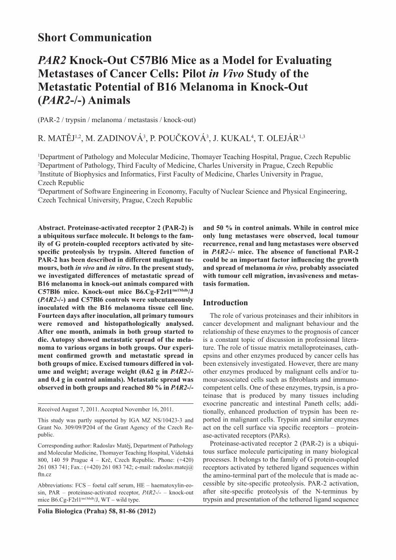
differencesbetweengroupsweresignificant(Pvalue=0.057).Microscopically,weobservedregressivechangesin primary tumours (pigmented melanoma) in bothgroups of investigated animals. Distant metastases were recordedin2/3(0.667)ofWTanimalsandin5/7(0.714)of PAR2-/-animalswithoutanystatisticallysignificantdifference. Metastatic spread was observed in both groupsandreached80%inPAR2-/-and50%incontrolanimals. In control mice, only lung metastases were ob-served(Fig.4);however,localtumourrecurrence,renal
and lung metastases were observed in PAR2-/- mice(Table 1).Onlymicewith recordedmetastatic spreadcontributed data to survival analyses. A difference in survival times between animal groups was observed;knock-out animals had shorter average survival (47.2days)comparedtothecontrols(57.5days).Statistically,therewereno significantdifferencesbetween survivalcurves(Pvalue=0.8271)ofWTandPAR2-/-groups.However, it is worth mentioning that these data are only preliminary and cannot be statistically evaluated any further because of the small numbers of investigated animals(Fig.5).
Despite the fact that our study was not designed to evaluate differences between the groups, one interesting observation jumps to centre-stage and fairly seeks schol-arly attention. Tumours in the PAR2-/-micewerelargerandheavier;additionally,themetastaticspreadofthesetumoursinvolvedotherorgans(kidney)andlocalrecur-rences were observed in this group of mice as well. ThesefindingsmayraiseimportantquestionsabouttheroleofPAR-2intheenvironmentfortumourgrowthanddissemination despite its presence or absence on tumour cells themselves. PAR-2 acts as a pro-inflammatoryagent inducing production of different mediators (Al-Ani et al., 2010;Yoshida et al., 2011), vasodilatation(Bhattetal.,2010)andincreasedvascularpermeability(Kawabataetal.,1998).Linksbetweencancerspread,inflammation,vasculargrowthandcoagulation/fibrino-lysisareobvious,andthedirectactionofPAR-2hasal-readybeenpartlyelucidated(McEachronetal.,2010).PAR-2,ingeneral,appearstobeinvolvedinmanypro-cesses related to membrane internalization and intracel-lularsorting(Déryetal.,1999;Sohetal.,2010);how-ever, it also seems to play an important role in the immune system. PAR-2 directly triggers developmentofdendriticcells(Fieldsetal.,2003;Jiangetal.,2010)and promotes dendritic cell antigen transport and T-cell activation in vivo (Ramelli et al., 2010). It is obviousthattheabsenceofPAR-2canleadtoattenuationofthespecific cellular anti-tumour activity, although furtherinvestigations are needed to verify this hypothesis.
ConclusionTheabsenceoffunctionalPAR-2couldbeanimpor-
tantfactor influencingthegrowthandspreadofmela-
Fig. 4. Metastasis of the tumours in the lungs in control animal(A)andinknock-outmouse(B).Pigmentedhighlymalignant tumour is compatible with melanoma. There are slightly more prominent regressive changes in the tumour of the wild-type animal.
Table 1. Evaluation of the metastatic spread in both groups of animals
WT PAR2-/-Lung Local forelimb and hindlimb lymph node, lung Lung Local forelimb and lung No metastasis recorded Local hindlimb lymph node, kidney, lung No metastasis recorded(diedonlyonedayaftertumourextirpation,notenteredintothesurvivalanalysis)
Lung Lung No metastasis recorded No metastasis recorded
R.Matějetal.

Vol.58 85
noma in vivo, probably through the process of tumour cell migration, invasiveness and metastasis formation. However, further studies involving larger samples sizes willbeneededtoconfirmourresults.
AcknowledgementAuthors would like to thank Tom Secrest for revi-
sions in the English version of this article.
ReferencesAdams, M. N., Ramachandran, R., Yau, M. K., Suen, J. Y.,
Fairlie, D. P., Hollenberg, M. D., Hooper, J. D. (2011)Structure, function and pathophysiology of protease acti-vated receptors. Pharmacol. Ther. 130,248-282.
Al-Ani, B., Hewett, P. W., Cudmore, M. J., Fujisawa, T., Saifeddine, M., Williams, H., Ramma, W., Sissaoui, S., Jayaraman, P. S., Ohba, M., Ahmad, S., Hollenberg, M. D., Ahmed,A. (2010)Activation of proteinase-activated re-ceptor 2 stimulates soluble vascular endothelial growthfactor receptor 1 release via epidermal growth factor recep-tor transactivation in endothelial cells. Hypertension 55, 689-697.
Bhatt, D. K., Ploug, K. B., Ramachandran, R., Olesen, J., Gupta,S.(2010)ActivationofPAR-2elicitsNO-dependentand CGRP-independent dilation of the dural artery. Headache 50, 1017-1030.
Déry, O., Thoma, M. S., Wong, H., Grady, E. F., Bunnett, N. W. (1999) Trafficking of proteinase-activated receptor-2and β-arrestin-1 tagged with green fluorescent protein.β-Arrestin-dependentendocytosisofaproteinasereceptor.J. Biol. Chem. 274,18524-18535.
Elste,A.P.,Petersen,I.(2010)Expressionofproteinase-acti-vated receptor 1-4 (PAR 1-4) in human cancer. J. Mol. Histol. 41, 89-99.
Fields, R. C., Schoenecker, J. G., Hart, J. P., Hoffman, M. R., Pizzo,S.V.,Lawson, J.H. (2003)Protease-activated re-ceptor-2signalingtriggersdendriticcelldevelopment.Am. J. Pathol. 162,1817-1822.
Hansen, K. K., Oikonomopoulou, K., Baruch, A., Rama-chandran, R., Beck, P., Diamandis, E. P., Hollenberg, M. D.
(2008)Proteinasesashormones: targetsandmechanismsfor proteolytic signaling. Biol. Chem. 389,971-982.
Jiang, B., Grage-Griebenow, E., Csernok, E., Butherus, K., Ehlers,S.,Gross,W.L.,Holle, J.U. (2010)The roleofproteinase 3 (PR3) and the protease-activated receptor-2(PAR-2)pathwayindendriticcell(DC)maturationofhu-man-DC-like monocytes and murine DC. Clin. Exp. Rheumatol. 28 (Suppl 57), 56-61.
Kawabata, A., Kuroda, R., Minami, T., Kataoka, K., Taneda, M. (1998) Increased vascular permeability by a specificagonistofprotease-activatedreceptor-2inrathindpaw.Br. J. Pharmacol. 125,419-422.
Mannowetz, N., Würdinger, R., Zippel, A., Aumüller, G., Wennemuth,G.(2010)Expressionofproteinase-activatedreceptor-2 (PAR2) is androgen-dependent in stromal cellline (hPCPs) frombenignprostatic hyperplasia.Prostate 70,1350-1358.
Matej, R., Mandáková, P., Netíková, I., Poucková, P., Olejár, T. (2007) Proteinase-activated receptor-2 expression inbreast cancer and the role of trypsin on growth and metabo-lismofbreastcancercelllineMDAMB-231.Physiol. Res. 56,475-484.
McEachron, T. A., Pawlinski, R., Richards, K. L., Church, F. C.,Mackman,N.(2010)Protease-activatedreceptorsme-diatecrosstalkbetweencoagulationandfibrinolysis.Blood 116,5037-5044.
Nakanuma, S., Tajima, H., Okamoto, K., Hayashi, H., Nakagawara, H., Onishi, I., Takamura, H., Kitagawa, H., Fushida, S., Tani, T., Fujimura, T., Kayahara, M., Ohta, T., Wakayama,T.,Iseki,S.,Harada,S.(2010)Tumour-derivedtrypsin enhances proliferation of intrahepatic cholangio-carcinoma cells by activating protease-activated recep-tor-2.Int. J. Oncol. 36,793-800.
Ramelli, G., Fuertes, S., Narayan, S., Busso, N., Acha-Orbea, H.,So,A.(2010)Protease-activatedreceptor2signallingpromotes dendritic cell antigen transport and T-cell activa-tion in vivo. Immunology 129,20-27.
Soh,U. J.,Dores,M.R.,Chen,B.,Trejo, J. (2010) Signaltransduction by protease-activated receptors. Br. J. Phar-macol. 160,191-203.
Suen,J.Y.,Gardiner,B.,Grimmond,S.,Fairlie,D.P.(2010)Profiling gene expression induced by protease-activated
Fig. 5.Survivalcurvesofinvestigatedgroupsofmice(WT–3mice,PAR2-/-–7mice).Thereisnostatisticallysignifi-cant difference between the two groups, namely due to small numbers of investigated animals.
Melanoma Spread in Knock-Out (PAR2-/-)Animals

86 Vol.58
receptor2(PAR2)activationinhumankidneycells.PLoS One 5,e13809
Wald, M., Olejár, T., Sebková, V., Zadinová, M., Boubelík, M.,Poucková,P.(2001)Mixtureoftrypsin,chymotrypsinand papain reduces formation ofmetastases and extendssurvival timeofC57Bl6micewith syngeneicmelanomaB16.Cancer Chemother. Pharmacol. 47 (Suppl), S16-22.
Wang,X.,Liu,H.T.,Li,S.,Li,K.,Lin,N.,Fan,Q.X.,Zheng,Y.L.(2010)Prognosticvalueofprotease-activatedrecep-tor2expressioninoesophagealsquamouscellcarcinoma.J. Int. Med. Res. 38,1381-1388.
Yang, X. P., Li, Y., Wang, Y., Wang, Y., Wang, P. (2010)β-Tryptaseup-regulatesvascularendothelialgrowthfactor
expression via proteinase-activated receptor-2 and mito-gen-activated protein kinase pathways in bone marrow stromal cells in acute myeloid leukemia. Leuk. Lymphoma 51,1550-1558.
Yoshida, N., Takagi, T., Isozaki, Y., Suzuki, T., Ichikawa, H., Yoshikawa, T. (2011) Proinflammatory role of protease-activated receptor-2 in intestinal ischemia/reperfusion in-jury in rats. Mol. Med. Report. 4,81-86.
Zhang,X.,Wang,W.,True,L.D.,Vessella,R.L.,Takayama,T.K.(2009)Protease-activatedreceptor-1isupregulatedinreactive stroma of primary prostate cancer and bone metas-tasis. Prostate 69,727-736.
R.Matějetal.