Oxidative stress, DNA damage, and antioxidant enzyme activity induced by hexavalent chromium in...
8
Oxidative Stress, DNA Damage, and Antioxidant Enzyme Activity Induced by Hexavalent Chromium in Sprague-Dawley Rats Anita K. Patlolla, Constance Barnes, Clement Yedjou, V. R. Velma, Paul B. Tchounwou Molecular Toxicology Research Laboratory, NIH-Center for Environmental Health, College of Science, Engineering and Technology, CSET, Jackson State University, Jackson, Mississippi, USA Received 21 August 2007; revised 9 March 2008; accepted 13 March 2008 ABSTRACT: Chromium is a widespread industrial compound. The soluble hexavalent chromium Cr (VI) is an environmental contaminant widely recognized as carcinogen, mutagen, and teratogen toward humans and animals. The fate of chromium in the environment is dependent on its oxidation state. The reduction of Cr (VI) to Cr (III) results in the formation of reactive intermediates leading to oxidative tissue damage and cellular injury. In the present investigation, Potassium dichromate was given intraperitoneally to Sprague-Dawley rats for 5 days with the doses of 2.5, 5.0, 7.5, and 10 mg/kg body weight per day. Oxida- tive stress including the level of reactive oxygen species (ROS), the extent of lipid peroxidation and the ac- tivity of antioxidant enzymes in both liver and kidney was determined. DNA damage in peripheral blood lymphocytes was determined by single-cell gel electrophoresis (comet assay). The results indicated that administration of Cr (VI) had caused a significant increase of ROS level in both liver and kidney after 5 days of exposure, accompanied with a dose-dependent increase in superoxide dismutase and catalase activities. The malondialdehyde content in liver and kidney was elevated as compared with the control animals. Dose- and time-dependent effects were observed on DNA damage after 24, 48, 72, and 96 h posttreatment. The results obtained from the present study showed that Cr (VI) could induce dose- and time-dependent effects on DNA damage, both liver and kidney show defense against chromium-induced oxidative stress by enhancing their antioxidant enzyme activity. However, liver was found to exhibit more antioxidant defense than the kidney. # 2008 Wiley Periodicals, Inc. Environ Toxicol 24: 66–73, 2009. Keywords: chromium; malondialdehyde; antioxidant enzymes; superoxide dismutase; catalase; Sprague-Dawley rats; liver; kidneys INTRODUCTION Chromium (Cr) is a naturally occurring heavy metal found commonly in the environment in two valence states: triva- lent Cr (III) and hexavalent Cr (VI). It is widely used in numerous industrial processes and as a result, is a contami- nant of many environmental systems (Cohen et al., 1993). Commercially chromium compounds are used in industrial welding, metal finishes, leather tanning, and wood preser- vation and it has been a nonnegligible pollutant in the world (Norseth, 1981; Wang et al., 2006). Studies by animal model also found many harmful effects of Cr (VI) on mam- mals. Subcutaneous administration of Cr (VI) to rats caused severe progressive proteinuria, urea nitrogen, and creati- nine, as well as elevation in serum alanine aminotransferase activity and hepatic lipid peroxide formation (Kim and Na, 1991). Similar studies reported by Gumbleton and Nicholls Correspondence to: P. B. Tchounwou; e-mail: paul.b.tchounwou@ jsums.edu Contract grant sponsor: The National Institutes of Health – RCMI. Contract grant number: 1G12RR13459. Published online 28 May 2008 in Wiley InterScience (www. interscience.wiley.com). DOI 10.1002/tox.20395 C 2008 Wiley Periodicals, Inc. 66
-
Upload
anita-k-patlolla -
Category
Documents
-
view
212 -
download
0
Transcript of Oxidative stress, DNA damage, and antioxidant enzyme activity induced by hexavalent chromium in...
Oxidative Stress, DNA Damage, and AntioxidantEnzyme Activity Induced by HexavalentChromium in Sprague-Dawley Rats
Anita K. Patlolla, Constance Barnes, Clement Yedjou, V. R. Velma, Paul B. Tchounwou
Molecular Toxicology Research Laboratory, NIH-Center for Environmental Health,College of Science, Engineering and Technology, CSET, Jackson State University, Jackson,Mississippi, USA
Received 21 August 2007; revised 9 March 2008; accepted 13 March 2008
ABSTRACT: Chromium is a widespread industrial compound. The soluble hexavalent chromium Cr (VI) isan environmental contaminant widely recognized as carcinogen, mutagen, and teratogen toward humansand animals. The fate of chromium in the environment is dependent on its oxidation state. The reductionof Cr (VI) to Cr (III) results in the formation of reactive intermediates leading to oxidative tissue damageand cellular injury. In the present investigation, Potassium dichromate was given intraperitoneally toSprague-Dawley rats for 5 days with the doses of 2.5, 5.0, 7.5, and 10 mg/kg body weight per day. Oxida-tive stress including the level of reactive oxygen species (ROS), the extent of lipid peroxidation and the ac-tivity of antioxidant enzymes in both liver and kidney was determined. DNA damage in peripheral bloodlymphocytes was determined by single-cell gel electrophoresis (comet assay). The results indicated thatadministration of Cr (VI) had caused a significant increase of ROS level in both liver and kidney after 5days of exposure, accompanied with a dose-dependent increase in superoxide dismutase and catalaseactivities. The malondialdehyde content in liver and kidney was elevated as compared with the controlanimals. Dose- and time-dependent effects were observed on DNA damage after 24, 48, 72, and 96 hposttreatment. The results obtained from the present study showed that Cr (VI) could induce dose- andtime-dependent effects on DNA damage, both liver and kidney show defense against chromium-inducedoxidative stress by enhancing their antioxidant enzyme activity. However, liver was found to exhibit moreantioxidant defense than the kidney. # 2008 Wiley Periodicals, Inc. Environ Toxicol 24: 66–73, 2009.
Keywords: chromium; malondialdehyde; antioxidant enzymes; superoxide dismutase; catalase;Sprague-Dawley rats; liver; kidneys
INTRODUCTION
Chromium (Cr) is a naturally occurring heavy metal found
commonly in the environment in two valence states: triva-
lent Cr (III) and hexavalent Cr (VI). It is widely used in
numerous industrial processes and as a result, is a contami-
nant of many environmental systems (Cohen et al., 1993).
Commercially chromium compounds are used in industrial
welding, metal finishes, leather tanning, and wood preser-
vation and it has been a nonnegligible pollutant in the world
(Norseth, 1981; Wang et al., 2006). Studies by animal
model also found many harmful effects of Cr (VI) on mam-
mals. Subcutaneous administration of Cr (VI) to rats caused
severe progressive proteinuria, urea nitrogen, and creati-
nine, as well as elevation in serum alanine aminotransferase
activity and hepatic lipid peroxide formation (Kim and Na,
1991). Similar studies reported by Gumbleton and Nicholls
Correspondence to: P. B. Tchounwou; e-mail: paul.b.tchounwou@
jsums.edu
Contract grant sponsor: The National Institutes of Health – RCMI.
Contract grant number: 1G12RR13459.
Published online 28 May 2008 in Wiley InterScience (www.
interscience.wiley.com). DOI 10.1002/tox.20395
�C 2008 Wiley Periodicals, Inc.
66

(1988) found that Cr (VI) induced renal damage in rats
when administered by single s.c. injections. Bagchi et al.
demonstrated that rats received Cr (VI) orally in water
induced hepatic mitochondrial and microsomal lipid per-
oxidation (LPO), as well as enhanced excretion of urinary
lipid metabolites including malondialdehyde (MDA)
(Bagchi et al., 1995). Moreover, some adverse health effects
induced by Cr (VI) have been reported in human. Reports
of epidemiological investigations have shown that respira-
tory cancers had been found in workers occupationally
exposed to Cr (VI) compounds (Costa, 1997; Dayan and
Paine, 2001). DNA strand breaks in peripheral lymphocytes
and LPO products in urine observed in chromium exposed
workers in many researches also showed evidence of the Cr
(VI)-induced toxicity to humans (Gambelunghe et al.,
2003; Goulart et al., 2005).
The carcinogenicity of specific chromium compounds is
influenced by both the valence and the solubility of the
chromium species. Chromium (VI) compounds have been
reported to be more toxic and carcinogenic than chromium
(III) compounds (Connett et al., 1983; De Flora et al.,
1990) because the former can pass through cell membranes
more easily than the latter (De Flora and Wetterhahn,
1989). Once inside the cell, Cr (VI) is reduced to its lower
oxidation states [Cr (V)] and [Cr (IV)] and then Cr (III) by
low molecular weight molecules, enzymatic, and nonenzy-
matic reductants (Shi et al., 1999). These reactive chromium
intermediates are capable of generating a whole spectrum of
reactive oxygen species (ROS), which is an important char-
acteristic of Cr (VI) metabolism (O’Brien et al., 2003). Ex-
cessive quantity of ROS generated by these reactions can
cause injury to cellular proteins, lipids, and DNA leading to
a state known as oxidative stress (Stohs and Bagchi, 1995;
Nordberg and Arner, 2001). Therefore, one of the most im-
portant damage arosed by extraneous Cr (VI) is massive pro-
duction of ROS during the reduction of Cr (VI) in the cell.
LPO, the oxidative catabolism of polyunsaturated fatty
acids, is widely accepted as a general mechanism for cellu-
lar injury and death (Gutteridge and Quinlan, 1983; Halli-
well, 1984). LPO and free radical generation are complex
and deleterious processes that are closely related to toxicity
(Murray et al., 1988). LOP has been implicated in diverse
pathological conditions, including atherosclerosis (Holvoet
and Collen, 1998), aging (Spiteller, 2001), rheumatoid ar-
thritis (Henrotin et al., 1992), and cancer (Marnett, 2000).
It is also involved in the toxicity of pesticides (Bismuth
et al., 1990), solvents (Brattin et al., 1985), and metals
(Kasprzak, 1995). The extension of the oxidative catabo-
lism of lipid membranes can be evaluated by several end-
points, but the most widely used method is the quantifica-
tion of MDA, one of the stable aldehydic products of lipo-
peroxidation, present in biological samples (Gutteridge,
1995; De Zwart et al., 1999).
Antioxidants enzymes are frequently used as markers of
oxidative stress (Gutteridge, 1995). Among these bio-
markers, superoxide dismutase (SOD), glutathione peroxi-
dase (GPX), and catalase (CAT) are important in the pres-
ervation of homeostasis for normal cell function. Enzymes
control the rate of metabolic reactions by lowering the
amount of activation energy needed to start a reaction.
Enzymes exhibit specificity by binding to particular sub-
strates. CAT is found in the peroxisomes of the liver and
kidney and acts to metabolize hydrogen peroxide, a toxic
by-product of metabolic reactions, to water and free oxygen
(Knight, 1997). SOD scavenges the highly toxic superoxide
and converts it to hydrogen peroxide (Fridovich, 1989).
The hydrogen peroxide that is produced by SOD is main-
tained at a safe concentration by the glutathione system.
GPX protects the membrane lipids from oxidative damage
(Kantola et al., 1988).
Although chromium and chromium-containing com-
pounds have been the subject of important toxicology
research, there exists a lack of appropriate animal model
for oxidative stress assessment, DNA damage, as well as
scarcity of scientific data describing the antioxidant defense
relationship with respect to their toxicity in in vivo systems.
Therefore, the present work was undertaken to study the
role of oxidative stress and DNA damage in hexavalent
chromium-induced toxicity and the modulation of intracel-
lular antioxidant defense mechanisms in Sprague-Dawley
rats.
MATERIALS AND METHODS
Chemicals
Potassium dichromate, sodium chloride, sucrose, Triton X-
100, hydrochloric acid, histopaque-1077, NaOH, ethanol,
trypan-blue, and EDTA were obtained from Sigma-Aldrich
(St. Louis, MO). They were of analytical grade or highest
grade available. Lipid peroxidation (LPO) and SOD, assay
kits were purchased from Calbiochem (San Diego, CA).
Phosphate buffer (pH 7.4) was obtained from GIBCO (New
York, NY). Comet assay kit was purchased from Trevigen
(Gaithersburg, MD).
Animal Maintenance
Healthy adult male Sprague-Dawley rats [5–7 weeks of
age, with average body weight (BW) of 60 6 2 g] were
used in this study. They were obtained from Harlan-
Sprague-Dawley Breeding laboratories (Indianapolis, IN).
The animals were randomly selected and housed in polycar-
bonate cages (three rats per cage) with steel wire tops and
corn-cob bedding. They were maintained in a controlled
atmosphere with a 12 h:12 h dark/light cycle, a temperature
of (22 6 2)8C and 50–70% relative humidity with free
access to pelleted feed and fresh tap water. The animals
were supplied with dry food pellets commercially available
67ANTIOXIDANT ENZYME ACTIVITY INDUCED BY HEXAVALENT CHROMIUM
Environmental Toxicology DOI 10.1002/tox

from PMI Feeds (St. Louis, MO). They were allowed to ac-
climate for 10 days before treatment. Whole blood samples
were collected from the tail vein at 24, 48, 72, and 96 h
posttreatment in heparinized tubes for the single-cell gel
electrophoresis [comet assay].
Experimental Design
Groups of five rats each were treated with four different po-
tassium dichromate dose levels. Potassium dichromate was
diluted with distilled water (as required) and intraperitone-
ally administered to animals at the doses of 2.5, 5, 7.5, and
10 mg/kg BW, one dose per 24 h given for 5 days. We
selected intraperitoneal injection because it is the most
commonly used method that is simple and also, for many
agents such as chromium compounds, it will tend to maxi-
mize chemical exposure to the target organs. Each rat
received a total of five doses at 24 h intervals. The cumula-
tive dose of potassium dichromate given to each group of
rats was thus 12.5, 25, 37.5, or 50 mg/kg BW. Distilled
water was administered to the 5 animals of control group in
the same manner as in the treatment groups. At the end of
the exposure, after overnight starvation, the liver and both
kidneys were removed under anesthesia. The organs were
washed thoroughly in ice-cold physiological saline and
weighed. The biological material not used immediately was
stored frozen at 2808C until further analysis.
The local Ethics committee for animal experiments
[Institutional Animal Care and Use Committee] at Jackson
State University, Jackson, MS, approved this study. Proce-
dures involving the animals and their care conformed to the
institutional guidelines, in compliance with national and
international laws and guidelines for the use of animals in
biomedical research (Giles, 1987).
Preparation of Homogenates
At the end of the 5 days exposure to potassium dichromate,
liver and both kidneys were excised under anesthesia. The
organs were washed thoroughly in ice-cold physiological
saline and weighed. A 10% homogenate of each tissue was
prepared separately in 0.05 M phosphate buffer (pH 7.4)
containing 0.1 mM EDTA using a motor driven Teflon-pes-
tle homogenizer (Fischer), followed by sonication (Branson
sonifer), and centrifugation at 4000 rpm for 10 min at 48C.
The supernatant was decanted and centrifuged at 16 000
rpm for 60 min at 48C. The supernatant fraction obtained
was called ‘‘homogenate’’ and used for the assays.
ROS Detection
Reactive oxygen species (ROS) production was quantified
by the DCFH-DA method (Lawler et al., 2003) based on
the ROS-dependent oxidation of DCFH-DA to DCF. An
aliquot of homogenates was centrifuged at 1000 3 g for 10
min (48C). The supernatants were re-centrifuged at 20 000
3 g for 20 min at 48C, and then the pellet was resuspended.
The DCFH-DA solution with the final concentration of
50 lM and resuspension were incubated for 30 min at
378C. Fluorescence of the samples was monitored at an ex-
citation wavelength of 485 nm and an emission wavelength
of 538 nm.
Comet Assay
Rat lymphocytes were isolated and resuspended in PBS.
Following isolation, the cells were mixed with 0.4% Trypan
blue solution, after 15–20 min cells were counted and
checked for viability. The remaining cells were immedi-
ately used for single-cell gel electrophoresis. The assay was
performed according to Singh et al. (1988) with slight mod-
ifications. All the steps were conducted under yellow lamp
in the dark to prevent additional DNA damage. Stained
slides are viewed under automated robotic epiflorescent
microscope.
A total of 150 individual cells were screened/sample
[duplicate, each with 75 cells}.
Slides were read using DNA damage analysis software
[Loats Associates, Westminster, MD].
Malondialdehyde Determination
Malondialdehyde (MDA) concentration was measured in
10% homogenates of liver and kidney using LPO assay kit
from Calbiochem (San Diego, CA). Briefly, 0.65 mL of
10.3 mM N-methyl-2-phenyl-indole in acetonitrile was
added to 0.2 mL of sample (10% homogenate of liver or
kidney). After vortexing for 3–4 s and adding 0.15 mL of
37% HCl, samples were mixed well and closed with a tight
stopper and incubated at 458C for 60 min. The samples
were then cooled on ice, centrifuged and the absorbance
was measured spectrophotometrically at 586 nm. A calibra-
tion curve of an accurately prepared standard MDA solu-
tion (from 2 to 20 nmol/mL) was also run for quantitation.
Measurements of each group were performed in triplicate.
The standard deviations were less than 6 10%.
Antioxidant Enzymes Assay
The activity of SOD was determined in 10% homogenates
of liver and kidney prepared in cold 0.25 M sucrose. The
homogenates were centrifuged at 8500 rpm for 10 min at
48C. Next 400 mL of cold extraction reagent (absolute etha-
nol/chloroform 62.5/37.5 v/v) were added into 250 mL of
supernatant, mixed for 30 s, and centrifuged at 3000 rpm
for 5 min at 48C. The activity of SOD was measured in the
aqueous phase using SOD assay kit from Calbiochem (San
Diego, CA). One unit of enzyme activity is defined as the
68 PATLOLLA ET AL.
Environmental Toxicology DOI 10.1002/tox

amount of enzyme that decreases the initial rate to 50% of
its maximal value for the particular tissue being assayed.
Catalase (CAT) is also known as hydrogen peroxide oxi-
doreductase. To determine its activity, 10% homogenates
of liver and kidney, prepared in 0.05 M phosphate buffer
were used. CAT activity was determined according to Aebi
(1984). Briefly, the reaction mixture consisted of 0.1 mL of
supernatant and 0.01 mL of absolute ethanol. This was vor-
texed well and kept on ice (Ice Bucket, Fischer-Scientific,
Pittsburg, PA, USA) for 30 min. Then, the tubes containing
the sample and the blank were brought to room temperature
and 0.01 mL of Triton X-100 was added and vortexed well
till the whole tissue extract was completely dissolved.
Next, 0.66 M H2O2 in phosphate buffer was prepared afresh
at the time of reaction and 0.1 mL of this buffer containing
H2O2 was added to the above reaction mixture, and the
decrease in the absorbance was read in a spectrophotometer
at 240 nm against a blank for 60 s. Care was taken to carry
out the reactions in the dark. The results were expressed in
nmoles of H2O2 metabolized/mg protein tissue. One unit
corresponds to 1 nmol CAT per gram tissue.
Total Protein Determination
The concentration of total protein in the 10% homogenates
(prepared in phosphate buffer, pH 7.4) of liver and kidney
was estimated according to Lowry et al. (1951) method
using bovine albumin (Sigma) as a standard.
Data Analysis and Statistics
Data were compared by ANOVA. Statistical analysis was
performed using SAS for Windows 2003 package program.
Using the Dunnett test, multiple comparisons were per-
formed. All values were reported as means 6 SD for all the
experiments. The significance level was set at P\ 0.05.
RESULTS
DNA Damage
Comet tail length is an important parameter in evaluating
the DNA damage. All the doses of potassium dichromate
induced statistically significant increase in mean comet tail
length [6.23–33.42 lM] indicating DNA damage when
compared with controls [2.79 lM]. Maximum increase in
mean comet tail length was observed at 10 mg/kg BW at 48
h posttreatment [33.42 lM]. The mean comet tail length
showed a clear dose-dependent increase from 2.5 to 10 mg/
kg BW. A gradual decrease in the tail lengths from 72 h
post-treatment [3.74 lM] was observed by the 96 h and val-
ues had returned almost to control levels at all doses, indi-
cating repair of the damaged DNA and/or loss of heavily
damaged cells. The percentage of DNA damaged cells that
were observed in chromium exposed leukocytes ranged
from (42.3–85.7%) when compared with the control
(15.3%). The results of DNA damage and percentage of
DNA damaged cells are illustrated in Figure 1(A–C),
respectively. Representative comet assay images of control
leukocytes (A) and potassium dichromate treated leuko-
cytes at 2.5 mg/kg (B); 5 mg/kg (C); 7.5 mg/kg (D); and
10 mg/kg (E) are presented in Figure 1. Genotoxicity as
characteristized by the length of the comet tail and the per-
centage of DNA damage are represented in Figures 2 and 3,
respectively.
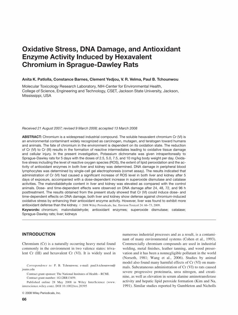
ROS Detection in Liver and Kidneys
ROS were determined in liver and kidney homogenates in
control and treatment groups after administration of potas-
sium dichromate to rats for 5 days. The administration of
Cr (VI) to rats significantly enhanced the ROS level at four
tested doses as compared with the control animals and
increases were dose-dependent. However, the level of ROS
in kidneys was lower compared with liver at all four dose
levels. Figure 2 represents the results of ROS detection.
MDA Concentration in Liver and Kidneys
One of the methods for evaluating LPO is measurement of
the circulating MDA concentration. Potassium dichromate
exposure significantly (P\ 0.05) increased the concentra-
tion of MDA in both liver (19.8–34.3 nmol/g liver) and kid-
neys (6.7–22.9 nmol/g kidney) when compared with the
control (12.3-liver; 5.22-kidney). Results are illustrated in
Figure 3. The increase in MDA concentration in both liver
and kidneys was found to be dose-dependent, indicating a
gradual increase with increasing dose of potassium dichro-
mate. However, the liver exhibited more oxidative stress
than the kidneys.
SOD Activity in Liver and Kidneys
SOD activity was determined in 10% homogenates of liver
and kidneys of Sprague-Dawley rats. There was a signifi-
cant increase in the activity of SOD in potassium dichro-
mate treated rats compared with their respective controls in
both organs. The results of the SOD activity are represented
in Figure 4. A similar trend of a significant increase in SOD
activity was found in both liver [4.43–8.82 U/mg of pro-
tein] and kidney [3.44–6.09 U/mg of protein] homogenates;
however, the liver exhibited more enzymatic activity than
the kidneys.
CATActivity in Liver and Kidneys
Potassium dichromate treatment showed an increase in the
activity of CAT in both liver and kidney homogenates. The
69ANTIOXIDANT ENZYME ACTIVITY INDUCED BY HEXAVALENT CHROMIUM
Environmental Toxicology DOI 10.1002/tox

highest doses (7.5 and 10 mg/kg) showed a significant
increase in the activity of CAT in both organs when com-
pared with the control. However, the trend was found to be
different, that is, kidney exhibiting a slightly more CAT ac-
tivity than the liver. The CAT activity was expected to rise
in response to the tissue trauma. Results of CAT activity
are illustrated in Figure 5.
DISCUSSION
The present study was undertaken to assess the extent of
DNA damage in peripheral blood leukocytes and the oxida-
tive status of liver and kidneys during exposure to various
doses of potassium dichromate. For this purpose, the level
of ROS, the activities of antioxidant enzymes such as SOD,
CAT, and the concentration of MDA as an indicator of
LPO were determined in both organs. We have selected
liver and kidneys in our investigation because these are
major target organs of toxicity. In addition, the liver is pri-
mary organ of biotransformation of xenobiotic compounds.
It contains metabolizing enzymes that change most toxi-
cants to less toxic and more water-soluble but sometimes
can lead to bioactivation. Liver is often important in tests
of oxidative stress because LPO is a major cause of liver
lesions. Kidneys on the other hand filter, detoxify, or bioac-
tivate toxicants (Reed, 1985; Plaa et al., 1997). They are
susceptible to the effects of oxidative stress or stress in gen-
eral because of their dependence on osmotic pressure and a
concentration gradient. The kidneys are also very important
because they are the sites of peroxisomes, which store the
enzyme CAT (Hook and Goldstein, 1993).
In this study, we observed that there was a significant
increase in the level of ROS in both the organs, however,
liver exhibited higher level than the kidneys. ROS have
been implicated in the toxicity of chromium (VI) by several
Fig. 1. Single Cell Gel Electrophoresis assessment of potassium dichromate toxicity inperipheral blood leukocytes of Sprague-Dawley rats: (A) Representative Comet images ofcontrol (A), and 2.5 (B), 5.0 (C), 7.5 (D), and 10 mg/kg (E); (B) Effect of various doses of po-tassium dichromate on DNA migration at various time intervals; and (C) Percentages ofDNA damaged cells observed in rat leukocytes treated with potassium dichromate at dif-ferent time points. Each experiment was done in triplicate. Data were represented asmeans 6 SDs. Statistical significance was indicated as (*) for P\ 0.05. [Color figure canbe viewed in the online issue, which is available at www.interscience.wiley.com.]
70 PATLOLLA ET AL.
Environmental Toxicology DOI 10.1002/tox
authors (Sugiyama, 1992; Bagchi et al., 1997; O’Brien
et al., 2003). Their formation with subsequent cellular dam-
age is considered as the common molecular mechanism of
Cr (VI)-induced toxicity. According to this hypothesis,
chromium (VI) itself is not a cytotoxic agent but rather an
oxygen free radical generator through cellular reduction to
chromium (VI) (Miesel et al., 1995). Chromium reduction
intermediates are believed to react with hydrogen peroxide
to form the hydroxyl radical (HO) (Kadiiska et al., 1994),
which may finally attack proteins, DNA, and membranes
lipids thereby disrupting cellular functions and integrity
(Bagchi et al., 1997).
In the present study, measuring the circulating MDA in
homogenates of liver and kidneys of potassium dichromate-
exposed and control rats assessed LPO. In both liver and
kidneys, there was a significant increase in the concentra-
tion of MDA after 5 days of potassium dichromate adminis-
tration. These results are in accordance with those obtained
by (Bagchi et al., 1995, 1997) who detected oxidative lipid
metabolites in the urine of Cr (VI) treated rats. The increase
observed in LPO may be because of the formation of HO
through a Fenton/Haber-Weiss reaction, catalyzed by
Fig. 3. Effect of potassium dichromate on malondialdehydeconcentration in liver and kidney of Sprague-Dawley rats.Each experiment was done in triplicate. Data were repre-sented as means 6 SDs. Statistical significance was indi-cated as (*) for P\ 0.05. [Color figure can be viewed in theonline issue, which is available at www.interscience.wiley.com.]
Fig. 4. Effect of Potassium dichromate on the activity ofSOD in liver and kidney of Sprague-Dawley rats. Eachexperiment was done in triplicate. Data were represented asmeans 6 SDs. Statistical significance was indicated as (*)for P\ 0.05. [Color figure can be viewed in the online issue,which is available at www.interscience.wiley.com.]
Fig. 5. Effect of potassium dichromate on the activity ofcatalase in liver and kidney of Sprague-Dawley rats. Eachexperiment was done in triplicate. Data were represented asmeans 6 SDs. Statistical significance was indicated as (*)for P\ 0.05. [Color figure can be viewed in the online issue,which is available at www.interscience.wiley.com.]
Fig. 2. Effect of potassium dichromate on detection of ROSin the liver and kidney of Sprague-Dawley rats. Each experi-ment was done in triplicate. Data were represented asmeans 6 SDs. Statistical significance was indicated as (*)for P\ 0.05. [Color figure can be viewed in the online issue,which is available at www.interscience.wiley.com.]
71ANTIOXIDANT ENZYME ACTIVITY INDUCED BY HEXAVALENT CHROMIUM
Environmental Toxicology DOI 10.1002/tox

chromium. This radical is capable of abstracting a hydrogen
atom from a methylene group of polyunsaturated fatty acids
enhancing LPO. However, Cr (VI) induced markedly
higher levels of LPO in liver compared with kidneys. This
was an unexpected finding as the liver was thought to have
more antioxidant defense because of its metabolic role in
detoxification.
The frequency of single-strand breaks showed a clear
dose-related increase up to 10 mg/kg bodyweight in our
investigation. Maximum DNA damage was observed at 48
h posttreatment when compared with the controls. Similar
results were reported with other heavy metals in rodents
using comet assay (Dana Devi et al., 2001; Wang et al.,
2006). Cr (VI) itself is not reactive to DNA, however, the
chromium metabolites radicals produced during reduction
can subsequently attack macromolecules and lead to multi-
form DNA damages, for example, strand breakage, DNA–
protein cross-links, DNA–DNA cross-links, Cr–DNA
adducts and base modifications in cells. Especially DNA
strand breaks are mainly ascribed to the ROS (Wang et al.,
2006). However, at later time intervals, from 72 h posttreat-
ment onward, a gradual decrease in mean comet tail lengths
of all doses was observed, showing a time-dependent
decrease in the DNA damage. Replication arrest may be a
common response of DNA polymerase to DNA–Cr lesions
and provide a plausible mechanism for the inhibition of
DNA synthesis and S-phase cell-cycle delay that occurs in
mammalian cells treated with genotoxic hexavalent chro-
mium (Bridgewater et al., 1998). Single cell gel electropho-
resis (comet assay) is a highly sensitive technique to evalu-
ate single strand breaks and alkali labile sites in DNA of
individual cells.
The activities of the antioxidant enzymes SOD and CAT
were increased by chromium treatment in both the liver and
kidneys. Since SOD catalyzes the dismutation of superox-
ide anion to H2O2, which is in turn the substrate of CAT,
this fact could explain the observed increment of the two
enzyme activities. It has been reported that CAT and SOD
inhibited chromate-induced DNA strand breaks in cultured
cell (Sugiyama, 1992). As these enzymes have a protective
role against oxygen free radical-induced damage, their
induction can be understood as an adaptive response to oxi-
dative stress. Sengupta et al. (1990) also observed similar
results in rats exposed to hexavalent chromium. Increased
generation of superoxide radicals could lead to LPO. SOD
activity also reflects the intensity of the stress because of
toxic action. Heavy metals are known to increase the bio-
chemical stress in the organisms because of deterioration of
metabolic cascade (Hudecova and Ginter, 1992).
In summary, the current study demonstrates that intra-
peritoneal administration of Cr (VI) to the rats during 5
days could induce DNA damage in peripheral blood lym-
phocytes and oxidative stress in liver and kidneys. Both
liver and kidney showed defense against chromium-induced
oxidative stress by enhancing their antioxidant enzymes.
However, the liver was found to exhibit a higher antioxi-
dant defense than the kidney. ROS may play an essential
role in DNA damage induced by Cr (VI) in vivo. Our results
show that the use of thiobarbituric acid reactive substances
as a marker of oxidative stress should be complemented
with antioxidant parameters both in liver and kidneys
namely SOD and CAT. The results support an involvement
of the oxidative damage pathway in the mechanism of tox-
icity of chromium. An understanding of the behavior of
these antioxidant enzymes can aid in the understanding of
chromium-induced toxicity.
The authors would like to thank Dr. Ronald Mason Jr., Presi-
dent, Dr. Abdul K. Mohamed, Dean Emeritus, and Dr. Mark
Hardy, CSET Dean, Jackson State University, for their technical
and administrative support in this project.
REFERENCES
Aebi HE. 1984. Catalase in vitro. Meth Enzymol 105:121–126.
Bagchi D, Hassoun EA, Bagchi M, Muldoon D, Stohs SJ. 1995.
Oxidative stress induced by chronic administration of sodium
dichromate (Cr VI) to rats. Comp Biochem Physiol 110C:281–
287.
Bagchi D, Vuchetich PJ, Bagchi M, Hassoun EA, Tran MX, Tang
L, Stohs SJ. 1997. Induction of oxidative stress by chronic
administration of sodium dichromate (chromium VI) and cad-
mium chloride (cadmium II) to rats. Free Rad Biol Med
22:471–478.
Bismuth C, Garnier R, Baud FJ, Muszynski J, Keyes C. 1990. Par-
aquat poisoning: An overview of current status. Drug Saf
5:243–251.
Brattin WJ, Glende EA, Recknagel RO. 1985. Pathological mech-
anisms in carbon tetrachloride hepatotoxicity. J Free Radic Biol
Med 1:27–38.
Bridgewater LC, Manning FC, Patierno SR. 1998. Arrest of repli-
cation by mammalian DNA polymerases alpha and beta caused
by chromium-DNA lesions. Mol Carcinog 23:201–206.
Cohen MD, Kargacin B, Klein CB, Costa M. 1993. Mechanisms
of chromium carcinogenicity and toxicity. Crit Rev Toxicol
23:255–281.
Connett PH, Wetterhahn KE. 1983. Metabolism of carcinogenic
chromate by cellular constituents. Struct Bond 54:93–124.
Costa M. 1997. Toxicity and carcinogenicity of Cr (VI) in animal
models and humans. Crit Rev Toxicol 27:431–442. Review.
Dana Devi K, Rozati R, Saleha Banu B, Jamil K, Grover P. 2001.
In vivo genotoxic effect of potassium dichromate in mice leuko-
cytes using comet assay. Food Chem Toxicol 39:859–865.
Dayan AD, Paine AJ. 2001. Mechanisms of chromium toxicity,
carcinogenicity and allergenicity: Review of the literature from
1985 to 2000. Hum Exp Toxicol 20:439–451.
De Flora S, Wetterhahn KE. 1989. Mechanisms of chromium me-
tabolism and genotoxicity. Life Chem Rep 7:169–244.
De Flora S, Bagnasco M, Serra D, Zanacchi P. 1990. Genotoxicity
of chromium compounds: A review. Mutat Res 238:99–172.
72 PATLOLLA ET AL.
Environmental Toxicology DOI 10.1002/tox

De Zwart LL, Meerman JH, Commandeur JN, Vermeulen NP.
1999. Biomarkers of free radical damage applications in experi-
mental animals and in humans. Free Radic Biol Med 26:202–
226. Review.
Fridovich I. 1989. Superoxide dismutase. An adaptation to a para-
magnetic gas. J Biol Chem 264:7761–7764.
Gambelunghe A, Piccinini R, Ambrogi M, Villarini M, Moretti M,
Marchetti C, Abbritti G, Muzi G. 2003. Primary DNA damage
in chrome-plating workers. Toxicology 188:187–195.
Giles AR. 1987. Guidelines for the use of animals in biomedical
research. Thromb Haemostasis 58:1078–1984.
Goulart M, Batoreu MC, Rodrigues AS, Laires A, Rueff J. 2005.
Lipoperoxidation products and thiol antioxidants in chromium
exposed workers. Mutagenesis 20:311–315.
Gumbleton M, Nicholls PJ. 1988. Dose-response and time-
response biochemical and histological study of potassium
dichromate-induced nephrotoxicity in the rat. Food Chem Toxi-
col 26:37–44.
Gutteridge JMC. 1995. Lipid peroxidation and antioxidants as bio-
markers of tissue damage. Clin Chem 41:1819–1828.
Gutteridge JMC, Quinlan GJ. 1983. Malondialdehyde formation
from lipid peroxides in thiobarbituric acid test. The role of lipid
radicals, iron salts and metal chelator. J Appl Biochem 5:293–
299.
Halliwell B. 1984. Oxygen radicals: A common sense look at their
nature and medical importance. Med Biol 62:71–77.
Henrotin Y, Deby-Dupont G, Deby C, Franchimont P, Emerit I.
1992. Active oxygen species, articular inflammation and carti-
lage damage. EXS 62:308–322.
Holvoet P, Collen D. 1998. Oxidation of low density lipoproteins
in the pathogenesis of atherosclerosis. Atherosclerosis 137
(Suppl):S33–S38.
Hook JB, Goldstein JR. 1993. Toxicology of the kidney, 2nd ed.
In: Target Organ Toxicology Series. Washington, DC: Taylor &
Francis. p 576.
Hudecova A, Ginter E. 1992. The influence of ascorbic acid on
lipid peroxidation in guinea pigs intoxicated with cadmium.
Food Chem Toxicol 30:1011–1015.
Kadiiska MB, Xiang QH, Mason RP. 1994. In vivo free radical
generation by chromium(VI): An electron spin resonance spin-
trapping investigation. Chem Res Toxicol 7:800–805.
Kantola M, Sarranen M, Vanha PT. 1988. Selenium and glutathi-
one peroxidase in seminal plasma of men and bulls. J Reprod
Fertil 83:785–794.
Kasprzak KS. 1995. Possible role of oxidative damage in metal
induced carcinogensis. Cancer Inv 13:411–430.
Kim E, Na KJ. 1991. Nephrotoxicity of sodium dichromate
depending on the route of administration. Arch Toxicol 65:537–
541.
Knight JA. 1997. Reactive oxygen species and the neuro-degener-
ative disorders. Ann Clin Lab Sci 27:11–25.
Lawler JM, Song W, Demaree SR. 2003. Hindlimb unloading
increases oxidative stress and disrupts antioxidant capacity in
skeletal muscle. Free Radical Biol Med 35:9–16.
Lowry OH, Rosebrough NJ, Farr AL, Randall RJ. 1951. Protein
measurement with the folin phenol reagent. J Biol Chem
193:265–275.
Marnett LJ. 2000. Oxyradicals and DNA damage. Carcinogenesis
21:361–370.
Miesel R, Kroger H, Kurpisz M, Wesser U. 1995. Induction of ar-
thritis in mice and rats by potassium peroxochromate and assess-
ment of disease activity by whole blood chemiluminescence and
99mpertechnetate-imaging. Free Radic Res 23:213–227.
Murray RK, Granner DK, Mayes PA, Rodwell VW. 1988.
Harper’s Biochemistry, 21st ed. Englewood Cliffs, NJ: Prentice
Hall. pp 138–139.
Nordberg J, Arner ES. 2001. Reactive oxygen species, antioxi-
dants, and the mammalian thioredoxin system. Free Radical
Biol Med 31:1287–1312.
Norseth T. 1981. The carcinogenicity of chromium—Review. En-
viron Health Perspect 40:121–130.
O’Brien TJ, Ceryak S, Patierno SR. 2003. Complexities of chro-
mium carcinogenesis: Role of cellular response, repair and re-
covery mechanisms. Mutat Res 533:3–36.
Plaa LG, Hewitt RW. 1997. Toxicology of the liver, 2nd ed. In:
Target Organ Toxicology Series. Washington, DC: Taylor &
Francis. pp 431.
Reed DJ. 1985. Cellular defense mechanisms against reactive
metabolites. In: Anders MW, editor. Bioactivation of Foreign
Compounds. Orlando: Academic Press. pp 71–108.
Sengupta T, Chattopadhyay D, Ghosh N, Das M, Chatterjee GC.
1990. Effect of chromium administration on glutathione cycle
of rat intestinal epithelial cells. Ind J Exp Biol 28:1132–1135.
Shi X, Chiu A, Chen CT, Halliwell B, Castranova V, Vallyathan
V. 1999. Reduction of chromium (VI) and its relationship to
carcinogenesis. J Toxicol Environ Health 2:87–104.
Singh NP, McCoy MT, Tice RR, Schneider EL. 1988. A simple
technique for quantitation of low levels of DNA damage in indi-
vidual cells. Exp Cell Res 175:184–191.
Spiteller G. 2001. Lipid peroxidation in aging and age-dependent
diseases. Exp Gerontol 36:1425–1457.
Stohs SJ, Bagchi D. 1995. Oxidative mechanisms in the toxicity
of metal ions. Free Radic Biol Med 18:321–336.
Sugiyama M. 1992. Role of physiological antioxidants in Cr (VI)
induced cellular injury. Free Radic Biol Med 12:397–407.
Wang XF, Xing ML, Shen Y, Zhu X, Xu LH. 2006. Oral adminis-
tration of Cr(VI) induced oxidative stress. DNA damage and ap-
optotic cell death in mice. Toxicology 228:16–23.
73ANTIOXIDANT ENZYME ACTIVITY INDUCED BY HEXAVALENT CHROMIUM
Environmental Toxicology DOI 10.1002/tox