Fenitrothion induced oxidative stress and morpholo- gical ... · Fenitrothion induced oxidative...
8
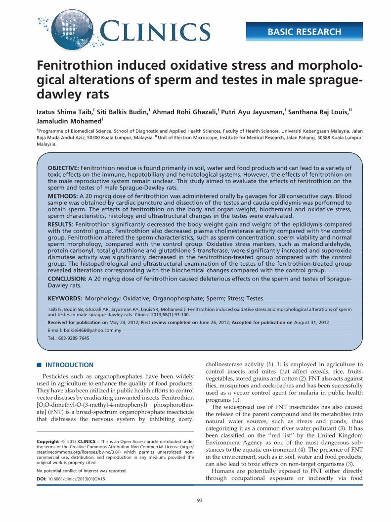
Fenitrothion induced oxidative stress and morpholo- gical alterations of sperm and testes in male sprague- dawley rats Izatus Shima Taib, I Siti Balkis Budin, I Ahmad Rohi Ghazali, I Putri Ayu Jayusman, I Santhana Raj Louis, II Jamaludin Mohamed I I Programme of Biomedical Science, School of Diagnostic and Applied Health Sciences, Faculty of Health Sciences, Universiti Kebangsaan Malaysia, Jalan Raja Muda Abdul Aziz, 50300 Kuala Lumpur, Malaysia. II Unit of Electron Microscope, Institute for Medical Research, Jalan Pahang, 50588 Kuala Lumpur, Malaysia. OBJECTIVE: Fenitrothion residue is found primarily in soil, water and food products and can lead to a variety of toxic effects on the immune, hepatobiliary and hematological systems. However, the effects of fenitrothion on the male reproductive system remain unclear. This study aimed to evaluate the effects of fenitrothion on the sperm and testes of male Sprague-Dawley rats. METHODS: A 20 mg/kg dose of fenitrothion was administered orally by gavages for 28 consecutive days. Blood sample was obtained by cardiac puncture and dissection of the testes and cauda epididymis was performed to obtain sperm. The effects of fenitrothion on the body and organ weight, biochemical and oxidative stress, sperm characteristics, histology and ultrastructural changes in the testes were evaluated. RESULTS: Fenitrothion significantly decreased the body weight gain and weight of the epididymis compared with the control group. Fenitrothion also decreased plasma cholinesterase activity compared with the control group. Fenitrothion altered the sperm characteristics, such as sperm concentration, sperm viability and normal sperm morphology, compared with the control group. Oxidative stress markers, such as malondialdehyde, protein carbonyl, total glutathione and glutathione S-transferase, were significantly increased and superoxide dismutase activity was significantly decreased in the fenitrothion-treated group compared with the control group. The histopathological and ultrastructural examination of the testes of the fenitrothion-treated group revealed alterations corresponding with the biochemical changes compared with the control group. CONCLUSION: A 20 mg/kg dose of fenitrothion caused deleterious effects on the sperm and testes of Sprague- Dawley rats. KEYWORDS: Morphology; Oxidative; Organophosphate; Sperm; Stress; Testes. Taib IS, Budin SB, Ghazali AR, Jayusman PA, Louis SR, Mohamed J. Fenitrothion induced oxidative stress and morphological alterations of sperm and testes in male sprague-dawley rats. Clinics. 2013;68(1):93-100. Received for publication on May 24, 2012; First review completed on June 26, 2012; Accepted for publication on August 31, 2012 E-mail: [email protected] Tel.: 603-9289 7645 & INTRODUCTION Pesticides such as organophosphates have been widely used in agriculture to enhance the quality of food products. They have also been utilized in public health efforts to control vector diseases by eradicating unwanted insects. Fenitrothion [O,O-dimethyl-O-(3-methyl-4-nitrophenyl) phosphorothio- ate] (FNT) is a broad-spectrum organophosphate insecticide that distresses the nervous system by inhibiting acetyl cholinesterase activity (1). It is employed in agriculture to control insects and mites that affect cereals, rice, fruits, vegetables, stored grains and cotton (2). FNT also acts against flies, mosquitoes and cockroaches and has been successfully used as a vector control agent for malaria in public health programs (1). The widespread use of FNT insecticides has also caused the release of the parent compound and its metabolites into natural water sources, such as rivers and ponds, thus categorizing it as a common river water pollutant (3). It has been classified on the ‘‘red list’’ by the United Kingdom Environment Agency as one of the most dangerous sub- stances to the aquatic environment (4). The presence of FNT in the environment, such as in soil, water and food products, can also lead to toxic effects on non-target organisms (3). Humans are potentially exposed to FNT either directly through occupational exposure or indirectly via food Copyright ß 2013 CLINICS – This is an Open Access article distributed under the terms of the Creative Commons Attribution Non-Commercial License (http:// creativecommons.org/licenses/by-nc/3.0/) which permits unrestricted non- commercial use, distribution, and reproduction in any medium, provided the original work is properly cited. No potential conflict of interest was reported. DOI: 10.6061/clinics/2013(01)OA15 BASIC RESEARCH 93
Transcript of Fenitrothion induced oxidative stress and morpholo- gical ... · Fenitrothion induced oxidative...
Fenitrothion induced oxidative stress and morpholo-gical alterations of sperm and testes in male sprague-dawley ratsIzatus Shima Taib,I Siti Balkis Budin,I Ahmad Rohi Ghazali,I Putri Ayu Jayusman,I Santhana Raj Louis,II
Jamaludin MohamedI
I Programme of Biomedical Science, School of Diagnostic and Applied Health Sciences, Faculty of Health Sciences, Universiti Kebangsaan Malaysia, Jalan
Raja Muda Abdul Aziz, 50300 Kuala Lumpur, Malaysia. II Unit of Electron Microscope, Institute for Medical Research, Jalan Pahang, 50588 Kuala Lumpur,
Malaysia.
OBJECTIVE: Fenitrothion residue is found primarily in soil, water and food products and can lead to a variety oftoxic effects on the immune, hepatobiliary and hematological systems. However, the effects of fenitrothion onthe male reproductive system remain unclear. This study aimed to evaluate the effects of fenitrothion on thesperm and testes of male Sprague-Dawley rats.
METHODS: A 20 mg/kg dose of fenitrothion was administered orally by gavages for 28 consecutive days. Bloodsample was obtained by cardiac puncture and dissection of the testes and cauda epididymis was performed toobtain sperm. The effects of fenitrothion on the body and organ weight, biochemical and oxidative stress,sperm characteristics, histology and ultrastructural changes in the testes were evaluated.
RESULTS: Fenitrothion significantly decreased the body weight gain and weight of the epididymis comparedwith the control group. Fenitrothion also decreased plasma cholinesterase activity compared with the controlgroup. Fenitrothion altered the sperm characteristics, such as sperm concentration, sperm viability and normalsperm morphology, compared with the control group. Oxidative stress markers, such as malondialdehyde,protein carbonyl, total glutathione and glutathione S-transferase, were significantly increased and superoxidedismutase activity was significantly decreased in the fenitrothion-treated group compared with the controlgroup. The histopathological and ultrastructural examination of the testes of the fenitrothion-treated grouprevealed alterations corresponding with the biochemical changes compared with the control group.
CONCLUSION: A 20 mg/kg dose of fenitrothion caused deleterious effects on the sperm and testes of Sprague-Dawley rats.
KEYWORDS: Morphology; Oxidative; Organophosphate; Sperm; Stress; Testes.
Taib IS, Budin SB, Ghazali AR, Jayusman PA, Louis SR, Mohamed J. Fenitrothion induced oxidative stress and morphological alterations of spermand testes in male sprague-dawley rats. Clinics. 2013;68(1):93-100.
Received for publication on May 24, 2012; First review completed on June 26, 2012; Accepted for publication on August 31, 2012
E-mail: [email protected]
Tel.: 603-9289 7645
& INTRODUCTION
Pesticides such as organophosphates have been widelyused in agriculture to enhance the quality of food products.They have also been utilized in public health efforts to controlvector diseases by eradicating unwanted insects. Fenitrothion[O,O-dimethyl-O-(3-methyl-4-nitrophenyl) phosphorothio-ate] (FNT) is a broad-spectrum organophosphate insecticidethat distresses the nervous system by inhibiting acetyl
cholinesterase activity (1). It is employed in agriculture tocontrol insects and mites that affect cereals, rice, fruits,vegetables, stored grains and cotton (2). FNT also acts againstflies, mosquitoes and cockroaches and has been successfullyused as a vector control agent for malaria in public healthprograms (1).
The widespread use of FNT insecticides has also causedthe release of the parent compound and its metabolites intonatural water sources, such as rivers and ponds, thuscategorizing it as a common river water pollutant (3). It hasbeen classified on the ‘‘red list’’ by the United KingdomEnvironment Agency as one of the most dangerous sub-stances to the aquatic environment (4). The presence of FNTin the environment, such as in soil, water and food products,can also lead to toxic effects on non-target organisms (3).
Humans are potentially exposed to FNT either directlythrough occupational exposure or indirectly via food
Copyright � 2013 CLINICS – This is an Open Access article distributed underthe terms of the Creative Commons Attribution Non-Commercial License (http://creativecommons.org/licenses/by-nc/3.0/) which permits unrestricted non-commercial use, distribution, and reproduction in any medium, provided theoriginal work is properly cited.
No potential conflict of interest was reported.
DOI: 10.6061/clinics/2013(01)OA15
BASIC RESEARCH
93
consumption. Previous research has found that variousconcentrations of FNT caused histopathological effects onthe liver and kidneys of rats, cytotoxic effects on the lungs ofrats and immunosuppressive effects in rats (5,6).Furthermore, after a seven-day exposure of FNT at dosesof 15 and 30 mg/kg, the compound acted as an anti-androgen that could reduce the growth of androgen-dependent tissues in rats (7). However, the pathophysiolo-gical effect of FNT on sperm and testes remains unclear andunderreported.
These toxic effects likely occur through the generation ofreactive oxygen species (ROS) that may be produced as aresult of the FNT metabolism by cytochrome P450s or due tothe high-energy consumption coupled with the inhibition ofoxidative phosphorylation (8). The imbalance between theformation of ROS and mechanism of enzymatic and non-enzymatic antioxidants as a body defense system can lead tooxidative stress. Oxidative stress has been reported to be theprimary mechanism of organophosphate toxicity afterprolonged exposure (9). In addition, oxidative stressconditions may cause alterations in sperm cells due to thehigh levels of polyunsaturated fatty acid (PUFA) in theirplasma membrane (10).
FNT at a dose of 20 mg/kg body weight (bw) by oraladministration caused significant damage to the liver andkidneys (5) and changes in the histology of lungs (6) in malealbino rats. Therefore, this study aimed to assess the potentialeffects of FNT at a dose of 20 mg/kg bw on the reproductiveparameters of male Sprague-Dawley rats to provide apotential extrapolation of the findings to humans. The purposeof this study was to determine the effects of FNT on spermcharacteristics, sperm and testicular morphology, biochemicaland oxidative stress markers in testes of male Sprague-Dawleyrats.
& MATERIALS AND METHODS
AnimalsTwenty adult male Sprague-Dawley rats weighing 230-
250 g were used in this study. All rats were obtained fromthe Laboratory Animal Resource Unit, Faculty of Medicine,Universiti Kebangsaan Malaysia, one week prior to thestudy for acclimatization purposes. The rats were kept inplastic cages, with two rats per cage under standardenvironmental conditions (12 h light/dark cycles, 25-28 C)and maintained on a standard pellet and water ad libitumdiet for the duration of the study. All of the animal handlingprotocols were approved by the Animal Ethics Committeeof Laboratory Animal Resource Unit, Faculty of Medicine,Universiti Kebangsaan Malaysia.
ChemicalsThe chemicals and reagents used in this study were of
high purity. FNT with a purity of 99.9% was purchasedfrom SUPELCO Analytical, USA with lot number: LB75917.Other chemicals used in this study were purchased fromSigma Chemicals Co., St. Louis, Missouri, USA, with theexception of 2,4-nitrophenylhydrazine, which was pur-chased from Mallinckrodt Chemicals, Lenzing AG,Salzburg, Austria. Nitro blue tetrazolium, reduced glu-tathione and other reagents were purchased from Merck,Darmstadt, Bundesland of Hesse, Germany.
Animal treatment scheduleAfter the one-week acclimatization, the rats were divided
randomly into control and treated groups, with ten rats pergroup. FNT (dissolved in corn oil) was administered at adose of 20 mg/kg bw (1/3 LD50) using oral gavages for 28consecutive days (five days/week), whereas the controlgroup received only corn oil (1 mg/kg bw). Toxicity signswere observed and recorded daily. Body weight wasrecorded weekly throughout the study duration.
At the end of the study, the rats were fasted overnight andanaesthetized with diethyl ether. Blood was collected in anEDTA tube via cardiac puncture. Plasma was obtained bycentrifugation of blood samples at 3,500 rpm at 4 C for 10minutes. Plasma samples were maintained at -40 C untilfurther biochemical analysis. At the end of the experiment,the rats were sacrificed and dissected. The weights of thecauda epididymis, epididymis and testes were recorded.Left and right of cauda epididymis were immediatelyplaced in 2 ml of Hank’s Buffered Salt Solution (HBSS)enriched with 0.5% BSA and pre-warmed at 37 C to obtainthe sperm. The cauda epididymis was cut into small piecesand centrifuged at 1,000 rpm at 4 C for 3 minutes to allowthe sperm to be released for further analysis. Testes werehomogenized in 0.15 M KCl solution (1:2) at 4 C andcentrifuged at 10,000 X g for 20 minutes at 4 C. Thehomogenated testes were maintained at -40 C until furtherbiochemical analysis.
Biochemical analysisPlasma cholinesterase (ChE) was assayed using the
Ellman method (11). The cholinesterase in the sampleshydrolyzed the propionythiocholine to form thiocholine,which was then reacted with 5,59-dithiobis-2-nitrobenzoicacid (DTNB) to form a yellow solution containing 5-thio-2-nitrobenzoate. The formation of the yellow color wasmonitored at 405 nm and the cholinesterase activity wascalculated in U/l.
Protein and cholesterol levels in the testes were deter-mined by the Bradford (12) and Franey and Amador (13)methods, respectively. Briefly, an aliquot of the homoge-nated testes was added to Bradford’s reagent and incubatedfor 5 minutes at room temperature. The protein reacted withBradford’s reagent to produce colored complexes and theintensity of these complexes was measured at 595 nm usinga microplate reader. Similarly, cholesterol detection wasbased on the reaction between cholesterol, ferric chlorideand concentrated sulfuric acid. Briefly, 100 ml of homo-genate testes was added to ethanol for cholesterol extractionpurposes. The obtained supernatant was treated with ferricchloride and sulfuric acid. The concentration of cholesterolin each sample was determined at 560 nm, where itexhibited a color change from yellow to brown. The proteinand cholesterol levels were calculated based on theirstandard curves in mg/dl and mg/ml, respectively.
The homogenated testes were evaluated for malondialde-hyde (MDA), protein carbonyl (PC), total glutathione(GSH), glutathione S-transferase (GST) and superoxidedismutase (SOD) to assess the redox processes. Theproduction of MDA was measured for lipid peroxidationbased on the Stocks and Dormandy methods (14). Briefly, analiquot of (0.5 ml) of homogenated testes was added to aTCA/HCl solution, vortexed and incubated at roomtemperature for 15 minutes. The mixture was added toTBA/NaOH, vortexed and heated in a boiling water bath
FNT alters male reproductive systemTaib IS et al.
CLINICS 2013;68(1):93-100
94
(100 C) for 30 minutes. The MDA in the sample reacted withthe thiobarbituric acid to form a pink chromogen containingthiobarbituric acid reactive substances (TBARS). The level ofTBARS in the supernatant was measured using a spectro-photometer at 532 nm. The MDA concentration wasexpressed as mM/mg of tissue protein.
The PC content was measured for oxidative stress in theprotein precipitate using the method described by Levine(15), with some modifications. Briefly, the homogenatedtestes were added to TCA (1:1) to precipitate the protein,incubated in ice for 15 minutes and then centrifuged at15,000 X g for 5 minutes at 4 C. The obtained pellet wasreacted with 10 mM DNPH and incubated for 1 hour atroom temperature in the dark. TCA was added to themixture to precipitate the protein; the mixture wasincubated in ice for 5 minutes and then centrifuged at15,000 X g for 5 minutes at 4 C. The obtained pellet was thenwashed twice using a mixture of ethanol and ethyl acetateand then diluted with 5 M urea. The derivatives obtainedafter centrifugation at 15,000 X g for 3 minutes at 4 C weremonitored between 375 and 380 nm. PC formation wasexpressed as nmol/mg of tissue protein.
GSH content was measured in the homogenated testesbased on the Ellman method (16), with some modification.Deproteinized homogenated testes were treated with ametaphosphoric acid solution and centrifuged at 3,000 X gfor 10 minutes at 4 C. The obtained supernatant was mixedwith a reaction buffer at pH 8.0 and DTNB for 15 minutesand measured at 412 nm by using a microplate reader. TheGSH content was expressed as mmol/mg protein.
SOD and GST enzyme activity was determined using theBeyer and Fridovich (17) and Habig and Jacoby (18)methods, respectively. Briefly, an aliquot of (20 ml) of thehomogenate was mixed with the substrate containing[PBS(EDTA); L-methionine; NBT.2HCl; Triton-X] and ribo-flavin. The mixture was incubated in an aluminum boxunder 20 watt lamp for 7 minutes. The SOD activity wasmeasured spectrophotometrically by monitoring the inhibi-tion of ferricytochrome reduction using a xanthine-xanthineoxidase as a source of peroxides. An SOD unit is defined asone unit of enzyme, which inhibits 50% of the nitro bluetetrazolium (NBT) reduction. SOD activity was calculatedand expressed in U/mg protein. GST was assayed bymeasuring the rate that the enzyme catalyzed the conjuga-tion of reduced glutathione with CDNB. Briefly, 50 ml ofhomogenated testes was added to 1 mM GSH and 75 mM of1-chloro-2,4-dinitro benzene (CDNB). The rate of GSTactivity was measured at 340 nm and expressed as U mM/min/mg protein.
Sperm viabilitySperm viability was determined by a routine gold
standard method suggested by WHO (19) involving thedetermination of eosin, which penetrates the membranes ofdamaged cells. Approximately 100 ml of sperm suspensionwas mixed with two drops of eosin 1% and allowed toremain undisturbed for 30 seconds. Then, three drops 10%nigrosin were mixed with the solution and within 30seconds, a thick smear was performed in triplicate. Theresults were determined by counting both motile andimmotile sperm and are presented as a percentage (%).
Sperm density and morphologySperm density was calculated by a Markler Counting
Chamber based on a WHO method (19). Briefly, 10 ml of asperm suspension was calculated for at least ten differentmicroscopic fields under 40X magnification using a lightmicroscope. Results are expressed as 106 cells/ml.
A smear was conducted by using a drop of spermsuspension. After the smear had dried, the slide was fixedwith absolute ethanol for 5 minutes. Then, the slide wasimmersed in Diff-Quik Stain I and II for 5 minutes each. Acover slip was applied and two hundred sperms werecalculated per animal to measure the morphological abnorm-alities under oil immersion. The abnormal sperm heads werecalculated in triplicate. Data are shown as a percentage ofsperm head abnormalities.
Morphological analysisThe testes in both groups were treated with 10% buffered
formalin solution and a routine histological procedure wasconducted. All testicular sections were stained with hema-toxylin and eosin (H & E) stains and monitored formorphological changes under X10 and X40 magnifications.All changes were verified by a histopathological expert.
A section of the testes was cut into small pieces (1 mm3),treated with 2.5% glutaraldehyde 0.1 N PBS at roomtemperature for one hour and post-fixed with osmiumtetraoxide for another hour. Testes tissue was dehydrated in70, 90 and 100% (twice) acetone for five minutes each,followed by 1:1 (acetone: resin) for five minutes and thenembedded in epoxy resin. Ultrathin slices (90 nm) wereobserved using a transmission electron microscope, TecnaiG2 (FEI, USA), at 100 kV.
Statistical analysisAll obtained data from the analysis were normally
distributed. The differences between the treated and controlgroups were statistically evaluated using an independentStudent’s t-test. All data are expressed as the meanvalues¡SD, with significant values at p,0.05.
& RESULTS
Signs of toxicityAdministration of a 20 mg/kg bw dose of FNT caused
cholinergic signs, such as hypoactivity, lacrimation, piloerec-tion and tremor. All of the toxicity signs occurred within oneor two hours after FNT administration. The toxicity signsremained in the same animals for approximately 48 hoursthroughout the experimental period. Approximately 60% oftreated rats showed signs of toxicity. No deaths wererecorded in either experimental group.
Body and organ weightThe body and organ weight of the FNT and control
groups are provided in Table 1. The treated and controlgroups showed increased body weight throughout theexperimental period. However, the rats treated with FNTshowed a significantly lower weight-gain compared withthe control group (p,0.05). The absolute and relativeepididymis weight decreased significantly for the FNTgroup compared with the control group, with p,0.05 andp,0.001, respectively. The relative weight of the testes wassignificantly higher in the FNT group than control group
CLINICS 2013;68(1):93-100 FNT alters male reproductive systemTaib IS et al.
95
(p,0.05). No statistically significant differences were notedin absolute and relative weight of cauda epididymisbetween the treated and untreated groups.
Biochemical analysisThe results of the biochemical analysis are provided in
Table 2. A marked reduction in the cholinesterase activity wasfound for the FNT group compared with the control group(p,0.05). However, testicular cholesterol and protein levelswere not significantly different between the treated anduntreated groups. FNT not only increased the formation ofMDA (p,0.05) and PC (p,0.01) but also increased the contentof GSH and activity of GST compared with the control group(p,0.05). However, SOD activity decreased in the FNT treatedgroup compared with the control group (p,0.001).
Sperm parametersThe sperm concentration, sperm viability and sperm head
abnormality data are provided in Table 3. A significantdecrease in sperm concentration was found in the FNTgroup (p,0.05). Furthermore, FNT significantly decreasedsperm viability and normal sperm morphology comparedwith the control group (p,0.05).
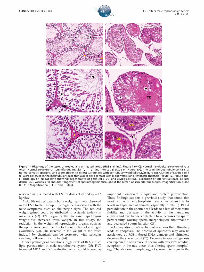
Histological examinationThe normal histological structure of rat testes is illustrated
in Figure 1 (A-C). The seminiferous tubule consists ofnormal somatic and spermatogenic cells and is surroundedby peritubular myoid cells (Figure 1B). Clusters of Leydig’scells were observed in the intertubular space that is in closecontact with blood vessels and lymphatic channels(Figure 1C). However, histopathological observations suchas degeneration of germ cells, expansion of interstitial space,disarrangement of spermatogonia in seminiferous tubule,degeneration of Leydig cells and the presence of cellulardebris throughout the lumen of the seminiferous tubule ofrat testes were observed in the FNT group (Figure 1 D–F).
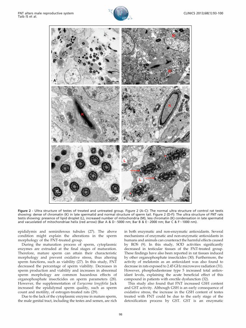
The ultra-structure of the rat testis showed morphologicalalterations in the treated groups compared with the controlgroup (Figure 2). Abnormalities included the presence of alipid droplet, increased number of mitochondria, lesschromatin condensation in the late spermatid and avacuolated mitochondrial helix (Figure 2 D-F). However,histopathological and ultra-structure alterations were onlyobserved in approximately 40% of the treated rat population.
& DISCUSSION
The extensive use of organophosphate in the environmenthas affected many non-target organisms, including humans.The high lipid solubility of fenitrothion may lead to thebioaccumulation of the parent compound and its toxicmetabolites in the adipose tissue. The release of thiscompound from the fat storage could be enhanced in stressfulsituations, such as during illness and diet changes (20). Thecurrent toxicity scenarios have attracted the attention ofresearchers studying the toxic effects of organophosphate onthe male reproductive system. FNT has been found to causesome reproductive abnormalities in Japanese sprayers, with areduction in the sperm quality (21).
This study showed that the administration of FNT at a doseof 20 mg/kg bw for 28 days in male Sprague-Dawley ratsinduced significant adverse effects, including toxicity signsand symptoms, alterations in the biochemical status, reduc-tions in sperm quality and histopathological and ultrastruc-tural changes in the testes. FNT administration inhibits theacetyl cholinesterase enzymes that cause acetylcholineaccumulation in cholinergic synapses. Therefore, cholinergicsigns were observed, including hypoactivity, lacrimation,piloerection and tremor. Significant inhibition of acetylcholinesterase enzyme by FNT directly supports the commonacute toxicity mechanism of organophosphate poisoning (1).These findings are generally consistent with the studies byTurner (22), who reported that the muscle tremor was
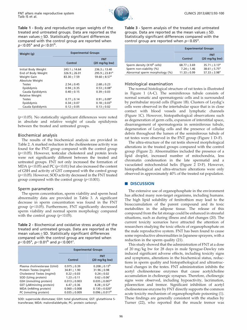
Table 1 - Body and reproductive organ weights of thetreated and untreated groups. Data are reported as themean values¡SD. Statistically significant differencescompared with the control group are reported whenp,0.05a and p,0.01b.
Weight (g) Experimental Groups
Control
FNT
(20 mg/kg bw)
Initial Body Weight 243.1¡14.64 236.3¡13.40
End of Body Weight 326.9¡26.01 295.9¡23.81a
Weight Gain 83.30¡7.59 59.60¡8.57a
Absolute Weight
Testes 2.54¡0.45 2.68¡0.23
Epididymis 0.94¡0.35 0.53¡0.08a
Cauda Epididymis 0.40¡0.15 0.39¡0.03
Relative Weight
Testes 0.77¡0.14 0.91¡0.09a
Epididymis 0.34¡0.07 0.18¡0.03b
Cauda Epididymis 0.12¡0.05 0.13¡0.02
Table 2 - Biochemical and oxidative stress analysis of thetreated and untreated groups. Data are reported as themean values¡SD. Statistically significant differencescompared with the control group are reported whenp,0.05a, p,0.01b and p,0.001c.
Experimental Groups
Control
FNT
(20 mg/kg bw)
Plasma cholinesterase (U/ml) 0.975¡0.39 0.208¡0.13b
Protein Testes (mg/ml) 34.81¡1.90 31.96¡0.98
Cholesterol Testes (mg/ml) 0.22¡0.03 0.24¡0.02
SOD (U/mg protein) 1.23¡0.11 0.62¡0.06c
GSH (mmol/mg protein) 0.013¡0.003 0.035¡0.007a
GST (mM/min/mg protein) 6.47¡0.36 8.28¡0.52a
MDA (mM/mg protein) 0.060¡0.008 0.130¡0.020a
PC (nmol/mg protein) 0.035¡0.009 0.096¡0.017b
SOD: superoxide dismutase; GSH: total glutathione; GST: glutathione S-
transferase; MDA: malondialdehyde; PC: protein carbonyl.
Table 3 - Sperm analysis of the treated and untreatedgroups. Data are reported as the mean values¡SD.Statistically significant differences compared with thecontrol group are reported when p,0.05a.
Experimental Groups
Control
FNT
(20 mg/kg bw)
Sperm density (X106 cells) 68.77¡3.69 35.71¡3.13a
Sperm non-viability (%) 7.24¡1.46 38.61¡5.75a
Abnormal sperm morphology (%) 11.33¡0.99 57.33¡3.98a
FNT alters male reproductive systemTaib IS et al.
CLINICS 2013;68(1):93-100
96
observed in rats treated with FNT at doses of 20 and 25 mg/kg/day.
A significant decrease in body weight gain was observedin the FNT treated group; this might be associated with thetoxic symptoms, such as cholinergic signs. The reducedweight gained could be attributed to systemic toxicity inmale rats (23). FNT significantly decreased epididymisweight but increased testis weight. In this study, thereduction in the weight of reproductive organs, such asthe epididymis, could be due to the reduction of androgenavailability (23). The increase in the weight of the testesinduced by chemicals can be correlated with testicularswelling, followed by atrophy (24).
Under pathological conditions, high levels of ROS inducelipid peroxidation in male reproductive system (25). FNTincreased MDA and PC production, which could be used as
important biomarkers of lipid and protein peroxidation.These findings support a previous study that found thatmost of the organophosphate insecticides altered MDAlevels in experimental animals, especially in rats (5). PUFAperoxidation in the sperm head leads to a loss of membranefluidity and decrease in the activity of the membraneenzyme and ion channels, which in turn increases the spermpermeability, causing sperm morphological abnormalitiesand decreased sperm function (26).
ROS may also initiate a chain of reactions that ultimatelyleads to apoptosis. The process of apoptosis may also beaccelerated by ROS-induced DNA damage and ultimatelydecrease the sperm count (26). Decrease in spermatogenesiscan explain the occurrence of sperm with excessive residualcytoplasm in the mid-piece, thus altering sperm morphol-ogy. The abnormal morphology of sperm may occur in the
Figure 1 - Histology of the testes of treated and untreated group (H&E staining). Figure 1 (A–C): Normal histological structure of rat’stestis. Normal structure of seminiferous tubules ( ) and interstitial tissue (*)(Figure 1A). The seminiferous tubule consists ofnormal somatic, sperm (S) and spermatogenic cells (G) surrounded with peritubularmyoid cells (My)(Figure 1B). Clusters of Leydig’s cells(L) were observed in the intertubular space that was in close contact with blood vessels and lymphatic channels (Figure 1C). Figure 1(D–F): Histology of FNT rat testis showing: degenerative of germ cells (DG) and Leydig cells (DL), expansion of interstitsial space, cellulardebris (CD), vacuoles (v) and disarrangement of spermatogonia throughout the lumen of seminiferous tubule. (Magnification A andD,X10; Magnification B, C, E and F,X40).
CLINICS 2013;68(1):93-100 FNT alters male reproductive systemTaib IS et al.
97
epididymis and seminiferous tubules (27). The abovecondition might explain the alterations in the spermmorphology of the FNT-treated group.
During the maturation process of sperm, cytoplasmicenzymes are extruded at the final stages of maturation.Therefore, mature sperm can attain their characteristicmorphology and prevent oxidative stress, thus alteringsperm functions, such as viability (27). In this study, FNTdecreased the percentage of sperm viability. Decreases insperm production and viability and increases in abnormalsperm morphology are common hazardous effects oforganophosphate insecticides on sperm parameters (28).However, the supplementation of Eurycoma longifolia Jackincreased the epididymal sperm quality, such as spermcount and motility, of estrogen-treated rats (29).
Due to the lack of the cytoplasmic enzyme in mature sperm,the male genital tract, including the testes and semen, are rich
in both enzymatic and non-enzymatic antioxidants. Severalmechanisms of enzymatic and non-enzymatic antioxidants inhumans and animals can counteract the harmful effects causedby ROS (9). In this study, SOD activities significantlydecreased in testicular tissues of the FNT-treated group.These findings have also been reported in rat tissues inducedby other organophosphate insecticides (30). Furthermore, theactivity of melatonin as an antioxidant was also found todecrease in rats exposed to 2.45 GHz microwave radiation (31).However, phosphodiesterase type 5 increased total antiox-idant levels, explaining the acute beneficial effect of thiscompound in patients with erectile dysfunction (32).
This study also found that FNT increased GSH contentand GST activity. Although GSH is an early consequence ofoxidative stress, the increase in the GSH content of testestreated with FNT could be due to the early stage of thedetoxification process by GST. GST is an enzymatic
Figure 2 - Ultra structure of testes of treated and untreated group. Figure 2 (A–C): The normal ultra structure of control rat testisshowing: dense of chromatin (K) in late spermatid and normal structure of sperm tail. Figure 2 (D-F): The ultra structure of FNT ratstestis showing: presence of lipid droplet (L), increased number of mitochondria (M), less chromatin (K) condensation in late spermatidand vacuolated of mitochondriae helix (red arrow) (Bar A & D,5000 nm; Bar B & E,2000 nm; Bar C & F,1000 nm).
FNT alters male reproductive systemTaib IS et al.
CLINICS 2013;68(1):93-100
98
antioxidant that is involved in the detoxification oforganophosphate insecticides to form a non-toxic product(33). Organophosphate insecticides have been reported toalter the biochemical status in experimental animalsdepending on the various types of organophosphates,exposures durations and doses (34). There is a slightdifference in the total protein and cholesterol content intestes of the FNT group. Changes in protein and cholesterollevels in the testes have been attributed to the inhibition ofthe androgen concentration (34).
The above findings were supported by the histopatholo-gical changes in sperm and testis induced by FNT. FNTcaused pathological changes in seminiferous tubules follow-ing a 28-day exposure. The apoptosis induced by ROShelped remove abnormal germ cells in the testes andprevent their overproduction by activating caspases (10).This explained the degenerative nature of germ andLeydig’s cells found in the seminiferous tubule andinterstitial tissues of the FNT-treated group. FNT alsoallowed the crossing of the blood-testes barrier, therebydecreasing the spermatogenesis. These findings supportedthe study performed by Turner (22), who found that FNTinhibited the maturation and differentiation of germ cells.Toxicants can directly disturb the function of Sertoli cells,thereby causing the disorganization of germ cells in theseminiferous tubule of the FNT-treated group. In this study,FNT increased the formation of vacuoles and number ofmitochondria in the testes. Vacuoles can be described as anearly stage of damage induced by any toxicants (35). Inaddition, the increase in the number of mitochondria maybe attributed to the high-energy consumption of the redoxprocess in testes (9). FNT also caused fewer chromatins inlate spermatids and vacuolated mitochondria of the spermtail. A decrease in the chromatin in late spermatids may bedue to genetic abnormalities that occurred during thedifferential process in spermatogenesis (28).
In conclusion, FNT at a dose of 20 mg/kg bw has adetrimental effect on the sperm and testes of Sprague-Dawley rats.
& ACKNOWLEDGMENTS
The authors would like to thank the Ministry of Higher Education and
Universiti Kebangsaan Malaysia for research funds (UKM-GUP-2011-
126). We also extend our gratitude to the Director of Institute for Medical
Research (IMR), Malaysia, for the permission to use the electron
microscopy facilities. The authors gratefully acknowledge the staff of
Programme of Biomedical Sciences, School of Diagnostic and Applied
Health Sciences, Faculty of Health Sciences, Universiti Kebangsaan
Malaysia, and Electron Microscopy Unit, Institute for Medical Research
(IMR), Malaysia, for providing the research facilities. We would like to
acknowledge all of the lecturers, researchers and those who directly or
indirectly supported this research.
& AUTHOR CONTRIBUTIONS
Taib IS performed the majority of the experiments and drafted the
manuscript. Budin SB was the project leader and was responsible for the
project design. Ghazali AR and Mohamed J revised the manuscript for
important intellectual content. Jayusman PA and Louis SR performed
some of the experiments. All authors read and approved the final
manuscript.
& REFERENCES
1. Sarikaya R, Selvı M, Erkoc F. Investigation of acute toxicity offenitrothion on peppered corydoras (Corydoras paleatus) (Jenyns,
1842). Chemosphere. 2004;56(7):697-7002, http://dx.doi.org/10.1016/j.chemosphere.2004.04.008.
2. Uygun U, Koksel H, Atli A. Residue levels of malathion and itsmetabolites and fenitrothion in post-harvest treatment wheat duringstorage, milling and baking. Food Chem. 2005;92:643-7, http://dx.doi.org/10.1016/j.foodchem.2004.08.045.
3. Derbalah AS, Nakatani N, Sakugawa H. Photocatalytic removal offenitrothion in pure and natural waters by photo-Fenton reaction.Chemosphere. 2004;57(7):635-44, http://dx.doi.org/10.1016/j.chemosphere.2004.08.025.
4. Crathorne B, Dobbs AJ. Chemical pollution of the aquatic environmentby priority pollutants and its control. In: Harrison RM, editor. Pollution:Causes, Effcets and Control. 2 ed. Cambridge, United Kingdom.: RoyalSociety of Chemistry; 1990.p.1 - 18.
5. Elhalwagy MEA, Darwish NS, Zaher EM. Prophylactic effect of green teapolyphenols against liver and kidney injury induced by fenitrothioninsecticide. Pest Biochem Physiol. 2008;91(2):81-9, http://dx.doi.org/10.1016/j.pestbp.2008.01.006.
6. Hayes WJJ, Laws ERJ. Fenitrothion. Handbook of Pesticide Toxicology,Classes of Pesticides. San Diego: Academic Press; 1991,1020-3.
7. Tamura H, Maness SC, Reischmann K, Dorman DC, Gray LE, Gaido KW.Androgen receptor antagonism by the organophosphate insecticidefenitrothion. Toxicol Sci. 2001;60(1):56-62, http://dx.doi.org/10.1093/toxsci/60.1.56.
8. Milatovic D, Gupta RC, Aschner M. Anticholinesterase toxicity,oxidative stress. Sci World J. 2006;6:295-310, http://dx.doi.org/10.1100/tsw.2006.38.
9. Lukaszewicz-Hussain A. Role of oxidative stress in organophosphateinsecticide toxicity - Short review. Pest Biochem Physiol. 2010;98:145-50,http://dx.doi.org/10.1016/j.pestbp.2010.07.006.
10. Agarwal A, Allamaneni S. Oxidative stress and human reproduction. In:Singh K, editor. Oxidative stress, disease and cancer. Singapore:Mainland Press; 2006. 687-703.
11. Ellman G, Courtney K, Andres VJ, Featherstone M. A new and rapidcolorimetric determination of acetylcholinesterase activity. BiochemPharmacol. 1961;7:88-95, http://dx.doi.org/10.1016/0006-2952(61)90145-9.
12. Bradford M. Rapid and sensitive method for the quantitation ofmicrogram quantities of protein utilizing the principle of protein-dyebinding. Analytical Biochemistry. 1976;72:248-54, http://dx.doi.org/10.1016/0003-2697(76)90527-3.
13. Franey R, Amador E. Serum cholesterol measurement based on ethanolextraction and ferric chloride-sulphuric acid. Clin Chim Acta.1968;21(2):255-63, http://dx.doi.org/10.1016/0009-8981(68)90135-6.
14. Stocks J, Dormandy TL. The autooxidation of human red cell lipidsinduced by hydrogen peroxide. J Hematol. 1971;20(1):95-111, http://dx.doi.org/10.1111/j.1365-2141.1971.tb00790.x.
15. Levine RL, Garland D, Oliver C, Amici A, Climent I, Lenz A, et al.Determination of carbonyl content in oxidatively modified proteins.Enzymol. 1990;186:464-78, http://dx.doi.org/10.1016/0076-6879(90)86141-H.
16. Ellman G. Tissue sulfhydryl groups. Arch Biochem Biophys. 1959;82(1):70-7, http://dx.doi.org/10.1016/0003-9861(59)90090-6.
17. Beyer W, Fridovich I. Assaying for superoxide dismutase activity: Somelarge consequences of minor changes in conditions. Anal Biochem.1987;161(2):559-66, http://dx.doi.org/10.1016/0003-2697(87)90489-1.
18. Habig WH, Pabst MJ, Jakoby WB. Glutathione S-Transferase. The firstenzymatic step in mercapturic acid formation. J Biol Chem.1974;249(22):7130-9.
19. WHO. Laboratory Manual for the Examination of Human Semen andSperm-Cervical Mucus Interactions. 4th, editor. Cambridge, UnitedKingdom: Cambridge University Press; 1999.
20. Ecobichon DJ, Ozere RL, Reid E, Crocker JFS. Acute fenitrothionpoisoning. Can Med Assoc J. 1977;166(4):377-9.
21. Kamijima M, Hibi H, Gotoh M, Taki K, Saito I, Wang H, et al. A survey ofsemen indices in inecticide sprayes. J Occup Health. 2004;46(2):109-18,http://dx.doi.org/10.1539/joh.46.109.
22. Turner KJ, Barlow NJ, Struve MF, Wallace DG, Gaido KW, Dorman DC,et al. Effects of in Utero Exposure to the Organophosphate InsecticideFenitrothion on Androgen-Dependent Reproductive Development in theCrl:CD(SD)BR Rat. Toxicol Sci. 2002;68(1):74-83.
23. Mylchreest E, Malley LA, O’Neill AJ, Kegelman TA, Sykes GP, ValentineR. Reproductive and developmental toxicity of inhaled 2,3-dichloro-1,3-butadiene in rats. Reprod Toxicol. 2006;22(4):613-22, http://dx.doi.org/10.1016/j.reprotox.2006.04.002.
24. Hess R, Bunick D, Lee K, Bahr J, Taylor J, Korach K, et al. A role forestrogens in male reproductive system. Nature. 1997;390(6659):509-12,http://dx.doi.org/10.1038/37352.
25. Kalender Y, Kaya S, Durak D, Uzun FG, Demir F. Protective effects ofcatechin and quercetin on antioxidant status, lipid peroxidation andtestis-histoarchitecture induced by chlorpyrifos in male rats. EnvironToxicol Pharmacol. 2010;3(2):141-8.
26. Agarwal A, Said TM. Role of sperm chromatin abnormalities and DNAdamage in male infertility. Human reproductive Update 2003;9(4):331-45, http://dx.doi.org/10.1093/humupd/dmg027.
CLINICS 2013;68(1):93-100 FNT alters male reproductive systemTaib IS et al.
99
27. Gil-Guzman E, Ollero M, Lopez MC, Sharma RK, Alvarez JG, ThomaAJJ, et al. Differential production of reactive oxygen species by subsets ofhuman spermatozoa at different stages of maturation. Hum Reprod.2001;16(9):1922-30, http://dx.doi.org/10.1093/humrep/16.9.1922.
28. Burruel VR, Raabe OG, Overstreet JW, Wilson BW, Wiley LM. PaternalEffects from Methamidophos Administration in Mice. Toxicol ApplPharmacol. 2000;165(2):148-57, http://dx.doi.org/10.1006/taap.2000.8933.
29. Wahab NA, Mokhtar NM, Halim WNHA, Das S. The effect of eurycomalongifolia Jack on spermatogenesis in estrogen-treated rats. Clinics.2010;65(1):93-8, http://dx.doi.org/10.1590/S1807-59322010000100014.
30. Cemek M, Buyukben A, Buyukokuroglu ME, Aymelek F, Tur L.Protective roles of vitamin E (a-tocopherol), selenium and vitamin Eplus selenium in organophosphate toxicity in vivo: A comparative study.Pest Biochem Physiol. 2010;96(3):113-8, http://dx.doi.org/10.1016/j.pestbp.2009.09.009.
31. Kumar S, Kesari KK, Behari J. The therapeutic effect of a pulsedelectromagnetic field on the reproductive patterns of male Wistar rats
exposed to a 2.45-GHz microwave field. Clinics. 2011;66(7):1237-45,http://dx.doi.org/10.1590/S1807-59322011000700020.
32. Savas M, Yeni E, Verit A, Gulum M, Aksoy N, Ciftci H, et al. Acute effect ofphosphodiesterase type 5 inhibitor on serum oxidative status andprolidase activities in men with erectile dysfunction. Clinics.2010;65(12):1311-4, http://dx.doi.org/10.1590/S1807-59322010001200014.
33. Ranjbar A, Solhi H, Mashayekhi F, Susanabdi A, Rezaiec A, Abdollahi M.Oxidative stress in acute human poisoning with organophosphorusinsecticides: a case control study. Environment Toxicology andPharmacology. 2005;20(1):88-91, http://dx.doi.org/10.1016/j.etap.2004.10.007.
34. Sharma AK, Kapadia A, Fransis P, Rao M. Reversible effects of mercuricchloride on reproductive organs of the male mouse. Reprod Toxicol.1996;10(2):153-9, http://dx.doi.org/10.1016/0890-6238(95)02058-6.
35. Creasy D. Pathogenesis of male reproductive toxicity. Toxicol Pathol.2001;29(1):64-76, http://dx.doi.org/10.1080/019262301301418865.
FNT alters male reproductive systemTaib IS et al.
CLINICS 2013;68(1):93-100
100