Osteoblastoma of the Sternum - Tumor Surgery of the... · · 2012-08-27bones of the foot and...
5
55 Bulletin of the NYU Hospital for Joint Diseases 2010;68(1):55-9 Villalobos CE, Rybak LD, Steiner GC, Wittig JC. Osteoblastoma of the sternum: case report and review of the literature. Bull NYU Hosp Jt Dis. 2010;68(1):55-9. Abstract Osteoblastoma is an extremely rare entity that represents less than 1% of all bone tumors, and affects twice as many males as females with peak incidence between 15 and 20 years. Osteoblastomas commonly affect axial bones, long bones, bones of the foot and hand, and less commonly the pelvis, scapula, ribs, and clavicle. Osteoblastoma does not have a classic presentation, but can vary with the location and size of the tumor. The main complaint is often progressive pain localized at the tumor site. Osteoblastoma is a benign tumor with an aggressive behavior. The treatment is wide surgical resection, otherwise it continues to enlarge and destroy the bone and surrounding structures. We report a 32-year-old male with an osteoblastoma of this sternum who was treated with an en-bloc resection and reconstruction with Marlex ® and a methylmethacrylate plate. O steoblastoma is a rare, benign primary tumor of bone. There have been only three cases of osteo- blastoma in the sternum reported in the literature. The current case report is of a benign osteoblastoma in the sternum of a 32-year-old male. Tumors arising from the bones of the chest wall are uncommon, but, when they occur, they are usually malignant. Initially described by Jaffe, in 1932, 1 and later, again, by Jaffe 2 and Lichtenstein, 3 in 1956, osteoblastoma also was known as giant osteoid osteoma, as its histology resembled that of an osteoid osteoma. Osteo- blastoma affects twice as many males as females and has a peak incidence in patients between 15 and 20 years of age; 90% of cases are diagnosed before the age 30. Osteoblas- toma commonly affects the vertebral column; long bones, such as the femur and tibia; and also the foot and hand. Less commonly, the pelvis, scapula, ribs, and clavicle are sites. Osteoblastoma is a benign entity but sometimes demon- strates aggressive growth; however, it can be differentiated radiologically and histologically from osteosarcoma. We report the clinical presentation, radiologic and pathologic findings, and surgical and medical management, with a review of the literature. Case Report A 32-year-old male presented with a mass in the region of his left sternoclavicular joint. He complained of swelling in the area for 5 months prior to his consultation. The mass was mildly painful and had progressively increased in size. He experienced chest pain at night, which would awaken him from sleep and was only partially relived by nonsteroidal antiinflammatory medication. There was no history of fever, though he did complain of occasional night sweats and some weight loss. There was no parasthesia, except in his left up- per extremity. His past medical history was significant for asthma. He had no history of infections, intravenous drug abuse, tuberculosis, or malignancy. He never had had sur- gery. His mother had a history of breast cancer; otherwise, there was no further significant family history. On physical examination, the patient had a very firm and immobile mass (approximately, 5 cm) over his manubrium and left sternoclavicular joint, with mild tenderness on palpation. He had full range of motion throughout his left upper limb. Osteoblastoma of the Sternum Case Report and Review of the Literature Camilo E. Villalobos, M.D., Leon D. Rybak, M.D., German C. Steiner, M.D., and James C. Wittig, M.D. Camilo E. Villalobos, M.D., is a Research Assistant in the Division of Orthopaedic Oncology, Department of Orthopaedic Surgery, Mount Sinai Medical Center, New York, New York. Leon D. Ry- bak, M.D., is an Assistant Professor, NYU School of Medicine, and an Attending in the Division of Radiology, NYU Hospital for Joint Diseases, New York, New York. German C. Steiner, M.D., is Professor of Pathology, NYU School of Medicine, and Director of Pathology, NYU Hospital for Joint Diseases, New York, New York. James C. Wittig, M.D., is an Associate Professor of Orthopaedic Surgery, and Chief of the Division of Orthopaedic Oncology, Mount Sinai Medical Center, New York, New York; and is Chief of the Division of Orthopaedic Oncology, Hackensack University Medical Center, Hackensack, New Jersey. Correspondence: James C. Wittig, M.D., Department of Ortho- paedic Surgery, Box 1188, Mount Sinai Medical Center, 5 East 98th Street, New York, New York 10029; [email protected].
Transcript of Osteoblastoma of the Sternum - Tumor Surgery of the... · · 2012-08-27bones of the foot and...

55Bulletin of the NYU Hospital for Joint Diseases 2010;68(1):55-9
Villalobos CE, Rybak LD, Steiner GC, Wittig JC. Osteoblastoma of the sternum: case report and review of the literature. Bull NYU Hosp Jt Dis. 2010;68(1):55-9.
Abstract
Osteoblastoma is an extremely rare entity that represents less than 1% of all bone tumors, and affects twice as many males as females with peak incidence between 15 and 20 years. Osteoblastomas commonly affect axial bones, long bones, bones of the foot and hand, and less commonly the pelvis, scapula, ribs, and clavicle. Osteoblastoma does not have a classic presentation, but can vary with the location and size of the tumor. The main complaint is often progressive pain localized at the tumor site. Osteoblastoma is a benign tumor with an aggressive behavior. The treatment is wide surgical resection, otherwise it continues to enlarge and destroy the bone and surrounding structures. We report a 32-year-old male with an osteoblastoma of this sternum who was treated with an en-bloc resection and reconstruction with Marlex® and a methylmethacrylate plate.
Osteoblastoma is a rare, benign primary tumor of bone. There have been only three cases of osteo-blastoma in the sternum reported in the literature.
The current case report is of a benign osteoblastoma in the sternum of a 32-year-old male. Tumors arising from the
bones of the chest wall are uncommon, but, when they occur, they are usually malignant. Initially described by Jaffe, in 1932,1 and later, again, by Jaffe2 and Lichtenstein,3 in 1956, osteoblastoma also was known as giant osteoid osteoma, as its histology resembled that of an osteoid osteoma. Osteo-blastoma affects twice as many males as females and has a peak incidence in patients between 15 and 20 years of age; 90% of cases are diagnosed before the age 30. Osteoblas-toma commonly affects the vertebral column; long bones, such as the femur and tibia; and also the foot and hand. Less commonly, the pelvis, scapula, ribs, and clavicle are sites. Osteoblastoma is a benign entity but sometimes demon-strates aggressive growth; however, it can be differentiated radiologically and histologically from osteosarcoma. We report the clinical presentation, radiologic and pathologic findings, and surgical and medical management, with a review of the literature.
Case ReportA 32-year-old male presented with a mass in the region of his left sternoclavicular joint. He complained of swelling in the area for 5 months prior to his consultation. The mass was mildly painful and had progressively increased in size. He experienced chest pain at night, which would awaken him from sleep and was only partially relived by nonsteroidal antiinflammatory medication. There was no history of fever, though he did complain of occasional night sweats and some weight loss. There was no parasthesia, except in his left up-per extremity. His past medical history was significant for asthma. He had no history of infections, intravenous drug abuse, tuberculosis, or malignancy. He never had had sur-gery. His mother had a history of breast cancer; otherwise, there was no further significant family history. On physical examination, the patient had a very firm and immobile mass (approximately, 5 cm) over his manubrium and left sternoclavicular joint, with mild tenderness on palpation. He had full range of motion throughout his left upper limb.
Osteoblastoma of the SternumCase Report and Review of the Literature
Camilo E. Villalobos, M.D., Leon D. Rybak, M.D., German C. Steiner, M.D., and James C. Wittig, M.D.
Camilo E. Villalobos, M.D., is a Research Assistant in the Division of Orthopaedic Oncology, Department of Orthopaedic Surgery, Mount Sinai Medical Center, New York, New York. Leon D. Ry-bak, M.D., is an Assistant Professor, NYU School of Medicine, and an Attending in the Division of Radiology, NYU Hospital for Joint Diseases, New York, New York. German C. Steiner, M.D., is Professor of Pathology, NYU School of Medicine, and Director of Pathology, NYU Hospital for Joint Diseases, New York, New York. James C. Wittig, M.D., is an Associate Professor of Orthopaedic Surgery, and Chief of the Division of Orthopaedic Oncology, Mount Sinai Medical Center, New York, New York; and is Chief of the Division of Orthopaedic Oncology, Hackensack University Medical Center, Hackensack, New Jersey. Correspondence: James C. Wittig, M.D., Department of Ortho-paedic Surgery, Box 1188, Mount Sinai Medical Center, 5 East 98th Street, New York, New York 10029; [email protected].

Bulletin of the NYU Hospital for Joint Diseases 2010;68(1):55-956
No motor or sensory deficits were noted in the extremities bilaterally. Pulses and reflexes were normal. Blood work, including complete blood count, chemistries, coagulation analysis, erythrocyte sedimentation rate (ESR) and C-reactive protein (CRP) were all normal. Plain radiographs of the chest were interpreted as normal. Axial (Fig. 1A) and coronally reconstructed (Fig. 1B) comput-ed tomography (CT) images demonstrated a large geographic lobulated lesion involving the left side of the manubrium. The lesion was expansile, extending into the overlying ventral soft tissues, as well as posteriorly into the mediastinum. The soft tissue component was surrounded by a rim of reactive bone. Faint areas of internal mineralization were noted. No periosteal reaction was identified. The lesion measured ap-proximately 4x3.3x4.4 cm. On magnetic resonance imaging (MRI), the lesion demonstrated a lobular configuration with slight heterogeneity. The lesion was predominantly isointense to muscle, with a hyperintense central signal and a thin sur-rounding rim of low signal on axial (Fig. 2A) and sagittal (Fig. 2B) T1-weighted images. On axial T2-weighted images (Fig. 2C), the lesion also was heterogeneous, but predominantly hyperintense in signal, with mild surrounding soft tissue edema. The lesion demonstrated uptake on bone scan, with no evidence of multicentric or metastatic disease. The patient underwent a CT-guided biopsy. Multiple cores of soft tissue were obtained from the lesion. Patho-logical analysis revealed an osteoblastoma. The patient then underwent en bloc resection of the tumor (sternum, medial clavicular head, and part of pectoralis major). The sternum was reconstructed with Marlex® mesh and methylmethacrylate (Fig. 3). A plate of methylmethacrylate was placed between two sheets of abdominal mesh. The methylmethacrylate plate was trimmed to fit the size of the defect. Heavy nonabsorbable sutures were utilized to suture the medial ends of the ribs and clavicles to the abdominal mesh. A contralateral rotational rectus abdominis muscle flap was used to cover the plate of
methacrylate. The pectoralis major was sutured to the edges of the rectus abdominis muscle. There were no intraoperative or postoperative complications. The entire specimen measured 10.5x9x5.5 cm. The tis-sue had a red-tan appearance and gritty consistency. The cut gross section revealed a well demarcated mass in the sternum measuring 4.5x3.7x3.8 cm. Histological examination revealed a bone-forming tumor, composed of anastomosing immature bone trabeculae, surrounded by a rim of osteoblasts, and separated by a fibrovascular stroma. There were variable amounts of osteoid and bone. Some osteoblasts showed atypi-cal changes. The mitotic activity was low. The margins of the host bone-tumor interface were sharp, and well circumscribed. There was no evidence of tumor permeation. The above his-tologic features were consistent with osteoblastoma (Fig. 4). There have been no complications or complaints related to the reconstruction after 3 years postoperatively. The patient is pain free and has full function of both upper extremities. He has no respiratory problems, and he exercises without shortness of breath.
DiscussionOsteoblastoma is a rare, benign primary bone tumor, likely related to osteoid osteoma. Initially, it was described by Jaffe and Mayer,1 in 1932, under the descriptive name of “An osteo-blastic osteoid tissue-forming tumor of a metacarpal bone.” Later, both Jaffe2 and Lichtenstein,3 in 1956, and Lichtenstein and Sawyer,4 in 1964, described osteoblastoma in various ar-ticles under the title “Benign osteoblastoma.” Osteoblastomas are rare and represent less than 1% of all bone tumors and approximately 3.5% of all benign bone tumors.5-7 In Osteoblastoma, males are affected more often than females, with a respective ratio of 2:1. Most patients are between 15 and 20 years of age and approximately 90% of cases are diagnosed before the age of 30.6,7 The mean age at presentation in the largest series studied was 20.4 years, with
BA
Figure 1 A, An axial CT image with bone windows demonstrates a large somewhat expansile and lobulated lytic mass involving the left side of the manubrium. The lesion extends into the overlying ventral soft tissues as well as posteriorly into the mediastinum. There is thinning of the cortex without evidence of cortical discontinuity, pathologic fracture, or associated soft tissue mass. Faint areas of internal calcification are noted. B, A coronal CT reconstruction demonstrating the craniocaudal dimensions of the lesion.
57Bulletin of the NYU Hospital for Joint Diseases 2010;68(1):55-9
wide range from 6 months to 75 years.8 Osteoblastoma occurs four-times less frequently than osteoid osteoma.9 There is a predilection for the axial skeleton, particularly the posterior elements of the vertebral column. The next most common site is the long bones of the extremities, although it also has been reported in the hands, feet, skull, maxilla, scapula, clavicle, and ribs.5-7,10 Osteoblastomas usually arise from the medullary cavity of the bones; periosteal origin is rare.4 There have been only three cases reported in the literature of osteoblastoma in the sternum: two in adults and one in a child.5,11,12 Osteoblastoma does not have a specific clinical presenta-tion. The presentation varies with the location and size of the tumor. The main complaint is often progressive pain, localized at the tumor site. It is often a dull aching pain, sometimes worse at night and is not relieved with salicylates, as with osteoid osteomas.13 The affected bone may be expanded and appear as a palpable mass, associated with tenderness and swelling. The lower extremity may cause gait problems. Pa-tients with an osteoblastoma localized in the vertebral column may complain of radiating pain, tingling, weakness, pares-thesias, and even paraplegia.3,6,7,9 Sometimes, osteoblastomas may be asymptomatic and are diagnosed incidentally.13,14 Radiologic studies are useful in establishing the diagnosis. Plain radiographs may vary according to the stage of tumor
at the time of presentation.2,14 Generally, the lesions appear as well-defined expansile radiolucent lesions, sometimes as-sociated with delicate, thin internal trabeculae and a rim of surrounding sclerosis.8,9 Osteoblastomas may demonstrate subtle internal calcifications, but mineralization is generally not detectable on plain radiograph. There also may be sur-
C
BA
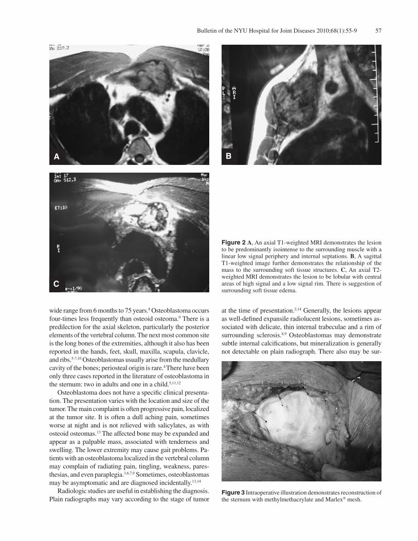
Figure 2 A, An axial T1-weighted MRI demonstrates the lesion to be predominantly isointense to the surrounding muscle with a linear low signal periphery and internal septations. B, A sagittal T1-weighted image further demonstrates the relationship of the mass to the surrounding soft tissue structures. C, An axial T2-weighted MRI demonstrates the lesion to be lobular with central areas of high signal and a low signal rim. There is suggestion of surrounding soft tissue edema.
Figure 3 Intraoperative illustration demonstrates reconstruction of the sternum with methylmethacrylate and Marlex® mesh.

Bulletin of the NYU Hospital for Joint Diseases 2010;68(1):55-958
rounding periosteal new bone formation.2,5 The size of an osteoblastoma is usually greater than 2 cm and can range from 2 to 8 cm. This is generally larger than the presentation of an osteoid osteoma.2,5,9 Lucas and colleagues8 reported on 306 cases of osteoblastoma, in which the size ranged from 1 to 11 cm, with a mean of 3.2 cm. CT scans can provide useful information regarding the size, precise location, and any soft tissue extension of the lesion. This may be particularly useful with spinal lesions. CT scans may also delineate the presence of subtle mineralization; thus, aiding in diagnosis, as well as providing information neces-sary for the planning of a surgical procedure. Osteoblastomas demonstrate increased uptake on a bone scan. Bone scans are useful to localize the lesion and detect multicentric lesions, but have limited resolution in some cases.5 MRI is helpful for determining intraosseous and soft tissues extension and in terms of vertebral tumors and en-croachment on the spinal cord and neural elements.5,15 Kroon and Schumars16 and Cervoni and coworkers15 described low to intermediate signal intensity on T1-weighted images and high signal intensity on T2-weighted images. The degree of enhancement on post Gadolinium images also has been vari-ably reported from none to strong enhancement.15 Thus, there does not appear to be a specific, characteristic appearance of osteoblastoma with MRI. The pathological features of osteoblastoma are similar to osteoid osteoma. Both are bone producing lesions that arise from osteoblastic origin. Osteoblastomas are larger than osteoid osteomas and appear to have unlimited growth potential. Usually, the lesion is reddish-brown, even pinkish; friable, with a gritty consistency; and hemorrhagic in appear-ance.2,4,9.14 The cortex is frequently thinned and expanded.9 Osteoblastomas are highly vascularized and sometimes can show secondary aneurysmal bone cyst formation.8,10 The osteoblast cells are often plump and pleomorphic. The nuclei vary from small to big, and there is usually abundant cytoplasm. The osteoblasts are responsible for osteoid forma-
tion and usually are arranged in an orderly manner, within a lining of primitive trabecular bone (osteoblastic rimming).6 The absence of dysplastic changes, and mitoses, as well as the orderly arrangement of normal appearing osteoblasts, are important features that help distinguish osteoblastoma from osteosarcoma.14 The pathology should always be interpreted after careful review of the radiological studies. There have been some cases of osteoblastomas reported to behave more aggressively. These tumors have been asso-ciated with large epithelioid osteoblasts. These “aggressive osteoblastomas” are more mitotically active than conventional osteoblastoma.8,14 Malignant degeneration has been reported, although it is speculative as to the initial nature of the lesion. At long-term follow-up (10 years), Jackson reported 10% recurrence after incomplete resection.17 Marsh and associ-ates10 and Lucas and colleagues8 collected 197 and 305 cases, respectively, and reported local recurrence in 16% to 21% of cases. The size, location, and ability to resect the entire lesion are important factors related to recurrence, morbidity, and mortality, although metastasis has been rarely documented.8,14 The natural history of osteoblastomas when they are not excised surgically is to continue to enlarge and damage the bone and adjacent structures. They are benign and aggres-sive in nature. Therefore, the treatment of an osteoblastoma is wide surgical resection. Whenever feasible, spine lesions should be removed en bloc. Extensive curettage of long bone lesions may be possible in most cases. Postoperative radiation is usually not recommended6; however, it may be considered for tumors that have recurred multiple times, particularly around the spine. The tumor, in this case, was treated with wide en bloc resection, because it was extending into the surrounding soft tissues and was close to vital structures in the mediastinum. We believe that this approach provided the best surgical treat-ment for minimizing the chance of a local recurrence in this critical region. The sternum was reconstructed with a plate of methylmethacrylate that was sandwiched between two
BA
Figure 4 A, Typical histology features of osteoblastoma showing anastomosing trabeculae of woven bone separated by a fibrovascular stroma. B, High power view of the trabecular bone with prominent osteoblastic rimming.

59Bulletin of the NYU Hospital for Joint Diseases 2010;68(1):55-9
layers of Marlex® mesh. The ribs and clavicles were sutured to the mesh and a rectus abdominus rotational muscle flap was employed to cover the prosthesis. The patient is 4-years postoperative. He is pain free and has no functional limitations and no respiratory difficulties. Many techniques have been described for the closure of chest wall defects to minimize paradoxical movement of the chest and protect underling mediastinal structures after sternal resection. Since 1972, Marlex® mesh filled with methylmeth-acrylate was utilized for complete sternectomy reconstruction, providing excellent stability and cosmetic acceptability.18,19
Chapelien and coworkers20 reported 38 who patients under-went sternal reconstruction. They found that Marlex® mesh alone (without methylmethacrylate) was sufficient for recon-struction following partial sternectomy.21,22 Marlex® mesh filled with methylmethacrylate has been recommended fol-lowing total sternectomy to limit paradoxical chest motion.21 Soysal and associates22 recommended muscle flaps in cases with upper partial sternectomy (manubrium with ribs 1 to 3) and rigid reconstructions (mesh with methylmethacrylate) for cases with lower sternal resections to protect the heart and stabilize the thorax. This has also been proposed by several other authors.19-21 Muscle or myocutaneous flaps are frequently used to repair defects of the chest wall alone or in conjunction with rigid replacements.19,20 The most common muscle flaps used are the latissimus dorsi, pectoralis major, rectus abdominis, ser-ratus anterior, external oblique, trapezius.19 Omental muscle flaps are often used for infected sternal wounds or in cases of previously failed muscle flaps.19 Osteoblastomas are challenging for the physician to diagnose and manage. They should be biopsied carefully to provide enough material to make an accurate diagnosis. High quality imaging studies aid in the diagnosis and surgical planning, and should be correlated with histological material to ensure an accurate diagnosis. Osteoblastomas are char-acterized by both benignity and aggressive behavior, with a propensity for local destruction and recurrence. There have been a few reported cases presenting in the sternum. Surgery is the mainstay of treatment, with the type of surgery depending on the tumor’s size and anatomic location. En bloc resection is recommended for lesions of the sternum that expand or violate the cortex.
Disclosure StatementNone of the authors have a financial or proprietary interest in the subject matter or materials discussed, including, but not limited to, employment, consultancies, stock ownership, honoraria, and paid expert testimony.
References1. Jaffe HL, Mayer L. An osteoblastic osteoid tissue-forming
tumor of a metacarpal bone. Arch Surg. 1932;24:550. 2. Jaffe HL. Benign osteoblastoma. Bull Hosp Jt Dis.
1956;1:141-51.
3. Lichtenstein L. Benign osteoblastoma: a category of osteoid-and bone-forming tumors other than classical osteoid osteoma which may be mistaken for giant-cell tumor or osteogenic sarcoma. Cancer. 1956;9(5):1044-52.
4. Lichtenstein L, Sawyer WR. Benign osteoblastoma: further observations and report of twenty additional cases. J bone Joint Surg Am. 1964;46:755-65.
5. Golant A, Lou J, Erol B, et al. Pediatric osteoblastoma of the sternum: a new surgical technique for reconstruction after removal. Case report and review of literature. J Pediatr Orthop. 2004;24(3):319-22.
6. Huvos AG. Osteoblastoma. In: Bone Tumors: Diagnosis, Treatment and prognosis. (2nd ed). Philadelphia: WB Saun-ders, 1991, pp. 67-83.
7. Unni KK. Benign osteoblastoma (giant osteoid osteoma). In: Unni KK (ed). Dahlin’s Bone Tumors: General Aspect and Data on 11,087 Cases. (5th ed). Philadelphia: Lippincott-Raven, 1996, pp. 131-142.
8. Lucas DR, Unni KK, McLeod RA, et al. Osteoblastoma: clini-copathologic study of 306 cases. Hum Pathol. 1994;25:117.
9. Frassica FS, Waltrip RL, Sponseller PD, et al. Clinicopatho-logic features and treatment of osteoid osteoma and osteo-blastoma in children and adolescents. Orthop Clin North Am. 1996;27(3):559-74.
10. Marsh BW, Bontiglio M, Brandy LP, Enneking WF. Benign osteoblastoma: range of manifestations. J bone Joint Surg Am. 1975;57:1-9.
11. Roberts A, Long J, Wickstrom J. A metacarpal giant cell tumor, a sternal osteoblastoma and pubic osteogenic sarcoma in the same patient. South Med J. 1976;69:660-2.
12. Lin E, Oliver S, Liberman Y, et al. Osteoblastoma of sternum. Arch Orthop Traum Surg. 1987;106:132-4.
13. Greenspan A, Remagen W: Differential Diagnosis of Tumors and Tumor-Like Lesions of Bones and Joints. Philadelphia: Lippincott-Raven, 1998.
14. Moon KS, Jung S, Lee JH, et al. Benign osteoblastoma of the occipital bone: case report and literature review. Neuropathol-ogy. 2006;26:141-6.
15. Cervoni L, Innocenzi G, Raguso M, et al. Osteoblastoma of calvaria: report of two cases diagnoses with MRI and clinical review. Neurosurg Rev. 1997;20:51-4.
16. Kroon HM, Schumars J. Osteoblastoma: clinical and radio-logical findings in 98 new cases. Radilology. 1990;175:783-90.
17. Jackson RP. Recurrent osteoblastoma: a review. Clin Orthop Relat Res. 1978;(131):229-33.
18. Lardinois D, Muller M, Ferrer M, et al. Functional assessment of chest wall integrity after methylmetacrylate reconstruction. Ann Thorac Surg. 2000;69:919-23.
19. Sabathan S, Shah R, Mearns AJ. Surgical treatment of primary chest wall tumors. Eur J Cardiothorac Surg. 1997;11:1011-6
20. Chapelier A, Missana MC, Couturaud B, et al. Sternal resec-tion and reconstruction for primary malignant tumors. Ann Thorac Surg. 2004;77:1001-7.
21. McCormack PM. Use of prosthetic materials in chest wall reconstruction. Surg Clin North Am. 1989;69:965-76.
22. Soysal O, Walsh G, Nesbitt J, et al. Resection of sternal tu-mors: extent, reconstruction and survival. Ann Thorac Surg. 1995;60:1353-9.