On the typical development of stereopsis: Fine and coarse processing
7
On the typical development of stereopsis: Fine and coarse processing Deborah Giaschi a,⇑ , Sathyasri Narasimhan a , Aliya Solski b , Emily Harrison a , Laurie M. Wilcox b a University of British Columbia, Department of Ophthalmology and Visual Sciences, Vancouver, BC, Canada b Centre for Vision Research, Department of Psychology, York University, Toronto, Ontario, Canada article info Article history: Received 16 August 2012 Received in revised form 12 July 2013 Available online 24 July 2013 Keywords: Depth perception Stereopsis Visual development Psychophysics abstract Stereoscopic depth perception may be obtained from small retinal disparities that can be fused for single vision (fine stereopsis), but reliable depth information is also obtained from larger disparities that produce double vision (coarse stereopsis). While there is some evidence that stereoacuity improves with age, little is known about the development and maturation of coarse stereopsis. Here we address this gap by assessing the maturation of stereoscopic depth perception in children (4–14 years) and adults over a large range of disparities from fused (fine) to diplopic (coarse). The observer’s task was to indicate whether a stereoscopic cartoon character was nearer or farther away than a zero-disparity reference frame. The test disparities were grouped into fine (0.02, 0.08, 0.17, 0.33, 0.68, 1.0 deg) and coarse (2.0, 2.5, 3.0, 3.5 deg) ranges based on an initial determination of the diplopia threshold for each observer. Next, percent correct depth direction was determined as a function of disparity. In the coarse range, accu- racy decreased slightly with disparity and there were no differences as a function of age. In the fine range, accuracy was constant across all disparities in adults and increased with disparity in children of all ages. Performance was immature in all children at the finest disparity tested. We conclude that stereopsis in the coarse range is mature at 4 years of age, but stereopsis in the fine range, at least for small disparities, continues to mature into the school-age years. Ó 2013 Elsevier Ltd. All rights reserved. 1. Introduction The conventional view of stereoscopic processing is that the visual input from the two eyes is fused to produce the percept of a single scene. In doing so, the stereoscopic system also provides extremely high-resolution information about the relative depth of objects in space. This ‘fine’ stereopsis has been well documented, and is assessed by clinical tests of stereoacuity (e.g. Randot stereo- test). However, stereoscopic depth is also obtained when viewing images with very large disparities (horizontal offsets) that cannot be fused into a single image and hence appear diplopic. The exis- tence of a ‘coarse’ disparity processing mechanism was studied in early investigations of stereopsis (Mitchell, 1969; Ogle, 1953; Tschermak & Hoefer, 1903), but remains poorly understood. More recently, Wilcox and Hess (1995, 1996, 1997, 1998) showed that different neural mechanisms seem to support fine (1st order) and coarse (2nd order) stereopsis. This distinction is upheld by other psychophysical (Kovacs & Feher, 1997; Langley, Fleet, & Hibbard, 1999; McKee, Verghese, & Farell, 2004, 2005) and physiological (Tanaka & Ohzawa, 2006) research. To date, investigation of stereo- scopic dichotomies based on disparity range has been restricted to adult populations. However it is possible that the coarse and fine mechanisms have different developmental timelines, which leads us to evaluate the development of stereopsis in children. Stereopsis is not present at birth, but appears by approximately 4 months of age in most infants (Birch & Petrig, 1996; Birch, Shim- ojo, & Held, 1985; Brown & Miracle, 2003; Fawcett, Wang, & Birch, 2005; Shea et al., 1980; Takai et al., 2005). Binocular fusion follows a similar time course (Birch & Petrig, 1996; Birch, Shimojo, & Held, 1985). Sensitivity to monocular pictorial depth cues appears later in development by 7 months of age (e.g. Arteberry, Yonas, & Ben- sen, 1989). Electrophysiological experiments with infant monkeys suggest that the development of stereopsis is limited not by a lack of disparity selective mechanisms in early visual cortex (V1/V2), but by their relatively coarse spatial frequency tuning, lower response rate and low contrast sensitivity (Chino et al., 1997; Maruko et al., 2008). This is consistent with human psychophysical experiments suggesting that the critical immaturity limiting infant stereopsis is contrast sensitivity (Brown et al., 2007). The develop- ment of stereopsis appears to be very dependent on visual experi- ence because it appears at the same time after birth in both preterm and full-term infants (Jandó et al., 2012). Most previous studies on the maturation of fine stereopsis have used commercially available tests such as the Titmus, Randot, Fris- by or TNO. Estimates of the age at which stereoacuity reaches adult levels vary considerably and depend on the test used, however, most studies agree that fine stereopsis is still immature at 5 years of age and reaches adult levels between 6 and 9 years of age (Ciner 0042-6989/$ - see front matter Ó 2013 Elsevier Ltd. All rights reserved. http://dx.doi.org/10.1016/j.visres.2013.07.011 ⇑ Corresponding author. Address: 4480 Oak St., Room A146, Vancouver, BC V6H 3V4, Canada. Fax: +1 1 604 875 2683. E-mail address: [email protected] (D. Giaschi). Vision Research 89 (2013) 65–71 Contents lists available at SciVerse ScienceDirect Vision Research journal homepage: www.elsevier.com/locate/visres
Transcript of On the typical development of stereopsis: Fine and coarse processing
Vision Research 89 (2013) 65–71
Contents lists available at SciVerse ScienceDirect
Vision Research
journal homepage: www.elsevier .com/locate /v isres
On the typical development of stereopsis: Fine and coarse processing
0042-6989/$ - see front matter � 2013 Elsevier Ltd. All rights reserved.http://dx.doi.org/10.1016/j.visres.2013.07.011
⇑ Corresponding author. Address: 4480 Oak St., Room A146, Vancouver, BC V6H3V4, Canada. Fax: +1 1 604 875 2683.
E-mail address: [email protected] (D. Giaschi).
Deborah Giaschi a,⇑, Sathyasri Narasimhan a, Aliya Solski b, Emily Harrison a, Laurie M. Wilcox b
a University of British Columbia, Department of Ophthalmology and Visual Sciences, Vancouver, BC, Canadab Centre for Vision Research, Department of Psychology, York University, Toronto, Ontario, Canada
a r t i c l e i n f o
Article history:Received 16 August 2012Received in revised form 12 July 2013Available online 24 July 2013
Keywords:Depth perceptionStereopsisVisual developmentPsychophysics
a b s t r a c t
Stereoscopic depth perception may be obtained from small retinal disparities that can be fused for singlevision (fine stereopsis), but reliable depth information is also obtained from larger disparities thatproduce double vision (coarse stereopsis). While there is some evidence that stereoacuity improves withage, little is known about the development and maturation of coarse stereopsis. Here we address this gapby assessing the maturation of stereoscopic depth perception in children (4–14 years) and adults over alarge range of disparities from fused (fine) to diplopic (coarse). The observer’s task was to indicatewhether a stereoscopic cartoon character was nearer or farther away than a zero-disparity referenceframe. The test disparities were grouped into fine (0.02, 0.08, 0.17, 0.33, 0.68, 1.0 deg) and coarse (2.0,2.5, 3.0, 3.5 deg) ranges based on an initial determination of the diplopia threshold for each observer.Next, percent correct depth direction was determined as a function of disparity. In the coarse range, accu-racy decreased slightly with disparity and there were no differences as a function of age. In the fine range,accuracy was constant across all disparities in adults and increased with disparity in children of all ages.Performance was immature in all children at the finest disparity tested. We conclude that stereopsis inthe coarse range is mature at 4 years of age, but stereopsis in the fine range, at least for small disparities,continues to mature into the school-age years.
� 2013 Elsevier Ltd. All rights reserved.
1. Introduction
The conventional view of stereoscopic processing is that thevisual input from the two eyes is fused to produce the percept ofa single scene. In doing so, the stereoscopic system also providesextremely high-resolution information about the relative depthof objects in space. This ‘fine’ stereopsis has been well documented,and is assessed by clinical tests of stereoacuity (e.g. Randot stereo-test). However, stereoscopic depth is also obtained when viewingimages with very large disparities (horizontal offsets) that cannotbe fused into a single image and hence appear diplopic. The exis-tence of a ‘coarse’ disparity processing mechanism was studiedin early investigations of stereopsis (Mitchell, 1969; Ogle, 1953;Tschermak & Hoefer, 1903), but remains poorly understood. Morerecently, Wilcox and Hess (1995, 1996, 1997, 1998) showed thatdifferent neural mechanisms seem to support fine (1st order) andcoarse (2nd order) stereopsis. This distinction is upheld by otherpsychophysical (Kovacs & Feher, 1997; Langley, Fleet, & Hibbard,1999; McKee, Verghese, & Farell, 2004, 2005) and physiological(Tanaka & Ohzawa, 2006) research. To date, investigation of stereo-scopic dichotomies based on disparity range has been restricted toadult populations. However it is possible that the coarse and fine
mechanisms have different developmental timelines, which leadsus to evaluate the development of stereopsis in children.
Stereopsis is not present at birth, but appears by approximately4 months of age in most infants (Birch & Petrig, 1996; Birch, Shim-ojo, & Held, 1985; Brown & Miracle, 2003; Fawcett, Wang, & Birch,2005; Shea et al., 1980; Takai et al., 2005). Binocular fusion followsa similar time course (Birch & Petrig, 1996; Birch, Shimojo, & Held,1985). Sensitivity to monocular pictorial depth cues appears laterin development by 7 months of age (e.g. Arteberry, Yonas, & Ben-sen, 1989). Electrophysiological experiments with infant monkeyssuggest that the development of stereopsis is limited not by a lackof disparity selective mechanisms in early visual cortex (V1/V2),but by their relatively coarse spatial frequency tuning, lowerresponse rate and low contrast sensitivity (Chino et al., 1997;Maruko et al., 2008). This is consistent with human psychophysicalexperiments suggesting that the critical immaturity limiting infantstereopsis is contrast sensitivity (Brown et al., 2007). The develop-ment of stereopsis appears to be very dependent on visual experi-ence because it appears at the same time after birth in bothpreterm and full-term infants (Jandó et al., 2012).
Most previous studies on the maturation of fine stereopsis haveused commercially available tests such as the Titmus, Randot, Fris-by or TNO. Estimates of the age at which stereoacuity reaches adultlevels vary considerably and depend on the test used, however,most studies agree that fine stereopsis is still immature at 5 yearsof age and reaches adult levels between 6 and 9 years of age (Ciner
66 D. Giaschi et al. / Vision Research 89 (2013) 65–71
et al., 1989; Cooper, Feldman, & Medlin, 1979; Fox, Patterson, &Francis, 1986; Heron et al., 1985; Leat et al., 2001; Romano, Roma-no, & Puklin, 1975; Simons, 1981; Tomac & Altay, 2000). In con-trast, Birch and Petrig (1996) reported that VEP responses tostereoscopic stimuli approached adult levels by 6–7 months whenassessed using dynamic random dot patterns. Note that in theirwork, Birch and Petrig (1996) refer to disparities greater than20 arcmin as ‘coarse’ because they are large relative to their chosendisparity conditions; they did not use the diplopia-based classifica-tion applied here.1 In fact, while they are very useful for avoiding thepresence of monocular features, diplopia is not perceived in random-dot stereograms due to the presence of multiple false matches.
Very little is known about the development and maturation ofcoarse stereopsis. Our recent finding that disruption of binocularvision by amblyopia during childhood can spare stereopsis for dip-lopic stimuli (Giaschi et al., in press), could reflect the earlier mat-uration of coarse stereopsis relative to fine.
A number of potential roles for coarse stereopsis in humanvision have been proposed (reviewed in Wilcox & Allison, 2009).For instance, most objects in a visual scene lie outside Panum’sfusion zone and so are diplopic or double. Since fine stereopsiscannot signal depth for such stimuli, the coarse mechanism couldbe essential in providing depth information for a large region of vi-sual space. Another possible role for coarse stereopsis is in theearly development of coordinated binocular eye movements. Assummarized by Simons (1993), a coarse stereoscopic mechanismcould be used by the visual system to help align the two eyes, per-mitting the development of the high-resolution, fine stereoscopicsystem. The developing visual system initially faces enormous cal-ibration challenges and considerable internal noise, these com-bined with relatively poor visual acuity and contrast sensitivity,will jeopardize its ability to make fine stereoscopic matches. Earlydevelopment of a coarse stereoscopic signal may provide localdepth information until the high-resolution information is avail-able. This hypothesis has not yet been tested empirically. It follows,however, that if the coarse mechanism is used in this way, it mustdevelop prior to the fine mechanism, and possibly mature earlier.
Here we assess the maturation of stereoscopic depth perceptionin children and adults using a computerized test. The aim is todetermine the age at which performance reaches adult levels overa large range of disparities from fused (fine) to diplopic (coarse) inchildren aged 4–14 years with normal vision. We chose this agerange based on the fine stereopsis studies summarized above, aswell as previous studies showing that several other aspects ofvisual perception mature during the school age years (e.g. Gunnet al., 2002; Hadad, Maurer, & Lewis, 2011; Hayward et al., 2011;Levi & Carkeet, 1993; Parrish et al., 2005). As outlined above, theliterature on the development of stereopsis has focused almostexclusively on the measurement of stereoacuity. While it is impor-tant to establish the minimum discriminable disparity, thisapproach leads to a focus on relatively small disparities. Here weassess accuracy of depth discrimination for a large range of supra-threshold disparities that are not typically included in thresholdexperiments. This will give us insight into the full range of stereo-scopic depth perception, not just the lower limits.
2. Methods
2.1. Participants
Thirty-two adults, aged 18–40 years (mean 26 years) and 134children, aged 4–14 years, were recruited. Prior to testing,
1 The terms ‘coarse’ and ‘fine’ stereopsis have been associated with a variety ofdisparity ranges in the literature. Here we adopt a strict operational definition basedon each observer’s diplopia point.
informed consent was obtained from each adult or parent, and ver-bal or written assent was obtained from each child. Visual acuitywas assessed by the Regan high-contrast letter chart (Regan,1988), and stereoacuity was assessed by the Randot Circles testand the Randot Preschool test (Stereo Optical Co.). The Lighthousepicture chart (Lighthouse Low Vision Products) was used to assessvisual acuity if a child had not yet mastered the alphabet. All par-ticipants had best corrected decimal visual acuity of at least 0.82 onthe Regan chart or at least 0.67 on the picture chart (Chen et al.,2006; Dobson et al., 2009), stereoacuity of 60 arcsec or better onboth stereo tests (Birch et al., 2008), and no known eye or visionproblems. Six children and 2 adults were excluded from the dataanalysis because their best corrected visual acuity was poorer thanthe cutoffs specified above. Three children (age 7, 9 and 11) and 4adults were excluded from the data analysis because their stereoacu-ity was poorer than the cutoff of 60 arcsec. The remaining childrenwere divided into five groups according to age: 4–5 (N = 25), 6–7(N = 25), 8–9 (N = 27), 10–11 (N = 26) and 12–14 (N = 22).
2.2. Apparatus
The stimuli were generated using a Macintosh G4 computer andpresented on a ViewSonic Graphic series G225f CRT monitor with aresolution of 1024 � 768 and a refresh rate of 120 Hz. Stereoscopicimages were displayed through liquid crystal shutter glasses (Crys-talEyes 4) synchronized to the computer. Participant responseswere collected using a Gravis game pad pro controller and theroom was diffusely illuminated to avoid glare.
2.3. Stimulus
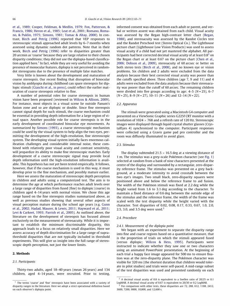
The display subtended 21.5 � 16.5 deg at a viewing distance of1 m. The stimulus was a grey-scale Pokémon character (see Fig. 1)selected at random from a bank of nine characters presented at thecentre of the display and surrounded by a rectangular, zero-dispar-ity reference frame. The stimulus was presented on a grey back-ground, at a moderate intensity to avoid crosstalk between thetwo eye’s images. Two small black, zero-disparity squares werepositioned above and below the reference frame to aid fusion.The width of the Pokémon stimuli was fixed at 2.2 deg while theheight varied from 1.6 to 3.1 deg according to the character. Tomaintain a fixed distance of 0.6 deg between the outside edge ofthe stimulus and the reference frame, the width of the frame wasscaled with the test disparity while the height varied with thecharacter. Test disparities of 0.02, 0.08, 0.17, 0.33, 0.67, 1.0, 2.0,2.5, 3.0, and 3.5 deg were used.3
2.4. Procedure
2.4.1. Measurement of the diplopia pointWe began with an experiment to separate the disparity range
into fine and coarse regions based on a quantitative measure, thatis the proportion of trials on which the stimuli appeared fused(versus diplopic; Wilcox & Hess, 1995). Participants wereinstructed to indicate whether they saw one or two charactersusing an animated PowerPoint presentation. At the beginning ofeach trial a happy face image appeared for 500 ms to ensure fixa-tion was at the zero-disparity plane. The Pokémon character wasvisible for 320 ms (the shortest duration that children would toler-ate in pilot studies) and trials were self-paced. A mid-range subsetof the test disparities was used and presented randomly on each
2 A decimal visual acuity of 0.8 is equivalent to a Snellen ratio of 20/25 or 0.1LogMAR. A decimal visual acuity of 0.67 is equivalent to 20/30 or 0.2 LogMAR.
3 For comparison with other tests, these disparities are 72, 288, 612, 1188, 2412,3600, 7200, 9000, 10,800, and 12,600 s.
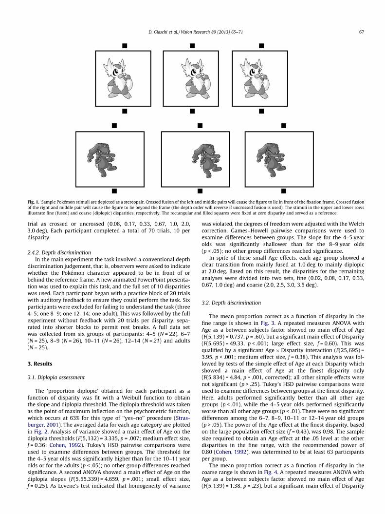
Fig. 1. Sample Pokémon stimuli are depicted as a stereopair. Crossed fusion of the left and middle pairs will cause the figure to lie in front of the fixation frame. Crossed fusionof the right and middle pair will cause the figure to lie beyond the frame (the depth order will reverse if uncrossed fusion is used). The stimuli in the upper and lower rowsillustrate fine (fused) and coarse (diplopic) disparities, respectively. The rectangular and filled squares were fixed at zero disparity and served as a reference.
D. Giaschi et al. / Vision Research 89 (2013) 65–71 67
trial as crossed or uncrossed (0.08, 0.17, 0.33, 0.67, 1.0, 2.0,3.0 deg). Each participant completed a total of 70 trials, 10 perdisparity.
2.4.2. Depth discriminationIn the main experiment the task involved a conventional depth
discrimination judgement, that is, observers were asked to indicatewhether the Pokémon character appeared to be in front of orbehind the reference frame. A new animated PowerPoint presenta-tion was used to explain this task, and the full set of 10 disparitieswas used. Each participant began with a practice block of 20 trialswith auditory feedback to ensure they could perform the task. Sixparticipants were excluded for failing to understand the task (three4–5; one 8–9; one 12–14; one adult). This was followed by the fullexperiment without feedback with 20 trials per disparity, sepa-rated into shorter blocks to permit rest breaks. A full data setwas collected from six groups of participants: 4–5 (N = 22), 6–7(N = 25), 8–9 (N = 26), 10–11 (N = 26), 12–14 (N = 21) and adults(N = 25).
3. Results
3.1. Diplopia assessment
The ‘proportion diplopic’ obtained for each participant as afunction of disparity was fit with a Weibull function to obtainthe slope and diplopia threshold. The diplopia threshold was takenas the point of maximum inflection on the psychometric function,which occurs at 63% for this type of ‘‘yes–no’’ procedure (Stras-burger, 2001). The averaged data for each age category are plottedin Fig. 2. Analysis of variance showed a main effect of Age on thediplopia thresholds (F(5,132) = 3.335, p = .007; medium effect size,f = 0.36; Cohen, 1992). Tukey’s HSD pairwise comparisons wereused to examine differences between groups. The threshold forthe 4–5 year olds was significantly higher than for the 10–11 yearolds or for the adults (p < .05); no other group differences reachedsignificance. A second ANOVA showed a main effect of Age on thediplopia slopes (F(5,55.339) = 4.659, p = .001; small effect size,f = 0.25). As Levene’s test indicated that homogeneity of variance
was violated, the degrees of freedom were adjusted with the Welchcorrection. Games–Howell pairwise comparisons were used toexamine differences between groups. The slope for the 4–5 yearolds was significantly shallower than for the 8–9 year olds(p < .05); no other group differences reached significance.
In spite of these small Age effects, each age group showed aclear transition from mainly fused at 1.0 deg to mainly diplopicat 2.0 deg. Based on this result, the disparities for the remaininganalyses were divided into two sets, fine (0.02, 0.08, 0.17, 0.33,0.67, 1.0 deg) and coarse (2.0, 2.5, 3.0, 3.5 deg).
3.2. Depth discrimination
The mean proportion correct as a function of disparity in thefine range is shown in Fig. 3. A repeated measures ANOVA withAge as a between subjects factor showed no main effect of Age(F(5,139) = 0.737, p = .60), but a significant main effect of Disparity(F(5,695) = 49.33, p < .001; large effect size, f = 0.60). This wasqualified by a significant Age � Disparity interaction (F(25,695) =3.95, p < .001; medium effect size, f = 0.38). This analysis was fol-lowed by tests of the simple effect of Age at each Disparity whichshowed a main effect of Age at the finest disparity only(F(5,834) = 4.84, p = .001, corrected); all other simple effects werenot significant (p > .25). Tukey’s HSD pairwise comparisons wereused to examine differences between groups at the finest disparity.Here, adults performed significantly better than all other agegroups (p < .01), while the 4–5 year olds performed significantlyworse than all other age groups (p < .01). There were no significantdifferences among the 6–7, 8–9, 10–11 or 12–14 year old groups(p > .05). The power of the Age effect at the finest disparity, basedon the large population effect size (f = 0.43), was 0.98. The samplesize required to obtain an Age effect at the .05 level at the otherdisparities in the fine range, with the recommended power of0.80 (Cohen, 1992), was determined to be at least 63 participantsper group.
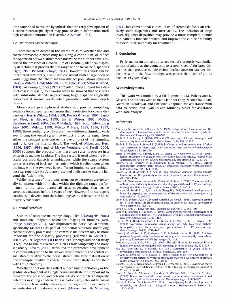
The mean proportion correct as a function of disparity in thecoarse range is shown in Fig. 4. A repeated measures ANOVA withAge as a between subjects factor showed no main effect of Age(F(5,139) = 1.38, p = .23), but a significant main effect of Disparity
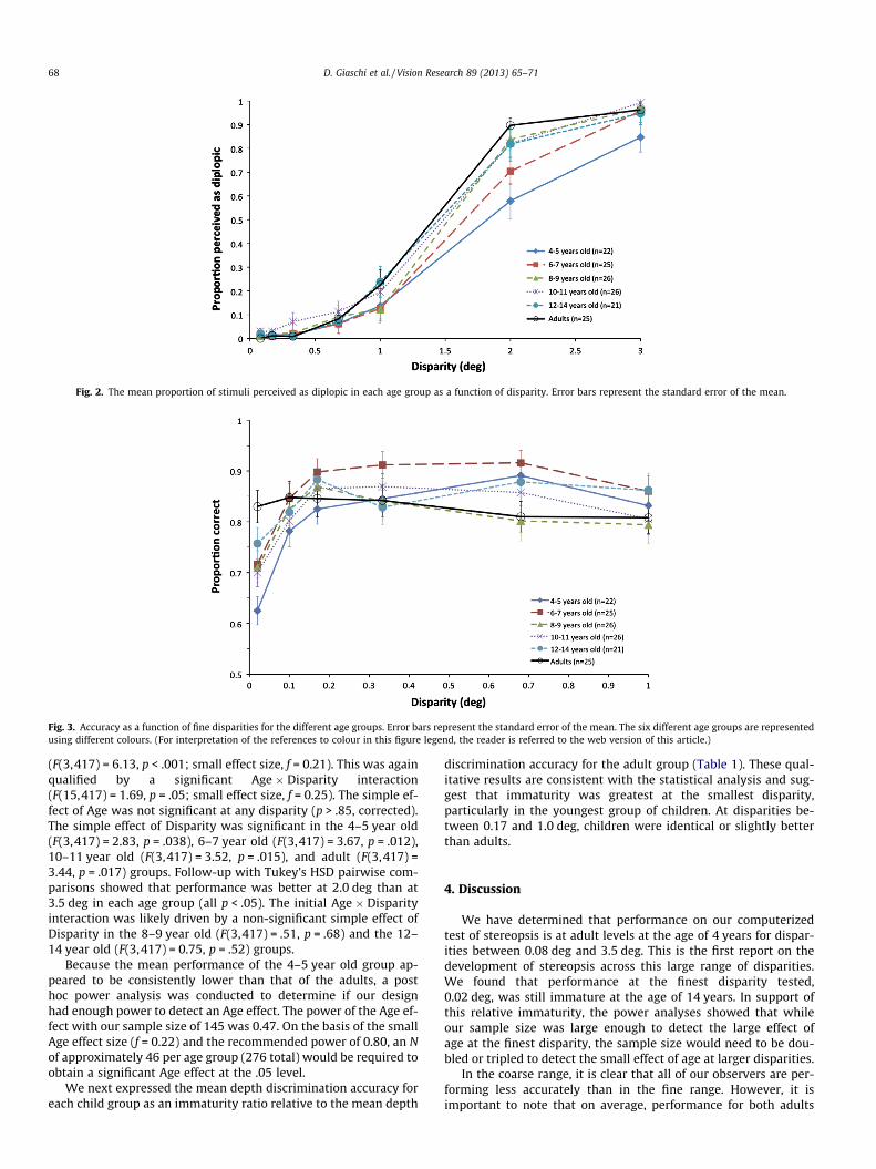
Fig. 2. The mean proportion of stimuli perceived as diplopic in each age group as a function of disparity. Error bars represent the standard error of the mean.
Fig. 3. Accuracy as a function of fine disparities for the different age groups. Error bars represent the standard error of the mean. The six different age groups are representedusing different colours. (For interpretation of the references to colour in this figure legend, the reader is referred to the web version of this article.)
68 D. Giaschi et al. / Vision Research 89 (2013) 65–71
(F(3,417) = 6.13, p < .001; small effect size, f = 0.21). This was againqualified by a significant Age � Disparity interaction(F(15,417) = 1.69, p = .05; small effect size, f = 0.25). The simple ef-fect of Age was not significant at any disparity (p > .85, corrected).The simple effect of Disparity was significant in the 4–5 year old(F(3,417) = 2.83, p = .038), 6–7 year old (F(3,417) = 3.67, p = .012),10–11 year old (F(3,417) = 3.52, p = .015), and adult (F(3,417) =3.44, p = .017) groups. Follow-up with Tukey’s HSD pairwise com-parisons showed that performance was better at 2.0 deg than at3.5 deg in each age group (all p < .05). The initial Age � Disparityinteraction was likely driven by a non-significant simple effect ofDisparity in the 8–9 year old (F(3,417) = .51, p = .68) and the 12–14 year old (F(3,417) = 0.75, p = .52) groups.
Because the mean performance of the 4–5 year old group ap-peared to be consistently lower than that of the adults, a posthoc power analysis was conducted to determine if our designhad enough power to detect an Age effect. The power of the Age ef-fect with our sample size of 145 was 0.47. On the basis of the smallAge effect size (f = 0.22) and the recommended power of 0.80, an Nof approximately 46 per age group (276 total) would be required toobtain a significant Age effect at the .05 level.
We next expressed the mean depth discrimination accuracy foreach child group as an immaturity ratio relative to the mean depth
discrimination accuracy for the adult group (Table 1). These qual-itative results are consistent with the statistical analysis and sug-gest that immaturity was greatest at the smallest disparity,particularly in the youngest group of children. At disparities be-tween 0.17 and 1.0 deg, children were identical or slightly betterthan adults.
4. Discussion
We have determined that performance on our computerizedtest of stereopsis is at adult levels at the age of 4 years for dispar-ities between 0.08 deg and 3.5 deg. This is the first report on thedevelopment of stereopsis across this large range of disparities.We found that performance at the finest disparity tested,0.02 deg, was still immature at the age of 14 years. In support ofthis relative immaturity, the power analyses showed that whileour sample size was large enough to detect the large effect ofage at the finest disparity, the sample size would need to be dou-bled or tripled to detect the small effect of age at larger disparities.
In the coarse range, it is clear that all of our observers are per-forming less accurately than in the fine range. However, it isimportant to note that on average, performance for both adults
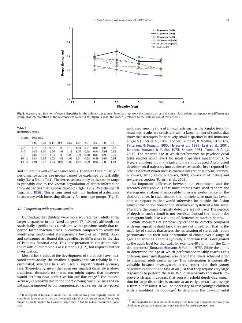
Fig. 4. Accuracy as a function of coarse disparities for the different age groups. Error bars represent the standard error of the mean. Each colour corresponds to a different agegroup. (For interpretation of the references to colour in this figure legend, the reader is referred to the web version of this article.)
Table 1Immaturity ratios.
Group Disparity
0.02 0.08 0.17 0.33 0.67 1.0 2.0 2.5 3.0 3.5
4–5 0.75 0.92 0.97 1.0 1.10 1.03 0.91 0.85 0.89 0.876–7 0.86 1.00 1.06 1.08 1.13 1.07 0.98 0.94 0.98 0.938–9 0.86 0.97 1.03 1.0 1.0 0.99 0.88 0.87 0.96 0.9510–11 0.84 0.95 1.02 1.03 1.06 1.0 0.98 0.89 0.94 0.9412–14 0.91 0.97 1.04 0.98 1.08 1.07 0.99 0.92 1.05 1.03
D. Giaschi et al. / Vision Research 89 (2013) 65–71 69
and children is well above chance levels. Therefore the similarity inperformance across age groups cannot be explained by task diffi-culty (i.e. a floor effect). The decreased accuracy in the coarse rangeis probably due to the known degradation of depth informationfrom disparities that appear diplopic (Ogle, 1952; Westheimer &Tanzman, 1956). This is consistent with our finding of a decreasein accuracy with increasing disparity for most age groups (Fig. 4).
4.1. Comparison with previous studies
Our finding that children were more accurate than adults at thelarger disparities in the fused range (0.17–1.0 deg), although notstatistically significant, is consistent with a previous study that re-ported faster reaction times in children compared to adults foridentifying random-dot stereograms (Dowd et al., 1980). Dowdand colleagues attributed this age effect to differences in the sizeof Panum’s fusional area. This interpretation is consistent withthe results of our diplopia assessment (Fig. 2), but requires furtherinvestigation.
Most other studies of the development of stereopsis have mea-sured stereoacuity, the smallest disparity that can reliably be dis-criminated, whereas here we used a suprathreshold accuracytask. Theoretically, given that even our smallest disparity is abovetraditional threshold estimates, one might expect that observerswould perform near perfect within our fine range.4 The reducedaccuracy is probably due to the short viewing time (320 ms) and ra-pid pacing imposed by our computerized test versus the self-paced,
4 It is important to bear in mind that the scale of any given disparity should beconsidered in relation to the size (horizontal width) of the test stimulus. A relatively‘small’ disparity applied to a narrow target may in fact be outside Panum’s fusionalarea.
unlimited viewing time of clinical tests such as the Randot tests. In-stead, our results are consistent with a large number of studies thatshow that stereopsis for relatively small disparities is still immatureat age 5 (Ciner et al., 1989; Cooper, Feldman, & Medlin, 1979; Fox,Patterson, & Francis, 1986; Heron et al., 1985; Leat et al., 2001;Romano, Romano, & Puklin, 1975; Simons, 1981; Tomac & Altay,2000). The reported age at which performance on psychophysicaltasks reaches adult levels for small disparities ranges from 6 to9 years, and depends on the task and the stimulus used. A protracteddevelopmental trajectory into adolescence has also been reported forother aspects of vision such as contour integration (Gervan, Berencsi,& Kovacs, 2011; Kaldy & Kovacs, 2003; Kovacs et al., 1999) andtexture perception (Parrish et al., 2005).
An important difference between our experiment and theresearch cited above is that most studies have used random dotstereograms making it impossible to assess performance in thediplopic range. In such stimuli, the multiple false matches (avail-able at disparities that would otherwise be outside the fusionrange) provide solutions to the stereoscopic system at a fine scale.Therefore the coarse disparity detectors are not used. The perceptof depth in such stimuli is not veridical, instead the random dotstereogram looks like a volume of elements at random depths.
While measures of stereoacuity cannot be directly comparedwith our suprathreshold task, they are not unrelated. That is, themajority of studies that assess the maturation of stereopsis reportperformance on their task or stimulus of choice over a range ofages and abilities. There is typically a criterion that is designatedas the adult level for that task, for example 40 arcmin for the Ran-dot stereotest (Romano, Romano, & Puklin, 1975). While the aim isto determine the age at which performance reliably reaches thiscriterion, most investigators also report the levels achieved priorto attaining adult performance. This information is potentiallyvaluable because investigators rarely report that their youngobservers cannot do the task at all, just that they require very largedisparities to perform the task. While stereoacuity thresholds im-prove with age, it appears that suprathreshold depth discrimina-tion for large disparities is mature at an early age (at least by age4 from our results). It will be necessary to test younger childrenwith a modified methodology5 to determine the developmental
5 The computerized task and methodology used here was designed specifically forchildren as young as 4 years, but is not suitable for testing younger ages.
70 D. Giaschi et al. / Vision Research 89 (2013) 65–71
time course and to test the hypothesis that the early development ofa coarse stereoscopic signal may provide depth information untilhigh-resolution information is available (Simons, 1993).
4.2. Fine versus coarse stereopsis
There has been debate in the literature as to whether fine andcoarse stereoscopic processing fall along a continuum, or reflectthe operation of two distinct mechanisms. Some authors have sup-ported the presence of a continuum of essentially identical dispar-ity detectors that process the full range of fine to coarse disparities(Ogle, 1953; Richards & Kaye, 1974). However, this work can beinterpreted differently, and is also consistent with a large body ofwork suggesting that there are two distinct populations involved(Hess & Wilcox, 1994; Mitchell, 1969; Ogle, 1953; Schor & Wood,1983). For example, Jones (1977) provided strong support for a dis-tinct coarse disparity mechanism when he showed that observerswith substantial deficits in processing large disparities typicallyperformed at normal levels when presented with small depthoffsets.
More recent psychophysical studies also provide compellingevidence for a disparity mechanism that is selective for coarse dis-parities (Hess & Wilcox, 1994, 2008; Kovacs & Feher, 1997; Lang-ley, Fleet, & Hibbard, 1999; Lin & Wilson, 1995; McKee,Verghese, & Farell, 2004; Sato & Nishida, 1994; Schor, Edwards, &Sato, 2001; Wilcox, 1999; Wilcox & Hess, 1995, 1996, 1997,1998). These studies typically present very different stimuli to eacheye, forcing the visual system to extract a disparity signal fromeither the contrast envelope or the overall area of the stimulus,and to ignore the interior detail. The work of Wilcox and Hess(1996, 1997, 1998) and of McKee, Verghese, and Farell (2004,2005) supports the proposal that these two systems operate in asynergistic manner in adults. The fine system is used when the bin-ocular correspondance is unambiguous, while the coarse systemserves as a type of back-up mechanism which is relied upon whenthe images in the two eyes have different luminance, are ambigu-ous (e.g. repetitive bars), or are presented at disparities that are be-yond the fusion limit.
While not a test of this dissociation, our experiments are gener-ally consistent with it, in that within the diplopic range perfor-mance is the same across all ages suggesting that coarsestereopsis matures before 4 years of age. However fine stereopsiscontinues to develop into the school-age years, at least at the finestdisparity we tested.
4.3. Neural correlates
Studies of macaque neurophysiology (Uka & DeAngelis, 2006)and functional magnetic resonance imaging in humans (Neri,Bridge, & Heeger, 2004) have implicated the dorsal visual stream,specifically MT/hMT+ as part of the neural substrate underlyingcoarse disparity processing. The ventral visual stream may be moreimportant for fine disparity processing (reviewed in Roe et al.,2007; Schiller, Logothetis, & Charles, 1990) though additional studyis required to rule out variables such as task complexity and noisesensitivity. Kovacs (2000) attributed the protracted developmentof contour integration to the slower development of the ventral vi-sual stream relative to the dorsal stream. The later maturation offine stereopsis relative to coarse in the current study is consistentwith this dichotomy.
Whether or not our data reflect a mechanistic dichotomy or thegradual development of a single neural substrate, it is important torecognize the presence and potential utility of these large disparitydetectors in young children. This is particularly relevant to visualdisorders such as amblyopia where the degree of binocularity isan indicator of treatment success (McKee, Levi, & Movshon,
2003), but conventional clinical tests of stereopsis focus on rela-tively small disparities and stereoacuity. The inclusion of large(even diplopic) disparities may provide a more complete pictureof a patient’s binocular status, and improve the clinician’s abilityto assess their suitability for treatment.
5. Conclusion
Performance on our computerized test of stereopsis was similarto that of adults at the youngest age tested (4 years) for large dis-parities that produce double vision. Performance for smaller dis-parities within the fusable range was poorer than that of adultseven at 14 years of age.
Acknowledgments
This work was funded by a CIHR grant to L.M. Wilcox and D.Giaschi. The authors wish to thank Jennifer Fong, Nicole Chaudhari,Ghazaleh Farrokhyar and Christine Chapman for assistance withdata collection, and Ryan Lo and Kimberly Meier for assistancewith data analysis.
References
Arteberry, M., Yonas, A., & Bensen, A. S. (1989). Self-produced locomotion and thedevelopment of responsiveness to linear perspective and texture gradients.Developmental Psychology, 25, 976–982.
Birch, E. E., & Petrig, B. (1996). FPL and VEP measures of fusion, stereopsis andstereoacuity in normal infants. Vision Research, 36, 1321–1327.
Birch, E. E., Shimojo, S., & Held, R. (1985). Preferential-looking assessment of fusionand stereopsis in infants aged 1 to 6 months. Investigative Ophthalmology &Visual Science, 26, 366–370.
Birch, E., Williams, C., Drover, J., Fu, V., Cheng, C., Northstone, K., et al. (2008).Randot preschool stereoacuity test: Normative data and validity. Journal of theAmerican Association for Pediatric Ophthalmology and Strabismus, 12, 23–26.
Brown, A. M., Lindsey, D. T., Satgunam, P., & Miracle, J. A. (2007). Criticalimmaturities limiting infant binocular stereopsis. Investigative Ophthalmology& Visual Science, 48, 1424–1434.
Brown, A. M., & Miracle, J. A. (2003). Early binocular vision in human infants:Limitations on the generality of the superposition hypothesis. Vision Research,43, 1563–1574.
Chen, S. I., Chandna, A., Norcia, A. M., Pettet, M., & Stone, D. (2006). The repeatabilityof best corrected acuity in normal and amblyopic children 4 to 12 years of age.Investigative Ophthalmology & Visual Science, 47(2), 614–619.
Chino, Y. M., Smith, E. L., III, Hatta, S., & Cheng, H. (1997). Postnatal development ofbinocular disparity sensitivity in neurons of the primate visual cortex. Journal ofNeuroscience, 17, 296–307.
Ciner, E. B., Scheiman, M. M., Schanel-Klitsch, E., & Weil, L. (1989). Stereopsis testingin 18- to 35-month old children using operant preferential looking. Optometry &Vision Science, 66, 782–787.
Cohen, J. (1992). A power primer. Psychological Bulletin, 112, 155–159.Cooper, J., Feldman, J., & Medlin, D. (1979). Comparing stereoscopic performance of
children using the Titmus, TNO, and Randot stereo tests. Journal of the AmericanOptometric Association, 50, 821–825.
Dobson, V., Clifford-Donaldson, C. E., Green, T. K., Miller, J. M., & Harvey, E. M.(2009). Normative monocular visual acuity for early treatment diabeticretinopathy study charts in emmetropic children 5 to 12 years of age.Ophthalmology, 116(7), 1397–1401.
Dowd, J. M., Clifton, R. K., Anderson, D. R., & Eichelman, W. H. (1980). Childrenperceive large-disparity random-dot stereograms more readily than adults.Journal of Experimental Child Psychology, 29, 1–11.
Fawcett, S., Wang, Y.-Z., & Birch, E. (2005). The critical period for susceptibility ofhuman stereopsis. Investigative Ophthalmology & Visual Science, 46, 521–525.
Fox, R., Patterson, R., & Francis, E. (1986). Stereoacuity in young children.Investigative Ophthalmology & Visual Science, 27, 598–600.
Gervan, P., Berencsi, A., & Kovacs, I. (2011). Vision first? The development ofprimary visual cortical networks is more rapid than the development of primarymotor networks in humans. PLoS One, 6, 1–9.
Giaschi, D., Lo, R., Narasimhan, S., Lyons, C., & Wilcox, L. (2013). Sparing of coarsestereopsis in stereodeficient children with a history of amblyopia. Journal ofVision (in press).
Gunn, A., Cory, E., Atkinson, J., Braddick, O., Wattam-Bell, J., Guzzetta, A., et al.(2002). Dorsal and ventral stream sensitivity in normal development andhemiplegia. Cognitive Neuroscience and Neuropsychology, 13, 843–847.
Hadad, B., Maurer, D., & Lewis, T. L. (2011). Long trajectory for the development ofsensitivity to global and biological motion. Developmental Science, 14,1330–1339.
D. Giaschi et al. / Vision Research 89 (2013) 65–71 71
Hayward, J., Truong, G., Partanen, M., & Giaschi, D. (2011). Effects of speed, age, andamblyopia on the perception of motion-defined form. Vision Research, 51,216–223.
Heron, G., Dholakia, S., Collins, D. E., & McLaughlan, H. (1985). Stereoscopicthreshold in children and adults. American Journal of Optometry & PhysiologicalOptics, 62, 505–515.
Hess, R., & Wilcox, L. M. (1994). Linear and non-linear filtering in stereopsis. VisionResearch, 18, 2431–2438.
Hess, R., & Wilcox, L. M. (2008). The transient nature of 2nd-order stereopsis. VisionResearch, 11, 1327–1334.
Jandó, G., Mikó-Barátha, E., Markóa, K., Hollódyb, K., Törökc, B., & Kovacs, I. (2012).Early-onset binocularity in preterm infants reveals experience-dependentvisual development in humans. Proceedings of the National Academy of Sciencesof the United States of America, 109, 11049–11052.
Jones, R. (1977). Anomalies of disparity detection in the human visual system.Journal of Physiology, 264, 621–640.
Kaldy, Z., & Kovacs, I. (2003). Visual context integration is not fully developed in 4-year-old children. Perception, 32, 657–666.
Kovacs, I. (2000). Human development of perceptual organization. Vision Research,40, 1301–1310.
Kovacs, I., & Feher, A. (1997). Non-Fourier information in bandpass noise patterns.Vision Research, 37, 1167–1175.
Kovacs, I., Kozma, P., Feher, A., & Benedek, G. (1999). Late maturation of visualspatial integration in humans. Proceedings of the National Academy of Sciences ofthe United States of America, 96, 12204–12209.
Langley, K., Fleet, D. J., & Hibbard, P. B. (1999). Stereopsis from contrast envelopes.Vision Research, 39, 2313–2324.
Leat, S. J., St Pierre, J., Hasan-Abadi, S., & Faubert, J. (2001). The moving dynamicrandom dot stereosize test: Development, age norms, and comparison with theFrisby, Randot and Stereo Smile tests. Journal of Pediatric Ophthalmology &Strabismus, 38, 284–294.
Levi, D. M., & Carkeet, A. D. (1993). Amblyopia: A consequence of abnormal visualdevelopment. In K. Simons (Ed.), Early visual development: Normal and abnormal(pp. 391–408). New York: Oxford University Press.
Lin, L., & Wilson, H. R. (1995). Stereoscopic integration of Fourier and non-Fourierpatterns. Investigative Ophthalmology & Visual Science, 36, S364.
Maruko, I., Zhang, B., Tao, T., Tong, J., Smith, E. L., III, & Chino, Y. M. (2008). Postnataldevelopment of disparity sensitivity in visual area 2 (V2) of Macaque monkeys.Journal of Neurophysiology, 100, 2486–2495.
McKee, S., Levi, D., & Movshon, A. (2003). The pattern of visual deficits in amblyopia.Journal of Vision, 3, 380–405.
McKee, S. P., Verghese, P., & Farell, B. (2004). What is the depth of a sinusoidalgrating? Journal of Vision, 4, 524–538.
McKee, S. P., Verghese, P., & Farell, B. (2005). Stereo sensitivity depends on stereomatching. Journal of Vision, 5, 783–792.
Mitchell, D. E. (1969). Qualitative depth localization with diplopic images ofdissimilar shape. Vision Research, 9, 991–994.
Neri, P., Bridge, H., & Heeger, D. J. (2004). Stereoscopic processing of absolute andrelative disparity in human visual cortex. Journal of Neurophysiology, 92(3),1880–1891.
Ogle, K. N. (1952). Disparity limits of stereopsis. Archives of Ophthalmology, 48(1),50–60.
Ogle, K. (1953). Precision and validity of stereoscopic depth perception from doubleimages. Journal of the Optical Society of America, 43, 906–913.
Parrish, E., Giaschi, D., Boden, C., & Dougherty, R. (2005). The maturation of form andmotion perception in school age children. Vision Research, 45, 827–837.
Regan, D. (1988). Low contrast letter charts and sinewave grating tests inophthalmological and neurological disorders. Clinical Vision Science, 2, 235–250.
Richards, W., & Kaye, M. G. (1974). Local versus global stereopsis: Twomechanisms? Journal of the Optical Society of America, 64, 1703–1705.
Roe, A. R., Parker, A. J., Born, R. T., & DeAngelis, G. C. (2007). Disparity channels inearly vision. Journal of Neuroscience, 27(44), 11820–11831.
Romano, P. E., Romano, J. A., & Puklin, J. E. (1975). Stereoacuity development inchildren with normal binocular single vision. American Journal ofOphthalmology, 79(6), 966–971.
Sato, T., & Nishida, S. (1994). Does an envelope-detecting mechanism mediatestereopsis for band-limited stimuli? Investigative Ophthalmology & VisualScience, 35, S1916.
Schiller, P. H., Logothetis, N. K., & Charles, E. R. (1990). Role of the color-opponentand broad-band channels in vision. Visual Neuroscience, 5, 321–346.
Schor, C. M., Edwards, M., & Sato, M. (2001). Envelope size tuning for stereo-depthperception of small and large disparities. Vision Research, 41, 2555–2567.
Schor, C. M., & Wood, I. (1983). Disparity range for local stereopsis as a function ofluminance spatial frequency. Vision Research, 23, 1649–1654.
Shea, S. L., Fox, R., Aslin, R. N., & Dumais, S. T. (1980). Assessment of stereopsis inhuman infants. Investigative Ophthalmology & Visual Science, 19, 1400–1404.
Simons, K. (1981). Stereoacuity norms in young children. Archives of Ophthalmology,99, 439–445.
Simons, K. (1993). Stereoscopic neurontropy and the origins of amblyopia andstrabismus. In K. Simons (Ed.), Early visual development: Normal and abnormal(pp. 409–453). New York: Oxford University Press.
Strasburger, H. (2001). Converting between measures of slope of the psychometricfunction. Perception & Psychophysics, 63, 1348–1355.
Takai, Y., Sato, M., Tan, R., & Hirai, T. (2005). Development of stereoscopic acuity:Longitudinal study using a computer-based random-dot stereo test. JapanJournal of Ophthalmology, 49, 1–5.
Tanaka, H., & Ohzawa, I. (2006). Neural basis for stereopsis from second-ordercontrast cues. Journal of Neuroscience, 26, 4370–4382.
Tomac, S., & Altay, Y. (2000). Near stereoacuity: Development in preschool children;normative values and screening for binocular vision abnormalities; a study of115 children. Binocular Vision Strabismus Quarterly, 15, 221–228.
Tschermak, A., & Hoefer, P. (1903). Ueber binoculare Tiefenwahrnehmung aufGrund von Doppelbildern. European Journal of Physiology, 98, 299–321 (inGerman).
Uka, T., & DeAngelis, G. C. (2006). Linking neural representation to function instereoscopic depth perception: Roles of the middle temporal area in coarseversus fine disparity discrimination. Journal of Neuroscience, 26, 6791–6802.
Westheimer, G., & Tanzman, I. (1956). Qualitative depth localization with diplopicimages. Journal of the Optical Society of America, 46, 116–117.
Wilcox, L. M. (1999). 1st and 2nd-order contributions to surface interpolation.Vision Research, 39, 2335–2347.
Wilcox, L. M., & Allison, R. S. (2009). Coarse-fine dichotomies in human stereopsis.Vision Research, 49, 2653–2665.
Wilcox, L. M., & Hess, R. (1995). Dmax for stereopsis depends on size not spatialfrequency content. Vision Research, 35, 1061–1069.
Wilcox, L. M., & Hess, R. F. (1996). Is the site of non-linear filtering in stereopsisbefore or after binocular combination? Vision Research, 36, 391–399.
Wilcox, L. M., & Hess, R. F. (1997). Scale selection for second-order (non-linear)stereopsis. Vision Research, 37, 2981–2992.
Wilcox, L. M., & Hess, R. (1998). When stereopsis does not improve with increasingcontrast. Vision Research, 38, 3671–3679.