Examination of the Patient -Stereopsis
10
CHAPTER 15 Examination of the Patient—V DEPTH PERCEPTION S tereopsis is an epiphenomenon of normal bin- ocular vision (see Chapter 2). Its presence or absence is an important indicator of the state of binocularity in patients with ocular motility disor- ders. Barring a few notable exceptions (see Chap- ter 16), patients with essential infantile esotropia are stereoblind or, at best, have markedly reduced stereopsis, and the potential for regaining it is practically nil. In childhood strabismus with a later onset or in adults with acquired strabismus it is an important therapeutic goal to reestablish stere- opsis. Whether this can be accomplished depends on many variables, among them the age of onset and the duration of the strabismus and the com- pleteness of ocular realignment. Development of Stereopsis Depth perception on the basis of binocular dispar- ity is not fully developed at birth. Several studies using different paradigms such as line stereograms and a preferential looking procedure, random dots with a forced-choice preferential looking tech- nique, and random dots with visually evoked re- sponses have shown remarkably consistent find- ings: stereopsis is absent in almost all infants less than 3 months old, after which it rapidly develops 298 to normal levels which are reached by the sixth month of life. Interestingly, this rapid rate of matu- ration far exceeds that of visual acuity. 11 The dura- tion of the plasticity period of stereopsis in hu- mans still needs to be established. For a review of the literature, see Teller 31 and Birch. 5 Stereopsis and Strabismus Patients with a large manifest deviation do not have useful stereopsis in casual seeing. Neverthe- less, they can function quite well in space, making use of nonstereoscopic clues to depth perception, especially if the strabismus is of early origin. They may have trouble with fast-moving objects, such as flying balls, and this experience may be frustrat- ing to young children. However, when the strabis- mus is acquired later in life the loss of stereopsis is felt acutely and may present a real handicap. It appears as if stereopsis is useful in the comprehen- sion of complex visual presentations and those requiring good hand-eye coordination. Although the importance of stereopsis is often stressed, stud- ies addressing the functional effects of stereo- scopic deficits are sparse. 8 It is always interesting and useful to determine whether a patient with strabismus has stereopsis
description
Examination of the Patient - Stereopsis
Transcript of Examination of the Patient -Stereopsis
C H A P T E R 15Examination of thePatient—V
DEPTH PERCEPTION
Stereopsis is an epiphenomenon of normal bin-ocular vision (see Chapter 2). Its presence or
absence is an important indicator of the state ofbinocularity in patients with ocular motility disor-ders. Barring a few notable exceptions (see Chap-ter 16), patients with essential infantile esotropiaare stereoblind or, at best, have markedly reducedstereopsis, and the potential for regaining it ispractically nil. In childhood strabismus with a lateronset or in adults with acquired strabismus it isan important therapeutic goal to reestablish stere-opsis. Whether this can be accomplished dependson many variables, among them the age of onsetand the duration of the strabismus and the com-pleteness of ocular realignment.
Development of Stereopsis
Depth perception on the basis of binocular dispar-ity is not fully developed at birth. Several studiesusing different paradigms such as line stereogramsand a preferential looking procedure, random dotswith a forced-choice preferential looking tech-nique, and random dots with visually evoked re-sponses have shown remarkably consistent find-ings: stereopsis is absent in almost all infants lessthan 3 months old, after which it rapidly develops
298
to normal levels which are reached by the sixthmonth of life. Interestingly, this rapid rate of matu-ration far exceeds that of visual acuity.11 The dura-tion of the plasticity period of stereopsis in hu-mans still needs to be established. For a review ofthe literature, see Teller31 and Birch.5
Stereopsis and Strabismus
Patients with a large manifest deviation do nothave useful stereopsis in casual seeing. Neverthe-less, they can function quite well in space, makinguse of nonstereoscopic clues to depth perception,especially if the strabismus is of early origin. Theymay have trouble with fast-moving objects, suchas flying balls, and this experience may be frustrat-ing to young children. However, when the strabis-mus is acquired later in life the loss of stereopsisis felt acutely and may present a real handicap. Itappears as if stereopsis is useful in the comprehen-sion of complex visual presentations and thoserequiring good hand-eye coordination. Althoughthe importance of stereopsis is often stressed, stud-ies addressing the functional effects of stereo-scopic deficits are sparse.8
It is always interesting and useful to determinewhether a patient with strabismus has stereopsis
Examination of the Patient—V 299
or the potential for such. Some patients may re-spond to disparate stimulations with a degree ofstereopsis if the targets are placed at the objectiveangle, as in a major amblyoscope. Some patients(e.g., intermittent exotropes) may respond withgood stereoscopic acuity even when a stereoscopeis used, although they seemingly may be unableto superimpose dissimilar targets. Such patientsrequire strong fusional stimuli to keep their eyesaligned and to fuse. When they do, they gainmotor and sensory fusion, often with a high degreeof stereopsis.
Some ophthalmologists use stereoscopic teststo determine whether patients with small or inter-mittent deviations have foveal suppression. If thestereoscopic threshold is low enough, they con-clude that there is no foveal suppression.27 A posi-tive result is certainly conclusive, but a negativeresult does not necessarily mean that foveal im-ages are completely suppressed. There are patientswho fuse all but disparate retinal stimuli, whichare selectively suppressed.
A positive stereoscopic response of a patientwith a neuromuscular anomaly of the eyes at anyfixation distance and in any part of the binocularfield is of paramount importance prognosticallyand in directing treatment. This finding makes itmandatory that every effort be made, both nonsur-gically and surgically, to restore to the patient fullbinocular cooperation with stereopsis at all fixa-tion distances and in every part of the field.
Testing for stereopsis should always be doneafter operations have properly aligned the eyes.The findings may give indications whether andhow to follow up the operation by nonsurgicaltreatment.
Testing for Stereopsis
Equipment for testing stereopsis ranges from sim-ple equipment to complex laboratory apparatus.Only tests that the ophthalmologist can conve-niently apply in the office are discussed in thissection. A test for stereopsis must incorporate twoessential features. The two eyes must be dissoci-ated; that is, each eye must be presented with aseparate field of view, and each of the two fieldsor targets must contain elements imaged on corre-sponding retinal areas. Thus a frame of referenceis provided, and disparately imaged elements canbe fused and seen stereoscopically. In addition,
there should be fiducial marks that permit theexaminer to check whether both eyes are usedsimultaneously.
Major Amblyoscope or Stereoscope
The targets may be opaque or transparent and maybe used in a major amblyoscope or stereoscope.Both devices have mechanically separated fieldsof view, are set optically at infinity, and use ex-changeable targets. The advantage of the majoramblyoscope is that its arms can be set at thepatient’s angle of deviation, thus allowing controlof the retinal area being stimulated. Similarly thestereoscope may be used with prisms, but thisprocedure may not be accurate, and the distortionsinduced by prisms may become bothersome.
The number and variety of targets are limitedonly by the ingenuity of the designer and user, butstandard sets of targets and cards are commerciallyavailable for the different major amblyoscopesand stereoscopes. Targets of special interest in thepresent context are those that contain objects withdiffering amounts of disparity (e.g., the KeystoneDB6 card), so that they appear at different relativedepth distances. The object seen in depth, whichhas the least disparity, denotes the patient’s stereo-scopic threshold.
Stereogram
A useful clinical application can be made of thesimple stereogram consisting of eccentric circles,one set seen with each eye (see Fig. 2–15). If thepatient reports that two fiducial marks and twocircles are seen, but not in depth, one shouldinquire whether the two circles are concentric.They cannot be seen concentrically unless theyare also seen stereoscopically. If they are seeneccentrically, one may now ask whether the innercircles are closer to the right or left of the outercircle. The patient’s answer determines whetherthe disparate elements are suppressed in the rightor the left eye.
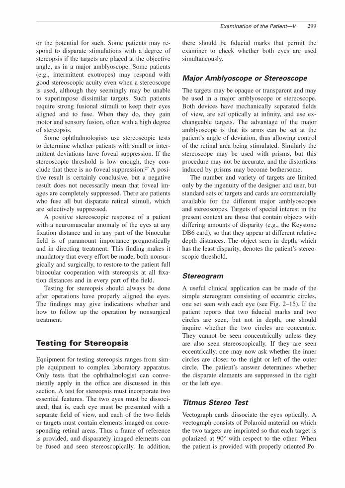
Titmus Stereo Test
Vectograph cards dissociate the eyes optically. Avectograph consists of Polaroid material on whichthe two targets are imprinted so that each target ispolarized at 90� with respect to the other. Whenthe patient is provided with properly oriented Po-

300 Introduction to Neuromuscular Anomalies of the Eyes
FIGURE 15–1. The Titmus Stereo Test.
laroid spectacles, each target is seen separatelywith the two eyes. This principle is used in theTitmus Stereo Test (Fig. 15–1). In this test a grossstereoscopic pattern representing a housefly is pro-vided to orient the patient and to establish whetherthere is gross stereopsis (threshold: 3000 secondsof arc). In testing young children, one must askquestions the child will understand. For example,one may ask the child to take hold of the wingsof the fly. If the child sees them stereoscopically,the child will reach above the plate. It is amusingto watch the child’s startled look when he or shedoes so. It is indeed an eerie feeling not to havea tactile sensation of a seen object. Some children,though they have stereopsis, will touch the wingson the plate because they ‘‘know’’ they are there.The examiner must explain to these children thathe or she does not inquire about what they know,but what they see.
The Polaroid test also contains three rows ofanimals, one animal in each row imaged dispa-rately (thresholds: 100, 200, and 400 seconds ofarc, respectively). The child is asked which oneof the animals stands out. The animal figures con-tain a misleading clue. In each row one of theanimals, correspondingly imaged in two eyes, isprinted heavily black. A child without stereopsiswill name this animal as the one that stands out.
Last, the Titmus test contains nine sets of fourcircles arranged in the form of a lozenge. In thissequence the upper, lower, left, or right circleis disparately imaged at random with thresholdsranging from 800 to 40 seconds of arc. If the childhas passed the other tests, he or she is now askedto ‘‘push down’’ the circle that stands out, begin-ning with the first set. When the child makesmistakes or finds no circle to push down, thelimits of stereopsis are presumably reached.
If there is doubt whether the patient actuallydoes see stereoscopically, one may occlude oneeye and inquire whether there is a difference inappearance, say, of the housefly, with one or botheyes open. And since only horizontal disparityproduces stereopsis, one can also turn the plate90�, which should block out the stereoscopic ef-fect.
Because of its simplicity, the Titmus StereoTest is widely used. On the basis of this test alone,however, one is not always justified in statingsimply that ‘‘the patient has no stereopsis,’’ thatis, that there is no sensitivity for disparate stimuli.One must keep in mind that the vectograph test isused for testing near vision. Some patients sup-press disparate stimuli at near but respond to themin distance fixation, or vice versa, usually whenthe deviation is intermittent at one fixation dis-
Examination of the Patient—V 301
tance and constant at the other. If such a patternis suspected, it is always wise to supplement thevectograph test with a projected vectograph test atdistance fixation (Polaroid Vectographic Project-O-Chart, American Optical Reichert) or with theB-VAT (Mentor) projection device.
In recent years much emphasis has been placedon the use of stereoacuity testing as a screeningmethod to detect anomalies of binocular function.9,
29, 30 Normal stereoacuity is said to preclude sup-pression, amblyopia, or heterotropia, and a sub-normal test result may indicate the presence ofsuch anomalies. In applying the Titmus test as ascreening device, Simons and Reinecke29 foundthat, with the exception of the fine stereoacuitycircles 5 to 9, this test often is unreliable in differ-entiating patients with amblyopia and heterotropiafrom those with normal vision. Moreover, the Tit-mus test is capable of indicating an artifactualstereocapability when none actually exists (seealso Kohler and Stigmar14). Some of the circles ofthe Titmus test may be selected even by stereoblindobservers because they look ‘‘different’’ and notbecause they are seen stereoscopically. Some pa-tients notice an image jump in the disparate por-tions of the test target (e.g., the wings of the fly)
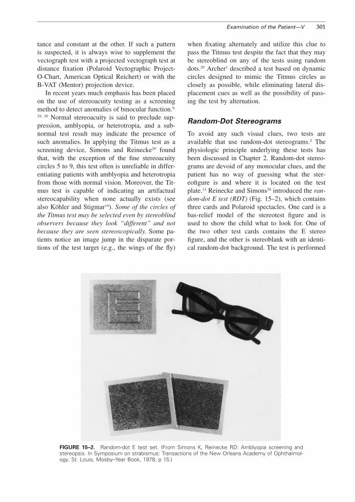
FIGURE 15–2. Random-dot E test set. (From Simons K, Reinecke RD: Amblyopia screening andstereopsis. In Symposium on strabismus: Transactions of the New Orleans Academy of Ophthalmol-ogy. St. Louis, Mosby–Year Book, 1978, p 15.)
when fixating alternately and utilize this clue topass the Titmus test despite the fact that they maybe stereoblind on any of the tests using randomdots.29 Archer1 described a test based on dynamiccircles designed to mimic the Titmus circles asclosely as possible, while eliminating lateral dis-placement cues as well as the possibility of pass-ing the test by alternation.
Random-Dot Stereograms
To avoid any such visual clues, two tests areavailable that use random-dot stereograms.2 Thephysiologic principle underlying these tests hasbeen discussed in Chapter 2. Random-dot stereo-grams are devoid of any monocular clues, and thepatient has no way of guessing what the ster-eofigure is and where it is located on the testplate.13 Reinecke and Simons28 introduced the ran-dom-dot E test (RDT) (Fig. 15–2), which containsthree cards and Polaroid spectacles. One card is abas-relief model of the stereotest figure and isused to show the child what to look for. One ofthe two other test cards contains the E stereofigure, and the other is stereoblank with an identi-cal random-dot background. The test is performed

302 Introduction to Neuromuscular Anomalies of the Eyes
FIGURE 15–3. A, The TNO test. B, The random-dot stereogram offers no monocular clues as tothe presence of a large circle in the center of the upper right quadrant and a smaller circle in thecenter of the lower left quadrant. (From Noorden GK von: Present status of sensory testing instrabismus. In Symposium on Strabismus: Transactions of the New Orleans Academy of Ophthalmol-ogy. St. Louis, Mosby–Year Book, 1978, p 51.)
by holding both test cards 50 cm in front of thepatient, who is then requested to indicate whichcard contains the letter E. The test is simple toperform, and the patient will give a ‘‘pass’’ or‘‘fail’’ response. It can be quantitated by increas-ing the testing distance from the patient. Manymodifications of the RDT have become availablein the meantime.6 Random-dot stereopsis can bemeasured also for distance with the Mentor B-VAT II-SG computerized testing system (MentorO & O, Norwell, MA). This is particularly usefulin intermittent exotropia.32
TNO Test
Another procedure, the TNO test, is based on asimilar principle but has the advantage of elicitingquantitative responses without changing the test-ing distance. This test uses a pair of red-greenspectacles and a test booklet (Fig. 15–3). Eachtest plate in the booklet consists of a stereogramin which the half-images have been superimposedand printed in complementary colors (anaglyphs).The test plates, when viewed binocularly with red-green spectacles by a normal subject, will elicit
Examination of the Patient—V 303
perception of an image in depth. The TNO test isgraded to provide retinal disparities ranging from15 to 480 seconds of arc. Comparative studieshave shown that this test compares favorably withthe Titmus test when used as a screeningdevice.26, 33 Together with the Lang test (see be-low) it is the preferred test in our clinic. It mustbe emphasized, however, that even random-dottesting of stereopsis is not a fail-safe method toassess visual acuity and binocular function in pre-school and school-age children, since normal lev-els of stereoacuity have been observed in anisome-tropic and visual deprivation amblyopia.3, 7, 21 Howshould stereopsis, determined with any of thetests, be recorded? Cards and vectographs thatattempt to qualify stereopsis are graded in differ-ent ways. Some use artificial scales (such as theSheppard scale); many speak of percentage ofstereopsis, assuming a certain threshold to mean100%. All this is misleading and arbitrary. Theonly proper way to record stereopsis is by theamount of disparity incorporated into the target. Itis unequivocal, and it should be generally under-stood when it is stated that a patient has stereopsiswith a threshold of 400 or 100 or 40 seconds ofarc or whatever the threshold may be.
Lam and coworkers15 evaluated the response ofnormal subjects to various visual function tests,including stereopsis. They found a wide rangeof responses in completely normal subjects, thusraising the question which level of stereopsis re-flects normalcy (see also Fisher9). It appears as adifficult task to identify a cutoff value separating
FIGURE 15–4. Stereoscopic images embedded in random dots of the Lang test. (From Lang J: Anew stereotest. J Pediatr Ophthalmol Strabismus 20:72, 1983.)
normals from abnormals. These authors furtherstated that because of the fact that more than 40%of normal children demonstrate stereoacuity ofless than 40 seconds of arc, random-dot testing isnot a real measure of a biological function.
Awaya et al.4 studied the effects of aniseikoniaon stereopsis measurements. With their aniseiko-nia test, they found that aniseikonia of 7% to 13%is still compatible with binocular fusion. However,aniseikonia of greater than 5% is incompatiblewith testing higher levels of stereoacuity with theTitmus and TNO tests.
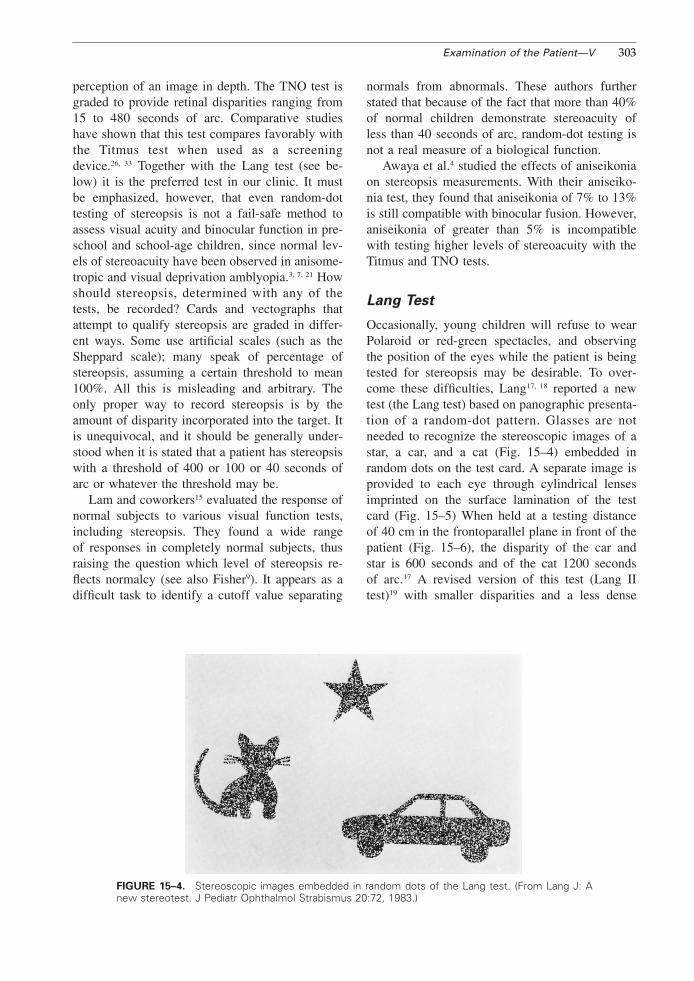
Lang Test
Occasionally, young children will refuse to wearPolaroid or red-green spectacles, and observingthe position of the eyes while the patient is beingtested for stereopsis may be desirable. To over-come these difficulties, Lang17, 18 reported a newtest (the Lang test) based on panographic presenta-tion of a random-dot pattern. Glasses are notneeded to recognize the stereoscopic images of astar, a car, and a cat (Fig. 15–4) embedded inrandom dots on the test card. A separate image isprovided to each eye through cylindrical lensesimprinted on the surface lamination of the testcard (Fig. 15–5) When held at a testing distanceof 40 cm in the frontoparallel plane in front of thepatient (Fig. 15–6), the disparity of the car andstar is 600 seconds and of the cat 1200 secondsof arc.17 A revised version of this test (Lang IItest)19 with smaller disparities and a less dense
304 Introduction to Neuromuscular Anomalies of the Eyes
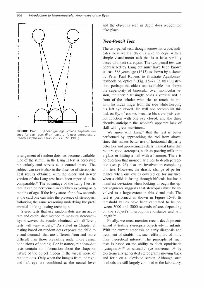
FIGURE 15–5. Cylinder gratings provide separate im-ages for each eye. (From Lang J: A near stereotest. JPediatr Ophthalmol Strabismus 20:72, 1983.)
arrangement of random dots has become available.One of the stimuli in the Lang II test is perceivedbinocularly and serves as a control mark. Thesubject can see it also in the absence of stereopsis.Test results obtained with the older and newerversion of the Lang test have been reported to becomparable.25 The advantage of the Lang I test isthat it can be performed in children as young as 6months of age. If the baby stares for a few secondsat the card one can infer the presence of stereopsis,following the same reasoning underlying the pref-erential looking testing technique.
Stereo tests that use random dots are an accu-rate and established method to measure stereoacu-ity; however, the results obtained with differenttests will vary widely.21 As stated in Chapter 2,testing based on random dots exposes the child tovisual demands that are different from and moredifficult than those prevailing under more casualconditions of seeing. For instance, random-dottests contain no information about the shape ornature of the object hidden in the visual noise ofrandom dots. Only when the images from the rightand left eye are combined at the neural level
and the object is seen in depth does recognitiontake place.
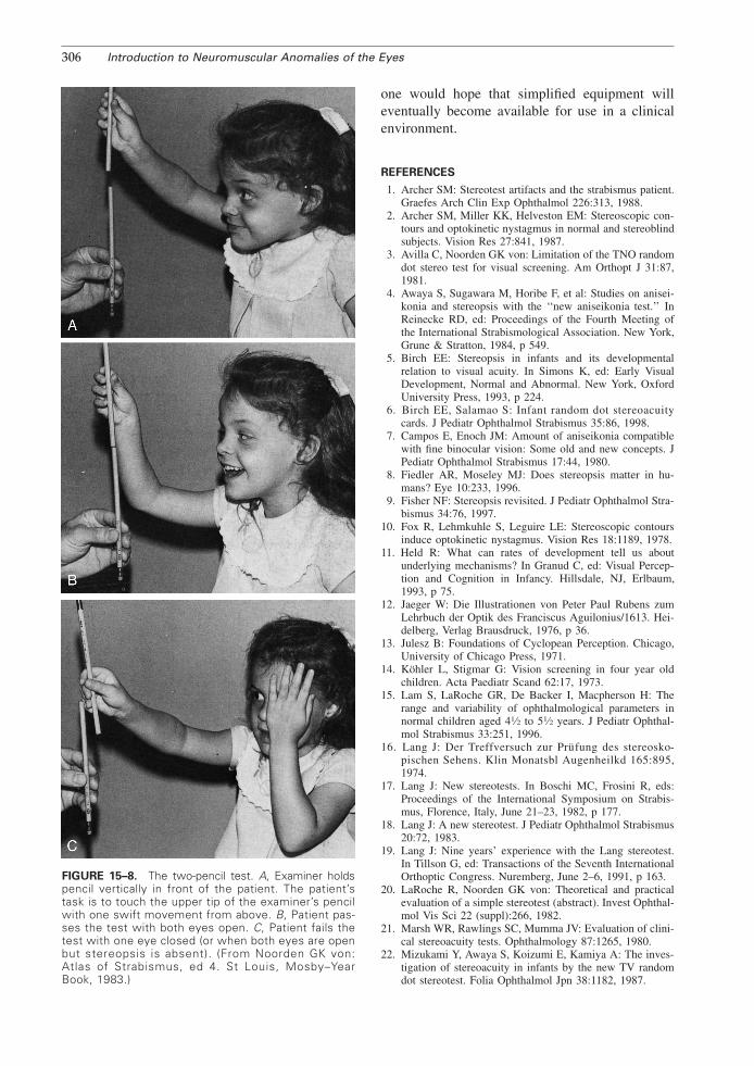
Two-Pencil Test
The two-pencil test, though somewhat crude, indi-cates how well a child is able to cope with asimple visual-motor task that is at least partiallybased on intact stereopsis. The two-pencil test waspopularized by Lang but must have been knownat least 388 years ago (1613) as shown by a sketchby Peter Paul Rubens to illustrate Aguilonius’textbook on optics12 (Fig. 15–7). In this illustra-tion, perhaps the oldest one available that showsthe superiority of binocular over monocular vi-sion, the cherub teasingly holds a vertical rod infront of the scholar who tries to touch the rodwith his index finger from the side while keepinghis left eye closed. He will not accomplish thistask easily, of course, because his stereopsis can-not function with one eye closed, and the threecherubs anticipate the scholar’s apparent lack ofskill with great merriment.
We agree with Lang16 that the test is betterperformed by approaching the rod from above,since this makes better use of horizontal disparitydetectors and approximates daily manual tasks thatrequire good stereopsis, such as pouring milk intoa glass or hitting a nail with a hammer. There isno question that monocular clues to depth percep-tion (see p. 25) also are involved in completingthis test. However, the drastic change of perfor-mance when one eye is covered or, for instance,when a child is fusing through bifocals but has amanifest deviation when looking through the up-per segments suggests that stereopsis must be in-volved to a large extent in this visual task. Thetest is performed as shown in Figure 15–8. Itsthreshold values have been estimated to be be-tween 3000 and 5000 seconds of arc, dependingon the subject’s interpupillary distance and armlength.20
Finally, we must mention recent developmentsaimed at testing stereopsis objectively in infants.With the current emphasis on early diagnosis andtreatment of strabismus, such efforts are of morethan theoretical interest. The principle of suchtests is based on the ability to elicit optokineticnystagmus2, 10 or saccadic eye movements22 byelectronically generated stereograms moving backand forth on a television screen. Although suchmethods are still largely confined to the laboratory,

Examination of the Patient—V 305
FIGURE 15–6. The Lang test. The child points to the stereoscopic image. (From Lang. J: A newstereotest. J Pediatr Ophthalmol Strabismus 20-72, 1983.)
FIGURE 15–7. Illustration by Peter Paul Rubens in Aguilonius’ textbook on optics. (From Jaeger W:Die Illustrationen von Peter Paul Rubens zum Lehrbuch der Optik des Franciscus Aguilonius/1613.Heidelberg, Verlag Brausdruck, 1976, p 36.)
306 Introduction to Neuromuscular Anomalies of the Eyes
FIGURE 15–8. The two-pencil test. A, Examiner holdspencil vertically in front of the patient. The patient’stask is to touch the upper tip of the examiner’s pencilwith one swift movement from above. B, Patient pas-ses the test with both eyes open. C, Patient fails thetest with one eye closed (or when both eyes are openbut stereopsis is absent). (From Noorden GK von:Atlas of Strabismus, ed 4. St Louis, Mosby–YearBook, 1983.)
one would hope that simplified equipment willeventually become available for use in a clinicalenvironment.
REFERENCES
1. Archer SM: Stereotest artifacts and the strabismus patient.Graefes Arch Clin Exp Ophthalmol 226:313, 1988.
2. Archer SM, Miller KK, Helveston EM: Stereoscopic con-tours and optokinetic nystagmus in normal and stereoblindsubjects. Vision Res 27:841, 1987.
3. Avilla C, Noorden GK von: Limitation of the TNO randomdot stereo test for visual screening. Am Orthopt J 31:87,1981.
4. Awaya S, Sugawara M, Horibe F, et al: Studies on anisei-konia and stereopsis with the ‘‘new aniseikonia test.’’ InReinecke RD, ed: Proceedings of the Fourth Meeting ofthe International Strabismological Association. New York,Grune & Stratton, 1984, p 549.
5. Birch EE: Stereopsis in infants and its developmentalrelation to visual acuity. In Simons K, ed: Early VisualDevelopment, Normal and Abnormal. New York, OxfordUniversity Press, 1993, p 224.
6. Birch EE, Salamao S: Infant random dot stereoacuitycards. J Pediatr Ophthalmol Strabismus 35:86, 1998.
7. Campos E, Enoch JM: Amount of aniseikonia compatiblewith fine binocular vision: Some old and new concepts. JPediatr Ophthalmol Strabismus 17:44, 1980.
8. Fiedler AR, Moseley MJ: Does stereopsis matter in hu-mans? Eye 10:233, 1996.
9. Fisher NF: Stereopsis revisited. J Pediatr Ophthalmol Stra-bismus 34:76, 1997.
10. Fox R, Lehmkuhle S, Leguire LE: Stereoscopic contoursinduce optokinetic nystagmus. Vision Res 18:1189, 1978.
11. Held R: What can rates of development tell us aboutunderlying mechanisms? In Granud C, ed: Visual Percep-tion and Cognition in Infancy. Hillsdale, NJ, Erlbaum,1993, p 75.
12. Jaeger W: Die Illustrationen von Peter Paul Rubens zumLehrbuch der Optik des Franciscus Aguilonius/1613. Hei-delberg, Verlag Brausdruck, 1976, p 36.
13. Julesz B: Foundations of Cyclopean Perception. Chicago,University of Chicago Press, 1971.
14. Kohler L, Stigmar G: Vision screening in four year oldchildren. Acta Paediatr Scand 62:17, 1973.
15. Lam S, LaRoche GR, De Backer I, Macpherson H: Therange and variability of ophthalmological parameters innormal children aged 41⁄2 to 51⁄2 years. J Pediatr Ophthal-mol Strabismus 33:251, 1996.
16. Lang J: Der Treffversuch zur Prufung des stereosko-pischen Sehens. Klin Monatsbl Augenheilkd 165:895,1974.
17. Lang J: New stereotests. In Boschi MC, Frosini R, eds:Proceedings of the International Symposium on Strabis-mus, Florence, Italy, June 21–23, 1982, p 177.
18. Lang J: A new stereotest. J Pediatr Ophthalmol Strabismus20:72, 1983.
19. Lang J: Nine years’ experience with the Lang stereotest.In Tillson G, ed: Transactions of the Seventh InternationalOrthoptic Congress. Nuremberg, June 2–6, 1991, p 163.
20. LaRoche R, Noorden GK von: Theoretical and practicalevaluation of a simple stereotest (abstract). Invest Ophthal-mol Vis Sci 22 (suppl):266, 1982.
21. Marsh WR, Rawlings SC, Mumma JV: Evaluation of clini-cal stereoacuity tests. Ophthalmology 87:1265, 1980.
22. Mizukami Y, Awaya S, Koizumi E, Kamiya A: The inves-tigation of stereoacuity in infants by the new TV randomdot stereotest. Folia Ophthalmol Jpn 38:1182, 1987.
Examination of the Patient—V 307
23. Noorden GK von: Present status of sensory testing instrabismus. In Symposium on Strabismus: Transactions ofthe New Orleans Academy of Ophthalmology. St Louis,Mosby–Year Book, 1978, p 51.
24. Noorden GK von: Atlas of Strabismus, ed 4. St Louis,Mosby–Year Book, 1983.
25. Nussgens Z, Czerwonka B, Roggenkamper P: Examinationof the new Lang test. Strabismus 1:69, 1993.
26. Okuda F, Apt L, Wanter B: Evaluation of the TNOrandom-dot stereogram test. Am Orthopt J 27:124,1977.
27. Parks MM: Stereoacuity as an indicator of bifixation. InArruga A, ed: International Strabismus Symposium, Uni-versity of Giessen, Germany 1966. Basel, S Karger, 1968,p 258.
28. Reinecke R, Simons K: A new stereoscopic test for ambly-opia screening. Am J Ophthalmol 78:714, 1974.
29. Simons K, Reinecke RD: A reconsideration of amblyopiascreening and stereopsis. Am J Ophthalmol 78:707, 1974.
30. Simons K, Reinecke RD: Amblyopia screening and stere-opsis. In Symposium on Strabismus: Transactions of theNew Orleans Academy of Ophthalmology. St Louis,Mosby–Year Book, 1978, p 15.
31. Teller DY: First glances: The vision of infants (Frieden-wald lecture). Invest Ophthalmol Vis Sci 38:2183, 1997.
32. Yildrim C, Altinsoy I, Yakut E: Distance stereoacuitynorms for the mentor B-VAT II-SG video acuity tester inyoung children and young adults. J AAPOS 2:26, 1998.
33. Walraven J: Amblyopia screening with random-dot stereo-gram. Am J Ophthalmol 80:893, 1975.