On Minor Salivary Gland Secretion - GUPEA: Home · In 1956, Schneyer (18) expressed the opinion...
55
On Minor Salivary Gland Secretion Lars Eliasson Department of Cariology Institute of Odontology Sahlgrenska Academy at Göteborg University Göteborg 2006 ISBN 91-628-6947-7 Lars Eliasson On Minor Salivary Gland Secretion 2006
Transcript of On Minor Salivary Gland Secretion - GUPEA: Home · In 1956, Schneyer (18) expressed the opinion...

OnMinor Salivary Gland Secretion
Lars Eliasson
Department of CariologyInstitute of Odontology
Sahlgrenska Academy at Göteborg University
Göteborg 2006ISBN 91-628-6947-7
Lars Eliasson
On M
inor Salivary Gland Secretion
2006

On Minor Salivary Gland Secretion
Lars Eliasson
Department of Cariology Institute of Odontology
Sahlgrenska Academy at Göteborg University
Göteborg 2006

2

3
ABSTRACT On Minor Salivary Gland Secretion Lars Eliasson, Department of Cariology, Institute of Odontology, Sahlgrenska Academy at Göteborg University, Box 450, SE-405 30 Göteborg, Sweden The overall aim of this series of studies has been to examine palatal, buccal and labial minor salivary gland secretions in relation to age and gender and other factors and con-ditions that could have effects on the saliva. Further aims were to analyze minor sali-vary gland secretions in relation to feelings of oral dryness and dental plaque pH. The studies are based on the Periotron method, which measures minor fluid volumes col-lected in absorbent filter papers. The concentrations of proteins in the saliva samples, recovered from the filter papers, were also examined using ELISA techniques. The ac-curacy and variability of the Periotron measurements were evaluated (Paper I). Mucosal gland secretion rates were analyzed in relation to age and gender, common diseases and medications (Papers I and V), during oestrogen treatment (Paper II) and hyposalivation due to Sjögren’s syndrome or head and neck radiation (Paper III). The secretion rates were also related to denture wearing (Papers I and V), pregnancy (Paper V) and tobacco use (Papers I and V). The salivary concentration of IgA was examined in relation to various factors in Paper V, while albumin and lactoferrin were examined together with IgA in relation to hyposalivation (Paper III). Acidogenic microorganisms and pH in dental plaque after a sugar challenge were determined in hyposalivation subjects and matched controls (Paper IV). The results showed high accuracy for the Periotron meas-urements but large inter- and intra-individual variations in minor gland secretions. The secretion rate per mucosal surface area was highest at buccal sites and lowest at palatal sites. In the palatal mucosa, the secretion rate was higher at medial sites than at lateral sites. Age was not correlated to the minor gland secretion rates but was positively corre-lated to the IgA concentration. Women displayed lower minor gland flow rates and lower buccal saliva IgA concentrations than men. The buccal mucosal secretion rate was reduced during the use of diuretics and anti-hypertensive medication, while elderly women had an increased labial salivary secretion rate during oestrogen treatment. Sub-jective feelings of oral dryness were reduced with increased labial salivary flow. Indi-viduals with removable dentures had a significantly higher secretion rate in palatal glands compared with those without dentures. Lower secretion rates and higher protein concentrations were seen in minor gland saliva during hyposalivation and this applied especially to irradiated subjects. Compared with their respective matched controls, irra-diated patients had a more acidic plaque after a sugar challenge than the Sjögren’s syn-drome patients. A low buccal gland secretion rate was correlated to increased plaque acidity in hyposalivation subjects. The number of acidogenic microorganisms had a more important effect on this acidity in the healthy controls. The present studies also showed that buccal gland secretion rates and IgA concentrations were positively corre-lated to whole saliva secretion rates and IgA. Key words: age, dental plaque pH, dentures, gender, health, IgA, medication, minor salivary glands, oral dryness, saliva, salivary secretion rate. ISBN 91-628-6947-7

4

5
CONTENTS
ORIGINAL PAPERS ......................................................................................................... 7 INTRODUCTION
Salivary glands .................................................................................................... 9 Factors affecting salivary secretions................................................................... 14 Oral dryness and salivary secretion rate ............................................................. 16 Co-variation between major and minor salivary gland secretion rates ................ 17 Proteins in minor gland saliva............................................................................ 17
AIMS........................................................................................................................... 19 MATERIALS AND METHODS
Test populations ................................................................................................ 21 Salivary secretion and plaque pH measurements ................................................ 22 Minor salivary gland measurements................................................................... 22 Whole salivary secretion rate and buffer pH measurements................................ 23 Tests of bacterial adherence and aggregation ..................................................... 23 Feelings of oral dryness ..................................................................................... 24 Determination of protein concentration in saliva ................................................ 24 Plaque-pH measurements................................................................................... 25 Microorganisms in plaque.................................................................................. 25 Statistical methods............................................................................................. 25
RESULTS
Salivary secretion rates ...................................................................................... 27 Feelings of oral dryness ..................................................................................... 28 Protein concentrations in minor gland saliva...................................................... 29 Plaque pH.......................................................................................................... 30 Microorganisms in plaque.................................................................................. 31 Stimulated whole saliva buffer capacity and bacterial adherence and aggregation during oestrogen treatment ............................................................. 32
DISCUSSION................................................................................................................. 35 MAIN CONCLUSIONS .................................................................................................... 43 ACKNOWLEDGEMENTS................................................................................................. 45 REFERENCES ............................................................................................................... 47 APPENDIX (Papers I-V)

6

7
ORIGINAL PAPERS This thesis is based on the following papers, which are referred to in the text by their
Roman numerals:
I Eliasson L, Birkhed D, Heyden G, Strömberg N. Studies on human minor sali-
vary gland secretions using the Periotron® method. Arch Oral Biol
1996;41:1179-1182.
II Eliasson L, Carlén A, Laine M, Birkhed D. Minor gland and whole saliva in
postmenopausal women using a low potency oestrogen (oestriol). Arch Oral
Biol 2003;48:511-517.
III Eliasson L, Almståhl A, Lingström P, Wikström M, Carlén A. Minor gland sa-
liva flow rate and proteins in subjects with hyposalivation due to Sjögren’s syn-
drome and radiation therapy. Arch Oral Biol 2005;50:293-299.
IV Eliasson L, Carlén A, Almståhl A, Wikström M, Lingström P. Dental plaque pH
and microorganisms during hyposalivation. J Dent Res 2006;85:334-338.
V Eliasson L, Birkhed D, Österberg T, Carlén A. Minor salivary gland secretion
rates and IgA in adults and elderly. Eur J Oral Sci 2006; accepted for pub-
lication.

8

9
INTRODUCTION
Saliva is rich in mechanisms and functions that are important not only for oral health
and well-being but also for general health and well-being. It facilitates oral functions by
lubricating the oral surfaces and it contains a number of protective mechanisms against
microbial and chemical assaults. These functions are established by the salivary content
of a number of inorganic and organic constituents (1). What we in daily speech refer to,
as saliva is whole saliva. It is a mixture of secretions that is excreted into the oral cavity
from a number of major and minor salivary glands. In all, they excrete between 0.6 and
one liter of saliva per day (2).
Salivary glands There are three major pairs of salivary glands. The parotid glands are the largest, and
they are situated in front of the ear and behind the lower jaw. The glands deliver the sa-
liva through ducts penetrating the buccal mucosa near the second upper molar. The
submandibular glands are smaller and are situated in the posterior part of the floor of
the mouth close to the inner aspect of the mandible. Their excretory ducts open under-
neath the tongue lateral to the lingual frenulum. The sublingual glands, which are also
situated in the floor of the mouth, are the smallest of the three paired major salivary
glands. Their secretions enter the oral cavity through a series of small ducts opening
under the tongue. These three pairs of glands produce the largest part (>90%) of the to-
tal saliva volume (3). The remaining component of saliva originates from the mucosal,
minor salivary glands (4). The minor salivary glands are located in the buccal, labial,
palatal and lingual regions, including the base of the tongue (von Ebner’s glands). They
are estimated to occur in humans in numbers varying between 600 and 1,000 (3). The
only regions of the mouth in which no glands have been found are the gingiva and the
anterior part of the hard palate. The glands develop during embryonic life through the
proliferation of a cord of cells from the epithelium into the underlying mesenchyma,
followed by a branching process to form the grape-like arrangements of the secretory
lobes (5). Unlike the major salivary glands, the minor salivary glands lack a branching

10
network of draining ducts. Instead, each salivary gland unit has its own simple duct
(Fig.1).
a b
Fig. 1. Schematic pictures of secretory end pieces and excretory ducts from mi-nor (a) and parotid (b) salivary glands (adapted from Orban: ORAL HISTOLOGY AND EMBRYOLOGY, 4th ed © 1957 Mosby, with the kind per-mission of Elsevier and Springer Science and Business Media).
Salivary secretion The secretion of saliva is an energy consuming and active two-stage process. Salivary
glands produce their secretions by transforming capillary blood into interstitial fluid,
which is secreted by the acini as isotonic primary saliva in the terminal end pieces of the
gland parenchyma. This saliva is finally modulated to a hypotonic secretion in the duc-
tal systems (3). The secretion process of the major salivary glands is activated by both
the sympathetic and the parasympathetic nervous systems. This mechanism regulate the
secretory capacity in complicated interactions, varying between the separate glands (6).
Since most of the minor glands have little or no sympathetic innervations (7), parasym-
pathetic signals with cholinergic transmission (8) constitute the main nervous drive to
their salivary secretions. Also in contrast to the major glands, a small volume of saliva
is continuously and spontaneously secreted from the minor glands.

11
The nature of saliva from different glands has traditionally been described as serous
or mucous. Parotid secretion is referred to as serous whereas the submandibular/sub-
lingual secretions are referred to as sero-mucous. Minor gland saliva is traditionally re-
garded as mucous in nature. Recent data from ultrastructural investigations have, how-
ever, shown that the separate minor glands differ in their secretions. Lingual glands are
predominantly serous and the other minor glands, together with the mucous cells, pos-
sess a variable number of sero-mucous cells, with the relatively highest number in buc-
cal glands (9). The protein content is also different in the minor gland secretions com-
pared with that from the major glands (10, 11).
Saliva function Major gland saliva has several functions that are of special importance for the teeth. The
saliva is e.g. rich in buffering bicarbonate and in proteins that bind calcium and phos-
phate The flow of the saliva solution after stimulation from eating facilitates oral clear-
ance of food remnants and acids. These mechanisms allow for the maintenance of tooth
mineral integrity. At reduced flow of saliva, the risk of oral mucosal lesions and caries
are increased (1, 12). Reduced pH and higher numbers of acidogenic micro-organisms
are also seen in hyposalivators (13-17).
In 1956, Schneyer (18) expressed the opinion that minor salivary glands make no sig-
nificant contribution to the total unstimulated volume of saliva. It was subsequently re-
ported that minor salivary gland secretion accounts for 7-10% of the total volume of
whole saliva (4). The minor gland secretions are rich in high-molecular weight glyco-
proteins and secretory IgA (10, 19), which are important constituents of the protective
layer covering the hard and soft oral tissues. This layer moistures the oral tissues and
protects them from bacteria and mechanical wear and from chemical penetration of dif-
ferent substances. It also facilitates mastication, swallowing and speech by minimizing
the friction between the teeth and the oral mucosa (20-22). In spite of the small volumes
that are secreted, minor gland saliva may thus have a significant effect on salivary func-
tions.

12
Considerable information on the secretion rate, composition and function of whole
saliva and major gland saliva (parotid and submandibular/sublingual) is available in pa-
pers and textbooks. Fewer data have been published on minor gland function.
Methods for studying minor salivary gland secretions
Some investigations of minor salivary glands are based on histomorphologic studies
(23-28). The difficulties involved in the collection of the small amounts of viscous sa-
liva from these glands have hampered studies of their secretions. Micro-pipettes, capil-
lary tubes, sponges, synthetic discs and filter papers have been used to collect minor
salivary gland secretions for qualitative studies (22, 29-40). Weighing methods or semi-
quantitative estimations as measurements of the secreted droplets (18, 27, 31, 33, 39,
41-49), as well as calculations based on measuring colored spots on chromatology pa-
pers (50, 51), have been applied in order to estimate minor gland flow rates.
The Periotron method
Following the introduction of an electronic device for measuring the oral mucosal flow
of saliva (Periotron®, Pro-Flow™, Inc., Amityville, NY, USA), quantitative estimates of
minor gland secretions have become fairly easy to perform. The method was originally
designed for gingival fluid measurements (52), but it was subsequently modified for sa-
liva research.
The introduction of this device has greatly improved precision and versatility when es-
timating minor gland secretion rates. Recent publications on mucosal salivary flow rates
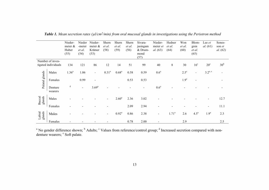
are based on this technique (53-65). The mean values for mucosal gland secretion rates
presented in these publications are summarized in Table 1.
13
Table 1. Mean secretion rates (µl/cm2/min) from oral mucosal glands in investigations using the Periotron method
Nieder-meier & Huber (55)
Nieder-meier et al. (54)
Nieder- meier & Krämer (53)
Shern et al. (58)
Shern et al. (59)
Shern et al. (56)
Sivara-jasingam & Drum-mond (57)
Nieder- meier et al. (63)
Hedner et al. (64)
Won et al. (60)
Blom-gren et al. (65)
Lee et al. (61)
Sones-son et al. (62)
Number of inves-tigated individuals 134 121 86 12 14 51 99 40 8 30 16c 20c 30b
Males 1.06 - 0.58 0.59 2.5e - -
Females
1.36a
0.99 -
0.31a 0.68a
0.53 0.53
0.6a
1.9e -
3.2a, e
-
Pala
tal g
land
s
Denture wearers
d - 3.69a - - - - 0.6a - - - - -
Males - - - - 2.36 3.02 - - - - - 12.7
Buc
cal
glan
ds
Females - - - -
2.60a
2.09 2.94 - - - - - 11.1
Males - - - - 0.86 2.38 - 2.6 2.3
Labi
al
glan
ds
Females - - - -
0.92a
0.78 2.00 -
1.71a
2.9
4.5a 1.9a
2.5
a No gender difference shown; b Adults; c Values from reference/control group; d Increased secretion compared with non- denture wearers; e Soft palate.

14
Principles of the Periotron method
Moisture from the oral mucosa is harvested by applying a piece of blotting paper to the
mucosa. The paper is placed between two plates on the measuring instrument and a
voltage is applied across the plates. Due to the dielectric nature of saliva molecules, the
instrument is capable of calculating the volume of moisture which has been absorbed by
the blotting paper. In practice, the instrument is first zeroed to compensate for any mois-
ture retained in the paper during storage. The harvesting of mucosal fluid is then per-
formed and the paper is again placed in the instrument. The volume absorbed is calcu-
lated from a standard curve obtained with known volumes of water added to the papers.
Due to the technique the saliva amount is usually expressed in µl/cm2/min.
Factors affecting salivary secretions Several factors that can affect secretion have been identified from studies of major
glands and whole saliva. Our knowledge of the effects of these factors on minor gland
saliva is, however, scarce or non-existent.
Age and gender
Conflicting data have been reported when it comes to the correlation between age and
gender and major salivary gland and whole salivary secretion rates (66-71). Published
studies of minor salivary glands have the same diversity, although several investigators
have found an age-related lower salivary flow rate or structural, degenerative changes in
labial glands, suggesting reduced function with increasing age (25, 26, 42). However,
one study showed no age-related effect on labial salivary flow (56). In the case of pala-
tal and buccal gland saliva, a decreasing or non-existent effect by age on the secretion
rate was reported (27, 53-57). Apart from one study in which a higher palatal secretion
rate was found in men compared with women (27), there are reports of no gender-
related differences in minor gland secretion rates (43, 53-57). On the other hand, a study
of the total sum of labial, buccal and palatal mucosal gland secretion rates revealed
lower secretion rates for women than for men and reduced secretion with increasing age
(72). This discussion is further complicated by data showing fewer secreting salivary
glands per labial mucosal area in adults compared with children and a lower secretion

15
rate in buccal and labial mucosal glands in children compared with adults and adoles-
cents (62). In this study of young adults, adolescents and children, no gender differences
were found.
Stimulation
It has been reported that the minor glands display a poor response to stimulation (30,
31), in contrast to major glands (73). However, subsequent data report that the local ap-
plication of physostigmine (64) and continuous speaking (48) could stimulate the labial
mucosal gland secretion rate. The mechanical stimulation of a denture base plate on the
palatal mucosa also appears to stimulate the underlying minor glands to increase their
salivary secretion rate (53, 55).
Disease and medication
Medication, especially with drugs causing oral dryness symptoms, are known to reduce
whole salivary secretion rates (74). An impaired palatal gland secretion rate among
medicated persons has also been reported (53).
In longitudinal studies, medication with female sex hormones increased the flow rate
of whole (75-77) and palatal (63) saliva, although this effect on whole saliva was not
seen in cross-sectional investigations (78-80).
It is known that diseases, such as diabetes, hypertension (81), cystic fibrosis (41), and
rheumatic diseases (82), regardless of treatment, can affect the salivary secretions. In
Sjögren’s syndrome, patients’ salivary gland structures are infiltrated by lymphatic cells
(23, 24). This interferes with glandular function and results in a lower salivary secretion
rate (83). It is, however, believed that a residual function exists during the inflammation
(84), which explains why stimulation of the function is possible (85).
Cancer patients are often treated with chemotherapy or irradiation. During chemother-
apy, the labial minor salivary gland secretion rate but not the stimulated whole salivary
secretion rate is hampered (65). Radiation therapy directed against the head and neck
region causes permanent damage to the radiosensitive salivary gland structures. This
leads to the depression of salivary flow (86), an increase in electrolyte and protein con-
centrations and a decrease in bicarbonate (87) and phosphate (88) concentrations. An

16
increase in whole saliva total protein, albumin and lactoferrin concentrations in both
radiation therapy and Sjögren’s syndrome patients is further reported (89). There are no
reports of minor gland saliva in subjects with Sjögren’s syndrome or irradiated patients.
Tobacco use
Tobacco habits are known to be hazardous to oral health. The relationship between to-
bacco use and a significantly lower number of remaining teeth (90, 91), a higher preva-
lence of periodontitis, the aggravation of the disease by increased pocket depth (92-94),
a higher plaque index (95), lower salivary buffer effects, a higher number of lactobacilli
and Streptococcus mutans (96) and an increase in caries incidence (97) among tobacco
users has been reported. To some extent, these effects of tobacco could directly depend
on the local or vaso-active effects in the oral cavity of noxious components from to-
bacco products (98), but effects on salivary glands with adverse effects on gland secre-
tions must also be considered (99-101).
Oral dryness and salivary secretion rate
The prevalence of oral dryness is calculated to be 20-30% in an adult population, with a
tendency to increase with increasing age (102, 103). This condition is more common
among elderly women than among elderly men (104, 105) and is reported to be allevi-
ated during hormone replacement therapy (106). A low secretion rate among persons
complaining of oral dryness has been reported in some studies (107, 108), while others
report a poor correlation between these oral discomfort symptoms and the whole sali-
vary secretion rate (109, 110). During Sjögren’s syndrome, a change in the viscosity of
saliva, in addition to the reduced salivary secretion rate, could aggravate the feeling of
oral dryness by increasing mucosal friction (83). Minor gland secretion in individuals
with feelings of oral dryness has interested some authors, who report reduced palatal
(54, 55) and labial (44) gland secretion rates among these persons. A correlation be-
tween oral dryness symptoms and reduced salivary output was also found when the sum
of labial, buccal and palatal gland output was studied (72).
17
Co-variation between major and minor salivary gland secretion rates A lack of co-variation between major and minor gland flow rates has been reported (54,
56, 58). On the other hand, a positive correlation was found between labial and resting
whole salivary secretions in a study in which an iodo-starch reaction in paper strips was
used to calculate the labial salivary secretion rate (51). A similar correlation was also
reported for labial gland and parotid secretion rates (31).
Proteins in minor gland saliva A few studies of proteins in minor gland saliva have been published. The major salivary
immunoglobulin, IgA, is found in high concentrations in labial (29, 32-35, 43) and pala-
tal (111) gland secretions. Amylase and lysozyme in labial saliva (112) and levels of
amylase, cystatin and IgA in palatal saliva are also reported (111). The current knowl-
edge of the influence of factors such as age, health status and medication on proteins in
saliva is mainly restricted to whole saliva studies. In this saliva, lactoferrin (113) and
IgA (71) concentrations were found to be elevated with increasing age. This correlation
for age and whole saliva IgA was, however, not seen among persons aged 75 years and
older (113). Albumin and lactoferrin concentrations are elevated in hyposalivating indi-
viduals (89) and IgA is reported to increase during pregnancy (114) and in rheumatic
patients (82). No change in albumin concentrations was seen during hormone replace-
ment therapy (76).
It would be true to say that the knowledge of minor gland secretions is limited. The
studies in this thesis are inspired by the fact that minor salivary gland secretions may be
more important than previously believed and that the new Periotron technique has en-
abled easy estimations of small fluid volumes.

18

19
AIMS
The main objective of these series of studies was to investigate some factors that could influence palatal, buccal and labial minor gland secretions. Whole saliva examinations
were performed in parallel. The specific aims were:
- to evaluate the Periotron method and minor gland secretion rate variability (Paper I)
and measure the salivary secretion rates from palatal, buccal and labial salivary glands
(Papers I and V),
- to relate these secretion rates (Papers I and V) to age, gender and the effect of common
diseases, medication, pregnancy and wearing removable dentures,
- to examine minor gland secretion rates and whole saliva buffer capacity and bacterial
adherence and aggregation during oestrogen medication (Paper II),
- to examine IgA concentrations in minor gland and whole saliva and to relate these to
various factors (Paper V),
- to investigate labial and buccal minor salivary gland secretion rates and concentra-
tions of IgA, albumin and lactoferrin (Paper III) as well as plaque pH (Paper IV) in
subjects with hyposalivation due to primary Sjögren’s syndrome and head and neck
radiation therapy, and
- to investigate minor gland saliva in relation to subjective oral dryness (Papers I and
II).

20
21
MATERIALS AND METHODS Test populations
The population included in Paper I consisted of 127 individuals, 61 females and 66
males (mean age 55 yr, range 22-89 yr). A total of 30 of these individuals were daily
users of tobacco. Nine individuals were daily users of diuretics. Twenty individuals
wore full upper dentures. Thirty-six individuals (20 females and 16 males) were ran-
domly selected for duplicate measurements in order to calculate the variance of the Pe-
riotron method (Paper I). The two measurements at the different mucosal sites were per-
formed with a 20-minute delay.
In the one-year study of oestriol treatment (Paper II), postmenopausal women were
recruited through advertisements. Eighteen women with a mean age of 68 years (range
61-76 yr), with no medical problems or medication affecting salivary gland secretion
during the last 3 months, were included in the study. Two women discontinued the
medication after 6 months. Nine peri- and postmenopausal women (mean age 57 yr,
range 53-61 yr) in good health and with no medication were included as a reference
group.
In Papers III and IV, 10 subjects with primary Sjögren’s syndrome and 10 subjects
irradiated in the head and neck region and their age- and gender-matched healthy con-
trols were investigated. All the subjects in the primary Sjögren’s syndrome group were
women with a mean age of 63 years (range 42-76 yr). In the irradiated group, four were
women and the mean age in the group was 55 years (range 38-76 yr). Both patient
groups were hyposalivators.
The test population in Paper V consisted of 96 women and 46 men, aged 18–82 years.
Fifty-one individuals had no conditions affecting health, 35 suffered from circulatory
diseases, 10 from diabetes mellitus and 12 from thyroid hypofunction. Twenty pregnant
women and 25 daily smokers and their matched controls were included. Among the eld-
erly individuals, 8 persons wore removable dentures.

22
Salivary secretion and plaque pH measurements All saliva sampling in Papers I-V was performed between 8 am and 1 pm. Saliva sam-
pling was performed at the same time of the day for each individual during the study
period in Paper II. The individuals were instructed not to eat or drink and not to perform
oral hygiene for at least 1 h prior to sampling. For the participants in the studies in Pa-
per III and IV, where proximal plaque was measured and collected, the instructions
were not to clean their teeth proximally for 3 days, not to brush their teeth the same day
and not to eat and drink for at least 2 h prior to the test.
Minor salivary gland measurements A Periotron 6000, model II (modified for minor salivary gland measurements) (Pro-
Flow™, Inc.), and pre-cut pieces of filter paper (Munktell, Stora Kopparberg, Sweden)
were used to measure unstimulated secretion rates. Prior to use, the reliability of the in-
strument was tested by checking the standard curve obtained with known volumes of
water added to the filter papers. The wetted area of the paper was placed between the
sensors of the device and the three-digit readings (0-200) were recorded according to
the Periotron manual.
The values obtained when calculating the standard curve, together with an additional
check-up one month after the study, showed both high reproducibility (SD=0.08
µl/cm2/min) and stability in the readings of the instrument (Paper I).
The instrument was able to register harvested volumes of moisture of <1.2 µl. In order
not to exceed the measuring range of the instrument, readings were made after collec-
tion times of 5 s from the buccal mucosa, 15 s from the labial mucosa and 30 s from the
palatal mucosa in Papers I, II and V. In Paper III, 10 s was used for buccal and 30 s for
labial measurements because of the low salivary flow. The filter paper was placed in the
instrument for the adjustment of zero and, after drying the actual mucosal area with a
cotton gauze pad, it was placed on the mucosa. The filter paper was handled using sur-
gical gloves and was held in place with light finger pressure to ensure mucosal contact
and block the paper from contact with moisture from the breathing air. Lower labial se-
cretion was collected near the mid-line and 3 mm from the other border of the mucosa.
Buccal secretion was sampled on the left side at the level of the parotid excretory duct
23
orifice, approximately 2 cm in front of it and ≥1 cm posterior to the angulus oris when
the buccal mucosa was held and slightly stretched with a finger. Palatal secretions were
measured bilaterally 5 mm from the first molars and medially at the border of the soft
and hard palate (Paper I) and 5 mm medially from the first molar on the right hand side
(Papers II and V). Four samples were collected from each mucosal area and the mean of
the different readings was used for all calculations of minor gland secretion rates (Pa-
pers II and V). In Paper III, eight to twelve samples were collected from each mucosal
site. No systematic variations were seen for the readings of secretion rates during the
collections.
Whole salivary secretion rate and buffer pH measurements Unstimulated whole saliva was collected for 5 min using the draining method according
to Dawes (115). Paraffin-stimulated saliva was collected in ice-chilled tubes after one minute of pre-stimulation and during 5 min or until 8 ml had been collected. Determina-
tions of secretion rates were performed immediately after the saliva collections and buffering capacity was determined according to Ericsson 1959 (116) within 15 min (Pa-
pers II and IV).
Tests of bacterial adherence and aggregation
Stimulated whole saliva from the test persons in Paper II was clarified by centrifugation
(1200g; 7 min) and kept over night at 4ºC for adherence and aggregation tests the fol-
lowing day.
Streptococcus mutans strain Ingbritt, Streptococcus sanguis strain ATCC 10556 and
Actinomyces næslundii strain ATCC 12104 were labeled by anaerobic growth over
night in broth supplemented with 35S-methionine. For the aggregation tests, bacteria
were cultured over night in the broth.
Bacterial adherence and aggregation was tested at baseline and at 6 and 12 months.
The adherence tests were performed in a tube with 40 mg of hydroxyapatite beads that
were incubated for 60 min with 1.0 ml of saliva. After washing, the saliva-coated beads
were incubated for 60 min with 1.0 ml of bacterial solution and the number of bound
bacteria were determined by scintillation counting. Adherence was expressed as the

24
percentage of bound cells in relation to the number of cells added. For the aggregation
test, aliquots of 0.6 ml of the saliva sample were mixed with 1.2 ml of the bacterial so-
lution and the absorbance at 720 nm was registered every three minutes during one hour
in a Shimadzu UV-160 spectrophotometer. The rate of aggregation was deter-mined as
the time in minutes required to reduce the absorbency by 0.2 units.
Feelings of oral dryness
In the study described in Paper I, the test persons were asked whether or not they had
any subjective feelings of oral dryness. In Paper II, the participants were asked about
their sensations of oral dryness at baseline using the following questions: do you expe-
rience feelings of oral dryness permanently or temporarily? If temporarily, do you expe-
rience them i) during the daytime? ii) during the night? After 3, 6 and 12 months of
medication, the participants were asked if their sensations of oral dryness had changed.
Determination of protein concentration in saliva
The samples of stimulated whole saliva and filter papers used in the Periotron meas-
urements in Papers III and V were kept frozen (- 70ºC). A method for recovering and
measuring the proteins IgA, albumin and lactoferrin in the filter papers was evaluated.
Pilot tests were performed using known volumes of whole saliva added to and recov-
ered from the filter papers. It was shown that the majority of proteins could be recov-
ered from samples of approximately 1µl saliva using the applied method. For the IgA
estimations, 20 µl of 2% SDS solution were added to the tube with minor gland saliva
samples and, after heating the tube (90ºC; 5 min), the solution was centrifuged into a
larger, outer tube. The centrifugation was repeated after the addition of phosphate buff-
ered saline (PBS) to the filter paper in the tube. The recovered saliva sample was diluted
to 1/800 and further serially diluted in microtiter plates for ELISA analysis with specific
antibodies. For the other protein tests, papers were eluted without heating using PBS
supplemented with 0.1% Tween 20 and 1% gelatin for albumin and with 0.1% Tween
20 and 0.1% gelatin for lactoferrin. The volumes were adjusted to 160 µl (albumin) and
200 µl (lactoferrin) before further serial dilutions in the ELISA analyses.

25
Plaque-pH measurements Plaque pH was measured using the microtouch method (117) (Paper IV). Measurements
were performed at two proximal dental sites; in the front and in the premolar/molar re-gion. With the exception of two individuals, both sites were in the upper jaw. No metal
fillings were present at the sites of measurements. After registering the resting pH
(baseline = 0 min), a mouth rinse was performed with 10 ml of 10% sucrose for 1 min. Plaque pH was recorded at 2, 5, 10, 15, 20, 30, 40, 50 and 60 min thereafter. During this
period, the subjects were told to remain still and avoid talking.
Microorganisms in plaque Directly after the pH measurements (Paper IV), plaque was collected from the same two
sites using sterile toothpicks. After diluting the samples, 100 µl was inoculated on agar
plates for the enumeration of the total number of bacteria, mutans streptococci, lactoba-
cilli and Candida spp.
Statistical methods
Repeated calibration curves for the Periotron instrument showed direct proportionality
between the test volumes of water and the instrument readings. The raw data from the
Periotron display could therefore be used for the statistical calculations of minor gland
saliva in all papers. The co-variations in fluid output between the different mucosal sites
in Paper I were analyzed using simple, linear regression. Simple regression analyses
were also used to test the correlation between the secretion rates and concentrations of
proteins in separate types of saliva in Paper III. The correlation between fluid output
and age, gender, upper dentures, use of diuretics and tobacco in Paper I and the possible
influence of age and groups on the secretion rates in Paper II were estimated using mul-
tiple regression tests.
In Paper V, simple regression tests were initially used to examine whether any of the
examined factors correlated with the saliva outputs and IgA concentrations. A correla-
tion was seen for some drug categories and they were included as independent factors in
further stepwise multiple regression analyses to test the effect of age, gender and the
other factors on saliva.
26
A stepwise regression model was also used in Paper IV to test the importance of mi-
nor gland and stimulated and resting whole salivary secretion rates, stimulated whole
saliva buffer capacity and the numbers of acidic microorganisms in relation to plaque
acidogenicity.
Paired t-tests were applied for the intra-individual comparisons of minor gland secre-
tion rates or protein concentrations in separate mucosal sites in Papers I, II, III and V.
The influence of mucosal fluid output on the subjective feeling of oral dryness in Pa-
per I was tested with a one-tailed, unpaired t-test. Unpaired, two-tailed t-tests were used
for comparisons at baseline between the hormone and reference groups in Paper II, for
the comparisons of all test variables between the primary Sjögren’s syndrome and the
irradiated patients and their respective control groups in Paper III and for differences
between pregnant women or smokers and their respective matched controls in Paper V.
Individual means of plaque pH and the logarithmically transformed microbial data in
Paper IV obtained from two dental sites were also compared between the test and their
control groups using unpaired, two-tailed t-tests.
In Paper II, the data were subjected to two-way analysis of variance (ANOVA, re-
peated measures). When ANOVA rejected the multiple hypotheses of equal means,
Fisher’s PLSD post hoc test was applied for comparisons between baseline and the lon-
gitudinal measurements.
StatView (Abacus, Berkeley, CA) and SPSS 11.0 for Macintosh computer software
were used. p-values below 0.05 were regarded as statistically significant.

27
RESULTS
Salivary secretion rates
The standard deviations for minor gland secretion rates that were calculated
(µl/cm2/min), including both intra-individual and method variations, were 0.6 (43%) for
the palatal mucosa, 2.7 (17%) for the buccal mucosa and 0.9 (18%) for the labial mu-
cosa. As the Periotron method was highly reproducible (see above), the intra-individual
variations constituted the main parts of the values. Large inter-individual variations
were also found in Papers I, II, III and V.
The ranges of mean mucosal outputs of fluid (µl/cm2/min) found in Papers I, II, III
and V were 11.6-16.0 for buccal glands, 2.3-4.8 for labial glands and 0.8-0.9 for palatal
glands. The site-related differences were statistically significant in all studies.
The co-variation of fluid output from the various sites of the oral mucosa was calcu-
lated. A correlation was observed for the buccal and labial mucosa (Papers I and III). A
strong co-variation was also observed for the output from three different sites of the
palatal mucosa. However, medial sites displayed a higher secretion rate (about 45%)
than lateral sites (Paper I).
The influence of age on the minor gland salivary flow rates was examined in Papers I
and V. In both studies, a number of other factors, which could affect salivary secretion
rates, were accounted for. Multiple (Paper I) or stepwise multiple (Paper V) regression
tests did not reveal any age-related effect on palatal, buccal or labial minor gland secre-
tion rates. In contrast, a lower stimulated (Paper II) and resting whole (Paper V) salivary
secretion rate was seen with increasing age.
In Paper I, females were found to have significantly lower (10-20%) secretion rates in
all minor glands compared with men. In Paper V, these differences were statistically
verified for the buccal and labial secretions, but not for the palatal secretions. In this
study, women also had a lower stimulated whole salivary secretion rate than men.
Diabetics had an increased buccal gland and a reduced resting whole salivary secre-
tion rate. During pregnancy, the resting whole salivary secretion rate was lowered. In
patients with Sjögren’s syndrome or irradiated due to cancer of the head and neck re-
gion, we found significantly impaired whole and minor salivary gland secretion rates,

28
with one exception. The buccal mucosal gland salivary secretion rate was not signifi-
cantly reduced in the Sjögren’s syndrome group, compared with their controls (Paper
III). Medication with diuretics significantly reduced the secretion rate of the buccal mu-
cosa (about 15%) (Paper I). This was not seen for the study population in Paper V,
where diuretics correlated to a lower stimulated whole salivary secretion rate. However,
in this study, the buccal secretion was significantly reduced by anti-hypertensive medi-
cation, other than diuretics and beta-receptor blockers. Compared with a reference
group, the labial but not the buccal or palatal secretion was increased by 3 months of
medication in women taking a low dose of unopposed oestrogen preparation (Paper II).
An increase in the stimulated whole saliva flow rate was seen in both groups after 3
months. The resting whole saliva flow rates did not vary significantly during the 12-
month test period in any of the groups.
The use of tobacco (smoking and snuff) resulted in a significantly higher palatal fluid
output (27%) (Paper I). A similar observation was not seen in Paper V, where only
smoking was accounted for. The wearing of upper dentures resulted in a considerably
elevated (300%) palatal gland secretion rate (Paper I). A statistically verified increase
was also found in Paper V, in spite of the fact that there were fewer denture wearers in
this study.
The multivariate statistics in Paper V showed a significant co-variation between buc-
cal mucosal gland secretion rate and both resting and stimulated whole salivary secre-tion rates. An overview of the findings for the mean minor gland secretion rates, found
for all the participants in the studies, and affecting factors is given in Table 2.
Feelings of oral dryness
A possible relationship between mucosal fluid output and subjective feelings of oral dryness was investigated. Of 127 individuals in Paper I, 14 experienced feelings of oral
dryness. In these subjects, the labial fluid output was reduced by 21%, while the buccal
and palatal outputs did not show any differences compared with others. Ten women in the hormone replacement group (Paper II) experienced permanent feelings of oral dry-
ness, while 8 experienced temporary feelings of oral dryness at baseline. After 3 months of medication and throughout the test period, nine of the women with permanent dis-

29
comfort and three with temporary discomfort reported a noticeable relief of this symp-tom. In the reference group, there were no reports of permanent feelings of oral dryness
and one woman reported temporary feelings of oral dryness at baseline and throughout the 1-year study period.
Protein concentrations in minor gland saliva
Our studies revealed that the buccal salivary IgA concentration was significantly higher
than the labial concentration (Papers III and V), especially during hyposalivation (Paper
III) (Table 2). In Paper V, even higher IgA concentrations were found in palatal secre-
tions and the highest values were seen for the elderly (≥65 yr), both men and women.
The concentration of IgA was statistically significantly elevated in buccal saliva in irra-
diated subjects and in stimulated whole saliva in both irradiated and Sjögren’s syn-
drome patients, compared with their controls (Paper III).
The multivariate statistical tests in Paper V revealed that increasing age was correlated
to increasing concentrations of IgA in all measured minor gland secretions, when possi-
ble interfering factors were accounted for (Fig. 2; Table 2). Additional observations
were that the buccal saliva IgA was correlated to the stimulated whole saliva concentra-
tion of IgA and it was higher in men than in women (Table 2). When the pregnant
women were compared with a matched control group, a significantly reduced IgA con-
centration was observed in buccal saliva. This observation was, however, not verified in
the multivariate statistical test.

30
Fig. 2. Minor salivary gland saliva concentrations of IgA in relation to age. Linear regression lines calculated from scatter diagrams.
Albumin was significantly elevated in buccal secretions from the Sjögren’s syndrome and irradiated subjects and in whole saliva from the latter group. Lactoferrin was sig-
nificantly increased in both the minor and whole salivary secretions, but labial saliva was increased in the Sjögren’s syndrome individuals. Statistical tests, including data
from all the examined hyposalivation and control subjects, revealed higher concentra-
tions of albumin and lactoferrin in buccal secretions than in labial secretions (Paper III) (Table 2).
Plaque pH Compared with the healthy controls, the primary Sjögren’s syndrome group displayed
an almost identical plaque-pH response to a sugar challenge in proximal dental plaque.
In the irradiated group, a lower minimum pH, lower final pH and larger area under the
curve were seen (Fig. 3) (Paper IV). Multivariate statistics revealed that the buccal minor gland secretion rate was nega-tively correlated to the plaque-pH response, expressed as both AUC6.2 and AUC5.7 (area
under the curve; pH x min) in the hyposalivators (Fig. 4) (Paper IV).
20 40 60 80
Age (yr)
µg/ml
Labial saliva
Buccal saliva
Palatal saliva
100
200

31
.
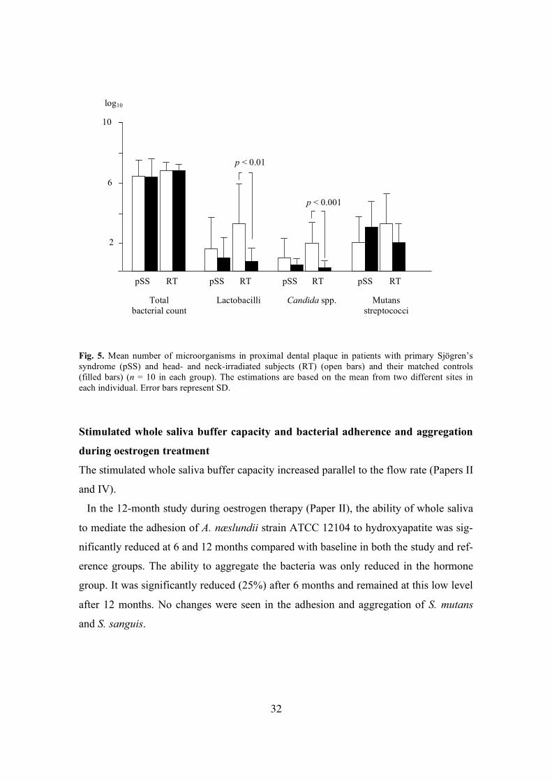
Microorganisms in plaque
In microbial analyses of the proximal dental plaque (Paper IV), the total number of mi-
croorganisms was of the same magnitude in the hyposalivation and control groups. To-
gether with a more acidogenic plaque, a shift towards a higher number of acidogenic
microorganisms was seen in the irradiated individuals. These patients had significantly
higher numbers of lactobacilli and Candida spp. in their plaque compared with their
controls (Fig. 5). In a stepwise regression model, it was revealed that the number of mu-
tans streptococci was correlated to plaque pH in the controls.
Time (min) µl/cm2/min
Controls
Irradiated
5
6
7
0 20 40 60
pH
5 20
AUC 6.2
AUC 5.7
10 15
0
20
40
60
AUC
Fig. 3. Mean values of plaque-pH response to sugar challenge among head- and neck- irradiated subjects and their matched con-trols (n = 10 in each group). The estimations are based on the mean from two different sites in each individual. Bars at 5, 30 and 50 min represent SD
Fig. 4. Plaque acidogenicity (AUC; area under the curve; pH x min) in relation to buccal secretion rate among the hyposali-vators. Linear regression lines calculated from scatter diagrams.
32
Fig. 5. Mean number of microorganisms in proximal dental plaque in patients with primary Sjögren’s syndrome (pSS) and head- and neck-irradiated subjects (RT) (open bars) and their matched controls (filled bars) (n = 10 in each group). The estimations are based on the mean from two different sites in each individual. Error bars represent SD.
Stimulated whole saliva buffer capacity and bacterial adherence and aggregation
during oestrogen treatment
The stimulated whole saliva buffer capacity increased parallel to the flow rate (Papers II
and IV). In the 12-month study during oestrogen therapy (Paper II), the ability of whole saliva
to mediate the adhesion of A. næslundii strain ATCC 12104 to hydroxyapatite was sig-
nificantly reduced at 6 and 12 months compared with baseline in both the study and ref-
erence groups. The ability to aggregate the bacteria was only reduced in the hormone
group. It was significantly reduced (25%) after 6 months and remained at this low level
after 12 months. No changes were seen in the adhesion and aggregation of S. mutans
and S. sanguis.
Total bacterial count
Candida spp. Mutans streptococci
10
6
2
log10
RT pSS RT pSS RT pSS RT
Lactobacilli
p < 0.01
p < 0.001
pSS

33
Table 2. Summary of findings in this thesis on minor salivary gland secretions
Secretion rates (µl/cm2/min±SD)
IgA (µg/ml±SD)
Albumin (mg/ml±SD)
Lactoferrin (µg/ml±SD)
Palatal
Buccal Labial Palatal Buccal Labial Buccal Labial Buccal Labial
Paper I 0.9±0.5 16.0±4.5 4.8±1.9 Paper II 0.8±0.6 14.2±3.7 2.0±1.0 Paper III 11.6±5.3 2.3±1.6 200.3±155.5 87.2±70.2 0.5±0.5 0.3±0.2 39.7±38.1 13.9±14.9 Paper V 0.9±0.5 14.8±4.0 3.0±1.5 149.8±185.8 114.6±113.8 50.3±34.6 Intra-individual variations 43%a 17%a 18%a
Age +e +e +e Gender xa xa, e xa, e xe Pregnancy -e Tobacco +a Dentures +a, e +e Oestrogen +b Diuretics -a Anti-hypertensive medication -e Oral dryness -a, b Plaque acidity -d Sjögren’s syndrome -c +c Radiation therapy -c -c +c +c +c +c
Whole saliva secretion rate +e Whole saliva IgA +e
Cor
rela
ting
fact
ors
Buccal secretion rate -e
+ Positive correlation; - Negative correlation; x Lower in women; a Paper I; b Paper II; c Paper III; d Paper IV; e Paper V.

34

35
DISCUSSION
The studies in this thesis were initiated by observations of small red dots in the some-
what pale palatal mucosa of some individuals. It was soon revealed that these dots were
the expression of more or less intense inflammation around the excretory ducts of mi-
nor, mucosal salivary glands (118). Until this point, a few research reports had been
published on these glands. These reports mainly focused on the labial glands and their
excretions, primarily because this saliva could be harvested from small droplets, which
are easily seen when the lower lip is pulled slightly outwards. In 1990, however, Shern
et al. (59) published their report on a method using which mucosal secretions could be
collected and measured fairly easily and from almost any mucosal site.
The Periotron method is a considerable improvement compared with older methods
(as described in the introduction) for measuring the secretion rate in mucosal salivary
glands. The simplicity and accuracy of the instrument are major advantages. When col-
lecting saliva from mucosal surfaces, it is, however, always important to consider the
risk of contamination of saliva from the major salivary glands. This is especially obvi-
ous for buccal measurements. The excretory duct of the parotid gland reaches the oral
cavity via the lateral surface of the masseter muscle, turns dorsally at the medial edge of
the muscle, pierces the buccinator and delivers the parotid saliva through the orifice at
the level of the second molar (119). The measurements in this thesis of buccal minor
gland saliva were performed well away from the parotid orifice and the very short col-
lection period further eliminates the risk of contamination by parotid saliva. In addition,
a fingertip with an operating glove was held over the measuring filter paper during the
collection of saliva. Moreover, a study of minor gland secretions, collected in filter pa-
pers and measured using the Periotron method, and conventionally collected major
gland saliva reported distinct protein patterns and differences in the ability to mediate
bacterial adherence between the individual saliva collections (11). This further supports
the opinion that mucosal secretions, collected in the same way as in this thesis, may not
be contaminated to any significant extent by major gland saliva.
The lower secretion rates in the minor salivary glands in women compared with men
and the finding that age did not appear to affect the secretion (Papers I and V) are not in

36
agreement with most of the previous studies using the Periotron method (Tables 1 and
2). One explanation for these deviating results could be that individuals with a different
distribution of age groups were included in the studies. The number of individuals in-
cluded in the studies could also vary and many of the studies shown in Table 1 are
based on relatively few individuals compared with Paper I and V (Table 2). Another
explanation could be that in other studies the possible effects of health variables and
measuring variability were not considered. As has been reported for parotid saliva
(120), the study in Paper I reports large intra-individual variations when measuring the
secretion rates of the minor salivary glands. Repeated measurements are therefore pref-
erable in population studies. In the studies in Papers II-V, the mean of at least four dif-
ferent readings from each site was used for the calculations.
In accordance with previously published investigations, we found a large variation in
the secretion rates between different sites of the mucosa. Even within anatomically
well-defined mucosal regions, there could be large variations. Paper I reveals that the
secretion rate is higher in the medial part of the palate than in the lateral part. The mean
values for the secretion rates in the buccal and labial areas of the mucosa, reported in
the present studies, are higher than those in other investigations (Tables 1 and 2). This
may be due to measuring in different parts of the mucosa where the number of glands
can vary (45, 60, 121). There might be an additional explanation for the relatively high
labial secretion values in Paper I. These subjects participated in a larger population study on the Koster islands, Strömstad and measurements were made directly after an
extensive medical anamnesis with a lot of questions and discussions. Talking in this
way may have stimulated the labial mucosal glands and resulted in relatively high se-
cretion rate values, in accordance with the findings of Boros et al. (48). The labial se-
cretion rates reported in Papers II–V were obtained during more resting conditions and
these are not very different from the secretion rates reported by others (Table 1).
The palatal glands may also be mechanically stimulated (111). In 1994, when the
study in Paper I was performed, the wearing of removable dentures was quite common
in rural populations. The increased palatal gland flow rate in denture wearers reported in
this and earlier studies (54) could be verified ten years later in Paper V, in spite of the
more limited number of denture wearers that were included. It can therefore be stated

37
that the minor salivary glands could be mechanically stimulated and are not uniformly
distributed throughout the mucosal membranes. In measurements of minor salivary
gland function, it is therefore important to define the sites that are used and, as for all
saliva sampling, standardize the overall examination procedure.
Medication is known to affect saliva and the most known oral side-effect is reduced
salivation. Some medications, although few in number, appear to increase the whole
salivary secretion rate or buffer capacity. They include physostigmine (64), pilocarpine
(122) and female sex hormones (75, 76). The longitudinal findings in Paper II of whole
salivary secretion rates and buffer pH during a 12-month period of oestrogen treatment
appeared to support these earlier data. However, the whole resting and stimulated sali-
vary secretion rate and buffer pH, as well as the minor palatal and buccal secretion
rates, showed variations during the test period that were not significantly different from
those seen for a reference group of unmedicated women. We therefore concluded that a
low dose of oestrogen had no effect on secretion rate and buffer capacity of whole sa-
liva. The different conclusions from our study, compared with the previous ones, may
not only be explained by the lack of a control or reference group in the previous studies.
Another explanation could be that the previous studies used more potent oestrogen
preparations and in higher doses than in Paper II.
Our findings of decreased whole saliva aggregation of early colonizing Actinomyces
næslundii after 6 and 12 months of a low dose oestrogen treatment suggested that the oestrogen treatment affected the biological activity of saliva. Female hormones has been
reported to modulate the whole saliva content of sialic acid (123). This carbohydrate structure is involved in the binding of oral streptococci and actinomyces to aggregating
salivary glycoproteins (124). It remains to be examined if the observed changes in A.
naeslundii aggregation resulted from changed expression of bacteria-binding receptor structures in saliva.
At baseline, the women taking oestrogen had a significantly lower secretion rate from
their minor labial salivary glands than the reference group. The longitudinal data
showed a significantly increased flow rate for this saliva after 3, 6 and 12 months of
medication. This increase resulted in a labial salivary secretion rate of approximately
the same magnitude as that in the reference group. The concomitant relief of feelings of
38
oral dryness, which were more common in the test group than in the reference group at
baseline, was registered. The relief of oral dryness symptoms during hormone replace-
ment therapy has also previously been reported (106). An observation of a correlation
between subjective feelings of oral dryness and the labial mucosal salivary secretion
rate was also made in Paper I. The explanation of these observations may be that saliva
from minor mucosal glands is rich in mucins that bind water and have a high viscosity.
Moreover, some mucins are bound to mucosal cells (125) and therefore remain for long
periods on the oral mucosal surfaces. Reduced minor gland secretion rates (72) and in-
creased friction on the labial mucosa (126) in individuals complaining of oral dryness
have also been found in other studies. It could be summarized that reduced labial gland
salivary secretion seems to be correlated to feelings of oral dryness.
Sjögren’s syndrome patients and individuals who have undergone irradiation of the
head and neck region for the treatment of cancer are known to suffer from oral dryness
(87, 127). Accordingly, a reduced labial minor gland secretion rate was found among
these patients, together with reduced resting and stimulated whole saliva (Paper III).
These two groups of patients differed, however, in that buccal saliva was only signifi-
cantly reduced in the irradiated group. Earlier research on Sjögren’s syndrome patients
has revealed reduced major gland salivary secretion rates and inflamed tissues in the
labial glands (24). The data in Paper III suggest that the buccal glands are relatively less
affected by sialoadenitis among these patients. In contrast, irradiation may cause the
generalized destruction and impaired function of all salivary glands (88).
As mentioned in the introduction, there are a few reports in the literature on proteins
in labial and palatal secretions. To my knowledge, there are none on protein concentra-
tions in buccal secretions. In Paper III, proteins recovered from filter papers were used
for the determination of IgA, albumin and lactoferrin concentrations in buccal and labial
minor gland saliva. In our pretests, we found that the vast majority of these proteins
could be recovered from the filter papers with samples of whole saliva. This is in accor-
dance with a previous report of the successful elution and determination of proteins
from filter papers (60). The minor gland and whole salivary secretions are, however,
differently composed and mucins, for example, could retain complex bound proteins,
such as IgA, in the filter papers (128). However, the aim of our investigations was not

39
to establish exact quantifications of minor gland proteins but to make comparisons be-
tween groups, which we think the method may permit. The protein concentrations re-
ported in Papers III and V are also largely within the ranges of previous reports (Tables
2 and 3), although data from minor mucosal secretions are scarce.
The IgA concentration in labial minor and parotid gland saliva was examined by Crawford et al. (32) and Smith et al. (34, 35, 43), who found that the concentration was
higher in labial saliva than in parotid saliva. In Papers III and V, the levels of labial IgA were found to be of the same magnitude as in these previous studies (Tables 2 and 3).
High concentrations of IgA were also revealed in palatal and buccal salivary secretions.
In view of the large area of the buccal mucosa, its secretion may be an important con-tributor to the whole saliva IgA. It should be mentioned that, as in the previous studies,
the quantification of IgA in Papers III and V was performed with alpha-chain specific antibodies that recognize not only secretory IgA but also IgA from serum. Under normal
conditions, the major part of IgA in secretions from the labial mucosa is secretory IgA
(32). Serum IgA may, however, leak out from a damaged or fragile mucosa. The mu-cosal status was not recorded in the studies and the possibility that some of the IgA in
the mucosal secretions emanated from the mucosal cells, especially in the hyposalivat-ing subjects (Paper III), cannot therefore be excluded. Our findings and previous find-
ings (89, 129-132) of high lactoferrin and albumin levels in minor gland and stimulated
whole saliva during hyposalivation suggest that the mucosa or mucosal cells are dam-aged in these subjects.

40
Table 3. Mean protein concentrations (µg/ml) reported in whole, parotid and minor
gland saliva
IgA
Albumin
Lactoferrin
Saliva
n
Whole Parotid Palatal Labial Whole Whole Parotid Crawford et al. (32)
17 62 194
Bennet & Reade (133)
10 30-260 c
Wiederström & Bratthall (114)
15a 48.3 d
Izutsu et al. (33)
Gandara et al. (46)
25a 32.3 d 60.9 d 3
Smith et al. (34)
20 35.5 e 36.2 e
Brandtzaeg (134)
194 c
Smith et al. (35)
18 79 88 111
Kugler et al. (135)
18b ≈230
Smith et al. (43)
264 32 – 35 e 52 – 145 e
Sundh et al. (136)
12a 237 c 22 c
Henskens et al. (137)
19a 80 c
Henskens et al. (138)
25a 164 c 123 c
Närhi et al. (104)
71 53 d 13.2 d
Veerman et al. (111)
5 26 85
Leimola-Virtanen et al. (106)
27 41
Almståhl et al. (89)
26a 200 d 3.7 d
Nagler & Hershkovich (71)
45 44.9
Helenius et al. (82)
77a 30.9 d
a Controls; b Adults; c Resting whole saliva; d Stimulated whole saliva; e Median val-
ues shown.

41
Saliva is important for the humoral protection of the oral cavity and the upper respira-
tory tract. The salivary concentration of immunoglobulin is essential in this respect and
could be expected to increase with increasing age, due to challenges from resident or
transient microorganisms. Studies of whole saliva in children reveal a rapid increase in
IgA concentration soon after birth (139), which is followed by a slowly reduced in-
crease during the first year of infancy (140). Data from childhood, adolescence (135)
and in adults (71) also show this increase in whole saliva IgA with increasing age. Our
data in Paper V did not show this correlation, when minor gland IgA was included as an
independent factor in the statistical analyses. In contrast to an earlier finding of stimu-
lated labial saliva (43), Paper V showed a clear positive correlation between age and the
IgA concentrations in palatal, buccal and labial mucosal gland saliva in individuals aged
from 18 to 83 years. Thus, our findings suggest that increased levels of whole saliva
IgA with age in adults could be a reflection of increased minor gland IgA. The relation-
ship between minor gland IgA and age in younger age groups of individuals remains to
be examined.
It is known that the concentration of whole saliva IgA is negatively correlated to the
secretion rate (113, 135) and this was also seen in Papers III and V. For the minor gland
secretions, such a relationship was only found for buccal saliva.
In Paper IV, plaque pH during hyposalivation was examined in relation to acidogenic
microorganisms in that plaque and to minor labial and buccal gland secretion rates. Re-
lationships to whole saliva buffer pH and secretion rate were also examined. The oral
microflora is reported to be differently affected by hyposalivation due to different con-
ditions, i.e. Sjögren’s syndrome or irradiation directed against the head and neck (16,
89). In Paper IV, the microbiota in proximal dental plaque was examined in these pa-
tients and matched controls. The numbers of mutans streptococci did not differ signifi-
cantly between the hyposalivation subjects and their controls. However, a shift towards
higher proportions of lactobacilli and Candida albicans was seen in the irradiated group
but not in the Sjögren’s syndrome patients. The irradiated patients also had a reduced
whole saliva buffer capacity and higher acidogenic capacity in dental plaque, which
were not seen among patients with Sjögren’s syndrome. According to previous data (13,
141), it could be expected that a reduction in stimulated whole salivary secretion rate in

42
both hyposalivation groups (Paper III) would result in more acidogenic plaque in both
groups, compared with their controls. There were, however, two striking differences in
salivary secretion rates when comparing the Sjögren’s syndrome and the irradiated
groups. The irradiated patients had a comparably more pronounced reduction in the
stimulated whole salivary secretion rate compared with the Sjögren’s syndrome group.
The buccal minor gland salivary secretion rate was only significantly reduced in the ir-
radiated group. In the stepwise multiple regression tests, which were performed to test
the influence of all measured saliva variables, it was revealed that the buccal salivary
secretion rate was the only factor correlating with plaque acidogenicity after a sugar
rinse in individuals with generally reduced salivation. In the controls, the numbers of
mutans streptococci was the most important factor for plaque acidogenicity. These find-
ings show that buccal secretion may be critical for caries protection during hyposaliva-
tion.
This series of studies demonstrated previously unknown functions of the minor sali-
vary gland secretions. Interesting observations included the fact that buccal secretion
may be of special importance during hyposalivation and for IgA in whole saliva. More
studies of these glands in larger population groups are therefore of future interest.

43
MAIN CONCLUSIONS
The Periotron method enabled reliable and simple measurements of minor mucosal
gland secretion rates. The secretion rates showed large intra- and inter-individual varia-
tions and differed between separate mucosal sites.
The minor mucosal gland secretion rates did not seem to be reduced with increasing
age. The secretion rates were, however, affected by medication, diabetes and wearing of
dentures, but not by pregnancy. Women had lower minor mucosal gland secretion rates
than men.
During medication with a low dose oestrogen, the labial secretion rate increased and the
whole saliva aggregation of a strain of A. næslundii decreased.
The concentrations of IgA in minor gland saliva increased with increasing age. The
buccal salivary secretion rate and IgA content were correlated to the secretion rate and
the IgA content in whole saliva.
The minor gland secretion rates were reduced and the concentrations of IgA, albumin
and lactoferrin in minor gland saliva were generally increased with hyposalivation, and
especially in irradiated subjects. Especially the buccal saliva seems to be of importance
for plaque pH in hyposalivated subjects.
The complaints of oral dryness were related to a low labial gland secretion rate.

44
45
ACKNOWLEDGEMENTS
First of all, I would like to thank my supervisors Anette Carlén and Dowen Birkhed for
their support and willing help.
I would also like to extend my thanks to Ann-Charlott Börjesson for her excellent
technical assistance with methods and samples and her patience with laboratory matters.
I am also grateful to my co-authors, Annika Almståhl, Guy Heyden, Merja Laine, Pe-
ter Lingström, Nicklas Strömberg, Maude Wikström and Tor Österberg, for fruitful dis-
cussions and help during the work and in the preparation of the manuscripts.
I would also like to express my sincere gratitude to all my friends among the staff at
the Departments of Cariology and Oral Microbiology, Institute of Odontology, Univer-
sity of Göteborg, who have contributed in different ways to the studies in this thesis.
Many thanks also to Henrik, for explaining the theories of dielectric measurements
and for assistance with the photographs, and to Lilian and Jonas for their support and
patience.
Göteborgs Tandläkaresällskap, The Institute of Odontology, Sahlgrenska academy at
Göteborg University, Patentmedelsfonden för Odontologisk Profylaxforskning, Praktik-
ertjänst AB, Sigge Persson och Alice Nybergs Stiftelse, Svenska Tandläkarnes
Inköpsförenings Stiftelse för forskning och studier and the Swedish Dental Association
provided research grants.

46

47
REFERENCES 1. DODDS MW, JOHNSON DA, YEH CK. Health benefits of saliva: a review. J Dent 2005;
33: 223-33. 2. LAGERLÖF F, LENANDER-LUMIKARI M, TENOVUO J. Saliven - en nödvändighet för
tandhälsan. Tandläkartidningen 1997; 89: 49-55. 3. TEN CATE AR. Oral histology. Development, structure and function. Toronto: C. V.
Mosby Company; 1985. 4. DAWES C, WOOD CM. The contribution of the oral minor mucous gland secretions to
the volume of whole saliva in man. Arch Oral Biol 1973; 18: 337-42. 5. MULLER M, JASMIN JR, MONTEIL RA, LOUBIERE R. Embryology and secretory activity
of labial salivary glands. J Biol Buccale 1991; 19: 39-43. 6. EMMELIN N. Nervous control of salivation. Ala J Med Sci 1981; 18: 294-9. 7. EKSTRÖM J. Salivationens reglering. Tandläkartidningen 2000; 92: 36-43. 8. ROSSONI RB, MACHADO AB, MACHADO CRS. A histochemical study of catechola-
mines and cholinesterases in the autonomic nerves of the human minor salivary glands. Histochem J 1979; 11: 661-8.
9. RIVA A, PUXEDDU R, URAS L, LOY F, SERRELI S, TESTA RIVA F. A high resolution sem study of human minor salivary glands. Eur J Morphol 2000; 38: 219-26.
10. SHIBA A, SANO K, NAKAO M, YOSHIDA J, CHO H, HAYASHI T. Electrophoretic analy-sis of the protein in palatine saliva. J Prosthet Dent 1980; 43: 385-91.
11. CARLÉN A, ELIASSON L, ARONSSON G, BIRKHED D. Human minor and major gland saliva proteins and ability to mediate Actinomyces naeslundii adherence. Arch Oral Biol 2004; 49: 177-81.
12. SAMARANAYAKE LP, ROBERTSON AG, MACFARLANE TW, HUNTER IP, MACFARLANE G, SOUTAR DS, et al. The effect of chlorhexidine and benzydamine mouthwashes on mucositis induced by therapeutic irradiation. Clinical Radiology 1988; 39: 291-4.
13. LINGSTRÖM P, BIRKHED D. Plaque-pH and oral retention after consumption of starchy snack products at normal and low salivary secretion rate. Acta Odontol Scand 1993; 51: 379-88.
14. ALMSTÅHL A, WIKSTRÖM M, KRONELD U. Microflora in oral ecosystems in primary Sjögren's syndrome. J Rheumatol 2001; 28: 1007-13.
15. VUOTILA T, YLIKONTIOLA L, SORSA T, LUOTO H, HANEMAAIJER R, SALO T, et al. The relationship between MMPs and pH in whole saliva of radiated head and neck cancer patients. J Oral Pathol Med 2002; 31: 329-38.
16. ALMSTÅHL A, WIKSTRÖM M. Electrolytes in stimulated whole saliva in individuals with hyposalivation of different origins. Arch Oral Biol 2003; 48: 337-44.
17. RAVALD N, LIST T. Caries and periodontal conditions in patients with Sjögren's syn-drome. Swed Dent J 1998; 22: 97-103.
18. SCHNEYER LH. Source of resting total mixed saliva of man. J Appl Physiol 1956; 9: 79-81.
19. MÄKINEN KK, VIRTANEN KK, SÖDERLING E, KOTIRANTA J. Composition of human palatine gland secretions and evidence for the presence of specific arylamidases. Arch Oral Biol 1983; 28: 893-4.
20. GREEN DRJ, EMBERY G. Incorporation of inorganic (35S)-sulphate into glycoproteins of rat buccal and palatal salivary glands in vivo and in vitro. Arch Oral Biol 1984; 29: 335-41.
21. TABAK LA, LEVINE MJ, MANDEL ID, ELLISON SA. Role of salivary mucins in the pro-tection of the oral cavity. J Oral Pathol 1982; 11: 1-17.
48
22. HENSTEN-PETTERSEN A. Biological activities in human labial and palatine secretions. Arch Oral Biol 1975; 20: 107-10.
23. JONSSON R, KRONELD U, TARKOWSKI A. Histological and functional features of sali-vary glands in rheumatic patients with oral sicca symptoms. Scand J Rheumatol 1988; 17: 387-91.
24. SAITO T, FUKUDA H, ARISUE M, MATSUDA A, SHINDOH M, AMEMIYA A, et al. Rela-tionship between sialographic findings of parotid glands and histopathologic finding of labial glands in Sjögren's syndrome. Relation to clinical and immunologic findings. Oral Surg Oral Med Oral Pathol 1991; 72: 675-80.
25. DRUMMOND JR, CHISHOLM DM. A qualitative and quantitative study of the ageing hu-man labial salivary glands. Arch Oral Biol 1984; 29: 151-5.
26. SYRJÄNEN S. Age-related changes in structure of labial minor salivary glands. Age Age-ing 1984; 13: 159-65.
27. ÖSTLUND SG. Palatine glands and mucin. Factors influencing the retention of complete dentures. Odont Tidskr 1954; 62: 1-128.
28. HIRSCH J-M, HEYDEN G, THILANDER H. A clinical, histomorphological and histo-chemical study on snuff-induced lesions of varying severity. J Oral Pathol 1982; 11: 387-98.
29. KRASSE B, GAHNBERG L, BRATTHALL D. Antibodies reacting with Streptococcus mu-tans in secretions from minor salivary glands in humans. Adv Exp Med Biol 1978; 107: 349-54.
30. MÄKINEN KK, VIRTANEN KK, SÖDERLING E, KOTIRANTA J. Effect of xylitol-, sucrose- and water-rinses on the composition of human palatine gland secretions. Scand J Dent Res 1985 ;93: 253-61.
31. SPEIRS RL. Secretion of saliva by human lip mucous glands in response to gustatory stimuli and chewing. Arch Oral Biol 1984; 29: 945-8.
32. CRAWFORD JM, TAUBMAN MA, SMITH DJ. Minor salivary glands as a major source of secretory immunoglobulin A in the human oral cavity. Science 1975; 190: 1206-9.
33. IZUTSU KT, MENARD TW, SCHUBERT MM, ENSIGN WY, SULLIVAN K, TRUELOVE EL, et al. Graft versus host disease-related secretory immunoglobulin A deficiency in bone marrow transplant recipients. Findings in labial saliva. Lab Invest 1985; 52: 292-7.
34. SMITH DJ, TAUBMAN MA, KING WF. Immunological features of minor salivary gland saliva. J Clin Immunol 1987; 7: 449-55.
35. SMITH DJ, TAUBMAN MA, ALI-SALAAM P. Immunoglobulin isotypes in human minor gland saliva. J Dent Res 1991; 70: 167-70.
36. HENSTEN-PETERSEN A, KORNSTAD L. The contribution of the minor mucous glands to the concentrations of blood group specific substances, carbohydrates and proteins in human mixed saliva. Arch Oral Biol 1976; 21: 485-9.
37. KUTSCHER AH, MANDEL ID, ZEGARELLI EV, DENNING C, ERIV A, RUIZ L, et al. A technique for collecting the secretion of minor salivary glands: 1. Use of capillary tubes. J Oral Therap Pharmacol 1967; 3: 391-2.
38. DAWES C, WOOD CM. The composition of human lip mucous gland secretion. Arch Oral Biol 1973; 18: 343-50.
39. KAABER S. Sodium and potassium content in human palatine gland secretion. Arch Oral Biol 1977; 22: 529-32.
40. KRISTIANSEN B-E. Collection of mucosal secretion by synthetic discs for quantitation of secretory IgA and bacteria. J Immunol Methods 1984; 73: 251-7.
41. WIESMANN UN, BOAT TF, DI SANT'AGNESE PA. Flow-rates and electrolytes in minor-salivary-gland saliva in normal subjects and patients with cystic fibrosis. Lancet 1972; 2: 510-2.

49
42. GANDARA BK, IZUTSU KT, TRUELOVE EL, ENSIGN WY, SOMMERS EE. Age-related salivary flow rate changes in controls and patients with oral lichen planus. J Dent Res 1985; 64: 1149-51.
43. SMITH DJ, JOSHIPURA K, KENT R, TAUBMAN MA. Effect of age on immunoglobulin content and volume of human labial gland saliva. J Dent Res 1992; 71: 1891-4.
44. GREAVES IC, HUME WJ, NISBET T. Stereophotomicroscopic assessment of labial sali-vary gland flow. J Oral Med 1986; 41: 172-4.
45. HAMADA T, KAWASOE Y, SEKINO K, NAGASAWA T, TSURU H. Palatal gland distribu-tion. J Dent Res 1974; 53: 944.
46. GANDARA BK, IZUTSU KT, TRUELOVE EL, MANDEL ID, SOMMERS EE, ENSIGN WY. Sialochemistry of whole, parotid, and labial minor gland saliva in patients with oral li-chen planus. J Dent Res 1987; 66: 1619-22.
47. GAUBENSTOCK LM. Dental caries and the secretory activity of human labial minor sali-vary glands. Arch Oral Biol 1995; 40: 525-8.
48. BOROS I, KESZLER P, ZELLES T. Study of saliva secretion and the salivary fluoride con-centration of the human minor labial glands by a new method. Arch Oral Biol 1999; 44: 59-62.
49. MARTON K, BOROS I, FEJERDY P, MADLENA M. Evaluation of unstimulated flow rates of whole and palatal saliva in healthy patients wearing complete dentures and in pa-tients with Sjogren's syndrome. J Prosthet Dent 2004; 91: 577-81.
50. SHOJI N, SASANO T, INUKAI S, SATOH-KURIWADA S, LIKUBO M, FURUUCHI T, et al. A simple, yet accurate method for detecting and quantifying secretions from human minor salivary glands using the iodine-starch reaction. Arch Oral Biol 2003; 48: 761-5.
51. OGAMI K, SAKURAI K, ANDO T. A method of measuring salivary flow rate in the lower labial mucosal region. J Oral Rehabil 2004; 31: 861-5.
52. SUPPIPAT N, SUPPIPAT N. Evaluation of an electronic device for gingival fluid quantita-tion. J Periodontol 1977; 48: 388-94.
53. NIEDERMEIER WH, KRÄMER R. Salivary secretion and denture retention. J Prosthet Dent 1992; 67: 211-6.
54. NIEDERMEIER W, SCHALLER K-H, FISCHER D. Die sekretion der speichel- und schleimdrüsen im quantitativen vergleich. D Zahnärtzl Z 1989; 44: 52-6.
55. NIEDERMEIER W, HUBER M. Quantitative untersuchung zur sekretionsleistung der gaumenspeicheldrüsen. D Zahnärtzl Z 1989; 44: 37-40.
56. SHERN RJ, FOX PC, LI SH. Influence of age on the secretory rates of the human minor salivary glands and whole saliva. Arch Oral Biol 1993; 38: 755-61.
57. SIVARAJASINGAM V, DRUMMOND JR. Measurements of human minor salivary gland secretions from different oral sites. Arch Oral Biol 1995; 40: 723-9.
58. SHERN RJ, GUCKES AD, LI SH. A method for measuring secretions from the palatal minor salivary glands. J Prosthet Dent 1993; 69: 624-9.
59. SHERN RJ, FOX PC, CAIN JL, LI SH. A method for measuring the flow of saliva from the minor salivary glands. J Dent Res 1990; 69: 1146-9.
60. WON S-H, KHO H-S, KIM Y-K, CHUNG S-C, LEE S-W. Analysis of residual saliva and minor salivary gland secretions. Arch Oral Biol 2001; 46: 619-24.
61. LEE S-K, LEE S-W, CHUNG S-C, KIM Y-K, KHO H-S. Analysis of residual saliva and minor salivary gland secretions in patients with dry mouth. Arch Oral Biol 2002; 47: 637-41.
62. SONESSON M, ELIASSON L, MATSSON L. Minor salivary gland secretion in children and adults. Arch Oral Biol 2003; 48: 535-9.
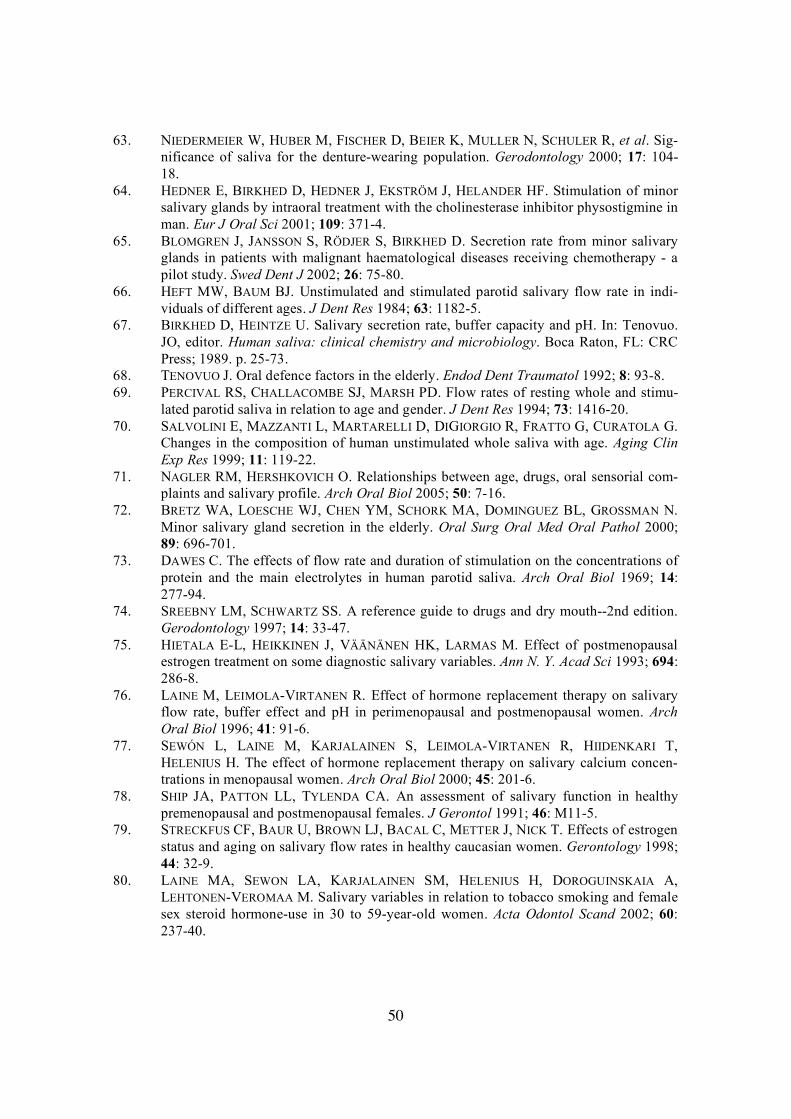
50
63. NIEDERMEIER W, HUBER M, FISCHER D, BEIER K, MULLER N, SCHULER R, et al. Sig-nificance of saliva for the denture-wearing population. Gerodontology 2000; 17: 104-18.
64. HEDNER E, BIRKHED D, HEDNER J, EKSTRÖM J, HELANDER HF. Stimulation of minor salivary glands by intraoral treatment with the cholinesterase inhibitor physostigmine in man. Eur J Oral Sci 2001; 109: 371-4.
65. BLOMGREN J, JANSSON S, RÖDJER S, BIRKHED D. Secretion rate from minor salivary glands in patients with malignant haematological diseases receiving chemotherapy - a pilot study. Swed Dent J 2002; 26: 75-80.
66. HEFT MW, BAUM BJ. Unstimulated and stimulated parotid salivary flow rate in indi-viduals of different ages. J Dent Res 1984; 63: 1182-5.
67. BIRKHED D, HEINTZE U. Salivary secretion rate, buffer capacity and pH. In: Tenovuo. JO, editor. Human saliva: clinical chemistry and microbiology. Boca Raton, FL: CRC Press; 1989. p. 25-73.
68. TENOVUO J. Oral defence factors in the elderly. Endod Dent Traumatol 1992; 8: 93-8. 69. PERCIVAL RS, CHALLACOMBE SJ, MARSH PD. Flow rates of resting whole and stimu-
lated parotid saliva in relation to age and gender. J Dent Res 1994; 73: 1416-20. 70. SALVOLINI E, MAZZANTI L, MARTARELLI D, DIGIORGIO R, FRATTO G, CURATOLA G.
Changes in the composition of human unstimulated whole saliva with age. Aging Clin Exp Res 1999; 11: 119-22.
71. NAGLER RM, HERSHKOVICH O. Relationships between age, drugs, oral sensorial com-plaints and salivary profile. Arch Oral Biol 2005; 50: 7-16.
72. BRETZ WA, LOESCHE WJ, CHEN YM, SCHORK MA, DOMINGUEZ BL, GROSSMAN N. Minor salivary gland secretion in the elderly. Oral Surg Oral Med Oral Pathol 2000; 89: 696-701.
73. DAWES C. The effects of flow rate and duration of stimulation on the concentrations of protein and the main electrolytes in human parotid saliva. Arch Oral Biol 1969; 14: 277-94.
74. SREEBNY LM, SCHWARTZ SS. A reference guide to drugs and dry mouth--2nd edition. Gerodontology 1997; 14: 33-47.
75. HIETALA E-L, HEIKKINEN J, VÄÄNÄNEN HK, LARMAS M. Effect of postmenopausal estrogen treatment on some diagnostic salivary variables. Ann N. Y. Acad Sci 1993; 694: 286-8.
76. LAINE M, LEIMOLA-VIRTANEN R. Effect of hormone replacement therapy on salivary flow rate, buffer effect and pH in perimenopausal and postmenopausal women. Arch Oral Biol 1996; 41: 91-6.
77. SEWÓN L, LAINE M, KARJALAINEN S, LEIMOLA-VIRTANEN R, HIIDENKARI T, HELENIUS H. The effect of hormone replacement therapy on salivary calcium concen-trations in menopausal women. Arch Oral Biol 2000; 45: 201-6.
78. SHIP JA, PATTON LL, TYLENDA CA. An assessment of salivary function in healthy premenopausal and postmenopausal females. J Gerontol 1991; 46: M11-5.
79. STRECKFUS CF, BAUR U, BROWN LJ, BACAL C, METTER J, NICK T. Effects of estrogen status and aging on salivary flow rates in healthy caucasian women. Gerontology 1998; 44: 32-9.
80. LAINE MA, SEWON LA, KARJALAINEN SM, HELENIUS H, DOROGUINSKAIA A, LEHTONEN-VEROMAA M. Salivary variables in relation to tobacco smoking and female sex steroid hormone-use in 30 to 59-year-old women. Acta Odontol Scand 2002; 60: 237-40.
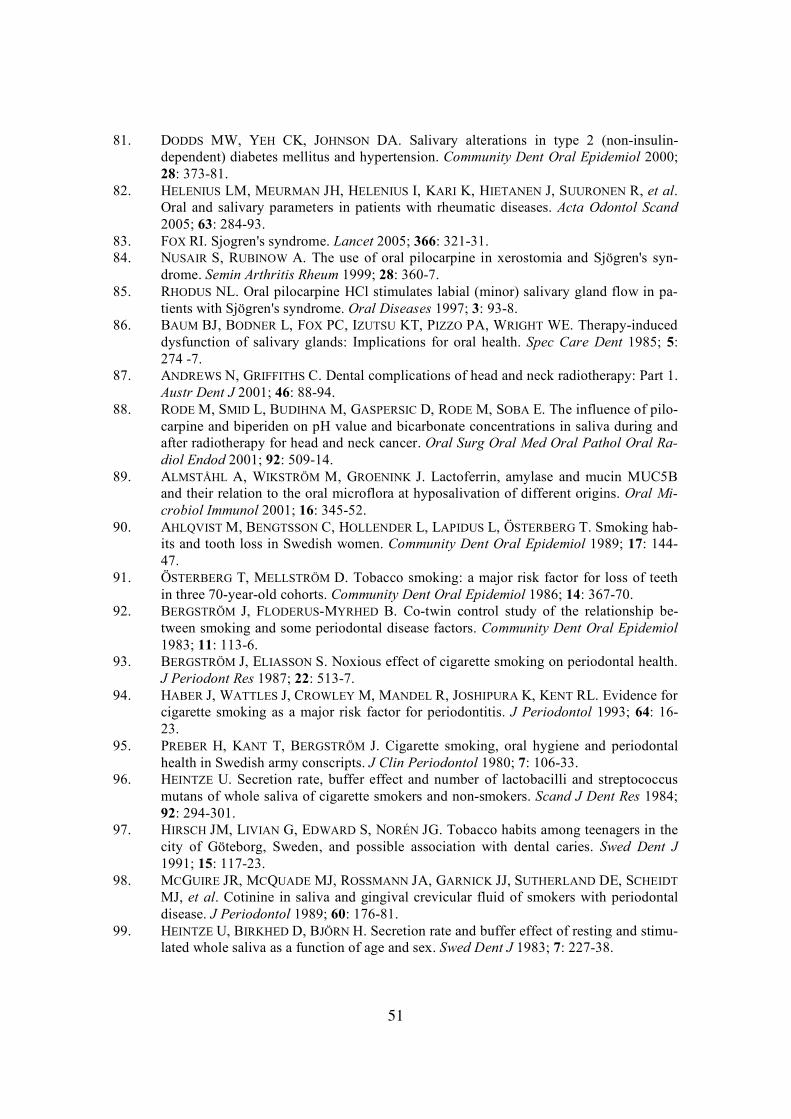
51
81. DODDS MW, YEH CK, JOHNSON DA. Salivary alterations in type 2 (non-insulin-dependent) diabetes mellitus and hypertension. Community Dent Oral Epidemiol 2000; 28: 373-81.
82. HELENIUS LM, MEURMAN JH, HELENIUS I, KARI K, HIETANEN J, SUURONEN R, et al. Oral and salivary parameters in patients with rheumatic diseases. Acta Odontol Scand 2005; 63: 284-93.
83. FOX RI. Sjogren's syndrome. Lancet 2005; 366: 321-31. 84. NUSAIR S, RUBINOW A. The use of oral pilocarpine in xerostomia and Sjögren's syn-
drome. Semin Arthritis Rheum 1999; 28: 360-7. 85. RHODUS NL. Oral pilocarpine HCl stimulates labial (minor) salivary gland flow in pa-
tients with Sjögren's syndrome. Oral Diseases 1997; 3: 93-8. 86. BAUM BJ, BODNER L, FOX PC, IZUTSU KT, PIZZO PA, WRIGHT WE. Therapy-induced
dysfunction of salivary glands: Implications for oral health. Spec Care Dent 1985; 5: 274 -7.
87. ANDREWS N, GRIFFITHS C. Dental complications of head and neck radiotherapy: Part 1. Austr Dent J 2001; 46: 88-94.
88. RODE M, SMID L, BUDIHNA M, GASPERSIC D, RODE M, SOBA E. The influence of pilo-carpine and biperiden on pH value and bicarbonate concentrations in saliva during and after radiotherapy for head and neck cancer. Oral Surg Oral Med Oral Pathol Oral Ra-diol Endod 2001; 92: 509-14.
89. ALMSTÅHL A, WIKSTRÖM M, GROENINK J. Lactoferrin, amylase and mucin MUC5B and their relation to the oral microflora at hyposalivation of different origins. Oral Mi-crobiol Immunol 2001; 16: 345-52.
90. AHLQVIST M, BENGTSSON C, HOLLENDER L, LAPIDUS L, ÖSTERBERG T. Smoking hab-its and tooth loss in Swedish women. Community Dent Oral Epidemiol 1989; 17: 144-47.
91. ÖSTERBERG T, MELLSTRÖM D. Tobacco smoking: a major risk factor for loss of teeth in three 70-year-old cohorts. Community Dent Oral Epidemiol 1986; 14: 367-70.
92. BERGSTRÖM J, FLODERUS-MYRHED B. Co-twin control study of the relationship be-tween smoking and some periodontal disease factors. Community Dent Oral Epidemiol 1983; 11: 113-6.
93. BERGSTRÖM J, ELIASSON S. Noxious effect of cigarette smoking on periodontal health. J Periodont Res 1987; 22: 513-7.
94. HABER J, WATTLES J, CROWLEY M, MANDEL R, JOSHIPURA K, KENT RL. Evidence for cigarette smoking as a major risk factor for periodontitis. J Periodontol 1993; 64: 16-23.
95. PREBER H, KANT T, BERGSTRÖM J. Cigarette smoking, oral hygiene and periodontal health in Swedish army conscripts. J Clin Periodontol 1980; 7: 106-33.
96. HEINTZE U. Secretion rate, buffer effect and number of lactobacilli and streptococcus mutans of whole saliva of cigarette smokers and non-smokers. Scand J Dent Res 1984; 92: 294-301.
97. HIRSCH JM, LIVIAN G, EDWARD S, NORÉN JG. Tobacco habits among teenagers in the city of Göteborg, Sweden, and possible association with dental caries. Swed Dent J 1991; 15: 117-23.
98. MCGUIRE JR, MCQUADE MJ, ROSSMANN JA, GARNICK JJ, SUTHERLAND DE, SCHEIDT MJ, et al. Cotinine in saliva and gingival crevicular fluid of smokers with periodontal disease. J Periodontol 1989; 60: 176-81.
99. HEINTZE U, BIRKHED D, BJÖRN H. Secretion rate and buffer effect of resting and stimu-lated whole saliva as a function of age and sex. Swed Dent J 1983; 7: 227-38.

52
100. PARVINEN T. Stimulated salivary flow rate, pH and lactobacillus and yeast concentra-tions in non-smokers and smokers. Scand J Dent Res 1984; 92: 315-8.
101. REDDY MS, NAIK SR, BAGGA OP, CHUTTANI HK. Effect of chronic tobacco-betel-lime ''quid'' chewing on human salivary secretions. Am J Clin Nutr 1980; 33: 77-80.
102. NEDERFORS T, ISAKSSON R, MORNSTAD H, DAHLOF C. Prevalence of perceived symp-toms of dry mouth in an adult Swedish population--relation to age, sex and pharma-cotherapy. Community Dent Oral Epidemiol 1997; 25: 211-6.
103. NÄRHI TO, MEURMAN JH, AINAMO A. Xerostomia and hyposalivation. Causes, conse-quences and treatment in the elderly. Drugs Aging 1999; 15: 103-16.
104. NÄRHI T. Prevalence of subjective feelings of dry mouth in the elderly. J Dent Res 1994; 73: 20-5.
105. THOMSON WM, CHALMERS JM, SPENCER AJ, SLADE GD. Medication and dry mouth: findings from a cohort study of older people. J Public Health Dent 2000; 60: 12-20.
106. LEIMOLA-VIRTANEN R, HELENIUS H, LAINE M. Hormone replacement therapy and some salivary antimicrobial factors in post- and perimenopausal women. Maturitas 1997; 27: 145-51.
107. JOHNSON G, BARENTHIN I, WESTPHAL P. Mouthdryness among patients in longterm hospitals. Gerodontology 1984; 3: 197-203.
108. BARDOW A, NYVAD B, NAUNTOFTE B. Relationships between medication intake, com-plaints of dry mouth, salivary flow rate and composition, and the rate of tooth deminer-alization in situ. Arch Oral Biol 2001; 46: 413-23.
109. THORSELIUS I, EMILSON C-G, ÖSTERBERG T. Salivary conditions and drug consump-tion in older age groups of elderly Swedish individuals. Gerodontics 1988; 4: 66-70.
110. SPIELMAN A, BEN-ARYEH H, GUTMAN D, SZARGEL R, DEUTSCH E. Xerostomia - Di-agnosis and treatment. Oral Surg Oral Med Oral Pathol 1981; 51: 144-7.
111. VEERMAN ECI, VAN DEN KEYBUS PAM, VISSINK A, NIEUW AMERONGEN AV. Human glandular salivas: their separate collection and analysis. Eur J Oral Sci 1996; 104: 346-52.
112. FERGUSON DB. The flow rate and composition of human labial gland saliva. Arch Oral Biol 1999; 44: S11-4.
113. NÄRHI TO, TENOVUO J, AINAMO A, VILJA P. Antimicrobial factors, sialic acid, and protein concentration in whole saliva of the elderly. Scand J Dent Res 1994; 102: 120-5.
114. WIDERSTRÖM L, BRATTHALL D. Increased IgA levels in saliva during pregnancy. Scand J Dent Res 1984; 92: 33-7.
115. DAWES C. Physiological factors affecting salivary flow rate, oral sugar clearance, and the sensation of dry mouth in man. J Dent Res 1987; 66: 648-53.
116. ERICSSON Y. Clinical investigations of the salivary buffering action. Acta Odontol Scand 1959; 17: 131-65.
117. LINGSTRÖM P, IMFELD T, BIRKHED D. Comparison of three different methods for measurement of plaque-pH in humans after consumption of soft bread and potato chips. J Dent Res 1993; 72: 865-70.
118. ELIASSON L, HEYDEN G, LANDAHL S, STEEN B. Effects of tobacco and diuretics on human palatal salivary glands. J Oral Pathol Med 1991; 20: 126-9.
119. KUTTA H, MAY J, JAEHNE M, MUNSCHER A, PAULSEN FP. Antimicrobial defence mechanisms of the human parotid duct. J Anat 2006; 208: 609-19.
120. BURLAGE FR, PIJPE J, COPPES RP, HEMELS ME, MEERTENS H, CANRINUS A, et al. Variability of flow rate when collecting stimulated human parotid saliva. Eur J Oral Sci 2005; 113: 386-90.
121. GAUBENSTOCK LM, LEONTYEV VK. Quantitative and topographic characteristics of minor salivary glands. Stomatologia 1990; 69: 28-31.
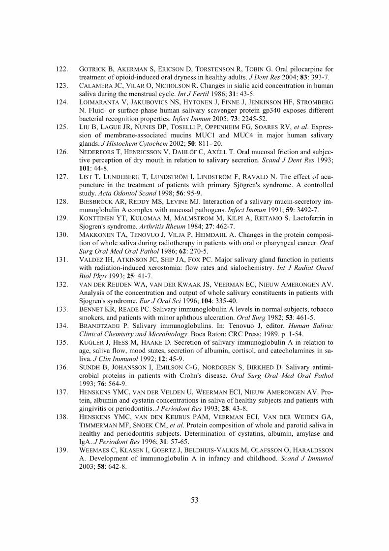
53
122. GOTRICK B, AKERMAN S, ERICSON D, TORSTENSON R, TOBIN G. Oral pilocarpine for treatment of opioid-induced oral dryness in healthy adults. J Dent Res 2004; 83: 393-7.
123. CALAMERA JC, VILAR O, NICHOLSON R. Changes in sialic acid concentration in human saliva during the menstrual cycle. Int J Fertil 1986; 31: 43-5.
124. LOIMARANTA V, JAKUBOVICS NS, HYTONEN J, FINNE J, JENKINSON HF, STROMBERG N. Fluid- or surface-phase human salivary scavenger protein gp340 exposes different bacterial recognition properties. Infect Immun 2005; 73: 2245-52.
125. LIU B, LAGUE JR, NUNES DP, TOSELLI P, OPPENHEIM FG, SOARES RV, et al. Expres-sion of membrane-associated mucins MUC1 and MUC4 in major human salivary glands. J Histochem Cytochem 2002; 50: 811- 20.
126. NEDERFORS T, HENRICSSON V, DAHLÖF C, AXÉLL T. Oral mucosal friction and subjec-tive perception of dry mouth in relation to salivary secretion. Scand J Dent Res 1993; 101: 44-8.
127. LIST T, LUNDEBERG T, LUNDSTRÖM I, LINDSTRÖM F, RAVALD N. The effect of acu-puncture in the treatment of patients with primary Sjögren's syndrome. A controlled study. Acta Odontol Scand 1998; 56: 95-9.
128. BIESBROCK AR, REDDY MS, LEVINE MJ. Interaction of a salivary mucin-secretory im-munoglobulin A complex with mucosal pathogens. Infect Immun 1991; 59: 3492-7.
129. KONTTINEN YT, KULOMAA M, MALMSTROM M, KILPI A, REITAMO S. Lactoferrin in Sjogren's syndrome. Arthritis Rheum 1984; 27: 462-7.
130. MAKKONEN TA, TENOVUO J, VILJA P, HEIMDAHL A. Changes in the protein composi-tion of whole saliva during radiotherapy in patients with oral or pharyngeal cancer. Oral Surg Oral Med Oral Pathol 1986; 62: 270-5.
131. VALDEZ IH, ATKINSON JC, SHIP JA, FOX PC. Major salivary gland function in patients with radiation-induced xerostomia: flow rates and sialochemistry. Int J Radiat Oncol Biol Phys 1993; 25: 41-7.
132. VAN DER REIJDEN WA, VAN DER KWAAK JS, VEERMAN EC, NIEUW AMERONGEN AV. Analysis of the concentration and output of whole salivary constituents in patients with Sjogren's syndrome. Eur J Oral Sci 1996; 104: 335-40.
133. BENNET KR, READE PC. Salivary immunoglobulin A levels in normal subjects, tobacco smokers, and patients with minor aphthous ulceration. Oral Surg 1982; 53: 461-5.
134. BRANDTZAEG P. Salivary immunoglobulins. In: Tenovuo J, editor. Human Saliva: Clinical Chemistry and Microbiology. Boca Raton: CRC Press; 1989. p. 1-54.
135. KUGLER J, HESS M, HAAKE D. Secretion of salivary immunoglobulin A in relation to age, saliva flow, mood states, secretion of albumin, cortisol, and catecholamines in sa-liva. J Clin Immunol 1992; 12: 45-9.
136. SUNDH B, JOHANSSON I, EMILSON C-G, NORDGREN S, BIRKHED D. Salivary antimi-crobial proteins in patients with Crohn's disease. Oral Surg Oral Med Oral Pathol 1993; 76: 564-9.
137. HENSKENS YMC, VAN DER VELDEN U, WEERMAN ECI, NIEUW AMERONGEN AV. Pro-tein, albumin and cystatin concentrations in saliva of healthy subjects and patients with gingivitis or periodontitis. J Periodont Res 1993; 28: 43-8.
138. HENSKENS YMC, VAN DEN KEIJBUS PAM, VEERMAN ECI, VAN DER WEIDEN GA, TIMMERMAN MF, SNOEK CM, et al. Protein composition of whole and parotid saliva in healthy and periodontitis subjects. Determination of cystatins, albumin, amylase and IgA. J Periodont Res 1996; 31: 57-65.
139. WEEMAES C, KLASEN I, GOERTZ J, BELDHUIS-VALKIS M, OLAFSSON O, HARALDSSON A. Development of immunoglobulin A in infancy and childhood. Scand J Immunol 2003; 58: 642-8.

54
140. RUHL S, RAYMENT SA, SCHMALZ G, HILLER KA, TROXLER RF. Proteins in whole sa-liva during the first year of infancy. J Dent Res 2005; 84: 29-34.
141. ABELSON DC, MANDEL ID. The effect of saliva on plaque pH in vivo. J Dent Res 1981; 60: 1634-8.