Pitfalls in minor salivary gland neoplasms
57
Pitfalls in the Biopsy Diagnosis Of Intraoral Minor Salivary Gland Neoplasms: Diagnostic Considerations and Recommended Approach (Adv Anat Pathol Volume 21, Number 1, January 2014) BY EKTA JAJODIA
-
Upload
ekta-taparia -
Category
Health & Medicine
-
view
300 -
download
0
Transcript of Pitfalls in minor salivary gland neoplasms

Pitfalls in the Biopsy Diagnosis Of Intraoral Minor Salivary Gland
Neoplasms: DiagnosticConsiderations and Recommended
Approach
(Adv Anat Pathol Volume 21, Number 1, January 2014)
BY EKTA JAJODIA

Sites of minor salivary glands
-Most numerous at junction of hard and soft palate-Buccal mucosa-Lips-Tongue
All of these glands are predominantly mucous
VON EBNER’S GLANDS – these are purely serousLocated in dorsum of tongue in relation to circumvallate papillae
Minor salivary glands are not encapsulated
Intraoral minor salivary gland neoplasms include • Benign tumors -1.Pleomorphic adenoma 2.Monomorphic adenoma, basal cell type (also known as basal cell adenoma)
• Malignant tumors-1.Mucoepidermoid carcinoma2.Adenoid cystic carcinoma3.Polymorphous low-grade adenocarcinoma

• Initial diagnostic modality for a patient with anintraoral mass - fine-needle aspiration biopsy or incisional biopsy
• Limitations-
1. Small tissue sampling2. Similar growth patterns3. Similar cytomorphology4. Similar immunoreactivity
Both benign and malignant
Exception to this problem- Mucoepidermoid carcinoma(MEC)
Characterized by mucocytes, epidermoid cells, and intermediate cells
As no other salivary gland neoplasm consists of these cellular components, identification of these cell types permits a diagnosis of MEC even in context of limited sampling
Other intraoral minor salivary gland tumors share light microscopic and IHC features
Distinction between benign and malignant lesions depends upon identification of invasive growth
But incisional biopsy of intraoral minor salivary gland tumors consists of lesional material without surrounding tissues - assessment of invasive growth cannot be done
So specific diagnosis rendered on the basis of FNAB or incisional biopsy may result in discordant diagnosis after complete surgical resection

1. Circumscription without encapsulation2. Multiple growth patterns3. Bland cytomorphology4. Combination of epithelial and myoepithelial cell differentiation5. Absent to minimal increase in mitotic activity 6. To a large extent similar IHC reactivity
Shared findings among minor salivary gland neoplasms

A and B, Incisional biopsy shows a cellular neoplasm comprised of tubules with isomorphic basaloid-appearing nuclei without features allowing for determining whether it is benign or malignant. C, After complete excision showing circumscription without invasion, a diagnosis of basal cell adenoma can be rendered
Circumscription, Encapsulation, and Invasive growth
• All minor salivary gland neoplasms, whether benign or malignant, are unencapsulatedSo presence or absence of capsule does not differentiate benign and malignant minor salivary gland tumors
• Both benign and malignant intraoral minor salivary gland neoplasms may be circumscribed

Extension of tumor to (and even involvement of) surface (squamous) epithelium does not represent criterion for malignancy
Malignant lesions show invasive growth, including invasion into non-neoplastic seromucous glands , soft tissues , perineural invasion (PNI) , and lymph-vascular invasion (LVI) But such findings are uncommon in small biopsies of intraoral minor salivary gland neoplasms

All minor salivary gland neoplasms are unencapsulated and often even malignant neoplasms such as polymorphous low-grade adenocarcinoma appears circumscribed in its superficial aspect
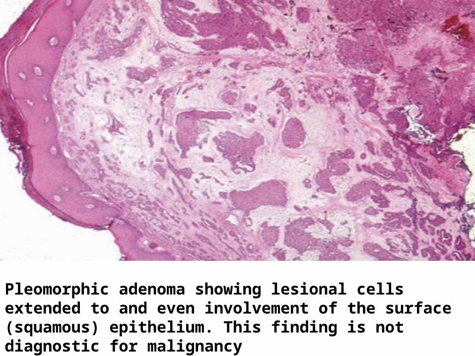
Pleomorphic adenoma showing lesional cells extended to and even involvement of the surface (squamous) epithelium. This finding is not diagnostic for malignancy

GROWTH PATTERN
Salivary gland neoplasms of both major and minor salivary gland show >1 growth pattern includingtubular-ductular, solid, (micro)cystic, cribriform, trabecular, nodular, lattice-like, fascicular, and/or papillary
Cribriform growth may suggest a diagnosis ofadenoid cystic carcinoma, but is not pathognomonic of this lesion This finding may also be present in other intraoralminor salivary gland neoplasms including PLGA, pleomorphic adenoma and basal cell adenoma

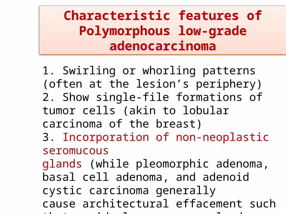
1. Swirling or whorling patterns (often at the lesion’s periphery)2. Show single-file formations of tumor cells (akin to lobular carcinoma of the breast)3. Incorporation of non-neoplastic seromucousglands (while pleomorphic adenoma, basal cell adenoma, and adenoid cystic carcinoma generallycause architectural effacement such that residual seromucous glands cannot be identified within the tumor
Characteristic features of Polymorphous low-grade
adenocarcinoma
Diagnosis of PLGA(ie, diagnosis of minorsalivary gland carcinoma) requires identification of PNI, LVI, and/or invasive growth
Nuclear palisading of basaloid cells along the stromal interface is often seen in basal cell adenoma, although this feature is not specifically diagnostic of basal cell adenoma
Intraoral minor salivary gland tumors show architectural overlap to permit definitive diagnosisbased on any single growth pattern, or combinationof architectural features

• Adenoid cystic carcinoma characteristicallycontains abundant myoepithelial (abluminal) cells with increased nuclear-to-cytoplasmic ratio, and basaloid (hyperchromatic) angulated nuclei without identifiable nucleoli
• Similar nuclear features can be present in basal cell adenoma and even in pleomorphic adenoma
• But basaloid hyperchromatic nuclei are not evident in PLGA which typically consists of cells with vesicular chromatin and inconspicuoussmall nucleoli
CYTOMORPHOLOGY

• Although myoepithelial cells are predominantin basal cell adenoma and adenoid cystic carcinoma,these tumors may display small ductular or tubularstructures lined by luminal cells with round-to-oval nuclei and eosinophilic cytoplasm • In basal cell adenoma, ductal (luminal) cells aremost conspicuous in the tubular subtype
• Myoepithelial cells can be present in all the above tumor types, although myoepithelial cells are not characteristic of PLGA

• Neoplastic (modified) myoepithelial cells may display various morphology including plasmacytoid, spindle shaped (so-called “hyaline cells”) , epithelioid, cuboidal, stellate, or clear cells
• Plasmacytoid myoepithelial cells often occur in pleomorphic adenoma and may be informative in limited tissue sampling, although they are not pathognomonic

A- In adenoid cystic carcinoma, small ductular or tubular structures lined by luminal cells among the predominant myoepithelial (abluminal) cell type.B- Ductular or tubular structures are readily apparent in this basal cell adenoma, tubular subtype

PLGA show characteristic but not pathognomonic growth patterns including: (A)swirling/whorling cellular arrangements at the lesion’s periphery (B) single cell file formation (akin to lobular carcinoma of the breast (C) incorporation or envelopment (rather than effacement) of residual non-neoplastic seromucous gland

A- In adenoid cystic carcinoma, the predominant cell type is the myoepithelial (abluminal) cells comprised of isomorphic, basaloid (hyperchromatic), and angulated nuclei without identifiable nucleoli. B- Polymorphous low-grade adenocarcinoma also comprised of isomorphic nuclei but in contrast show vesicular appearingnuclei with small identifiable nucleoli.

A- The presence of squamous eddies (with and without keratinization) seen in basal cell adenoma B- Plasmacytoid cells in pleomorphic adenoma C- spindle shaped in pleomorphic adenoma

• Chondromyxoid stroma - definitive histologiccomponent of pleomorphic adenomaFacilitates distinction of pleomorphic adenoma from monomorphic adenoma
• Due to limitations of tissue sampling, chondromyxoid stroma of PA may not be present
• Consequently, absence of chondromyxoid stroma does not preclude a diagnosis of PA, nor should lack of it prompt a diagnosis of monomorphic adenoma
• It may appear similar to the slate blue-gray mucohyaline matrix sometimes seen in PLGA
TUMOR STROMA

• Adenoid cystic carcinoma shows pseudocysts with basophilic and eosinophilic-appearing material
• Hyalinized (collagenized) stroma can be seen inbasal cell adenoma, particularly the membranous subtype as well as in PA and adenoid cystic carcinoma
• Basal cell adenoma sometimes shows small“droplets” of matrix within nests of neoplastic cells
• Stromal features are useful but definitive diagnosis cannot be based on it
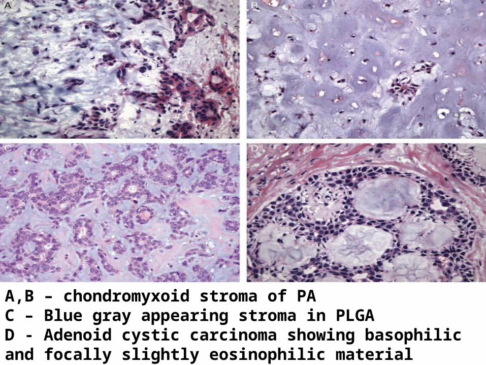
A,B – chondromyxoid stroma of PAC – Blue gray appearing stroma in PLGAD - Adenoid cystic carcinoma showing basophilic and focally slightly eosinophilic material within the pseudocysts
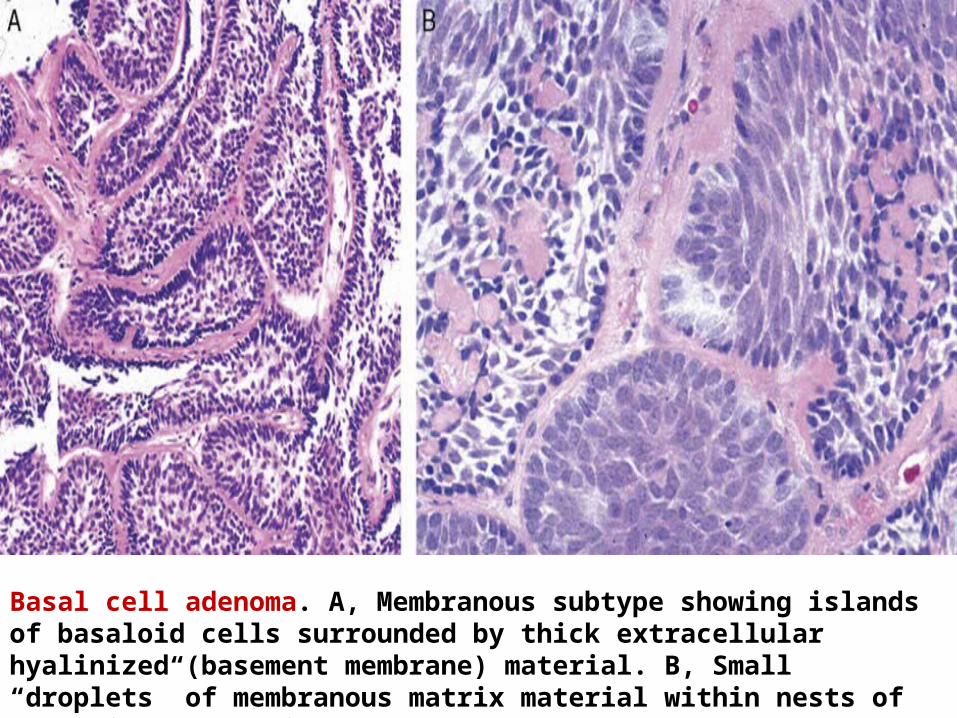
Basal cell adenoma. A, Membranous subtype showing islands of basaloid cells surrounded by thick extracellular hyalinized (basement membrane) material. B, Small “droplets” of membranous matrix material within nests of basaloid neoplastic cells.

• Metaplasia may occur spontaneously or after FNAB
• Squamous metaplasia in the form of cells with keratinization and intercellular bridges are seen in PLGA, PA and membranous variant of BCA
• In contrast to other tumor types, adenoid cystic carcinoma rarely displays squamous metaplasiaSo, metaplasia may be informative, particularly in ruling out adenoid cystic carcinoma
METAPLASIA

• These neoplasms are frequently devoid of mitotic activity by light microscopy
• Malignant lesions may show marked increase in mitotic activity with atypical mitoses, but mitotic figures do not distinguish malignant lesions from their benign counterparts
• Ki-67 (MIB-1) IHC also does not definitively distinguish adenoid cystic carcinoma and/or PLGAfrom PA or basal cell adenomaSo Ki-67 IHC is of limited value in the distinction of these tumors
MITOTIC ACTIVITY
MISCELLANEOUS FINDINGSTyrosine-rich crystalloids - characteristic ofPleomorphic adenoma but may also be seen in PLGA and other tumors
Psammomatoid concretions can be present in a variety of salivary gland neoplasms like PLGA, pleomorphic adenoma, and monomorphic adenoma

HISTOCHEMISTRY
• All of these lesions lack intracytoplasmic mucin - show negative intracytoplasmic staining with mucicarmine
• But, any of these lesions may contain extracellular (eg, intraluminal) mucicarmine positive material
• Any of these lesions may also show intracytoplasmic glycogen (ie, positive intracytoplasmic staining with DPAS).
IHC markers for epithelial component include – 1. Low–molecular weight cytokeratins (eg, CK7,CK19, and CAM5.2)2. Epithelial membrane antigen(EMA)3. Carcinoembryonic antigen(CEA)
CD117/c-kit is negative in normal salivary gland cells but is positive in luminal cells of various salivary gland tumors
IMMUNOHISTOCHEMISTRY

All the above mentioned tumors show both epithelialand myoepithelial components and also shareoverlapping IHC featuresSo IHC does not generally facilitate distinction between these lesions
IHC markers for myoepithelial component include- 1.high–molecular weight cytokeratin2.Vimentin3.p634.Calponin5. muscle-specific and smooth muscle actin (SMA)6. S100 protein7. glial fibrillary acidic protein (GFAP)8.maspin(serine protease inhibitor)

• The only IHC stain that might prove beneficial in limited biopsies is S100 - to identify neurotropism- a finding that would confer a diagnosis of carcinoma
• However, the absence of PNI in limited tissue sampling does not exclude a diagnosis of carcinoma
• c-Kit (CD117) IHC – potential marker of adenoid cystic carcinoma
• Differences in c-Kit staining represent the most striking distinction between adenoid cystic carcinoma and PLGA, suggesting potential utility of c-Kit in the evaluation of small biopsies

NEOPLASM CHROMOSOMAL TRANSLOCATION
GENE FUSION
PA 8q REARRANGEMENT PLAG1
12q REARRANGEMENT HMGA2
MEC t(11;19) CRTC1-MAML2
AdCC t(6;9) MYB-NFIB
HCCC t(12;22) EWSR1-ATF1
MASC t(12;15) ETV6-NTRK3
MOLECULAR GENETICS

Recommendations for the Diagnosis of Intraoral Minor Salivary Gland
Neoplasms
• Presence of metastatic disease would be diagnostic for a malignant neoplasm, but most intraoral minor salivary gland carcinomas, including PLGA and adenoid cystic carcinoma (as well as low-grade mucoepidermoid carcinoma), are infrequently associated with metastatis at time of diagnosis
• Also definitive features of malignancy are predicated on infiltrative growth

• Infiltrative growth includes invasion into non neoplastic seromucous glands, soft tissues and/or bone, PNI, and LVIBy definition, invasive growth occurs at a lesion’s peripheryDue to limited sampling of the lesion’s periphery, invasion cannot be assesed
• Due to histopathologic and IHC overlap between various intraoral minor salivary gland neoplasms interpretation of incisional biopsies is complicated• In this scenario, pathologists are advised to render relatively broad diagnosis while providing as much information as feasible regarding differential diagnosis

• When such cases which lack overtly malignant features, but does not permitexclusion of malignancy, appropriate diagnostic terminology would resemble “Minor salivary gland neoplasm, not further specified,” with recommendation of conservative but complete surgical excision with tumor-free margins
• On basis of limited material, a more specific diagnosis whether benign or malignant, may be discordant with diagnosis resulting from a subsequent resection specimen

CONCLUSION
• Limited biopsies of intraoral minor salivary gland neoplasms are frequently insufficient for definitive diagnosis
• Our clinical colleagues may believe that biopsy material should facilitate distinction between benign and malignant neoplasms
• Unfortunately, this expectation is frequently unrealistic due to sampling issues
• Terminology such as “Minor salivary gland neoplasm, not further specified,” should be used, confirming presence of a neoplastic proliferation that necessitates complete surgical resection with clear margins
• After resection, evaluation of excised specimen facilitates assessment of invasive growth, and establishment of definitive diagnosis
• An intraoral minor salivary gland neoplasm lacking invasive growth is benign (eg, pleomorphic adenoma or basal cell adenoma)• Complete resection of benign tumors is curative

• Identification of invasive growth warrants a malignant diagnosis (eg, PLGA or adenoid cystic carcinoma)
• In case of PLGA, complete resection with tumor-free margins is considered curative and additional therapy (eg, radiotherapy) is generally notindicated
• Potential complications of radiotherapy in the setting of PLGA may include osteoradionecrosis
• Adenoid cystic carcinoma, in contrast, necessitates adjuvant therapy, specifically radiation, in addition to complete surgical resection

MAMMARY ANALOGUE SECRETORY CARCINOMA (MASC)
• To date, approx 70 reported cases
• M:F – 1.5:1
• Majority of cases reported in major salivary glands
• ETV6-NTRK3 gene fusion – hallmark factor/pathognomonic for differentiating MASC from other salivary gland tumors

MICROSCOPICALLY – 1. Lobulated growth pattern c/o microcystic,
tubular and papillary structures2. Vesicular nuclei, centrally placed nucleoli and
finely vacuolated/ granular eosinophilic cytoplasm
3. Abundant eosinophilic secretory material within luminal spaces
4. No necrosis, atypical mitosis or pleomorphism
IHC- Neoplastic cells strongly positive for Cam5.2 , S-100 and vimentin
PAS and PAS-D highlight the luminal material

D/D-
1. PLGA (MASC weakly stains for Ki-67 while PLGA strongly stains for Ki-67 and GFAP)
2. Low grade cribriform cystadenocarcinoma
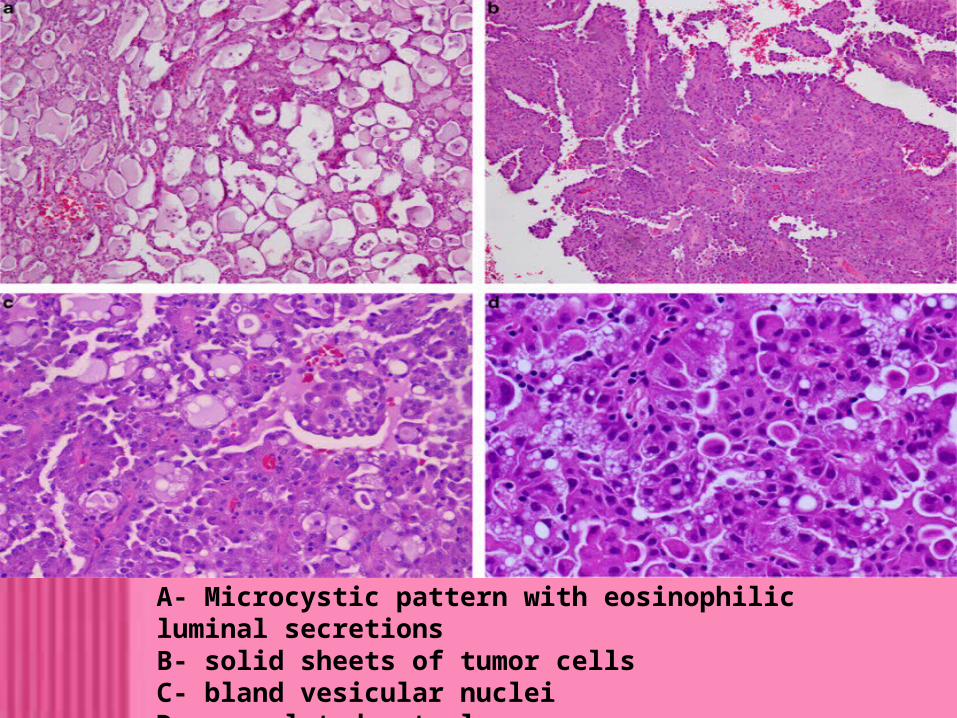
A- Microcystic pattern with eosinophilic luminal secretionsB- solid sheets of tumor cellsC- bland vesicular nucleiD- vacuolated cytoplasm seen

INFLAMMATORY MYOFIBROBLASTIC TUMOR (IMT)• Previously known as inflammatory pseudotumor – as tumor like conditions• Added as IMT in the AFIP series 4• Tumor with intermediate or low malignant potentialOther sites- Lung, mesentery , omentumMICROSCOPICALLY- 1. Circumscribed but unencapsulated2. Spindle shaped myofibroblasts arranged in
storiform / fascicular pattern3. Inflammatory infiltrate composed of lymphocytes,
plasma cells and eosinophils 4. Myxoid to hyaline stroma
D/D – 1. Leiomyoma2. Leiomyosarcima3. Nodular fascitis4. Myofibroma
• All of this lack the inflammatory cell component of IMT
• Inflammatory cells seen in nodular fascitis but lymphocytes, rather than plasma cells predominate
• IHC- spindle cells in IMT shows positive for SMA - About half of the tumors show positive for
Anaplastic lymphoma kinase 1(ALK)

IMT – numerous inflammatory cells among spindle cells
SCLEROSING MUCOEPIDERMOID CARCINOMA WITH EOSINOPHILIA
• Uncommon tumor of thyroid gland• 2 cases reported as primary tumor of major salivary gland• Behave as low grade malignant neoplasm• Unclear whether a distinctive tumor type or a variant of conventional MEC1. Sclerotic stroma heavily infiltrated by chronic
inflammatory cells and eosinophils2. Island and trabeculae of tumor cells which have
a squamoid appearance and focal keratinisation3. Admixed mucinous cells and glandular
structures seen

SCLEROSING MUCOEPIDERMOID CARCINOMA WITH EOSINOPHILIA

KERATOCYSTOMA
• Very rare benign tumor with only 3 cases reported
• Histologically mistaken for a well differentiated squamous cell carcinoma
• Grossly, tumor is a multilocular cystic lesion filled with creamy material
Histologically – 1. Randomly distributed cystic and solid nests of
squamous cells

2. Cysts are lined by bland looking stratified squamous epithelium with ortho- and parakeratosis and lacking a granular layer
3. Lumens are filled with lamellated keratin
4. Epithelium is demarcated from fibrotic stroma by basement membrane
5. Foreign body giant cell reaction may be present against keratin extruded from ruptured cyst

Showing multiple cystic formations filled with lamellar keratin material

A- variably sized and shaped cystic lesions filled with lamellar keratin materialB- Portion of the cyst wall showing stratified squamous epithelium with keratinization and parakeratotic cells. Note the lack of a granular cell layerC- Solid squamous cell islands with sharply defined margins enclosed within collagenous stromaD- Solid masses composed of squamous epithelium in conjunction with parotid ducts. Note a residual duct lumen in squamous cell nests

CRIBRIFORM ADENOCARCINOMA OF THE TONGUE• Unclear whether a distinct tumor type or a variant of PLGA• Occurs exclusively in base of tongue• Frequency of cervical lymph node metastasis at presentation is high1. Diverse growth patterns like solid, microcystic, cribriform and papillary2. Tumor cells are bland looking and possess uniform nuclei with ground glass/vesicular chromatin3. No significant mitotic activity, hemorrhage or necrosis

Cribriform adenocarcinoma of the tongue. The tumour is characterized by invasive cribriform islands comprising cells with uniform ground glass nuclei and low mitotic activity

THANK YOU