![Propositions as [Types] - Andrej](https://static.fdocuments.us/doc/165x107/62074c7f49d709492c3006fe/propositions-as-types-andrej.jpg)
On a bender—BARs, ESCRTs, COPs, and finally - Andrej Sali Lab
10
The Rockefeller University Press J. Cell Biol. Vol. 193 No. 6 963–972 www.jcb.org/cgi/doi/10.1083/jcb.201102042 JCB 963 JCB: Review Introduction The elaborate internal membrane compartments of modern eu- karyotic cells are testament to both the functional flexibility and the selective advantage that these structures provide, despite the significant energetic burdens that building such structures places on an organism. Although the presence of internal membrane com- partments is not strictly restricted to eukaryotes (Stolz, 1998; Seufferheld et al., 2004; Fuerst, 2005), the sheer number and complexity of these compartments is unique to the Eukaryota (Fig. 1). Yet, a prokaryotic progenitor lacking elaborate internal membranes clearly evolved into modern eukaryotes. How did this transition occur? Intracellular organelles could originate via two mecha- nisms: (i) exogenously, by acquiring a preformed biological struc- ture, i.e., endosymbiont; or (ii) autogenously, from preexisting intrinsic structures, requiring duplications of existing genes and subsequent acquisition of new functions in the duplicates (Margulis, 1993; Martin, 1999). Both mechanisms undoubtedly contributed to eukaryotic origins, and it is accepted that the mitochondrion has an exogenous origin, probably through a single endosymbiosis event (Fig. 1; Tovar et al., 1999, 2003; Dolezal et al., 2005). Similarly, the chloroplast arose by endosymbiosis, initially from a cyanobacteria and then with multiple subsequent endosymbiotic events in the different lineages (Gould et al., 2008). Both chloroplasts and mitochondria have experienced significant secondary reduction (Wilson et al., 1996; Funes et al., 2002; McFadden, 2010). However, all remaining internal or- ganelles, i.e the endoplasmic reticulum, Golgi complex, vacuoles/ lysosomes, and the nucleus most likely have autogenous origins (Martin, 1999). Among the huge diversity in endomembrane compart- ments, some of the most striking and well-characterized exam- ples are found in protists. This diversity has arisen by several distinct routes. First is innovation, whereby duplication of a pre- existing organelle allows a new function; for example, the ma- laria parasite possesses three specialized regulated exocytic organelles that appear to have arisen through such innovation, and discharge their contents in a strict order to facilitate host cell invasion (Kats et al., 2008). Second is sculpting, where membrane compartments are simplified by secondary loss; in Toxoplasma gondii ER exit sites are fused to the nuclear enve- lope (Hager et al., 1999). Third is modification, where compart- ment function is modified, but retains original functions; thus trypanosome peroxisomes have become glycosomes, and ac- quired the glycolytic apparatus (Hart et al., 1984; Michels and Opperdoes, 1991), whereas the exocytic pathway of Emiliana huxleyi can export colossal cargoes such as calcium carbonate coccoliths that are of similar diameter to the cell itself (Marsh, 1999). More complex patterns, likely from a combination of pro- cesses, occur in Giardia lamblia, where biosynthetic and degra- dative processes occupy the same compartment (Abodeely et al., 2009). A remarkable feature of these pathways is that many of the large number of proteins involved in them, representing some 5–10% of the total cellular proteome (Koumandou et al., 2008), can be organized into a comparatively small number of families. Taking a mainly molecular view, we consider several questions concerning origins and subsequent evolution of membrane traf- ficking systems at the level of the protein player, specifically: Tremendous variety in form and function is displayed among the intracellular membrane systems of different eu- karyotes. Until recently, few clues existed as to how these internal membrane systems had originated and diversi- fied. However, proteomic, structural, and comparative geno- mics studies together have revealed extensive similarities among many of the protein complexes used in controlling the morphology and trafficking of intracellular mem- branes. These new insights have had a profound impact on our understanding of the evolutionary origins of the internal architecture of the eukaryotic cell. Evolution On a bender—BARs, ESCRTs, COPs, and finally getting your coat Mark C. Field, 1 Andrej Sali, 2 and Michael P. Rout 3 1 Department of Pathology, University of Cambridge, Cambridge CB2 1QT, England, UK 2 Department of Bioengineering and Therapeutic Sciences, Department of Pharmaceutical Chemistry, and California Institute for Quantitative Biosciences, University of California, San Francisco, San Francisco, CA 94158 3 Laboratory of Cellular and Structural Biology, The Rockefeller University, New York, NY 10065 © 2011 Field et al. This article is distributed under the terms of an Attribution– Noncommercial–Share Alike–No Mirror Sites license for the first six months after the pub- lication date (see http://www.rupress.org/terms). After six months it is available under a Creative Commons License (Attribution–Noncommercial–Share Alike 3.0 Unported license, as described at http://creativecommons.org/licenses/by-nc-sa/3.0/). Correspondence to Mark Field: [email protected] THE JOURNAL OF CELL BIOLOGY on June 23, 2011 jcb.rupress.org Downloaded from Published June 13, 2011
Transcript of On a bender—BARs, ESCRTs, COPs, and finally - Andrej Sali Lab
The Rockefeller University PressJ. Cell Biol. Vol. 193 No. 6 963–972www.jcb.org/cgi/doi/10.1083/jcb.201102042 JCB 963
JCB: Review
IntroductionThe elaborate internal membrane compartments of modern eu-karyotic cells are testament to both the functional flexibility and the selective advantage that these structures provide, despite the significant energetic burdens that building such structures places on an organism. Although the presence of internal membrane com-partments is not strictly restricted to eukaryotes (Stolz, 1998; Seufferheld et al., 2004; Fuerst, 2005), the sheer number and complexity of these compartments is unique to the Eukaryota (Fig. 1). Yet, a prokaryotic progenitor lacking elaborate internal membranes clearly evolved into modern eukaryotes. How did this transition occur?
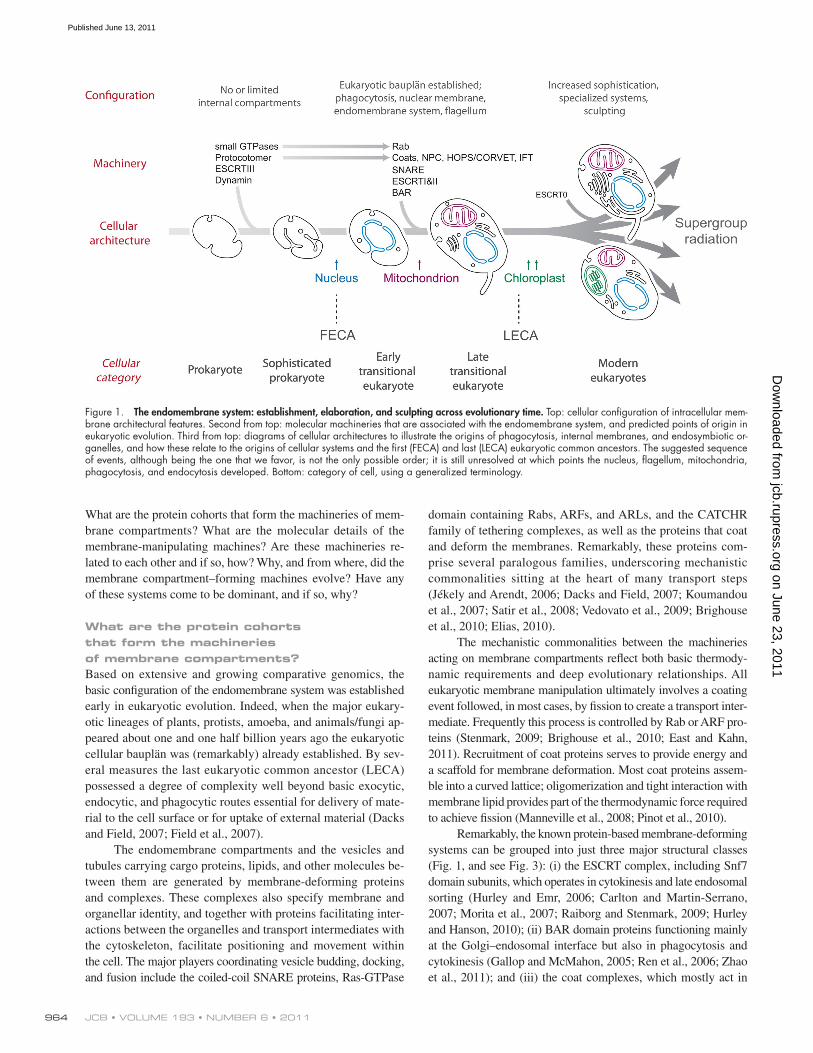
Intracellular organelles could originate via two mecha-nisms: (i) exogenously, by acquiring a preformed biological struc-ture, i.e., endosymbiont; or (ii) autogenously, from preexisting intrinsic structures, requiring duplications of existing genes and subsequent acquisition of new functions in the duplicates (Margulis, 1993; Martin, 1999). Both mechanisms undoubtedly contributed to eukaryotic origins, and it is accepted that the mitochondrion has an exogenous origin, probably through a single endosymbiosis event (Fig. 1; Tovar et al., 1999, 2003; Dolezal et al., 2005). Similarly, the chloroplast arose by endosymbiosis,
initially from a cyanobacteria and then with multiple subsequent endosymbiotic events in the different lineages (Gould et al., 2008). Both chloroplasts and mitochondria have experienced significant secondary reduction (Wilson et al., 1996; Funes et al., 2002; McFadden, 2010). However, all remaining internal or-ganelles, i.e the endoplasmic reticulum, Golgi complex, vacuoles/ lysosomes, and the nucleus most likely have autogenous origins (Martin, 1999).
Among the huge diversity in endomembrane compart-ments, some of the most striking and well-characterized exam-ples are found in protists. This diversity has arisen by several distinct routes. First is innovation, whereby duplication of a pre-existing organelle allows a new function; for example, the ma-laria parasite possesses three specialized regulated exocytic organelles that appear to have arisen through such innovation, and discharge their contents in a strict order to facilitate host cell invasion (Kats et al., 2008). Second is sculpting, where membrane compartments are simplified by secondary loss; in Toxoplasma gondii ER exit sites are fused to the nuclear enve-lope (Hager et al., 1999). Third is modification, where compart-ment function is modified, but retains original functions; thus trypanosome peroxisomes have become glycosomes, and ac-quired the glycolytic apparatus (Hart et al., 1984; Michels and Opperdoes, 1991), whereas the exocytic pathway of Emiliana huxleyi can export colossal cargoes such as calcium carbonate coccoliths that are of similar diameter to the cell itself (Marsh, 1999). More complex patterns, likely from a combination of pro-cesses, occur in Giardia lamblia, where biosynthetic and degra-dative processes occupy the same compartment (Abodeely et al., 2009). A remarkable feature of these pathways is that many of the large number of proteins involved in them, representing some 5–10% of the total cellular proteome (Koumandou et al., 2008), can be organized into a comparatively small number of families. Taking a mainly molecular view, we consider several questions concerning origins and subsequent evolution of membrane traf-ficking systems at the level of the protein player, specifically:
Tremendous variety in form and function is displayed among the intracellular membrane systems of different eukaryotes. Until recently, few clues existed as to how these internal membrane systems had originated and diversified. However, proteomic, structural, and comparative genomics studies together have revealed extensive similarities among many of the protein complexes used in controlling the morphology and trafficking of intracellular membranes. These new insights have had a profound impact on our understanding of the evolutionary origins of the internal architecture of the eukaryotic cell.
Evolution
On a bender—BARs, ESCRTs, COPs, and finally getting your coat
Mark C. Field,1 Andrej Sali,2 and Michael P. Rout3
1Department of Pathology, University of Cambridge, Cambridge CB2 1QT, England, UK2Department of Bioengineering and Therapeutic Sciences, Department of Pharmaceutical Chemistry, and California Institute for Quantitative Biosciences, University of California, San Francisco, San Francisco, CA 94158
3Laboratory of Cellular and Structural Biology, The Rockefeller University, New York, NY 10065
© 2011 Field et al. This article is distributed under the terms of an Attribution–Noncommercial–Share Alike–No Mirror Sites license for the first six months after the pub-lication date (see http://www.rupress.org/terms). After six months it is available under a Creative Commons License (Attribution–Noncommercial–Share Alike 3.0 Unported license, as described at http://creativecommons.org/licenses/by-nc-sa/3.0/).Correspondence to Mark Field: [email protected]
TH
EJ
OU
RN
AL
OF
CE
LL
BIO
LO
GY
on June 23, 2011jcb.rupress.org
Dow
nloaded from
Published June 13, 2011
JCB • VOLUME 193 • NUMBER 6 • 2011 964
domain containing Rabs, ARFs, and ARLs, and the CATCHR family of tethering complexes, as well as the proteins that coat and deform the membranes. Remarkably, these proteins com-prise several paralogous families, underscoring mechanistic commonalities sitting at the heart of many transport steps (Jékely and Arendt, 2006; Dacks and Field, 2007; Koumandou et al., 2007; Satir et al., 2008; Vedovato et al., 2009; Brighouse et al., 2010; Elias, 2010).
The mechanistic commonalities between the machineries acting on membrane compartments reflect both basic thermody-namic requirements and deep evolutionary relationships. All eukaryotic membrane manipulation ultimately involves a coating event followed, in most cases, by fission to create a transport inter-mediate. Frequently this process is controlled by Rab or ARF pro-teins (Stenmark, 2009; Brighouse et al., 2010; East and Kahn, 2011). Recruitment of coat proteins serves to provide energy and a scaffold for membrane deformation. Most coat proteins assem-ble into a curved lattice; oligomerization and tight interaction with membrane lipid provides part of the thermodynamic force required to achieve fission (Manneville et al., 2008; Pinot et al., 2010).
Remarkably, the known protein-based membrane-deforming systems can be grouped into just three major structural classes (Fig. 1, and see Fig. 3): (i) the ESCRT complex, including Snf7 domain subunits, which operates in cytokinesis and late endosomal sorting (Hurley and Emr, 2006; Carlton and Martin-Serrano, 2007; Morita et al., 2007; Raiborg and Stenmark, 2009; Hurley and Hanson, 2010); (ii) BAR domain proteins functioning mainly at the Golgi–endosomal interface but also in phagocytosis and cytokinesis (Gallop and McMahon, 2005; Ren et al., 2006; Zhao et al., 2011); and (iii) the coat complexes, which mostly act in
What are the protein cohorts that form the machineries of mem-brane compartments? What are the molecular details of the membrane-manipulating machines? Are these machineries re-lated to each other and if so, how? Why, and from where, did the membrane compartment–forming machines evolve? Have any of these systems come to be dominant, and if so, why?
What are the protein cohorts that form the machineries of membrane compartments?Based on extensive and growing comparative genomics, the basic configuration of the endomembrane system was established early in eukaryotic evolution. Indeed, when the major eukary-otic lineages of plants, protists, amoeba, and animals/fungi ap-peared about one and one half billion years ago the eukaryotic cellular bauplän was (remarkably) already established. By sev-eral measures the last eukaryotic common ancestor (LECA) possessed a degree of complexity well beyond basic exocytic, endocytic, and phagocytic routes essential for delivery of mate-rial to the cell surface or for uptake of external material (Dacks and Field, 2007; Field et al., 2007).
The endomembrane compartments and the vesicles and tubules carrying cargo proteins, lipids, and other molecules be-tween them are generated by membrane-deforming proteins and complexes. These complexes also specify membrane and organellar identity, and together with proteins facilitating inter-actions between the organelles and transport intermediates with the cytoskeleton, facilitate positioning and movement within the cell. The major players coordinating vesicle budding, docking, and fusion include the coiled-coil SNARE proteins, Ras-GTPase
Figure 1. The endomembrane system: establishment, elaboration, and sculpting across evolutionary time. Top: cellular configuration of intracellular mem-brane architectural features. Second from top: molecular machineries that are associated with the endomembrane system, and predicted points of origin in eukaryotic evolution. Third from top: diagrams of cellular architectures to illustrate the origins of phagocytosis, internal membranes, and endosymbiotic or-ganelles, and how these relate to the origins of cellular systems and the first (FECA) and last (LECA) eukaryotic common ancestors. The suggested sequence of events, although being the one that we favor, is not the only possible order; it is still unresolved at which points the nucleus, flagellum, mitochondria, phagocytosis, and endocytosis developed. Bottom: category of cell, using a generalized terminology.
on June 23, 2011jcb.rupress.org
Dow
nloaded from
Published June 13, 2011
965Evolution of membrane curvature mechanisms • Field et al.
The presence of multiple paralogous families in mem-brane-targeting systems and their importance in defining organ-elle identity provides a potential modular route for evolution of new compartments, as described by the organellar paralogy model (Dacks and Field, 2007). The model proposes that individual components arise by paralogous expansion and may function within preexisting complexes, but diverge into new complexes by step-wise subunit replacement/sequence divergence, creat-ing diversity and new compartments. Organellar paralogy sug-gests this ratchet-like mechanism as far more flexible for the diversification of new organelles than defining organelles with disparate groups of unrelated proteins or requiring coevolution of a large cohort of proteins to produce new specificity (Dacks and Field, 2007).
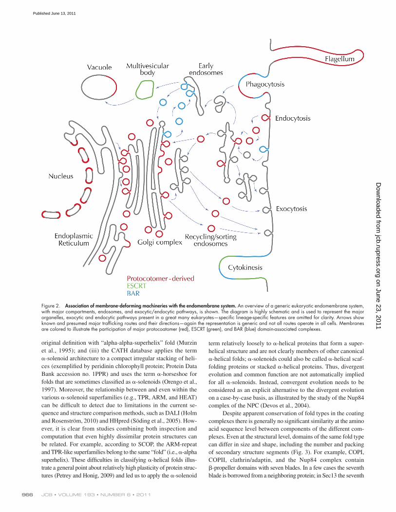
What are the molecular details of membrane-manipulating machines?Atomic structures for many components of the protocoatomer complexes have been determined experimentally or predicted by comparative structure modeling (Fig. 3), and encompass whole proteins, domains, or subcomplexes. In some cases these components have been assembled into coat lattice models based on electron density maps, protein contacts determined from pro-teomics and biochemistry, and molecular modeling. The clathrin/adaptin (Fotin et al., 2004), COPII (Fath et al., 2007), and COPI (Lee and Goldberg, 2010) complexes have an outer lattice made of coat proteins anchored to the membrane via adaptor proteins. Complexes of other membrane coats have not yet been deter-mined at atomic resolution, though portions of some, e.g., sec-tions of the Nup84 complex of the NPC, have been modeled (Kampmann and Blobel, 2009).
These coat proteins consist of one or more domains of -propeller and -solenoid–like (Fig. 3). The -propellers have been determined or predicted to consist of four to eight blades of four -strands arranged in a barrel (Chaudhuri et al., 2008; Smith, 2008), whereas the -solenoids consist of a varying number of -helices in a more or less compact arrangement (Kobe and Kajava, 2000; Karpenahalli et al., 2007). Intrigu-ingly, the order of the domains tends to be conserved, with one -propeller domain preceding the -solenoid domain (Devos et al., 2004, 2006); some sequences, such as -COP, have two -propeller domains (Lee and Goldberg, 2010), and additional coat proteins, e.g., IF122, are predicted to also have this configur-ation (Jékely and Arendt, 2006).
As an aside, a consideration of the -solenoid–like proteins illustrates general problems with assigning evolutionary re-lations between proteins of similar fold types. Thus, it is no-table, and confusing, that the term -solenoid is not used consistently (Murzin et al., 1995; Orengo et al., 1997; Groves and Barford, 1999; Kobe and Kajava, 2000; Andrade et al., 2001; Devos et al., 2004; Söding et al., 2005; Karpenahalli et al., 2007). Initially, -solenoids were defined to consist of repeating helix-turn-helix motifs, with these units arranged into a contin-uous, rather noncompact superhelix (Kobe and Kajava, 2000). Since then, (i) additional structures with irregular helical repeats have been classified as -solenoids; (ii) the SCOP database completely omits the term -solenoid, coming closest to the
exocytosis and endocytosis. These, which include the COPI, COPII, HOPS/CORVET, SEA (Seh1-associated), and clathrin complexes (see below), are important for the production of trans-port vesicles and are recruited at the early stages of vesicle forma-tion. Additionally, membership of this coat protein complex family extends to proteins that do not form transport vesicles but coat or associate with membranes, such as the nuclear pore com-plex (NPC) core scaffold proteins and intraflagellar transport (IFT) complexes (Devos et al., 2004, 2006; Jékely and Arendt, 2006). All of these systems were present in LECA, with molecu-lar configurations predicted to be essentially indistinguishable from modern cells. Interestingly, many adaptor proteins that bring coat complexes to specific membrane microdomains, for exam-ple Epsin/AP180, were also present in LECA, albeit in a simpler configuration (Holstein and Oliviusson, 2005; Gabernet-Castello et al., 2009). There are also several lineage-specific instances of later evolution in specific taxa, including the RON complex of T. gondii, which is involved in membrane deformation and ap-pears structurally unrelated to other coats (Alexander et al., 2005), and caveolin, acting in clathrin-independent endocytic mechanisms, and specific to metazoan organisms (Field et al., 2007). Importantly, although there may exist unidentified coat/membrane-deforming systems, most of the major families of players have, in all likelihood, been identified.
A structural relationship between multiple components of membrane coats, specifically COPI and clathrin/adaptin com-plexes, has been recognized for some time (Boehm and Bonifacino, 2001). Both complexes contain proteins consisting of one or two iterations of a -propeller fold, an -solenoid–like fold, or both in the order - (Devos et al., 2004). More recently, this architecture was recognized in the NPC, COPII, and likely other coating com-plexes, even extending to Sec13, being a bona fide subunit shared by both COPII and the NPC. Significantly, in the heart of the NPC is a “core scaffold” assembly entirely composed of -propeller and -solenoid–like proteins, and comprising 50% of the NPC mass. Based on these similarities the protocoatomer hypothesis proposed that NPCs and clathrin, COPI, and COPII vesicle coats share a common evolutionary origin in an early membrane-curving module, the “protocoatomer” (Fig. 2; Devos et al., 2004, 2006; Dokudovskaya et al., 2006; Field and Dacks, 2009). This hypothesis has been extended to IFT, SEA, and HOPS/CORVET complexes (Jékely and Arendt, 2006; Nickerson et al., 2009; Dokudovskaya et al., 2011). Protein structures determined by x-ray crystallography and electron microscopy have strongly sup-ported the hypothesis, and greatly reduce the likelihood that these complexes arose via convergent evolution (Fig. 3; Fotin et al., 2004; Wilbur et al., 2005; Hsia et al., 2007; Debler et al., 2008; Stagg et al., 2008; Brohawn and Schwartz, 2009; Brohawn et al., 2009; Kampmann and Blobel, 2009; Leksa et al., 2009; Nagy et al., 2009; Seo et al., 2009; Whittle and Schwartz, 2009; Lee and Goldberg, 2010; Sampathkumar et al., 2011). Furthermore, similar architectures are shared with additional complexes associated with coating systems, though their evolutionary origins are currently unclear; for example, the -solenoid is present in NPC-interacting karyopherins (Devos et al., 2006) and even subunits of the ret-romer complex, involved in Golgi/late endosomal transport (Cook et al., 2007; Collins, 2008).
on June 23, 2011jcb.rupress.org
Dow
nloaded from
Published June 13, 2011
JCB • VOLUME 193 • NUMBER 6 • 2011 966
term relatively loosely to -helical proteins that form a super-helical structure and are not clearly members of other canonical -helical folds; -solenoids could also be called -helical scaf-folding proteins or stacked -helical proteins. Thus, divergent evolution and common function are not automatically implied for all -solenoids. Instead, convergent evolution needs to be considered as an explicit alternative to the divergent evolution on a case-by-case basis, as illustrated by the study of the Nup84 complex of the NPC (Devos et al., 2004).
Despite apparent conservation of fold types in the coating complexes there is generally no significant similarity at the amino acid sequence level between components of the different com-plexes. Even at the structural level, domains of the same fold type can differ in size and shape, including the number and packing of secondary structure segments (Fig. 3). For example, COPI, COPII, clathrin/adaptin, and the Nup84 complex contain -propeller domains with seven blades. In a few cases the seventh blade is borrowed from a neighboring protein; in Sec13 the seventh
original definition with “alpha-alpha-superhelix” fold (Murzin et al., 1995); and (iii) the CATH database applies the term -solenoid architecture to a compact irregular stacking of heli-ces (exemplified by peridinin chlorophyll protein; Protein Data Bank accession no. 1PPR) and uses the term -horseshoe for folds that are sometimes classified as -solenoids (Orengo et al., 1997). Moreover, the relationship between and even within the various -solenoid superfamilies (e.g., TPR, ARM, and HEAT) can be difficult to detect due to limitations in the current se-quence and structure comparison methods, such as DALI (Holm and Rosenström, 2010) and HHpred (Söding et al., 2005). How-ever, it is clear from studies combining both inspection and computation that even highly dissimilar protein structures can be related. For example, according to SCOP, the ARM-repeat and TPR-like superfamilies belong to the same “fold” (i.e., -alpha superhelix). These difficulties in classifying -helical folds illus-trate a general point about relatively high plasticity of protein struc-tures (Petrey and Honig, 2009) and led us to apply the -solenoid
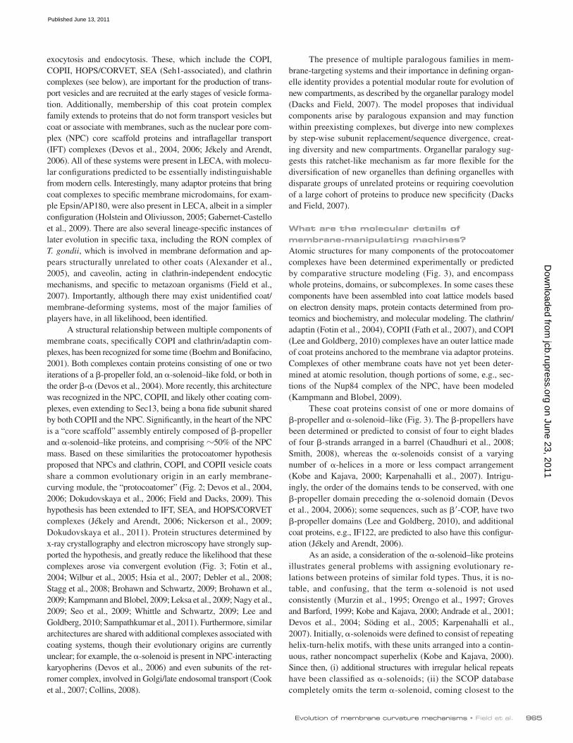
Figure 2. Association of membrane-deforming machineries with the endomembrane system. An overview of a generic eukaryotic endomembrane system, with major compartments, endosomes, and exocytic/endocytic pathways, is shown. The diagram is highly schematic and is used to represent the major organelles, exocytic and endocytic pathways present in a great many eukaryotes—specific lineage-specific features are omitted for clarity. Arrows show known and presumed major trafficking routes and their directions—again the representation is generic and not all routes operate in all cells. Membranes are colored to illustrate the participation of major protocoatomer (red), ESCRT (green), and BAR (blue) domain-associated complexes.
on June 23, 2011jcb.rupress.org
Dow
nloaded from
Published June 13, 2011
967Evolution of membrane curvature mechanisms • Field et al.
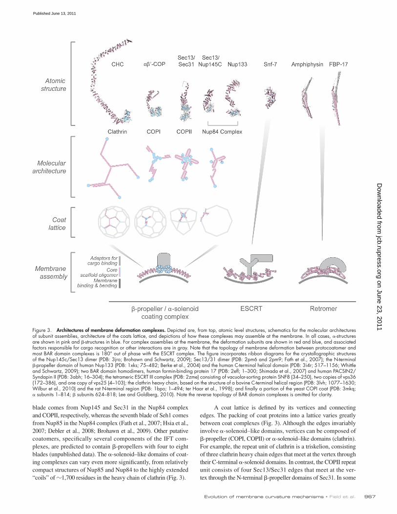
A coat lattice is defined by its vertices and connecting edges. The packing of coat proteins into a lattice varies greatly between coat complexes (Fig. 3). Although the edges invariably involve -solenoid–like domains, vertices can be composed of -propeller (COPI, COPII) or -solenoid–like domains (clathrin). For example, the repeat unit of clathrin is a triskelion, consisting of three clathrin heavy chain edges that meet at the vertex through their C-terminal -solenoid domains. In contrast, the COPII repeat unit consists of four Sec13/Sec31 edges that meet at the ver-tex through the N-terminal -propeller domains of Sec31. In some
blade comes from Nup145 and Sec31 in the Nup84 complex and COPII, respectively, whereas the seventh blade of Seh1 comes from Nup85 in the Nup84 complex (Fath et al., 2007; Hsia et al., 2007; Debler et al., 2008; Brohawn et al., 2009). Other putative coatomers, specifically several components of the IFT com-plexes, are predicted to contain -propellers with four to eight blades (unpublished data). The -solenoid–like domains of coat-ing complexes can vary even more significantly, from relatively compact structures of Nup85 and Nup84 to the highly extended “coils” of 1,700 residues in the heavy chain of clathrin (Fig. 3).
Figure 3. Architectures of membrane deformation complexes. Depicted are, from top, atomic level structures, schematics for the molecular architectures of subunit assemblies, architecture of the coats lattice, and depictions of how these complexes may assemble at the membrane. In all cases, -structures are shown in pink and -structures in blue. For complex assemblies at the membrane, the deformation subunits are shown in red and blue, and associated factors responsible for cargo recognition or other interactions are in gray. Note that the topology of membrane deformation between protocoatomer and most BAR domain complexes is 180° out of phase with the ESCRT complex. The figure incorporates ribbon diagrams for the crystallographic structures of the Nup145c/Sec13 dimer (PDB: 3jro; Brohawn and Schwartz, 2009); Sec13/31 dimer (PDB: 2pm6 and 2pm9; Fath et al., 2007); the N-terminal -propeller domain of human Nup133 (PDB: 1xks; 75–482; Berke et al., 2004) and the human C-terminal helical domain (PDB: 3i4r; 517–1156; Whittle and Schwartz, 2009); two BAR domain homodimers, human formin-binding protein 17 (PDB: 2efl; 1–300; Shimada et al., 2007) and human PACSIN2/Syndapin II (PDB: 3abh; 16–304); the tetrameric ESCRT III complex (PDB: 2zme) consisting of vacuolar-sorting protein SNF8 (34–250), two copies of vps36 (172–386), and one copy of vps25 (4–103); the clathrin heavy chain, based on the structure of a bovine C-terminal helical region (PDB: 3lvh; 1077–1630; Wilbur et al., 2010) and the rat N-terminal region (PDB: 1bpo; 1–494; ter Haar et al., 1998); and finally a portion of the yeast COPI coat (PDB: 3mkq; subunits 1–814; subunits 624–818; Lee and Goldberg, 2010). Note the reverse topology of BAR domain complexes is omitted for clarity.
on June 23, 2011jcb.rupress.org
Dow
nloaded from
Published June 13, 2011
JCB • VOLUME 193 • NUMBER 6 • 2011 968
unstructured and contain a number of interaction motifs for other partners. Although comparatively little is known about ESCRT and retromer quaternary structures, components of both the BAR and ESCRT represent intricate, essentially nonrepetitive fold structures dedicated to forming a very particular set of shapes.
From where did the membrane compartment–forming machineries evolve?Of the three known membrane-bending mechanisms, only the ESCRT system can be unequivocally traced back to prokaryotes (Samson et al., 2008), and this is restricted to the membrane de-formation ESCRT III subunits and Vps4, the associated ATPase. However, we can likely infer that the protocoatomer and BAR domain proteins arose from prokaryotic precursors, but these have yet to be identified due to weak sequence similarities. Higher order structural data may reveal these relationships, but even a structure-based argument is complicated by the ubiquity of -propeller, -solenoid–like, and -helical bundle (BAR-like) domains across both eukaryotes and prokaryotes (Kobe and Kajava, 2000; Karpenahalli et al., 2007; Chaudhuri et al., 2008; Smith, 2008). Nevertheless, characteristic combinations of these domains uniquely define membrane-deforming complexes, so the absence of such combinations from the vast majority of prokaryotes argues for an origin approximately coincident with the first eukaryotic common ancestor (FECA).
Recent findings have blurred the distinction between true coat complexes that build membrane-deforming lattices and tethering complexes, traditionally considered to be involved in vesicle docking. The HOPS/CORVET complex is classed as a tethering complex, but possesses /-domain subunits, recog-nizes curved membranes, acts in late endocytic targeting, is sub-ject to sophisticated kinase-mediated control (Nickerson et al., 2009; Cabrera et al., 2010), and is a near-universal eukaryotic feature (Koumandou et al., 2007). It is unknown if HOPS/ CORVET forms a lattice contributing to formation of transport intermediates, or is restricted to participation in the later stages of vesicle transport. Similarly, IFT complexes are found in essen-tially all eukaryotic flagellates and form raft-like structures trans-porting molecules along the flagellar membrane; the structural parallels of several IFT subunits to protocoatomer proteins are striking (Jékely and Arendt, 2006; Satir et al., 2008), but it is un-clear if the IFT rafts act to coat and deform membrane or act in some other manner. It has yet to be determined if the newly char-acterized SEA complex, with some structural similarities to the HOPS/CORVET complex, is involved in tethering, coat forma-tion, or another function (Dokudovskaya et al., 2011). Remark-ably, since LECA, there has apparently been little innovation of new vesicle coat and membrane deformation systems (Dacks and Field, 2007).
In addition to movement between endomembrane com-partments, eukaryotes require membrane deformation for mul-tiple functions, including cell division and replication of both endosymbiotic and autogenously derived organelles. Also, sev-eral compartments require precise delineation of membrane subdomains; for example, the Golgi complex and fenestrations within ER membrane networks. There are several examples of spe-cific budding processes taking place at specialized subdomains,
respects, the COPI lattice may be intermediary between clathrin and COPII lattices, and was modeled to consist of three edges like clathrin, but with the vertex corresponding to the packing of -propellers, like COPII (Lee and Goldberg, 2010). Most signifi-cantly, a single lattice architecture can stabilize membranes of differing curvature and hence vesicles of different sizes, as illus-trated by the varied clathrin/adaptin vesicles (Fig. 3); here the dif-ferent curvatures are achieved by varying packing angles between the edges of neighboring triskelions, while maintaining each indi-vidual triskelion structure, including the vertices, essentially un-changed (Fotin et al., 2004). In contrast, expansion of the COPII cage from the cuboctahedron to the icosidodecahdron is associ-ated with the plasticity of the vertices rather than the edges, made possible by relatively limited physical contacts between the -propeller domains forming these vertices (Stagg et al., 2008). Although the molecular details are still not known, it is clear that the Nup84 complex forms a pair of head-to-tail rings in each NPC rather than forming a homo-oligomeric cage (Alber et al., 2007a,b), again underscoring the plasticity available to this fam-ily of coating proteins. Evolutionarily the coat architecture bene-fits from the ability of -solenoid–like domains to relatively easily add or delete helices or to change their packing (Kobe and Kajava, 2000; Devos et al., 2004; Karpenahalli et al., 2007; Forwood et al., 2010). Thus, it appears that the -propeller and -solenoid domains are permissive for evolving a large variety of geometries. This flexibility is consistent with these folds being involved in additional cellular processes; for example, -propeller domains participate in G protein signal transduction and chroma-tin assembly (Chaudhuri et al., 2008; Smith, 2008).
The -helical domains of several ESCRT and retromer components are of altogether distinct fold types as compared with each other and the -solenoid–like protein coats (above; Fig. 3). Crystallographic studies revealed the BAR (Bin/Amphi-physin/Rvs-homology) domain as -helix rich, with a core six-helix bundle (Masuda and Mochizuki, 2010). BAR domains form dimers with long extended curves and appear to be outstandingly suitable modules for enabling the formation of tubes or buds from a nearly flat membrane. The elongated, banana-shape struc-ture has a positively charged concave surface of 22-nm diameter that is directly responsible for generating and/or sensing a highly curved membrane. The 18 known atomic structures reveal subtle variations in dimerization angles and helical kinks, adapting the BAR architecture to a variety of membrane-sculpting functions.
Six subcomplexes of the ESCRT complex were recently reviewed, relying largely on crystallographic and EM studies (Hurley, 2010). These subcomplexes vary in domain content, overall organization, and function (Ghazi-Tabatabai et al., 2008). For example, the yeast ESCRT-0 complex is a heterodimer of Hse1 and Vps27 that interact with each other via an elongated anti-parallel coiled-coil motif and swapped GAT domains. Hse1 also contains ubiquitin-binding VHS and UIM domains (8 helix bundles) as well as an SH3 domain (a -barrel). Vps27 contains a membrane-binding FYVE domain that interacts with phospha-tidylinositol 3-phosphate in the membrane (a zinc finger with two Zn2+-binding clusters, two double-stranded antiparallel sheets, and a C-terminal -helix), one VHS and two UIM domains, and a clathrin-binding domain. The C termini of both proteins are
on June 23, 2011jcb.rupress.org
Dow
nloaded from
Published June 13, 2011
969Evolution of membrane curvature mechanisms • Field et al.
orthologues are essential for membrane scission (Graumann, 2007; Lindås et al., 2008; Samson et al., 2008). Interestingly, ESCRT- and FtsZ-mediated cytokinesis systems seem to be mu-tually exclusive. Significantly, ESCRT retains the function in cytokinesis in eukaryotes, being targeted to and extending the cleavage furrow in metazoan cells (Makarova et al., 2010). There-fore, the roles of DLPs, FtsA/actin, FtsZ/tubulin, and ESCRT components in the endomembrane system may represent acquisi-tion of expanded functions (Low and Löwe, 2006), in addition to partly (or wholly) retaining the original roles in cytokinesis.
There remain two dominant roles for the original eukaryote-specific function of the endomembrane system; phagocytosis and establishment of the nucleus (Cavalier-Smith, 2002; Martin and Koonin, 2006). Both roles require compartmentalization of the cellular interior, and there are compelling arguments for each as the first to have arisen. Specifically, the selective advantage that phagocytosis offers a primitive eukaryote is significant, as it would be able to engulf its competitors and is a presumed precondition for acquisition of endosymbionts. Conversely, seg-regation of genetic material from the cytoplasm separates tran-scription and translation, protecting the genetic material and facilitating increasingly sophisticated mechanisms for gene con-trol. Thus, regardless of the precise order of events at the dawn of the eukaryotes, there is compelling evidence for why eukaryotes became compartmentalized.
Why has one system come to dominate the eukaryotic cell?Structurally, the membrane coat repertoire is dominated by one class, namely the -propeller/-solenoid–like proteins (Fig. 2). Like the scales of our ancestral primitive fish have evolved into teeth, horns, feathers, and fur, so the original “protocoatomer” has diverged widely into many complexes, including some such as the NPC with roles seemingly far removed from the original. Why did these systems come to dominate the eukaryotic cell, in-stead of other complexes that are clearly capable of deforming membranes? As the major known role for these proteins is form-ing scaffolds, we presume there to be some aspect of their mo-lecular architecture especially well suited to adapting them to scaffolding a variety of different membranes. This selective advantage would allow expansion and diversification of this fam-ily, over and above the other membrane deformation systems.
BAR domain proteins assemble principally as dimers, and the individual subunits are comprised of helical bundles assem-bled into a banana shape (Fig. 3). Although BAR domain curva-ture can vary somewhat, the architecture of the BAR domain is relatively invariant (Masuda and Mochizuki, 2010). For ESCRT, only a single set of coat proteins is encoded in the genome (Leung et al., 2008), and hence only a single architecture is possible (Fig. 3). Therefore, for both BAR and ESCRT, there appears to be limited scope for structural variation; they are locked into a single molecular architecture. By contrast, the / coat pro-teins are more like a molecular LEGOTM (Fig. 3). Both -solenoids and -propellers are repetitive, making addition or removal of elements potentially facile in evolutionary terms with signifi-cant variation in primary structure allowed without disrupting the overall conformation of the protein, but instead varying the
including the ER and Golgi complex (Ladinsky et al., 1999; Budnik and Stephens, 2009), but a potent example is the nuclear pore complex (Fig. 1; Alber et al., 2007a,b).
Both the nuclear envelope and phagocytosis are ancient eukaryotic features (Cavalier-Smith, 2009, 2010; DeGrasse et al., 2009), and established in the transition between FECA and LECA. However, there are no molecular components specific to either the nuclear envelope or the phagocytic system with clear prokaryotic origin. The presence of internal membranes associ-ated with the nucleoid in the Planctomycete bacterium Gemmata obscuriglobus, a structure segregating DNA from the remaining cytoplasm, stands as a good example of an apparently nuclear-like compartment in a prokaryote (Fuerst, 2005). More recently, parallels between the nucleoid and the nucleus were possibly extended by detection of possible nucleoid-associated proteins with a putative protocoatomer - architecture in these bacteria (Santarella-Mellwig et al., 2010). However significant to eukary-otic origins this proves, our view of the origins of endomembrane coats is incomplete, as protocoatomer proteins are not found in the majority of prokaryotes including Archaea. If major com-ponents of an ancestral membrane deformation machine were carried over from prokaryotes, then the absence of a conventional phagocytic/endocytic system from most prokaryotes is problem-atic, as many models assume that phagocytosis accounts for the initial endosymbiotic event (Embley and Martin, 2006). Further, the eukaryotic flagellum is also recognized as a feature of the very earliest eukaryotes and probably predates LECA, but again lacks an obvious prokaryotic molecular signature. Thus, where the - coating module originated from preLECA (and preFECA) is still unclear, and given its tremendous structural plasticity may even have been repurposed from a protein originally used for some prokaryotic function now lost to eukaryotes.
Why did membrane trafficking machinery evolve?To grow and develop, all cells require their membranes to be dynamic and moldable. At its most basic, these features are re-quired for the post-mitotic separation of daughter cells at cyto-kinesis. In addition, cells also benefit from packaging their interior into more efficient and/or functionally specialized com-partments. Cell division represents a fundamental rationale for bending membrane, as even the most primitive cell has a require-ment to replicate. Unsurprisingly then, these processes retain significant molecular conservation between prokaryotes and eu-karyotes, which were overlooked by earlier studies due to very low sequence similarities. The evolutionary relationships were instead exposed by the structures of several prokaryotic and eu-karyotic players, including FtsZ, the prokaryotic tubulin ortho-logue essential for the scission steps in cytokinesis, FtsA/MreB, an actin orthologue (Graumann, 2007), and bacterial dynamin-like proteins (BDLP; Low and Löwe, 2006), for which the ances-tral function was likely also in cytokinesis (Miyagishima et al., 2008). All of these examples share less than 20% sequence iden-tity with their eukaryotic orthologues but are structurally quite similar. Significantly, there is apparently more than one cyto-kinesis mechanism present in prokaryotes. For example, in the Archaeon Sulpholobus ESCRT III complex (Vps2 and Vps4)
on June 23, 2011jcb.rupress.org
Dow
nloaded from
Published June 13, 2011
JCB • VOLUME 193 • NUMBER 6 • 2011 970
example of natural selection operating to select for functional-ity, and hence fitness, within the internal workings of the cell, rather than, as is more traditionally regarded, between organ-isms or populations.
The progress in the last decade in our understanding of the origins of many of the structures present within eukary-otes has been astonishing. These cellular features have left virtually no geological record, yet genome sequencing to-gether with molecular modeling and solution of the molecular and atomic structures of key components has given us many of the necessary tools and evidence to reconstruct a probable trajectory of eukaryotic cellular evolution. Direct analysis of trafficking systems in diverse but evolutionarily key organ-isms is essential in providing an unbiased view of how pres-ent diversity arose, and may identify novel components absent from more traditional model organisms, further increasing our appreciation of the range of diversity produced by evolu-tion, together with providing an improved grasp of the cell biology of many pathogens and ecologically important or-ganisms. Similarly, structural biology efforts will take phylo-genetic comparisons to the atomic level, revealing structural similarities that elude sequence-based methods. Taken together, these approaches should continue to revolutionize our view of how the eukaryotic cell came to be.
We thank Joel B. Dacks for many fruitful discussions on eukaryotic origins and trafficking, and Jeremy Phillips for help preparing Figure 3. Finally, we apolo-gize to the very great many people whose primary work we were unable to cite due to limitations in space.
Work in the authors’ laboratories is supported by the following agen-cies; the Wellcome Trust, the Medical Research Council (UK), and the Na-tional Institutes of Health (Bethesda, MD).
Submitted: 8 February 2011Accepted: 5 May 2011
ReferencesAbodeely, M., K.N. DuBois, A. Hehl, S. Stefanic, M. Sajid, W. DeSouza, M.
Attias, J.C. Engel, I. Hsieh, R.D. Fetter, and J.H. McKerrow. 2009. A contiguous compartment functions as endoplasmic reticulum and endo-some/lysosome in Giardia lamblia. Eukaryot. Cell. 8:1665–1676. doi:10 .1128/EC.00123-09
Alber, F., S. Dokudovskaya, L.M. Veenhoff, W. Zhang, J. Kipper, D. Devos, A. Suprapto, O. Karni-Schmidt, R. Williams, B.T. Chait, et al. 2007a. Determining the architectures of macromolecular assemblies. Nature. 450:683–694. doi:10.1038/nature06404
Alber, F., S. Dokudovskaya, L.M. Veenhoff, W. Zhang, J. Kipper, D. Devos, A. Suprapto, O. Karni-Schmidt, R. Williams, B.T. Chait, et al. 2007b. The molecular architecture of the nuclear pore complex. Nature. 450:695–701. doi:10.1038/nature06405
Alexander, D.L., J. Mital, G.E. Ward, P. Bradley, and J.C. Boothroyd. 2005. Identification of the moving junction complex of Toxoplasma gondii: a collaboration between distinct secretory organelles. PLoS Pathog. 1:e17. doi:10.1371/journal.ppat.0010017
Andrade, M.A., C. Perez-Iratxeta, and C.P. Ponting. 2001. Protein repeats: struc-tures, functions, and evolution. J. Struct. Biol. 134:117–131. doi:10.1006/ jsbi.2001.4392
Berke, I.C., T. Boehmer, G. Blobel, and T.U. Schwartz. 2004. Structural and functional analysis of Nup133 domains reveals modular building blocks of the nuclear pore complex. J. Cell Biol. 167:591–597. doi:10.1083/ jcb.200408109
Boehm, M., and J.S. Bonifacino. 2001. Adaptins: the final recount. Mol. Biol. Cell. 12:2907–2920.
Brighouse, A., J.B. Dacks, and M.C. Field. 2010. Rab protein evolution and the history of the eukaryotic endomembrane system. Cell. Mol. Life Sci. 67:3449–3465. doi:10.1007/s00018-010-0436-1
number of repeats. The -solenoid is highly variable in length, with for example clathrin being approximately twice the molec-ular weight of Nup133 (Fig. 3). Though the -propeller is inher-ently more constrained than the -solenoid, the four to eight blades provide multiple sites for protein–protein interactions, and allow incorporation of additional loops between each compact -sheet blade (Fig. 3).
This is consistent with comparative genomics allowing us to propose a configuration for the membrane-trafficking sys-tem in LECA (Dacks and Field, 2007; Field and Dacks, 2009). First, the full ESCRT I/II/III system was present but subsequent retention of the complex was then variable between lineages, with evidence for secondary loss of several of the ESCRT III and III-associated subunits (Leung et al., 2008). Second, although BAR domains are universally represented and present in LECA, many organisms have very small families with Trypanosoma for example having a single retromer-associated BAR domain protein (Koumandou et al., 2011). Lastly, the -propeller/ -solenoid–like coat family contrasts sharply with BAR and ESCRT in their extensive representation; clathrin, COPI, COPII, IFT, the NPC, and HOPS/CORVET are all present in nearly every eukaryotic lineage we have examined, and therefore were estab-lished as independent complexes before LECA. This suggests that post-FECA organisms found in the protocoatomer family an evolutionary flexible template for development of the multiple mechanisms and compartments required to build a highly so-phisticated and adaptable endomembrane system.
ConclusionsThe emergence of eukaryotes is probably the most dramatic evolutionary transition after the origin of life, and represents a revolution in cellular architecture, gene expression mechanisms, and many other features, but with obvious continuity with the pro-karyotic ancestors. The last eukaryotic common ancestor (LECA), which probably lived about one billion years ago, was a very so-phisticated organism and bore many innovations specific to eu-karyotes. Much of the innovation therefore predates LECA, and must be placed within the transitional period between FECA and LECA. Representing the most extreme bottleneck possible, LECA is the sole representative known to have survived from the transitional period. Basic metabolic pathways and biosynthetic mechanisms are excellent examples of persistent prokaryotic contributions, whereas membrane transport and the construction of intracellular compartments probably represents the major eu-karyotic innovation, and facilitated massive increases to cellular complexity and functional flexibility. Few systems seem directly derived from prokaryotes, with the exception of essential roles in cytokinesis and perhaps endosymbiotic organelle replication. The majority of intracellular membranes are likely a result of evo-lutionary expansion of an ancestral protocoatomer complex and also additional paralogous families, which occurred pre-LECA. We suggest that the complex diverged massively to produce nuclear pore complexes, intraflagellar transport, and a great proportion of the endomembrane system. Structural flexibility, allowing incorporation of small, functionally significant variations within these proteins may explain how these proteins came to dominate the endomembrane system. Significantly, this provides a potent
on June 23, 2011jcb.rupress.org
Dow
nloaded from
Published June 13, 2011
971Evolution of membrane curvature mechanisms • Field et al.
Field, M.C., C. Gabernet-Castello, and J.B. Dacks. 2007. Reconstructing the evolution of the endocytic system: insights from genomics and mo-lecular cell biology. Adv. Exp. Med. Biol. 607:84–96. doi:10.1007/ 978-0-387-74021-8_7
Forwood, J.K., A. Lange, U. Zachariae, M. Marfori, C. Preast, H. Grubmüller, M. Stewart, A.H. Corbett, and B. Kobe. 2010. Quantitative structural analysis of importin- flexibility: paradigm for solenoid protein struc-tures. Structure. 18:1171–1183. doi:10.1016/j.str.2010.06.015
Fotin, A., Y. Cheng, P. Sliz, N. Grigorieff, S.C. Harrison, T. Kirchhausen, and T. Walz. 2004. Molecular model for a complete clathrin lattice from electron cryomicroscopy. Nature. 432:573–579. doi:10.1038/nature03079
Fuerst, J.A. 2005. Intracellular compartmentation in planctomycetes. Annu. Rev. Microbiol. 59:299–328. doi:10.1146/annurev.micro.59.030804.121258
Funes, S., E. Davidson, A. Reyes-Prieto, S. Magallón, P. Herion, M.P. King, and D. González-Halphen. 2002. A green algal apicoplast ancestor. Science. 298:2155. doi:10.1126/science.1076003
Gabernet-Castello, C., J.B. Dacks, and M.C. Field. 2009. The single ENTH-domain protein of trypanosomes; endocytic functions and evolutionary relationship with epsin. Traffic. 10:894–911. doi:10.1111/j.1600-0854 .2009.00910.x
Gallop, J.L., and H.T. McMahon. 2005. BAR domains and membrane curvature: bringing your curves to the BAR. Biochem. Soc. Symp. (72):223–231.
Ghazi-Tabatabai, S., S. Saksena, J.M. Short, A.V. Pobbati, D.B. Veprintsev, R.A. Crowther, S.D. Emr, E.H. Egelman, and R.L. Williams. 2008. Structure and disassembly of filaments formed by the ESCRT-III subunit Vps24. Structure. 16:1345–1356. doi:10.1016/j.str.2008.06.010
Gould, S.B., R.F. Waller, and G.I. McFadden. 2008. Plastid evolution. Annu. Rev. Plant Biol. 59:491–517. doi:10.1146/annurev.arplant.59.032607.092915
Graumann, P.L. 2007. Cytoskeletal elements in bacteria. Annu. Rev. Microbiol. 61:589–618. doi:10.1146/annurev.micro.61.080706.093236
Groves, M.R., and D. Barford. 1999. Topological characteristics of heli-cal repeat proteins. Curr. Opin. Struct. Biol. 9:383–389. doi:10.1016/ S0959-440X(99)80052-9
Hager, K.M., B. Striepen, L.G. Tilney, and D.S. Roos. 1999. The nuclear enve-lope serves as an intermediary between the ER and Golgi complex in the intracellular parasite Toxoplasma gondii. J. Cell Sci. 112:2631–2638.
Hart, D.T., O. Misset, S.W. Edwards, and F.R. Opperdoes. 1984. A compari-son of the glycosomes (microbodies) isolated from Trypanosoma brucei bloodstream form and cultured procyclic trypomastigotes. Mol. Biochem. Parasitol. 12:25–35. doi:10.1016/0166-6851(84)90041-0
Holm, L., and P. Rosenström. 2010. Dali server: conservation mapping in 3D. Nucleic Acids Res. 38:W545–W549. doi:10.1093/nar/gkq366
Holstein, S.E., and P. Oliviusson. 2005. Sequence analysis of Arabidopsis thali-ana E/ANTH-domain-containing proteins: membrane tethers of the clathrin-dependent vesicle budding machinery. Protoplasma. 226:13–21. doi:10.1007/s00709-005-0105-7
Hsia, K.C., P. Stavropoulos, G. Blobel, and A. Hoelz. 2007. Architecture of a coat for the nuclear pore membrane. Cell. 131:1313–1326. doi:10.1016/ j.cell.2007.11.038
Hurley, J.H. 2010. The ESCRT complexes. Crit. Rev. Biochem. Mol. Biol. 45:463–487. doi:10.3109/10409238.2010.502516
Hurley, J.H., and S.D. Emr. 2006. The ESCRT complexes: structure and mecha-nism of a membrane-trafficking network. Annu. Rev. Biophys. Biomol. Struct. 35:277–298. doi:10.1146/annurev.biophys.35.040405.102126
Hurley, J.H., and P.I. Hanson. 2010. Membrane budding and scission by the ESCRT machinery: it’s all in the neck. Nat. Rev. Mol. Cell Biol. 11:556–566. doi:10.1038/nrm2937
Jékely, G., and D. Arendt. 2006. Evolution of intraflagellar transport from coated vesicles and autogenous origin of the eukaryotic cilium. Bioessays. 28:191–198. doi:10.1002/bies.20369
Kampmann, M., and G. Blobel. 2009. Three-dimensional structure and flex-ibility of a membrane-coating module of the nuclear pore complex. Nat. Struct. Mol. Biol. 16:782–788. doi:10.1038/nsmb.1618
Karpenahalli, M.R., A.N. Lupas, and J. Söding. 2007. TPRpred: a tool for pre-diction of TPR-, PPR- and SEL1-like repeats from protein sequences. BMC Bioinformatics. 8:2. doi:10.1186/1471-2105-8-2
Kats, L.M., B.M. Cooke, R.L. Coppel, and C.G. Black. 2008. Protein trafficking to apical organelles of malaria parasites - building an invasion machine. Traffic. 9:176–186. doi:10.1111/j.1600-0854.2007.00681.x
Kobe, B., and A.V. Kajava. 2000. When protein folding is simplified to protein coiling: the continuum of solenoid protein structures. Trends Biochem. Sci. 25:509–515. doi:10.1016/S0968-0004(00)01667-4
Koumandou, V.L., J.B. Dacks, R.M.R. Coulson, and M.C. Field. 2007. Control systems for membrane fusion in the ancestral eukaryote; evo-lution of tethering complexes and SM proteins. BMC Evol. Biol. 7:29. doi:10.1186/1471-2148-7-29
Brohawn, S.G., and T.U. Schwartz. 2009. Molecular architecture of the Nup84-Nup145C-Sec13 edge element in the nuclear pore complex lattice. Nat. Struct. Mol. Biol. 16:1173–1177. doi:10.1038/nsmb.1713
Brohawn, S.G., J.R. Partridge, J.R. Whittle, and T.U. Schwartz. 2009. The nu-clear pore complex has entered the atomic age. Structure. 17:1156–1168. doi:10.1016/j.str.2009.07.014
Budnik, A., and D.J. Stephens. 2009. ER exit sites—localization and control of COPII vesicle formation. FEBS Lett. 583:3796–3803. doi:10.1016/ j.febslet.2009.10.038
Cabrera, M., L. Langemeyer, M. Mari, R. Rethmeier, I. Orban, A. Perz, C. Bröcker, J. Griffith, D. Klose, H.J. Steinhoff, et al. 2010. Phosphorylation of a membrane curvature-sensing motif switches function of the HOPS subunit Vps41 in membrane tethering. J. Cell Biol. 191:845–859. doi:10.1083/jcb.201004092
Carlton, J.G., and J. Martin-Serrano. 2007. Parallels between cytokinesis and retroviral budding: a role for the ESCRT machinery. Science. 316:1908–1912. doi:10.1126/science.1143422
Cavalier-Smith, T. 2002. The phagotrophic origin of eukaryotes and phylogenetic classification of Protozoa. Int. J. Syst. Evol. Microbiol. 52:297–354.
Cavalier-Smith, T. 2009. Predation and eukaryote cell origins: a coevolution-ary perspective. Int. J. Biochem. Cell Biol. 41:307–322. doi:10.1016/ j.biocel.2008.10.002
Cavalier-Smith, T. 2010. Origin of the cell nucleus, mitosis and sex: roles of intracellular coevolution. Biol. Direct. 5:7. doi:10.1186/1745-6150-5-7
Chaudhuri, I., J. Söding, and A.N. Lupas. 2008. Evolution of the -propeller fold. Proteins. 71:795–803. doi:10.1002/prot.21764
Collins, B.M. 2008. The structure and function of the retromer protein complex. Traffic. 9:1811–1822. doi:10.1111/j.1600-0854.2008.00777.x
Cook, A., F. Bono, M. Jinek, and E. Conti. 2007. Structural biology of nucleo-cytoplasmic transport. Annu. Rev. Biochem. 76:647–671. doi:10.1146/ annurev.biochem.76.052705.161529
Dacks, J.B., and M.C. Field. 2007. Evolution of the eukaryotic membrane- trafficking system: origin, tempo and mode. J. Cell Sci. 120:2977–2985. doi:10.1242/jcs.013250
Debler, E.W., Y. Ma, H.S. Seo, K.C. Hsia, T.R. Noriega, G. Blobel, and A. Hoelz. 2008. A fence-like coat for the nuclear pore membrane. Mol. Cell. 32:815–826. doi:10.1016/j.molcel.2008.12.001
DeGrasse, J.A., K.N. DuBois, D. Devos, T.N. Siegel, A. Sali, M.C. Field, M.P. Rout, and B.T. Chait. 2009. Evidence for a shared nuclear pore complex architecture that is conserved from the last common eukary-otic ancestor. Mol. Cell. Proteomics. 8:2119–2130. doi:10.1074/mcp .M900038-MCP200
Devos, D., S. Dokudovskaya, F. Alber, R. Williams, B.T. Chait, A. Sali, and M.P. Rout. 2004. Components of coated vesicles and nuclear pore complexes share a common molecular architecture. PLoS Biol. 2:e380. doi:10.1371/journal.pbio.0020380
Devos, D., S. Dokudovskaya, R. Williams, F. Alber, N. Eswar, B.T. Chait, M.P. Rout, and A. Sali. 2006. Simple fold composition and modular architec-ture of the nuclear pore complex. Proc. Natl. Acad. Sci. USA. 103:2172–2177. doi:10.1073/pnas.0506345103
Dokudovskaya, S., R. Williams, D. Devos, A. Sali, B.T. Chait, and M.P. Rout. 2006. Protease accessibility laddering: a proteomic tool for probing pro-tein structure. Structure. 14:653–660. doi:10.1016/j.str.2006.02.006
Dokudovskaya, S., F. Waharte, A. Schlessinger, U. Pieper, D.P. Devos, I.M. Cristea, R. Williams, J. Salamero, B.T. Chait, A. Sali, et al. 2011. A con-served coatomer-related complex containing Sec13 and Seh1 dynami-cally associates with the vacuole in Saccharomyces cerevisiae. Mol. Cell. Proteomics. doi:10.1074/mcp.M110.006478
Dolezal, P., O. Smíd, P. Rada, Z. Zubácová, D. Bursac, R. Suták, J. Nebesárová, T. Lithgow, and J. Tachezy. 2005. Giardia mitosomes and trichomonad hydrogenosomes share a common mode of protein tar-geting. Proc. Natl. Acad. Sci. USA. 102:10924–10929. doi:10.1073/ pnas.0500349102
East, M.P., and R.A. Kahn. 2011. Models for the functions of Arf GAPs. Semin. Cell Dev. Biol. 22:3–9. doi:10.1016/j.semcdb.2010.07.002
Elias, M. 2010. Patterns and processes in the evolution of the eukaryotic en-domembrane system. Mol. Membr. Biol. 27:469–489. doi:10.3109/ 09687688.2010.521201
Embley, T.M., and W. Martin. 2006. Eukaryotic evolution, changes and chal-lenges. Nature. 440:623–630. doi:10.1038/nature04546
Fath, S., J.D. Mancias, X. Bi, and J. Goldberg. 2007. Structure and organization of coat proteins in the COPII cage. Cell. 129:1325–1336. doi:10.1016/ j.cell.2007.05.036
Field, M.C., and J.B. Dacks. 2009. First and last ancestors: reconstructing evolu-tion of the endomembrane system with ESCRTs, vesicle coat proteins, and nuclear pore complexes. Curr. Opin. Cell Biol. 21:4–13. doi:10 .1016/j.ceb.2008.12.004
on June 23, 2011jcb.rupress.org
Dow
nloaded from
Published June 13, 2011
JCB • VOLUME 193 • NUMBER 6 • 2011 972
Raiborg, C., and H. Stenmark. 2009. The ESCRT machinery in endosomal sorting of ubiquitylated membrane proteins. Nature. 458:445–452. doi:10.1038/nature07961
Ren, G., P. Vajjhala, J.S. Lee, B. Winsor, and A.L. Munn. 2006. The BAR domain proteins: molding membranes in fission, fusion, and phagy. Microbiol. Mol. Biol. Rev. 70:37–120. doi:10.1128/MMBR.70.1.37-120.2006
Sampathkumar, P., T. Gheyi, S.A. Miller, K.T. Bain, M. Dickey, J.B. Bonanno, S.J. Kim, J. Phillips, U. Pieper, J. Fernandez-Martinez, et al. 2011. Structure of the C-terminal domain of Saccharomyces cerevisiae Nup133, a component of the nuclear pore complex. Proteins. 79:1672–1677. doi:10.1002/prot.22973.
Samson, R.Y., T. Obita, S.M. Freund, R.L. Williams, and S.D. Bell. 2008. A role for the ESCRT system in cell division in archaea. Science. 322:1710–1713. doi:10.1126/science.1165322
Santarella-Mellwig, R., J. Franke, A. Jaedicke, M. Gorjanacz, U. Bauer, A. Budd, I.W. Mattaj, and D.P. Devos. 2010. The compartmentalized bac-teria of the planctomycetes-verrucomicrobia-chlamydiae superphylum have membrane coat-like proteins. PLoS Biol. 8:e1000281. doi:10.1371/journal.pbio.1000281
Satir, P., D.R. Mitchell, and G. Jékely. 2008. How did the cilium evolve? Curr. Top. Dev. Biol. 85:63–82. doi:10.1016/S0070-2153(08)00803-X
Seo, H.S., Y. Ma, E.W. Debler, D. Wacker, S. Kutik, G. Blobel, and A. Hoelz. 2009. Structural and functional analysis of Nup120 suggests ring for-mation of the Nup84 complex. Proc. Natl. Acad. Sci. USA. 106:14281–14286. doi:10.1073/pnas.0907453106
Seufferheld, M., C.R. Lea, M. Vieira, E. Oldfield, and R. Docampo. 2004. The H(+)-pyrophosphatase of Rhodospirillum rubrum is predominantly located in polyphosphate-rich acidocalcisomes. J. Biol. Chem. 279:51193–51202. doi:10.1074/jbc.M406099200
Shimada, A., H. Niwa, K. Tsujita, S. Suetsugu, K. Nitta, K. Hanawa-Suetsugu, R. Akasaka, Y. Nishino, M. Toyama, L. Chen, et al. 2007. Curved EFC/F-BAR-domain dimers are joined end to end into a fila-ment for membrane invagination in endocytosis. Cell. 129:761–772. doi:10.1016/j.cell.2007.03.040
Smith, T.F. 2008. Diversity of WD-repeat proteins. Subcell. Biochem. 48:20–30.
Söding, J., A. Biegert, and A.N. Lupas. 2005. The HHpred interactive server for protein homology detection and structure prediction. Nucleic Acids Res. 33:W244–W248. doi:10.1093/nar/gki408
Stagg, S.M., P. LaPointe, A. Razvi, C. Gürkan, C.S. Potter, B. Carragher, and W.E. Balch. 2008. Structural basis for cargo regulation of COPII coat assembly. Cell. 134:474–484. doi:10.1016/j.cell.2008.06.024
Stenmark, H. 2009. Rab GTPases as coordinators of vesicle traffic. Nat. Rev. Mol. Cell Biol. 10:513–525. doi:10.1038/nrm2728
Stolz, J.F. 1998. Bacterial intracellular membranes. In Nature Encyclopedia of Life Sciences. A. Mitchell, J. Trapnell, S. Hadfield, V. Kerguelen, and F. Richmond, editors. Wiley, Chichester, UK. pp 1–5.
ter Haar, E., A. Musacchio, S.C. Harrison, and T. Kirchhausen. 1998. Atomic structure of clathrin: a beta propeller terminal domain joins an alpha zig-zag linker. Cell. 95:563–573. doi:10.1016/S0092-8674(00)81623-2
Tovar, J., A. Fischer, and C.G. Clark. 1999. The mitosome, a novel organelle related to mitochondria in the amitochondrial parasite Entamoeba histolytica. Mol. Microbiol. 32:1013–1021. doi:10.1046/j.1365-2958.1999.01414.x
Tovar, J., G. León-Avila, L.B. Sánchez, R. Sutak, J. Tachezy, M. van der Giezen, M. Hernández, M. Müller, and J.M. Lucocq. 2003. Mitochondrial rem-nant organelles of Giardia function in iron-sulphur protein maturation. Nature. 426:172–176. doi:10.1038/nature01945
Vedovato, M., V. Rossi, J.B. Dacks, and F. Filippini. 2009. Comparative analysis of plant genomes allows the definition of the “Phytolongins”: a novel non-SNARE longin domain protein family. BMC Genomics. 10:510. doi:10.1186/1471-2164-10-510
Whittle, J.R., and T.U. Schwartz. 2009. Architectural nucleoporins Nup157/170 and Nup133 are structurally related and descend from a second ancestral element. J. Biol. Chem. 284:28442–28452. doi:10.1074/jbc .M109.023580
Wilbur, J.D., P.K. Hwang, and F.M. Brodsky. 2005. New faces of the familiar clath-rin lattice. Traffic. 6:346–350. doi:10.1111/j.1600-0854.2005.00277.x
Wilbur, J.D., P.K. Hwang, J.A. Ybe, M. Lane, B.D. Sellers, M.P. Jacobson, R.J. Fletterick, and F.M. Brodsky. 2010. Conformation switching of clathrin light chain regulates clathrin lattice assembly. Dev. Cell. 18:841–848. doi:10.1016/j.devcel.2010.04.007
Wilson, R.J., P.W. Denny, P.R. Preiser, K. Rangachari, K. Roberts, A. Roy, A. Whyte, M. Strath, D.J. Moore, P.W. Moore, and D.H. Williamson. 1996. Complete gene map of the plastid-like DNA of the malaria parasite Plasmodium falci-parum. J. Mol. Biol. 261:155–172. doi:10.1006/jmbi.1996.0449
Zhao, H., A. Pykäläinen, and P. Lappalainen. 2011. I-BAR domain proteins: linking actin and plasma membrane dynamics. Curr. Opin. Cell Biol. 23:14–21. doi:10.1016/j.ceb.2010.10.005
Koumandou, V.L., S.K.A. Natesan, T. Sergeenko, and M.C. Field. 2008. The trypanosome transcriptome is remodelled during differentiation but dis-plays limited responsiveness within life stages. BMC Genomics. 9:298. doi:10.1186/1471-2164-9-298
Koumandou, V.L., M.J. Klute, E.K. Herman, R. Nunez-Miguel, J.B. Dacks, and M.C. Field. 2011. Evolutionary reconstruction of the retromer complex and its function in Trypanosoma brucei. J. Cell Sci. 124:1496–1509. doi:10.1242/jcs.081596
Ladinsky, M.S., D.N. Mastronarde, J.R. McIntosh, K.E. Howell, and L.A. Staehelin. 1999. Golgi structure in three dimensions: functional in-sights from the normal rat kidney cell. J. Cell Biol. 144:1135–1149. doi:10.1083/jcb.144.6.1135
Lee, C., and J. Goldberg. 2010. Structure of coatomer cage proteins and the rela-tionship among COPI, COPII, and clathrin vesicle coats. Cell. 142:123–132. doi:10.1016/j.cell.2010.05.030
Leksa, N.C., S.G. Brohawn, and T.U. Schwartz. 2009. The structure of the scaf-fold nucleoporin Nup120 reveals a new and unexpected domain architec-ture. Structure. 17:1082–1091. doi:10.1016/j.str.2009.06.003
Leung, K.F., J.B. Dacks, and M.C. Field. 2008. Evolution of the multivesicular body ESCRT machinery; retention across the eukaryotic lineage. Traffic. 9:1698–1716. doi:10.1111/j.1600-0854.2008.00797.x
Lindås, A.C., E.A. Karlsson, M.T. Lindgren, T.J. Ettema, and R. Bernander. 2008. A unique cell division machinery in the Archaea. Proc. Natl. Acad. Sci. USA. 105:18942–18946. doi:10.1073/pnas.0809467105
Low, H.H., and J. Löwe. 2006. A bacterial dynamin-like protein. Nature. 444:766–769. doi:10.1038/nature05312
Makarova, K.S., N. Yutin, S.D. Bell, and E.V. Koonin. 2010. Evolution of di-verse cell division and vesicle formation systems in Archaea. Nat. Rev. Microbiol. 8:731–741. doi:10.1038/nrmicro2406
Manneville, J.B., J.F. Casella, E. Ambroggio, P. Gounon, J. Bertherat, P. Bassereau, J. Cartaud, B. Antonny, and B. Goud. 2008. COPI coat assem-bly occurs on liquid-disordered domains and the associated membrane deformations are limited by membrane tension. Proc. Natl. Acad. Sci. USA. 105:16946–16951. doi:10.1073/pnas.0807102105
Margulis, L. 1993. Symbiosis in cell evolution. W.H. Freeman, New York. 452 pp.
Marsh, M.E. 1999. Biomineralization in coccolithophores. Gravit. Space Biol. Bull. 12:5–14.
Martin, W. 1999. A briefly argued case that mitochondria and plastids are descen-dants of endosymbionts, but that the nuclear compartment is not. Proc. R. Soc. London Sci. Ser. B. 266:1387–1395. doi:10.1098/rspb.1999.0792
Martin, W., and E.V. Koonin. 2006. Introns and the origin of nucleus-cytosol compartmentalization. Nature. 440:41–45. doi:10.1038/nature04531
Masuda, M., and N. Mochizuki. 2010. Structural characteristics of BAR domain superfamily to sculpt the membrane. Semin. Cell Dev. Biol. 21:391–398. doi:10.1016/j.semcdb.2010.01.010
McFadden, G.I. 2010. The apicoplast. Protoplasma.
Michels, P.A., and F.R. Opperdoes. 1991. The evolutionary origin of gly-cosomes. Parasitol. Today (Regul. Ed.). 7:105–109. doi:10.1016/ 0169-4758(91)90167-M
Miyagishima, S.Y., H. Kuwayama, H. Urushihara, and H. Nakanishi. 2008. Evolutionary linkage between eukaryotic cytokinesis and chloroplast division by dynamin proteins. Proc. Natl. Acad. Sci. USA. 105:15202–15207. doi:10.1073/pnas.0802412105
Morita, E., V. Sandrin, H.Y. Chung, S.G. Morham, S.P. Gygi, C.K. Rodesch, and W.I. Sundquist. 2007. Human ESCRT and ALIX proteins interact with proteins of the midbody and function in cytokinesis. EMBO J. 26:4215–4227. doi:10.1038/sj.emboj.7601850
Murzin, A.G., S.E. Brenner, T. Hubbard, and C. Chothia. 1995. SCOP: a struc-tural classification of proteins database for the investigation of sequences and structures. J. Mol. Biol. 247:536–540.
Nagy, V., K.C. Hsia, E.W. Debler, M. Kampmann, A.M. Davenport, G. Blobel, and A. Hoelz. 2009. Structure of a trimeric nucleoporin complex reveals alternate oligomerization states. Proc. Natl. Acad. Sci. USA. 106:17693–17698. doi:10.1073/pnas.0909373106
Nickerson, D.P., C.L. Brett, and A.J. Merz. 2009. Vps-C complexes: gate-keepers of endolysosomal traffic. Curr. Opin. Cell Biol. 21:543–551. doi:10.1016/j.ceb.2009.05.007
Orengo, C.A., A.D. Michie, S. Jones, D.T. Jones, M.B. Swindells, and J.M. Thornton. 1997. CATH—a hierarchic classification of protein domain struc-tures. Structure. 5:1093–1108. doi:10.1016/S0969-2126(97)00260-8
Petrey, D., and B. Honig. 2009. Is protein classification necessary? Toward alternative approaches to function annotation. Curr. Opin. Struct. Biol. 19:363–368. doi:10.1016/j.sbi.2009.02.001
Pinot, M., B. Goud, and J.B. Manneville. 2010. Physical aspects of COPI ves-icle formation. Mol. Membr. Biol. 27:428–442. doi:10.3109/09687688 .2010.510485
on June 23, 2011jcb.rupress.org
Dow
nloaded from
Published June 13, 2011