of the SCANDINA VIAN SOCIETY FOR PARASITOLOGY · 2017-12-12 · bulletin of the scandina vian...
107
Bulletin of the SCANDINAVIAN SOCIETY FOR PARASITOLOGY WITH PROCEEDINGS OF BALTIC-SCANDA VIAN SPOSIUM on PARASITIC ZOONOSES AND T ECOLOGY OF PARASITES, VNIUS, LITHUANIA 7- 8 SEPT. 1994 Vol. 5 No. 1 1995
Transcript of of the SCANDINA VIAN SOCIETY FOR PARASITOLOGY · 2017-12-12 · bulletin of the scandina vian...

Bulletin
of the
SCANDINA VIAN SOCIETY
FOR PARASITOLOGY
WITH PROCEEDINGS OF THE BALTIC-SCANDINA VIAN SYMPOSIUM on
PARASITIC ZOONOSES AND THE ECOLOGY OF PARASITES, VILNIUS, LITHUANIA 7- 8 SEPT. 1994
Vol. 5 No. 1 1995
BULLETIN OF THE SCANDINAVIAN SOCIETY FOR PARASITOLGY
Editor: Jorun Tharaldsen, State Veterinary Laboratories, P.O. Box 8156 Dep, N-0033 Oslo, NORWAY. Telephone: +47 22964617 Fax: +47 22468890
Denmark: Flemrning Frandsen, Royal Vet. and Agric. Univ., Sect. for Zool., Inst. for Ecol. and Molec. Biology, Bulowsvej 13, DK-1870 Fredriksberg C (Tel: +45 35282775, Fax: +45 35282774)
Finland: E. Tellervo Valtonen, University of Jyvaskyhi, Dept. of Biology, P.O. Box 35, FIN-40351 JyvaskyHi. (Tel: +358 41 602329, Fax: +358 41 602321)
Editorial board:
Iceland: Sigurour Richter, University of Iceland, Institute for Experimental Pathology - Keldur, P.O. Box 8540, IS-128 Reykjavik (Tel: +354 1 674712, Fax: +354 1 673979)
Norway: Tor A Bakke, Zoological Museum, University of Oslo, Sarsgt. 1, N-0562 Oslo (Tel: +47 22851678, Fax: +47 22851837)
Sweden: Lars-Ake Nilsson, University of Goteborg, Inst. of Med. Microbial. & Immunol., Guldhedsgatan 10, S-413 46 Goteborg (Tel: 46 31 604717, Fax +46 31 604688)
Editor of Baltic News: Peter N ansen, Danish Ctr. of Exp. Parasitol, Royal Vet. and Agric. Univ., Bulowsvej 13, DK-1870 Fredriksberg C, (Tel: +45 3528 2780, Fax: +45 3528 2774)
The Bulletin is a membership journal of the Scandinavian Society for Parasitology. Besides membership information, it also presents articles on all aspects of parasitology, with priority given to contributors from the Nordic countries and other members of the Society. It will include review articles, short articles/communications. Comments on any topic within the field of parasitology may be presented as Letters to the Editor. The Bulletin is also open for a short presentation of new projects. All contributions should be written in English. Review articles are commissioned by the editor, however, suggestions for reviews are welcomed.
Subscriptions are available to non-members upon request from the Publisher. The subscription rate is SEK 300 per year (two issues annually).
Scandinavian Society for Parasitology (Nordisk Forening for Parasitologi) Society Board:
President: Inger Ljungstrom (Sweden) Vice-President: E. Tellervo Valtonen (Finland) General-Secretary: Sven Nikander (Finland) Treasurer: Birgitte Vennervald (Denmark) Board Member: Jan Thulin (Sweden)
Cover: In Norse mythology, the giant ash tree - Yggdrasill - spreads its limbs over the entire mankind. The ash has three roots, each of them sucking water from its own spring. The first spring- Hvergelmir - is found in the ice cold North; next to the spring, the serpent NiOhoggr is ceaselessly gnawing at the roots of the ash. The second spring -Mimisbrunnr - is the source of wisdom and is guarded by Mimir. The third spring -Uroarbrunnr - is guarded by three women, the Noms, which mete out man's thread of life.
Printed in Norway by HS-Trykk A/S ISSN 0803-4907

PROCEEDINGS
of the
BALTIC-SCANDINA VIAN SYMPOSIUM
on
PARASITIC ZOONOSES AND THE ECOLOGY OF PARASITES
7-8 September 1994, Vilnius, Lithuania
Organizing Board:
V. Kontrimavicius, The Baltic Society for Parasitology (Chairman) L. Viksna, The Baltic Society for Parasitology V. Paulikas, The Baltic Society for Parasitology B. Juodka, Lithuanian Academy of Sciences I. Ljungstrom, Scandinavian Society for Parasitology B. Vennervald, Danish Society for Parasitology E. Petersen, Danish Centre for Parasitic Zoonoses P. Nansen, Danish Centre for Experimental Parasitology
Organizing Committee in Lithuania:
V. Kontrimavicius (Chairman) V. Paulikas G. Valkiunas B. Vosylyte S. Petkevicius
Organizing Committee in Scandinavia (Denmark): P. Nansen (Chairman) Chr. Kapel H. Ktirstein
Editor of Proceedings:
Peter Nansen, with technical assistance from Jane R0ken, Fritz 0stergaard and Helle Ktirstein

ii
THE BALTIC SOCIETY FOR PARASITOLOGY THE SCANDINA VIAN SOCIETY FOR PARASITOLOGY THE LITHUANIAN ACADEMY OF SCIENCES THE LITHUANIAN INSTITUTE OF ECOLOGY DANISH SOCIETY FOR PARASITOLOGY THE DANISH CENTRE FOR EXPERIMENTAL PARASITOLOGY DANISH CENTRE FOR PARASITIC. ZOONOSES
ORGANIZING COMMITTEE ADDRESSES:
Organizing Committee in Lithuania:
Professor V. Kontrimavicius Lithuanian Academy of Sciences Gedirnino pr. 3 - 2600 Vilnius -Lithuania Telephone: +370 2 61-40-11 - Telefax: +370 2 61-84-64
Organizing Committee in Scandinavia (Denmark): Professor Peter Nansen Centre for Experimental Parasitology The Royal Veterinary and Agricultural University Biilowsvej 13 - DK-1870 Frederiksberg C - Denmark Telephone: +45 35 28 27 75 - Telefax: +45 35 28 27 74

iii
PREFACE
On 7 and 8 September 1994 the Baltic-Scandinavian symposium on Parasitic Zoonoses
and Ecology of Parasites was held in Vilnius , Lithuarua. It wa the first parasitological meeting jointly organized by the Baltic and the Scandinavian societie for para irology. All societies involved - i.e. Baltic Society for Para. i.tology, Lithuanian Academy of Sciences, Lithuanian Institute of Ecology candinavian ciety for ParasHology Danish Society for Parasitology, ani 11 Centre for xperimental Para itology and Danish Centre for Parasitic Zoonoses - had strongly supported the idea of bringing together the many parasitologists in this region to promote exchange of information and to encourage collaborative arrangements. The symposium attracted more than 100 persons, mainly from Baltic and Scandinavian countries, but fortunately also from
neighbouring countries, and even from overseas, as will appear from the present
proceedings. The main themes of the symposium, i.e. parasitic zoonoses and ecology of parasites, were chosen because of the relevance for human and animal health problems and because of their basic role in the understanding of parasitism and parasite tran mi sion. AL the same time, the e rather broad themes provided an opportunity for interdiscipHnary di cus ion with contributions from parasitologists having biological, meclical and veterinary ba kground . Additional themes allowed parasitologists with
other intere r to present U1eir r ulls and to exchange information and opinions. The organizing board and committees express their deepest thanks to all
participants, not lea t the invited peaker for their valuable contributions to the success of the ympo ium. We are not only thinking of the scientific achievements, but also the
open and warm ocial atmosphere which prevailed despite the language barriers that
exist. We also owe deep gratitude to rhe active and helpful individuals involved in the practical planning and carrying through of the symposium.
The symposium in Vilnius was altogether a great success, and a first step towards future Baltic-Scandinavian meetings with similar or other themes, hopefully rotating between the countries.
The present proceedings are based on the various contributions, in the form
of invited lectures, oral presentations or poster presentations. Through an editorial process some language corrections have been made, but the individual authors are
responsible for the presented figures, interpretations, terminology etc.
December 1994
V. Kontrimavicius P. Nansen

iv
CONTENTS
INVITED PAPERS
Foodbome helminth zoonoses Murrell KD .............................................. .
Toxoplasmosis: The animal reservoir van Knapen F . . . . . . . . . . . . . . . . . . . . . . . . . . . . . . . . . . . . . . . . . . . . . 3
Trichinella systematics and its practical importance in epidemiology and pathology Pozio E . , . . .. . , . , . . ... , . ... . . . . . . . . . .. . . . . . . . . . . . . . . . . .. __ 4
Agents of diseases and vectors of zooanthropozoonoses as a system with new features Alekseev AN . . . . . . . . . . . . . . . . . . . . . . . . . . . . . . . . . . . . . . . . . . . . . . 6
The role of FAO in the control of zoonotic helminth parasites and other parasitic diseases
Hansen JW . . . . . . . . . . . . . . . . . . . . . . . . . . . . . . . . . . . . . . . . . . . . . . . 7
SUBMITTED PAPERS - ORAL PRESENTATIONS
Parasitic Zoonoses
Problem of natural nidus of opisthorchosis in Lithuania
Biziulevicius S . . . . . . . . . . . . . . . . . . . . . . . . . . . . . . . . . . . . . . . . . . . . l2
Toxoplasmosis in various professional groups of human beings Gaidamaviciene L-L . . . . . . . . . . . . . . . . . . . . . . . . . . . . . . . . . . . . . . . . 13
Cryptosporidium sp. in Danish rodents Helwigh AB . . . . . . . . . . . . . . . . . . . . . . . . . . . . . . . . . . . . . . . . . . . . . . 14
Dynamics of antibody titers in spontaneous toxoplasmosis in children Juozapaitiene I . . . . . . . . . . . . . . . . . . . . . . . .. . . . .. . . . . . . . . . . . . . 14

V
Seroprevalence studies of toxoplasmosis in Danish pig populations Lind P, Haugegaard J, Heisel C, Wingstrand A, Henriksen SA . . . . . . . . . . 15
Echinococcosis in Lithuania: Epidemiological and surgical aspects LuneviCius R, Rockiene A, Racinas M . . . . . . . . . . . . . . . . . . . . . . . . . . . . 16
Infant hymenolepiosis Marcinkute A, NarkeviCiute I, Jurkoniene S . . . . . . . . . . . . . . . . . . . . . . . . 17
Diagnosis and treatment of toxoplasmosis Petersen E . . . . . . . . . . . . . . . . . . . . . . . . . . . . . . . . . . . . . . . . . . . . . . . 18
Spreading of Echinococcus granulosus in Lithuania; its causes PetkeviCius S, DanileviCius E . . . . . . . . . . . . . . . . . . . . . . . . . . . . . . . . . . 19
Taeniasis and cysticercosis in a rural village in Honduras Sanchez AL, G6mez 0, Soto N, Allebeck P, Cosenza H, Ljungstrom 119
Problems on Trichinellosis
Animal trichinosis in Lithuania BiziuleviCius S, Burakauskas A, Lukauskas K . . . . . . . . . . . . . . . . . . . . . . . 21
Observations on the predilection sites of Trichinella nativa muscle larvae in arctic foxes (Alopex lagopus ) caught wild
Kapel C M, Henriksen SA, Nansen P . . . . . . . . . . . . . . . . . . . . . . . . . . . . . 22
Focus of trichinellosis and factors determining its mild clinical course Koci�cka W, van Knapen F, Kortbeek T . . . . . . . . . . . . . . . . . . . . . . . . . . 22
On the occurrence and diagnosis of trichinellosis in wild animals in Estonia Miller I, Jiirvis T . . . . . . . . . . . . . . . . . . . . . . . . . . . . . . . . . . . . . . . . . . 23
Trichinella spiralis and density of host populations in Lithuania Paulikas V, Sarkilnas M . . . . . . . . . . . . . . . . . . . . . . . . . . . . . . . . . . . . . 24
Data on the seroepidemiology of human trichinellosis in Lithuania Rockiene A, KazakeviCius R, Rocka VS, Januleviciute N . . . . . . . . . . . . . . . 24
Investigation on the immune activity of Trichinella isolates Senutaite J, Sruoga A, BiziuleviCius S, Kutkiene L, Smagina N . . . . . . . . . . 25

vi
Comparative trichinoscopic examination of stained and unstained frozen meat Vysniauskas A . . . . . . . . . . . . . . . . . . . . . . . . . . . . . . . . . . . . . . . . . . . . 26
Ecology, Morphology and Taxonomy of Parasites
Mussels, mites and midges Baker RA . . . . . . . . . . . . . . . . . . . . . . . . . . . . . . . . . . . . . . . . . . . . . . . 27
New data on the life cycle and taxonomy of Sarcocystis Grikieniene J, Arnastauskiene T, Kutkinene L . . . . . . . . . . . . . . . . . . . . . . 27
New signs in the taxonomy of haemoproteids lezhova T, ValkiUnas G . . . . . . . . . . . . . . . . . . . . . . . . . . .... . . . . . . . . 28
Some ecological aspects of diplostomosis in Lithuania Kiseliene V, Niewiadomska K . . . . . . . . . . . . . . . . . . . . . . . . . . . . . . . . . 29
On the population dynamics of helminths in a bank vole population Mazeika V . . . . . . . . . . . . . . . . . . . . . . . . . . . . . . . . . . . . . . . . . . . . . . . 29
Origin and differentiation of the oncospheral tegument in the cestode Echinococcus multilocularis (Cyclophyllidea, Taeniidae)
Swiderski Z . . . . . . . . . . . . . . . . . . . . . . . . . . . . . . . . . . . . . . . . . . . . . . 30
Influence of haemosporidians on wild birds Valkiunas G . . . . . . . . . . . . . . . . . . . . . . . . . . . . . . . . . . . . . . . . . . . . . 32
Ecology, Morphology and Karyology of Parasites
Karyotype conservatism and other chromosome set peculiarities of trematodes Barsiene J . . . . . . . . . . . . . . . . . . . . . . . . . . . . . . . . . . . . . . . . . . . . . . . 32
Is Hexamita salmonis (Flagellata) causing widespread systemic disease in fish? Fagerholm H-P, Bylund G . . . . . . . . . . . . . . . . . . . . . . . . . . . . . . . . . . . 33
Cercariae of the genus Echinochasmus in Poland and Lithuania Grabda-Kasubska B, Kiseliene V . . . . . . . . . . . . . . . . . . . . . . . . . . . . . . . 34
New data on the karyotypes of Pseudophyllidea (Cestoda) PetkeviCiute R . . . . . . . . . . . . . . . . . . . . . . . . . . . . . . . . . . . . . . . . . . . . 35

The karyotypes of Brachylaima mesostoma and Rhipidocotyle illense (Trematoda: B rachy laemoidae)
vu
StaneviCiiite G . . . . . . . . . . . . . . . . . . . . . . . . . . . . . . . . . . . . . . . . . . . . 35
Exchange of metazoan parasites between three whitefish ( Coregonus lavaretus, L.s.l.) stocks in Central Finland
Valtonen ET, Valkeajiirvi P . . . . . . . . . . . . . . . . . . . . . . . . . . . . . . . . . . . 36
Parasite Populations
Ecology and modelling of the free-living stages of Trichostrongylus colubriformis Barnes EH. Dobson R J . . . . . . ... . . . . . . . . . . . . .. . . . . . . . . . . . . . . . 37
Comparison of three methods of experimental transfer of adult Oesophagostomum dentatum from donor to recipient pigs
Bj�rn H, Roepstorff A, Gr�ndahl C, Eriksen L, Bjerregllrd J, Nansen P. . . . 38
Studies of the population biology of experimental Oesophagostomum dentatum infections in pigs
Christensen C M, Nansen P, Roepstorff A, Frandsen F, Bresciani J . . . . . . . 38
Transplacental transmission as one of the ways of circulation of Sarcocystis Kutkiene L, Grikieniene J . . . . . . . . . . . . . . . . . . . . . . . . . . . . . . . . . . . . 39
Potential larval reduction capacity of the nematode-trapping fungus Duddingtonia flagrans in dung cultures following passage through the GI-tract of horses
Larsen M, Nansen P, Wolstrup J, Gr�nvold J, Henriksen SA . . .. . . . . . ... 40
Biological control of free-living larvae of parasitic trichostrongyles in calves on pasture Larsen M, Nansen P, Wolstrup J, Gr�nvold J, Henriksen SA ..... . . . ... . 41
Effects of diet on the development and location of experimental Ascaris suum and Oesophagostomum dentatum infection in pigs
PetkeviCius S, Bj�rn H, Roepstorff A, Nansen P, Knudsen KEB, Jensen K ... . . . . . . .. . . . . . . . . . . . . . . . . . ... . . . . . . . .. . .. . . . . . 42
Observations on early larval populations of Ascaris suum in pigs Roepstorff A, Nansen P, Eriksen L . . . . . . . . . . . . . . . . . . . . . . . . . . . . . . 42
Peculiarities of the development of Fasciola hepatica in lambs Vysniauskas A, Paketuriene D . . . . . . . . . . . . . . . . . . . . . . . . . . . . . . . . . 43
viii
Chemotherapy and Control
Efficiency of Rintal against gastrointestinal nematodes in pigs MatuseviCius A . . . . . . . . . . . . . . . . . . . . . . . . . . . . . . . . . . . . . . . . . . . 44
Vaccinoprophylaxis against coccidiosis in chickens Simovart H, Schattschneider T, Parre J . . . . . . . . . . . . . . . . . . . . . . . . . . 45
On the diagnosis and control of nematodes in horses in Estonia Villemson A, Jiirvis T . . . . . . . . . . . . . . . . . . . . . . . . . . . . . . . . . . . . . . . 45
Parasites, Miscellaneous
Schistosoma bovis in goats: Circulating antigen and antibody responses to egg and adult worm antigens
Johansen MV, @rnbjerg N, Monrad J, Deelder A . . . . . . . . . . . . . . . . . . . . 46
Hypersensitivity reactions during primary and re-infections with Schistosoma mansoni in mice
}(Jrgensen T R, Skov P S, Vennervald B J . . ... . . . ... . . .. . . . .... . . . . . 47
Long-term investigations of Tylodelphys clavata (Trematoda) metacercariae in the perch ( Perca fluviatilis ) population
Oskinis V . . . . . . . . . . . . . . . . . . . . . . . . . . . . . . . . . . . . . . . . . . . . . . . 48
Some questions concerning the occurrence of trichodine protozoans on fish in pond culture
Pojmanska T . . . . . . . . . . . . . . . . . . . . . . . . . . . . . . . . . . . . . . . . . . . . . 49
Bovine eimeriosis in Estonia Praks J, Parre J, Jiirvis T . . . . . . . . . . . . . . . . . . . . . . . . . . . . . . . . . . . . 49
Investigations of immunologic and biochemical markers relative to resistance and susceptibility to parasitic diseases
Sruoga A, Paulauskas A, Slavenaite S . . . . . . . . . . . . . . . . . . . . . . . . . . . 50
Influence of inoculum density on genotype interactions of the phytonematode Heterodera trifolii Goffart, 1932 and its host Trifolium repens L.
Stuni.enas V . . . . . . . . . . . . . . . . . . . . . . . . . . . . . . . . . . . . . . . . . . . . . . 50

Molecular characterization, localization, and vaccine potential of a rr-class glutathione-transferase from Onchocerca volvulus
Taylor D W, Sinha KA, Salinas G, Ffoulkes- Jones C, Braun G,
lX
Hormaeche CE, Khan C MA, Marechel P, Petit G, Bain 0 . . . . .. . . . . . . . 51
Ruminant helminth research project - Research collaboration in veterinary helminthology between East African countries and Denmark
Thamsborg S M, Br/Jgh H, Kyvsgaard N, Nansen P . . . . . . . . . . . . . . . . . . . 52
Detection of antibodies against Encephalitozoon cuniculi in the blue fox (Alopex lagopus ) by ELISA
Tharaldsen J . . . . . . . . . . . . . . . . . . . . . . . . . . . . . . . . . . . . . . . . . . . . . 53
Nematode infections in organic dairy cattle herds in Denmark Vaarst M, Thamsborg S M . . . . . . . . . . . . . . . . . . . . . . . . . . . .. . . . . . . . 54
Observations on the epidemiology of Lyme borreliosis in Denmark Webster P, Frandsen F . . . . . . . . . . . . . . .. . . . . . . . . . . . . . . . . . . . . .. 55
SUBMITTED PAPERS - POSTER PRESENTATIONS
Formation of helminthocenoses in small mammals in the Chemobyl accident zone Anisimova E . . . . . . . . . . . . . . . . . . . . . . . . . . . . . . . . . . . . . . . . . . . . . 56
Ecology of parasites in some wild animals in Lithuania Arnastauskiene T, Kazlauskas J . . . . . . . . . . . . . . ... . . .. . . . . . . . . . . . 56
Outbreak of trichinellosis in Kaunas during the years 1993-1994
BalCiunaitiene R, Laiskonis A, Cepulis E . . . . . . . . . . . . . . . . . . . . . . . . . 57
Structural disturbances of helminth tissues under the effect of different anthelmintics Bogojavlenskij J, Slikas A . . . . . . . . . . . . . . . ...... . .. . .. . . . .. . .. . 58
Oligochaetes in the life cycles of helminths of the woodcock ( Scolopax rusticola L.) Bondarenko S . . . . . . . . . . . . . . . . . . . . . . . . . . . . . . . . . . . . . . . . . . . . 58
Formation of helminthocenoses of wild birds under conditions of anthropogenic impact on the ecosystems
Bychkova E . . . . . . . ....... . . . . . . . . . . . . . . . . . . . . .. ... . . . . . . . 59

X
Mites (Acarina) of bumble-bees (Bombus latr.) Chmielewski W . . . . . . . . . . . . . . . . . . . . . . . . . . . . . . . . . . . . . . . . . . . 60
Acaroses of honey bees Chmielewski W . . . . . . . . . . . . . . . . . . . . . . . . . . . . . . . . . . . . . . . . . . . 61
House dust mites in the homes of allergic patients in Lithuania Dubakiene R, Dubinina J, Kazlauskaite V . . . . . . . . . . . . . . . . . . . . . . . . . 61
Antibodies and circulating antigens in human trichinellosis Dziemian E, Machnicka B, Prokopowicz D . . . . . . . . . . . . . . . . . . . . . . . . 62
Distribution of Braconidae (Hymenoptera) in the landscapes of Lithuania JakimaviCius A . . . . . . . . . . . . . . . . . . . . . . . . . . . . . . . . . . . . . . . . . . . . 63
Mink coccidiosis in Belorussia Jatusevich A, Gerasimchik V . . . . . . . . . . . . . . . . . . . 0 0 0 0 0 0 0 0 0 0 0 0 0 0 0 63
Schistosoma bovis in goats: Evaluation of serological disease markers during infection and following treatment with Praziquantel
Johansen MV, Monrad J, (/)mbjerg N 0 0 • • 0 • 0 • • 0 0 • 0 0 0 0 0 0 • 0 • • • 0 • 0 • 0 64
Dermocystidiosis on pond fishes in Estonia Kasesalu J, Lotman K 0 0 0 0 0 0 0 0 0 0 0 0 0 0 0 0 0 0 0 0 0 0 0 0 0 0 0 0 0 0 0 0 0 0 0 0 0 0 0 65
Investigation of the parasite fauna in fishes of Russian waters of the Baltic Sea Kovaliova AA 0 0 • • 0 0 • 0 0 0 • 0 • • • • • • • • • • • • • • • • • • • • • 0 • • 0 • • • • • • • 65
Pathomorphological findings in bird intestines invaded by nematodes of the family Capillariidae Neveu-Lemaire, 1936
Kublickiene 0, Slikas A . 0 • • • • • • • • • • • • • • • • • • • • • • 0 • • • • 0 • • 0 • • • • 66
Trichinellosis at the Kaunas Clinical Infection Hospital during the period 197 4-1993
Laiskonis A, Velyvyte D, Svilainiene S 0 • • • • • • • 0 • • • 0 • • • • • • • • • • 0 • • • 67
Clinical peculiarities of giardiasis in children Lapasinskas A, Panavas S, Celiesiene J, Stelmokiene R . . . . 0 0 • • 0 0 0 0 0 • 0 67
Seroepidemiological data of human toxoplasmosis in Lithuania Macijauskiene A, Rockiene A 0 0 0 • • 0 0 • • • 0 0 0 0 0 0 0 0 • 0 • • 0 0 • 0 0 • • 0 0 0 0 0 68
Human cryptosporidial diarrhoea in Vilnius Marcinkute A . . . . 0 0 0 0 0 0 0 0 0 0 0 0 0 0 0 • • 0 0 • 0 • • • 0 0 0 • • • 0 0 0 • 0 • • • 0 • 0 68

xi
Hierarchic classification of hosts based on their role in the parasite life cycle Nigmatullin CM . . . . . . . . . . . . . . . . . . . . . . . . . . . . . . . . . . . . . . . . . . . 69
Peculiarities of the fish parasitofauna from the West Antarctic and epizootological condition of the region
Rodjuk GN . . . . . . . . . . . . . . . . . . . . . . . . . . . . . . . . . . . . . . . . . . . . . . 70
Peculiarities of the helminth-fauna of different ecological. groups of squids from the open sea of South America (south-east Pacific and south-west Atlantic)
Schuhgalter OA . . . . . . . . . . . . . . . . . . . . . . . . . . . . . . . . . . . . . . . . . . . 71
Ecology of the Tabanidae (Diptera) from urban territories Tereshkina N . . . . . . . . . . . . . . . . . . . . . . . . . . . . . . . . . . . . . . . . . . . . . 71
Haemorrhagic fever with renal syndrome in Estonia in 1990-1993
Vasilenko V, llmoja V, KCfnnapuu S, Einberg 0, Orgulas K, Kasvand M . . . 72
Serological investigation of Lyme disease Virbaliene R . . . . . . . . . . . . . . . . . . . . . . . . . . . . . . . . . . . . . . . . . . . . . 73
Parasites of flounder ( Platichthys jlesus) in the eastern part of the Baltic Sea Vismanis K, Kondratovitch E . . . . . . . . . . . . . . . . .. . . . . . . . ... . . . . . . 74
Some data on tick infectiousness with Borrelia spirochaeta Zygwiene M . . . . . . . . . . . . . . . . . . . . . . . . . . . . . . . . . . . . . . . . . . . . . 74

XII
Alekseev AN (6)
Allebeck P ( 19)
Anisimova E (56)
Amastauskiene T (28, 56)
Bain 0 (51)
Baker RA (27)
Balciunaitiene R (57)
Bames EH (37)
Barsiene J (32)
Biziulevicius S (12, 21, 25)
Bjerregard J (38)
Bj�m H (38, 42)
Bogojavlenskij J (58)
Bondarenko S (58)
Braun G (51)
Bresciani J (38)
Burakauskas A (21)
Bychkova E (59)
Bylund G (33)
B�gh H (52)
Celiesiene J (67)
Chmielewski W (60, 61)
Christensen CM (38)
Cosenza H (19) Cepulis E (57)
Danilevicius E (19)
Deelder A ( 46)
Dobson RJ (37)
Dubakiene R (61)
Dubinina J (61)
Dziemian E (62)
Einberg U (72)
Eriksen L (38, 42)
Fagerholm H-P (33)
Ffoulkes-Jones C (51)
Frandsen F (39, 55)
AUTHOR INDEX
Gaidamaviciene L-L (13)
Gerasimchik V (63)
G6mez 0 (19)
Grabda-Kasubska B (34)
Grikieniene J (28, 39)
Gr�ndahl C (38)
Gr�nvold J (40, 41)
Hansen JW (7)
Haugegaard J ( 15)
Heisel C (15)
Helwigh AB (14)
Henriksen SA (15, 22, 40, 41)
Hormaeche CE (51)
Iezhova T (28)
Ilmoja V (72)
JakimaviCius A (63)
Janulevicifite N (23)
Jatusevich A (63)
Jensen K (42)
Johansen MV (46, 64)
Juozapaitiene I (14)
Jurkoniene S (17)
Jarvis T (23, 45, 49)
J�rgensen TR (47)
Kapel CM (22)
Kasesalu J (65)
Kasvand M (72)
Kazakevicius R (24)
Kazlauskaite V (61)
Kazlauskas J (56)
Khan CMA (51)
Kiseliene V (29, 34)
van Knapen F (3, 22)
Knudsen KEB (42)
Koci�cka W (22)
Kondratovitch E (74)

Kortbeek T (22)
Kovaliova AA (65)
Kublickiene 0 (66)
Kutkiene L (25, 28, 39)
Kunnapuu S (72)
Kyvsgaard N (52)
Laiskonis A (57, 67)
Lapasinskas A (67)
Larsen M (40, 41)
Lind P (15)
Ljungstrom I ( 19)
Lotman K (65)
Lukauskas K (21)
Lunevicius R ( 16)
Machnicka B (62)
Macijauskiene A (68)
Marcinkute A (17, 68)
Man�chel P (51)
MatuseviCius A (44)
Mazeika V (29)
Miller I (23)
Monrad J (46, 64)
Murrell KD (1)
Nansen P (22, 38, 40, 41, 42, 52)
Narkeviciute I (17)
Niewiadomska K (29)
Nigmatullin CM (69)
Orgulas K (72)
Oskinis V (48)
Paketuriene D (43)
Panavas S (67)
Parre J (45, 49)
Paulauskas A (50)
Paulikas V (24)
Petersen E (18)
Petit G (5 1)
Petkevicius S (19, 42)
Petkeviciute R (35)
Pojmanska T ( 49)
Pozio E (4)
Praks J (49)
Prokopowicz D (62)
Racinas M (16)
Rocka VS (24)
Rockiene A (16, 24, 68)
Rodjuk GN (70)
Roepstorff P (38, 42)
Salinas G (51)
Sanchez AL (19)
Schattschneider T ( 45)
Schuhgalter OA (71)
Senutaite J (25)
Simovart H (45)
Sinha KA (5 1)
Skov PS (47)
Slavenaite S (50)
Smagina N (25)
Soto N (19)
Sruoga A (25, 50)
Staneviciute G (35)
Stelmokiene R (67)
Stunzenas V (50)
Svilainiene S (67)
Swiderski Z (30)
Sarkunas M (24)
Slikas A (58, 66)
Taylor DW (51)
Tereshkina N (71)
Thamsborg SM (52, 54)
Tharaldsen J (53)
Vaarst M (54)
Valkeajarvi P (36)
Valkiunas G (28, 32)
Valtonen ET (36)
Vasilenko V (72)
Velyvyte D (67)
Vennervald BJ (49)
Villemson A (45)
Virbaliene R (73)
Vismanis K (74)
Vysniauskas A (26, 43)
Webster P (55)
Wingstrand A (15)
Wolstrup J (40, 41)
Zygutiene M (74)
0rnbjerg N (46, 64)
xiii

1
INVITED PAPERS
FOODBORNE HELMINTH ZOONOSES
K.D. Murrell
Beltsville Agricultural Research Center, USDA-ARS, Beltsville, Maryland, USA
Introduction
Foodborne diseases are major causes of morbidity and mortality throughout the world, including the so-called developed countries. A recent report of the
Food and Agricultural Organization and the International Atomic Energy Agency
on food safety concluded that "illness due to contaminated food is perhaps the most widespread health problem in the contemporary world and an important cause of reduced economic productivity" (WHO
1984). Diarrhoea! diseases, 70% of which
result from contaminated food, account
for about 25% of all deaths in developing
countries (WHO 1992). In western countries such as the United States, annually up to 14% of the population may contract a foodborne infection, with more than 9,000 associated fatalities. The costs of these diseases, for both the United States
and Canada, are estimated at $ 9,700 million. Of the numerous causative
agents of foodborne diseases, parasites represent an important, but difficult to
quantify, burden. Comprehensive and reliable statistics
on the incidence and impact of foodborne parsitic zoonoses are generally not available for all but a few countries . However,
the data available from the United States and Canada, which have relatively high levels of food safety and quality, are provocative. Studies on the incidence and impact of major foodborne diseases such
as toxoplasmosis, trichinellosis, and cysticercosis reveal that the public health burden imposed by these diseases are substantial although not well recognized.
The rising public anxiety over food safety, and the concomitant distrust of
safeguards has caused a re-examination of both government policy and industry
practices. The perception of industry's
ability to deliver safe food and its basis in fact is also a concern for the scientific community. The public trust in our food
production systems will only be gained from the development of effective safe
guards which in turn will depend upon a
greater understanding of the nature and
epidemiology of these zoonoses. Because of public concern, the opportunity to achieve this is greater now than at any time in the recent past.
This review will present the current understanding of the biology and epidemiology of the major foodborne helminth zoonoses and the issues they
present to authorities responsible for establishing a comprehensive system for safeguarding public food and water supplies.
Trichinellosis
Although all five species of Trichinella
are potentially zoonotic, T. spiralis is

2
responsible for most human infections. It
has a 'domestic' epidemiology and can be transmitted from livestock (pig, horse and cattle (?)) and wild game. The distribution of human trichinellosis is worldwide with recent outbreaks in Europe, North and South America, Africa, the Middle
East, China, and Thailand. Interestingly,
the predominant sources in Western Europe are infected horse meat and wild boar, while outbreaks in Eastern Europe are attributed to domestic pigs and game.
The economic costs of human trichinellosis are both direct and indirect. In the United States, health costs for an average of 52 cases per year are nearly US$ 800,-
000, but the costs for prevention (regulatory oversight of meat processing) is nearly US$ 1,000 million/year. Domestic pig infections are strongly associated with poor management and extensive rearing facilities. Under these conditions, transmission via rodents, cannibalism, and
feed containing pork scraps can occur.
Frequently, wild animals associated with
such farms become infected, thereby
serving as a reservoir for both pig and human infections.
Effective control includes both stringent husbandry practices and meat inspection (including game meat). Meat irradiation is also under serious consideration in regions where the above control stategies have either not been enforced or
have not been completely effective.
Cysticercosis/taeniasis
The important features of this particu
lar zoonosis are that the cestode larvae are meat-borne (beef or pork), and the adult stages develop only in the intestines
of humans (obligate host). There are two species, Taenia saginata ('beef tapeworm'), and T. solium ('pork tapeworm'). The latter species, T. solium, is of greatest clinical importance because, unlike T.
saginata, man may also serve as the host for the larval (cysticercus) stage if the adult worm's eggs are accidentally
ingested. Neurocysticercosis is a major public health problem affecting especially Latin America, Asia, and Africa. In Mexico, T. solium cysticercosis accounts for 1%
of all deaths in general hospitals and 25% of all intracranial tumours. It is estimated that the costs to Mexico in 1992 for medical treatment of neurocysticercosis and
lost wages totalled US$ 195 million. The World Health Organization (WHO) estimates that these infections affect about 50 million people and that 50,000 deaths occur worldwide each year due to neurocysticercosis (WHO 1983). Animal infections are also costly. In Mexico, the equivalent of 58% of the country's total invest
ment in pig production was lost due to cysticercosis condemnations in 1980. Bovine cysticercosis is estimated to cost Africa up to US$ 2,000 million/year.
Control currently relies on detection of infection at slaughter. However, high-risk
animal husbandry practices should be part of any control stategy, and these
aspects will be discussed. Irradiation of pork is also being evaluated in Latin
America.
Fishbome parasites
There are a number of zoonotic fish
borne parasites that, although generally not widely distributed, may be of signifi
cance in certain regions or countries, and

their potential impact should be understood by those with responsibilities for food safety. It has been estimated by WHO that there are 750 million people at
risk to fish and invertebrate-borne trematode parasites found in 50 countries; the number infected may exceed 40 million. Chief among these zoonotic diseases are anisakiosis, clonorchosis/ opisthorchosis, diphyllobothriasis and heterophylosis. The epidemiology and control of diseases will be discussed.
TOXOPLASMOSIS:
THE ANIMAL RESERVOIR
F. van Knapen Laboratory for Parasitology and Mycology,
National Institute of Public Health and En
vironmental Protection, Bilthoven, The Nether
lands
Toxoplasma gondii, a protozoan parasite, is found in a great variety of mam
mals and birds. Also humans are vulnerable to infection. Definitive hosts are certain feline species, among them the domestic cat. Oocysts are shed with the cat faeces and the parasite is transmitted to the intermediate host by ingestion of
the viable, sporulated oocyst. A second important transmission route is the consumption of raw or insufficiently cooked meat of T. gondii infected animals. T.
gondii is considered to be widespread among farm animals. Although infection
in healthy adult animals seldom leads to clinical symptoms, it may cause severe
damage in young animals. Also, in preg-
3
nant animals the infection may lead to abortion, miscarriage and stillbirth.
Based on the presence of sporulated oocysts in the environment, which are
very resistant to climate conditions, the transmision to livestock and wild-life animals is very frequent.
The prevalence of toxoplasma infection in various species is merely based on serzological surveys. The seroprevalence may vary from country to country, between animal species and even varies between investigators. The laboratory
methods of choice are likely due to the latter variations. There seems to be a general pattern which may be recognized:
the seroprevalence in animals (and man) increases with age. the seroprevalence within Europe decreases with the geographical lati
tude. In horses, the seroprevalence is
between 5 and 10%, in pigs up to 65% may be positive, and in the toxoplasmasensitive sheep the percentages are
between 75 and 100%. Cattle are relative
ly often seropositive; however, they are the only animal species which reckon
with toxoplasma, i.e. after some 1-2 years they become negative again. So far, it has been very difficult to demonstrate toxoplasma tissue cysts in various tissues,
particularly in meat from chronically infected cattle.
The majority of cats which have access to free wandering around are seropositive within their first year of life.
Generally, whenever cats are infected
for the first time, oocysts are excreted for only a limited period of time. After a
second infection, no more than 20% of the

4
cats may again shed oocysts for 1 to 2 weeks, but thereafter immunity of the cats prevents reshedding, even when they become infected again. The contamination of the environment (pastures, gardens, parks) leads to infection of small anirilals, birds, livestock and man. So far, no reliable methods exist for the study of the degree of contamination in the environment.
TRICHINELLA SYSTEMATICS AND
ITS PRACTICAL IMPORTANCE IN
EPIDEMIOLOGY AND PATHOL
OGY
E. Pozio Laboratory of Parasitology, Istituto Superiore de Sanita, Rome, Italy
The parasite Trichinella spiralis was
described in 1835 by Owen; and for a long time, domestic pigs and synanthro
pic rats were considered as main reservoirs, and the presence of this nematode
in other mammals was considered cir
cumstantial. Only one century later, there was evidence of the presence of Trichinel
la in other mammals, especially carni
vores and omnivores. Since 1943, some authors evidenced
biological differences among Trichinella
strains collected from different regions and hosts. In particular in 1961, Nelson et al. evidenced biological and clinical dif
ferences between African isolates and
European pig isolates. Other differences
were observed by Rausch et al. between
isolates from arctic carnivores and pig
isolates. These authors considered these strains as geographical variants of the cosmopolitan species T. spiralis.
In 1972, Britov and Boev described two new sibling species within the genus Trichinella: Trichinella nativa and Trichinella
nelsoni, and in the same year Garkavi described Trichinella pseudospiralis.
Since then, controversy has reigned over the taxonomy of the genus Trichinel
la, primarily over the criteria to be
employed for the diagnosis of species.
In the last eight years, more than 350 isolates were studied using biochemical (27 allozymes), molecular (Dot and Southem blots with specific DNA probes, PCR
with specific primers, RAPD) and biological methods, including environmental data at the Trichinella Reference Centre. These studies were intercongruent and
showed the presence of eight differentiated clusters within the genus Trichinella, five of which, T. spiralis, T.
nativa, T. britovi, T. pseudospiralis and T.
nelsoni at species level, and three Trichi
nella T5, T6 and T8 at uncertain taxonomic level. All isolates examined belong to one or other of these clusters (Pozio et al.,
J Parasitol 1992; 78: 654-659). The main biological parameters utilized in differen
tial diagnosis are: i) newborn larvae production; ii) infectivity to birds, rats and pigs; iii) resistance to freezing; iv) nurse cell development; v) intestinal phase. The
environmental parameters utilized in differential diagnosis are the isotherms in
January and July in the place of isolate origin.
Most experimental studies on this genus were carried out on laboratory
mice and/ or rats. Rodents, in particular

rats, are good hosts for T. spiralis and to a lesser degree for T. pseudospiralis. In contrast, only a few individuals of the 'sylvatic' group are able to reproduce in the guts of rodents, and the majority of newborn larvae are rapi4ly destroyed.
Usually, no living larvae are detectable in mice and rats 12-15 and 3 months after
infection, respectively. In pigs, 'sylvatic' Trichinella species behave similarly as in rats. Circulating specific IgGs rapidly disappear in rats and pigs infected with 'sylvatic' trichinellas, while they persist in T. spiralis infected rats (or pigs) for a
longer period of time. These biological characteristics explain
why 'sylvatic' trichinellas are seldom
present in the traditional domestic hosts (i.e. rats and pigs). When these parasites infect domestic pigs, the number of infective larvae established in these hosts muscles is very low and their number drops rapidly. Consequently, there are no reports of domestic H domestic or do
mestic �synanthropic transmission of
'sylvatic' parasites; i.e. 'sylvatic' trichinellas cannot be maintained by a domestic cycle involving rats and pigs. A similar picture is observed among sylvatic rats and wild boars where the infection with 'sylvatic' trichinellas is only circumstantial (for example, in Italy the prevalence of T. britovi infection is less than 0.1% in wild boars in areas where the prevalence of infection in foxes is 20-25%).
The main reservoirs of 'sylvatic' trichinellas are carnivores belonging to the
families of Canidae, Ursidae, Hyaenidae and Felidae. In those hosts, the 'sylvatic'
trichinellas have established a good para-
5
site/host relationship characterized by: i) production of a low number of newborn larvae per female; ii) survival of muscle larvae in host tissue for a long time
(years); iii) minimum or absent immunological protective reaction of the host due to the low number of parasites; iv) presence of a low number of muscle lar
vae/ g. Only in deprived (i.e. immunodepressed) animals the host/parasite balance is altered.
The host range of Trichinella species
have implications on their world-wide distribution. The distribution of 'sylvatic'
trichinellas is correlated with the distribution of sylvatic carnivores. The trans
mission areas of these parasites are reduced by progressive changes of the natural habitat of these hosts that are banished in protected areas and parks or in the mountains. Unlike 'sylvatic' trichinellas, T. spiralis seems to infect the majority of mammals. This wide host spectrum and the global distribution of domestic pigs and synanthropic rats account
for the cosmopolitan distribution of this species among domestic, synanthropic and sylvatic animals. These data bear strong evidence that the Trichinella
biomass is greater in sylvatic than in domestic animals.
A rigorous comparison of clinical and
pathological differences in human infections due to different Trichinella parasites
is impossible, because the number of living larvae ingested by patients is gen
erally unknown. However, there is strong evidence of clinical and biological differ
ences observed in humans infected by different species of Trichinella. The percentage of patients with intestinal

6
symptomatology appears to differ among species. In T. spiralis human infections eosinophilia persists over six months, while in T. britovi infections the duration
of eosinophilia is less than three months. Similar differences were observed in
specific IgG. The Western blot analysis shows some differences in protein profiles among T. spiralis, T. britovi and T.
pseudospiralis.
This new classification of Trichinella
parasites shows a very important and
practical spin-off in the epidemiology
(identifying different cycles and reser
voirs, evaluating the htunan risk and carrying out epidemiological surveys) in the diagnosis of htunan and animal infection (identifying the probable source of infection and its geographical origin, interpreting laboratory features, and evaluating the time of development of muscle larvae, the clinical course of infection and the need of treatment) and in control programmes (establishing appropriate strategies and methods for the
diagnosis of infection in animals, identifying the target reservoirs, and evaluating the resistance to freezing).
AGENTS OF DISEASES AND VEC
TORS OF ZOOANTHROPOZOO
NOSES AS A SYSTEM WITH NEW
FEATURES
A.N. Alekseev Zoological Institute, Russian Academy of Sciences, St. Petersburg, Russia
The notion of the helminth/host
couple as a system is now generally
accepted. The purpose of this communication is to prove that vector-borne disease agents and their specific vectors are also systems with new features and feedbacks which increase the probability of the successful circulation of the pathogen in the environment. Among these are analyzed not only such simple pathogen actions as suppression of apyrase production in malaria-infected Anopheles salivary
glands (Ribeiro et al., J Insect Physiol, 1985; 31: 689-692) or trypanosoma
infected tse-tse mechanoreceptors blocking (Jenni et al., Nature 1980; 283: 383-
385), but such complicated features as the deep physiological changes which influence the blood digestion, water balance, reactions to environmental stimuli, moulting periods, etc.
Flea I plague couple. According to our investigation, the bactericidal flea gut factor (Alekseev et al., Parazitologia 1969; 3: 228-235) decreased microbe quantity in the sucked blood and triggered the selec
tion process, as a result of which newborne coccal forms of Yersinia pestis block the flea proventriculus. Specific plague plasmids not only strengthen block formation, but depress blood digestion and, feedback-acting, stimulate the host to try to feed more frequently.
Tick-borne encephalitis virus (TBEV) I ixodid tick couple. For the first time in the world science it was demonstrated, according to our investigations (Alekseev, Tick-tick-borne pathogen system and its emergent qualities, ed. Daiter AB, St. Petersburg, Zoological Institute 1993), that TBEV is reproduced in the most active and viable tick-vector specimens and is
changing their moving activity and reac-

tions to the host and plant odours (result: on the host (man), infected Ixodes persul
catus are found 7 times more often than on the vegetation); and that TBEV suppresses water losses in the infected specimens which are able to move higher on the vegetation and to hunt longer than naive ones. The specificity of TBEV reproduction in the specific Ixodes tick host salivary glands permits virus to be transmitted by the transsalival and distant routes during naive and infected tick eofeeding on the nonviraemic hosts and by the omovampiric route of transmission by the bite of males with infected saliva during copulation with females in the nature. Feedback-physiological peculiarities of Ixodes salivary gland cells determine the TBEV strains qualities along their area (e.g. DS + strains from I. per
sulcatus) and distant virus exchange ability. Our investigations and analyzed literature permit us to suppose that similar systemic features do exist among other pathogen/vector couples.
THE ROLE OF FAO IN THE
C O N T R O L O F Z O O N O T I C
HELMINTH PARASITES AND
OTHER PARASITIC DISEASES
J.W. Hansen Animal Health Service, Animal Production and Health Division, The Food and Agriculture Organization
The Food and Agriculture Organization (FAO) is one of the specialized agencies of the United Nations, similar to the World Health Organization (WHO),
7
UNESCO, UNICEF and others. The major objective of FAO is to promote the improvement of production and productivity in all areas of agriculture, fishery and forestry. This takes place at several levels, but the main activities of the Organization are in the areas of:
collecting, analyzing and disseminating information; and through FAO, the member countries have access to some of the world's largest data bases covering all areas of interest to the Organization;
advising the governments on development policies and the formulation, planning and implementation of these;
the FAO Field Programme including the formulation and implementation of development projects and programmes for adaptation and transfer of appropriate technology to the developing countries.
FAO cannot initiate any activities in the the two latter areas without receiving a request from member countries.
Development of activities, projects and programmes related to the above- mentioned fields as they relate to livestock production and health, is the responsibility of the Animal Production and Health Division, which comprises three services, the Animal Production Service, the Meat and Milk Service and the Animal Health Service. The latter is subdivided into: 1) The Veterinay Services Group, which is primarily responsible for the field of epidemiology, assisting in the development of veterinary services, veterinary education, and the Animal Health Yearbook. 2) The Infectious Diseases Group, which is in charge of viral and bacterial diseases, emergency disease prevention, and vaccine production. 3)

8
The Parasitic Diseases Group, which is responsible for all aspects of control of tsetse and trypanosomosis, ticks and tickborne diseases, and helminth infections.
While the world in general has experienced a rather astonishing increase in crop production during the last 20 years, livestock production in many areas of the world, particularly in Africa, has stagnated or even declined. This lack of progress is somewhat surprising in view of the fact that the great effort in controlling the major infectious and parasitic diseases ( r in d erp e s t , c ontagious b o vine pleuropneumonia, tsetse and trypanosomosis, ticks and tick-borne diseases) has to a large extent been successful.
The reasons for this lack of progress are many and varied, and include a partial or complete neglect of the control of a number of important production-related diseases (helminth infections, reproductive disorders, nutritional factors and other non-infectious conditions). This is, however, not the sole reason, but is interrelated with a complex set of circumstances, such as lack of government incentives, erroneous price policies, and a failure to understand the socio-economic background of many of the livestock producers.
FAO has of course been aware of the importance of helminth infections and non-infectious diseases for years, but has only recently decided to focus more on the production diseases and has to that effect created positions for two Animal Health Officers in helminthology and non-infectious diseases. Since the establishment of the positions four years ago, the main task has been to establish a FAO
programme for the control of helminth infections and non-infectious diseases.
With only one helrninthologist in the Animal Health Service, input from colleagues around the world with regard to the importance of helminths in various countries, the need for basic research on epidemiology and control programmes, the latest developments within the field of diagnostics, and many other aspects related to animal health and production, is extremely important. It has, therefore, been one of the priorities to establish a network of individuals and laboratories through which information can be received and the awareness of the economic importance of helminth infections be promoted.
A first important step in implementing this strategy was the organizing of an FAO Expert Consultation on Helminth Infections of Livestock in the Developing Countries, which was held in Rome in September 1991. At this meeting, 20
experts on helminthology, representing various geographical regions and fields of interest, formulated global guidelines and recommendations for FAO activities in all areas of this subject in short, medium and long-term plans. The report from this meeting is available upon request to the Animal Health Service.
Another key component is the establishment of links between FAO and wellknown laboratories with activities in the fields of epidemiology, diagnosis and control of helminths. The excellence of these laboratories is monitored regularly, and the network can be expanded according to the need for specific expertise. At present, the following laboratories have

been designated FAO Collaborative Centres for Helminth Infections:
Commonwealth Agricultural Bureau, St. Albans, UK. Areas of expertise: taxonomy, information, training.
USDA, Parasitology Laboratory, Beltsville, USA Areas of expertise: immunology, diagnostics, trichinellosis, development of vaccines.
CSIRO, Division of Animal Health, Australia (several sites). Areas of expertise: epidemiology, anthelmintic, resistance, control, genetic resistance, modelling.
Danish Centre for Experimental Parasitology, Royal Veterinary and Agricultural University, Copenhagen, Denmark. Areas of expertise: epidemiology, control, biological control, diagnostics.
It has also been part of the strategy for obtaining information on the current status of helminthology in various regions of the world to use consultants, who at the end of their visit will prepare detailed reports including recommendations for follow-up activities. South-East Asia and West Africa have been targets for such consultancies which have, together with the experience of the Animal Health Officer in charge of helminthology, resulted in the creation of a substantial data base comprising information about the priorities and the needs of the Veterinary Services in the countries visited with regard to the control of helminth diseases.
A number of publications covering various fields and aspects of helminth infections have been or will be prepared. An inventory of activities in this field in developing countries, with the title "Dis-
9
tribution and Impact of Helminth Diseases of Livestock in Developing Countries", was published early in 1992. This book lists more than 1,000 references (1975-1990) of published research and can be used as a guide to past and ongoing activities and may possibly stimulate collaboration between neighbouring countries. The Animal Health Service has been updating (1990-1993) this reference publication, and it is now available on disk with abstracts, enabling scientists to perform their own literature search.
The second edition of the manual "Epidemiology, Diagnosis and Control of Gastro-Intestinal Parasites of Ruminants in Africa" has been published and is now available. This new edition has been expanded to cover all helminths and geographically comprise all developing countries. These changes naturally required a modification of the title, and it is now called "Epidemiology, Diagnosis and Control of Helminth Parasites of Ruminants". The manual will be available in French later this year.
A contract for the preparation of a similar manual for helminth infections of pigs has been signed with the FAO Collaborative Centre on Helminth Infections, Copenhagen, Denmark, and Professor Nansen and his colleagues have already started the preparatory work on this much-needed handbook.
A series of extensively illustrated publications regarding fluke diseases of livestock have been prepared for different target groups and is in the process of being translated into French and Spanish and printed, and will be available around December/January. The pamphlets pre-

1 0
pared for extension personnel and farmers are suitable for translation into local (tribal) languages.
The rapidly increasing problem of anthelmintic resistance in sheep parasites is of great concern to FAO, and funds have been allocated to activities which attempt to map the extent of the problem in the developing countries. A consultant from CSIRO was hired for an evaluation of the situation in the southern part of Latin America, and a similar consultancy covered selected countries in Africa last autumn. As a follow-up to the consultancy to Argentina, Brazil and Uruguay, a regional project comprising the three countries and Paraguay was designed and funding was secured for the start of the project in October 1993.
Of other ongoing or planned projects specifically related to helminth infections and control, a small project in Tanzania where different control stategies for parasites in small ruminants are being tested at village level, should be mentioned. A project which has been designed to establish the epidemiology of helminth infections and test various control strategies for measuring of production benefits has recently been started in Mozambique. This, together with another project designed to measure the economic impact of helminth infections on ruminant productivity in villages in Vietnam, will reveal much needed data on the economic importance of these infections.
Historically, FAO has had a commitment to assist member countries in the control of zoonotic diseases. This has naturally been in collaboration with the World Health Organization (WHO), par-
ticularly the Veterinary Public Health Unit. With regard to parasitic zoonoses, the focus was previously only on e ch in o c o c c o s i s / h y d a t i d o s i s a n d taenia/ cysticercosis. However, during the last four years FAO has expanded the activities into the area of trichinellosis, fasciolosis and other food -borne trematode infections.
Among some of the projects and activities that FAO has been and is currently involved in, is a hydatidosis control project in Uruguay with a potential expansion into a regional programme. The Organization is also in the process of preparing a project proposal for a regional control programme for echinococcosis/hydatidosis in the North West African countries. In collaboration with WHO and O.I.E., FAO is participating in the revision of the guidelines for hydatidosis control. An FAO representative recently participated in a WHO established working group on food-borne trematode infections. At present the Organization is assisting the World Trichinellosis Reference Centre in identifying funding possibilities, and FAO is planning to officially recognize the Centre.
Training is obviously an important part of FAO activities, and all projects usually contain a considerable training component. This can be as individual scholarships and fellowships, or in the form of workshops and seminars. A regional workshop on epidemiology, diagnosis and control of helminth infections was recently held in Sri Lanka for participants from South-East Asian coun-

tries. A similar workshop will be held in
East Africa in December 1994.
While these activities will be consoli
dated and expanded in the future, others,
including support to research on genetic
resistance and modelling, . will be initi
ated.
This short review is intentionally
focusing on the activities related to hel
minth infections, but it should be kept in
mind that these diseases are only one
component of animal health and produc
tion issues and should not be viewed in
isolation. It is important that scif:mtists
work together with the national Veterin
ary Services in establishing priorities and
subsequently develop appropriate disease
control programmes.
1 1

1 2
SUBMITTED PAPERS - ORAL PRESENTATIONS
Parasitic Zoonoses
PROBLEM OF NATURAL NIDUS
OF OPISTHORCHOSIS IN
LITHUANIA
S. Biziulevicius
Institute of Ecology, Vilnius, Lithuania
The first to have described human opisthorchosis in Lithuania was Askanazy (1900, 1906), who had observed patients in the Konigsberg clinic. They had come from the coastal regions of the Curonian Lagoon, Kintai, Rusne, Silute and from other settlements. Rindfleisch (1910) claimed that Braun had also observed opisthorchosis among Lithuanian inhabitants in the western settlements (near the Curonian Lagoon, coastal regions as well as in the lower reaches of the river Nemunas) at the beginning of the 20th century. Braun and Seifert (1925-1926), Vogel (1929, 1937), Ischucke, Szidat and Wigand (1932), Steiner (1933), Erhardt (1934), Blumberg (1938), Erhardt, Germer, Homing (1962) and many others mention the cases of human opisthorchosis in Lithuania's western settlements.
The evidence of the existence of the pathogen of opisthorchosis (near the Curonian Lagoon and the lower reaches of the Nemunas) can be found in the works of Vogel (1934), Sulman (1949),
Geceviciute (1954), Sulman and Krotas (1959), who claimed to have found opisthorchan larvae in the fish of the Nemunas and of the Curonian Lagoon. Having investigated 29 species of fish from the Curonian Lagoon, GeceviciO.te stated that she had found cercariae of this helminth in five species. After autopsies of some of the fishermen's cats in those parts, she found that their livers harboured the larvae of this parasite.
After helminthological examination of more than 6,000 inhabitants in the coastal region of the Lagoon, of which 283 inhabitants were complaining of pains in the liver area, as well as 40 human corpses, opisthorchas and their elements were not detected.
In order to follow the incidence of opisthorchosis among the domestic and wild fauna in the coastal regions of the Curonian Lagoon and the Baltic Sea, helminthological investigations were made: 228 cats, 5 pigs, 3 foxes and 2 wild boars were autopsied: 180 cats, foxes and wild boars of the Curonian coastal regions and the lower reaches of the Nemunas, and 42 cats from the settlements of the Baltic coastal regions. 70.7% of the examined cats, 2 foxes, 1 wild boar and 2 pigs from the Curonian coastal settlements, harboured opisthorchas in their livers. The intensity varied from animal

to animal. This parasite was not found in the cats of the Baltic coastal homesteads.
The investigations carried out bear evidence of the long-standing existence of the natural nidus of opisthorchosis in the coastal region of the Curonian Lagoon and the lower reaches of the river Nemu-nas.
(List of references may be obtained from the
author on request. - Ed.)
TOXOPLASMOSIS IN VARIOUS
PROFESSIONAL GROUPS OF
HUMAN BEINGS
L.-L. Gaidamaviciene
Institute of Ecology, Vilnius, Lithuania
Toxoplasma gondii is an obligate intracellular protozoan parasite widespread in all mammals, including human beings. The parasite causes severe disease -toxoplasmosis - especially in immunocompromised people, like those with malignancies, transplanted organs, AIDS, etc. - by reactivation of latent toxoplasma infection. Manifestations of reactivated toxoplasmosis as an opportunistic infection can be encephalitis, myocarditis, pneumonia, with lethal outcome. This shows the great importance of early diagnosis of toxoplasmosis, in its latent as well as acute forms.
The aim of this work was to examine various human professional groups for toxoplasmosis and to determine high-risk personnel in Lithuania.
1 3
2,366 persons of various professions were examined serologically for toxoplasmosis by Indirect Immunofluorescent
Test (1FT) and were also examined by specialists.
The lowest percentage of infected individuals was in donors: 10.6±2.3% (control group) and among pupils and students: 14.8±3.9%. The significantly higher percentage of toxoplasma infection was found in the professional groups who handled animals and raw meat; it was within the limit of 57.8±4.2 -
58.9±4.5%. What is noteworthy here is the high infection percentage which was observed in medical personnel: 72.5±4.3%, and in those who were faced with many stress situations in their daily work, such as teachers, lecturers, economists, accountants; the latter were infected within the limits of 67.0±4.7 -84.6±7.1%.
Thus, the conclusion can be drawn that high-risk personnel groups for toxoplasmosis in Lithuania are those persons who handle animals and raw meat, as well as those who are confronted with many stress situations in their daily work Thus, first of all, we must suspect toxoplasmosis in these groups in all unclear cases of disease.

1 4
CRYPTOSPORIDIUM SP.
IN DANISH RODENTS
A.B. Helwigh
Danish Centre for Experimental Parasitology,
Royal Veterinary and Agricultural University,
Copenhagen, Derunark
Until now, investigations of cryptosporidiosis in Denmark have mainly focused on the epidemology in humans and livestock (e.g. Holten-Andersen et al., J Infect 1984, 9: 277-282; Henriksen and Krogh, Nord Vet-Med 1985, 37: 34-41). In the present investigation, the prevalence of Cryptosporidium sp. in rodents was investigated in Denmark for the first time.
Rodents, totally 727 (Clethrionomys gla
reolus (C.g.), Microtus agrestis (M.a.), Apo
demus flavicollis (A.f.), A. sylvaticus (A.s.) and Micromys minutus (M.m.)), were caught from 6 locations in Denmark during an 18-rnonth period. Faecal smears were taken and fixed in methanol/ 1% HCL and thereafter stained by a modified Ziehl-Neelsen technique (Henriksen and Polenz, Acta Vet Scand 1981; 22: 594-596).
The overall prevalence of Cryptospo
ridium sp. was 7.2%. At all the locations C. glareolus was significantly more often infected than all the other species except M. minutus. Location 2 had the highest infection rate, not only because C. glareo
lus had a high infection, but also the other species were more often infected at this location.
This study showed that 7.2% of the investigated rodents harboured Crypto-
% Infection 20 18 74
16 14 12 10 8 6 4 2
2
e.g. M.a.
3
* number of mice caught
6 Location
sporidium sp., and there were some differences between different species of mice and different locations. However, the investigation cannot conclude whether the species found in the rodents also act as a reservoir for infections in humans and livestock
DYNAMICS OF ANTIBODY TI
TERS IN SPONTANEOUS TOXO
PLASMOSIS IN CHILDREN
I. Juozapaitiene
Institute of Ecology, Vilnius, Lithuania
The aim of our work was to reveal the fluctuations of antitoxoplasmic antibody titers in blood serum of children with spontaneous toxoplasmosis (without any effect of specific medicine).

233 children with ocular pathology were examined according to the intradermal test (IDT), complement fixation test (CFT), and indirect fluorescent antibody test (IFAT). The afflicted children were from 8 to 17 years old. 50 (21.4±2.6%) children were found to be seropositive. 111 healthy children were examined for control. 6 (5.4±2.1 %) of these were found to be seropositive. Thus, compared with healthy children, children with ocular pathology responded to toxoplasmin more frequently (P<O.OS).
Dynamics of antitoxoplasmic antibody titers was tested in blood serum of 33 invaded children. Serological studies were carried out in May, September, October and November. During our studies, CFT and IFAT antibody titers were at the same level or changed insignificantly (within the range of one dilution) in blood serum of 13 (5.6±1.5%) children. In 7 (3.0±1 .1 %) children, antitoxplasmic antibody titers increased gradually, while in 9 (3.8±1 .2%) children they decreased gradually. In 2 (0.8±0.5%) children, CFI antibody titers decreased considerably and low IFAT titers increased at the end of the study. In the blood of 2 children, CFI and IFAT high antitoxoplasmic antibody titers decreased gradually and increased again at the end of the study.
In all cases, IFAT antibody titers were higher by 2-5 dilutions as compared to CFT.
After the examinations were completed, children with high or increased antitoxoplasmic antibody titers under-
1 5
went a treatment with specific preparations.
Our studies indicated that in cases of spontaneous toxoplasmosis, antibody titers in blood serum in children fluctuate; thus, the process of toxoplasmosis can grow acute or become chronic.
SEROPREVALENCE STUDIES OF
TOXOPLASMOSIS IN DANISH
PIG POPULATIONS
P. Lind1, J. Haugegaard2, C. HeiseF, A. Wing
strand1 and S.A. Henriksen1
1 National Veterinary Laboratory, Copenhagen,
Denmark
2 Federation of Danish Pig Producers and
Slaughterhouses, Denmark
Toxoplasmosis may be transmitted to man from domestic animals such as pigs and sheep, by way of unfrozen and undercooked meat. Serology is an alternative to direct demonstration of viable Toxoplasma gondii by e.g. mouse inoculation, which involves time-consuming and expensive procedures. The present study investigated the usefulness of monitoring T. gondii seroprevalence in pigs by means of herd and slaughterhouse samples.
An indirect ELISA, measuring pig IgG antibodies to T. gondii tachyzoite lysate was established. A cut-off OD value of 0.36 for positive sero-reaction was determined on the basis of 69 sera from 4 herds, investigated by Dye-Test (serum dilution 1:10) and ELISA. The chosen cutoff gave optimum combined sensitivity and specificity of 0.94 and 0.92, re-

1 6
spectively, using the Dye-Test as a standard. In experimental infections with different doses of cyst-forming strains, 40/42 pigs, positive by mouse inoculation, were seropositive in ELISA. Sera from a total of 87 pigs, experimentally infected with bacteria of the genera Sal
monella, Yersinia or Actinobacillus and with the parasites Isospora suis, Trichinella
spiralis or Ascaris suum, did in no case produce cross-reactions in the ELISA. However, 3/9 pigs inoculated with 50,000 sporocysts of Sarcosystis miescheriana gave maximum OD readings of 0.40-0.45 during the 13-15 weeks observation period.
The seroprevalence in 30 Danish sow herds was 11 .9% (N=807) with a significant age-related increase from gilts (5.9%, N=443) to sows with parity ;;:::5 (23.5%, N=102). Between herds, seropositivity varied from 0 (14 herds) up to 46%. When 16 of these herds were re-examined two years later, 9 I 10 herds remained seronegative and 5 I 6 herds remained seropositive. Seropostivity persisted in 15/21 individual sows on retest.
The seroprevalence in 4 quarterly samples of serum from 4,016 slaughter pigs, taken at random as a pre-fixed percentage of slaughterings from all Danish abattoirs during a 2-week period each quarter, was 3.1%. This figure agrees well with a recent finding of 3.3% seroprevalence in 1,398 Danish pigs.
Meat juice, drained from the frozen and rethawed hearts and tongues of slaughtered pigs, was investigated for the content of anti-Toxoplama antibodies. When tested by ELISA at a 10 times
lower dilution step, meat juice samples produced excellent correlations with corresponding serum samples (r=0.97, slope=l.ll, y-intercept=0.06, N=38).
The cost/ efficiency ratio of integrating toxoplasmosis testing into a quality control programme for Danish slaughter pigs will depend i.a. on whether the same degree of clustering of seroreactors exists in slaughterhouse herds as in breeding herds, and whether feasible control measures can be established on individual farms for reducing the transmission of T. gondii from cats to pigs.
ECHINOCOCCOSIS IN LITHU
ANIA: EPIDEMIOLOGICAL AND
SURGICAL ASPECTS
R. Lunevicius, A. Rockiene and M. Racinas
Vilnius University Emergency Hospital,
Lithuanian National Center of Hygiene, Vil
nius, Lithuania
In Lithuania, immunological reactions have been carried out since 1979: latex agglutination reaction (1979-1988), ELISA (1989-1992), nondirect haemagglutination reaction (from 1993). Echinococcosis caused increased morbidity. This disease was confirmed in 1979 in 4 patients, 1980 - in 3, 1981 - 4, 1982 - 4, 1983 - 2, 1984 -2, 1985 - 0, 1986 - 1, 1988 - 12, 1989 - 1, 1990 - 9, 1991 - 8, 1992 - 19 (one died), 1993 - 22 (one died).
The liver is the organ most commonly involved, but the lungs and the brain may be affected. In rare cases,

hydatidosis is localized in the muscular or skeletal system, ovaries, or spleen. Surgical treatment is performed to avoid cyst complications and anaphylactic reactions. Operations are carried out as soon as possible. The indications for operation are absolute in complicated cases.
From 1993 to June 1994, 2 patients with hepatic echinococcosis underwent operations at the 1st abdominal surgery department of the Vilnius University Emergency Hospital. One of them, a 22-year old man, was operated for a traumatic rupture of a hepatic echinococcal cyst in the abdominal cavity. After trauma, diffusive peritonitis was diagnosed. During laparotomy, rupture of the cyst (5-6 cm in diameter) of the left hepatic lobe near the anterior border was found. A marginal resection was performed. The epidemiological anamnesis was as follows: the man served in the SU Army in 1989-1990 in Uzbekistan. During night service in the mountains he slept together with the dogs to keep warm. On another patient (a 65- year old woman) an elective operation for a gigantic hepatic cyst was carried out in May 1994: cystectomy. She died after the operation.
Echinococcosis has become a problem in Lithuania. People can contract the disease by migration, not only in places that are considered to be endemic centres, but also in our republic. Care should be taken, not only by the medical personnel, but by the entire society. This means that this particular area of surgical work will be increased. In cases of echinococcosis, any complications are possible, including those described above.
1 7
INFANT HYMENOLEPIOSIS
A. Marcinkute, I. Narkeviciute and S. Jurkon
iene
Vilnius University, Children's Clinic of Infec
tious Diseases, Vilnius, Lithuania
Invasion by Hymenolepis diminuta is very rare in Lithuania. Only individual cases of this invasion are described in Lithuanian medical literature (Lenkauskaite, 1990), but until now it has not been diagnosed in infants. We are presenting the case report of an infant with the diagnosis of H. diminuta invasion.
The girl G .E., at the age of 11 months, was not of normal weight for her age and had sporadic episodes of diarrhoea. When she was 13 months old, worm ova were found during an examination of faeces at the polyclinic laboratory. That was why the patient was hospitalized in the Children's Clinic of Infectious Diseases. She arrived in a satisfactory state, without diarrhoea, bodyweight being 8,200 g (birth weight 4,050 g). On the 3rd day in clinic, diarrhoea repeated and persisted for four days. The temperature of the patient was normal. In the peripheral blood test, Hb 111 g/l, leu 8.4*109 /l, eo 4, neutr 13, lymph 79, mon 4, ESR 9 mm/h. Urine tests without abnormalities. In coproscopy tests, H. diminuta ova were found. EIEC 0151 was also found in two faecal bacteriology tests.
Epidemiological anamnesis: The infant was brought up in a village with poor sanitary conditions, where rodents were observed. The infant had direct contact

1 8
with flour and crops. After the diagnosis was confirmed, the patient was treated with 1 g of phenasalum once per day. After 40 days of treatment, the same H.
diminuta ova were found. The patient had no complaints. After that the girl was treated with 150 mg biltricide per day three times a day. Repeated dehelmintization was effective.
Conclusion: H. diminuta invasion at the age of one year is very rare. The episodes of diarrhoea and abnormal age weight may be one of the reasons for this invasion. Biltricide proved to be effective in the treatment of the H. diminuta
invasion.
DIAGNOSIS AND TREATMENT
OF TOXOPLASMOSIS
E. Petersen
Laboratory of Parasitology, National Serum
Institute, Copenhagen, Denmark
The diagnosis of toxoplasmosis is easy in the grown-up patient with an acute, acquired infection where high IgG antibodies can easily be demonstrated and IgM antibodies are present. However, the diagnosis of toxoplasmosis poses special problems in pregnant women and adults with late onset congenital toxoplasmosis.
During the last few years, several new tools have been introduced into the routine diagnosis of acquired and congenital toxoplasmosis, especially sensitive assays for toxoplasma-specific IgM and IgA
antibodies and the toxoplasma IgG avidity assay.
Infections in pregnancy present special problems in cases where it is important to know whether the infection has passed from the mother to the child. With the development of the polymerase chain reactor (PCR), it is now possible to determine within a few days whether a sample of amniotic fluid contains Toxo
plasma gondii, whereas previously it took at least six weeks to perform an in vivo
culture in mice. At the same time it is becoming evident that transmission rates in the beginning of the pregnancy are lower than previously expected, and it now hardly seems indicated any longer to perform induced termination of a pregnancy, based on the presence of specific IgM antibodies, without proving that the foetus is infected.
Treatment of toxoplasmosis has for many years relied on the traditional drug combination of sulfadiazin and pyrimethamin. This drug combination is used in pregnancy and congenitally infected children, when the infection is certain, and spiramycin is used as a suppressive treatment in the same category of patients in cases of uncertainty whether infection has actually taken place.
Patients after the neonatal period will only very rarely need treatment, but occasional severe, prolonged infections and especially myocarditis and encephalitis will need treatment.
New drugs on the horizon are azithromycin and atovaquone, which are at present undergoing clinical trials in HIV-

infected patients, and will hopefully also be of use in pregnancy and neonatals.
SPREADING OF ECHINOCOCCUS
GRANULOSUS IN LITHUANIA · I
ITS CAUSES
S. Petkevicius and E. Danilevicius
Lithuanian Veterinary Institute, KaiSiadorys,
Lithuania
It is an old tradition in Lithuania to slaughter domestic animals, especially pigs and sheep, at private farmsteads possibly meeting sanitary requirements.
Sometimes farmers do not pay attention to Echinococcus larvae vesicles in the liver, being ignorant of their parasitic origin. Such liver is readily accessible to dogs. When the infected liver is eaten I
the cycle of Echinococcus is repeated. Pigs in Lithuania are usually infected with Echinococcus oncospherus in spring and autumn.
Pigs are the main intermediate hosts of the cestode E. granulosus in Lithuania. The larval stage of this helminth can occur in sheep, but the epizootological role of sheep is rather negligible. In 1993, 143242 pigs were slaughtered at the Lithuanian slaughterhouses. Approximately 0.4% of the pigs were infected with Echinococcus. In comparison with data from the preceding 10 years, the number of cases with these helminths have been reduced by 2-3 times.
Dogs in Lithuania are dewormed according to annual plans developed by
1 9
the state veterinary service, which are not always fulfilled. Often the owners of dogs ask veterinary services to examine their dogs for intestinal helminths and deworm for nematodes and cestodes.
The opinion is held that the number of domestic animals infected with Echi
nococcus should be reduced in the future.
TAENIOSIS AND CYSTICERCOSIS
IN A RURAL VILLAGE IN HON
DURAS
A.L. Sanchez1, 0. G6mez1, N. Soto1, P. Alle
beck2, H. Cosenza1 and I. Ljungstrom3
1 Department of Microbiology, Universidad
Nacional Aut6noma de Honduras, Tegucigal
pa, Honduras
2 Department of Community Medicine, Hud
dinge Hospital, Karolinska Institutet, Hud
dinge, Sweden
3 Laboratory of Parasitology, Swedish Institute
for Infectious Disease Control, Microbiology
and Tumorbiology Center, Karolinska Insti
tutet, Stockholm, Sweden
Taenia solium infection in man is acquired by ingesting the larval stage (the cysticercus) in raw or undercooked meat from pigs that acquire the infection by ingesting T. solium eggs in faeces from a human tapeworm carrier. Accidentally, man can be infected by eggs, and the larval stage develops, which can lead to neurocysticercosis, a life-threatening disease.
The aim of this study was to assess the prevalence of taeniasis and cysticercosis and to determine risk factors associ-

20
ated with their transmission in a rural community of Honduras where 60.5% of the population live in rural areas. The selected village had a population of 1,384 inhabitants in 217 households. Of these households, 110 were randomly selected to be included in the study.
ELISA was used to determine antibodies against T. solium cystercercus vesicular fluid antigen. Sera were obtained from 526 humans and 179 pigs. Three stool samples were collected from each person at 3-day intervals. The samples were examined for taeniosis and other intestinal parasites, using formalether concentration technique. Pigs were examined for lingual cysticerci. Taenia
spp. positive persons were given niclosamide. Expelled worms were identified using carmine staining.
Sensitivity, specificity, and predictive values for the ELISA were calculated. The association between seropositivity and various background characteristics was expressed as odds ratio with 95% exact confidence interval. Data analyses were performed by means of Epi info software package, version 5, 1990.
Ninety-six per cent of the human population was parasitized by one or more of 17 identified parasites. Of 11 persons (2%, 9 females, 2 males) found to be infected with Taenia spp., nine were treated and four expelled Taenia segments were identified as T. solium. In humans, the seroprevalence of anti-cysticercus antibodies was 38%, in which a statistically significant association between seraprevalence and age was found. Risk factors associated with seropositivity were:
having earthen floor, overcrowding, previous taeniasis, and living in the same household as a person with previous taeniosis. In pigs, the seroprevalence of antibodies was 43%, whereas tongue examination revealed 7% infected animals. These findings suggest that a few tapeworm carriers are enough to spread the infection to humans and pigs. Furthermore, the spread of the infection is strongly associated with socio-economic and sanitary conditions.

2 1
Problems on Trichinellosis
ANIMAL TRICHINOSIS IN
LITHUANIA
S. Biziulevicius, A. Burakauskas and K.
Lukauskas
Institute of Ecology, Veterinary Department of
the Lithuanian Republic, Vilnius, Lithuania
No data existed about trichinosis among the wild and synantropic fauna in Lithuania until 1957. Kazlauskas and Pnlsaih'� (1958) were the first to investigate trichinosis in wild animals. Having investigated 21 homeless cats, 2 foxes and 36 wolves that had been caught in various regions, they found that one cat, one fox and 12 wolves were trichinized. Most of the animals infected with trichinous larvae were from south-east Lithuania. According to data collected by Musteikaite and Prusaite (1959), 33.4% of wolves, 10.7% of foxes and 21% of wild dogs were trichinized. Trichinosis among wild boars was registered only in 4 regions of Lithuania until 1968 (Biziulevicius et al., 1969). Fur-bearing animals from 20 farms were investigated, but trichinae were not detected (Dubnickis, 1967; Stankevicius et al., 1973). Trichinoscopy was performed on 8,827 domestic, synantropial and wild animals (29 species in all) in the course of 1969-1974 by StankeviCius, who found that 9 of these species harboured trichinae.
The Institute of Zoology and Parasitology carried out investigations in various regions of Lithuania in 1970-1982· I
of 29,491 investigated animals, comprising 28 species, 20 species were found to be infested with trichinae.
The first data about trichinosis among pigs in Lithuania were published in 1926-1932; six pig carcasses were registered as harbouring trichinae. After World War 11 I
pig trichinosis was detected only in the south-east regions. In 1971-1975, more than 8 million pigs were investigated at meat-processing plants, and trichina larvae were found in 23 slaughtered pigs. Over 9 million pig carcasses were investigated in 1976-1980, 23 of them were found trichinized.
In the course of 1952-1967, 5 wild boars were found to harbour trichinae. 34,648 hunted wild boars underwent a veterinary examination in 1965-1973, and the meat of 67 wild boars was trichinous. An obligatory trichinoscopy of wild boar meat was resorted to in 1973. It has become widely known that not only pig meat may be trichinous, but also that of wild boars. Wild boar trichinosis is evenly spread in the Republic, while that of pigs is found mostly in south-east and eastern regions.
The incidence of trichinosis among humans having become a more frequent phenomenon, active measures have been taken in the campaign against trichinosis in Lithuania.

22
OBSERVATIONS ON THE PREDILEC
TION SITES OF TRICHINELLA NATIVA MUSCLE LARVAE IN ARCTIC FOXES
(ALOPEX IAGOPUS) CAUGHT WILD
C.M. Kapel1, S.A. Henriksen2 and P. Nansen1
1 Danish Centre for Experimental Parasitology,
Royal Veterinary and Agricultural University,
Copenhagen, Denmark
2 National Veterinary Laboratory, Copenhagen,
Denmark
It is known that the predilection sites of muscle larvae of Trichinella depend on the host species. However, few reports exist on the the predilection sites of Trichinella larvae in carnivores.
For the diagnosis of a Trichinella infection, it is of major practical importance, in particular in field situations, to know the predilection sites of the parasite, since this will make the diagnosis more sensitive.
Of 270 arctic foxes (Alopex lagopus), all caught wild in Greenland, 16 were positive for Trichinella. The foxes were stored frozen for several weeks before examination. 18 selected muscles/muscle groups, freed from all tendons and fasciae, were examined using a HCL/Pepsin digestion in a Stomacher blender.
No differences were found when comparing the average weight between infected and uninfected animals. The level of infection varied considerably between foxes, where average numbers of larvae per gram (1/ g) showed values from 1 to 148. The number of muscle larvae did not exceed 460 1/ g in any of the samples examined. The highest relati-
ve larval densities were found in the following muscle/muscles groups: The muscles of the eye (m. rectus dor
salis/medialis/lateralis/ventralis + m. obliquus
dorsalis/ventralis), the lower part of the hind leg (m. gastrocnemius), the upper part of the front leg (m. biceps brachii/m. triceps
brachii), the sublumbar muscles (m. psoas
minor), and the upper part of the hind leg (m. rectus femoris).
In the present study, the larval distribution shared the same characteristics as those found in other studies on closely related wild-living carnivores and on experimentally infected arctic fox (A.
lagopus), but differed from experimentally infected herbivorous species. This observation supports the assumption that the relative density of T. spiralis muscle larvae will depend on the functional importance to which the muscles are predisposed in that particular genus/species of host, whether the muscles are being frequently used or not.
FOCUS OF TRICHINELLOSIS AND
FACTORS DETERMINING ITS MILD
CLINICAL COURSE
W. Kociecka1, F. van Knapen2 and T. Kortbeek2
1 Department of Parasitic and Tropical Diseases,
Medical School, Poznan, Poland
2 Laboratory for Parasitology and Mycology, �ation
al Institute of Public Health and Environment
Protection, Bilthoven, The Netherlands
An outbreak of trichinellosis near Poznan provided the opportunity to describe the clinical course of the disease.

Out of 81 reported persons, 68 patients were studied, all of whom were subjected to a set of clinical and laboratory tests. Serological tests aimed at trichinellosis included the determination of total, IgG, IgM and IgA class antibodies by ELISA techniques. Three groups were distinguished: the patients of group I (10 patients) had consumed raw pork. In this group, trichinellosis had a moderate course in 1 patient, abortive course in 7
patients, and was asymptomatic in 1 patient. IgG class antibody titers were low in 8 patients (1:10 to 1 :80) and only in 2 patients, IgG total titers were 1:640 and 1:320, associated with IgM ab titers of 1 :80, and 1:320. All the patients were treated with mebendazole (vermox) at 200 mg twice daily with good effects. In group II (13 patients) who also had consumed raw or semi-raw pork, no symptoms were noted and eosinophil levels in blood were normal. Only in 1 case IgM ab titer was 1 :80, in 2 other patients IgG an IgM titers were at the borderline of significance. All the patients were prophylactically treated with mebendazole. In group Ill (45 persons), who had consumed cooked pork only or pork products, neither symptoms of the disease nor eosinophilia were detected, and the group was excluded from further observation. Parasitological examination of the consumed pork detected only 5 T. spiralis
larvae/ g meat. Trichinoscopy showed that most of the larvae were uncapsulated, digestion of meat samples showed that all T. spiralis larvae were uncoiled and immobile, probably dead. Biological test (xenodiagnosis) aimed at detecting
23
reproductive capacity of the larvae yielded a negative result: 3 months after infection with 60 larvae, parasitological examination of rat muscles demonstrated no larvae of T. spiralis.
In the focus trichinellosis was diagnosed in only 16.1% of the cases (out of 68 persons examined) and its clinical course was mild. This was explained by low intensity of invasion in the consumed pork and low invasiveness of T. spiralis
larvae most of which were uncapsulated and unable to reproduce in a subsequent host. This was probably due to the early stage of invasion in the animals from which the pork originated. Devitalization of larvae after digestion of the meat sample points to their injury, possibly due to the meat processing.
The parasitological characters of the Trichinella invasion determined its mild clinical course in the epidemical focus.
ON THE OCCURRENCE AND DIAG
NOSIS OF TRICHINELLOSIS IN WILD
ANIMALS IN
ESTONIA
I. Miller and T. Jiirvis
Estonian Agricultural University, Tartu, Estonia
The objective of this study was to establish the extent of Trichinella invasion in Estonian wild animals, in order to determine the most intensively invaded muscles and to identifity Trichinella spp.
Under examination were raccoon dogs (Nyctereutes procyonoides) foxes (Vulpes

24
vulpes), badgers (Meles meles), wild boars (Sus scrofa), pine martens (Martes martes),
squirrels (Sciurus vulgaris), lynxes (Felis
lynx), wolves (Canis lupus), bears (Ursus
arctos), black rats (Rattus rattus), polecats (Mustela putorius), mink� (Mustela lutreo
la), dogs and cats. Muscle samples from 13 muscles and muscle groups were examined by an artificial digestion method. Trichinella spp. were identified by the RAPD method at the Trichinella Reference Centre.
Trichinellosis was diagnosed in boars, raccoon dogs, foxes, lynxes, pine martens, wolves, and cats in 14 rural districts. Raccoon dogs and cats were most invaded. M. masseter, m. temporalis, corpus and radix linguae, m. extensor digitorum communis, m. extensor digitorum longus, m. flexor digitorum lateralis, and m. flexor digitorum profundus were most infected. Raccoon dogs were infected with T. nativa and T. britovi, foxes with T. britovi and wild boars with T.
nativa. One raccoon dog was infected with T. nativa and T. britovi.
TRICHINELLA SPIRALIS AND DEN
SITY OF HOST POPULA TIONS IN
LITHUANIA
V. Paulikas and M. Sarkiinas
Veterinary Academy of Lithuania, Kaunas, Lithuania
The extensive spreading of Trichinella
spiralis among wild and domestic pigs has been registered in Lithuania in the past years. Of the 7,876 wild and 2.1 million domestic pigs investigated in 1993, 119
wild and 692 domestic pigs were found to be infected.
A great number of infected domestic pigs was from large-scale collective farms. Domestic pigs were infected mainly from rodents.
Only 30 domestic pigs were found infected with T. spiralis, of 1 .95 million pigs investigated during the period of 1933-1937.
The T. spiralis infection rates in wild and domestic pigs have increased correspondingly with the increasing densities of susceptible wildlife, rodents, and domestic pig populations in Lithuania.
The densities of wild and domestic pig populations increased 2.5-10 times from 1933 to 1990. The densities of rodent populations in fields also increased during that time.
The forming of large-scale collective farms has disrupted the balance of ecosystems, and has caused serious problems with the T. spiralis parasite in Lithuania.
DATA ON THE SEROEPIDEMIOLOGY
OF HUMAN TRICHINELLOSIS IN
LITHUANIA
A. Rockiene, R. KazakeviCius, V.S. Rocka and N.
J anuleviciiite
National Centre of Hygiene, Vilnius University,
Vilnius, Lithuania
Until recently, the distribution of human trichinellosis in Lithuania was determined according to evident cases (i.e. foci and number of patients). However, such a method does not completely

reveal the distribution of all cases of trichinellosis, due to the fact that most of them, subclinical cases especially, remain unknown. Our task was to estimate the immunological aspect of human trichinellosis in Lithuania.
Blood serum samples were taken all over Lithuania from 562 persons who were not suspected by their physicians to be suffering from trichinellosis. The sera were tested by ELISA using excretion/ secretion antigen and DAKO reagents.
25
1:10 for 68.1% of seropositive persons; it was 1:20 for 22.7% of such persons, 1 :40 for 7.57% and 1:80 for 1 .62%. The blood of 95.65% of seropositive healthy donors manifested IgM antibodies, and 4.35% of these had IgG.
INVESTIGATION ON THE IMMUNE
A C T IV I T Y O F TRI CHINEL L A ISOLATES
The results obtained are given in the J. Senutaite, A. Sruoga, S. Biziulevicius, L. Kutkiene
table below. and N. Smagina
Distribution of tested persons according to
age:
Sero- Percentage Age positive sero-(years) Total suspected positive
0-1 15 0 0 .0
1 -5 31 7 22.6
6-10 65 23 35.4
1 1-15 64 26 40.6
16-20 53 19 35.9
21-30 76 20 26.3
31-40 111 33 29.7
4 1-50 79 32 40.5
5 1 -60 43 13 30.2
60 and more 25 12 48.0
TOTAL 562 185 32.9
According to our tests, trichinellosis antibodies are found in the blood of 1/3 of the Lithuanian population. However, such antibodies were negative in the blood of babies under 1 year of age. The titer of the most common antibodies was
Institute of Ecology, Vilnius, Lithuania
The objective of this paper is to examine the immune activity of the trichina isolates, both rat-adapted and extraced from a pig, a wild boar, and a cat.
The investigation was carried out dynamically in the course of the first and the second trichina passages. The humoral antibodies were determined by applying double diffusion to agar gel and immuno-electrophoresis methods with a standard antigen. The antibody titers were determined from a complement linkage reaction. Subsequent to infection of white rats with adapted isolate, the precipitates were formed already after the 7th-14th day and remained until the 60th day of invasion. We obtained a positive reaction on the 14th-28th day of invasion, the antibody titers being 1:5 and 1:20. Formation of expressed precipitation patterns were observed in the area of globulins.
Upon infection with a pig isolate, the antibodies were determined in 1 :5 titer in

26
the period between 21 and 60 days of the
first passage and between 14 and 30 days
of the second passage. In agar gel the
antibodies were expressed on the 30th
day and remained until the 60th day of
invasion. The precipitation was
particulary expressed on the 45th day.
The precipitation remained expressed
until the 60th day during the second
passage.
Upon infection of rats with a wild
boar isolate, immune precipitates were
determined by the two above-mentioned
methods until the 60th day of invasion
during the first and the second passages;
however, they were more expressed in
the latter case.
Upon infection with an isolate ex
tracted from cat muscles, precipitating
antibodies in agar gel were determined
on the 30th day with antibody titers being
1:5. A particularly expressed reaction was
observed on the 60th day of invasion.
Our investigations have shown that
the trichina isolates, though not being
adapted to a new host, are able to induce
significant antibody responses.
COMPARATIVE TRICHINOSCOPIC
EXAMINATION OF STAINED AND
UNSTAINED FROZEN MEAT
A. Vysniauskas
Lithuanian Veterinary Institute,
Vilnius, Lithuania
In order to evaluate the efficacy of a
compressor slide technique in detecting
trichina in stained and unstained frozen
meat, samples of wild boar with Trichinel
la spiralis larvae were used for examin
ation. 140 samples were prepared and
placed in 5 compressors of 28 samples
each. At first unstained frozen meat
samples were examined microscopically
and with a portable projective trichino
scope, then the same samples were
stained for 5 min. with methylene blue
(1:500) and washed with 5% acetic acid
and distilled water. After this procedure,
the samples were again examined micro
scopically and with a portable projective
trichinoscope. Cuts of unstained meat
from 140 samples were studied: 24 of
them contained 27 larvae. The examin
ation of these samples took 59 min. The
examination of the same samples with a
portable projective trichinoscope revealed
29 larvae of T. spiralis in 25 samples. The
examination took 60 min. The microscopi
cal examination of dyed meat cuts took
only 47 min., and 45 min. with a portable
projective trichinoscope. Microscopical in
vestigation of dyed samples revealed 43
larvae of T. spiralis in 38 samples. Investi
gations with a portable projective trichi
noscope showed 46 larvae of T. spiralis in
39 samples.
Therefore, the examination of dyed
frozen meat samples allowed us to detect
1 .6 times more invaded samples and 1.6-
1 .7 times more larvae of T. spiralis. The
investigations described revealed that the
larvae of T. spiralis are more distinct in
dyed samples. This method is less tire
some to the eyes of the analyst and
makes the examination faster. Taking into
consideration the possibility of missing
invaded carcasses in the examination of
I
, I
unstained meat samples, we think the
staining of meat samples should be com
pulsory.
Ecology, Morphology and Taxonomy of Parasites
MUSSELS, MITES AND MIDGES
RA. Baker
Deparhnent of Pure and Applied Biology,
University of Leeds, United Kingdom
A unique association occurs between
freshwater mussels (Anodonta spp.), mites
(Unionicola spp.) and midges (Chironomi
nae). The larval mites are parasitic on
adult midges, whereas the nymphal and
adult stages of some Unionicolids live on
and between the gills of freshwater bival
ves. The water mite life cycle can now be
successfully completed in the laboratory.
Infected adult Chironomids are produced
by maintaining mud, containing larval
midges and one species of mussel, in a
tank covered with muslin. The mud,
covered to a depth of a few cm with
water, is aerated and maintained at con
stant temperature (13°C) and a 16-hour
daylight regime. Emerging adult midges
are collected by aspiration using a pooter.
Mite larvae released from Anodonta
cygnea, occur mainly in the intersegmental
region between the thorax and abdomen
(54%) or on the first and second abdomi
nal segments (25%) of midges [Infestation
rate is 17% and average mite load 2.4].
27
Those in the intersegmental area are
Unionico/a ypsilophora and those on the
anterior abdominal segments are U. acule
ata. Unionicolid larvae released from A.
anatina are found mainly on the first and
second abdominal segments (83%) [Infes
tation rate is 37% and mite load 2.5] and
are U. aculeata. Although U. intennedia
also occurs in A. anatina, the larval stage
has not been recorded from Chironomids,
leading to speculation that this species
does not require a midge host.
NEW DATA ON THE LIFE CYCLE
AND TAXONOMY OF SARCOCYS
TIS
J. Grikieniene, T. Arnastauskiene and L. Kut
kiene
Institute of Ecology, Vilnius, Lithuania
Species of Sarcocystis Lankester, 1882,
are distinguished by their obligate-hetero
xenous life cycles. An opinion exists that
an intermediate host can become infected
with parasites only after having ingested
sporocysts and oocysts excreted into the
environment by definitive hosts. There
are scarce data about the role of such
ecological factors as cannibalism and
predation among the intermediate hosts
in the spreading of sarcosporidians.
Our work was devoted to the investi
gation of sarcosporidians of rodents in
Lithuania. It is known that cannibalism is
common among rodents. We have proved
that Sarcocystis rodentifelis from the bank
vole (Clethrionomys glareolus) and the
Norway rat (Rattus norvegicus) can circu-

28
late among rats and mice without the
participation of the definitive host (cat).
Transmission of this parasite from an
intermediate host to another occurs by an
alimentary route through the sarcocystic
stages. This characteristic feature of a
parasite survived after four passages. Ty
pical sarcocysts are developing in muscles
of an intermediate host infected by this
route. In addition, our research revaled
that S. rodentifelis can be transmitted to
laboratory rat (Rattus norvegicus var. alba)
young by transplacental transmission
after infection of female rats with sar
cocysts.
The analysis of our original data, as
well as data from literature, enables us to
expand the diagnosis of the genus Sarco
cystis (Grikieniene, Arnastauskiene, Kutki
ene, Ekologija 1993; 1 : 16-24). We suggest
dividing this genus into 3 subgenera: 1)
Sarcocystis, 2) Levitia, 3) Matuschkia. The
first subgenus should include those spe
cies of Sarcocystis which are not notable
by cannibalistic and predatory relations
among the intermediate hosts. The newly
described S. rodentifelis species is included
in the Levitia subgenus. The representati
ves of this subgenus can circulate among
intermediate hosts of the same or related
species through the cyst stages, by canni
balism or predatory routes. Asexual and
sexual development of the parasites of
the Matuschkia subgenus takes place in a
single host or in different individuals of
the same host species ( dihomoxenous life
cycle). Cannibalism and autotomy are the
main routes of transmission of these
sarcosporidians.
We are convinced that to understand
the life cycle of parasites, features of spe
cific ecological hosts should be taken into
account.
NEW SIGNS IN THE TAXONOMY
OF HAEMOPROTEIDS
T. Iezhova and G. Valkiunas
Institute of Ecology, Vilnius, Lithuania
The identification of species of hae
moproteids (Haemosporidia: Haemopro
teidae) is based on very limited data of
gametocytes morphology and specificity
of the parasites. The main aim of the
investigation is to determine new signs
which can be used in the taxonomy of
haemoproteids.
Observations were made through
examination of thin blood films which
were prepared at appropriate intervals
from citrated fresh blood of infected
birds. The comparative investigation of
gametogenesis, zygote and ookinete for
mation of Haemoproteus tartakovskyi, H. belopolskyi, H. fringillae, H. pallidus, H. balmorali, H. dolniki, and H. majoris under
a ligth microscope was performed for the
first time. The most informative signs
which can be used for haemoproteid
systematics, are microgamete length,
zygote structure, morphological pecu
liarities of developing ookinete as well as
the rate of ookinete formation.
H. pallidus microgametes are almost
half the length of the microgametes of
other species. A large clear vacuole is
formed in the zygote of H. balmorali and
H. fringillae only. Three types of initial
stages in ookinete formation were distin
guished. The first type is characteristic of
H. tartakovskyi, H. belopolsyi, H. balmorali,
H. dolniki, and H. majoris. Ookinete forma
tion in these species starts with the ap
pearance of thin long digital growth. The
second type is characteristic of H. fringil
lae. A short blunt growth appears in the
middle part of the parasite. The growth
expands, and the forming ookinete attains
the shape of a pear. The third type is
observed in H. pallidus. The ookinete of
this species acquires a triangle form. One
side of the 'triangle' accumulates some
pigment and the opposite angle becomes
prolonged, thus giving the beginning of
the ookinete.
The peculiarities of gametogenesis,
zygote and ookinete formation under
stable conditions in vitro can be useful
additional signs for the determination of
haemoproteid species.
SOME ECOLOGICAL ASPECTS OF
DIPLOSTOMOSIS IN
LITHUANIA
V. Kiseliene1 and K. Niewiadomska2
1 Institute of Ecology, Vilnius, Lithuania
2 W. Stefanski Institute of Parasitology, Polish
Academy of Sciences, Warszawa, Poland
In a study on the distribution of cer
cariae of Diplostomum in Lithuanian water
bodies (Curonian lagoon, Nemunas delta,
water reservoirs and lakes included in the
cooling system of the electric power
station, various water bodies of east
region of Lithuanian), 5,233 limnaeid
29
snails were examined. Six Diplostomum
species were found: D. pseudospathaceum,
D. paracaudum, D. spathaceum, D. mergi, D.
parviventosum, and D. baeri, in five species
of Limnaea snails. Higher degrees of
infection of snails and foci of diplostomo
sis were obtained in the littoral of the
Curonian lagoon (24.0%). The limnaeid
snails were concentrated in small bays
sometimes drying-up. These were resting
places for migrating birds as well as
resting and feeding places for wintering
Mergus merganser. The foci of diplosto
mosis were obtained in the Nemunas
delta in the river of Ulmas (16.9%) and in
the water reservoirs of Elektrenai. The
influence of heated water created favour
able conditions for the development of
fish diplostomosis.
ON THE POPULATION DYNA
MICS OF HELMINTHS IN A BANK
VOLE POPULATION
V. Mazeika
Institute of Ecology, Vilnius, Lithuania
In the population biology of parasites,
factors regulating population density are
of major importance. Generally it is
accepted that both density-dependent and
density-independent factors are involved
in the regulation of helminth populations,
but their relative importance is still being
discussed and differs in various host
parasite systems. Here I provide data
about population dynamics of three
helminth species in bank vole (Clethriono
mys glareolus), and discuss factors deter-

30
mining the dynamics.
The material was collected in South
West Lithuania (Sakiai district). The
biotope is the wood-cutting areas of
mixed coniferous-deciduous forest.
Rodents were caught in seasonal assays
in 1983-1986 and in the winter of 1990. In
1983-1986, total helrninthological autop
sies of 340 bank voles were carried out.
For the 58 voles caught in the winter of
1990, only the large intestine, including
the caecum, was examined.
The maximum density of the bank
vole population was found in the autumn
of 1983 (56.0 voles per 100 trap-days). In the winter the density decreased and in
the spring of 1984 it was 1 .1 . Thereafter
the population density slowly increased
(autumn 1984: 10.7; autumn 1985: 18.9;
autumn 1986: 20.5).
Seventeen helminth species were
found in the investigated population of
bank voles. The population dynamics of
Notocotylus noyeri (Trematoda), Taenia
mustelae (Cestoda) and Heligmosomum
mixtum (Nematoda) was studied.
N. noyeri: No evident seasonal
dynamics was detected. The rodents
become infected with this fluke only
when the environmental conditions for
the transmission of infection are favour
able.
T. mustelae: The maximum levels of
infection parameters were found in 1984
(spring: prevalence of infection E=76.2%,
abundance M=l.6 parasite per host; sum
mer: E=17.9%, M=2.4), i.e. in the period
of low vole population density. Later the
values of the parameters were lower. The
dynamics of the T. mustelae population is
influenced by processes of interaction of
its hosts: voles and mustelids.
H. mixtum: The levels of the H. mixtum
infection parameters in the autumn of
1983 were not high: E=17.1 %, M=0.2). In 1984 the values increased considerably
and were at a maximum in the autumn of
1984 (E=52.6%, M=1.28) and in the winter
of 1985 (E=61.5 %, M=l.31). Later the
values decreased, but were higher than in
the autumn of 1983.
It should be noted that the population
dynamics patterns and regulation factors
differ in the examined host-parasite sys
tems. The high parasite burdens when the
host population density was low can be
explained by the delay in density-depen
dent regulation.
ORIGIN AND DIFFERENTIATION
OF THE ONCOSPHERAL TEGU
MENT IN THE CESTODE ECHI
NOCOCCUS MULTILOCULAR!$
(CYCLOPHYLLIDEA, TAENIIDAE)
Z. Swiderski
Department of Comparative Anatomy and
Physiology, University of Geneva, Switzerland
W. Stefanski Institute of Parasitology, Polish
Academy of Sciences, Warszawa, Poland
Rybicka' s description (Rybicka, Parasi
tology 1973; 66: 9-18) of the oncospheral
tegurnent in Hymenolepis diminuta exhibit
ing a distal cytoplasm with an internal
cyton makes an attractive theory, suggest
ing homologies between the teguments of
the oncosphere, the metacestode, and the
adult worm. The oncospheral tegument

plays an important role at the stage of
infective, activated oncosphere (Swider
ski, La Cellule 1972; 69: 207-237); its distal
cytoplasm, after hatching and rupture of
the hook region membrane, spreads
around the hexacanth. The aim of the
present study is to describe the origin,
differentiation and ultrastructure of the
oncospheral tegument in the taeniid
cestode Echinococcus multilocularis.
Tissue samples from mature and
gravid proglottids of E. multilocularis were
fixed for 7 h in 4% glutaraldehyde buf
fered to pH 7.4 with cacodylate buffer
and post-fixed for 2 h in 1% osmium
tetroxide. The samples were then routine
ly processed for TEM and embedded in
Spurr's resin. Thin sections were double
stained with lead citrate and uranyl acet
ate and examined with Zeiss EM-10 elec
tron microscope.
The oncospheral tegument of E. m
ultilocularis originates from a syncytial
binucleate complex, which appears at the
early stage of morphogenesis and
organogenesis of the hexacanth. At this
stage, the binucleate complex primordium
appears as a syncytial cup or "calotte"
situated beneath the inner envelope at
one pole of the developing embryo. Dur
ing oncosphere differentiation, the cyton
of the syncytium migrates progressively
into the central part of the embryo, but
remains always connected with the distal
cytoplasm by a tendrillar cytoplasmic
connection. Following cyton migration,
numerous cytoplasmic vesicles appear in
the distal cytoplasm. The vesicles join,
finally forming by a fusion a single large
cavity or lacuna, the walls of each becom-
3 1
ing the walls of two delaminated layers:
the oncospheral tegument distal cyto
plasm and the so-called "hook-region
membrane". This delamination of the
initially compact layer of binucleate com
plex cytoplasm into two layers seems to
be closely associated with differentiation
of oncospheral hooks, the elongating
blades of which protrude progressively
into a newly formed cavity. The pression
of hook blades on the hook region mem
brane, appears to facilitate its further
separation from the distal cytoplasm. In the mature oncosphere the surface of the
distal cytoplasm forms a regular brush
border of cytoplasmic processes or mi
crovilli. The distal cytoplasm accumulates
numerous membrane-bound, dense gran
ules, mitochondria and vesicles, and
represents the true body covering of the
hexacanth. The cytoplasmic connection
between the distal cytoplasm and the
cyton, or so-called "medullary binucleate
cell", appears on the longitudinal and
cross-sections as a narrow cytoplasmic
strand, the plasma membrane of which is
reinforced by numerous microtubules.
The above results indicate that the
origin and differentiation of the onco
spheral tegument in E. multilocularis is
essentially similar to that described by
Rybicka (Rybicka, Parasitology 1973; 66:
9-18) in H. diminuta, but differs from that
reported by Gabrion (Gabrion, Z. Parasi
tenk. 1981; 65: 191-205) and Kashin (Kas
hin, Parazitologija 1990; 24: 23-27) in
other cyclophyllidean species.

32
INFLUENCE OF HAEMOSPORIDI
ANS ON WILD BIRDS
G. Valkiiinas
Institute of Ecology, Vilnius, Lithuania
Haemosporidians (Sporozoa: Haemosporidia) are common parasites of wild birds in the Holarctic region. Data on the relationship between these parasites and free-living birds are very scanty. A notion of the harmlessness of haemosporidians to wild birds dominates in the literature nowadays.
The long-term populational investigation of the Curonian Spit in the Baltic Sea provides several pieces of evidence that the haematozoa harm their free-living avian hosts.
(1) At the peak of parasitaemia by Haemoproteus fringillae for a 3-day period, young chaffinches (Fringilla coelebs)
become weakly mobile and secretive. They are usually not trapped by net catching, but may be shot. The limited mobility of the birds leads to an increased probability of the elimination of infected individuals.
(2) The high parasitaemia by Haemo
proteus sp. influences negatively the accumulation of migratory fat in small passerine birds during spring migration. The migratory fat is a main energy material for migratory flight. So, heavy parasitaemia cannot be neutral for migrants.
(3) The prevalence of Leucocytozoon sp. in chaffinches increases significantly at the late stages of the autumnal migration. This can be regarded as a peculiar para-
sitic filter delaying the infected birds in their migration.
(4) The rarity of Haemoproteus sp . . in very old (4-11 years) chaffinches in comparison with younger ones may indicate a reduced longevity of infected birds.
Most probably, the notion of the harmlessness of the haemosporidian parasites to wild free-living birds is not correct. Data on the non-pathogenicity of some species of haemosporidian parasites for wild birds, and probably also of other parasites, obtained under experimental conditions, cannot be directly applied to wild animals under field conditions. Special ecological investigations may reveal pathogenic effects of parasites which are known to be non-dangerous to their hosts in captivity.
Ecology, Morphology and Karyology of Parasites
KARYOTYPE CONSERVATISM
AND OTHER CHROMOSOME SET
PECULIARITIES OF TREMATODES
J. Barsiene
Institute of Ecology, Lithuanian Academy of
Sciences, Vilnius, Lithuania
Up till now, karyological studies have not been applied to the orders Heronimi
formes, Haploporiformes, and Lepocreadifor
mes. Digeneans of the orders Hemiurifor
mes, Opisthorchiiformes and Plagiorchiifor
mes appear to be insufficiently studied. I
have had the opportunity to study the

chromosome sets of 95 trematode species.
The overwhelming majority of the
species was studied during the parth
enogenetic stage of the development, i.e.
chromosome sets were described in em
bryonic cells of rediae or sporocysts.
Molluscs infected with the parthogenetic
generation of trematodes were collected
in different water bodies in Lithuania,
Poland, Bulgaria, Kazakhstari, Azerbaijan
and Chukotka, and in the White Sea. Air
dried slides were examined under a Jena
Med cytology microscope.
The rate of karyotype evolution was
relatively low. The diploid number of
chromosomes varied from 12 to 28. Chro
mosome sets with 22 and 20 elements
predominated (52.7% of the species).
There was an extraordinary left asymme
try in the distribution of trematode chro
mosome classes (2n = 18, 16, or 12
included 40.4% of the species; while 2n =
24, 26, 28, or 3n included only 6.9% of the
species). There was additional evidence of
the chromosome junction in the evolutio
nary processes of trematodes. Besides,
there were no gaps and disruptive pat
terns in the distribution of trematode
chromosome classes. The minimal inter
population karyotype differences showed
also a high conservatism level of
trematodes. Pericentric inversions and
accumulation of DNA repetitive
sequences are the driving forces at the
early stages of karyotype evolution. The
next stages of evolution are marked by
Robertsonian translocations, reduction in
chromosome numbers and the
symmetrization of karyotypes. There are
at least four main phyletic lineages of
33
trematodes: Echinostomatoidea, Strigeoidea,
Opisthorchoidea, and Plagiorchoidea. Their
ancient forms probably contained 22, 20,
18 and 24 one-armed chromosomes, res
pectively. The formation of giant chromo
somes is marked in highly specialized
trematodes and in those being nearer to
the stem of the phylogenetic tree.
I S HEXAMITA S A L M ONIS
( F L A G E L L A T A ) C A U S I N G
WIDESPREAD SYSTEMIC DISEASE
IN FISH?
H.-P. Fagerholrn and G. Bylund
Institute of Parasitology, Abo Academy Univer
sity, BioCity, Abo, Finland
Recent reports by several authors
describing systemic infections with the
protozoan flagellate Hexamita salmonis in
farmed salmonid fish, as well as the
involvement of this parasite in the devel
opment of the 'hole-in-the-head'-disease
and its hypothetical involvement in the
development of the 'head and lateral line
erosion syndrome' (HLLE), raise the
question of how common systemic hexa
mitosis might be.
An analysis of the occurence of fish
parasites, including microscopical exam
inations of intestinal contents, gall blad
der contents, fresh smears of liver tissue
and of fresh and fixed Giemsa-stained
blood smears, was carried out throughout
Finland in fishes from fresh and brackish
water sites (both wild and farmed fish). A
total of 1,700 specimens of fish, including

34
37 different species, was analyzed.
The study revealed Hexamita salmonis
to be common only in a few salmonid
and gadoid fishes, the parasites occuring
free in the intestinal lumen, in the gall
bladder, and rarely in liver tissue. Only
Onchorhynchus mykiss, Thymallus thymal
lus, Gadus morhua and Lata lata were
found infected. In turbot, analyzed for
seasonal occurrence of infection, the pa
rasite was present throughout the year.
Hexamita was never observed in the blood
samples, although flagellates of the gen
era Trypanosoma and Cryptobia were fre
quently encountered.
Conclusion: The present study indi
cates that in the region studied, Hexamita
is common only in a few fish groups.
Systemic infections were not observed.
However, further studies are needed to
evaluate any role of Hexamita in the deve
lopment of the HLLE syndrome in cod, a
disease recorded also in south-western
Finland.
CERCARIAE OF THE GENUS ECHI
NOCHASMUS IN POLAND AND
LITHUANIA
B. Grabda-Kasubska1 and V. Kiseliene2
1 W. Stefanski Institute of Parasitology, Polish
Academy of Sciences, Warszawa, Poland
2 Institute of Ecology, Vilnius, Lithuania
Trematodes of the genus Echinochas
mus Dietz, 1909 occur mainly in
piscivorous birds, Podicipedidae and
Ardeidae. Their intermediate hosts are
prosobranch snails of the genus Bithynia,
and metacercariae encyst in the gills and
mouth cavity of various cyprinid fish. Of
about 10 species recorded in Central
Europe, the life cycles have been recog
nized in 4 species (Karmanova, Trudy
Gelm Lab, 1973; 23: 71-76; Helmintho
logia, Bratislava, 1974; 15: 669-678; Trudy
Gelm Lab, 1974; 24: 45-52), and chaeto
taxy in 3 (Grabda-Kazubska et al., Ch
Ann Parasitol Hum Comp, 1991; 66: 263-
2 6 8 ; D y m i t r o v a n d K a n e v ,
Helminthology, Sofia, 1992; 32: 19-23).
The cercariae show great similarity in
body structure and dimensions, except
the tail, which may be of various sizes
and structures. Short tail (shorter than the
body) was observed in E. (Echinochasmus)
beleocephalus and E. (E.) coaxatus, long tail
(1.5-2 times longer than the body) in E.
(Monilifer) spinosus, and in Echinochasmus
sp. - probably the larva of E. (M.) spinulo
sus and large (much longer than the
body) in E. (Episthmium) shigini. The
length of the tail seems to be in accord
ance with the subgeneric qualification of
the species, except the cercaria of E.
(Episthmium) bursicola which, according to
Karmanova (Trudy Gelm Lab, 1973; 23:
71-76), has a short tail.
Chaetotaxy in both representatives of
the subgenus Echinochasmus is very simi
lar; only small differences may be noted
in group C IV5 and in ventral and lateral
rows on the body. The tails of both
cercariae bear the same number of sensil
lae: 4 pairs on the dorsal and 1 pair on
the ventral side, near the end. Echinochas
mus sp. cercariae have a greater number
of sensillae in C II and C IV rings, in
lateral rows and on the tail. The tail bears

5 pairs on the dorsal and 5 pairs on the
ventral side plus 2 lateral pairs on its
proximal part. Despite the above-men
tioned differences, the pattern of chaeto
taxy is the same and seems to be charac
teristic of the whole family Echinochasmi
dae. It is much more similar to the chae
totaxy recognized in representatives of
Psilotrema and Sphaeridiotrema than to
Echinostomatidae.
NEW DATA ON THE KARYOTY
PES OF P SEUDOPHYLLIDEA
(CESTODA)
R. Petkeviciute
Institute of Ecology, Vilnius, Lithuania
Karyological studies in the mitotic
complements of pseudophyllidean
cestodes were carried out in order to
extend the very scanty knowledge exist
ing on karyology of this group and to
contribute to the elucidation of a number
of questions on the systematics and
phylogeny of cestodes. So far, karyotypes
of 15 species of pseudophyllideans, inclu
ding literature and present new data,
have been described. In the present work
the preliminary data on karyotypes of 3
species are presented for the first time.
Analysis of Giemsa-stained metaphase
plates of plerocercoids of Schistocephalus
solidus Muller, 1776 (from the body cavity
of Gasterosteus aculeatus) revealed the
mode diploid number of chromosomes
2n=18. Chromosome pairs 1, 5, 6, 8 and 9
are metacentric; pairs 2, 4 and 7 are sub
telocentric, and pair 3 is subtelocentric to
35
acrocentric. The karyotype is character
ized by mean absolute chromosome
length between 1.9 and 5.9 pm.
A diploid set of Bothriocephalus gowkon
gensis Yen, 1955 (from Cyprinus carpio)
consists of 14 comparatively small biarmed
elements. A diploid chromosome set of
Eubothrium sp. (the parasite of Clupes har
engus membras) contains 16 biarmed
elements with the first two pairs of large
metacentric chromosomes.
The range of diploid chromosome
numbers among species of Pseudophyllidea
spans from 12 to 26; most of the species
have 18 chromosomes. The congeneric
species tend to have the same number and
the similar chromosome morphology. The
wide distribution of the karyotypes with
2n=18, not only between pseudophyllide
ans, but also between other groups of
cestodes - caryophyllideans, proteocephali
deans, and cyclophyllideans, leads to the
consideration that 2n=18 could be an
initial number for cestodes.
THE KARYOTYPES OF BRA
CHYLAIMA MESOSTOMA AND
R HIPID O C O TYLE I L L E N S E
(TREMATODA: BRACHYLAEMOI
DAE)
G. Staneviciute
Institute of Ecology, Vilnius, Lithuania
Karyotype analysis of two species,
Brachylaima mesostoma Rudolphi, 1803
(fam. Brachylaemidae) and Rhipidocotyle
illense Ziegler, 1883 (fam. Bucephalidae),
of the superfamily Brachylaemoidae

36
Joyeux and Foley, 1930, was performed in order to increase our karyological knowledge of the order Strigeiformes. The material was collected from naturally infected molluscs of the genus Unio from the river Neris (Lithuania) and from experimentally infected molluscs (Xeropicta
krynickii) from the Crimean peninsula. Chromosomes of somatic cells of the parthenites were examined after air-drying of the cells.
The chromosome complement of B.
mesostoma was found to consist of 9 pairs of uniarmed chromosomes which decreased gradually in length (from 3.87 to 1 .43 Jlll1). Chromosome pairs 1-4 were acrocentric, pairs 5-8 were acrocentric to subtelocentric, and pair 9 was subtelocentric. 68% of the cells of the parthenites obtained from the experimentally infected snails (infective eggs were obtained from one marita) contained B-chromosomes. The B-chromosomes were small elements with submedian to subterminal centromeres.
The chromosome complement of R.
illense consisted of 7 pairs. All chromosomes were biarmed. They had median (pairs 1, 4, 6, 7) and submedian (pairs 3,
5) centromeres, except the 2nd pair, which was submetacentric to subtelocentric. The total length of the chromosomes ranged from from 5.78 to 1 .55 J.lm.
The high degree of symmetrization of R. illense karyotypes indicates that bucephalids are not primitive trematodes. The karyotypic differences that were found between the species investigated show that their phylogenetic relationships may be close. Trematodes of the families Buce-
phalidae and Brachylaemidae form a karyologically distinct group within the order Strigeiformes.
EXCHANGE OF METAZOAN
PARASITES BETWEEN THREE
WHITEFISH (COREGONUS LAVA
RETUS, L . s . l . ) STOCKS I N
CENTRAL FINLAND
E.T. Valtonen and P. Valkeajarvi
University of Jyvaskyla, Department of Bio
logical and Environmental Science, Jyvaskyla,
Finland
Component communities of parasites of freshwater fishes are dependent on both evolutionary (specificity and immunological responses, for example) and ecological factors (as migrations and feeding behaviour).
The exchange of parasites has been documented to be more common between relative species and mainly generalists are exchanged between unrelated fish species. The significance of the differences between fish stocks and their taxonomical status are problematic, especially in coregonids.
Our purpose is to examine whether parasites can be used in understanding more thoroughly the ecological relationships between three species of whitefish (Coregonus lavaretus L.s.l.) stocks (C.l.
wartmanni, C.l. nilssoni and C.l. pallasi) in an oligotrophic large lake, Lake Konnevesi, in Central Finland.
Another aspect is to see how stable the system is in relation to the establishment

of each fish species over time. Whitefish
were studied in the early 1970s and again
in the early 1990s.
The present results indicated that the
composition and abundance of the 11
metozoan parasite species in the whitefish
stocks were similar in both study periods.
However, some surprising features were
found, including the dominance of Ergasi
lus sieboldi (96-100%) and Ichthyocotylurus
erraticus (96-100%), and the low level of
cestode infections.
Some parasite species, such as the
copepod Salmincola coregonorum and the
acanthocephalan Acanthocephalus lucii,
were found more rarely in the early 1990s
than in the 1970s.
These results give support to the idea
that only one Coregonus species occurs in
Lake Konnevesi. However, to separate
different stocks which differ, for example
in gillraker numbers and breeding behav
iour, is essential when the maintenance of
whitefishes in the area is to be conside
red.
Parasite Populations
ECOLOGY AND MODELLING OF
THE FREE-LIVING STAGES OF
TRICHOSTRONGYLUS COLUBRI
FORMIS
E.H. Barnes1 and R.J. Dobson2
1 Danish Centre for Experimental Parasitology,
Royal Veterinary and Agricultural University,
Copenhagen, Denmark
37
2 CSIRO Division of Animal Health, McMaster
Laboratory, Sydney, Australia
A long-term field study was carried
out at Badgery's Creek, NSW, Australia,
to investigate the ecology of the free
living stages of Trichostrongylus colubrifor
mis, a nematode parasite of sheep. Plots
were set up each month for three years.
Faeces containing known numbers of T.
colubriformis eggs were deposited once on
each plot, then samples of herbage were
collected weekly to determine the avail
ability of infective larvae on pasture.
Daily weather data were also recorded.
Faeces deposited in autumn and win
ter yielded more larvae that persisted for
longer on pasture, than faeces deposited
in spring and summer. Moisture avail
ability in the first week after eggs were
deposited was an important factor for the
development and survival of larvae.
The results of the study were used to
develop a mathematical model describing
the survival and development of eggs
and infective larvae and migration of
larvae to the herbage. The number of
larvae on pasture is predicted from
standard meteorological measurements -
maximum and minimum air tempera
tures, rainfall and evaporation. The model
was fitted to data from the artificially
contaminated plots, then adjusted to
predict larvae on pastures contaminated
by grazing sheep.
The model has been incorporated into
a model of the complete life-cycle of T.
colubriformis in sheep, including farm
management and the evolution of
anthelmintic resistance. This model is

38
used to simulate traditional worm control
program and new worm control technol
ogies, and their impact on worm popula
tions and the development of
anthelmintic resistance.
C O M P A R I S O N O F T H R E E
METHODS OF EXPERIMENTAL
TRANSFER OF ADULT OESOPHA
GOSTOMUM DENTATUM FROM
DONOR TO RECIPIENT PIGS
H. Bj0rn, A. Roepstorff, C. Gmndahl, L. Erik
sen, J. Bjerregard and P. Nansen
Danish Centre for Experimental Parasitology,
Royal Veterinary and Agricultural University,
Copenhagen, Denmark
Objective: Experimental establishment
of adult parasitic worms in hosts is
usually achieved by artificial infection
with the infective stages. However, some
experimental designs in helminthological
research may require establishment of
adult worms which have not undergone
the normal development from the infec
tive to adult stage, and where it is a pre
requisite that the established worm popu
lation has a known composition of his
tory, size and sex. The purpose of this
study was to examine 3 methods of trans
fer of Oesophagostomum dentatum, a com
mon large intestinal nematode of pigs,
from donor to helminth nai:ve recipient
pigs. Two non-surgical and one surgical
procedure were examined.
Materials and methods: Each of the
following methods was tested in 5 pigs:
A. Transfer of worms by stomach tube to
the ventricle of pigs treated with 20 mg
omeprazol to increase gastrointestinal
peristalsis; B. Transfer by stomach tube· to
the ventricle of pigs pre-treated with 20
mg omeprazol and cisaprid (0.5 mg/kg),
a drug blocking hydrochloric acid se
cretion; C. Surgical transfer of worms to
caecum of pigs.
Results: Procedures A and B resulted
in establishment rates corresponding to
only 0.5% and 7.6% of the transferred
worms. In contrast, surgical transfer
allowed 74.2% of the transplanted worms
to be established. In all groups the trans
planted worms migrated to the normal
predilection site, i.e. the middle part of
the large intestine. More female than
male worms established in all groups.
Discussion: Surgical transfer allowed
the highest establishment rate of worms,
and the variation in establishment was
reasonably low, indicating that this was a
reliable and reproducible procedure for
establishment of transplanted worms.
STUDIES OF THE POPULATION
BIOLOGY OF EXPERIMENTAL
OESOPHAGOSTOMUM DENTA
TUM INFECTIONS IN PIGS
C.M. Christensen, P. Nansen, A. Roepstorff, F.
Frandsen and J. Bresciani
Danish Centre for Experimental Parasitology,
Royal Veterinary and Agricultural University,
Copenhagen, Denmark
The development of Oesophagostomum
in pigs is known only to a limited extent.
Hence, the present work was undertaken

to describe the effect of different dose
levels of infection on the growth of the
parasite.
Three groups of 40 nine-week old,
helminth naive pigs were infected once
with either 2,000 (group A), and 20,000
(group B), or 200,000 (group C) infective
third-stage larvae of 0. dentatum. The
pigs were fed ad libitum throughout the
study, and their weight gain measured at
regular intervals. Subgroups of 5 pigs
from each group were slaughtered at
frequent intervals after the infection in
order to examine the composition of the
worm burdens as a function of the dose
level. The last slaughtering took place on
day 47 post infection (p.i.). Faecal samp
les were collected at regular intervals
after day 13 p.i., and faecal egg counts
were determined.
There were no overt clinical signs of
gastrointestinal helminthosis in the course
of the experiment. Faecal egg counts
became positive in groups A and B at
around day 19 p.i., whereas the high-dose
group (C) did not have positive egg
counts until day 27-33 p.i. Correspon
dingly, significant differences in the size
of the worms were observed, with adult
worms from the low-dose group (A)
being significantly larger than those from
group C. Moreover, towards the end of
the experiment the worm populations in
group C consisted mainly of immature
larval stages, while those of group A and
B were predominantly adult stages.
These results indicate a stunting of the
worms at high dose levels, and may be
applied to future studies on the phenom
enon of arrested development.
39
TRANSPLACENTAL TRANSMIS
SION AS ONE OF THE WAYS OF
CIRCULATION OF SARCOCYSTIS
L. Kutkiene and J. Grikieniene
Institute of Ecology, Vilnius, Lithuania
Transplacental transmission of sarco
sporidians as a phenomenon has not yet
been studied throughly.
For elucidation of the possibility of
transplacental transmission of sarcospori
dians, 5 experiments on 381 non-pedigree
rats and Norway rats (Rattus norvegicus
var. alba) of the line "Wistar" were carried
out, 13 females in the first and 9 in the
last week of pregnancy were infected by
alimentary with cysts of Sarcocystis roden
tifelis from the Norway rat. 88 young
were born from the females infected in
the first week of pregnancy. In 45 (51 . 1 %)
of the individuals, cysts of S. rodentifelis
were determined by sarcoscopy 78-210
days after birth. From the females
infected in the last week of pregnancy, 64
young were born. Sarcocysts were found
in 58 (90.6%) of the individuals. This
parasite was found in the young even of
those females which were infected 2.5
days before giving birth to their young. It
was shown that the parasite has been
transmitted to the foetus directly after the
infection of the females. Infected rats
were found in the broods of all 22
females. The index of extensity and inten
sity of infection in different broods varied
markedly. Sarcocysts were not found in
145 control ratlings born from 20 females.
The data obtained allow us to confirm
that S. rodentifelis can be exposed to trans-

40
placental transmission to foetuses of
laboratory rats after sarcocyst infection of
females during the first and last weeks of
pregnancy.
This route of transmission of some
Sarcocystis species is, most probably, of
epizootological significance.
POTENTIAL LARVAL REDUCTION
CAPACITY OF THE NEMATODE
TRAPPING FUNGUS DUDDING
TONIA FLAGRANS IN DUNG CUL
TURES FOLLOWING PASSAGE
THROUGH THE GI-TRACT OF
HORSES
M. Larsenl, P. Nansen1, J. Wolstrupu, J. Gnm
vold1·2 and S.A. Henriksen1.J
1 Danish Centre for Experimental Parasitology,
Royal Veterinary and Agricultural University,
Copenhagen, Denmark
2 Department of Ecology and Molecular Bio
logy, Royal Veterinary and Agricultural Uni
versity, Copenhagen, Denmark
3 National Veterinary Laboratory, Copenhagen,
Denmark
In recent years the role of small
strongyles (cyathostomes) in parasite
associated equine diarrhoea has been
highlighted. At the same time surveys
document that anthelmintic resistance in
cyathostomes against benzimidazoles is a
growing problem. Biological control of
free-living stages of horse strongyles by
using nematophagous microfungi could
be a way to lessen the present intensive
use of anthelmintics.
This study was undertaken to examine
the potential of the nematophagous mic
rofungus Duddingtonia Jlagrans to survive
passage through the gastro-intestinal tract
of horses and subsequently destroy free
living stages of cyathostomes in faecal
cultures.
Fungal material was cultivated on
autoclaved barley grains for 4-6 weeks,
washed off and filtrated through fine
gauze. Three different oral dose levels
(105, 106 and 107 fungal units/kg
bodyweight) were tested, two horses
being used for each level. Doses were
given orally as single doses. Faeces were
collected twice daily, and the numbers of
strongyle eggs per gram of faeces were
determined. The numbers of infective
third-stage larvae which developed in
faecal cultures were determined after the
cultures had been incubated for two
weeks at 24°C.
The fungus was found in the cultures
the following days after dosing, and the
numbers of positive cultures were posi
tively correlated with increasing dose
level. After dosing with both 106 and 107 units/kg, a significant reduction in the
number of larvae in the faecal cultures
was observed.
This experiment clearly demonstrated
that D. flagrans chlamydospores can sur
vive passage through the GI-tract of the
horse and significantly reduce the num
ber of larvae in faecal cultures. These
positive results form the basis for further
evaluation of the biological control poten
tial of this fungus against free-living
larvae of horse strongyles in a field expe
riment.

BIOLOGICAL CONTROL OF FREE
LIVING LARVAE OF PARASITIC
T R I C H O S T R O N G Y L E S I N
CALVES ON PASTURE
M. Larsen1, P. Nansenl, J. Wolstrup1•2, J. Gmn
vold2 and S.A. Henriksen1.3
1 Danish Centre for Experimental Parasitology,
Royal Veterinary and Agricultural University,
Copenhagen, Denmark
2 Department of Ecology and Molecular Bio
logy, Royal Veterinary and Agricultural Uni
versity, Copenhagen, Denmark
3 National Veterinary Laboratory, Copenhagen,
Denmark
Evidence to the potential of nemato
phagous fungi as biological control agents
of nematode parasites of livestock has
increased significantly over the last five
years. In vitro and in vivo experiments
have shown that particularly isolates of
the species Duddingtonia flagrans exhibit
very promising properties. The results of
three field experiments conducted in two
grazing seasons are presented here. The
aim was to test the ability of an isolate of
D. flagrans to reduce the number of infec
tive third-stage larvae of parasitic
nematodes on herbage and subsequently
reduce parasitism in grazing calves.
Each of the three experiments had two
equally sized groups (based on
bodyweight) of young first-year grazing
calves set to graze two plots of equal size.
At turnout in spring all animals were
given a primary infection dose of 5,000
infective larvae of a Danish Ostertagia os
tertagi strain to ensure a uniform initial
infection level. Fungal material was pro-
4 1
duced by growing D. flagrans on barley
grains (Gnmvold et al., J Helminthol
1993; 63: 115-126). In 1992 and one of the
1993 experiments, fungal material was
given daily to the fungus- treated group
of calves for two months from turnout. In
the second 1993 experiment, treatment
with fungal material lasted three months.
Throughout the grazing season, the fol
lowing data were collected fortnightly:
faeces, blood and bodyweight of the
animals, plus grass from the plots of the
fungus-treated and control animals. Fae
cal samples were used for faecal egg
counts (EPG) and faecal cultures to deter
mine the genera of nematodes present.
The potential reducing effect of the fun
gus was evaluated by comparing the
number of larvae resulting from the
faecal cultures of fungus-treated animals
with those of cultures from control ani
mals. In the 1992 experiment, one-half of
the animals in the two groups were
slaughtered in August and the rest in
October to determine the worm burdens.
In 1992, where the fungus feeding
period coincided with an unusually dry
summer, the fungus treatment had a
significant effect on EPG larvae on her
bage, animal worm burdens, and serum
pepsinogen levels. In 1993, the climatic
situation again was somewhat unusual,
with a very wet summer. The control
animals in both the two and three months
fungus trial developed clinical tricho
strongylosis, while the treated animals
were unaffected. In the two months fun
gus-feeding trial, herbage infectivity,
number of infective larvae developed in
faecal cultures, and pepsinogen levels

42
were lower in the fungus-treated group.
For the three months fungus feeding trial,
there was a significant difference in
weight gain and herbage infectivity by
the end of August. Also, a significantly
lower number of larvae developed in
faecal cultures of the fungus-treated
group than in those of the control group
during the feeding regime.
The trial demonstrates that treatment
with fungal material in the early grazing
season is able to prevent subclinical as
well as clinical trichostrongylosis.
EFFECTS OF DIET ON THE DE
VELOPMENT AND LOCATION OF
EXPERIMENTAL ASCARIS SUUM
AND OESOPHAGOSTOMUM DEN
TATUM INFECTION IN PIGS
S. Petkevicius, H. Bj0rn, A. Roepstorff, P.
Nansen, K.E. Bach Knudsen and K. Jensen
Danish Centre for Experimental Parasitology,
Royal Veterinary and Agricultural University,
Copenhagen, Denmark
The influence of nutritional diet on the
development and location of Ascaris suum
and Oesophagostomum dentatum in pigs
was investigated. Forty-eight initially
worm-free pigs, originating from a speci
fic pathogen-free (SPF) farm, were used.
The animals were randomly divided into
two groups which were infected with a
mixed dose of 600 infective A. suum eggs
and 6,000 infective larvae of 0. dentatum
per pig. One group received traditional
ground barley plus protein supplement
feed, while the other group was fed corn-
mercial, full-constituent, pelleted feed.
One-half of the pigs in each group were
slaughtered at 3 weeks post infection and
the other half at 12 weeks p.i.
At 3 weeks, A. suum larvae were
rather numerous in pigs fed ground
barley plus protein supplement, but were
absent in pigs fed a commercial full-con
stituent pelleted fodder. At 12 weeks this
marked difference had diminished, and
average adult worm burdens were low
and comparable. 0. dentatum worm bur
dens were significantly lower in the
group receiving commercial feed than in
the other group at both 3 and 12 weeks,
showing that the commercial diet exerted
a strong population regulatory influence.
In addition, this diet led to a more distal
location of the parasite and a reduced
fecundity of the female worms.
OBSERVATIONS ON EARLY
LARVAL POPULATIONS OF AS
CARIS SUUM IN PIGS
A. Roepstorff, P . Nansen and L. Eriksen
Danish Centre for Experimental Parasitology,
Royal Veterinary and Agricultural University,
Copenhagen, Denmark
The present study was carried out to
investigate the effect of a low-level pri
mary infection by the large roundworm,
Ascaris suum, on the establishment and
migration of a secondary challenge infec
tion with a moderate number of infective
A. suum eggs.
Forty Landrace x Yorkshire x Duroc
outbred !i! pigs from a helminth-free pig

herd were allocated into 10 groups of 4
pigs each according to weight, and all
pigs were housed in contamination-free
pens during the whole experiment. Twen
ty-four pigs (6 groups) were each inocu
lated with 50 infective A. suum eggs by
stomach tube at week 0. Two of these
groups were slaughtered after 4 weeks
and 10 weeks, respectively, in order to
evaluate this primary infection. At week
10 post infection, the pigs of the remain
ing 4 groups, together with the 4 groups
of previously non-infected pigs, were
challenged with 50 infective eggs per kg
body weight. Thereafter, one primarily
and one secondarily infected group were
slaughtered on day 4, 7, 10, and 14. At
slaughter, the numbers of white spots were enumerated, the total numbers of
migrating larvae in the livers and the
lungs were estimated by macro-
baermannization of the chopped tissues,
and subsamples of 20% of the small in
testinal contents were examined.
At 4 weeks after the primary infection,
a few intestinal immatures were found in
one pig, while later on only one mature
A. suum was found in the remaining 20
primarily infected pigs, and all faecal
samples were negative throughout. The
short-term primary infection did not seem to influence the establishment rate and
migration of the challenge infection, as
the size and distribution of the challenge
larval populations were quite similar in
helminth-nai:Ve and in previously infected
pigs. On days 4, 7, and 10 post challenge,
the numbers of larvae recovered in livers,
lungs, and small intestines were 23
(-0.6% recovery), 1,016 (-26.0% re-
43
covery), and 1,083 (-31.3% recovery),
respectively. On day 14 post challenge,
the intestinal worm burdens seemed to
have moved distally in the small intest
ines, and in some pigs almost all larvae
seemed to have been expelled.
Conclusion: Experimental infections
with low doses of A. suum eggs may result in liver and lung migration and
few intestinal immatures, but may not result in patent infections or acquired
resistance against challenge. The larval
populations resulting from moderate
doses of A. suum eggs may easily be
followed during the migration and in the
small intestine, where rather high bur
dens of tiny worms may be found shortly
after completed migration, but these large
intestinal worm burdens seemed to be
expelled rather quickly.
PECULIARITIES OF THE DE
VELOPMENT OF FASCIOLA HE
PATICA IN LAMBS
A. Vysniauskas and D. Paketuriene
Lithuanian Veterinary Institute,
Vilnius, Lithuania
While investigating the peculiarities of
F. hepatica development in the organism
of 34 lambs, we carried out two experi
ments. In the first experiment we studied
the sexual maturity of fasciolas under
different invasion intensities in cases of
single and repeated experimental infec
tion. Our investigations revealed that in
the case of intensive invasion, fasciolas
develop at slower rates, i.e. are smaller in

44
size, weigh less, and reach the stage of
sexual maturity much later. During the
autopsy of experimental lambs with 127-
164 day old intensive invasion of fasciolas
in their organisms, we found immature
forms in all cases. The livers contained
from 301 to 435 fasciolas, of which less
than 1% were immatures. It should be
noted that 4.4% of the fasciolas were only
4-6 mm long.
In the second experiment we investi
gated the development of fasciolas, their
sexual maturity and distribution in the
cases of initial invasion and re-invasion.
The lambs from group I were infected
during eight successive days (4,500
cercariae in all). 100 days after first day of
infection, the lambs were dehelminthized
with raphoxanide (10 mg/kg body
weight). Dehelmintized lambs were
infected repeatedly after 6 weeks, 200
cercariae at a time. The lambs from group
11 were infected only once, 200 cercariae
each. It was observed that in the case of
re-invasion and very strong degeneration
of the liver resulting from the intensive
fasciola invasion, fasciolas in the liver
were smaller in size and weight after a
repeated invasion. The number of im
mature fasciolas re-invaded in lamb liver
was 2.5 times greater, compared with the
initial invasion. In the case of initial
invasion most fasciolas were located in
the left half of the liver, whereas in the
case of re-invasion, they were in the right
half.
Chemotherapy and Control
EFFICIENCY OF RINTAL AGAINST
G A S T R O I N T E S T I N A L
NEMATODES IN PIGS
A. Matusevicius
Lithuanian Veterinary Academy, Kaunas,
Lithuania
A lot of investigators look for new
antihelmintic preparations in the benz
imidazole group. Numerous effective
preparations in this group have been in
vestigated. The purpose of our work was
to evaluate the therapeutical efficiency of
Rintal treatment of pigs infected with
intestinal nematodes.
We carried out our investigations on
Rintal efficiency with pigs naturally
infected with Oesophagostomum dentatum
and Ascaris suum. We administered the
preparation individually and by group
method with forage once a day in doses
of 40, 50, 70, and 80 mg per kg body
weight. Rintal contains the active ingredi
ent of 10% febantel. An experiment was
conducted by giving Rintal individually
to 20 pigs which were divided into 3 ex
perimental groups and one control group.
Rintal by group method with forage was
given to 30 pigs divided into 3 groups, 10
pigs each.
During the helminthological dissec
tions it was determined that in the case of
applying 40 mg/kg Rintal by individual
method, the lE (intensity efficiency) was
76.6%, EE (extensity efficiency) 60.0% for
Oesophagostomum, and lE 80.0%, EE 60.0%
for Ascaris; in the case of 50 mg/kg dose

lE was 80.6%, EE 80.0% for Oesophagosto
mum, and lE 89.6%, EE 60.0% for Ascaris.
The efficacy of 70 mg/kg of Rintal dose
was 80.0% for Oesophagostomum and 100%
for Ascaris.
The analysis of the results in the field
of trial indicates that when giving this
preparation with forage by group method
at a dose of 70 mg per kg of body weight,
the following efficiency was achieved: lE 91 .5%, EE 80.0% against Oesophagostomum,
and lE 95.0%, EE 80.0% against Ascaris.
Giving Rintal at a dose of 80 mg/kg, the
efficiency was 100% against Oesophago
stomum and lE 98.6%, EE 90.0% against
Ascaris.
After dehelminthization, Oeso
phagostomum is removed from the pigs'
organisms with faeces in 12-36 hours, and
Ascaris in 12-48 hours.
V ACCINOPROPHYLAXIS
AGAIN ST COCCIDIOSIS IN
CHICKENS
H. Simovart, T. Schattsdmeider and J. Parre
Estonian Agrobiocentre, Estonian Agricultural
University, Tartu, Estonia
Every floor-reared commercial flock of
chickens in Estonia will be exposed to
coccidia during their growing period.
Coccidiosis is frequently found in combi
nation with other diseases. The poultry
farmer can avoid coccidiosis by the use of
a coccidiostat or planned immunization.
Coccidiosis vaccine of the Estonian
Agrobiocentre is a measured suspension
of the sporulated oocysts, designed to
45
introduce into chickens at a controlled
number of commercially important spe
cies of coccidia to stimulate an immune
response.
The data demonstrate that vaccinated
birds had a significant resistance to
coccidiosis.
ON THE DIAGNOSIS AND CON
TROL OF NEMATODES IN HOR
SES IN ESTONIA
A. Villernson and T. Jarvis
Estonian Agricultural University, Tartu, Estonia
Horses in Estonia frequently have
nematode parasites. Data on the helmin
thofauna in horses have become obsolete.
The purpose of this study was to investi
gate the occurrence of helminths in
horses at two farms and to test some
anthelmintics.
Faecal samples from 82 horses were
investigated using a flotation method.
Subsequently, one group was treated
with Strongid-P at a dose rate of 12.5
mg/kg p.o. by syringe; one with Nema
tox vet. 15 mg/kg p.o. with concentrate; one with Ivomec 0.2 mg/kg s.c. by injec
tion; while the fourth group was an
untreated control. The results of the treat
ments were estimated 2 weeks later.
Seventy-eight % of the horses exa
mined were invaded. 4 species of
helminths: Parascaris equorum (prevalence
(P) 8%), Strongyloides westeri (P 8%), Oxy
uris equi (P 2%), Trichostrongylus axei and
other representatives of strongylides (P
100%) were diagnosed. The horses of the

46
corporation "Lennu" were much more in
vaded with strongylids than the horses
from the Thaste Riding Centre, due to in
sufficient veterinary service and stabling
conditions. The most effective drugs were
Strongid-P and lvomec.
Parasites, Miscellaneous
SCHISTOSOMA BOVIS IN GOATS:
CIRCULATING ANTIGEN AND
ANTIBODY RESPONSES TO EGG
AND ADULT WORM ANTIGENS
M.V. Johansen1, N. 0rnbjerg\ J. Monrad2 and
A Deelder
1 Danish Bilharziasis Laboratory, Charlotten
lund, Denmark
2 Danish Centre for Experimental Parasitology,
Royal Veterinary and Agricultural University,
Copenhagen, Denmark
3 Laboratorium voor Parasitologie, Rijksuni
versitet te Leiden, The Netherlands
The circulating antigen and antibody
responses in Schistosoma bovis infected
West African dwarf goats were evaluated
during patency and following treatment
with praziquantel. 30 goats were infected
with 2,000 cercariae each, and 15 of them
received praziquantel treatment ( 60
mg/kg) 13 weeks post infection. One day,
one week and four weeks post treatment,
representative goats from each group
were euthanized and perfused for worm
recovery. For comparison, parasite-free
control animals were monitored, some of
which received treatment with praziquan
tel. Every two weeks during the study,
blood and faecal samples were collected.
Two gut-associated antigens, circu
lating cathodic (CCA) and circulating
anodic antigen (CAA), were measured
using monoclonal antibody- based ELISA
technique. For specific antibody detection,
crude S. bovis adult worm and egg homo
genates were prepared. Total Ig, IgG, and
IgM were measured using an ELISA tech
nique. The level of CCA in the infected
groups rose significantly (p<0.05) from
the time of onset of ovideposition (six
weeks post infection). In the group recei
ving treatment, the CCA titers dropped to
control levels one week post treatment,
whereas the infected non-treated group
remained at a significantly elevated level
(p<O.OOl) throughout the study period.
On the individual level, strong correla
tions were found between CCA and
faecal egg counts from eight weeks post
infection (p<O.Ol) and between CCA and
worm counts in the infected non-treated
group (p<0.01). CAA failed to show
similar correlations. The total specific
antibody response (Ig) and the specific
IgG and IgM responses for both worm
and egg antigen followed nearly identical
patterns during the study period. Specific
Ig, IgG and IgM against worm antigen
were significantly elevated (p<0.05) four
weeks post infection, two weeks before
the onset of egg excretion, and the titers
gradually increased during the study. An additionally significant (p<0.01) increase
in antibody titers (IgG and IgM) was
observed one week after chemotherapy in
the treated group, but two weeks later no

differences were observed between the two infected groups, both being signi
ficantly elevated as compared to the
control groups (p<O.OS). The antibody re
sponses to egg antigen in the infected
non-treated group rose significantly
(p<O.OOl) from eight weeks post infection
and throughout the study period, where
as in the treated group the titers dropped
significantly (p<O.OS) three weeks after
chemotherapy. Correlations of antibody
titers to worm counts and faecal egg
counts were very poor, except for a rea
sonable correlation found three weeks
after treatment between antibody titers
and faecal egg counts (p<O.OS).
HYPERSENSITIVITY REACTIONS
DURING PRIMARY AND RE
INFECTIONS WITH SCHISTOSO
MA MANSONI IN MICE
T.R. J0rgensen1, P.S. Skov2 and B.J. Vennervald1
1 Danish Bilharziasis Laboratory, Charlotten
lund, Denmark
2 The Reference Laboratory, Copenhagen,
Denmark
The objective of the present study was
to examine hypersensitivity responses
during primary and re-infection with
Schistosoma mansoni in mice. Histamine
release from peritoneal mast cells from
mice infected with S. mansoni was
measured at different times post infection
using a modified Histamine Release
Micro Test. Mast cells were stimulated
with soluble S. mansoni worm antigen
preparation (SWAP), anti-IgE or anti-
47
IgGl. Total IgE and IgGl, and S. mansoni
specific IgE and IgGl responses were
measured by ELISA. Significant differen
ces in mast cell sensitivity was observed
between the S. mansoni-infected mice and
the non-infected mice. Mast cell sensitiv
ity to anti-IgGl were observed in the pre
patent infection (ns) followed by an
increase in mast cell sensitivity to anti-IgE
in the patent infection. A re-infection
increased mast cell sensitivity to anti
IgGl, and anti-IgE and SWAP increased
to a maximum level throughout the study
period. S. mansoni specific IgGl and IgE
responses increased 2-4-fold from week 3
p.i. At patency, the total IgE response in
creased 13-fold and the total IgGl respon
se increased two-fold in the primary
infection. The total IgE response in the reinfected group was 20-fold of the control
level. The specific IgGl response
increased 4-6-fold in the re-infected
group. The results of the present study
could indicate that IgGl plays a role in
hypersensitivity reactions in S. mansoni
infections in mice. The modified Hista
mine Release Micro method is a repro
ducible and sensitive method for measur
ing biologically active IgE in relevant
animal or human models.

48
LONG-TERM INVESTIGATIONS
OF 'IYLODELPHYS CLA VATA (TRE
MATODA) METACERCARIA£ IN
THE PERCH (PERCA FLUVIATILIS)
POPULATION
V. OSkinis
Deparhnent of Environment Protection, Vilnius
Technical University, Lithuania
Investigations were carried out in
Druksiai Lake (the basin of the river
Daugava), which in 1984 became the cooling reservoir of the Ignalina Nuclear
Power Plant. From 1984 to 1986, during
the operation of one power block and a
thermal loading (ThL) of 60 W /m2, the
defined area of water of 12 sq.km was
subjected to heat, while in 1987, during
the performance of two power-blocks and
a ThL of 120 W /m2, the area amounted to
42 sq.km.
Within ambient and thermally altered
locations of Druksiai Lake, the prevalence
and mean density of T. clavata metacerca
riae were monitored in perches for 50
months from March 1985 to April 1989.
At the ambient site, the prevalence of T.
clavata ranged from 96% in November
1985 to 4% in October-November 1988.
During the study period, peak seasonal
infection percentages were erratic. In the
heated area, seasonal changes in preva
lence were also erratic over the duration
of the study, although it tended to be
highest in spring and to decline in late
summer and early autumn.
Mean densites at the ambient area
fluctuated over time. There was, however,
no consistent pattern according to season.
Moreover, after reaching a maximum of
7.1 metacercariae/host in May 1987, there
was a sharp decline in density, and then
the numbers were persistently low. In the
heated area, the mean density fluctuated
over time. Seasonal patterns, although
apparent, were not sharp and distinct. In
general, the highest seasonal densities
were observed in spring, followed by a
decline in late summer and early autumn.
During 26 out of the 50 months, the
prevalence was significantly higher at one
of the two thermal sites. For 18 of these
26 months, the prevalence was significantly higher in the heated ar�a, and for
8 months it was highest at the ambient
site. During 29 of the 50 months, mean
densities were significantly higher at one
of the two sites: for 20 months in the
heated area, and for 9 months at the
ambient site. During the last 24 months of
investigations, when the power of the
Ignalina NPP increased, the metacercariae
prevalence and mean density were sig
nificantly higher in the heated area: pre
valence for 12 months and mean density
for 11 months. At the ambient site the
increase was insignificant.
During the second and the third years
of operation of the Ignalina NPP, a
marked increase in the variability of T.
clavata metacercariae invasion parameters
was recorded at both thermal sites. When
the second power-block was launched in
1987, the prevalence and mean densiy in
the thermally altered site became signifi
cantly higher. However, in the autumn of
1988, the autumnal invasion number of T. clavata metacercariae, which was recorded
every year, did not increase. The reason

can be found with the changes in the
populations of the first intermediate
hosts-molluscs - because of the increased
thermal pollution.
SOME QUESTIONS CONCERN
ING THE OCCURRENCE OF TRI
CHODINE PROTOZOANS ON
FISH IN POND CULTURE
T. Pojmanska
W. Stefanski Institute of Parasitology, Polish
Academy of Sciences, Warszawa, Poland
Four fish species were examined dur
ing three years: Cyprinus carpio (common
carp) and three introduced species, Cteno
pharyngodon idella (grass carp), Hypophthal
michthys molitrix (silver carp) and Aristich
thys nobilis (big-head carp). Seven species
of trichodines were recorded, but only
three of them (Trichodina mutabilis, Tricho
dinella subtilis, Chilodonella hexasticha)
occurred commonly on all fish under
study. All trichodines represented
autochthonous species. In spite of this,
they colonized more often the introduced
fish than the common carp. Four species
(Trichodina pediculus, Trichodinella subtilis,
Chilodonella hexasticha, C. piscicola) occur
red mainly on these fish, whereas 2 spe
cies (Trichodina domerguei, T. nigra) were
noted very rarely on all fish species. Only T. mutabilis occurred more often on com
mon carp than on introduced fish.
All trichodines showed a distinct
preference for young fish. They appeared
on fish very early (they were present on
the first fishes sampled, about 1 month
49
old), and all were maintained through the
first year of fish life. Three species (Tric
hodina mutabilis, Trichodinella subtilis, Chi
lodonella hexasticha) occurred also on fish
older than one year, but their prevalence
decreased with fish age.
There were also some relationships
between the level of trichodine preva
lence and seasons of the year. Spring
appeared to be the most favourable sea
son for their occurrence, summer being
the worst period. Out of 7 species, 5
(Trichodina domerguei, T. nigra, T.
pediculus, Chilodonella hexasticha, C. piscico
la) were absent in summer samples, and
only Trichodina domerguei was more abun
dant in autumn than in spring. Trichodi
nella subtilis and Trichodina mutabilis were
the only species found also in summer, but the peak of their occurrence was also
in spring.
BOVINE EIMERIOSIS IN
ESTONIA
J. Praks, J. Parre and T. Jarvis
Estonian Agricultural University, Tartu, Estonia
Bovine eimeriosis is an enzootic disease of calves in Estonia. Five Eimeria
species have been found in a research
project of bovine eimeriosis etiology and
epizootology. These are E. zuernii, E.
bovis, E. auburnensis, E. ellipsoidalis and E.
subspherica. The first two of the above
mentioned species were heavily
pathogenic in the large intestine, the next
two were midly pathogenic in the small
intestine, and the last one was of insigni-

50
ficant pathogenicity.
The average Eimeria prevalence in the
cattle was 78%, with calves of 3-4 months
and heifers of 12-18 months being
invaded at an equal extent of 87% and
cows of 27%. The Eimeria species most
frequently found were E. bovis (44%) and
E. ellipsoidalis (34%). Oocysts of E. zuernii
made up 12%, E. auburnensis 9%, and E.
subspherica only 1% of the total number of
oocysts found.
INVESTIGATION OF IMMUNO
LOGIC AND BIOCHEMICAL
MARKERS RELATIVE TO RESIS
TANCE AND SUSCEPTIBILITY TO
PARASITIC DISEASES
A. Sruoga, A. Paulauskas and S. Slavenaite
Institute of Ecology, Vilnius, Lithuania
The relationship between a parasite
and a host is stipulated by many factors.
Currently, a number of immunity investi
gators have determined that this relation
ship is subject to genetic control. To solve
some aspects of the parasite-to-host rela
tionship, we have infected goslings and
ducklings with adolescaria of two trema
tode species of the Notocotylus genus, N.
attenuatus and N. ephemera, obtained from
spontaneously infected intermediate
hosts, Lymnaea stagnalis (Lymnaeidae) and
Planorbarius corneus (Bulinidae). Five gos
lings and five ducklings were individ
ually infected with adolescaria produced
by a single mollusc. The marita gained
from the birds infected in this way consti
tuted one genetic clone.
The antigenic analysis was carried out
by applying rabbit antisera in immuno
electrophoresis and double diffusion agar
gel methods. The rabbit antisera were ob
tained after immunization of a rabbit by
using serum proteins of final hosts (dif
ferent species of the goose order).
The obtained test data indicated
antigenic similarities and differences
between two investigated species of noto
cotylids and their final hosts, as well as
different virulence of genetic adolescaria
clones within the investigated species.
Virulence was determined on the basis of
adaptation ability of notocotylids in a
final host. Genetic homogeneity of indi
vidual clones was proved on the basis of
enzymatic systems investigated by an
electrophoresis method in polyacrylamid
gel. It has been reliably determined that
the difference in morphological features
of the investigated notocotylids depends on the host species.
INFLUENCE OF INOCULUM DEN
S I T Y O N G E N O T Y P E I N
TERACTIONS OF THE PHYTONE
MATODE HETERODERA TRIFOLII
GOFFART, 1932 AND ITS HOST
TRIFOLIUM REPENS L.
V. Stun.Zenas
Insitute of Ecology, Vilnius, Lithuania
Different authors have described dif
ferent effects of phytonematode density
on phytonematode/plant interactions.
Phytonematode populations and sorts or
populations of plants were used in these

experiments.
The present paper deals with the
differences in interactions between clones
of parasite and host using six initial
inoculum densities (from 20 to 455 juven
iles and eggs). Three clones of clover (sort
Jegava-4) and two clones of H. trifolii
were used, and the survival of phytone
matodes was evaluated. The original
equipment and methods of experiments
and cultivation of phytonematodes were
used. Distribution of the data on survival
of H. trifolii on T. repens was not normal,
thus the method of Kolmogorov- Smimov
was applied for the evaluation of statisti
cally significant differences in survival.
The analysis of the results points to
the existence of two variants of survival
of phytonematodes: 1) no differences
between groups, 2) the second inoculation
group (51-80 juveniles and eggs) differs.
These differences occur in both nematode
clones, and depend on the pairs of phyto
nematode and plant clones used.
The used clones of plants showed
genetical differences. Also the clones of
phytonematodes are genetically different.
On the background of these data, it can
be concluded that some sorts of plants
cannot be used as purely genetic objects
in such experiments.
5 1
M O L ECULAR CHARACTE RI
ZATION, LOCALIZATION, AND
VACCINE POTENTIAL OF A n-
CLASS
FERASE
GLUT ATHlONE-TRANS
FROM ONCHOCERCA
VOLVULUS
D.W. Taylor1, K.A. Sinha1, G. Salinas1, C. Ffoul
kes-Jones1, G. Braun1, C.E. Hormaeche1, C.M.A.
Khan1, P. Marechel2, G. Petif and 0. Bain2
1 University of Cambridge, United Kingdom
2 Museum National d'Histoire, Paris, France
A full-length cDNA clone (Ov24)
coding for a 24 kD pie-class glutathione
S-transferase of 0. volvulus has been
isolated.
Immunohistochemistry performed
with antisera raised against Ov24 suggest
differential regulation of GST gene ex
pression in the gametocytes and in the
different stages of the life cycle of 0. volvulus.
Anti-Ov24 serum also recognizes a
similar sized antigen in the infective L3 larvae of Litomosoides sigmodontis which is
a rodent filarial parasite currently being
used as a model to study the immunolo
gy of filarial infections.
Tetanus toxoid has been used as an
adjuvant for chemically coupled antigens.
We have expressed two Ov24 constructs
from pTECH, a modified form of the
nirB-TetC plasmid, pTETnir 15. pTECH
Ov24C was designed to express a fusion
protein of TetC and the terminal portion
of Ov24 (12kD). pTECH-Ov24F was
designed to express a fusion protein of
TetC and the full-length GST (24kD). We
have obtained stable expression of these

52
constructs m vivo, and fusion proteins
were recognized by Western Blot by both
rabbit polyclonal sera raised against TetC
and against GST. There was a comparable
level of expression of fusion proteins
from E. coli TG2 and Salmonella typhimuri
um SL32261. Groups of 12 mice were
given a single i.v. dose of approximately
1 x 10⁶ live recombinant S. typhimurium
SL32261 expressing TetC, TetC-Ov24C
and TetC-Ov24F. There was stable in vivo
persistance of constructs in the Salmonella,
persisting up to 9 days post inoculation.
Circulating antibodies (total IgG) to both
TetC and GST were detected by ELISA.
Vaccination with TetC-Ov24F resulted
in a 30% reduction in the worm burden
of mice challenged with L3 of L. sigmo
dontis.
RUMINANT HELMINTH RE
SEARCH PROJECT - RESEARCH
COLLABORATION IN VETERIN
ARY HELMINTHOLOGY BETWEEN
EAST AFRICAN COUNTRIES AND
DENMARK
S.M. Thamsborg, H. B0gh, N. Kyvsgaard and P.
Nansen
Danish Centre for Experimental Parasitology,
Royal Veterinary and Agricultural University,
Copenhagen, Denmark
Strengthening of research activities
and recruitment of researchers are of
utmost importance for sustainable vet
erinary services and relevant veterinary
education in developing countries. Four
years ago, the Danish International Devel-
opment Agency (DANIDA) launched a
programme called ENRECA (EN
hancement of REsearch CApacity); and
our Ruminant Helminth Research Project
was one of the first projects to be estab
lished within the programme.
The major aim of the project is long
term research assistance in the field of
tropical helminth diseases of cattle, sheep
and goats of primarily small-scale
farmers. Apart from a few specific infec
tions, these infections are often concur
rent, subclinical, and chronic, and will
inevitably result in sub-optimal produc
tion levels, unthriftiness, and increased
susceptibility to other diseases. This area
has been grossly neglected due to the
main focus internationally on 'big killer
diseases' like Rinderpest and the
haemoparasitic diseases. So far, re
searchers from the veterinary faculties of
Kenya, Tanzania, and Denmark have
participated, but the project is extended
to cover also Zambia and Zimbabwe from
1994 onwards. The project comprises
postgraduate training involving local
supervisors and Danish eo-supervisors,
provision of sufficient funding for
research and for participation in interna
tional conferences, establishment of a
local network for information in this field
and, to a lesser degree, transfer of new
technology. Linking up with research in
animal science and research of local or
international agricultural institutions, e.g.
FAO, is also important.
Hallmarks of our project are: local
enrolment of M.Sc. and Ph.D. students,
extensive communication between partici
pating countries including a bi-annual

Newsletter, a 4 days annual workshop
hosted by participating African countries,
and ex-change of researchers, technical
staff, and knowledge between laboratories
involved.
It is the aim of our current research
project to demonstrate the pathogenicity
and influence of subclinical infections on
productivity, to provide prevalence and
basic epidemiological data on these infec
tions to be able to define sustainable
ways of control in the future, and not
least, to look into the problems of poor
efficacy of drugs due to either resistant
parasites or poor quality of pharmaceuti
cal products. At present, one Ph.D. stu
dent and one M.Sc. have finished, and 3
Ph.D. and 3 M.Sc. studies are ongoing.
Research is carried out on nematode
infections (e.g. Haemonchus spp.) in cattle,
sheep and goats, Schistosoma bovis in
cattle, Taenia saginata in cattle, Fasciola
gigantica in different breeds of sheep,
nematode- predacious fungi, and ant
helmintic resistance. Research on param
phistomes and interactions with nutrition
is in the planning phase.
Research collaboration creates interna
tional scientific understanding and com
munication, and helps in breaking down
barriers between North and South and
not least between African countries.
53
DETECTION OF ANTIBODIES
AGAINST ENCEPHALITOZOON
CUNICULI IN THE BLUE FOX
(ALOPEX LAGOPUS) BY ELISA
J. Tharaldsen
State Veterinary Laboratories, Central Veteri
nary Laboratory, Oslo, Norway
Heavy losses among pups of domes
ticated blue fox (Alopex lagopus) have
periodically been caused by Encephalito
zoon cuniculi in Norway since the first
cases were diagnosed in 1968 (Nordstoga,
Nord Vet-Med 1972; 24: 21-24). During
the period 1975-1982, research was per
formed to establish the routes of trans
mission, pathology and pathogenesis, and
measures to control spreading of the
parasite (Mohn, Thesis, National Veterin
ary Institute, Oslo, Norway, 1983). Diag
nosis in live foxes has until now been
based on detection of antibodies using
the CIA (Carbon lmmuno Assay) (Waller,
Lab Anim 1977; 11:93-97; Mohn, Acta Vet
Scand 1982; 23: 99-106), which is, how
ever, time-consuming and somewhat
difficult to interpret. An ELISA was
therefore developed to increase the diag
nostic capacity of our laboratory, and
hopefully also to avoid some of the oc
casional problems seen with the CIA. So
far, the ELISA has produced results
which seem to meet our requirements,
and a comparison between these and
those obtained with the CIA will be pres
ented. Further work to improve the
method will continue at our laboratory,
together with epidemiological studies.

54
NEMATODE INFECTIONS IN OR
GANIC DAIRY CATTLE HERDS IN
DENMARK
M. Vaarst1 and S.M. Thamsbori
1 National Institute of Animal Science, Foulum,
Denmark
2 Royal Veterinary and Agricultural University,
Copenhagen, Denmark
Organic farming in Denmark is char
acterized by the following rules laid
down by the Ministry of Agriculture,
1991: 1) Summmer grazing at least 150
days for all cattle. 2) Preventive use of
medicine is prohibited. 3) Withdrawal
time following medical treatment is
increased 3 times the statutory with
drawal time. 4) Herd size must not influ
ence health negatively. 5) If required,
anthelmintics must only be given orally.
Control of nematode infections in
traditionally reared dairy heifers in Den
mark is based largely on preventive or
curative use of anthelmintics. In the
organic farming system it is, on the con
trary, necessary to develop preventive
strategies not involving anthelmintics,
including certain grazing strategies and
supplementary feeding. However, due to
limitations in use of drugs, heifers in
organic herds were nevertheless expected
to be more likely affected by endopara
sites during the grazing season.
A study was performed to investigate
levels of nematode infections and grazing
management strategies applied in 11
Danish organic dairy herds. 1st- season
grazing calves were examined at housing
in 1991, and 1st and 2nd- season grazing
animals at turnout (May) and housing
(October-December) in 1992. Five farms
were also sampled in August. All animals
were blood sampled, weighed and body
condition scored. Clinical observations
were made monthly. Serum was analyzed
for antibodies against Ostertagia ostertagi,
Cooperia oncophora, and Dictyocaulus vivi
parus using an indirect ELISA test, and
selected samples for pepsinogen. Faeces
were sampled from 10-15 animals per age
group per farm and analyzed for tricho
strongyle eggs (EPG).
Strategies used were: moving to clean
pasture at mid-summer or later (1 and 5
herds, respectively), late tumout (after
June 1st) (4 herds), supplementary feed
ing of 1st-seasongrazers during dry
periods (9 herds), no introduction of new
stock into established flocks (6 herds),
and strict separation of 1st and 2nd-sea
son grazers (3 herds).
In 3 herds, faecal soiling of hind legs
indicating diarrhoea was observed (7-32%
of both 1st and 2nd-season grazers) . In 5
herds, coughing was observed (5-18%),
and D. viviparus was detected in 2 of
these. One herd was found infected but
without clinical signs. Two herds infected
with D. viviparus were treated with
anthelmintic in August, and 1st-season
grazers were housed. No other treatments
were given.
Positive faecal egg counts were
observed in many 2nd-season grazers at
turnout (41% of samples, max. 240 EPG)
and during the season mean counts were
low or nil. A similar pattern was
observed in serum pepsinogen. 1st-season
grazers had egg counts of 20-380 EPG in

August and 0-340 EPG at housing. Serum
pepsinogen was generally increasing dur
ing the season, and at housing up to 25% of the animals per herd had values in
excess of 1 .0 i.u. /1. The levels of anti
bodies corresponded relatively well with
the pattern described, but titres against
different nematode species were strongly
correlated, probably indicating some
cross-reaction.
Conclusion: Grazing management
routines in organic herds were not carried
out as meticulously as expected. How
ever, apart from cases of lungworm dis
ease, little or no clinical illness was
recorded, and judged by parasitological
parameters, only low or moderate levels
of gastrointestinal nematode infections
were encountered in 1992.
O B S E R V A T I O N S O N T H E
E P I D E M I O L O G Y O F L Y ME
BORRELIOSIS IN DENMARK
P. Webster1 and F. Frandsen2
1 Danish Centre for Experimental Parasitology,
Royal Veterinary and Agricultural University,
Copenhagen, Denmark
2 Zoology Section, Royal Veterinary and Agri
cultural University, Copenhagen, Denmark
It is estimated that at least 2,000 human cases of the tick-borne zoonosis
Lyme borreliosis occur each year in Den
mark. The disease is caused by the
spirochaete Borrelia burgdorferi, transmit
ted by the hard tick Ixodes ricinus.
In 1990 a research project was con
ducted, aimed at determining the infec-
55
tion level in ticks and the prevalence of
antibodies in the probable spirochaete
reservoir animals from different localities
in Denmark.
Ticks were collected from vegetation
and their mid-guts were examined for the
presence of spirochaetes by indirect im
munofluorescence. A total of 1,087 mice
(6 species) were caught in traps, and
blood samples from 156 deer (3 species)
were taken at hunts. The prevalence of
antibodies against Borrelia burgdorferi was
determined by indirect immunofluores
cence.
The infection rate of Ixodes ricinus
nymphs was 18%. 25% of the mice and
44% of the deer had elevated antibody
titers. Differences in antibody prevalence
between species and localities were
shown for both mice and deer.
Conclusion: Borrelia burgdorferi is pres
ent and widely distributed in the wildlife
fauna in Denmark.

56
SUBMITTED PAPERS - POSTER PRESENTATIONS
FORMATION OF HELMINTHO
CENOSES IN SMALL MAMMALS
IN THE CHERNOBYL ACCIDENT
ZONE
trend was observed in biocenoses infesta
tion of rodents inhabiting forests. The
alder forests constitute the base for the
formation of the helminthocenosis of the
micromammalian community in forest
E. Anisimova ecosystems. The main parasite host here
Institute of Zoology, Belorussian Academy of is the bank vole (Clethrionomys glareolus
Sciences, Minsk, Belorussia Schreb ). Helminthic infestation of rodents
The effect of radioactive contamination
on parasites is not yet enough researched.
The anthropogenic influence on eco
systems of strongly radioactively con
taminated areas had changed, and the
succession from anthropogenically trans
formed ecosystems to natural types of
ecosystems was observed. Under these
conditions, the biodiversity of zoocenosis
undergoes considerable changes. This is
especially significant for parasites.
Investigation of this problem was
carried out at the Polessky Radiation
Ecological Reserve. The same parasito
cenosis in an unpolluted area was
researched at the Pripyat Landscape and
Hydrology Reserve. The relevant biocen
osis typical of the southern part of Belo
russia was examined.
The unstable state of helminthocenosis
of the micromammalian community was
observed in the radionuclide-contami
nated area. In 1991, micromammalian
infestation was 2 times higher than in the
previous year. Then some stabilization of
the process took place, with the infesta
tion levels becoming lower. The same
in open areas was lower, but during the
research period it increased more than 10 times. One of the reasons is the increasing
density of the root vole (Microtus oecono
mus Pall). The study of helminth species
variety in micromammals of both reserves
showed that the helminthic complex is
being transformed, i.e. a decrease of
general species is recorded. The factor of
fauna similarity lowered from 28.6% within the research period. The general
ecological situation in the zone favoured
the greater diversity of species as com
pared to the situation in the Pripyat
Reserve. This is indicated by Shinnon
Wiver's index, amounting to 2.43 and
1.84, respectively.
ECOLOGY OF PARASITES OF
SOME WILD ANIMALS IN
LITHUANIA
T. Arnastauskiene and J. Kazlauskas
Institute of Ecology, Vilnius, Lithuania
The results of the long-standing
parasitological investigation into Li-

thuanian wild animals are briefly reviewed. During a 20-year period, thousands of aurochs (Bison bonasus), elks (Alces alces), red deer (Cervus elaphus), roe deer (Capreolus capreolus), wild boars (Sus
scrofa) and hares (Lepus europaeus) from different regions of Lithuania were examined.
Endoparasites were collected from the inner organs of dead animals. Samples of excrements were collected around the mangers at the feeding-places, where the largest gatherings of the wild animals were noticed, and examined. 56 species of helminths and 28 species of coccidia were diagnosed. The greatest variety was found in the helminths of roe-deer (28 species), and the highest extensity of invasion was found among the hares (up to 83.1% ). 7 species of coccidia were isolated from the roe-deer and also from the hares, and 9 from the wild boars. These coccidia are parasites of domestic pigs as well. The highest level of infection with coccidia was observed also among hares (up to 83.0%). A high percentage of the wild boars was affected by the Sar
cocystis sp. (49.4%), and recently the trichinellosis had spread widely.
The level of the infection is determined by the density of its hosts (intermediate and definitive) in a particular area, the changes in the structure of the host's population, the character of the biotope, the culture of the hunting economy, and the economic activity of the population. The influence of the climatic conditions was of no importance.
Seven species of the wild animals' parasites that are dangerous to the
57
human population have been discerned: Toxoplasma gondii, Sarcocystis suihominis,
Fasciola hepatica, Dicrocoelum lanceatum,
Trichostrongylus colubriformis, Trichinella
spiralis, and Echinoccocus granulosus.
OUTBREAK OF TRICHINELLOSIS
IN KAUNAS DURING THE YEARS
1993-1994
R. Balciunaitiene, A. LaiSkonis and E. Cepulis
Kaunas Clinical Infection Hospital, Kaunas
Medical Academy, Lithuania
The major outbreak of trichinellosis in Kaunas from December 1993 to January 1994 was caused by Trichinella-contaminated pork. 246 patients were suffering from trichinellosis (189 adults and 57 children), and 579 exposed individuals underwent preventive treatment (51% males and 49% females), the majority of them (58%) being under the age of 40. Patients were hospitalized as follows:
61% during the first 5 days, 19% - 6-10 days, 18% - 1 1-20 days, 3% over 21 days.
The main complaints were of dyspeptic character. Oedema of the face was present in 86%, conjunctivitis in 34%. At the peak of the disease, 16% of the patients had normal body temperature: 37-38°C in 67%, 38.1-39°C in 12%, and above 39.1°C in 5% of the patients.
The number of eosinophils was 5 in 13%, 6-10 in 15%, 10-20 in 36%, 21-40 in 34%, 41-60 in 4% of the patients. Serological tests were positive in 30% of the pa-

58
tients under study. The patients were
given Vermox and symptomatic drugs. Of
the patients observed, 62% had a mild
form of the disease. 32% moderate to se
vere, and 27% had a severe form. In 4% of the patients, the course was not appa
rent.
STRUCTURAL DISTURBANCES OF
HELMINTH TISSUES UNDER THE
EFFECT OF DIFFERENT ANTHEL
MINTICS
J. Bogojavlenskif and A. Slikas2
1 Moscow Academy of Medicine, Moscow,
Russia
2 Institute of Ecology, Vilnius, Lithuania
All the investigations of the histo
logical and electron microscopic studies
that we carried out were associated with
studies on the specificity of structural
disturbances in the tissues of Opisthorchis
after the application of anthelmintics:
Albendazol, Mebendazol, Triclabendazol,
and Bitricid 'Bayer AG' (Proziquantel) .
The anthelmintics have an effect on the
tissues of helminths. It was determined
that the greatest structural changes in all
Opisthorchis were induced by the Biltricid
from 'Bayer AG'. Under its effect in the
course of 6 hours, a loosening of the
external part of tegument takes place. The
interior part of the tegument becomes
vacualized. The organs of the reproduc
tive system are under destruction. Patho
logical changes (however, to a lesser
extent) take place in tissues of Opisthor
chis following 12 hours of the introduc-
tion of Triclabendazol. Destruction of
Opisthorchis tissues under the effect of Al
bendazol and Mebendazol was less
expressed and was less noticeable after a
longer period.
Thus, it should be assumed that the
anthelmintic Biltricid from 'Bayer AG' is
considered at present to be the most
effective preparation in opisthorchosis.
Opisthorchosis is considered to be the
most dangerous and widespread hel
minthiasis in Siberia. The main control
method against opisthorchosis is chemo
therapy. On the basis of the experimental
investigations it was determined that the
most effective anthelmintic preparations
were of great importance in the histo
logical and electron microscopic studies
of tissues and helminth systems.
OLIGOCHAETES IN THE LIFE
CYCLES OF HELMINTHS OF THE
WOODCOCK (SCOLOPAX RUSTI
COLA L.)
S. Bondarenko
Institute of Ecology, Vilnius, Lithuania
The woodcock (5. rusticola) is one in a
few Charadriiformes birds inhabiting leaf
bearing and mixed forests. Earthworms
(Lumbricidae) prevail in the feed of the
birds. According to literary data, the hel
minth fauna of the woodcock include 58 species (Trematoda 16, Cestoda 32, Nematoda 9, Acanthocephala 1). Some of
these are probably doubtful or sporadic
in the woodcock. There is, with a certain
degree of assumption, information about

25 species of helminths which use the oligochaetes as intermediary hosts. The cestode fauna was represented by species of the families Dilepididae (17) and Hy
menolepididae (15). 7 species of Dilepididae
develop on oligochaetes. During 1983-1987 I was specifically
focusing on cestodes of the genus Aplopa
raksis Clerc, 1903 of woodcocks from different parts of Russia and Lithuania and their life history. The results of studying the life cycles of 7 from 8 species of this genus are listed below.
Experimental intermediate hosts of cestodes of the genus Aploparaksis from woodcock: A. filum (Goeze, 1782) Clerc,
1903 (Intermediate host (IM): Eisenia
tetraedra, Octolasium lacteum); A. australis
Johnston, 1911 (IM: E. foetida, E. tetraedra);
A. pseudofilum (Clerc, 1902), syn. A. san
juanensis sensu Demschin, 1985 (IM: Bryo
drilus arctica, Henlia diverticula, (Enchae
traeidae:) 0. lacteum, Nicodrilus roseus, E.
foetida, E. nordenskioldi, E. tetraedra, Den
drobaena octaedra); A. scolopacis Yamaguti,
1935 (IM: E. nordenskioldi, D. octaedra); A.
sinensis Tseng Shen, 1933, syn. A. sachalien
sis sensu Demschin, 1 985 (IM: 0. lacteum,
N. roseus, E. foetida, E. tetraedra, D. octaed
ra); Aploparaksis sp. nov. 1 (IM: B. arctica,
E. foetida, E. nordenskioldi, D. octaedra);
Aploparaksis sp. nov. 2 (IM: D. octaedra).
Distinct dependence on the dietary habits of the woodcock was clearly demonstrable. Among the oligochaeta, prevalent species are represented by amphibiotic forms (B. arctica, H. diverticu
lata, E. tetraedra), litter dwellers (D. octa
edra) and by top soil surface (E. foetida, E.
nordenskioldi) . Among the soil-dwelling
59
forms are the following: 0. lacteum, N.
roseus.
FORMATION OF HELMINTHO
CENOSES OF WILD BIRDS UNDER
CONDITIONS OF ANTHROPO
GENIC IMPACT ON THE ECOSY
STEMS
E. Bychkova
Institute of Zoology, Belorussian Academy of
Sciences, Minsk, Belorussia
The broad-scale transformation of ecosystems resulting from drainage amelioration and radioactive contamination of the Belorussian territory induces violation of stability in the existing parasite-host systems and in some cases causes formation of new systems.
Most vividly this can be seen in birds' helminths, since at different stages of ontogenesis they change their vital forms which adapt to the varying ecological background. The running processes reveal either adaptability or vulnerability of helminth species to sudden ecological changes.
The problem was studied at research stations in the south of Belarus by investigating the biocenosis series passing from natural-life sites (Pripyat Landscape and Hydrological Reserve) to those affected by drainage and radioactivity (Polessky Radiation Ecological Reserve). 978 specimens of wild birds belonging to 10 characteristic species were processed using full dissection.

60
It has been found that helminthocenoses of birds in forest ecosystems are most resistant to all types of anthropogenic influence. The helminthic community includes stenoxenic and euryxenic species. Drainage ameljoration of innated grasslands causes the vanishing of stenoxenic species and a two-fold decrease in the total number of species. Dominating hosts acquire new helminth complexes. Radioactivity causes practically no change in helminthocenoses of birds. The wide specific variety is retained, and helminthic community preserves both stenoxenic and euryxenic species. As a result of the removal of anthropogenic pressure on drained grasslands after the Chemobyl accident, the species variety is being restored, and general helminth infestation of characteristic hosts is noted to be growing.
MITES (ACARINA) OF BUMBLE
BEES (BOMBUS LATR.)
W. Chmielewski
Division of Apiculture, Institute of Pomology
and Floriculture, Pulawy, Poland
A very important field of activity of modem apiculture is the pollination service of entomophilous plant cultures. Bumble-bees, along with honey bees and solitary bees, are very useful pollinators, especially in greenhouses (tomato cultures) . Therefore, colonies of bumble-bees are being mass produced. However, such artificial insect monocultures are often infested with pests.
Results of acarological analyses of bumble-bees and their nests in nature as well as in culture, confirm the hypothesis that these insects are usually accompanied by mites. The most frequent and numerous of these are the following:
Parasitellus fucorum (phoresy, transport parasitism, commensalism, coprophagy, predatism). Other mesostigmatic mites (Laelaptidae, Parasitidae, e.g. Parasitus bom
bophilus) are also quite commom in these habitats.
Scu tacarus acarorum (phoresy, commensalism; fungivorous pseudoparasite).
Kuzinia laevis, Cerophagus granulatus
(phoresy, transp ort p aras i t ism, commensalism, nest parasitism, noxiousness: pests of bee bread).
Artificial monocultures of bumble-bees feeding on bee-collected pollen are also infested with beehive acarids (Acaridae,
Carpoglyphidae, Glycyphagidae) which are harmful to their brood.
A typical endoparasite attacking the tracheal system of bumble-bees is Locusta
carus buchneri, not yet registered in Poland.
The estimated total number of mite species associated with Polish bumblebees in nature and in colonies under artificial conditions covers more than 20 species.

ACAROSES OF HONEY BEES
W. Chmielewski
Division of Apiculture, Institute of Pomology
and Floriculture, Pulawy, Poland
Many years of Polish as well as foreign investigations and observations of honey bee colonies, individual bees, and hive detritus, show that the species composition of the acarofauna associated with bees is very rich and varied (more than 50 species). Some of these species are parasites of insects. Bees are affected by external as well as internal mites.
In Poland and in other European countries, the following species are widely known:
Varroa jacobsoni - a world-wide common and harmful ectoparasite infesting adult bees and brood.
Acarapis woodi - a cosmopolitan endoparasitic tracheal mite causing socalled 'acarine disease' ('Isle of Wight disease') in adult bees; the other two allied species (A. dorsalis, A. externus) are rarer and not so harmful to honey bees.
Pyemotes ventricosus - a common species, mostly ectoparasitic on various insects - pests of bee nests, sometimes occurring on bee brood.
There are other ectoparasites of bees, such as Asiatic mites (e.g. Euvarroa sinhai,
Euvarroa wongsirii, Varroa underwoodi,
Tropilaelaps clareae, Tropilaelaps koenigerum)
and some controversial pseudoparasitic species (e.g. Mellitiphis alvearius, Neocypho
laelaps indica, Pseudoacarapis indoapis) .
Although they are rather exotic and have not so far been found on honey bees
6 1
in Baltic-Scandinavian region, it is worthwile being familiar with them, because they are likely to be a potential European and world-wide problem in the future, maybe beyond 2000.
HOUSE DUST MITES IN THE
HOMES OF ALLERGIC PATIENTS
IN LITHUANIA
R. Dubakienel, J. Dubinina2 and V. Kaz
lauskaite1
1 Allergy Centre, Vilnius University, Vilnius,
Lithuania
2 Institute of Zoology, St. Petersburg, Russia
The house dust acarofauna in Lithuania consists of 23 species with a great prevalence of Dermatophagoides pteronyssi
nus.
House dust samples from the homes of 110 asthmatic patients and 50 healthy persons were studied. The house dust mites were detected 4 times more often in asthmatic homes than in a control group (P<0.01). In patients' houses, the mite D.
pteronyssinus was found in 82.6% of cases, in great amounts - up to 4,600 mites per gram, on the average 399.2±170.2. This kind of mite was 15 times more abundant in beds than in other sites, on the average 551±237.3 as compared to 35.4±10.45 mites/ gram (P<0.05). The second is the genus Glycyphagidae - 9.8% with a prevalence of Glycyphagus destructor, 6.4%, 185±155.1 mites/ gram. Thyrophagus putre
scentia from the Acarides family cornprized 1 .05% of all the mites, on the average 38±13.8 mites/ gram. Species

62
from the family Cheyletidae were 6.6%, mainly Cheyletus eruditus, on the average 56.35±24.9 mites/gram. The other species were not significant.
The sensitivity to mite allergens was examined by prick tests which were performed on patients as well as healthy persons. The sensitivity to D. pteronyssi
nus was established in 96.4% of asthmatics and in 12% of the healthy population (P<O.OOl).
The present investigation indicates the great role of mites in the allergy problem in Lithuania.
ANTIBODIES AND CIRCULATING
ANTIGENS IN HUMAN TRICHI
NELLOSIS
E. Dziemian1, B . Machnicka1 and D. Prokopo
wicz2
1 W. Stefanski Institute of Parasitology, Polish
Academy of Sciences, Warszawa, Poland
2 Medical Academy, Clinic of Infectious Dis
eases, Bialystok, Poland
The object of the presented surveys was the evalution of reciprocal relationships among antibodies IgM (IgM-Ab), IgG (IgG-Ab) detected using somatic and metabolic larval antigens, as well as circulating antigens (CAs), to determine these by ELISA. The subject of examination were serum samples from patients included in "family outbreaks", either hospitalized or receiving out-patient treatment in the Clinical Hospital of Infectious Diseases in Bialystok.
235 serum samples obtained from 103 patients were examined, including 43 samples from 26 asymptomatic patie�ts. In the first testing after admission to hospital, 12 samples from asymptomatic patients were negative, likewise 19 samples from symptomatic patients. However, in the sera from 48 patients, IgM-Ab and IgG-Ab were detected upon the first examination. Antibodies of both classes were found simultaneously in 39 serum samples. IgG-Ab only was present in 22 samples.
Serum samples in which IgM-Ab and IgG-Ab were present simultaneously constituted 71% of the samples and derived from the period between the lOth and 20th day of illness. Until the lOth day of trichinellosis, the antibodies detected belonged mainly to one class. The specific antibody was never found before the 2nd day of illness.
CAs were detected in 42% of the serum samples examined, not before the 3rd day of illness, and not later than 12 months after the appearance of the first symptoms. In the first testing, CAs were detected in 32 serum samples, of which 10 were from asymptomatic patients. Between day 0 and 10 of illness, CAs were registered in 36% of the sera examined.
DISTRIBUTION OF BRACONIDAE
(HYMENOPTERA) IN THE LAND
SCAPES OF LITHUANIA
A. Jakimavicius
Institute of Ecology,
Vilnius, Lithuania
Reactions of the world's insects to anthropogenic impact have not been much studied. Parasitic insects are considered to be, on the one hand, regulators of the numbers of other Arthropoda, and,
on the other hand, components of animate nature reacting sensitively to any environmental changes. Therefore, from a scientific point of view, it is interesting to draw attention to the study of insects in natural reserves and changed landscapes.
Using parasitic insects, Hymenoptera, Braconidae, as a sample, an analysis was made of the results of investigations on the occurrence and distribution of these insects in agrobiocenoses and natural communities, their relations with hosts, and the effect of anthropogenic activity on them.
Analysis · of the distribution of the entomofauna has shown that definite species and their groups (even of the same biotope) are not considered to be constantly the same. The density of these insects usually depends on the character and combination of arboreal species and grass cover. The most abundant and diverse fauna is found in forest and meadow biocenoses, the more scanty in agrocenoses. The fauna of sandy and boggy biocenoses has shown a conditional specificity.
63
Landscape management has an unfavourable effect on numbers of parasitic species. However, according to data of many years, not a single aspect of this question was revealed: in agrobiocenoses under certain conditions, the chains between natural and cultivated biotopes were not entirely broken. For example, the orchard fauna consists of species from surrounding remote and near-region cenoses. The study of places and the distribution of adult stages of Braconidae according to grass covering and bushes, as well as the determination of habitat of their preimaginal phases through the definitive and intermediate hosts has shown the existence of regulating mechanisms and stabilizing natural systems. It is even possible to utilize this purposefully. In addition to intercenotic relations in agrocenoses, parallel, rather stable secondary chains were revealed which, according to their properties, are close to natural cenoses of a primitive character. However, the connections in agrocenoses are very vulnerable and sensitive to any impact.
MINK COCCIDIOSIS IN BELO
RUSSIA
A. Jatusevich and V. Gerasimchik
Vitebsk State Academy of Veterinary Medicine,
Vitebsk, Belorussia
Coccidiosis is one of the common parasite diseases of minks which are kept in cages. We found 4 species of coccidiae on fur farms in Belorussia: Eimeria vison

64
(Kingscote, 1934), 83.5%; E. furonis
(Hoare, 1927), 2.3%; Isospora laidlawi
(Hoare, 1927), 14.1 %; I. eversmanni (Svanbaev, 1956), 0.08%.
The prevalence in 3-month old pups was 57.63%; in adults 12.16% in spring, in summer 9.46%, in autumn 3.04%, and in winter 5.95%.
The disease manifests itself in depression, loss of appetite, thirst, and diarrhoea with admixture of mucus and blood.
The most effective drugs were 0.3% Bycox water solution for drinking instead of water, chimcoccidae 30 mg/kg body weight by 2 courses of 5 days' treatment at 4-day intervals.
For prophylaxis we used MKK-2 probione, 25 mg/kg body weight for 14 days; oxidate peat 0.5 ml/kg body weight for 10-14 days; and crude pine-needle biomass 1-2 g per animal for 7 days.
SCHISTOSOMA BOVIS IN GOATS:
EVALUATION OF SEROLOGICAL
DISEASE MARKERS DURING IN
FECTION AND FOLLOWING
T R E A T M E N T
PRAZIQUANTEL
W I T H
M.V. Johansenl, J. Monrad2 and N. 0rnbjerg1
1 Danish Bilharziasis Laboratorium, Charlotten
lund, Denmark
2 Danish Centre for Experimental Parasitology,
Royal Veterinary and Agricultural University,
Copenhagen, Denmark
Potential serological disease markers
for Schistosoma bovis infection in West African dwarf goats were evaluated
before and after treatment with praziquantel.
30 goats were infected with 2,000 cercariae each, and 15 of them received teatment with praziquantel (60 mg/kg) 13 weeks post infection. One day, one week and four weeks post treatment, representative goats from each group were euthanized. For comparison, parasite-free control animals were monitored, some of which received treatment with praziquantel. Every two weeks during the study, the goats were weighed, and blood and faecal samples were collected. The serological parameters included: haemoglobin, eosinophils, albumin, lactate dehydrogenase (LDH), gamma glutamyl transferase (GGT), amino terminal propeptide of type Ill procollagen (PIIINP), and antipyrine clearance (APC).
The levels of haemoglobin, albumin, LDH and PIIINP were significantly reduced (p<0.05) in the infected goats from the time of onset of ovideposition. In the infected non-treated animals, the levels remained significantly reduced throughout the study period (p<0.05), whereas the levels in the infected treated goats returned to control levels three weeks post treatment. Eosinophilia was observed in the infected animals during prepatency and again at the time of maximum ovideposition, 8 and 10 weeks post infection, being significantly elevated as compared to the controls (p<0.05). An additional eosinophilia peak was observed in the treated goats one week after treatment (p<0.05), but two weeks later the level resembled that of the controls. No statistical differences in the GGT

levels were found between the groups during the study period. The antipyrine clearance test was performed on a cohort of 17 goats one day, 4 days and 24 days after treatment. However, the test did not show any significant differences between infected and non-infected goats, nor between treated and non-treated goats.
None of the evaluated parameters indicate any side effect of praziquantel treatment except for a temporary rise in the level of eosinophils one week after treatment.
DERMOCYSTIDIOSIS ON POND
FISHES IN ESTONIA
J. Kasesalu and K. Lotrnan
Estonian Institute of Veterinary Science,
University of Tartu, Tartu, Estonia
The genus of Dermocystidium is of uncertain classification at present and has been assigned by investigators either to the lower fungi or to Protozoans.
This is the first report on Dermocysti
dium infection in gills of common carp and rainbow trout in Estonia. First cysts of Dermocystidium on carp were found in March 1983. Cysts were found only on fingerlings from January to April. Neither visible pathological changes of the gills nor mortality of the fish were observed. Micropathological changes included local haemorrhage, epithelial hypertrophy and hyperplasia, as well as local inflammations. Cysts and developing spores were investigated by electronmicroscopy. By morphology of matured spores the para-
65
site was identified as Dermocystidium
cyprini Cervinka et Lorn. 1974. During 1 1 years, from 0.5% to 33.5% of the fish were invaded. The difference between numbers of invaded fish during the years, and positive correlation with warmer winters and more active invasion, were both statistically significant (p<O.OOl ).
Derrnacystidiosis of rainbow trout caused the death of a large number of spawners in March and April 1991. From some tens to several hundreds of Dermo
cystidium sp. cysts were found on the gills of the fish.
INVESTIGATION OF THE PA
RASITE FAUNA IN FISHES OF
RUSSIAN WATERS OF THE BAL
TIC SEA
A.A. Kovaliova
Atlantic Scientific Research Institute of Marine
Fisheries and Oceanography, Kaliningrad,
Russia
An investigation of the parasite fauna in commercially important fishes of the waters of the Baltic sea and the Curonian gulf has been carried out since May 1992. The study covered 2,175 specimens of 5 species in the Baltic sea (Clupea harengus,
Sprattus sprattus, Gadus morhua, Scopthal
mus maximus, Platichthys jlesus) and 256 specimens of 6 species (Abramis brama,
Rutilus rutilus, Carassius carassius, Perca
jluviatilis, Stizostedion lucioperca) in the Curonian gulf. During researches of the specific parasites and diseases by incomplete analysis, 5,628 specimens were in-

66
vestigated. In the Baltic sea, 27 parasite species were discovered, none of which were limiting fisheries. However, in February and March 1993 and in February 1994 in Clupea near Baltyisk, the nematode Anisakis simplex was found. The invasion extensivity was 0.03% with an intensivity of 1-4 species. A sharp decrease in the abundance of the marine
parasites was found, which probably was related to the noticeable desalination of the Baltic in recent years. A deterioration of the ecological situation in the sea leads to the occurrence of several diseases in the Russian waters (ulcus syndrome and skeleton deformation in cod, lymphocystis in flatfish, Clupea and others). In the Curonian gulf we found 36 parasite species. Among these, the cestodes Ligula,
occurring only in bream (fish size 17-25 cm) with an invasion intensivity of 1-12 species, can influence the fish quality negatively. The metacercariae of the trematodes Postodiplostomum cuticula were discovered in juveniles of roach and bream (25-50%, depending on time and place of the fish sampling), where they induced black-spotted disease. The definitive hosts of these trematodes are herons, a colony of which exists in the area of our investigations. In recent years, local swellings of the epithelial tissues (papillomas) were found in roach and bream on the surface of the body, fins and opercle, in severe cases papillomatoses. The number of affected fish makes up some 0.5-5% of the catch, depending on the season and fish age. An increased content of heavy metals was found in these.
PATHOMORPHOLOGICAL
FINDINGS IN BIRD INTESTINES
INVADED BY NEMATODES OF
THE FAMILY CAPILLARIIDAE NE
VEU-LEMAIRE, 1936
0. Kublickiene and A. Slikas
Vilnius University, Institute of Ecology, Vilnius,
Lithuania
The purpose of this work was to study the developmental dynamics of a host organism reaction in the case of experimental capillarosis. The dynamics of micromorphological changes in the small intestine of experimental fowl (chickens and turkey-poults) infected with Aoncho
theca caudinjlata (Molin, 1858) Moravec, 1982 and Baruscapillaria obsignata (Madsen, 1945) Moravec, 1982, at various stages of the postembryonic development of helminths was investigated. The first species was biohelminth, the second was geohelminth. It was found that the reaction of a host organism and its dynamics depended on the capillaria species. Different species induced pathomorphological changes which are typical of the individual species. Significant changes in small intestine mucosa induced by A. caudinfiata
were observed. From the 15th (4th developmental stage) to the 35th day (adult), these helminths penetrate deeply into intestinal mucosa and provoke there an inflammatory process which manifests itself by infiltration, haemorrhage, and cellular swelling. Adult helminths were eliminated from their locations, leaving cavities covered by connective tissue, and in many places of intestine mucosa large

accumulations of lymphoid tissue were observed. B. obsignata capillariae induced less pronounced changes in the fowl intestines.
TRICHINE L L O S I S A T THE
KAUNAS CLINICAL INFEC
TION H O SPITAL DURING THE
PERI O D 1 974-1993
A. Laiskonis, D. Velyvyte and S . Svilainiene
Kaunas Medical Academy, Lithuania
Since 1974 the incidence of trichinellosis has been increasing, and before 1994 we had 998 cases. Until 1971 the main source of infection was contaminated pork, and in 1971-1984 it was boar meat. Since 1984 the occurrence of infection from contaminated pork has increased again and remained the same until recent years. During the first decade, only sporadic cases were present, but since 1984 trichinellosis started to occur in out
breaks, and in the recent years the outbreaks were rather severe (171 patients were hospitalized in 1992, 182 patients in 1993). Changes of the clinical picture were inconsiderable, symptoms being typical. Cases of the first decade were characterized by the fever syndrome (61% of patients had a temperature for up to 10 days) but in separate years of the second decade, from 16.2% to 12.3% of the individuals had normal body temperature. In 1974-1983 leucocytosis above 8.10 g/1 was found in 70%-90% of the patients, but in the recent decade it decreased. In 1993 the number of leucocytes was below 8.10
67
g/1 in 53% of patients. In the first decade eosinophilia was more marked than in the recent one. In the first decade, 8.2%-8.5% were severe forms of the disease, and in 1981-1983 severe forms reached 18.3%. In the second decade severe forms started to decrease, and in 1993 they constituted only about 2%. The serological reaction was positive in onethird of the patients.
CLINICAL PECULIARITIES OF
GIARDIOSIS IN CHILDREN
A. Lapasinskas, S. Panavas, J. Celiesiene and R.
Stelmokiene
Clinic of Infectious Diseases, Kaunas, Lithuania
Giardia lamblia is a microorganism which can cause disease, according to data of a scientific group of WHO (World Health Organization). 4,759 patients with enteric dysfunctions were treated in chil
dren's departments of the Kaunas clinical hospital of infectious diseases in 1991-1993. 288 (6.1 %) of the patients had an initial form of giardiasis. 24 of these were infants, 201 were pre-school children, and 63 were in the school age. Giardiasis was accompanied by acute viral respiratory infection with complications (6 cases in infants) or without complications (10 cases in pre-school children). Hypotrophy was observed in 4, anaemia in 4, and rachitis in 2 infants.
Enteric diarrhoea, anorexia, nausea, flatulence and abdominal pains in older children continued for 5 days in 264 cases. The duration of these symptoms

68
was from 5 to 10 days in 20 patients and more than 10 days in 4 patients. Protracted diarrhoea can be explained by late diagnosis and late antiprotozoic treatment. Other invasive and enteric diseases were excluded in these patients.
Mild forms of disease were predominant in 284 patients, and 4 infants had accompanying diseases (hypotrophy and complicated acute viral respiratory infection). Changes in peripheral blood were not common. 226 patients were treated with Metronidazole or Tinidazole and 12 with Furazolidone. The duration of the treatment was 5 days, doses were common. The treatment with Metronidazole or Tinidazole was effective for 100%. Three stools examinations were made after the treatment, and Giardia lamblia
were not found. It may be concluded that Giardia
lamblia is a conditional protozoan which can cause disturbances of the digestive system.
SEROEPIDEMIOLOGICAL DATA
OF HUMAN TOXOPLASMOSIS IN
LITHUANIA
A. Macijauskiene and A. Rockiene
National Centre of Hygiene, Ministry of Health,
Vilnius, Lithuania
Data on the distribution of toxoplasmosis among the healthy population of Lithuania are scanty. Our task was to clarify the immunological situation during an incidence of toxoplasmosis. 750 persons were examined; 285 donors, 405
pregnant women, and 60 sailors. Blood sera were taken in various areas of Lithuania and were tested by the IF method using the Odessa-made lyophilised antigen and DAKO conjugates.
The results of our tests were as follows:
400 persons out of 750 tested, i.e. 53.3±2.5%, were found seropositive. They had IgG and IgM antibodies of 51 .4±2.5% and 9.86±3.5%, respectively.
Thus, preliminary immunological tests manifest that about one-half of the examined persons in Lithuania may be carriers of antibodies against Toxoplasma and are primarily IgG antibody carriers.
HUMAN CRYPTOSPORIDIAL
DIARRHOEA IN VILNIUS
A. Marcinkute
Vilnius University Hospital of Infectious Dis
eases, Vilnius, Lithuania
Organisms of the genus Cryptospori
dium are small coccidian parasites that infect the microvillous region of epithelial cells lining the digestive and respiratory organs of vertebrates. Cryptosporidium is an important, widespread cause of diarrhoea! disease in humans and some domestic animals. In immunocompetent persons, Cryptosporidium parvum may cause a short-term (3-20 days) diarrhoea! illness that resolves spontaneously. However, in immunocompromised patients, cryptosporidiosis usually presents as a life-threatening, prolonged, cholera-like disease. At the time of this

writing, no effective therapy for cryptoporidiosis has been identified.
The first case of human cryptosporidiosis in Lithuania was reported in 1986. The specific routine diagnosis of cryptosporidiosis was applied in a medical practice in Vilnius in 1989.
The purpose of the study was to determine the presence of Cryptosporidium
infection among acute and chronic diarrhoeic patiens.
In the period of 1988-1991, all samples of stools of 1,492 acute diarrhoeic and of 246 chronic diarrhoeic patients were examined for the presence of cryptosporidial oocysts. All samples were also tested for Salmonella, Shigella, Yersinia, Escherichia, Lamblia, Rotavirus, etc.
By modified Ziehl-Neelsen staining, cryptosporidial oocysts were found in stools from 3.2% of the acute patients and in 1 .3% of the chronic patients.
The results showed that acute cryptosporidiosis is more frequent among children under 5 years of age. Cryptospori
dium spp. ranks with Salmonella and Shigella as a leading cause of non-viral diarrhoea in Vilnius. Transmission of cryptosporidium infection has been noticed as occurring directly from animal reservoirs (calves), via a waterborne route, and person-to-person via the faecal-oral route, especially among children. Diarrhoea, vomiting, abdominal cramps and fever were the most common clinical syptoms, which were observed for 1-2 weeks. The prevalence of infection was highest in March, April and June. Most of the patients with clinical acute
69
cryptosporidiosis were treated symptomatically, a few of them with antibiotics.
Other results showed that chronic cryptosporidiosis is more frequent among adults under 60 years of age with symptoms of persistent diarrhoea and with premorbidity, such as immunosuppression after chemotherapy and frequent use of antibiotics, and with chronic symptoms of gastroduodenitis. Persistent diarrhoea and weight loss were the most common clinical symptoms. No data have been presented to suggest a carrier stage among tested patients and control groups.
Thus, these results indicate the significance of Cryptosporidium infection among diarrhoeic patients in Lithuania.
HIERARCHIC CLASSIFICATION
OF HOSTS BASED ON THEIR
ROLE IN THE PARASITE LIFE
CYCLE
C.M. Nigmatullin
Atlantic Scientific Research Institute of Marine
Fisheries and Oceanography, Kaliningrad,
Russia
Proposed for discussion is the classification of hosts (H) based on their actual role in the realization of the life cycles (LC) of parasites, with endoparasitic helminths (P) taken as an example. Three levels of H in LC of P were revealed:
1 . Primary structure of H. Genetically strictly determined circles of obligate hosts, where preparatory morpho-physio-

70
logical processes and achievement of the adult stage occurs in P. 1 .1 Intermediate (1st, 2nd and following levels of H) and 1.2. definitive H.
2. Secondary structure of H (intermediate only) . There is no morphogenesis of P inside them. Ecologically determined H promote the realization of LC of P and sometimes determine its possibility. As to the role in the enclosement of LC there are:
2.1 reservoir H, where P larvae accumulation and transfer to the next H occur. 2.2. Accessory H: transfer of P larvae without accumulation.
3. Tertiary structure of H. Ecologically determined definitive H. P may be found; they do not participate in the realization of LC. As to the longevity of the life there are:
3.1 Deadlock H: P can live for a long time, but the possibility of invasion of the next H is close to zero. 3.2 H-killers: P inevitably die soon after invasion.
H of the 1st and 2nd levels are cyclogenic, of 3rd, acyclogenic. The formation of secondary and tertiary structures in LC of P and their complicacy is a function of the complicacy of the trophic channels used by parasites that integrate a community. One of the main directions of the evolution of LC of P is a closer adaption of P to H and transition of H in the range: 3 - 2.1 - 2.2 - 1 in different variants or, more rarely, 2 or 1 .1 - 1 .2 (neoteinia). Application of this approach will be fruitful only when parasitological investigations are used in the general context of
a synecological study of the concrete communities. This can further the understanding of the actual structures of LC of H and the peculiarities of their evolution.
PECULIARITIES OF THE FISH
PARASITE FAUNA FROM THE
WEST ANTARCTIC AND EPI
ZOOTOLOGICAL CONDITION OF
THE REGION
G.N. Rodjuk
Atlantic Scientific Research Institute of Marine
Fisheries and Oceanography, Kaliningrad,
Russia
A special position of the West Antarctic fauna in the World Ocean - its high endemism, the original history of the formation, and also a keen interest in separate objects of the fishing industry, are the causes of a profound detailed study of the parasite fauna and peculiarities of the trophoparasitic relations of the region.
Examination of the fishes was carried out in the region of South Georgia Island, South-Shetland Islands and the Antarctic Peninsula. A total of 997 fishes belonging to 38 species of 12 families was studied. 53 species of parasites were found.
The influence of the specific variety of the hydrofauna and the hydrological conditions of the local region on the distribution of the parasites by their hosts in every region and all the area studied was determined. The dependence of the fish parasite fauna peculiarities of the trophic relationhips in the Antarctic was
shown. For example, the fish from more
southern Antarctic localities showed an
increased number of species and growing
quantitative indices of infection caused by
helminths having euphausiids in their life
cycles. Euphausiids are the main food
components of the Antarctic fishes.
The epizootological significance of the
fish parasites of the region was elicited.
11 of the parasite species were dangerous
to the health of human beings, and 3 species were not too dangerous but affec
ting the trade qualities of the raw fish or
fish products.
PECULIARITIES OF THE HEL
MINTH-FAUNA OF DIFFERENT
E C O L O G I C A L G RO U P S OF
SQUIDS FROM THE OPEN SEA OF
SOUTH AMERICA (SOUTH-EAST
PACIFIC AND SOUTH-WEST AT
LANTIC)
O.A. Schuhgalter
Atlantic Scientific Research Institute of Marine
Fisheries and Oceanography, Kaliningrad,
Russia
During the years 1985-1992, 13 species
of squids of 6 ecological groups in the
area of South-east Pacific and South-west
Atlantic were investigated. These groups
are: oceanic squids (Sthenoteuthis pteropus,
Ommastrephes bartrami and Eucleoteuthis
luminosa), neritic-oceanic (Todarodes ango
lensis and Dosidicus gigas), bottom-oceanic
(Illex argentinus, Martialia hyadesi, Morote
uthis ingens and Pholidoteuthis boschmai),
bathypelagic (Lycoteuthis diadema), pelagic
7 1
(Architeuthis dux and Tetronychoteuthis
dussumiery), and inhabitants of the open
shelf (Loligo patagonica).
In general, the helminth-fauna of
squids has some common traits, such as
high-percentage invasion of a didymo
zoid metacercariae, the presence of plero
cercoids from the fam. Phyllobothriidae
or Trypanorhynchidae, and larval
nematodes from the fam. Anisakidae.
However, these common traits have a
kind of specific realization due to the
difference in areas and ecological groups
of squids. The helminth-fauna of the
oceanic, neritic-oceanic and bottom
oceanic squids is roughly similar. Among
the others, pelagic squids of the fam.
Lepidoteuthidae seemed to be closer to
the above-mentioned. The helminth-fauna
of oceanic squids presented by the most
widespread species includes more than
80% of the parasites recorded for the neri
tic-oceanic, bottom-oceanic and pelagic
squids. The poorest fauna (a few species
only) was recorded for bathypelagic and
neritic squids.
ECOLOGY OF THE TABANIDAE
(DIPTERA) FROM URBAN TERRI
TORIES
N. Tereshkina
Institute of Zoology, Belorussian Academy of
Sciences, Minsk, Belorussia
Increasing of urban areas involves
alteration of faunistic complexes of para
sitic organisms, including bloodsucking

72
dipterian insects, and causes new medical problems in these territories.
Investigations were undertaken in 1992-1993 in three parts of a big industrial city (Minsk): (1) in a town garden situated in the centre of the city, (2) in the park zone of outskirts (both without water reservoir), and (3) in the park zone around Komsomolskoe-lake. At the same time, a study took place in the identical forest areas of the Berezina National Reserve. Flies were collected with an insect net and traps modified from prototypes used in Canada (Manitoba horse fly trap).
Collections of Tabanidae in urban territories yielded 14 species - 3 of Chrysops Mg., 3 of Tabanus L., 1 of Atylo
tus O.S., 4 of Hybomitra End., 1 of Heptato
ma Mg., and 2 of Haematopota Mg. In the reserve areas, Tabanidae were represented by 18 species, of which the genera Atylotus and Heptatoma were absent. Eight species were common for both areas. In urban territories the greatest number of species - 14 - was collected in the park zone of outskirts; 7 species were taken in the park zone around the lake, and 4 in the town garden. The average relative abundance was the same in all parts of the city (about 18 specimens for a test). Abundance of Tabanidae in the reserve areas was 1.6 times larger than in urban territories. T. bromius L., the dominant species in urban territories, and A. fulvus
Mg. and H. pellucens F., the common species, were absent in the collection from the reserve areas. This suggests a synanthropic tendency of their ecology.
HAEMORRHAGIC FEVER WITH
RENAL SYNDROME IN ESTONIA
IN 1990-1993
V. Vasilenko, V. Ilmoja, S. Kfumapuu, D. Ein
berg, K. Orgulas and M. Kasvand
Institute of Preventive Medicine, Ministry of
Social Affairs, Pelgulinna Hospital, Mustamae
Children's Hospital, Tallinn, Estonia
The examination by enzyme-linked immunosorbent assay (ELISA) of samples from 3,501 small mammals of 12 species, trapped in the territory of 14 municipal districts of Estonia, revealed the presence of Hantaan virus antigen in ·five species of the small mammals investigated. It is known that the bank vole (Clethrionomys
glareolus) is the main source of HFRS infection in European foci. In Estonia, the species most frequently captured was Clethrionomys glareolus (1,191), followed by Microtus arvalis (837) and Apodemus
agrarius (789). Also Hantaan virus antigen was most frequently found in Clethriono
mys glareolus, 14.4% (51 /1,191); in Apode
mus agrarius, 2.4% (19 /789); in Microtus
arvalis, 1.7% (15/837); and in Apodemus
flavicollis, 1 .2% (4/319). It should be pointed out that Hantaan virus antigen has been found in the specimens of Cleth
rionomys glareolus in all municipal districts of Estonia. These data demonstrate that practically each landscape zone has natural foci of Hantaan virus, which provided the ground for the study of the incidence of HFRS, as not a single clinical case of HFRS has been registered until 1991. In 1992, in order to recognize cases of HFRS, we carried out a study of the serological

and clinical signs in 296 adult patients
with various diseases, and also in 15 sick
children. According to the results of the
detection of antibodies to Puumala virus
and clinical symptoms, the diagnosis of
HFRS was confirmed in 4 patients in
1990-1991, in 1 1 patients in 1992, and in
15 patients in 1993. As there have been
only 30 cases of NE, we have only pre
liminary data on clinical manifestations,
epidemiological characteristics as well as
some other problems of HFRS in Estonia.
All cases occurred during autumn and
winter in five towns of Estonia. Hantaan
and Seoul virus infection have not yet
been diagnosed in Estonia. That needs
further studies.
SEROLOGICAL INVESTIGATIONS
OF L YME DISEASE
R. Virbaliene
Republican Centre of Hygiene, Vilnius,
Lithuania
The number of Lyme disease cases per
100,000 inhabitants has increased 5.2 times between 1992 and 1993. Serological
diagnosis has become important in
Lithuania at present. The number of
persons subjected to serological examin
ations has also increased, from 825 per
sons in 1992 to 2,075 in 1993. The number of examinations has increased threefold.
An increase in serologically confirmed
diagnoses is also observed.
An indirect immunofluorescence (IF)
method was employed when investigat-
73
ing the various categories of persons. The
following results were obtained:
Sero-Categories Persons Sero- positive of persons examined positive (%)
1. Pregnant women 277 92 33.2
2. Foresters 57 7 12.3 3. Patients
with erythema 149 39 26.2
4. Patients with dermatitis 30 7 23.3
5. Patients with syphilis 12 10 83.3
In the first group, pregnant women re
presented the highest seropositive percen
tage (33.2%) of those having antibodies.
Among foresters, seropositives
amounted to 12.3%; among patients with
dermatitis, 23.3%; among patients with
erythema whose titers vary from 1 :10 to
1 :160, 26.17%. We also investigated a group of syph
ilitic patients, since Borrelia burgdorferi, the
agent of the Lyme disease, belongs to the
Spirochaetales series. These cross-reacted,
emphasizing that differential diagnosis is
necessary.

74
PARASITES OF FLOUNDER (PLAT
ICHTHYS FLESUS) IN THE EAST
ERN PART OF THE BALTIC SEA
K. Vismanis and E. Kondratovitch
Latvian University, Latvian Fisheries Research
Institute, Riga, Latvia
During the last 13 years, 22 species of flounder parasites have been found in the Eastem Baltic. They belong to the following systematic groups: Virales - Lymphocystis disease, Protozoa - 4 species, Monogenea - 1, Trematoda - 3, Cestoda - 3,
Nematoda - 6, and Acanthocephala - 4 species.
The most frequent parasites in flounder populations are Trichodina raabei Lom, Diplostomum sp., Cucullanus heterochrous
Rudolphi, Dichelyne minutus (Rudolphi), Hysterothylacium aduncum (Rudolphi), Ra
phidascaris acus (Bloch), Echinorhynchus
gadi (Muller), Corynosoma semerme (Prossell), and Pomphorhynchus laevis (Muller). More rarely flounders were infected by Trichodina jadranica (Raabe), Gyrodactylus
flexibiliradix (Malmberg), Cryptocotyle
concavum (Creplin), Bothriocephalus scorpii
(Muller), and Scolex pleuronectis (Muller). The rarest parasites of flounders among those established are Myxobilatus platessae
(Hagenmuller), Posthodiplostomum bre
vicaudatum (Nordmann), Eubothrium sp., Ascarophis sp., Porrocoecum sp., and Co
rynosoma strumosum (Rudolphi). The occurrence of lymphocystosis was established more rarely in the last years. Probably this is due to changes of the host fish population. The extensity and intensity of infection by fish parasites among
flounders depends on their age, sex, season and habitats - the Gulf of Riga, coastal zone, or open sea.
SOME DATA ON TICK INFEC
TIOUSNESS WITH BORRELIA
SPIROCHAETA
M. Zygutiene
Republican Centre of Hygiene,
Vilnius, Lithuania
Ixodes ricinus and Dermacentor pictus
are the prevailing species of ticks in Lithuania. I. ricinus is found in all the administrative regions of the country.
Observations carried out in recent years show an increase in the number of ticks. I. ricinus are found not only in the woods, but also in city parks and recreational zones.
In the Republican Centre of Hygiene, ticks are examined annually for the presence of the tick encephalitis virus, the
Lyme disease and the carrying of Pasteurella tularensis.
During the two-year period of 1992-93, a total of 835 I. ricinus ticks, 768 adults and 67 nymphs, were collected from different localities in 14 regions and 2 towns in Lithuania. All ticks were examined for the presence of Borrelia
spirochaeta by means of fixed stained smears.
388 females (5.26% infected), 380 males (2.63% infected), and 67 nymphs (0.71% infected) were examined. Overall infectiousness of the ticks examined was 8.62%.

Infectiousness of ticks varies from forest to forest within the limits of 0-15.7%.
With the increasing number of ticks and the possibilities of their contact with human beings getting stronger, an increase in the number of Lyme disease cases may be expected.
- 0 -
FINAL ANNOUNCEMENT
of the
17th SCANDINA VIAN
SYMPOSIUM
OF PARASITOLOGY
UNIVERSITY OF JYV ASKYLA, FINLAND 15-17 JUNE 1995
75
has now been sent to those who responded to the first announcement.
For further information, Please contact:
Dr. E. Tellervo Valtonen, Department of Biological and Environmental Sciences, University of JyvaskyHi, P.O. Box 35, SF-40351 Jyvaskyla, Finland, Phone +358-41-602329, Telefax +358-41-602321, e-mail etvalto@tukki. jyu.fi

Bull Scand Soc Parasitol 1 995; 5: 76-88
OCCURRENCE OF GYRODACTYLUS (MONOGENEA) ON SALMON AND RAINBOW TROUT IN FISH FARMS IN
NORTHERN FINLAND
Perttu Koski' and Goran Malmbergb ' National Veterinary and Food Research Institute, Oulu Regional Laboratory, P.O.Box 517,
FIN-90101 Oulu, Finland b Department of Zoology, Stockholm University, S-10691 Stockholm, Sweden
Abstract
35 sahnonid farms within N orthem Finland (between latitudes 64° and 69° north) were investigated for Gyrodactylus
during the years 1990, 1991 and 1992. The investigations were performed during the cold water period (September-June, water temperature ::;;+ 10°C) and generally included the pectoral fins of 60 specimens per fish species, farm and year. A total of 3976 fish were examined.
G. salaris was found on sahnon, Salmo
salar, and rainbow trout, Oncorhynchus
mykiss, in a total of 13 out of 33 farms, including 9 out of 27 sahnon or rainbow trout farms investigated within the Baltic area (both fresh and brackish water), 1 out of 3 farms in the Arctic Ocean area and 3 rainbow trout farms in the White Sea area. G. lavareti was found only in freshwater farms and only on rainbow trout, being present in 11 out of 27 rainbow trout farms investigated, including 10 out of 21 in the Baltic area and 1 out of 3 in the Arctic Ocean area, but none in the White Sea area. Clinical gyrodacty; losis was not observed in any of the farms. No Gyrodactylus specimens were found on the Saimaa salmon, a land-
locked form of Salmo salar, the brown trout, 5. trutta, or the arctic char, Salveli
nus alpinus.
The results indicate that G. salaris,
and even G. lavareti, reproduce on rainbow trout and are more likely to spread between farms in Finland via the frequent interchanges of live sahnonids than via the outflow water and rivers. Thus transport of live salmonids between farms is of special interest to fish health screening programs for preventing a further spread of G. salaris to wild populations of the Atlantic group of 5. salar.
Introduction
Gyrodactylus salaris Malmberg, 1957 has been highly detrimental to wild populations of the Atlantic salmon, Salmo
salar L. in Norway, where it has been found since 1975 (Johnsen & Jensen, 1991). Before that it was reported in the Baltic drainage areas of Sweden (Mahnberg & Malmberg, 1993) and the former Soviet Union (Ergens, 1983). G. salaris
was identified on sahnon parr specimens in a Russian White Sea water system for the first time in 1992, a heavy infection in

the river Keret (Kierettijoki) (Ieshko, 1993, unpublished). So far it has not been found in the water systems of the Kola Peninsula, however (Mitenev, 1989; Ieshko, 1993, personal communication).
The first report of G. salaris in Finland dates back to 1984 and concerns findings on 1 + year-old parr specimens of S. salar in a fish farm on the River Kemijoki, which drains into the Baltic in Northern Finland (Rintamaki, 1989). Later on, Rimaila-Parniinen & Wiklund (1987) and Keriinen et al. (1992) reported the parasite in a number of salmonid fish farms in different water systems likewise draining into the Baltic Sea. In 1987, it was found in a salmon farm in the brackish water of the Baltic Sea itself (Malmberg and Malmberg, 1991a).
Gyrodactylus lavareti Malmberg, 1957 was originally described on whitefish, Coregonus lavaretus (L.), captured in a Swedish lake, and was later reported on coregonids in natural waters in Karelia and Kamchatka, the former Soviet Union (Ergens, 1983). At low magnification , the parasite species can be easily mistaken for G. salaris, however, which may cause irrelevant measures. Thus increased information on this species is important for health screening programmes in connection with farming of the rainbow trout, Oncorhynchus mykiss (Walbaum).
The present paper deals with findings of G. salaris and G. lavareti in salmonid fish farms in Northern Finland in 1990-92. The paper also deals with the hostparasite relationship between G. salaris
and both the rainbow trout and the salmon.
77
Material and methods
The investigations were performed during the cold water period (SeptemberJune; water temperature not exceeding + 10°C) in the years 1990-92 with the exception of October and December. The material was obtained from 35 salmonid farms in three drainage areas in Northern Finland, between latitudes 64° and 69° north (Table 1) : I. 28 farms in six basins draining into the Bothnian Bay, northern tip of the Baltic Sea, together with one farm (net cages) in the Bothnian Bay itself (brackish water, salinity ea. 0.2 %); II. 3 farms in one basin draining into the Arctic Ocean; Ill. 3 farms in one basin draining into the White Sea. The map of the drainage areas (Fig. 1) is based on the 1980 Hydrological Yearbook of the National Board of Waters (1983).
A total of 3976 live salmonid specimens were taken from the fish farms by the owners and sent to the Oulu Regional Laboratory of the National
Veterinary and Food Research Institute. The majority of the consignments consisted of rainbow trout, 2416 specimens and Baltic salmon (the Baltic group of Salmo salar L.), 1019 specimens, and the remainer of other salmonids: Saimaa salmon, a land-locked form of Salmo
salar, 300 specimens, arctic char, Salveli
nus alpinus (L.) 181 specimens, and of brown trout, Salmo trutta L., 60 specimens (Table 1). Generally at least 60 specimens per fish species, farm and year were investigated. A negative result was not included in the calculations of the frequency of infection if the number of fish examined was less than 60.
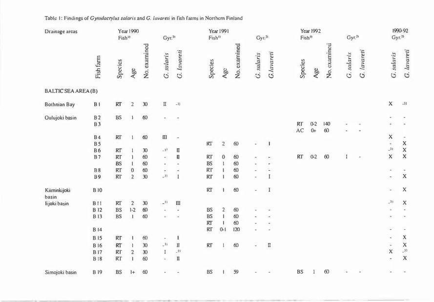
Table I : Findi ngs of Gyrodacrylus salaris and G. lavareti in fish farms in Northern Finland
Drainage areas Year 1 990 Year 1 99 1 Year 1992 199(}.92
Fish11 Gyr.ll Fish11 Gyr.l' Fish11 Gyr.21 Gyr.ll
"8 -c -c Q) V
c c: ·- c ·- ·-
"§ -� � .E . ::; .E .... -·E �
� � ·..: �
� i: .... � � � <.: :8 <.: .s � VJ «l � ::::: >< ::> >< V >< t:! ::>
"(3 Q) l::i � -� Q) �
"[ Q) ;::: I:; � ...c:: Q) � 0 "' ._. � 0 "' � � !).
"' - "' "' 0.. 0 c..:5 0. 0 cS {j c..:5 0 0 lE (/.) <( z (/.) <( z (/.) <(
BALTIC SEA AREA (B)
Bothnian Bay 8 I RI' 2 30 II -3) X _))
Oulujoki basin 8 2 BS I (JJ
8 3 Rf ().2 140 AC Ot 00
8 4 Rr I (JJ m X
B 5 Rf 2 00 I X
B 6 RT I 30 _ )) II _, X
B 7 Rr I (JJ - IJ Rr 0 (JJ Rr ().2 00 I X X
:ss I (fJ BS I (fJ
B B Rr 0 (fJ Rf I (fJ
8 9 Rf 2 30 )) I Rf 1 (fJ - I X
Kiiminkijoki B IO RI I (fJ - I - X
basin lijoki basin 8 1 1 RT 2 30 )) m .ll X
8 12 BS 1 -2 (fJ - BS 2 00
8 1 3 BS I (JJ BS I (JJ
Rf I (fJ
B l4 Rr (). I 120
B l5 Rf I ({) - I X
8 1 6 liT I 30 _)1 IT Rf I (fJ rr X
B 17 Rf 2 30 I _)) X _ll
B I B Rf 1 00 - n X
Simojoki basin 8 19 BS I+ 00 BS I 59 BS 1 (JJ

Kemijoki basin B 20 BS 2 00 m - BS 2 00 I BS 1 + -2 120 1 X BT 2 00
B 21 BS 0+ - 1 + 00 B22 Rf 1 45 _3) m RT I 00 n X X B23 Rf Ot-3 150 I X B 24 Rf 0-1 112 I - X B25 RT I 00 1 RT 2 00 I - X B 26 BS 2 00 B27 RI' 0 00
B 28 BS 1 -2 fl) B29 I.S L (:{) - lS 2 00
ARCTIC OCEAN AREA (A)
PaatSjoki basin A I AC 1- 1+ L21 A 2 RT 0.1 151 I 1 RT 0+ 188 I - X X A 3 lS I (:{) lS I 00 - lS I 00
WHITE SEA AREA (W)
Koutajoki basin W l RI' I 00 m X W 2 RT 0+ - 1 70 n X W 3 RI' I (:{) m - RI' I 00 nr - RI' I 00 m X
TOTAL 1478 1 199 1299
Fish 1 ) : Species: RT = rainbow trout (tot. 24 1 6 specimens); BS = Baltic salmon (tot. 1 0 1 9 specimens); LS = land-locked salmon (tot. 300 specimens); BT = brown trout (tot. 60 specimens); AC = arctic char (tot. 1 8 1 specimens).
cyr2> : Age: 0 =fry, 0+ = one summer old, I =one year old. I + = one year and one summer old, a.s.o. Amounts of Gyrodacrylus: -: no Gyrodactylus I = < 4 specimens per one pectoral fin; 1 1 = 4- 1 0 specimens ; Ill = > 1 0 specimens per pectoral fin. X: Gyrodactylus found
_3): Negative result not included in the calculation of prevalence.

80
Each fish specimen was decapitated or euthanised with benzocain Ph. Eur. (Tamro, Finland) in the laboratory. Its pectoral fins were then cut off, placed in the water of the fish farm and examined for Gyrodactylus under a dissecting microscope (Olympus VMT-4F-W) against a black background. The examinatory results (Table 1) were ranked as follows:
no Gyrodactylus specimens
I less than 4 specimens per pectoral fin
II 4-10 specimens per pectoral fin
Ill more than 10 specimens per pectoral fin
For subsequent species identification, 1-3 Gyrodactylus specimens from each infected fish species per fish farm sample were fixed in ammonium picrate-glycerin between a slide and a cover glass (Malmberg, 1970). Totally, 87 Gyrodactylus specimens were prepared. With one exception, the species determinations were made by the second author. The statistical tests were performed according to Ranta et al.
(1989) by the first author.
Results
The investigation revealed two Gyro
dactylus species, G. salaris and G. lavareti,
- - '
FINLAND RUSSIA
FAR M : DRA I NAGE AREAS .. Arctic Ocean area e with Gyrodactyfus safaris
l://}d White Sea area
� Baltic Sea area
0 no G. safaris
Fig. 1 . Findings of Gyrodactylus salaris at sahnonid fish farms in three drainage areas in Northern Finland (Baltic Sea, Arctic Ocean and White Sea) in 1990-92.

in the salmonid fish farms in Northern Finland. No Gyrodactylus specimens were found on the pectoral fins of the landlocked Saimaa salmon, the brown trout or the arctic char (Table 1).
G. salaris:
The species was identified in 13 of the 35 fish farms (ea. 39 % of the farms included in the calculation of prevalence, Table 1). G. salaris was found in both brackish water (Bothnian Bay, salinity ea. 0,2%; one infected farm) and freshwater farms in areas draining into the Baltic Sea (8 infected farms), the Arctic Ocean (one infected farm) and the White Sea (3 infected farms). The distribution of the infected fish farms is shown in Fig. 1 .
Both rainbow trout and salmon specimens could be infected by G. salaris, but no infection seemed to cause any disease. The rainbow trout had a higher intensity of infection than the salmon (Fig. 2), and the prevalence of infection was also
found to be higher in the populations of rainbow trout examined (ea. 49% of samples) than in the Baltic salmon (ea. 16% of samples, Table 1; chi-square test result X2=ca. 5.78, p<O.OS).
The records of the fish health monitoring programme undertaken by the National Veterinary and Food Research Institute contain nine farms which have a history of farming only rainbow trout (B 1, 4, 6, 14, 18, 27, A 2, W 1 and 2; Table 1), and G. salaris was found at five of these. The parasite was also present in one of the seven salmon farms without a history of raising rainbow trout (B 12, 19-21, 28-29 and A 3; Table 1) .
20
8 1
G. safaris on pectoral fins
• Rainbow trout (n=37)
Ill Baltic salmon (n=l 9)
't> 10 ... (I)
1 5
0 0 I fin 10 or less I fin > 10 I fin
Fig. 2. Findings of Gyrodactylus salaris on the pectoral fins of rainbow trout and Baltic salmon specimens in samples from salmonid farms in Northern Finland (1990-92). The G-test result (G2= ea. 6.29) shows a significant association between fish species and intensity of infection (p<O.OS).
Comparison of the fish specimens in their first and second winter showed no
significant association between either the amount or the prevalence of the infection and the age of the fish. Seven samples of fry (Age 0 in Table 1) were investigated, but no specimens of Gyrodactylus were found. No specimens of G. salaris were found in November, January and February (Fig. 3).
Both uninfected and infected farms were found within the same water system. In the River Kemijoki, G. salaris was found in an upstream farm but not in two other downstream farms (Table 1) :
farms B21 and B22, ea. 110 km apart. The farms were monitored for half a year.

82
farms B21 and B25, ea. 100 km apart. The farms were monitored for one year.
Jan
Feb
March
April
May
June
July
Aug
Sept
Oct
Nov
Dec
Jan
Feb
March
April
May
June
July
Aug
Sept
Oct
Nov
Dec
0
0
G. safaris
Pos. samples (tot. 2 1) • Neg. samples (tot. 32) O
5 1 0 1 5
G. lavareti
Pos. samples (tot. 1 1 ) • Neg. samples (tot. 26) !ill
5 10 1 5 Number of samples
20
20
Fig. 3. Samples collected at farms in Northem Finland in various months over three years (1990-92): findings of Gyrodactylus salaris on pectoral fins of rainbow trout and Baltic salmon and of Gyrodactylus lavareti on pectoral fins of rainbow trout.
G. lavareti:
The species was found only on rainbow trout, in 10 "Baltic" and one "Arctic" farm (totally ea. 33% of the farms investigated), but it was not found in the three
farms within the drainage area of the White Sea (Table 1). No specimens of G.
lavareti were found in September or March (Fig. 3). G. lavareti seemed not to be pathogenic for the rainbow trout.
Discussion
Material and methods
An earlier investigation on G. salaris
in Northern Finland (Keranen et al., 1992) concerned 16 farms where the sampling was performed by the authors themselves. In order to complete the present investigations within the stipulated time, the sampling had to be performed by a large number of persons, which raises the question of whether or not the number of samplers could have influenced the result. The farmers were supposed to use the same methods for sampling and transport as Keranen et al. (1992), however, and a comparison of the findings of Gyrodactylus shows that similar results were obtained in the two investigations.
When possible, 60 fish specimens per fish species, farm and year were examined. On the assumption that the prevalence of infection is 5%, such a sample size will give a 95% probability of finding at least one infected fish per sample (Ossiander and Wedemeyer, 1973) . Some of the 35 farms (Table 1) were not monitored for three years, however, or less than 60 fish specimens from some of them were examined, which reduced the chances of finding Gyrodactylus in such a farm. Even our results based on lower numbers of fish (30 or 45) may reflect the
degree of Gyrodactylus infection in a farm at the time of sampling, however.

In accordance with earlier Finnish investigations (Rintamaki, 1989; Keranen et al., 1992), the microscopic analysis was limited to the pectoral fins. This is based on observations that G. salaris prefers the fins rather than the body of its "true" host, S. salar, generally achieving its highest infection intensity on the dorsal and pectoral fins (Malmberg and Malmberg, 1991b; Jensen and Johnsen, 1992; Mo, 1992). Although the dorsal fin of salmon parr is habitually more infected, the prevalence of G. salaris on the dorsal and pectoral fins of salmon seems to be fairly similar Gensen and Johnsen, 1992; Malmberg, unpublished). Furthermore, in a Finnish investigation of farmed Baltic salmon, more G. salaris specimens were found on the pectoral fins than on the dorsal one (P. Rintamaki, personal communication). However, the examination of the two pectoral fins can be expected to be more revealing as to the presence of G.salaris than an analysis limited to the dorsal fin only.
G. lavareti was found exclusively on rainbow trout, and thus only farms rearing rainbow trout could be infected by both G. salaris and G. lavareti. In fact, the present investigations revealed a concurrPnt infection in only one rainbow trout fa rm . This may be an account of the
sma ll number of Gyrodactylus specimens avai labll� for identification.
'1'111' occure11ce of Gyrodactylus salaris in fish
.fii i'IIIS
G. safaris wns found here in 13 of 33 farms (ea. ;\l)%, Table 1 ), whereas in an
83
investigation two years earlier of salmonid farms in Northern Finland, likewise involving 60 specimens per fish species (Keranen et al. 1992), G. salaris was found in only 3 of the 16 farms investigated (ea. 19%). This proportionally low number of infected farms may be connected with the fact that the farms were investigated on only one occasion, while in the present study, a farm could have been examined on three occasions. At a cursory glance, the present results may be conceived as rather similar to those of Malmberg and Malmberg (1993) in salmonid farms in Sweden, where G. salaris
was found in 8 of 18 farms (ea. 44%). The two investigations were performed differently, however, and consequently the results are not fully comparable. In the Swedish study all the fins and the head, and in certain cases also the body, were investigated, which increased the opportunity of finding solitary parasites, and yielding higher prevalence figures. On the other hand, the smaller sample size in the Swedish investigation (generally 10 specimens per fish species and sample) will have reduced the possibility of an infected fish being included in a sample at a low prevalence of infection. Furthermore, the majority of Swedish farms were investigated between 1986 and 1990, but certain investigations date back to the 1970's (in one case to the 1950's). Moreover, most of the Swedish farms had been investigated on only one occasion, and certain farms that had been reinvestigated were not infected on all occasions.

84
Seasonality of Gyrodactylus in fish farms
The present investigations were per
formed during the cold water period in
Northern Finland, from September to
June, when the water temperature was
:::;+ l0°C. This limitation was based on the
observation that G. salaris in salmonid
farms in Finland (Rintamaki, 1993) and
Sweden (Malmberg and Malmberg, 1993)
seems to favour cold water. G. salaris was
found from March to June and in Sep
tember and G. lavareti from February to
June, but the small number of investiga
tions carried out at seasons other than
spring makes proper analysis of the
annual fluctuation impossible (Fig. 3).
The present results are nevertheless con
sistent with the usual seasonal pattern of
G. salaris infection in three salmon farms
in Northern Finland where monitoring
continued all the year round. Rintamaki
(1993) found G. salaris to be present on
the pectoral fins of Baltic salmon mainly
after the water temperature had fallen
below + l0°C. Her material of 15709 fish
showed prevalence and the intensity of
the infection to increase during the win
ter, leading to more heavily infected fish
in the spring than during the previous
summer. The present results are also in
accordance with observations in salmon
farms in Northern Sweden, where the
treatment of salmon parr for G. salaris
may have to be performed during the
period March-May (Malmberg and
Malmberg, 1991a) .
The seasonal occurrence of G. lavareti
in natural water is still unknown, but the
present results may indicate that in Nor
thern Finland the species is found more
frequently in a period around May than
during other times of the year.
Presence of G. safaris in Northern Finland:
The present paper is the first report
of G. salaris in Finnish waterways drain
ing into the Arctic Ocean and the White
Sea (Table 1). The salmon in both areas
belong to the Atlantic group of salmon,
which is less resistant to G. salaris than
the Baltic group (see Bakke et al., 1990) .
G. safaris can live on rainbow trout, and
the exchanging of live fish between farms
can cause the parasite to spread. In order
to eradicate the infection from the farm
in the Arctic Ocean area (farm A 2, Table
1) and in the hope of preventing it from
spreading to the Paatsjoki drainage basin,
all the fish in the farm were killed and a
programme was started for controlling
the G. safaris situation of the salmonid
farms and wild fish in the Paatsjoki basin
(Koski, 1993, unpubl.) .
There are no wild populations of
salmon, however, in either the River
Paatsjoki in the Arctic Ocean area or the
Finnish parts of the River Koutajoki in
the White Sea area (Koli, 1990). G. safaris
can spread directly upon contact between
two host specimens or via the substra
tum, but it cannot swim and is short
lived without a host (Malmberg, 1993).
This implies that live G. safaris specimens
in the outlet water from a salmonid farm
have little opportunity to infect wild fish
stocks, especially when a river system·
lacks suitable host specimens (salmon or
rainbow trout). Within the Baltic area of
Finland (Fig. 1; Table 1) it is only the
River Tornionjoki and the River Simojoki

that have wild populations of salmon (Koli, 1990) and the stocking of rivers with rainbow trout was uncommon at the time of the present work. Thus, even within this area G. salaris has limited opportunities for spreading further in natural waters. This may explain why G.
salaris could be present in the upstream salmon farms B22 and B25 but not in the downstream farm B21 on the River Kemijoki. The absence of suitable host specimens in the river may have prevented any spread downstream.
Rainbow trout farming and G. salaris in
Northern Finland:
The rainbow trout can harbour several Gyrodactylus species under farmed conditions in fresh water (e.g. G. salaris
and G. lavareti; see Malmberg and Malmberg, 1987), and G. safaris can even repro
duce on the rainbow trout under such conditions (Bakke, 1991). The present findings confirm this relationship, however, as G. salaris was in fact found on
rainbow trout in 11 out of 23 farms investigated (Table 1), five of which had never reared salmon and to our knowll'dge had no salmon in their respective rivers.
J ll l l'thcrmore, more specimens of G.
safaris WNl� found on rainbow trout spedtnl'llS t hnn on salmon parr (Fig. 2), the opposih� result of the expected, since the salmon Is regarded as the "true" host of G. safari.�. ' l 'lw present findings may be explained, however, by the different methods of rl.'tl l'illK Halmon and rainbow trout in lllnlnnd. Snhnon farms, for example, keep t lw Yl'll r dnsses in separate
85
plastic or glass fibre tanks with a parallel flow of water and rearing almost exclusively takes place on the "all in - all out" principle, whereas rainbow trout farms often keep different year classes in successive earth ponds with water passing through them. Thus the rearing of rainbow trout facilitates a constant presence of G. salaris in a farm, and may also enable an accumulation of parasite specimens on individual fish.
G. lavareti on rainbow trout:
G. lavareti was found in 11 out of 23 rainbow trout farms (Table 1), whereas investigations in Sweden revealed the parasite on the same temporary host in 2 out of 4 farms (Malmberg and Malmberg, 1987; 1991a; Malmberg, 1993). Its presence in rainbow trout farms must arise from an original infection from wild or farmed infected specimens of its true host Coregonus lavaretus. Extensive farming of coregonid fingerlings is very common in Finland, and according to Statistics Finland (1993) there were 9113 hectares of natural food rearing ponds mainly for coregonids in 1992 . One might assume that this farming would provide a continuous potential source of infection, allthough it is true that Finnish fish farmers very seldom keep coregonids and rainbow trout in the same farm. The newly hatched coregonid fry are usually moved to temporary, isolated natural food ponds and the young coregonids are transferred to natural waters in autumn at an age of 0+. In fact, parasitological analyses of coregonid stocks in such ponds revealed no speci-

86
mens of Gyrodactylus (see MannermaaKeranen, 1991). Thus the Finnish coregonid farms seem not to be directly involved in the spread of G. lavareti.
Spread of G. salaris and. G. lavareti between
Finnish rainbow trout farms:
Within a rainbow trout farm the water supply may act as a vehicle for the spread of G. salaris and G. lavareti
(above). On the assumption that the farms are situated within the same waterway system and suitable transport hosts are present, spreading via the outlet water (passive spreading) may be possible. Both Gyrodactylus species were found in farms within different areas or
drainage basins, however, which indicates that they had probably spread in connection with the exchange of live rainbow trout between the farms. Such movements of live rainbow trout have been taking place in Scandinavia since the end of the nineteenth century (Malmberg, 1993), which may imply that "farmed" populations of G. salaris and G.
lavareti exist that originate from specimens of old parasite populations adapted to the European rainbow trout farming.
Conclusions
The present results indicate that G.
salaris is introduced to rainbow trout farms in Northem Finland mainly in connection with exchanges of infected live fish between them. Passive spreading via the outlet water released from the farms into natural waters is improbable in waterway systems without suitable host specimens, mainly salmon.
The populations of G. salaris and G.
lavareti on rainbow trout specimens in farms may not be a recent introductions from wild Salmo salar and Coregonus
lavaretus, respectively, but may be of much earlier origin.
Aknowledgements
We thank all the people at the fish farms who helped us to obtain the material for this investigation. Dr. Tor Atle Mo of Oslo, Norway, kindly identified the Gyrodactylus salaris material from the farm on the River Paatsjoki. We are also grateful to Mrs. Bibbi Mayrhofer for making the figures by means of computer graphics and Mr. Malcolm Hicks for revising the language.
References
Bakke TA. A review of the inter- and intraspecific variability in salmonid hosts to laboratory infections with Gyrodactylus safaris
Malmberg. Aquaculture 1991; 98: 303-10
Bakke TA, Jansen PA, Hansen LP. Differences in the host resistance of Atlantic salmon, Salmo salar L., stocks to the monogenean Gyrodactylus salaris Malmberg, 1957. Journal of Fish Biology 1990; 37: 577-87
Ergens R. Gyrodactylus from Eurasian freshwater Salmonidae and Thymallidae. Folia Parasitologica 1983; 30: 15-26
Ieshko E. Parasite studies of Atlantic salmon G. salaris in Northwestern Russia, Karelia. Northern Rivers Atlantic Salmon Symposium. 24-25 August 1993. Teno River Fisheries Research Center, Utsjoki, Finland (unpubl.)
Jensen AJ, Johnsen BO. Site specificity of Gyrodactylus salaris Malmberg, 1957 (Monogenea) on Atlantic salmon (Salmo salar L.) in the River Lakselva, northern Norway. Canadian

Journal of Zoology 1992; 70: 264-67
Johnsen BO, Jensen AJ. The Gyrodactylus story in Norway. Aquaculture 1991; 98: 289-302
Keranen A-L, Koski P, Kulonen K, Ek-Kommonen C, Neuvonen E. Occurrence of infectious fish diseases in fish farms in Northern Finland. Acta veterinaria scandinavica 1992; 33: 161-67
Koli L. Suomen kalat (The fish of Finland, in Finnish). WSOY, Porvoo, Finland, 1990: 354 pp
Koski P. Parasites and diseases of salmonids in the northern region of Finland. Northern Rivers Atlantic Salmon Symposium. 24-25 August 1993. Teno River Fisheries Research Center, Utsjoki, Finland (unpubl.)
Malmberg G. The excretory systems and the marginal hooks as a basis for the systematics of Gyrodactylus (Trematoda, Monogenea) . Arkiv for Zoologi 1970; Ser. 2, 23 (1) : 1-235
Malmberg G. Gyrodactylidae and Gyrodactylosis of Salmonidae. Bulletin Francaise de la Peche et de la Pisciculture 1993; 328: 1-46
Malmberg G, Malmberg M. Gyrodactylus in salmon and rainbow trout farms. In: A. Stenmark and G. Malmberg, eds. Parasites and Diseases in Natural Waters and Aquaculture i n Nordic Countries. Naturhistoriska RiksmuSl'ds Reprocentral, Stockholm, Sweden, 1987: I l)IJ_204
M . d mht:rg C, Malmberg M. Undersokningar o l l l )',okndt• C:yrndactyllls pa laxfisk i fria vatten ' " '" od l i ngar under aren 1951-72 och 1986-maj ( ' I'l l ( l nvt •sl iga l i ons for Gyrodactylus on salmo" i d : ; i 1 1 n .d t l l ',d walcrs and fish farms during 1 1 1 1 • l "'r iods I <J;, I - I 'J72 and 1 986-May 1991, in �;w, ·d i.�h. w i l h l �ng l ish su mmary). Information �! . l l l s l i l t d t • of PrPs h wa ter Research, � ;l t w" l 1 1 1hn, Swt·dt' tl, I 'J'J i a : 1 -30
M . d t l l i H ' I ') ', ( ;, Mi l l mbt•rg M. Cyrodactylus spedt•:; " " :•,d tnonids in SwPdt:n. In: Abstracts of i{< •pot'l s, I l l l n h•rna l i ona l Symposium: Prob(, ' I l lS ot Fish I' a rasilology, i\ ugust 14-21, 1991,
87
Petrozavodsk, USSR, 1991b
Malmberg G, Malmberg M. Species of Gyro
dactylus (Platyhelminthes, Monogenea) on salmonids in Sweden. Fisheries Research 1993; 17: 59-68
Mannermaa-Keranen A-L. Luonnonravintolammikoissa kasvatettujen siianpoikasten rooli tautien ja loisten levittajina PohjoisSuomessa (The role of the fingerlings of whitefish farmed in natural food rearing ponds in the spreading of diseases and parasites in Northern Finland, in Finnish). Mimeograph of the N ational Veterinary Institute, Oulu Regional Laboratory, Oulu, Finland, 1991: 51 pp
Mitenev VK. Ecologic and geographic peculiarities of Monogenea in the Kola Peninsula subarctic zone. In: O.N. Bauer, ed. Parasites of Freshwater Fishes of North-West Europe. Materials of the International Symposium within the Program of the Soviet-Finnish Cooperation, 10-14 January, 1988, Inst. Biol. Acad. Sci., Karelia Petrozavodsk, USSR, 1989: 105-11
Mo TA. Variations of opisthaptoral hard parts of Gyrodactylus salaris Malmberg, 1957 (Monogenea: Gyrodactylidae) on rainbow trout Oncorhynchus mykiss (Walbaum) in a fish farm, with comments on the spreading of the parasite in South-Eastern Norway. Systematic Parasitology 1991; 20: 1-9
Mo TA. Seasonal variations in the prevalence and infestation intensity of Gyrodactylus salaris
Malmberg, 1957 (Monogenea: Gyrodactylidae) on Atlantic salmon parr, Salmo salar L., in the River Batnfjordselva, Norway. Journal of Fish Biology 1992; 41 : 697-707
National Board of Waters, Hydrological Yearbook 1980. Publications of the Water Research Institute, 53. National Board of Waters, Helsinki, 1983: 174 pp
Ossiander FJ. Wedemeyer G. Computer program for sample sizes required to determine
88
disease incidence in fish populations. Journal of the Fisheries Research Board of Canada 1973; 30: 1383-84
Ranta E, Rita H, Kouki J. Biometria, tilastotiedetta ekologeille (Biometrics and statistics for ecologists, in Finnish). Yliopistopaino, Helsinki, 1989: 569 pp
Rimaila-Pamanen E, Wiklund T. Gyrodactylus
salaris -loismadon levinneisyydesta makeanveden kalanviljelylaitoksissamme (The distribution of the parasite Gyrodactylus salaris at our freshwater fish farms, in Finnish). Suomen Elainlaakarilehti 1987; 93: 506-7
Rintamaki P. Gyrodactylus salaris at a fish farm in Northern Finland. In: O.N. Bauer, ed. Parasites of Freshwater Fishes of North-West
Europe. Materials of the International Symposium within the Program of the Soviet-Finnish Cooperation 10-14 January 1988, Inst Biol Acad Sci, Karelia Petrozavodsk, U.S.S.R., 1989: 123-30
Rintamaki P. Montan, Raasakan, Ossauskosken ja Keminmaan kalanviljelylaitosten kalaloiset ja taudit vuosina 1984-1991 (Fish parasites and diseases at the fish farms of Montta, Raasakka, Ossauskoski and Kerninmaa in Northern Finland, in 1984-91, in Finnish). Riista-ja kalatalouden tutkimuslaitos, Kalatutkimuksia - Fiskundersokningar 1993; 58: 1-44
Statistics Finland, Statistical Yearbook of Finland, 88 (new series). Painatuskeskus Oy, Helsinki, Finland, 1993: 620 pp
- 0 -
NOTES FROM THE EDITOR
This year there will be three issues of the Bulletin, instead of the normal two. The next issue will contain the proceedings of the SSP XVII in Jyvaskyla, Finland, 15-17 June 1995, and will be ready for this symposium.
The next ordinary issue will be printed in October.
Proceedings of the SSP-Paraqua-94, Heimaey, Iceland, 2 - 6 July, 1994:
These were printed in the previous issue of the Bull SSP (Vol. 4, No. 2, October 1994). We have extra copies for sale (Kr. 150 per copy), please contact Hans Peter Fagerholm. Present address:
The Royal Veterinary and Agricultural University Section for Zoologi Biilowsvej 13 DK-1870 Fredriksberg C DENMARK, Tel. +45 35 282677, Fax +45 35 282670

89
ANNOUNCEMENTS
Date:
Location:
Language:
FISH PARASITOLOGY - GRADUATE COURSE
August 12-23, 1995
Marine Biological Laboratory (University of Copenhagen), 3000 Helsing0r, Denmark
English
Number of Participants: 16
The course will cover accomodation expenses, but the participants must cover their own travel expences and food, kitchen facilities are available at the laboratory.
The course is primarely aimed at Danish Ph.D. students. To promote international cooperation, participants from other countries, primarely from the Scandinavian countries, may also participate.
Deadline for application: May 1, 1995. For further information, please contact: Dr. Marianne Keie, Marine Biological Laboratory, DK-3000 Heslinger, Denmark
SPRING SYMPOSIUM - March 4, 1995
Theme:
Organizers:
Time:
Venue:
R�giNtrat iun fee:
LEISHMANIA/TRYPANOSOMA
Danish Society for Parasitology & Danish Society for Tropical Medicine
Saturday March 4, 1995; 8.30 a.m. - 4.45 p.m.
August Krogh Instituttet, Universitetsparken 13, 2100, Copenhagen 0
DKK 150.-/75.- (with/without lunch)
TOXOPLASMA MEETING
' l 'hl' Swed ish lns t i t u ll' fm Disease Control is planning a meeting on Toxoplasma on Monday 24 A p ri l I 99.S. J lm fmt lwr i n fomml ion, plense contact Dr. Inger Ljungstrom, Parasitology l .nbumtury, Swt.•d iHh lnHtitute for Infectious Disease Control, S-105 21 Stuckhnlm, Sw�dt.•n t�l: +46 H n!i '1 429, fn " : +46 8 735 '1 162, c-mail : [email protected]

90
GUIDELINES FOR CONTRIBUTORS
All contributions should be submitted as word-processed manuscripts on floppy disk, accompanied by two exactly matching print-outs of good reading-quality. The preferred storage medium is a 3¥2 or 51,4 inch disk in MS-DOS or MS-DOS compatible format. The text should be written in WordPerfect or other word processing programs convertible to WordPerfect. With a Macintosh computer, save the file in the
MS-DOS compatible option. Please indicate the word processor (and version) used to generate the file, the type of computer, the operating system, and the formatted capacity of the diskette.
Short articles/ communications should not exceed 4 printed pages, including tables, figures, and references, and may contain a maximum of 2000 words if there are no figures or tables. The first page should show the title of the article, and the name(s) of the author(s). The authors' addresses should be given, and the complete correspondence address with telephone and telefax number (if available). The text should follow, without subheadings, but a short summary, maximum 100 words, may be included.
The text should be typed unjustified (unaligned right margins), without hyphenation (except for compound words), and at 1 Vz line spacing. Do not type page numbers. Label the hard copies by hand at the bottom of the page. Please ensure that the digit 1 and the letter '1' have been used properly, likewise with the digit 0 and the letter '0'. Do not use decorative formatting, such as boldface and centred headings, or underlining of titles or subheads.
Authors are obliged to follow the rules governing biological nomenclatures, as laid down in e.g. the International Code of Zoological Nomenclature. Disease names should follow the principles of Standardized Nomenclature of Parasitic Diseases
(SNOPAD). Figure legends must be included on the diskette, but the figures will be
handled conventionally. They should be marked on the back with the title of the article and name of the (first) author.
Line drawings should be provided as good quality hard copies suitable for reproduction as submitted.
Photographs must be provided as glossy prints, and be of sufficiently high quality to allow reproduction on standard (not glossy) paper. Colour plates will not be printed.
References in the text shold be stated by giving in brackets the name of the author and the year of publication, e.g. (Thomhill, 1987) or (Austin & Austin, 1987). If there are more than two authors, only the first name plus et al. is given (LundLarsen et al, 1977). The reference list should be in alphabetical order, and follow the style set forth in Uniform Requirements to Manuscripts Submitted to Biomedical Journals,
Br Med J 1988; 296: 401-05. References to journals should contain names and initials

of the authors, article title, the abbreviated name of the journal, year of publication, volume, and first and last page numbers of the paper. Journals should be abbreviated according to the "List of journals indexed in Index Medicus". Authors without access to this list may type the full name of the journal, and the Editor will take care of the abbreviations. If there are more than six authors, list only the first three and add 'et al'. Personal communications and unpublished data should not be used as references, but may be inserted in the text (within parenthesis marks).
Examples of correct forms of references are given below: Standard journal article:
Anonymous. Some facts on small animal practice. Vet Rec 1987; 120: 73
Horsberg TE, Berge GN, H0y T et al. Diklorvos som avlusningsmiddel for fisk: klinisk utpmving og toksisitetstesting. Nor Vet Tidsskr 1987; 99: 611-15
Lund-Larsen TR, Sundby A, Kruse V, Velle W. Relation between growth rate, serum somatomedin and plasma testosterone in young bulls. J Anim Sci 1977; 44: 189-94
Books and other monographs:
Austin B, Austin DA. Bacterial fish pathogens: disease in farmed and wild fish. Chichester: Ellis Horwood, 1987
Mcflcrran JB, McNulty MS, eds. Acute virus infections of poultry: a seminar in the CEC programme, Brussels 1985. Dordrecht: Martinus Nijhoff, 1986. (Current topics in Vl'll•rinary medicine and animal science 37)
Sosia ldepartemcntet. Tsjernobyl-ulykken: Rapport fra Helsedirektoratets radgivende faggruppe. Oslo: Un ivcrs itetsforlaget, 1987 (Norges offentlige utredninger NOU 1987:
I )
Thornhil l J A . l{l•na l mdocrinology. In: Drazner FH, ed. Small animal endocrinology. Nt•w Ymk: Churchi l l l .ivingstone, 1987: 315-39
Tlw lllllllWil'ri p l (d iskette and paper copies) should be sent to the editor in your nnantry, 111'1' in11idt• of front cover. Label the diskette with the name of the (first) author. Mamallnlpl11 nn• ntn•plt•d for publication after review and recommendation by the 1 \d l lol' ln l l lt l ll n l . A u l hor11 wi l l be notified by the Editor-in-Chief about final acceptance and t'X JWdt•d l l nw ol puhlka l ion.
IU!I'IU NTS W l l .l . NOT IJE AVAILABLE.
In lhl' ln lcorrNI uf Npcol'd, nu proofs will be sent to authors. It is therefore of vital l mpurl"'"'r lh"l lhr m1m UNcrlpts are carefully checked before submission.

VOL. 5 No. l
BULLETIN OF THE SCANDINA VIAN SOCIETY FOR PARASITOLOGY
CONTENTS FEBRUARY 1995
Proceedings of the Baltic-Scandinavian Symposium on Parasitic Zoonoses and the
Ecology of Paratites, 7-8 September 1994, Vilnius, Lithuania
Peter Nansen (Editor), with technical assistance from ]ane Raken, Fritz 0stergaard and
Helle Kiirstein t . . . . . . . . . . . . . . . . . . . . . . . . . . . . . . . . . . . . . . . . • . . . . . . . . . . . 1
Occurrence of Gyrodactylus (monogenea) on salmon and rainbow trout in fish farms
in northern Finland
Perttu Koski and Goran Malmberg , . . . . . . . . . . . . . . . . . . . . . . . . . . . . . . . . . . . . 76
NEWS
SSP XVII, Jyvaskyla 15-17 June 1995, Final Announcement . . . . . . . . . . . . . . . . . 75
From the Editor . . . . . . . . . . . . . . . . . . . . , . . . . . . . . . . . , . . , . . . . . . . . . . . . . . 88
Fish Parasitology - Graduate Course, Helsingor, 14-23 August 1995 . . . . . . . . . . 89
Spring Symposium, Copenhagen, 4 March 1995 . . . . . . . . . . . . . . . . . . . , . . . . . 89
Toxoplasma Meeting, Stockholm, 24 April 1995 . . . . . . . . . . . . . . . . . . . . . . . . . . . 89
Guidelines for contributors . . . . . . . . . . . . . . . . . . . . . . . . . . . . . . . . . . . . . . . . . 93