Novel Necrosis Inhibitor To prevent Myocardial Ischemia
59
Novel Necrosis Inhibitor to prevent Myocardial Ischemia-Reperfusion Injury Hyo-Soo Kim, MD/PhD/FAHA Cardiovascular Center & Department of Internal Medicine, Seoul National University Hospital
Transcript of Novel Necrosis Inhibitor To prevent Myocardial Ischemia

Novel Necrosis Inhibitor to prevent
Myocardial Ischemia-Reperfusion Injury
Hyo-Soo Kim, MD/PhD/FAHA
Cardiovascular Center & Department of Internal Medicine,
Seoul National University Hospital

INTRODUCTION

Myocardial I-R Injury: Neglected Therapeutic Target
J Clin Invest. 2013;123(1):92–100.

Three Roles of Mitochondria Abundant in CMC
N Engl J Med 2013;369:2236-51.
ROS generation
Ca2+ homeostasis
Chemical power plant

Structure and Mechanism of MPTP
Circ J 2013;77:1111 – 1122.
Cyclophilin D (CypD)
Mitochondrial
Permeability
Transition
Pore

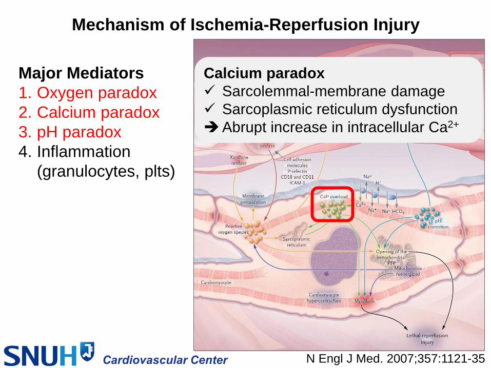
Mechanism of Ischemia-Reperfusion Injury
N Engl J Med. 2007;357:1121-35
Major Mediators
1. Oxygen paradox
2. Calcium paradox
3. pH paradox
4. Inflammation
(granulocytes, plts)
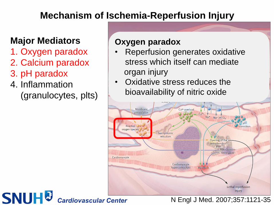
Mechanism of Ischemia-Reperfusion Injury
N Engl J Med. 2007;357:1121-35
Oxygen paradox
• Reperfusion generates oxidative
stress which itself can mediate
organ injury
• Oxidative stress reduces the
bioavailability of nitric oxide
Major Mediators
1. Oxygen paradox
2. Calcium paradox
3. pH paradox
4. Inflammation
(granulocytes, plts)
Mechanism of Ischemia-Reperfusion Injury
N Engl J Med. 2007;357:1121-35
Calcium paradox
Sarcolemmal-membrane damage
Sarcoplasmic reticulum dysfunction
Abrupt increase in intracellular Ca2+
Major Mediators
1. Oxygen paradox
2. Calcium paradox
3. pH paradox
4. Inflammation
(granulocytes, plts)

Mechanism of Ischemia-Reperfusion Injury
N Engl J Med. 2007;357:1121-35
pH paradox
• Reperfusion
Wash-out of lactic acid
Activation of Na+/H+ exchanger &
Na+/HCO3- symporter
Rapid restoration of physiologic pH
Major Mediators
1. Oxygen paradox
2. Calcium paradox
3. pH paradox
4. Inflammation
(granulocytes, plts)
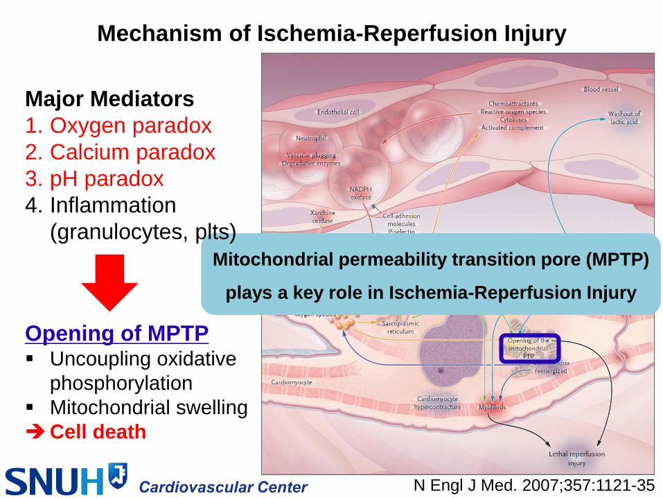
Mechanism of Ischemia-Reperfusion Injury
N Engl J Med. 2007;357:1121-35
Opening of MPTP Uncoupling oxidative
phosphorylation
Mitochondrial swelling
Cell death
Mitochondrial permeability transition pore (MPTP)
plays a key role in Ischemia-Reperfusion Injury
Major Mediators
1. Oxygen paradox
2. Calcium paradox
3. pH paradox
4. Inflammation
(granulocytes, plts)

APOPTOSIS vs. NECROSIS
• Mechanisms of mitochondrial membrane permeabilization
Nature. 2005;434:578-579.
Ischemia-Reperfusion • Mitochondrial ROS generation
• Mitochondrial Ca2+ overload
• Normalization of pH
MPTP opening Severe insult MPTPs stay open Necrosis
Moderate insult MPTPs transient opening Apoptosis

Novel Necrosis Inhibitor, NecroX,
Prevents myocardial Ischemia-
Reperfusion Injury

A Novel Necrosis Inhibitor: NecroX
• INTRODUCTION
Derivative and combination of products
NecroX-7; from LG life sciences (SH Kim, PhD)
Anti-necrotic effect by some mechanisms
via 1) Strong antioxidant
- Mitochondrial ROS and ONOO- scavenger
- Inhibition of ROS-generating enzyme (NADPH oxidase)
2) Inhibition of HMGB1

in vitro study

Hypoxia with 1% O2 Oxidative stress
with H2O2 400μM
Harvest
24 Hours of
incubation period
1.5 Hours of
Reoxygenation
Pre-treatment 1. Vehicle: 0.01% DMSO
2. Necrosis inhibitor: NecroX (20 μM)
3. Vitamin C (10 μM)
4. Vitamin C (20 μM)
5. Vitamin C (10 μM) + Vitamin E (20 μM)
6. N-acetylcysteine (250 μM)
7. Apoptosis inhibitor: Z-VAD-fmk (20 μM)
in vitro protocol Hypoxia-Oxidative stress/Reoxygenation (H-O/R) model
using H9C2 rat cardiomyoblasts (myoblast cell line)
Seeding :H9C2 cells 0.5X10⁵ Cells /35mm dish,
1.5X10⁵ Cells /60mm dish,
1.5X10⁶ Cells/100mm dish
24 Hours of Hypoxia
23.5 Hours 30min

Measurement of mitochondrial Ca2+ influx Vehicle
Vehicle treated
group showed
prominent
calcium influx
(red stain) in the
swollen
mitochondria via
mPTP opening
Hypoxia 24h + Oxidative stress with H2O2
Mitotracker : mitochondria Rhod-2 : Ca2+ influx
NecroX
Necrosis Inhibitor
revealed protective
effect on mPTP
opening under I/RI
Tim
e (
min
ute
s)
0
20

Action Mechanism of NecroX
: ROS scavenging activity
H-O/R : hypoxia-oxidative stress/reoxygenation.
NAC : N-acetylcysteine
H2DCF-DA assay
• Intracellular ROS probe
• Oxidization by ROS
DCF : highly fluorescent
NecroX has potent scavenging
activity on intracellular ROS.

Action Mechanism of NecroX
: ROS scavenging activity
H-O/R : hypoxia-oxidative stress/reoxygenation.
NAC : N-acetylcysteine
Dihydrorhodamine 123 (DHR 123) assay
• Mitochondria-specific ROS probe
• Oxidization by ROS
Rhodamine 123 : highly fluorescent
NecroX has potent mitochondria-
specific ROS scavenging activity.

Action Mechanism of NecroX
: Inhibition of ROS generating enzyme
Hypoxia
only
H2O2
only
Vehicle NecroX Vit C
(10 μM) Z-VAD
H-O/R
Vit C
(20 μM)
Vit C
+ Vit E
NAC
NecroX has inhibitory effect on NADPH oxidase,
an important ROS-generating enzyme.
H-O/R : hypoxia-oxidative stress/reoxygenation.
NAC : N-acetylcysteine
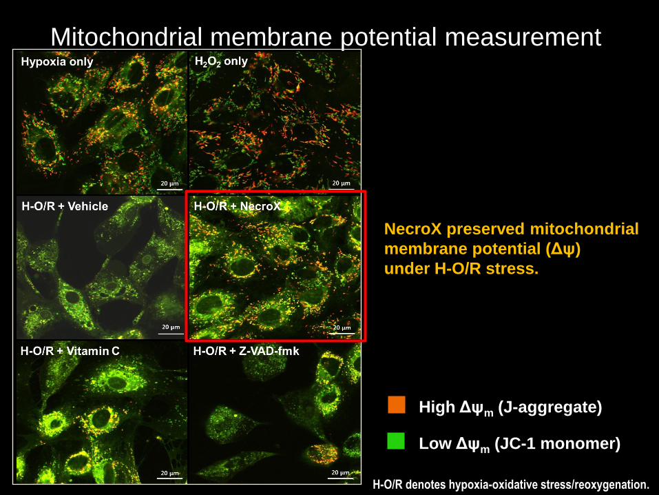
Mitochondrial membrane potential measurement
High Δψm (J-aggregate)
Low Δψm (JC-1 monomer)
H-O/R denotes hypoxia-oxidative stress/reoxygenation.
NecroX preserved mitochondrial
membrane potential (Δψ)
under H-O/R stress.

Mitochondrial membrane potential
During H-O/R injury, mitochondrial
transmembrane potential changed
from high to low gradient (Δψ),
except NecroX treated cells.
H-O/R : hypoxia-oxidative stress/reoxygenation.
NAC : N-acetylcysteine
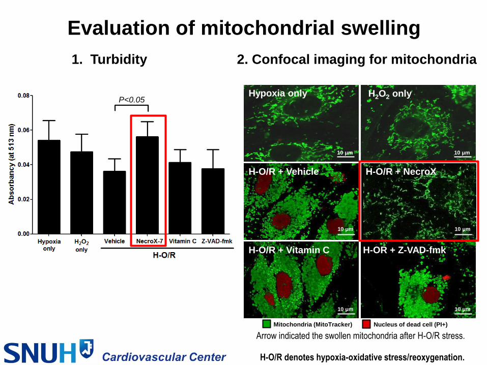
Evaluation of mitochondrial swelling
1. Turbidity 2. Confocal imaging for mitochondria
H-O/R denotes hypoxia-oxidative stress/reoxygenation.
Hypoxia only
H-OR + Z-VAD-fmk
H-O/R + NecroX H-O/R + Vehicle
H-O/R + Vitamin C
10 μm
H2O2 only
10 μm
10 μm 10 μm
10 μm 10 μm
Mitochondria (MitoTracker) Nucleus of dead cell (PI+)
Arrow indicated the swollen mitochondria after H-O/R stress.
P<0.05

TEM for mitochondrial swelling
Vehicle + H-O/R NecroX + H-O/R
━━ 500 nm ━━ ━━ 500 nm ━━
Intracytoplasmic vacuole
Swollen/ruptured mitochondria
and degenerated crista
Preserved normal mitochondria
H-O/R denotes hypoxia-oxidative stress/reoxygenation.

H-O/R : hypoxia-oxidative stress/reoxygenation.
NAC : N-acetylcysteine
NecroX protected H9C2 rat
cardiomyoblasts from necrotic
cell death after H-O/R stress.
PI/FDA staining & Counting the cell

Annexin-V
PI
2.6%
12.2%
6.1%
3.4%
68.4%
0.9%
39.5%
1.0%
65.4%
0.8%
63.9%
1.1%
67.2%
1.2%
44.2%
0.8%
H2O2 only H-O/R + Vehicle
H-O/R + NecroX H-O/R + Vit C (10 μM) H-O/R + Vit C (20 μM)
H-O/R + Z-VAD-fmk H-O/R + Vit C + Vit E
Hypoxia only
58.8%
0.8%
H-O/R + NAC
NecroX protected H9C2 rat
cardiomyoblasts from necrotic
cell death after H-O/R stress.
H-O/R : hypoxia-oxidative stress/reoxygenation.
NAC : N-acetylcysteine
Necrotic cells by FACS

in vivo study

in vivo protocol
IV bolus infusion
Like as Preconditioning (N=13)
1. Vehicle
2. Cyclosporin A (CsA; 5 mg/kg) for positive control
3. NecroX (1 mg/kg)
45min. of Ischemia
25min. 20min.
Reperfusion
Ischemia by
Ligation of LAD
Harvest
After EchoCG &
blood sampling
3 Days 11 Days
14 days after I/RI injury
EchoCG under anesthesia
EchoCG under anesthesia
NecroX (30 mg/kg)
P.O. daily
Formalin fixed tissue

NecroX preserved LVEF and inhibited LV remodeling

NecroX-treated rats: Reduction in Myocardial Fibrosis
Day 14
harvested
heart
Control (N=13) Cyclosporin A (N=13) NecroX (N=13)
Fibrotic myocardium
Intact myocardium

Necrotic myocardium quantification
P < 0.01
Control Cyclosporin A NecroX
Necrotic zone Non-necrotic zone Black arrow indicated necrotic myocyte.

Mechanism of anti-MHC antibody
Sarcolemma (WGA)
Anti-MHC antibody
MHC
Viable cardiomyocytes
Sarcolemma (WGA)
Anti-MHC antibody
MHC
Necrotic cardiomyocyte
In viable cardiomyocytes,
anti-MHC Ab cannot bind the cardiomyocytes.
Necrotic cardiomyocytes bind to anti-MHC
antibody through broken sarcolemma.
WGA : Wheat germ agglutinin for sarcolemma stain
MHC : myosin heavy chain
Necrotic myocardium specific quantification

Specific Quantification for Myocardial Necrosis
Why the anti-MHC Ab get injected before the ligation ?
IV bolus infusion
Like as Preconditioning (N=4)
1. Vehicle
2. Cyclosporin A (CsA; 5 mg/kg)
3. NecroX-7 (1 mg/kg)
45min. of Ischemia
25min. 20min.
Reperfusion
Ischemia by
Ligation of LAD
12 hours 20min.
Before
Ligation
Harvest &
OCT embedding
Anti-MHC antibody injection
via internal jugular vein

Necrotic myocardium specific quantification
P < 0.01
100 μm
50 μm 50 μm
Necrotic myocyte (MHC+) WGA (Sarcolemma) Nucleus
100 μm 100 μm
50 μm
Control Cyclosporin A NecroX
White arrow indicated necrotic myocyte.

Mechanism of NecroX on I/R injury
Myocardial
Infarction
&
Reperfusion
50 μm
Necrotic
cardiomyocyte
mPTP
Mitochondrial swelling
ROS burst Ca2
+
H2O + solute
Ischemia
/Reperfusion Ca2+
TCA cycle
& ETC
stimulation
⑥ ⑤ ④
② ③ ③’
NecroX
Necrotic cardiomyocyte
①

Clinical Trial: NEXsteMI RCT

Clinical trials…
Phase I trial : successfully finished !
Phase II multicenter RCT : on going

Phase I clinical trial

Phase II NEXsteMI RCT
• [NEXsteMI]
• NECRO-X to reduced reperfusion injury
• after emergent PCI in patients with STEMI
• Multicenter,
Randomized,
Double-blinded,
Placebo-controlled
• Efficacy & safety of single bolus IV infusion of Necro-X
Just before emergent PCI for patients with STEMI
• Area under curve of cardiac enzyme
• Dose-finding

Phase II NEXsteMI RCT
기관 번호 실시 기관명 시험책임자
01 경상대학교병원 황 진 용 교수
02 경희대학교병원 김 원 교수
03 서울대학교병원 김 효 수 교수(CI)
04 연세대학교 세브란스병원 최 동 훈 교수
05 전남대학교병원 정 명 호 교수

Phase II NEXsteMI RCT
Number of Planned Patients = 60
4 Gr Treatment group No. of Pt.
Control Placebo 1 vial 15
Tx 1 LC28-0126 1mg 15
Tx 2 LC28-0126 3mg 15
Tx 3 LC28-0126 10mg 15

Phase II NEXsteMI RCT
[1st screening criteria]
1) Age : 20 ~ 75
2) Onset of chest pain < 12 h
3) ST elevation > 0.1mV in two/more precordial leads
or new LBBB
[2nd inclusion criteria]
Occlusion at P.LAD or M.LAD (TIMI flow = 0 or 1)
Scheduled for emergent Primary PCI

Phase II NEXsteMI RCT
Pre-PCI
Screening
Hospitalization
Day 1~Day 4
V1(-3h~Random) V3 24h~48h
V2 Random~24h
<1st/2
nd criteria>
*pain < 12h
*ST elevation
or new LBBB
⇒ 무작위배정 번호
IP Kit no. 부여
PCI
Day 0
<2nd inclusion criteria> LAD occlusion on CAG/ Emergent PCI
Placebo
LC28-0126 1mg
LC28-0126 3mg
LC28-0126 10mg
V4 48h~72h
V5 72h~96h
V6 96h~120h
F/U
Day 30
V7
IP Injection
Cardio marker/ PK 채혈 (~ 72h)
Biomarker Biomarker Biomarker Biomarker
CMR
ECho
Discharge
CMR
ECho

Phase II NEXsteMI RCT
Primary Efficacy Endpoint
Secondary Efficacy Endpoint
1) PCI 후 72시간까지의 Troponin I의 AUC
2) PCI 후 72시간까지의 CK 의 AUC
3) PCI 후 Day 4, 30에 CMR로 평가된 Infarct size
4) PCI 후 Day 4, 30에 CMR로 평가된 LV function (LVEF, LVEDV, LVESV)
5) PCI 후 Day 4, 30에 심장초음파로 평가된 LV (LVEF,LVEDV, LVESV)
PCI 후 72시간까지의 CK-MB의 AUC

Phase II clinical trial: NEXsteMI RCT
• Planned Timeline
IND 승인 (2013년 12월 2일)
IRB 승인 (2013년 12월 27일)
개시방문 (2014년 01월~2014년 02월)
FPI (2014년 01월)
DM/STAT (2015년 2월)
CSR (2015년 04월)
LPO (2014년 12월)
INTERIM ANALYSIS (2014년 7월)

Novel Necrosis Inhibitor to prevent
Myocardial Ischemia-Reperfusion Injury
Hyo-Soo Kim, MD/PhD/FAHA
Cardiovascular Center & Department of Internal Medicine,
Seoul National University Hospital

Structure and Mechanism of MPTP
Cell Physiol Biochem 2007;20:01-22
Opening of MPTP • Triggered by
ROS overload
Ca2+ overload
pH normalization
Opening of MPTP Loss of ATP
Mitochondrial swelling
Necrotic cell death

Structure and Mechanism of MPTP
FEBS Lett. 2010;584(10):2161-6.
Molecular components of MPTP • Cyclophilin D (CypD)
• Adenine nucleotide translocase (ANT)
• Voltage-dependent anion channel (VDAC)
• Benzodiazepine receptor (BR)
• Creatine kinase (CK)
• Hexokinase (HK)
Opening of MPTP • Triggered by
ROS overload
Ca2+ overload
pH normalization
• Consequences
Loss of ATP
Mitochondrial swelling
Necrotic cell death

MPTP in Myocardial Ischemia-Reperfusion Injury
J Clin Invest. 2013;123(1):92–100.
During ISCHEMIA • Inhibited Ox-Phos
ATP depletion & Acidic pH
• Minimal Mitochondrial Ca2+ uptake
MPTP: closed
During REPERFUSION • Mitochondrial ROS generation
• Mitochondrial Ca2+ overload
• Normalization of pH
MPTP opening Cell Death

Myocardial Ischemia-
Reperfusion Injury
: Neglected Therapeutic Target

Prevention of Myocardial Reperfusion Injury
• Cardioprotection Trials
Lancet 2013; 382: 644–57
• Aims: To reduce the amount of necrosis
after myocardial ischemia-reperfusion.
• No cardioprotective interventions
have been included in guidelines or
clinical practice.
However, several are currently under investigation
- Cyclosporin, TRO40303, NO, Metformin, etc

Prevention of Myocardial Reperfusion Injury
• Cyclosporin A (CsA; inhibitor of MPTP opening)
• Mechanism of Action
– CsA binds to & inhibits cyclophilins
– CsA/CypD complex inhibits MPTP opening
Cell Physiol Biochem 2007;20:01-22

NECRO-X DATA IN OUR LAB
• Hypothesis / Aim of study
To reduce the infarct size of the heart
after ischemia-reperfusion injury
by using the necrosis inhibitor, NecroX-7
The necrosis inhibitor will reduce
the necrotic cell death and the infarct size
in the in vitro and in vivo models of
myocardial ischemia-reperfusion injury

In vitro evaluation
1. Mitochondria swelling measurement (Turbidity)
2. Mitochondria swelling using Confocal Images
3. TEM for Mitochondria swelling and Nucleus
4. PI / FDA staining and cell count
5. FACS analysis using PI / Annexin-V
6. Mitochondria membrane potential measurement using JC-1
7. Western blot for necrotic/apoptotic signaling pathways
8. Ca2+ influx in mitochondria under hypoxia-oxidative stress/reoxygenation stress
9. ROS scavenging activity measurement (DHR 123, DCF-DA)
10. NADPH oxidase activity measurement

Vehicle ( 3~5 minutes) Necrosis Inhibitor (over 30minutes)
Under hypoxia-oxidative stress/reoxygenation condition
Measurement of mitochondrial Ca2+ influx

Measurement of inflammatory cytokine
& cardiac enzymes at 12 hours after I/R injury

Measurement of inflammatory cytokine
& cardiac enzymes at 12 hours after I/R injury
Inflammatory cytokines & Cardiac enzymes were
significantly lower in the NecroX-treated rats.

IHC for HMGB1 at 12hr after I/RI
Control
Cyclosporin A
Necrosis inhibitor
Brownish
stained HMGB1
Why 12 hours
after MI/R injury?
: Necrosis is early
process in MI/R

NecroX blocked the necrotic signaling pathways
Veh: Vehicle
DR: Doxorubicin (apoptosis inducer)
H-O/R: Hypoxia-Oxidative stress/Reoxygenation
Oxidative stress (H2O2)
Harvest
24 hours of
incubation
45 min. of
Reoxygenation
Pre-treatment
Seeding with H9C2 cells
24 hours of Hypoxia
23.75 hours 15 min
Hypoxia
Veh NecroX Vit C Z-VAD
Cleaved caspase-3
Veh DR
Cleaved-PARP1
(89kDa; apoptosis)
Cleaved-PARP1
(55kDa; necrosis)
Apoptosis
Inducer
Full length PARP1
(116kDa)
α-tubulin
H-O/R
• Cleaved-PARP1 (55kDa), the necrotic fragment
was significantly expressed in the Vehicle-treated
H9C2 cells under H-O/R stress.
• The necrotic fragment was reduced by NecroX.
• The main mechanism of cell death after H-O/R
stress was not apoptosis but necrosis that was
prevented by NecroX.

NecroX blocked the necrotic signaling pathways
Phospho-JNK
Total JNK
Phospho-p38
Total p38
β-actin
Hypoxia
only
H2O2
only Veh NecroX Vit C Z-VAD
H-O/R
RIP1
RIP3
Veh: Vehicle
H-O/R: Hypoxia-Oxidative stress/Reoxygenation
• Necrotic signal pathways were effectively
blocked by the treatment with NecroX.