Normal oral mucosa - Delta...
107
Transcript of Normal oral mucosa - Delta...


Normal oral mucosa

Pale colour of normal mucosa It results from an interplay between four factors :
1.Vascularity
2. Epithelium thickness
3. Melanin pigment
4.keratinization

White lesions

Def.
White-appearing lesions of the oral mucosa, obtained their characteristic appearance from the scattering of light through an altered surface, e.g. such alterations may be the result of a thickened layer of keratin that may be due to :
1.Chronic physical trauma.
2. Mucocutaneous diseases.
3. Tobacco use.
4. Inflammatory reactions.
5. Genetic abnormalities.
Colour of White lesions results from 1. Hyperkeratosis (thickened layer of keratin ) 2. Acanthosis (epithelial hyperplasia as the thickened spinous layers masks the normal vascularity (redness). 3. Intracellular epithelial edema 4.Reduced vascularity of subjacent connective tissue 5. Fibrous exudate covering an :
- Submucosal deposits - ulcer - fungal colonies - surface debris
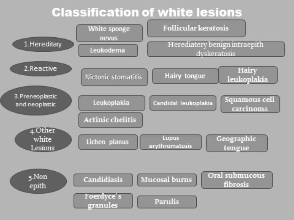
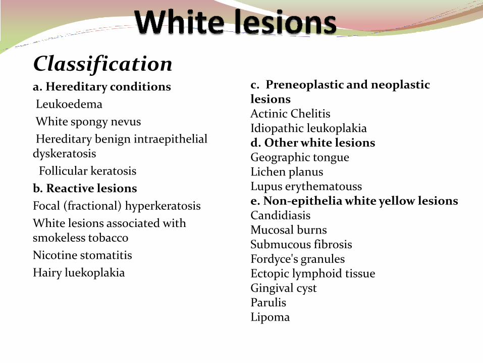
Classification a. Hereditary conditions
Leukoedema
White spongy nevus
Hereditary benign intraepithelial dyskeratosis
Follicular keratosis
b. Reactive lesions
Focal (fractional) hyperkeratosis
White lesions associated with smokeless tobacco
Nicotine stomatitis
Hairy luekoplakia
c. Preneoplastic and neoplastic lesions Actinic Chelitis Idiopathic leukoplakia d. Other white lesions Geographic tongue Lichen planus Lupus erythematouss e. Non-epithelia white yellow lesions Candidiasis Mucosal burns Submucous fibrosis Fordyce's granules Ectopic lymphoid tissue Gingival cyst Parulis Lipoma

1. Leukoedema
2. White spongy nevus
3. Hereditary benign intraepithelial dyskeratosis
4. Follicular keratosis

Leukoedema
Def.
Accumulation of fluid within the epithelial
cells of the buccal mucosa
Etiology
unknown
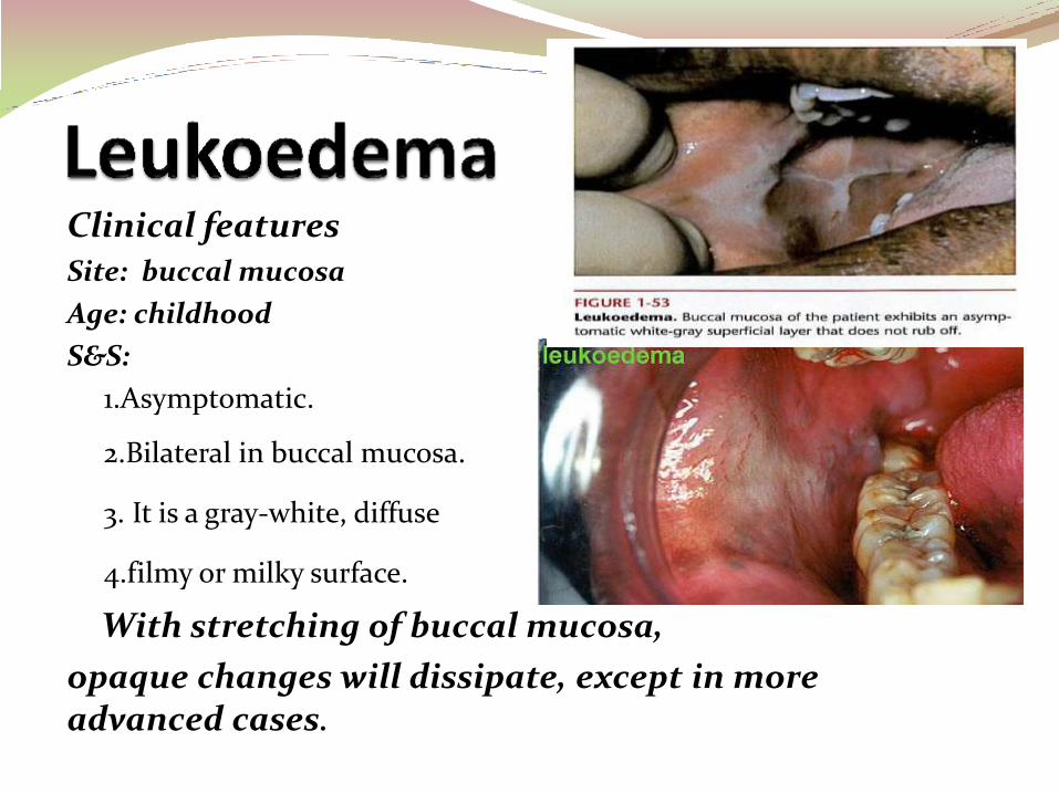
Clinical features Site: buccal mucosa
Age: childhood
S&S:
1.Asymptomatic.
2.Bilateral in buccal mucosa.
3. It is a gray-white, diffuse
4.filmy or milky surface.
With stretching of buccal mucosa,
opaque changes will dissipate, except in more advanced cases.
.

Histopathplogic features 1.Parakeratosis
2. Acanthosis
3.marked intracellular
edema of spinous cells.
4.Small pyknotic nuclei with clear cytoplasm.

Leukoedema

Differential diagnosis
1. White spongy nevus
2. leukoplakia,
3. hereditary benign intraepithelial dyskeratosis.
4. Response to chronic cheek biting.
Overall thickness of these lesions. Their persistence upon stretching, and specific microscopic features help separate them from leukoedema.
Treatment and prognosis
No treatment is essential and no malignant potential
White spongy nevus
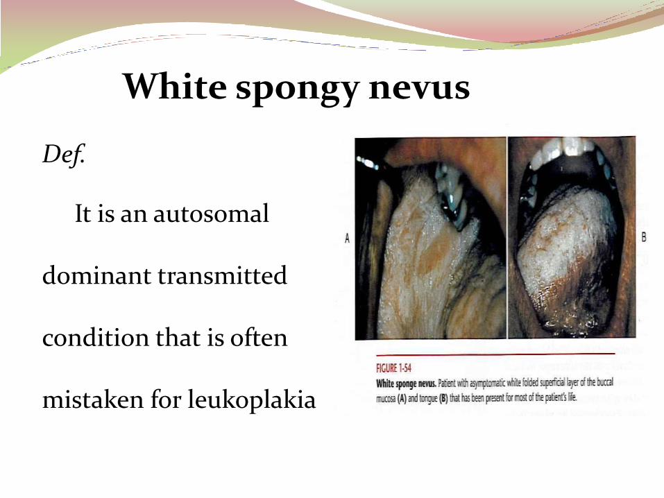
Def.
It is an autosomal
dominant transmitted
condition that is often
mistaken for leukoplakia
White spongy nevus
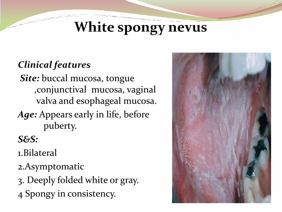
Clinical features
Site: buccal mucosa, tongue ,conjunctival mucosa, vaginal valva and esophageal mucosa.
Age: Appears early in life, before puberty.
S&S:
1.Bilateral
2.Asymptomatic
3. Deeply folded white or gray.
4 Spongy in consistency.
White spongy nevus
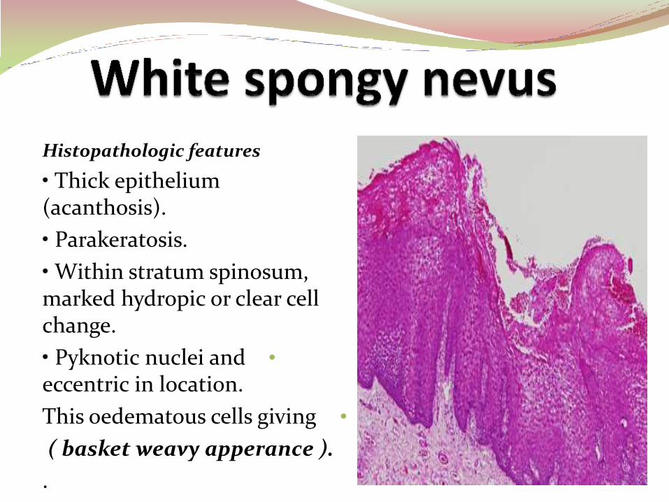
Histopathologic features
• Thick epithelium (acanthosis).
• Parakeratosis.
• Within stratum spinosum, marked hydropic or clear cell change.
•• Pyknotic nuclei and eccentric in location.
•This oedematous cells giving
( basket weavy apperance ).
.

White spongy nevus
White spongy nevus
White spongy nevus
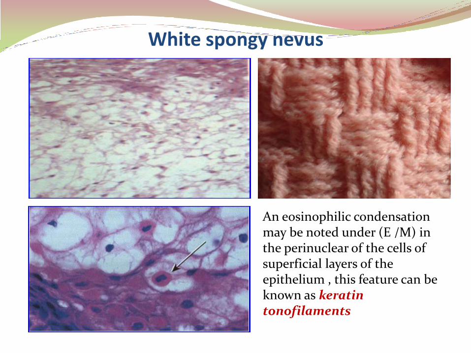
An eosinophilic condensation may be noted under (E /M) in the perinuclear of the cells of superficial layers of the epithelium , this feature can be known as keratin tonofilaments
Differential diagnosis
1. Heriditary begnin intraepithelial dyskeratosis 2.hypertrophic lichen planus
3. frictional keratosis
4. cheek biting.
5.Leukoedema
Treatment
No treatment since it is a begnin asymptomatic condition.
White spongy nevus

Hereditary benign intraepithelial dyskeratosis
(Hbid)(Witkop's disease)
Def.
It is actually a syndrome
Etiology
Heriditary autosomal dominant transmitted condition.
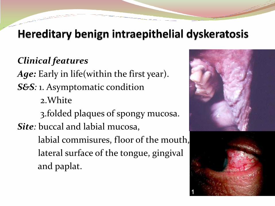
Clinical features
Age: Early in life(within the first year).
S&S: 1. Asymptomatic condition
2.White
3.folded plaques of spongy mucosa.
Site: buccal and labial mucosa,
labial commisures, floor of the mouth,
lateral surface of the tongue, gingival
and paplat.
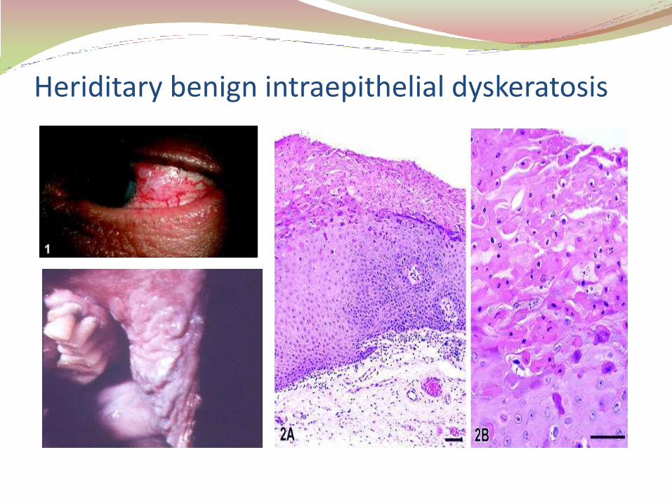
Histopathologic features
1.Acanthosis.
2.Hydropic degeneration of spinous cells.
3. Enlarged, hyline, waxy eosinophilic cells(which is the dyskeratotic elements).
4. Dyskeratotic cells may be surrounded by djacent cells producing(cell within cell).
5. Inflammatory cell infiltrate is minimal
Heriditary benign intraepithelial dyskeratosis

Differential diagnosis
1.White spongy nevus
2. hypertrophic lichen planus
3. frictional keratosis.
Follicular keratosis (Darier's Disease)

Differential diagnosis
features Histopathplogic
Clinical picture Etiology Parameters
.1. Vertical clefts
2. acantholytic
epithelial cells.
Microscopically,
these may be
represented under
the term "warty
dyskeratonia".
1. Oral mucosal sites include
keratinized regions such as:
attached gingiva, hard palate.
2. Lesions may extend to
oropharynx and pharynx.
3. Skin (symmetrically
distributed over the face and
thrunk).
Childhood or adolescence.
Thickening of the skin of palms
and soles.
• Papular lesions on the skin.
• Localized lesions may follow
sunburns, especially on the legs.
• oral lesions are in the form of
papules
Site
Age
S&S
Autosomal,
dominant mode
of inheritance. • 50% of.
Follicular keratosis (Darier's Disease)

Reactive lesions
1.Focal (fractional) hyperkeratosis
2.White lesions associated with smokeless
tobacco
3.Nicotine stomatitis
4.Hairy luekoplakia
5.Hairy tongue
Focal (frictional)hyperkeratosis
Def.
It is a white lesion that is often classified
under the general term "leukoplakia”.
The tissue response represents a protective
action against low-grade, long-term trauma

Etiology
1.Self-evident
2.Chronic rubbing or friction of an oral mucosal surface
Clinical features Site:
areas that are commonly traumatized such as lips, buccal mucosa along occlusal line, & edentulous ridges
S&S:
Friction-induced hyperparakeratosis or leukoplakia
Histopathlogic features 1.Hyperkeratosis
2. chronic inflammatory cells on connective tissue.
White lesions associated with smokeless tobacco

Causes
Chewing tobacco or snuff
Clinical features
alterations of oral cavity
Histopathology
1. hyperkeratosis
2. chronic inflammatory cells on connective tissue
Differential diagnosis
1..leukoplakia
If the etiology of a white lesion is in doubt, it should be regarded as idiopathic leukoplakia.

Nicotine stomatitis
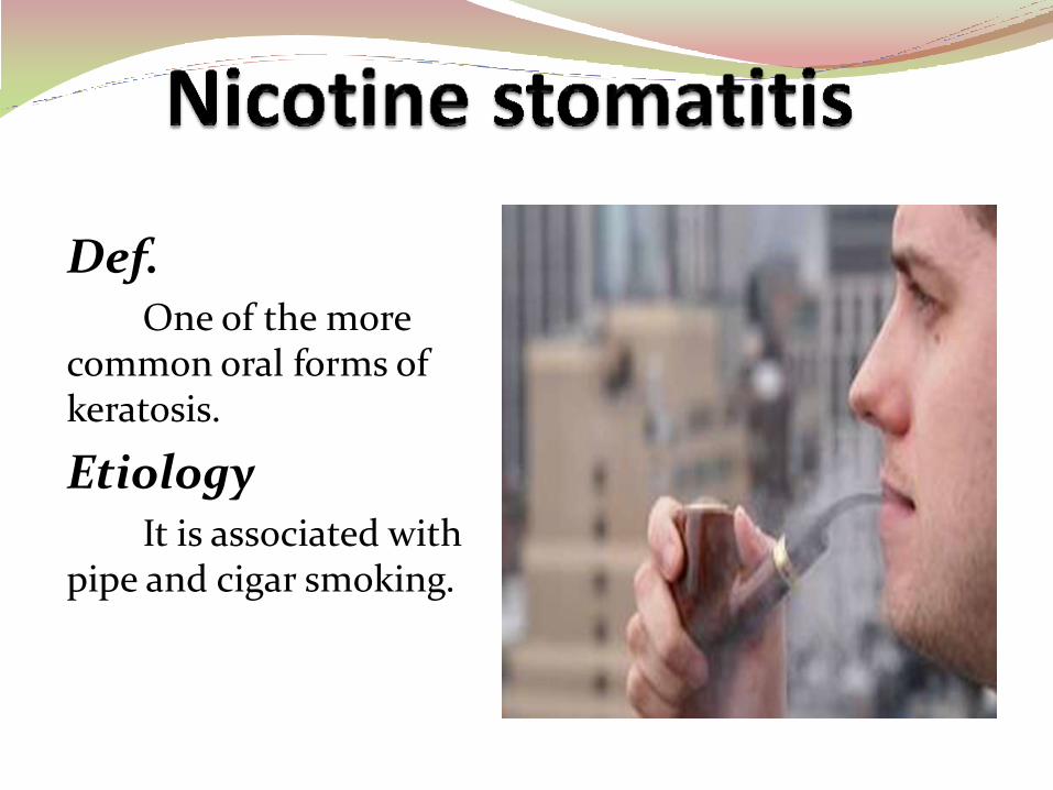
Def. One of the more common oral forms of keratosis.
Etiology It is associated with pipe and cigar smoking.
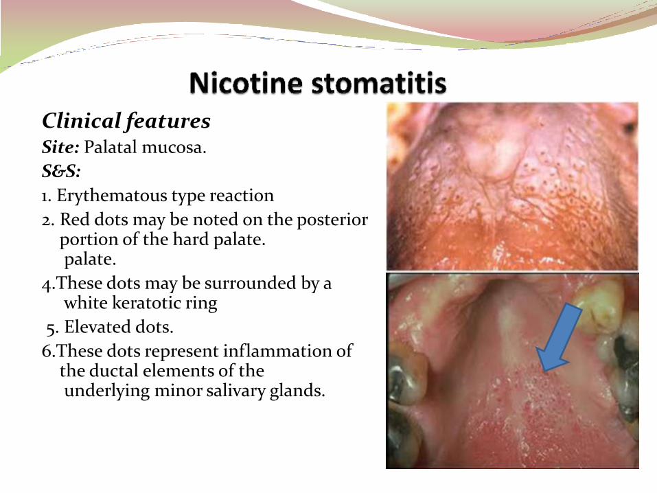
Clinical features Site: Palatal mucosa.
S&S:
1. Erythematous type reaction
2. Red dots may be noted on the posterior portion of the hard palate. palate.
4.These dots may be surrounded by a white keratotic ring
5. Elevated dots.
6.These dots represent inflammation of the ductal elements of the underlying minor salivary glands.

Histopathologic features
1. Orthokeratosis.
2. Acanthosis(Thick epithelium)
3. In minor salivary glands, excretory
ducts may show squamous
metaplasia
4.The glandular elements contain
chronic inflammatory cells.
Hairy leukoplakia

1.An opportunistic infection relates to Epstein-
Barr virus .
2.It is related to AIDS patients and in patients
with other forms of immunosuppression
3. male homosexuals.

Site : along lateral margins of tongue and may extend into dorsal surface.
S&S: - asymptomatic
- It may unilateral or bilateral
- folded flat
- plaque like lesion
- corrugated hairy like projections


1. Immunohistochemical staining technique using
anti-viral antibodies .
2. Ultrastructural study using electrone microscope
Differential Diagnosis 1. Idiopathic leukoplakia 2. lichen planus
3. leukoplakia associated with tobacco use
4. frictional keratosis
5. chronic hyperplastic candidiasis
Treatment
antiviral drugs such as Acyclovir

Preneoplastic and neoplastic lesions
Preneoplastic and neoplastic lesions
1. Actinic Chelitis
2. Idiopathic leukoplakia.
Actinic cheilitis
Def.
It represents accelerated tissue
degeneration of the lips, especially the
lower lip
Etiology
Secondary to regular and prolonged exposure to sunlight

Clinical Features
Site: vermilion portion of lips.
S&S:
1.atrophic
2.pale,glossy appearance
3. Fissuring
4.wrinkling at right angles to cutaneous vermilion junction.

Histopathplogic features
1.Atrophic epithelium
2.Hyperkeratosis.
3. Basal cells are generally hyperchromatic in nature.
4.Basophilic change of submucosa (lamina propria).
5.Telangiectasia
Prognosis
Development of carcinoma at this site.
Treatment
• No treatment.
Other white lesions
Other white lesions
1.Geographic tongue
2.Lichen planus
3.Lupus erythematouss
Def.
An asymptomatic, elongated, erythematous patch of atrophic mucosa
of the mid-dorsal surface of the tongue because of a chronic C. albicans
infection.
Etiology 1. Emotional stress
2. Fungal infection
3. Bacterial infection.
4. It is associated with several conditions as: psoriasis, seborrheic dermatitis, and
Reiter's syndrome.
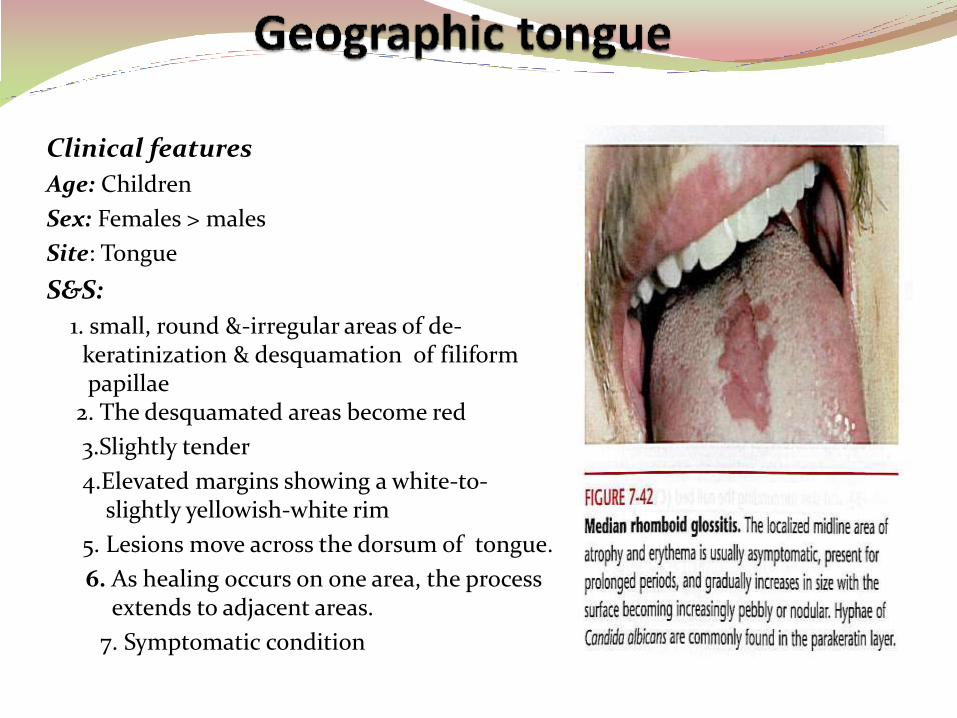
Clinical features
Age: Children
Sex: Females > males
Site: Tongue
S&S:
1. small, round &-irregular areas of de- keratinization & desquamation of filiform papillae 2. The desquamated areas become red
3.Slightly tender
4.Elevated margins showing a white-to- slightly yellowish-white rim
5. Lesions move across the dorsum of tongue.
6. As healing occurs on one area, the process extends to adjacent areas.
7. Symptomatic condition

Histpatholoqic feature
1. Reduced in number of filiform papillae
2. Hyperkeratosis and some acanthosis of the margins
3.Closer to the central portion of the lesion, corresponding to the
erythematous areas, there is often loss of superficial parakeratin, with
significant migration of polymorphic leukocytes and lymphocytes into
epithelium.
4. The leukocytes noted within micro-abscesses near the surface.
5. An inflammatory cell infiltrate within the underlying lamina
propria, consisting chiefly of neutrophils, lymphocytes and plasma
cells.
Differential diagnosis
• Candidiasis.
• Leukoplakia.
• Lichen planus.
• Lupus erythematosis.
Treatment
• Not required as condition is self-limited.
• Re-assuring of the patient that this condition is totally benign
Lichen planus
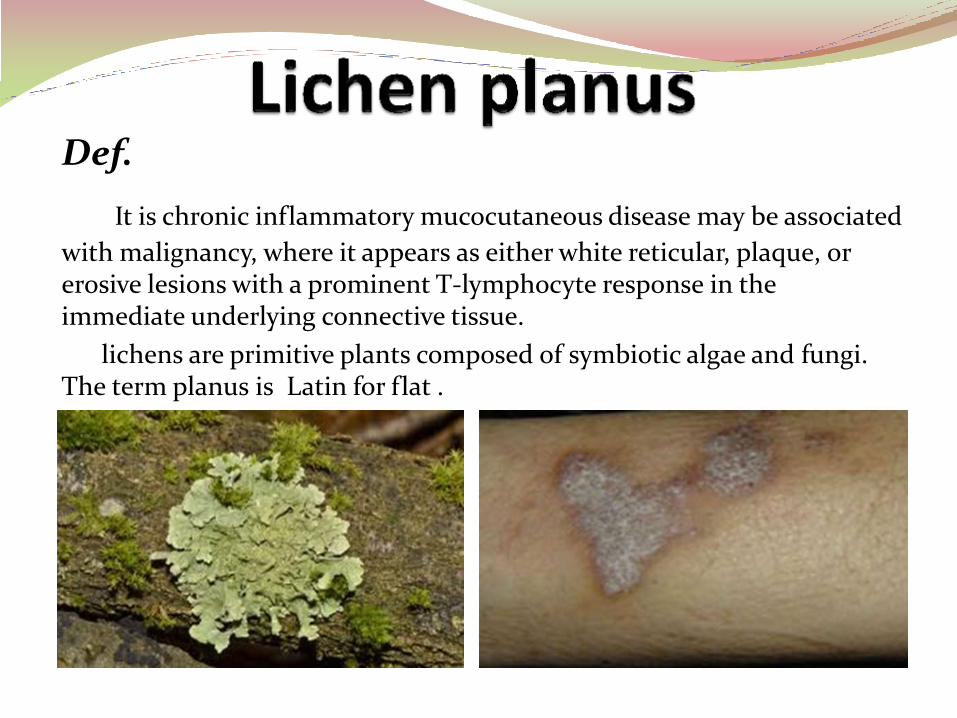
Def.
It is chronic inflammatory mucocutaneous disease may be associated
with malignancy, where it appears as either white reticular, plaque, or erosive lesions with a prominent T-lymphocyte response in the immediate underlying connective tissue.
lichens are primitive plants composed of symbiotic algae and fungi. The term planus is Latin for flat .
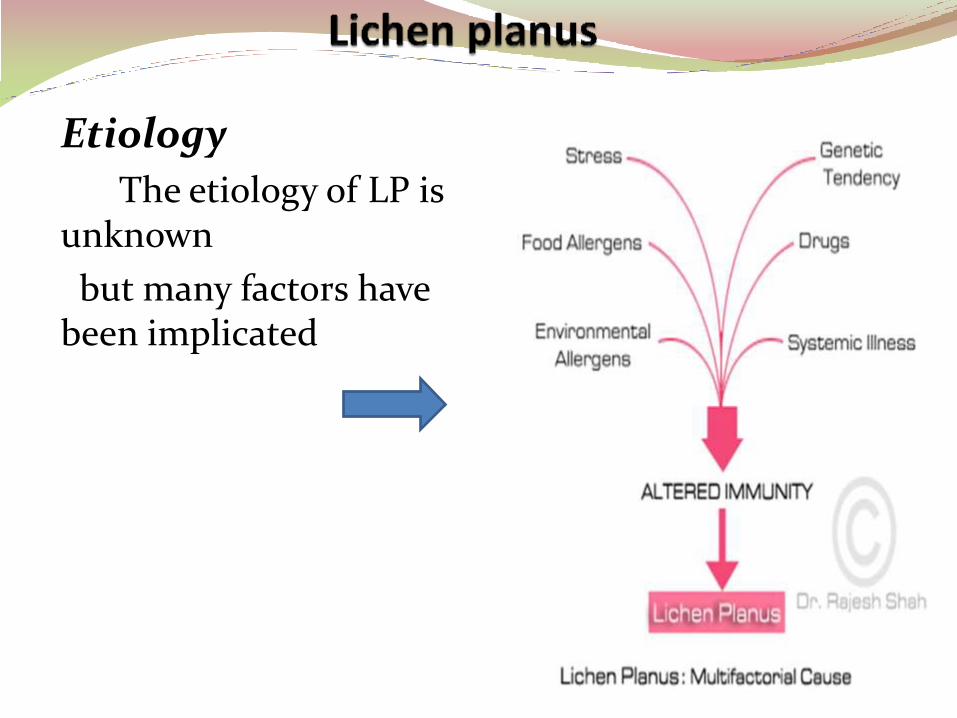
Etiology
The etiology of LP is unknown
but many factors have been implicated
Etiology
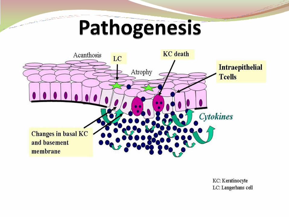
Epithelial basal cells are the primary target in
lichen planus (LP).
The mechanism of basal cell damage appears to
be related to a cell-mediated immune process
involving Langerhans' cell, T-!ymphocytes, and
macrophages

Langerhans' cells contact and "recognize" an antigen
Langerhans' cells process and present appropriate antigenic determinants to T-lymphocytes
T-lymphocytes attracted to the area by Langerhans / macrophages lymphokines known as "interleukin-1" (IL-1).
Stimulates
IL-1 produce IL-2 T -cell proliferation.
T-lymphocytes
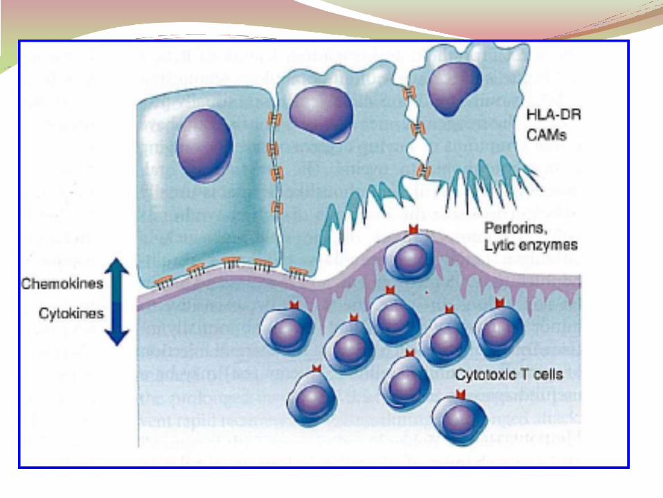
Activated lymphocytes are subsequently cytotoxic for basal cells & secrete gamma- interferon.

Gamma-interferon induces keratinocytes to express the Class II histocompatibility antigens (HLA-DR) & Lymphocytes normally expressing HLA-DR antigens
HLA-DR increase rate of differentiation of keratinocytes with formation of a thickened surface (This latter feature is
seen clinically as "white lesion") & explain the lymphocyte attraction and contact to the epithelium
During this contact, inappropriate epithelial antigenic information may be transferred from Langerhans' cells and macrophages to lymphocytes because of the HLA-DR linkage
With this mechanism, self-antigens may be recognized as "foreign", resulting in an "autoimmune" response s
keratinocytes, demonstrating antigens on basal cell surface that are structurally similar to foreign antigens and are recognized by host T-lymphocytes
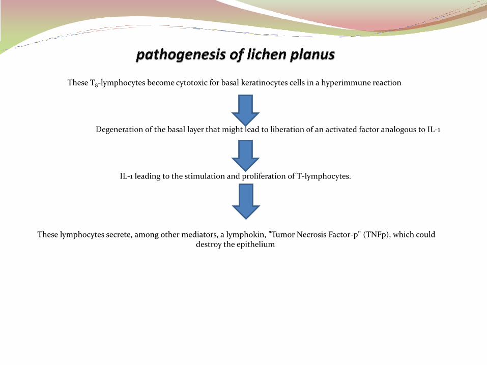
These T8-lymphocytes become cytotoxic for basal keratinocytes cells in a hyperimmune reaction
Degeneration of the basal layer that might lead to liberation of an activated factor analogous to IL-1
IL-1 leading to the stimulation and proliferation of T-lymphocytes.
These lymphocytes secrete, among other mediators, a lymphokin, "Tumor Necrosis Factor-p" (TNFp), which could destroy the epithelium

1.Langerhans cells and macrophages produce IL-1 .
2.IL-1 attracts and stimulates T helper (CD4) cells to produce IL-2 .
3.IL-2 causes proliferation and activation of T cytotoxic (killer)
(CD8) cells .
4. Activated T cells secretes gamma –interferon which induces
keratinocytes to express HLA-DR (class II histocompatibility antigens)
5.Normally lymphocytes express HLA-DR but now ,
keratinocytes expressing HLA-DR also
6.Linkage of these HLA-DR lead to improper epith antigenic
information and basal epith cells recognized as foreign body
by T cells and stimulate autoimmune response as T cells become cytotoxic for basal cells .
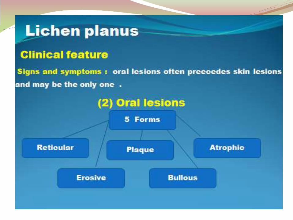

Oral manifestations The most common type
Site: 1. buccal mucosa.
2. tongue and less frequently on
gingiva and the lips, or they
may occur anywhere
S&S: - Presence of numerous
interlacing keratotic lines or striae (the
so-called "Wickham's Striae") that
produce lacy pattern.
- Symmetrical fashion

Site: 1.over the dorusm of
tongue
2.buccal mucosa
S&S:
1.It resembles leukoplakia
2. Elevated plaques
3. smooth surface.

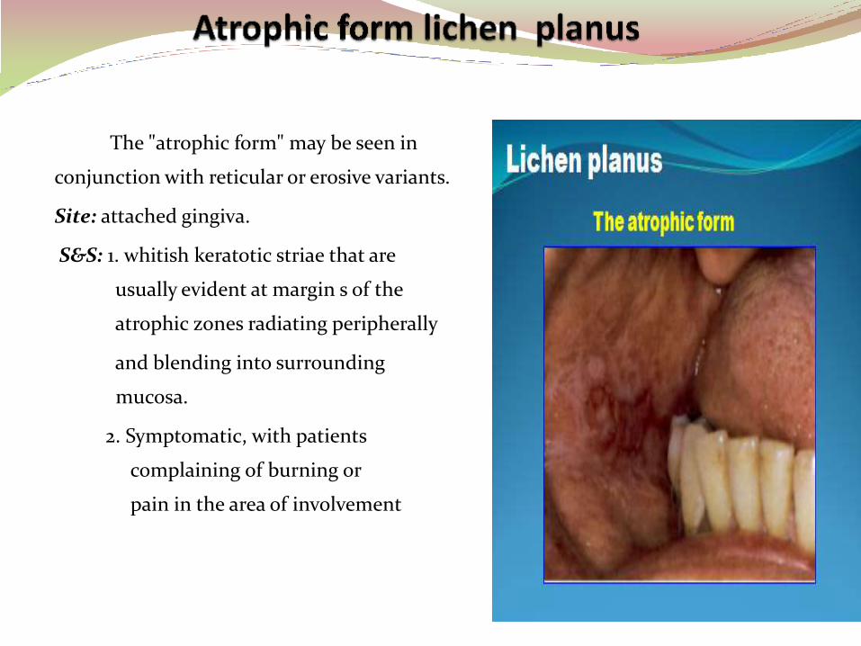
The "atrophic form" may be seen in
conjunction with reticular or erosive variants.
Site: attached gingiva.
S&S: 1. whitish keratotic striae that are
usually evident at margin s of the
atrophic zones radiating peripherally
and blending into surrounding
mucosa.
2. Symptomatic, with patients
complaining of burning or
pain in the area of involvement

Erosive form lichen planus
S&S: 1. Granular surface
2. Brightly erythematous
Careful examination usually
demonstrates a keratotic
component, generally peripheral to
the site of erosion, with either
reticular or finely radiating
keratotic striae.

Site: Buccal mucosa, especially in the posterior and inferior regions adjacent to the second and third
molars & lateral margin of the tongue. Rarely, gingiva and along the inner aspect of the lips.
S&S:
The bullae or vesicles range from a few millimeters to several centimeters in diameter.
2. Bullae are short-lived and, upon rupturing, leave an ulcerated, extremely painful surface

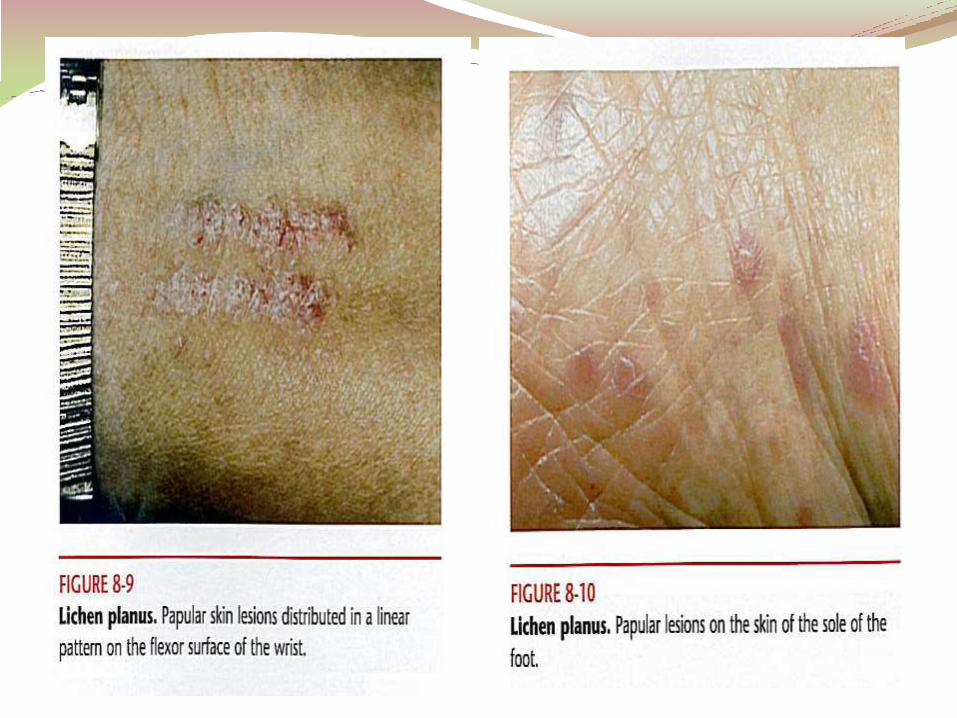
Skin lesions
1. Small, violaceous, polygonal, flat-topped
papules with a predilection for the flexor
surfaces.
2. Cutaneous lesions are noted in approximately
20 to 60% of patients presenting with oral
lichen planus.

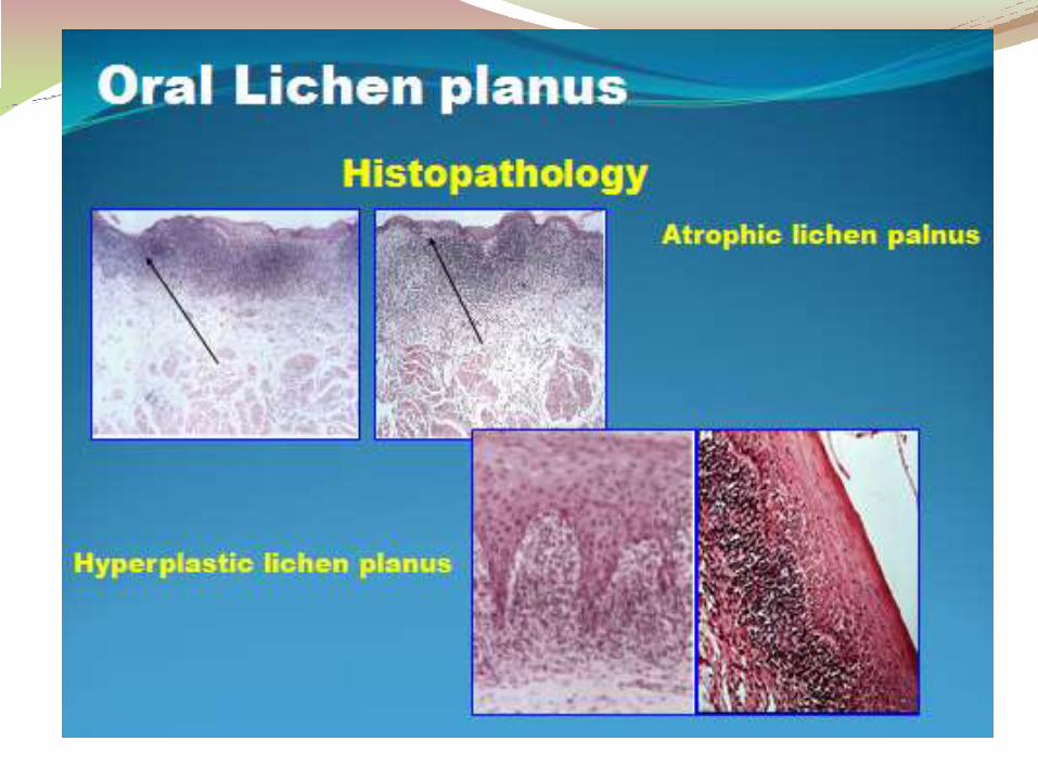
Histopathoiogic features reticular form
1. hyperorthokeratosis or hyperparakeratosis.
2. Variable degrees of acanthosis may be seen.
3. Liquefaction of basal layer to the extent of a near-total absence of basal cells.
4.Destruction of the epithelial-connective tissue interface is noted
5. lymphocyic band pattern found subepithelially in the lamina propria.
6. increased numbers of Langerhans' cells within the epithelium (as demonstrated with mmunohistochemistry).
7. Discrete eosinophilic ovoid bodies representing necrotic keratinocytes are occasionally noted at the basal cell level or within the surrounding
inflammatory cell infiltrate. Colloid (or the so-called "Civatte Bodies"). 8. Eosinophilic band adjacent to the basement membrane zone, often between the lymphocytic infiltrate and the epihtelial ells are also present.

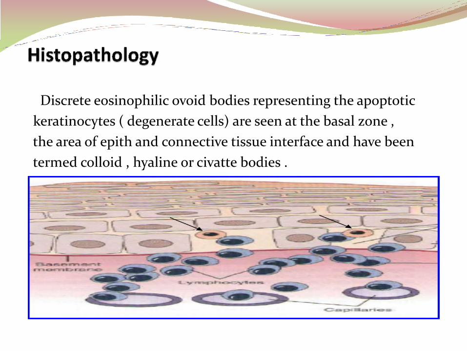
Discrete eosinophilic ovoid bodies representing the apoptotic
keratinocytes ( degenerate cells) are seen at the basal zone ,
the area of epith and connective tissue interface and have been
termed colloid , hyaline or civatte bodies .

A thin band of eosinophilic condensation eosinophilic
band may be seen at the junction of epith and connective tissue
and it may contain IgM and fibrin .
Presence of a dense band of inflammatory cells lymphocytic
band just beneath the epith / connective tissue junction .
The inflam cells are almost entirley lymphocytes
(T-lymphocytes) and macrophages
No significant degree of epith atypia .


Differential diagnosis 1.Atrophic candidiasis
2.leukoplakia
3.squamous cell carcinoma
4. Drug eruption
N.B
Erosive atrophic lichen planus affecting the attached gingiva must be differentiated from "circatricial pemphigoid
Treatment and prognosis • Topical and systemic corticosteroids are useful.
• Vitamin A (retinoids) has been used.
Non-epithelial white yellow lesions

Mucosal burns

Etiology
Chemical burns
Topical application of chemicals such as aspirin or caustic
agents to the mucosa.
Thermal Burn
It associated with sticky hot foods that adhere to the palate.
Electric burn
Electric
Clinical features chemical burns 1. localized mild erythema in case of short-term exposure to agent capable of inducing tissue necrosis. 2. Coagulation necrosis is more likely to occur as the concentration of the offending agent increases. 3. White slough or membrane. 4. Beneath the membrane, there will be a friable & painful surface that will bleed easily upon mobilization. Thermal Burn Site : hard palatal mucosa Features: as chemical burn
Electric burn 1. It is potentially quite serious because they are more destructive. 2. Tissue damage, followed by scarring and reduction in the size of the oral opening.
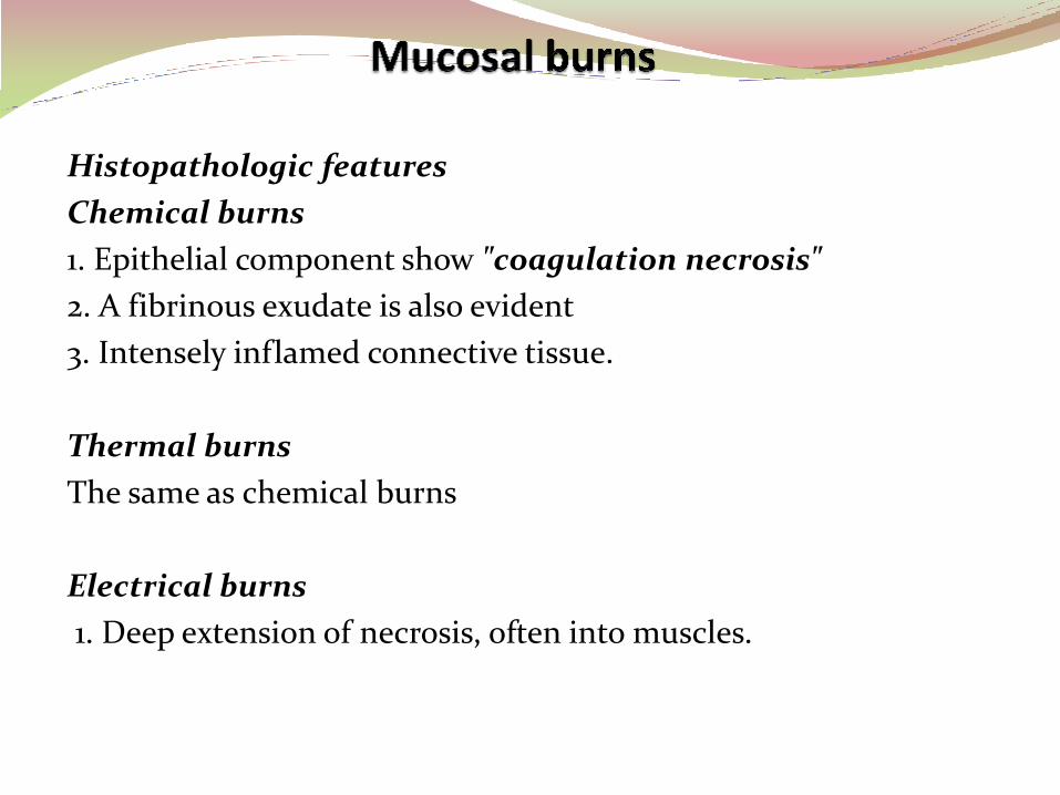
Histopathologic features
Chemical burns
1. Epithelial component show "coagulation necrosis"
2. A fibrinous exudate is also evident
3. Intensely inflamed connective tissue.
Thermal burns
The same as chemical burns
Electrical burns
1. Deep extension of necrosis, often into muscles.

Differential diagnosis
-- Acute necrotizing ulcerative gingivitis (ANUG).
In the absence of history of use of chemical or
thermal offender, ANUG must be included

Fordyace's granules

Def.
It represents ectopic sebaceous
glands or sebaceous choristomas (normal tissue in an abnormal site).
It is seen in approximately 80%of individuals).
Etiology
It is developmental in origin

Clinical features
Site: buccal mucosa, vermilion border of upper lip
Age : postpubertal.
S&S: 1.Asymptomatic.
2.multiple
3.symmetrically distributed
Histopathologic features
• Superficial located lobules of sebaceous glands are aggregated
around or adjacent to excretory ducts
• These ducts contain sebaceous and keratinous debris
Candidiasis


Def.
A clinical form of C.albicans infection that consists of
creamy, loose patches of desquamative epithelium
containing numerous matted mycelia over an
erythematous mucosa that is easily removed, common in
patients with more severe predisposing factors.

Predisposing factors
1. Local disturbance or systemic illness.
2. Antibiotic
3. Corticosteroid
4. Immunosuppressive drug therapy
5. Diabetes mellitus
6. Anaemia
7. Blood dyscrasias such as leukaemia
8. Advanced malignancy
9. Immunodefieicney states

Clinical features
Age: > 5 % of newborn infants & 10
% of elderly debilitated patients
Site: any mucosal surface of mouth
S&S: 1. Thick white coating (pseudo
membrane) can be
wiped away to leave a red,
raw & bleeding base
2. Variation in size from small
patches to confluent
lesions covering a wide
area.
Histological examination
1.Hyperplasitc epithelium with the superficial layers infiltrated both by candidal hyphae and spores, and by inflammatory cells which are predominantly neutrophil leucocytes.
2.The neutrophils may accumulate to form micro-abscesses.
3.Intense inflammatory infiltrate and oedema at the junction between the superficial infected layer (the pseudomembrane) and the deeper epithelium.
4. Infiltration of the lamina propria by acute and chronic inflammatory cells.
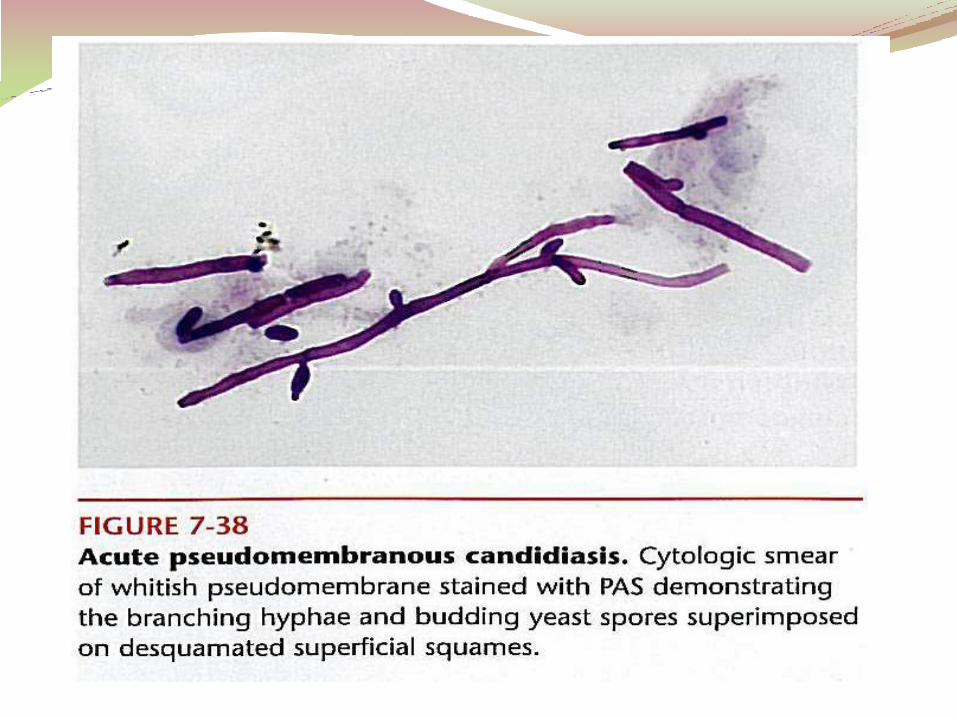
5.The candidal hyphae appear as weakly basophilic, thread-like structures with haematoxylin and esosin staining, but are seen much more clearly after special staining such as in periodic acid Schiff (PAS) preparations.
6.Examination of a smear made of the pseudomembrane shows necrotic material and leucocytes with epithelial cells partly matted together by candidal spors and hyphae.

Thank you