New Method for Antibiotic Susceptibility Testingaac.asm.org/content/2/2/51.full.pdf · NewMethodfor...
6
ANTIMICROBIAL AGENTS AND CHEMOTHERAPY, Aug. 1972, p. 51-56 Copyright © 1972 American Society for Microbiology Vol. 2, No. 2 Printed in U.S.A. New Method for Antibiotic Susceptibility Testing G. N. ROLINSON AND ELIZABETH J. RUSSELL Beecham Research Laboratories, Chemotherapeutic Research Centre, Betchworth, Surrey, England Received for publication 4 April 1972 The most widely used method for routine antibiotic susceptibility testing of clinical isolates of bacteria is the paper-disc method. This has the advantage of simplicity, but to obtain reliable results considerable care is required both in the standardization of the procedure and in the interpretation of the zones of inhibition. A new susceptibility test method is described in this report which retains the sim- plicity of the paper-disc method but which enables organisms to be tested directly for susceptibility to known concentrations of antibiotic in agar. Organisms may be tested against a single concentration of antibiotic or, altematively, a range of con- centrations may be used to determine the minimal inhibitory concentration. The method utilizes stable pre-prepared materials and is not appreciably more time con- suming than the conventional disc method. The two principal types of test used for routine antibiotic susceptibility testing of bacteria in clin- ical laboratories are the serial dilution test and the disc test. In the serial dilution test, doubling dilutions of antibiotic are prepared in a liquid or solid medium which is then inoculated with the test organism. After incubation, the minimal in- hibitory concentration is determined to the near- est dilution used in the series. This test is generally recognized as being the most satisfactory method of determining the antibiotic susceptibility of bac- teria, but since it is time-consuming most labora- tories are unable to use it as a routine procedure. In the disc test, paper discs impregnated with anti- biotic are placed on the surface of an agar plate inoculated with the test organism, and after incu- bation susceptibility is determined by observing the size of the resulting zone of inhibition sur- rounding each disc. This method has the advan- tage of simplicity, but for a given antibiotic disc the size of the zone of inhibition is markedly influ- enced by a number of variables including inocu- lum size, depth of agar, conditions of incubation, and medium composition. Unless these variables are carefully controlled, the results obtained may be misleading, and a number of surveys have shown that the results of routine susceptibility testing are quite frequently unreliable (2, 5, 6). Recommendations for the standardization and interpretation of disc susceptibility testing have been made, notably by Ericsson (3), Bauer et al. (1), and Ericsson and Sherris (4), but these recom- mendations involve such rigid control of the test conditions that the simplicity of the disc test is to some extent lost and many laboratories may be unable to adopt these recommendations in full. 51 In an attempt to overcome some of the limita- tions of the existing procedures, a new suscepti- bility test method is described in this report which combines the simplicity of the disc test with the quantitative results obtained by serial dilution. In this method, organisms are tested for susceptibil- ity to known concentrations of antibiotic, thus avoiding completely the question of interpretation of a zone of inhibition. The antibiotic concentra- tions are achieved by placing papers impregnated with antibiotic onto the surface of small volumes of agar contained in a plastic tray and allowing the antibiotic to diffuse from the paper into the agar. The experimental work has shown that the antibiotic diffuses almost completely into the agar and becomes almost uniformly distributed throughout the agar in a few hours. After the dif- fusion period, the papers are removed, and the agar surface is inoculated with the test organism. After overnight incubation, bacterial growth or inhibition of growth can be observed. In this way, it is possible to test directly for susceptibility to particular concentrations of antibiotic by a pro- cedure which is not significantly more time-con- suming than the paper-disc method. MATERIALS AND METHODS Diffusion of antibiotic through agar. Experiments were carried out to determine the time required for different antibiotics to diffuse from paper and become uniformly distributed throughout a layer of agar 3 mm in depth. Squares of Whatman no. 1 filter paper, 10 by 10 mm, were impregnated with known amounts of antibiotic by applying 0.02-ml volumes of the appro- priate antibiotic solutions. After drying at 37 C for 30 min, the papers were placed on the upper surface of blocks of agar 10 by 10 by 3 mm (Blood Agar Base on July 22, 2018 by guest http://aac.asm.org/ Downloaded from
Transcript of New Method for Antibiotic Susceptibility Testingaac.asm.org/content/2/2/51.full.pdf · NewMethodfor...
ANTIMICROBIAL AGENTS AND CHEMOTHERAPY, Aug. 1972, p. 51-56Copyright © 1972 American Society for Microbiology
Vol. 2, No. 2Printed in U.S.A.
New Method for Antibiotic Susceptibility TestingG. N. ROLINSON AND ELIZABETH J. RUSSELL
Beecham Research Laboratories, Chemotherapeutic Research Centre, Betchworth, Surrey, England
Received for publication 4 April 1972
The most widely used method for routine antibiotic susceptibility testing ofclinical isolates of bacteria is the paper-disc method. This has the advantage ofsimplicity, but to obtain reliable results considerable care is required both in thestandardization of the procedure and in the interpretation of the zones of inhibition.A new susceptibility test method is described in this report which retains the sim-plicity ofthe paper-disc method but which enables organisms to be tested directly forsusceptibility to known concentrations of antibiotic in agar. Organisms may betested against a single concentration of antibiotic or, altematively, a range of con-centrations may be used to determine the minimal inhibitory concentration. Themethod utilizes stable pre-prepared materials and is not appreciably more time con-suming than the conventional disc method.
The two principal types of test used for routineantibiotic susceptibility testing of bacteria in clin-ical laboratories are the serial dilution test andthe disc test. In the serial dilution test, doublingdilutions of antibiotic are prepared in a liquid orsolid medium which is then inoculated with thetest organism. After incubation, the minimal in-hibitory concentration is determined to the near-est dilution used in the series. This test is generallyrecognized as being the most satisfactory methodof determining the antibiotic susceptibility of bac-teria, but since it is time-consuming most labora-tories are unable to use it as a routine procedure.In the disc test, paper discs impregnated with anti-biotic are placed on the surface of an agar plateinoculated with the test organism, and after incu-bation susceptibility is determined by observingthe size of the resulting zone of inhibition sur-rounding each disc. This method has the advan-tage of simplicity, but for a given antibiotic discthe size of the zone ofinhibition is markedly influ-enced by a number of variables including inocu-lum size, depth of agar, conditions of incubation,and medium composition. Unless these variablesare carefully controlled, the results obtained maybe misleading, and a number of surveys haveshown that the results of routine susceptibilitytesting are quite frequently unreliable (2, 5, 6).Recommendations for the standardization andinterpretation of disc susceptibility testing havebeen made, notably by Ericsson (3), Bauer et al.(1), and Ericsson and Sherris (4), but these recom-mendations involve such rigid control of the testconditions that the simplicity of the disc test is tosome extent lost and many laboratories may beunable to adopt these recommendations in full.
51
In an attempt to overcome some of the limita-tions of the existing procedures, a new suscepti-bility test method is described in this report whichcombines the simplicity of the disc test with thequantitative results obtained by serial dilution. Inthis method, organisms are tested for susceptibil-ity to known concentrations of antibiotic, thusavoiding completely the question of interpretationof a zone of inhibition. The antibiotic concentra-tions are achieved by placing papers impregnatedwith antibiotic onto the surface of small volumesof agar contained in a plastic tray and allowingthe antibiotic to diffuse from the paper into theagar. The experimental work has shown that theantibiotic diffuses almost completely into the agarand becomes almost uniformly distributedthroughout the agar in a few hours. After the dif-fusion period, the papers are removed, and theagar surface is inoculated with the test organism.After overnight incubation, bacterial growth orinhibition of growth can be observed. In this way,it is possible to test directly for susceptibility toparticular concentrations of antibiotic by a pro-cedure which is not significantly more time-con-suming than the paper-disc method.
MATERIALS AND METHODSDiffusion of antibiotic through agar. Experiments
were carried out to determine the time required fordifferent antibiotics to diffuse from paper and becomeuniformly distributed throughout a layer of agar 3 mmin depth. Squares ofWhatman no. 1 filter paper, 10 by10 mm, were impregnated with known amounts ofantibiotic by applying 0.02-ml volumes of the appro-priate antibiotic solutions. After drying at 37 C for 30min, the papers were placed on the upper surface ofblocks of agar 10 by 10 by 3 mm (Blood Agar Base
on July 22, 2018 by guesthttp://aac.asm
.org/D
ownloaded from
ROLINSON AND RUSSELL
no. 1, Oxoid). At suitable time intervals over a periodof several hours at toom temperature, the papers wereremoved from two replicate blocks, and the distribu-tion of the antibiotic was determined as follows.
Residual antibiotic in the papers after removal fromthe agar blocks was assayed by placing the papers onagar seeded with a suitable test organism and measur-ing the inhibition zones obtained after incubation.Standard lines were obtained by applying knownamounts of antibiotic to 10-mm squares of paper andplacing these on the same assay plates. After incuba-tion, regression lines were prepared by plotting thesize of the inhibition zones against the quantity ofdrug, and from these lines the residual amounts ofantibiotic in the papers could be calculated.The antibiotic concentrations at the upper and
lower surfaces of the agar block were determined byapplying 10-mm squares of drug-free Whatman no. 1paper to both surfaces of the block and allowing themto absorb liquid from the agar surface. By this means,a standard volume of liquid was obtained in which theantibiotic concentration was representative of thatpresent at the agar surface. After 15 sec of contact, thepapers were removed from the blocks and placed onan agar assay plate seeded with a suitable test organ-ism. After incubation, the resulting zones of inhibitionwere used to calculate the antibiotic concentration byreference to regression lines. These were obtained byplacing 10-mm squares of filter paper on the surface ofsimilar blocks of agar containing known concentra-tions of antibiotic, transferring these to the assayplate and plotting the size of the resulting zones ofinhibition against drug concentration.
In the experiments shown in Table 1, papers con-taining 100 j,g of antibiotic were used. The assay or-ganisms used were Bacillus subtilis for benzylpenicillin,ampicillin, methicillin, cloxacillin, novobiocin, vanco-mycin, fusidic acid, and cephaloridine; Staphylococcusaureus for tetracycline, streptomycin, erythromycin,gentamicin, lincomycin, andkanamycin; and Bordetellabronchiseptica for polymyxin. Staphylococcus aureusand Sarcina lutea were used to assay the diffusion ofbenzylpenicillin when applied at 10 and 1 ,ug, respec-tively, and Bacillus cereus was used to assay the diffu-sion of 10 Ag of tetracycline.
Minimal inhibitory concentrations. Minimal inhibi-tory concentrations of antibiotics were determined bythe conventional method of serial dilution in agar, andthe results were compared with the values obtained byuse of the diffusion method described in this report.At the time these particular experiments were carriedout, a suitable plastic tray of the type shown in Fig.2-5 was not available; to obtain wells of a suitable sizewhich could be filled with agar, a sheet of sterile sili-cone rubber 3 mm thick containing 25 holes 13 mm indiameter was pressed firmly onto the surface of a ster-ile plate of glass cut to fit inside a 10-cm square plasticpetri dish. The wells so formed were then filled withagar flush with the surface of the rubber. Paper discscontaining known amounts of antibiotic were placedon the surface of the agar wells, and diffusion was al-lowed to take place at 37 C for 3 hr. The discs wereprepared by dropping 0.02-ml volumes of antibioticsolutions of appropriate concentration onto sterile
12-mm discs of Whatman no. 1 filter paper which werethen dried at 37 C for 30 min. The quantity of antibi-otic on each disc was that required to give the desiredagar concentration, assuming complete distributionthroughout the agar after the period of diffusion. Ex-perimental work had shown that diffusion of the anti-biotic from the paper into the agar was almost com-plete, and, since the volume of agar in the wells wasknown, the appropriate quantity of antibiotic on thedisc could readily be calculated. After the period ofdiffusion, the discs of paper were removed and the agarsurface was inoculated by use of a swab.
In the serial dilution test, the minimal inhibitoryconcentrations were determined by adding 2-ml vol-umes of the appropriate concentrations of antibioticto 18-ml volumes of cooled molten agar which werethen poured into petri dishes. The plates were dried at37 C and inoculated with the test organisms by use ofan automatic replicator delivering an inoculum of ap-proximately 0.003 ml. In both the serial dilution testand the diffusion test, Blood Agar Base (Oxoid) wasused, except in the case of sulfamethoxazole, for whichD.S.T. agar (Oxoid) containing 5% lysed horse bloodwas employed. In both tests, overnight nutrient brothcultures of the test organisms were used as inoculum,diluted as indicated in Table 2.
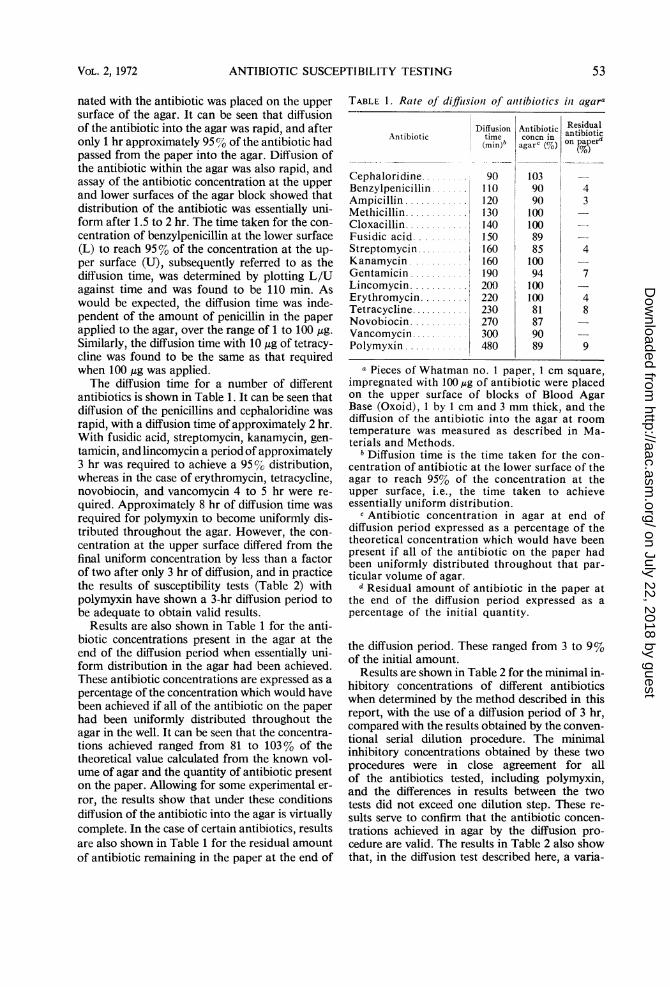
RESULTS AND DISCUSSIONResults are shown in Fig. 1 for the rate of dif-
fusion of benzylpenicillin in a block of agar 3 mmthick at room temperature, when paper impreg-
1ooo0
0)
C
C
0)(3
100 _
80 aC
60
Ec
0)
40 -C
2
20 0
20 *105 1.0 1-5 2-0 2-5
Time (hours)
FIG. 1. Diffusion ofbenzylpenicillin from paper intoagar 3 mm in depth. Squares of filter paper (I cm)containing 100 ,.g of benzylpenicillin were applied tothe surface ofblocks ofagar I by I cm and 3 mm deep.After intervals of time, the papers were removed andthe antibiotic concentration present at the upper andlower surfaces of the agar block was assayed as de-scribed in Materials and Methods. The amount ofantibiotic retained in the papers was also determined.Symbols: *, concentration of penicillin in the uppersurface of agar; 0, concentration of penicillin in thelower surface of agar; O, penicillin remaining in thefilterpaper.
52 ANTIMICROB. AG. CHEMOTHER.
on July 22, 2018 by guesthttp://aac.asm
.org/D
ownloaded from
ANTIBIOTIC SUSCEPTIBILITY TESTING
nated with the antibiotic was placed on the uppersurface of the agar. It can be seen that diffusionof the antibiotic into the agar was rapid, and afteronly 1 hr approximately 95%/ of the antibiotic hadpassed from the paper into the agar. Diffusion ofthe antibiotic within the agar was also rapid, andassay of the antibiotic concentration at the upperand lower surfaces of the agar block showed thatdistribution of the antibiotic was essentially uni-form after 1.5 to 2 hr. The time taken for the con-centration of benzylpenicillin at the lower surface(L) to reach 95% of the concentration at the up-per surface (U), subsequently referred to as thediffusion time, was determined by plotting L/Uagainst time and was found to be 110 min. Aswould be expected, the diffusion time was inde-pendent of the amount of penicillin in the paperapplied to the agar, over the range of 1 to 100 ,g.Similarly, the diffusion time with 10 ,g of tetracy-cline was found to be the same as that requiredwhen 100 Ag was applied.The diffusion time for a number of different
antibiotics is shown in Table 1. It can be seen thatdiffusion of the penicillins and cephaloridine wasrapid, with a diffusion time of approximately 2 hr.With fusidic acid, streptomycin, kanamycin, gen-tamicin, andlincomycin a period of approximately3 hr was required to achieve a 95% distribution,whereas in the case of erythromycin, tetracycline,novobiocin, and vancomycin 4 to 5 hr were re-quired. Approximately 8 hr of diffusion time wasrequired for polymyxin to become uniformly dis-tributed throughout the agar. However, the con-centration at the upper surface differed from thefinal uniform concentration by less than a factorof two after only 3 hr of diffusion, and in practicethe results of susceptibility tests (Table 2) withpolymyxin have shown a 3-hr diffusion period tobe adequate to obtain valid results.
Results are also shown in Table 1 for the anti-biotic concentrations present in the agar at theend of the diffusion period when essentially uni-form distribution in the agar had been achieved.These antibiotic concentrations are expressed as apercentage of the concentration which would havebeen achieved if all of the antibiotic on the paperhad been uniformly distributed throughout theagar in the well. It can be seen that the concentra-tions achieved ranged from 81 to 103% of thetheoretical value calculated from the known vol-ume of agar and the quantity of antibiotic presenton the paper. Allowing for some experimental er-ror, the results show that under these conditionsdiffusion of the antibiotic into the agar is virtuallycomplete. In the case of certain antibiotics, resultsare also shown in Table 1 for the residual amountof antibiotic remaining in the paper at the end of
TABLE 1. Rate of diffiusioni of anttibiotics in agara
Antibiotic
Cephaloridine .........
Benzylpenicillin .......
Ampicillin ............
Methicillin............Cloxacillin............Fusidic acid.Streptomycin..........Kanamycin ...........
Gentamicin ...........
Lincomycin...........Erythromycin .........
Tetracycline...........Novobiocin...........Vancomycin...........Polymyxin ............
Diffusiontime(min)b
90110120130140150160160190200220230270300480
Antibiotic Residualcnn antibioticcoacn in
on paper'4
103909010010089851009410010081879089
43
4
7
48
9
a Pieces of Whatman no. 1 paper, 1 cm square,impregnated with 100 mg of antibiotic were placedon the upper surface of blocks of Blood AgarBase (Oxoid), 1 by 1 cm and 3 mm thick, and thediffusion of the antibiotic into the agar at roomtemperature was measured as described in Ma-terials and Methods.
b Diffusion time is the time taken for the con-centration of antibiotic at the lower surface of theagar to reach 95% of the concentration at theupper surface, i.e., the time taken to achieveessentially uniform distribution.
c Antibiotic concentration in agar at end ofdiffusion period expressed as a percentage of thetheoretical concentration which would have beenpresent if all of the antibiotic on the paper hadbeen uniformly distributed throughout that par-ticular volume of agar.
d Residual amount of antibiotic in the paper atthe end of the diffusion period expressed as apercentage of the initial quantity.
the diffusion period. These ranged from 3 to 9%of the initial amount.
Results are shown in Table 2 for the minimal in-hibitory concentrations of different antibioticswhen determined by the method described in thisreport, with the use of a diffusion period of 3 hr,compared with the results obtained by the conven-tional serial dilution procedure. The minimalinhibitory concentrations obtained by these twoprocedures were in close agreement for allof the antibiotics tested, including polymyxin,and the differences in results between the twotests did not exceed one dilution step. These re-sults serve to confirm that the antibiotic concen-trations achieved in agar by the diffusion pro-cedure are valid. The results in Table 2 also showthat, in the diffusion test described here, a varia-
VOL. 2, 1972 53
on July 22, 2018 by guesthttp://aac.asm
.org/D
ownloaded from
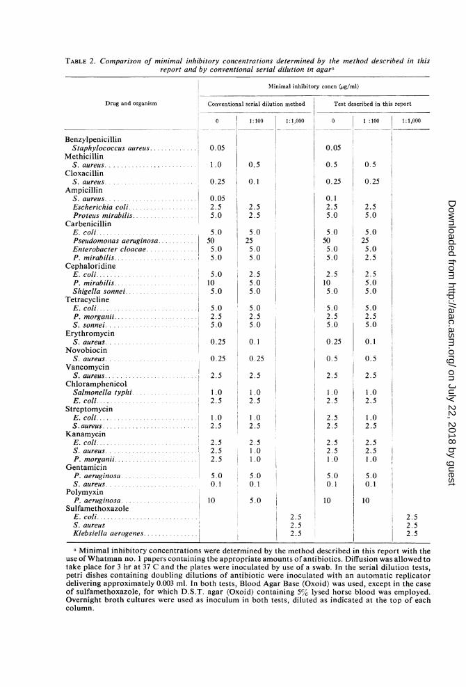
TABLE 2. Comparison of minimal inhibitory concentrations determined by the method described in thisreport and by conventional serial dilution in agara
Minimal inhibitory concn (,ug/ml)
Drug and organism Conventional serial dilution method Test described in this report
0 1:100 1:1,000 0 1:100 1:1,000
BenzylpenicillinStaphylococcus aureus ....... 0.05 0.05
MethicillinS. aureus ......................... 1.0 0.5 0.5 0.5
CloxacillinS. aureus ... 0.25 0.1 0.25 0.25
AmpicillinS. aureus......................... 0.05 0.1Escherichia coli................. 2.5 2.5 2.5 2.5Proteus mirabilis .............. . 5.0 2.5 5.0 5.0
CarbenicillinE. coli........................... 5.0 5.0 5.0 5.0Pseudomontas aeruginosa ...........1 50 25 50 25Enterobacter cloacae .............. 5.0 5.0 5.0 5.0P. mirabilis ................ 5.0 5.0 5.0 2.5
CephaloridineE. coli............ ....... 5.0 2.5 2.5 2.5P. mirabilis .... ..... . 10 5.0 10 5.0Shigella sonnei............... 5.0 5.0 5.0 5.0
TetracyclineE. coli.... .. . 5.0 5.0 5.0 5.0P. morganii...................... 2.5 2.5 2.5 2.5S. sonnei ... 5.0 5.0 5.0 5.0
ErythromycinS. aureus.....0.......0.25 0.1 0.25 0.1
NovobiocinS. aureus......................... 0.25 0.25 0.5 0.5
VancomycinS. aureus i 2.5 2.5
Chloramphenicol 2 2.5Salmonella typhi 1.0 1.0 1.0 1.0E. coli .................... 2.5 2.5 2.5 2.5
StreptomycinE. coli........................... 1.0 1.0 2.5 1.0S.aureus.......................... 2.5 2.5 2.5 2.5
KanamycinE. coli .... ... ..... 2.5 2.5 2.5 2.5S. aureus.. ....... 2.5 1.0 2.5 2.5P. morganii .......... 2.5 1.0 1.0 1.0
GentamicinP. aeruginosa ..................... 5.0 5.0 5.0 5.0S. aureus........... 0.1 0.1 0.1 0.1
PolymyxinP. aeruginosa .......... 10 5.0 10 10
SulfamethoxazoleE. coli.....l... 2.5 2.5S. aureus 2.5 . 2.5Klebsiella aerogenes.............. 2.5 2.5
a Minimal inhibitory concentrations were determined by the method described in this report with theuse of Whatman no. 1 papers containing the appropriate amounts of antibiotics. Diffusion was allowed totake place for 3 hr at 37 C and the plates were inoculated by use of a swab. In the serial dilution tests,petri dishes containing doubling dilutions of antibiotic were inoculated with an automatic replicatordelivering approximately 0.003 ml. In both tests, Blood Agar Base (Oxoid) was used, except in the caseof sulfamethoxazole, for which D.S.T. agar (Oxoid) containing 5% lysed horse blood was employed.Overnight broth cultures were used as inoculum in both tests, diluted as indicated at the top of eachcolumn.
on July 22, 2018 by guesthttp://aac.asm
.org/D
ownloaded from
ANTIBIOTIC SUSCEPTIBILITY TESTING
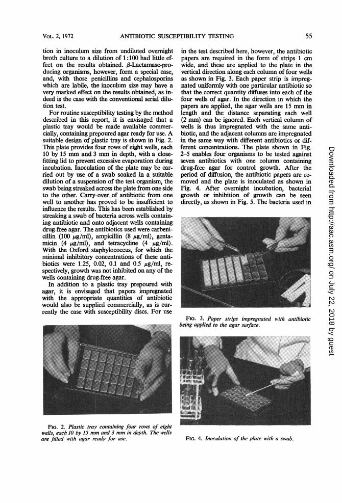
tion in inoculum size from undiluted overnightbroth culture to a dilution of 1 :100 had little ef-fect on the results obtained. (3-Lactamase-pro-ducing organisms, however, form a special case,and, with those penicillins and cephalosporinswhich are labile, the inoculum size may have avery marked effect on the results obtained, as in-deed is the case with the conventional serial dilu-tion test.For routine susceptibility testing by the method
described in this report, it is envisaged that aplastic tray would be made available commer-cially, containing prepoured agar ready for use. Asuitable design of plastic tray is shown in Fig. 2.This plate provides four rows of eight wells, each10 by 15 mm and 3 mm in depth, with a close-fitting lid to prevent excessive evaporation duringincubation. Inoculation of the plate may be car-ried out by use of a swab soaked in a suitabledilution of a suspension of the test organism, theswab being streaked across the plate from one sideto the other. Carry-over of antibiotic from onewell to another has proved to be insufficient toinfluence the results. This has been established bystreaking a swab of bacteria across wells contain-ing antibiotic and onto adjacent wells containingdrug-free agar. The antibiotics used were carbeni-cillin (100 ,ug/ml), ampicillin (8 ,g/ml), genta-micin (4,g/ml), and tetracycline (4 ,ug/ml).With the Oxford staphylococcus, for which theminimal inhibitory concentrations of these anti-biotics were 1.25, 0.02, 0.1 and 0.5 ,ug/ml, re-spectively, growth was not inhibited on any of thewells containing drug-free agar.
In addition to a plastic tray prepoured withagar, it is envisaged that papers impregnatedwith the appropriate quantities of antibioticwould also be supplied commercially, as is cur-rently the case with susceptibility discs. For use
FIG. 2. Plastic tray containing four rows of eightwells, each 10 by 15 mm and 3 mm in depth. The wellsare filled with agar ready for use.
in the test described here, however, the antibioticpapers are required in the form of strips 1 cmwide, and these are applied to the plate in thevertical direction along each column of four wellsas shown in Fig. 3. Each paper strip is impreg-nated uniformly with one particular antibiotic sothat the correct quantity diffuses into each of thefour wells of agar. In the direction in which thepapers are applied, the agar wells are 15 mm inlength and the distance separating each well(2 mm) can be ignored. Each vertical column ofwells is thus impregnated with the same anti-biotic, and the adjacent columns are impregnatedin the same way with different antibiotics or dif-ferent concentrations. The plate shown in Fig.2-5 enables four organisms to be tested againstseven antibiotics with one column containingdrug-free agar for control growth. After theperiod of diffusion, the antibiotic papers are re-moved and the plate is inoculated as shown inFig. 4. After overnight incubation, bacterialgrowth or inhibition of growth can be seendirectly, as shown in Fig. 5. The bacteria used in
FIG. 3. Paper strips impregnatedbeing applied to the agar surface.
with antibiotic
FIG. 4. Inoculation of the plate with a swab.
VOL. 2, 1972 55
on July 22, 2018 by guesthttp://aac.asm
.org/D
ownloaded from

ROLINSON AND RUSSELL
PG Cp Amn05 8 8
Ca Ge T C Ka100 2 4 4
FIG. 5. Inoculated plate after overniight incubation. Test organisms and antibiotics as indicated. Anitibioticconcentrations shown in micrograms per milliliter. PG = penicillin G; Cp = cephaloridine; Am = ampicillin;Ca = carbenicillin; Ge = gentamicin; TC = tetracycline; Ka = kanamycini.
the test shown in Fig. 5 were two strains ofEscherichia coli, a strain of Klebsiella aerogenes,and a strain of Pseudomonas aeruginosa. Theantibiotics used were benzylpenicillin (0.5 ug/ml),cephaloridine (8 ,ug/ml), ampicillin (8 ,ug/ml),carbenicillin (100,g/ml), gentamicin (2 Ag/ml),tetracycline (4 ,g/ml), and kanamycin (4 ,ug/ml).These antibiotics, and the particular concentra-tions, are purely arbitrary and were chosenmerely to illustrate the test. For general use,some agreement would be required as to what theantibiotic concentrations should be, but eachlaboratory could then choose a range of anti-biotics best suited to the particular organismsunder test. The antibiotic strips could provideseven different antibiotics or, if desired, morethan one concentration of a particular antibioticcould be employed and these could be related todosage of the drug, route of administration, andalso the site of infection. Antibiotic combina-tions such as carbenicillin and gentamicin orbenzylpenicillin and aminoglycosides could alsobe tested by using paper strips impregnated withboth drugs.The test described in this report may be used
for routine susceptibility testing against a range ofdifferent antibiotics, or it could be used to deter-mine the minimal inhibitory concentration of a
single antibiotic by using papers providing a rangeof twofold dilutions.The method described here for routine suscepti-
bility testing is not significantly more time-consuming than the conventional disc method
and has certain advantages. First, the test deter-mines susceptibility directly and avoids the needto interpret a zone of inhibition. Second, the re-sults obtained by this method are influenced lessby the test conditions, including inoculum size,than is the case with the disc test. This is becausediffusion of the antibiotic and growth of the testorganism do not proceed simultaneously as theydo in the disc test. The main advantage of themethod described here, however, is that orga-nisms can be tested for susceptibility to knownconcentrations of antibiotic, and these concentra-tions can be chosen in relation to the levels ofantibiotic likely to be achieved in the body.
ACKNOWLEDGMENTS
We are indebted to Peter R. Watt for his help in suggestingand making equipment involved in this work, to R. Sutherlandfor helpful discussion, and to Douglas F. Lawson for the photo-graphs.
LITERATURE CITED
1. Bauer, A. W., W. M. M. Kirby, J. C. Sherris, and M. Turck-1966. Antibiotic susceptibility testing by a standardized singledisk method. Amer. J. Clin. Pathol. 45:493-496.
2. College of Pathologists of Australia. 1968. A survey of anti-biotic sensitivity testing. Med. J. Aust. 2:171-172.
3. Ericsson, H. 1960. Rational use of antibiotics in hospitals.Scand. J. Clin. Lab. Invest. 12:Suppl. 50.
4. Ericsson, H. M., and J. C. Sherris. 1971. Antibiotic sensitivitytesting. Acta Pathol. Microbiol. Scand. Suppl. 217.
5. Report. 1960. A survey of antibiotic sensitivity tests. J. Med.Lab. Technol. 17:133-143.
6. Report. 1965. Report on antibiotic sensitivity test trial orga-nized by the Bacteriology Committee of the Association ofClinical Pathologists. J. Clin. Pathol. 18:1-5.
56
c:
,
ANTIMICROB. AG. CHEMOTHER.
E. colBiI
E.col i10418
Kleb.aerogeqnes
PseL do-moo as
on July 22, 2018 by guesthttp://aac.asm
.org/D
ownloaded from