New families of mesoporous materials - IOPscience
20
Science and Technology of Advanced Materials New families of mesoporous materials To cite this article: Ajayan Vinu et al 2006 Sci. Technol. Adv. Mater. 7 753 View the article online for updates and enhancements. You may also like A review: the utilization of mesoporous materials in wastewater treatment Badria M Al-Shehri, Abd El Rahman S Khder, Sheikha S Ashour et al. - Incorporation of anti-inflammatory agent into mesoporous silica Wilson Rodrigues Braz, Natállia Lamec Rocha, Emerson H de Faria et al. - Recycling of surfactant template in mesoporous MCM-41 synthesis J Y Lai, F Twaiq and L H Ngu - Recent citations Impact of Geometrical Disorder on Phase Equilibria of Fluids and Solids Confined in Mesoporous Materials Henry R. N. B. Enninful et al - Urea incorporated into ordered mesoporous silica for potential cosmetic application Vânia Rodrigues Leite-Silva et al - Laxmi Gond et al - This content was downloaded from IP address 5.44.199.90 on 13/01/2022 at 19:03
Transcript of New families of mesoporous materials - IOPscience

Science and Technology ofAdvanced Materials
New families of mesoporous materialsTo cite this article: Ajayan Vinu et al 2006 Sci. Technol. Adv. Mater. 7 753
View the article online for updates and enhancements.
You may also likeA review: the utilization of mesoporousmaterials in wastewater treatmentBadria M Al-Shehri, Abd El Rahman SKhder, Sheikha S Ashour et al.
-
Incorporation of anti-inflammatory agentinto mesoporous silicaWilson Rodrigues Braz, Natállia LamecRocha, Emerson H de Faria et al.
-
Recycling of surfactant template inmesoporous MCM-41 synthesisJ Y Lai, F Twaiq and L H Ngu
-
Recent citationsImpact of Geometrical Disorder on PhaseEquilibria of Fluids and Solids Confined inMesoporous MaterialsHenry R. N. B. Enninful et al
-
Urea incorporated into orderedmesoporous silica for potential cosmeticapplicationVânia Rodrigues Leite-Silva et al
-
Laxmi Gond et al-
This content was downloaded from IP address 5.44.199.90 on 13/01/2022 at 19:03

ARTICLE IN PRESSThe STAM archive is now available from the IOP Publishing website http://www.iop.org/journals/STAM
1468-6996/$ - se
doi:10.1016/j.st
�CorrespondE-mail addr
ariga.katsuhiko
Science and Technology of Advanced Materials 7 (2006) 753–771
www.elsevier.com/locate/stam
Review
New families of mesoporous materials
Ajayan Vinua,�, Toshiyuki Moria, Katsuhiko Arigab,�
aNano Ionics Materials Group, Fuel Cell Materials Center, National Institute for Materials Science (NIMS), JapanbSupermolecules Group, National Institute for Materials Science (NIMS), 1-1 Namiki, Tsukuba 305-0044, Japan
Received 14 August 2006; received in revised form 6 October 2006; accepted 25 October 2006
Available online 22 December 2006
Abstract
Mesoporous materials have been paid much attention in both scientific researches and practical applications. In this review, we focus
on recent developments on preparation and functionalization of new families of mesoporous materials, especially non-siliceous
mesoporous materials invented in our research group. Replica synthesis is known as the method to synthesize mesoporous materials
composed of various elements using originally prepared mesoporous replica. This strategy has been applied for the syntheses of novel
mesoporous materials such as carbon nanocage and mesoporous carbon nitride. Carbon nanocage has a cage-type structure with huge
surface area and pore volume, which exhibits superior capabilities for biomolecular adsorption. Mesoporous carbon nitride was
synthesized, for first time, by using mixed material source of carbon and nitrogen simultaneously. As a totally new strategy for synthesis
of mesoporous materials, the elemental substitution method has been recently proposed by us. Direct substitution of component
elements in original mesoporous materials, with maintaining structural regularity, provided novel mesoporous materials. According to
this synthetic strategy, mesoporous boron nitride and mesoporous boron carbon nitride have been successfully prepared, for first time. In
addition to these material inventions, hybridization of high functional materials, such as biomaterials, to mesoporous structure has been
also developed. Especially, immobilization of proteins in mesopores was systematically researched, and preparation of peptide-
hybridized mesoporous silica was demonstrated. These new families of mesoporous materials introduced in this review would have high
potentials in future practical applications in wide ranges from electronics and photonics to environmental and medical uses.
r 2006 NIMS and Elsevier Ltd. All rights reserved.
Keywords: Mesoporous material; Template synthesis; Replica synthesis; Elemental substitution; Biomaterial
Contents
1. Introduction . . . . . . . . . . . . . . . . . . . . . . . . . . . . . . . . . . . . . . . . . . . . . . . . . . . . . . . . . . . . . . . . . . . . . . . . . . . . . . . 753
2. Carbon nanocage and mesoporous carbon nitride via replica synthesis . . . . . . . . . . . . . . . . . . . . . . . . . . . . . . . . . . . . . . 756
3. Mesoporous boron nitride and mesoporous boron carbon nitride via elemental substitution method. . . . . . . . . . . . . . . . . 760
4. Biomaterial-hybridized mesoporous materials . . . . . . . . . . . . . . . . . . . . . . . . . . . . . . . . . . . . . . . . . . . . . . . . . . . . . . . . 762
5. Summary . . . . . . . . . . . . . . . . . . . . . . . . . . . . . . . . . . . . . . . . . . . . . . . . . . . . . . . . . . . . . . . . . . . . . . . . . . . . . . . . . 768
Acknowledgment . . . . . . . . . . . . . . . . . . . . . . . . . . . . . . . . . . . . . . . . . . . . . . . . . . . . . . . . . . . . . . . . . . . . . . . . . . . . 769
References . . . . . . . . . . . . . . . . . . . . . . . . . . . . . . . . . . . . . . . . . . . . . . . . . . . . . . . . . . . . . . . . . . . . . . . . . . . . . . . . 769
e front matter r 2006 NIMS and Elsevier Ltd. All rights rese
am.2006.10.007
ing authors. Tel.: +8129 860 4597; fax: +81 29 852 4832.
esses: [email protected] (A. Vinu),
@nims.go.jp (K. Ariga).
1. Introduction
Mesoporous materials with regular geometries havebeen recently paid much attention owing to their greatpotentials in practical applications such as catalysis,adsorption, separation, sensing, medical usage, ecology,
rved.

ARTICLE IN PRESS
Fig. 1. General concept for synthesis of mesoporous silica from micelle template.
A. Vinu et al. / Science and Technology of Advanced Materials 7 (2006) 753–771754
and nanotechnology [1–15]. According to IUPAC classifi-cation, they are defined as porous materials with diameterin the size range of 0.2–2.0 nm. In 1990, Kuroda andcoworkers first reported the preparation of mesoporoussilica with uniform pore size distribution from the layeredpolysilicate kanemite (FSM-16, Folded Sheet Materials)[16,17]. A significant breakthrough in the mesoporousmaterials research has come when Mobil scientists dis-closed the M41S family of materials, which have largeuniform pore structures, high specific surface areas andspecific pore volumes, including hexagonal-MCM-41[18,19], cubic-MCM-48 [20], and lamellar-MCM-50 [21].As illustrated in Fig. 1, the mesoporous materials werebasically prepared through silica formation around tem-plate micelle assemblies followed by template removal byappropriate methods such as calcination.
These pioneering findings were followed by variouskinds of mesoporous materials. For example, hexagonalmesoporous silica (HMS) prepared using neutral amine astemplate possesses slightly disordered hexagonal structureand thicker walls, superior thermal stability upon calcina-tion in air, and a smaller crystallite size, which affordscomplementary textural mesoporosity for improved accessto the framework-confined mesopores [22]. Michigan StateUniversity (MSU-1) synthesized by using polyethyleneoxide (PEO) as a structure directing agent also has adisordered channel structure [23]. This material possesseslarge wall thickness and small particle size with consider-able textural mesoporosity due to pores formed betweenthe relatively small particles. As widely used materials,highly ordered large pore mesoporous silica Santa BarbaraAmorphous-15 (SBA-15) with thicker pore walls and two-
dimensional hexagonal structure, by using amphiphilictriblock-copolymer of poly(ethylene oxide) and poly(pro-pylene oxide) (Pluronic P123) as the structure directingreagent in highly acidic media [24,25]. The pore diametersof the SBA materials are well tunable in the range of5–30 nm, and these materials exhibit higher hydrothermalstability as compared to that of other mesoporousmaterials. Meso cellular form (MCF) materials can beprepared by using triblock-copolymers stabilized oil inwater to result in aerogel-like structure, which offers thebenefits as catalyst supports and separation media [26]. Inaddition to preparation of various mesoporous silicastructures, incorporation of heteroatoms such as Cu, Zn,Al, B, Ga, Fe, Cr, Ti, V and Sn into mesoporous silicaframework has been widely investigated [27–45]. Further-more, it is possible to synthesize mesoporous structures ofthe materials other than silica. Methodology to preparemesoporous silica via the template synthesis is extended topreparation of a various mesoporous metal oxides [46–59]such as TiO2, Ta2O5, Nb2O5, ZrO2, Al2O3, and V2O5 aswell as synthesis of mesoporous aluminophosphate [60–63].Recent researches have revealed that use of silica sources
and templates designed by organic syntheses results inmesoporous materials with unique structural features. Twoexamples are shown in Fig. 2. One of the most uniqueapproaches upon modification of silica source is synthesisof periodic mesoporous organosilicates (PMO) (Fig. 2A).The PMO materials were invented independently by threegroups, Inagaki group [64], Ozin group [65], and Steingroup [66] in 1999. This method uses organic moleculeshaving multiple alkoxysilane groups as silica source andintroduces various organic components in frame-work.

ARTICLE IN PRESS
Fig. 2. Recent examples for modified syntheses of mesoporous silica materials: (A) periodic mesoporous organosilica from various multi-functionalized
silica sources; (B) chiral-shaped mesoporous silica synthesized with chiral surfactant.
A. Vinu et al. / Science and Technology of Advanced Materials 7 (2006) 753–771 755
As the one of the unique finding, Inagaki et al. successfullyprepared crystal-like pore-wall structure in PMO materialfrom benzene-bridged organosilane, 1,4-bis(triethoxysilyl)-benzene, where hydrophilic silicate layers and hydrophobicbenzene layers array alternately [67]. Unlike use ofconventional surfactants like cetyltrimethylammonium saltand triblock-copolymers, several examples of syntheses ofthe mesoporous materials with synthetically designedsurfactant have been recently reported [68–76]. For
example, chirality was first introduced in mesoporousmaterials by using chiraly defined surfactant template(Fig. 2B) [77–79]. A newly synthesized surfactant, N-aceyl-type alanine-based surfactant, was used as structure-directing reagent, resulting in mesoporous silica materialswith regularly twisted rod-like structures with diameter of130–180 nm and length of 1–6mm. In the obtainedstructure, hexagonally aligned mesoscopic channels withdiameter of 2.2 nm wound together in particular direction.

ARTICLE IN PRESSA. Vinu et al. / Science and Technology of Advanced Materials 7 (2006) 753–771756
As briefly summarized above, various approaches havebeen extensively proposed for development of technologiesbased on mesoporous materials mainly with mesoporoussilica and the related mesoporous metal oxide. However,preparation of non-oxide materials with mesoporousstructures has also played important roles especially in21st century, which is virtually initiated from invention ofmesoporous carbon materials. Such trends in researches ofmesoporous materials have successfully created newfamilies of mesoporous materials. In this review, weintroduce recent development of preparation of mesopor-ous materials of non-oxide materials, including mesopor-ous carbon, mesoporous carbon nitride, mesoporous boronnitride, and mesoporous boron carbon nitride. In addition,hybrid materials with mesoporous materials and bio-related structures are also described as a new direction ofresearches on mesoporous materials.
2. Carbon nanocage and mesoporous carbon nitride via
replica synthesis
From the later periods in 1990s, regular carbon materialshave been synthesized using regularly structured templatesuch as zeolite and mesoporous silica [80–84]. Such carbonmaterials with nanoscale pore sizes prepared from periodicinorganic silica templates have been receiving muchattention because of their versatility, and shape-selectivity.These characteristics are expected to be useful forchromatographic separation systems, catalysts, nanoreac-tors, battery electrodes, capacitors, energy-storage devices,and biomedical devices. The porous carbon materials withregular structures are usually obtained by the replicasynthesis (or nano-casting), which is preparation methodby replication of nano-structures to the other materials.For example, Kyotani and coworkers successfully synthe-sized highly regular microporous carbon [85,86]. Mesopor-ous carbon materials structures (CMK-x) were firstreported by Ryoo using sucrose as carbon source andmesoporous silicates template such as MCM-48, SBA-1and SBA-15 as templates and named as CMK-x [87–91].Hyeon et al. reported, independently and somewhat later,the similar approach for the well-ordered mesoporouscarbon materials designated SNU-x [92–94].
Fig. 3A schematically illustrated the synthetic process ofmesoporous carbon materials. In this method, appropriatecarbon sources, such as sucrose, are first impregnated intotemplate structures, followed by solidification and templateremoval, resulting in nano-structured porous carbonmaterials. Structure of template silica has crucial role insuccessful preparation of mesoporous carbon, becausereplicated carbon must have continuous phase to keeptheir structures. For example, mesoporous silica MCM-48in cubic phase is the one of the most appropriate template,because it has bicontinuous pore geometry, to result inrepresentative mesoporous carbon, CMK-1 (Fig. 3A(a)).In contrast, MCM-41 is not appropriate for the replicasynthesis, owing to independent nature of hexagonally
aligned meso-channel in MCM-41. Although SBA-15 hashexagonal array of cylindrical pores as similar to MCM-41,its interconnectivity of the silica channel with microporesleads to preservation of regular carbon structures (CMK-3)even after silica removal (Fig. 3A(b)). Recently, Vinu et al.reported successful tuning of SBA-15 structure uponcontrol of synthetic temperature [95] and applied thepore-tuned SBA-15 replica to synthesize CMK-3 mesopor-ous carbon materials with pore diameter ranging from 3 to6.5 nm [96].Wise selection of mesoporous silica template provides
regular carbon materials with superior textural character-istics. Vinu and coworkers very recently have reportedsynthesis of novel nanocarbon, ‘‘carbon nanocage’’[97–99], through replica synthesis using three-dimensionallarge cage-type face centered cubic mesoporous silicamaterials (KIT-5) [100] as inorganic templates. Image ofthe synthesis of carbon nanocage was illustrated in Fig. 3Bthat, however, shows just rough idea of the obtainedmaterials. The standard synthetic procedure for themesoporous carbon, CMK-3 is not suitable in the case tomake a highly ordered carbon nanocage due to the higherbulk density and lower pore volume of the KIT-5 ascompared to the other mesoporous silicates such as MCM-48 and SBA-15. Appropriate water to silica templateweight ratio (ca. 2.5) in the syntheses was found to be acrucial factor by systematic investigation on syntheticconditions. Upon these initial knowledge, carbon nanocagematerials with different pore diameter were prepared byusing different KIT-5 mesoporous silica synthesized atdifferent temperature (from 100 to 150 1C) as templatesand sucrose as the carbon source. Independently, thesamples were prepared at different sucrose to silica weightratio from 0.45 to 2.0. These experiments revealed thattunability of the pore structures and textural parameters incarbon nanocage materials. The powder XRD pattern ofall the samples show an intense (1 1 1) reflection and abroader (2 0 0) reflection, demonstrating that the mesopor-ous structure was preserved even after the removal of themesoporous. Thermogravimetric analysis under an oxygenconfirmed that the maximum silica residue is only in the1–1.5 wt% range. High-resolution transmission electronmicroscopy (HRTEM) images of the selected carbonnanocage material are shown in Fig. 4. The images wererecorded along two different crystallographic directions,and both of them confirm that the carbon nanocagepossesses highly ordered structure with uniform pore sizedistribution. A regular arrangement of bright spotssuggests that the mesoporous material is of the three-dimensional-cage type.Textural characteristics of the prepared carbon nanocage
materials were more deeply investigated by nitrogenadsorption–desorption measurement. Fig. 5A shows ex-amples of the nitrogen-adsorption isotherms. The analyzedparameters are summarized in the other three graphs inFig. 5B. Pore diameters (Fig. 5B(a)) change little dependingon sucrose to silica weight ratio, but specific surface area

ARTICLE IN PRESS
Fig. 3. (A) Synthesis of mesoporous carbon materials: (a) CMK-1 fromMCM-48; (b) CMK-3 from SBA-15. (B) Synthesis of carbon nanocage with cross-
sectional view for synthesis and simplified three-dimensional illustration.
A. Vinu et al. / Science and Technology of Advanced Materials 7 (2006) 753–771 757
(Fig. 5B(b)) and specific pore volume (Fig. 5B(c)) aresignificantly increased with decreasing the sucrose to silicaweight ratio. The specific surface area and specific porevolume reached to 1600m2 g�1 and 2.1 cm3 g�1, respec-tively, in the case of carbon nanocage at the lowest sucroseto silica ratio. These values are apparently larger than thosereported for conventional mesoporous carbon, CMK-3(surface area, 1260m2 g�1; pore volume, 1.1 cm3 g�1).A further analysis with the method proposed Ravikovitch
et al. [101] provided the cage diameter of 15 nm for thecorresponding carbon nanocage, which has pore diameterof 5.2 nm. Integrated structures with large differencebetween pore size and cage size would result in hugevalues of the surface area and pore volume. The carbonnanocage materials with excellent surface texture would behighly useful for material adsorption. Extensive researcheson adsorption of toxic, valuable, and especially bio-relatedmaterials are now carried out in our research group. Parts

ARTICLE IN PRESS
Fig. 4. HRTEM images of carbon nanocage: (A) cross-sectional projection; (B) longitudinal projection. Reprinted with permission from [98], A. Vinu
et al., J. Mater. Chem. 15 (2005) 5122–5127. r 2005, Royal Society of Chemistry.
Fig. 5. (A) Nitrogen adsorption (closed symbol) and desorption (open symbol) isotherms for carbon nanocage synthesized under different conditions
(a, b, c, and d). (B) Textural parameters of carbon nanocage materials analyzed from data shown in graph (A): (a) pore diameter; (b) specific surface area;
(c) specific pore volume. These parameters are plotted as a function of sucrose to silica weight ratio in preparative condition.
A. Vinu et al. / Science and Technology of Advanced Materials 7 (2006) 753–771758
of the results (protein immobilization) are described in latersection of this review.
The replica synthesis is not limited to synthesis of carbonmaterials with mesoporous structure. It can be utilized forsynthesis of novel mesoporous materials with elementsother than carbon. Instead of using single carbon source,use of sources with other elements and/or their mixtureswould provide mesoporous structures with differentmaterials as framework components. As one of thesuccessful examples along this strategy, synthesis ofmesoporous carbon nitride is here described, which wasvery recently reported by Vinu and coworkers [102].
For synthesis of mesoporous carbon nitride material(Fig. 6), co-material source, mixture of carbon sourceand nitrogen source, was impregnated into appropriatesilica template. Mesoporous silica SBA-15 template wasadded to a mixture of ethylenediamine and carbontetrachloride and the obtained composite was then heat-treated in a nitrogen flow. The mesoporous carbonnitride was recovered after dissolution of the silica frame-work in hydrofluoric acid. Thermogravimetric analysisunder an oxygen atmosphere revealed that the maximumsilica residue was confirmed to be less than 1wt%. Thestructure of mesoporous carbon nitride was investigated

ARTICLE IN PRESS
Fig. 6. Conceptual illustration for synthesis of mesoporous carbon
nitride.
A. Vinu et al. / Science and Technology of Advanced Materials 7 (2006) 753–771 759
by powder XRD, giving three clear peaks assignableto the (1 0 0), (1 1 0), and (2 0 0) diffractions of a two-dimensional hexagonal lattice with a lattice constant ofa100 ¼ 9.52 nm.‘This powder diffraction pattern of meso-porous carbon nitride showed a single broad diffractionpeak near 25.81, corresponding to interlayer d spacing of0.342 nm, which is similar to the d spacing obtained in thenon-porous carbon nitride spheres. The latter characteristicindicates turbostratic ordering of the carbon and nitrogenatoms in the graphene layers of the mesoporous carbonnitride.
Nitrogen adsorption–desorption measurement providedthe detailed data on pore structures of the mesoporouscarbon nitride materials. Fig. 7A displays the nitrogen-adsorption isotherms of the mesoporous carbon nitride(curve a) and of the parent mesoporous silica templateSBA-15 (curve b). The isotherm of the mesoporous carbonnitride is of type IV with a H1 hysteresis loop. Theadsorption pore size distributions of mesoporous carbonnitride are centered at 4.0 nm, the corresponding pore sizedistribution of SBA-15 is centered at 7.1 nm (Fig. 7B). Ascompared with SBA-15, the pose size distribution of themesoporous carbon nitride materials is somewhat broa-
dened but still exhibits narrow pore size distribution.Further analyses provided its specific surface area andspecific pore volume as 505m2 g�1 and 0.55 cm3 g�1,respectively. Pore structures of the mesoporous carbonnitride were directly investigated by HRTEM as shown inFigs. 7C and D. Only a stripe pattern could be detectedwhen viewed down the [1 0 0] direction (Fig. 7C). Brightcontrast strips on the under-focused image representimages of the pore walls and empty channels appear asdark contrast cores. Inserted figure displays the corre-sponding Fourier-transform optical diffraction patternderived from the image, which only shows a one-dimen-sional array of spots along the [1 0 0] direction. It can beindicated that there is no crystallographic ordering alongthe axis of the empty mesopores. The cross-sectionalHRTEM image shown in Fig. 7D clearly exhibits ahexagonal arrangement of the mesopores.In addition to the pore size analyses, elemental
composition of the mesoporous carbon nitride materialswas investigated by several methods. Figs. 8A and B shownano-scopic distribution of carbon and nitrogen, respec-tively, which clearly confirm that the both of the elementshomogeneously dispersed over the whole of the mesopor-ous structures. The traces of other elements were notdetected during these mapping processes. An electronenergy loss (EEL) spectrum of mesoporous carbon nitridematerial shows that carbon and nitrogen K-edges arelocated at 284 and 401 eV, respectively. The fine structureof the edges could be a fingerprint of sp2 hybridization, inparticular their left-hand shoulders revealing 1s�p*electronic transition. The X-ray photoelectron spectro-scopy (XPS) survey spectrum of mesoporous carbonnitride did not show any peaks for elements other thancarbon, nitrogen, and oxygen: the oxygen peak mayoriginate from unavoidable contaminants such moisture,ethanol, atmospheric O2, or CO2 adsorbed on the surface.As shown in Fig. 8C, the XPS C1s spectrum wasdeconvoluted into four peaks with binding energies of289.3, 287.5, 285.7, and 284.1 eV. The lowest energycontribution fitted for C1s is assigned to pure graphiticsites in the amorphous CN matrix, and the peak at285.7 eV is attributed to the sp2 carbon bonded to nitrogeninside the aromatic structure. The energy contributions at287.5 and 289.3 eV are assigned to the sp2 carbon and thesp2 carbon in the aromatic ring attached to NH2 groups,respectively. The XPS N1s spectrum was similarly decon-voluted into two peaks centered at 397.8 and 400.2 eV asshown Fig. 8D. The peak at higher binding energy(400.2 eV) corresponds to nitrogen atoms trigonallybonded to all sp2 carbons, or to two sp2 carbon atomsand one sp3 carbon atom in an amorphous CN network,while another peak at 397.8 eV is attributed to nitrogensp2-bonded to carbon. Furthermore, structure of themesoporous carbon nitride materials was investigated bythe other spectral techniques. Fourier-transform infrared(FTIR) spectrum of the mesoporous carbon nitridematerial contains the bands at 1257.3 and 1570.7 cm�1,

ARTICLE IN PRESS
Fig. 7. Characterization of pore structure of mesoporous carbon nitride: (A) nitrogen adsorption (closed symbol) and desorption (open symbol) isotherms of
parent SBA-15 (a) and mesoporous carbon nitride (b); (B) BJH pore size distribution of parent SBA-15 (a) and mesoporous carbon nitride (b);
(C) HRTEM image of mesoporous carbon nitride, longitudinal projection; (D) HRTEM image of mesoporous carbon nitride, cross-sectional projection.
A. Vinu et al. / Science and Technology of Advanced Materials 7 (2006) 753–771760
assignable to aromatic CN stretching bonds and aromaticring modes, respectively. The broad peak centered at3412 cm�1 in higher-energy region attributed to thestretching mode of NH groups in the aromatic ring. Theabsence of the peak around 2200 cm�1 in turn confirmsthat there are no C�N components in the mesoporouscarbon nitride material. From these IR characteristics, thepresence of a 1,3,5-triazine ring is highly suggested. Theband with maxima at 255 and 275 nm in UV–Vis spectrumof the mesoporous carbon nitride material can beunambiguously assigned to p�p* electronic transition inthe aromatic 1,3,5-triazine compounds.
As described above, successful preparation of mesopor-ous carbon nitride was confirmed by both from structuraland elemental analyses. This example demonstrates thewide possibility to synthesize non-oxide materials withmesoporous structures. They are not limited to carbon and
carbon nitride. Upon wise selection of material sources anddesign of template structures, various kinds of materialswith regular mesoporous structures would be created viathe replica synthesis.
3. Mesoporous boron nitride and mesoporous boron carbon
nitride via elemental substitution method
Mesoporous materials have been so far prepared by twomajor routes, soft template synthesis using micelle assem-blies and replica synthesis using hard mesostructuredtemplate. Very recently, Vinu and coworkers has proposedthe third method for synthesis of mesoporous materials[103]. This method can be called ‘‘elemental substitutionmethod’’, where component elements are substituted by theother elements with retaining mesoporous structure. Thesimilar concept was realized in syntheses of the other types

ARTICLE IN PRESS
Fig. 8. Characterization of elemental composition of mesoporous carbon nitride: (A) elemental mapping of mesoporous carbon nitride, carbon atom
distribution; (B) elemental mapping of mesoporous carbon nitride, nitrogen atom distribution; (C) XPS C1s spectrum (curve (a)) can be deconvoluted into
four peaks with binding energies of (b) 284.1 eV, (c) 285.7, (d) 287.5, and (e) 289.3 eV.(D) XPS nitrogen core-level 1s spectrum can be deconvoluted into
two peaks centered at (a) 397.8 and (b) 400.2 eV.
A. Vinu et al. / Science and Technology of Advanced Materials 7 (2006) 753–771 761
of nanostructures such as nanotubes. For example,nanotubes composed of boron nitride and boron carbonnitride can be easily prepared via substitution reactionfrom carbon nanotubes [104–106]. However, this conceptwas not applied to fields of mesoporous materials beforeVinu and coworkers first reported the synthesis ofmesoporous boron nitride and mesoporous boron carbonnitride [103,107]. In this section, syntheses and character-izations of mesoporous boron nitride and mesoporousboron carbon nitride are introduced as pioneering work forthe third-generation method for synthesis of mesoporousmaterials. Hexagonal boron nitride is structurally resem-bles graphite, and can be an insulator exhibiting a bandgap of ca. 5.5 eV, chemically inert, and thermally stable upto 1600 1C. These natures cannot be easily obtained fromthe other mesoporous materials. Therefore, boron nitrideand boron carbon nitride in ordered arrays of channelswould lead to novel applications through combiningcharacteristics of high surface area nature of mesoporous
structure and unique electronic and thermal properties ofboron nitride and boron carbon nitride.According to Vinu’s procedure, mesoporous boron
nitride and mesoporous boron carbon nitride with a veryhigh surface area and pore volume can be fabricated viasubstitution reactions at high temperatures, using a well-ordered hexagonal mesoporous carbon (CMK-3) as atemplate and boron trioxide as a boron source andnitrogen gas as a nitrogen source. Selective preparationof these materials can be done by careful tuning ofsynthetic temperature. As illustrated in Fig. 9, themesoporous boron nitride is usually synthesized at highertemperature such as 1750 1C, while the mesoporous boroncarbon nitride is obtained via the substitution reaction atlower temperature condition (1450–1550 1C) with rathershort reaction time. The textural parameters of thesemesoporous materials were obtained through nitrogenadsorption–desorption isotherms measurement Nitrogenadsorption–desorption isotherms of the mesoporous boron

ARTICLE IN PRESS
Fig. 9. Conceptual illustration of synthesis of (A) mesoporous boron
nitride and (B) mesoporous boron carbon nitride from mesoporous
carbon via elemental substitution method.
A. Vinu et al. / Science and Technology of Advanced Materials 7 (2006) 753–771762
nitride and the mesoporous boron carbon nitride areshown in Fig. 10A, where both the isotherms are classifiedas a type IV with a H1-type broad hysteresis loop, which istypical of mesoporous materials. These characteristicsconfirm the mesoporous nature of the obtained materials.Fig. 10B displays pore size distribution of mesoporousboron nitride and mesoporous boron carbon nitride.Although both the curves are somewhat broad ascompared with those of the parent mesoporous carbon,they are still sharp enough to ensure narrow pore sizedistribution. These materials contain an appreciableamount of mesopores of irregular shapes. The Brunauer,Emmett and Teller (BET) surface area and mesoporevolume decreased as the carbon content in the mesoporousmaterials lowered. The mesoporous boron carbon nitridesynthesized at 1450 and 1550 1C possess relatively highsurface areas of 740 and 650m2 g�1 and large pore volumesof 0.69 and 0.60 cm3 g�1, respectively. The specific surfacearea and the specific pore volume are decreased to565m2 g�1 and 0.53 cm3 g�1, respectively, for mesoporousboron nitride synthesized at 1750 1C.
HRTEM images of the mesoporous boron nitride in lowand high magnification are shown in Figs. 10C and D,respectively. Both the images suggest that the mesoporousboron nitride materials possess a less-ordered mesoporousstructure with a disordered boron nitride framework withhighly crystalline layers. In Fig. 11A, higher magnificationof the TEM image of the mesoporous boron carbon nitridematerial is exhibited. The image may suggest the well-ordered mesoporous structures with local interlinking of
crystalline boron carbon nitride layers, but actual phase ofboron carbon nitride has not been fully explored yet.Investigations on the state of this material are now underway, using several analytical approaches including NMRstudy at 21.8 T [107]. In order to confirm distribution ofcomponent elements, elemental mapping was carried out inthe same scope of the TEM image of mesoporous boroncarbon nitride, as shown in Fig. 11B for boron, in Fig. 11Cfor carbon, and in Fig. 11D for nitrogen. Comparison withthese images with the parent image in Fig. 11A clearlyrevealed that boron, carbon, nitrogen atoms homoge-neously distributed in all parts of the sample. In addition,the traces of other elements were not detected using thistechnique. The elemental composition of mesoporousboron nitride and the mesoporous boron carbon nitridewere also analyzed by EEL spectroscopy. The EELspectrum of the mesoporous boron nitride revealed thatthe boron-to-nitrogen ratio in the corresponding materialwas 1.0 and that carbon signal was virtually absent. TheEEL spectrum of mesoporous boron carbon nitrideshowed an increase of the carbon content (8.0% and20.1% for the corresponding materials synthesized at 1550and 1450 1C, respectively. The overall boron-to-nitrogenratio of the mesoporous boron nitride obtained from theXPS analysis is 0.94, which is again well consistent withideal composition of boron nitride. The XPS measurementprovided elemental composition of mesoporous boroncarbon nitride synthesized at 1450 1C as 21.1% of boron,37.7% of carbon, and 23.0 of nitrogen with 18.2% ofoxygen as unavoidable contaminant. Thermal stability ofthe obtained materials would be one of the most importantissues as their specific properties, which is now underinvestigation.Above-mentioned confirm successful preparation of
mesoporous boron nitride and mesoporous boron carbonnitride from regularly structured mesoporous carbon, viaelemental substitution method. This preparative concept isnew in fields of mesoporous materials and can be regardedas the third-generation method after template synthesis andreplica synthesis. With this concept, mesoporous materialswith various component elements will be prepared in nearfuture.
4. Biomaterial-hybridized mesoporous materials
Biomaterials have flexible nature with sophisticatefunction and attractive for many advanced applicationssuch as sensing, medical uses, and selective materialconversion. Immobilization of biomaterials into nano-organized structures, including supermolecules [108–113],lipid bilayers [114–126], self-assembled monolayers[127–129], Langmuir–Blodgett films [130–149], and layer-by-layer assemblies [150–163], have been paid muchattention. However, most of them have less mechanicalstability, and some of them have difficulty in materialdiffusion and unsuitability to make contact with artificialdevices. Unlike these materials, inorganic mesoporous

ARTICLE IN PRESS
Fig. 10. Characterization of pore structure of mesoporous boron nitride and mesoporous boron carbon nitride: (A) nitrogen adsorption (closed symbol)
and desorption (open symbol) isotherms of mesoporous boron nitride (a) and mesoporous boron carbon nitride (b); (B) BJH pore size distribution of
mesoporous boron nitride (a) and mesoporous boron carbon nitride (b); (C) HRTEM images of mesoporous boron nitride in low magnification; (D)
HRTEM images of mesoporous boron nitride in high magnification. Reprinted with permission from [103], A. Vinu et al., Chem. Mater. 17 (2005)
5887–5890. r2005, American Chemical Society.
A. Vinu et al. / Science and Technology of Advanced Materials 7 (2006) 753–771 763
materials have several advantages as media for biomaterialimmobilization. They are mechanically much stronger thanthe organic supports. Diffusion of materials should beensured in mesopore spaces. Mesoporous materials havehigh flexibility in fabrication including film formation[164–168], which is highly advantageous characteristic forpractical applications. Therefore, preparation of biomater-ial-hybridized mesoporous materials are highly worthy ofbeing investigated.
One of the simplest approaches to make biomaterial-hybridized mesoporous materials is use of physicaladsorption of biomaterials onto mesoporous structures.Adsorption of proteins to mesoporous materials, especiallyto mesoporous silica, has been rapidly paid attentions[169–185]. As shown below, Vinu et al. provide systematicresearches on protein adsorption onto mesoporous materi-als [96,186–196]. Typical results of adsorption of henegg white lysozyme and horse heart cytochrome c areshown in Fig. 12. Fig. 12A displays adsorption isothermof lysozyme onto three mesoporous silica materials
(C12-MCM-41, C16-MCM-41, and SBA-15) at pH 10.5[186]. The use mesoporous materials possess different texturalparameters: pore volume (C12-MCM-41, 0.70 cm3g�1;C16-MCM-41, 0.86 cm3g�1; SBA-15, 1.25 cm3g�1) and porediameter (C12-MCM-41, 3.54nm; C16-MCM-41, 4.10nm;SBA-15, 10.98 nm). All the isotherms obeyed Langmuir-typebehavior with 13.4, 28.1 and 35.3mmol g�1 for C12-MCM-41,C16-MCM-41, and SBA-15, respectively. Fig. 12B comparesthe adsorption isotherms of cytochrome c on the same silicamesoporous adsorbents at pH 9.6 [187]. Adsorption cap-ability of these materials in the cytochrome c adsorption is inthe similar tendency to the lysozyme adsorption (C12-MCM-41oC16-MCM-41oSBA-15). Both the results indicate pro-tein adsorption capability increases as pore volume and/orpore diameter increases. In order to determine predominantfactor (pore diameter or the specific pore volume) foreffective protein adsorption, SBA-15 materials synthesizedat different temperatures are used for adsorption experimentsof cytochrome c. It was known that increase of synthetictemperature results in an increase of the pore diameter of
ARTICLE IN PRESS
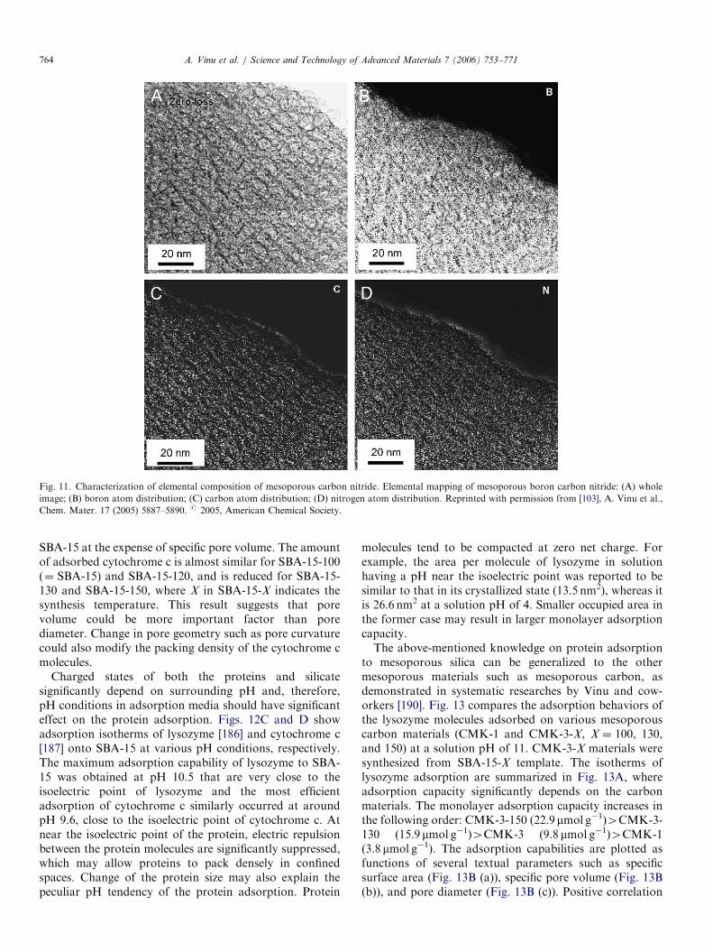
Fig. 11. Characterization of elemental composition of mesoporous carbon nitride. Elemental mapping of mesoporous boron carbon nitride: (A) whole
image; (B) boron atom distribution; (C) carbon atom distribution; (D) nitrogen atom distribution. Reprinted with permission from [103], A. Vinu et al.,
Chem. Mater. 17 (2005) 5887–5890. r 2005, American Chemical Society.
A. Vinu et al. / Science and Technology of Advanced Materials 7 (2006) 753–771764
SBA-15 at the expense of specific pore volume. The amountof adsorbed cytochrome c is almost similar for SBA-15-100(¼ SBA-15) and SBA-15-120, and is reduced for SBA-15-130 and SBA-15-150, where X in SBA-15-X indicates thesynthesis temperature. This result suggests that porevolume could be more important factor than porediameter. Change in pore geometry such as pore curvaturecould also modify the packing density of the cytochrome cmolecules.
Charged states of both the proteins and silicatesignificantly depend on surrounding pH and, therefore,pH conditions in adsorption media should have significanteffect on the protein adsorption. Figs. 12C and D showadsorption isotherms of lysozyme [186] and cytochrome c[187] onto SBA-15 at various pH conditions, respectively.The maximum adsorption capability of lysozyme to SBA-15 was obtained at pH 10.5 that are very close to theisoelectric point of lysozyme and the most efficientadsorption of cytochrome c similarly occurred at aroundpH 9.6, close to the isoelectric point of cytochrome c. Atnear the isoelectric point of the protein, electric repulsionbetween the protein molecules are significantly suppressed,which may allow proteins to pack densely in confinedspaces. Change of the protein size may also explain thepeculiar pH tendency of the protein adsorption. Protein
molecules tend to be compacted at zero net charge. Forexample, the area per molecule of lysozyme in solutionhaving a pH near the isoelectric point was reported to besimilar to that in its crystallized state (13.5 nm2), whereas itis 26.6 nm2 at a solution pH of 4. Smaller occupied area inthe former case may result in larger monolayer adsorptioncapacity.The above-mentioned knowledge on protein adsorption
to mesoporous silica can be generalized to the othermesoporous materials such as mesoporous carbon, asdemonstrated in systematic researches by Vinu and cow-orkers [190]. Fig. 13 compares the adsorption behaviors ofthe lysozyme molecules adsorbed on various mesoporouscarbon materials (CMK-1 and CMK-3-X, X ¼ 100, 130,and 150) at a solution pH of 11. CMK-3-X materials weresynthesized from SBA-15-X template. The isotherms oflysozyme adsorption are summarized in Fig. 13A, whereadsorption capacity significantly depends on the carbonmaterials. The monolayer adsorption capacity increases inthe following order: CMK-3-150 (22.9mmol g�1)4CMK-3-130 (15.9mmol g�1)4CMK-3 (9.8 mmol g�1)4CMK-1(3.8 mmol g�1). The adsorption capabilities are plotted asfunctions of several textual parameters such as specificsurface area (Fig. 13B (a)), specific pore volume (Fig. 13B(b)), and pore diameter (Fig. 13B (c)). Positive correlation

ARTICLE IN PRESS
Fig. 12. (A) Adsorption isotherms of lysozyme onto mesoporous silica materials at pH 10.5: (a) C12-MCM-41; (b) C16-MCM-41; (c) SBA-15. (B)
Adsorption isotherms of cytochrome c onto mesoporous silica materials at pH 9.6: (a) C12-MCM-41; (b) C16-MCM-41; (c) SBA-15. (C) Adsorption
isotherms of lysozyme onto SBA-15 at various pH conditions: (a) pH 6.5; (b) pH 9.6; (c) pH 10.5; (d) pH 12.0. (D) Adsorption isotherms of cytochrome
onto SBA-15 at various pH conditions: (a) pH 3.0; (b) pH 6.5; (c) pH 9.6; (d) pH 10.6. Reprinted with permission from [186], A. Vinu et al., J. Phys. Chem.
B 108 (2004) 7323-7330 and [187], A. Vinu et al., Chem. Mater. 16 (2004) 3056-3065. r 2004, American Chemical Society.
A. Vinu et al. / Science and Technology of Advanced Materials 7 (2006) 753–771 765
is obviously recognized between the adsorption capacityand the pore volume and pore diameter, while theadsorption capacity does not show clear relevance withthe surface area. Interestingly, unavoidable negativedeviation from the expected line can be detected for theadsorption capacity to CMK-1. This result would beexplained by the size exclusion effect at mesoporous media.A smaller pore diameter (2.3 nm for CMK-1) comparedwith dimensions of the lysozyme molecule (crystallographicdimension of 4.5� 3� 3 nm in ellipsoidal) resulted in verysmall adsorption capacity.
Similar to mesoporous silica materials, a maximumadsorption of the proteins was observed at near theisoelectric point. In addition to compact packing natureof the proteins at the isoelectric point, effective hydro-phobic interaction between carbon surface and neutralproteins may accelerate adsorption process. Vinu andcoworkers also extended the adsorption researches ofbiomaterials to smaller molecules such as vitamins[197,198] and amino acids [199,200], where superiority ofthe mesoporous carbon in these adsorption processes ascompared to mesoporous silica and activated non-structure

ARTICLE IN PRESS
Fig. 13. (A) Adsorption isotherms of lysozyme onto mesoporous carbon materials at pH 11: (a), CMK-1; (b) CMK-3; (c) CMK-3-130; (d) CMK-3-150.
(B) Effect of structural parameters on monolayer adsorption capacities of lysozyme onto mesoporous carbon materials: (a) specific surface area;
(b) specific pore volume; (c) pore size.
Fig. 14. Binding isotherms of lysozyme to mesoporous materials:
(a) carbon nanocage; (b) mesoporous carbon CMK-3.
A. Vinu et al. / Science and Technology of Advanced Materials 7 (2006) 753–771766
carbon is demonstrated. Because of large pore volume ofthe carbon nanocage, it would show superior capability inthe biomaterials adsorption [98]. As a simple demonstra-tion, adsorption behaviors of lysozyme to the carbonnanocage materials were compared with those of conven-tional mesoporous carbon (Fig. 14). The maximal mono-layer adsorption capacity of carbon nanocage was26.5 mmol g�1, respectively, while that of CMK-3 isassigned to only 9.8 mmol g�1. Not limited to proteinadsorption, carbon nanocage would have superior cap-ability in molecular adsorption, extraction, and removal,which are currently investigated in our research group.Because mesoporous materials provide well-defined
nanostructures, which bare resemblances to the nanospacesfound in biological systems in their size and accuracy.Therefore, dense assembling of biological components inmesopores could mimic structures and functions of rathercomplicated biomaterials. According to this concept, the

ARTICLE IN PRESS
Fig. 15. (A) Conceptual illustration of proteosilica. (B) HRTEM image of proteosilica (magnification increases from (a) to (c)).
A. Vinu et al. / Science and Technology of Advanced Materials 7 (2006) 753–771 767
mesoporous materials confining the peptide segments inhighly organized mesopore nanospace were newly devel-oped by Ariga and coworkers [201–203]. The obtainedbiomaterial-hybridized mesoporous silica was named asproteosilica (Fig. 15A). Peptide surfactants, which havepolar quaternary ammonium salt at the peptide N-terminaland hydrophobic alkyl chain at its C-terminal were used astemplate for synthesis of proteosilica, which was obtainedin both transparent film and powder form. HRTEM imageof the films indicate highly ordered regular pore arrays(Fig. 15B). Proteosilica provides optically asymmetricenvironment for doped molecules. Photoisomerizationbehaviors of spiropyran doped in the transparent proteo-silica films were investigated spectroscopically. Isomeriza-tion between the spiropyran form and the merocyanineform can be repeated upon alternate irradiation of thevisible light (420 nm) and UV light (280 nm) to the films,respectively. Alternate irradiation with the UV light andthe visible light induced repeated changes in the circulardichroism (CD) spectra, and a complete mirror image ofthe CD spectra was obtained when the chirality ofcomponent peptides was reversed. The CD signals fromthe guest were driven by the chiral environment of thesurrounding host. The presented materials are expected tobe applied to memory device with non-destructive read-outcapability.
Unlike the above-mentioned examples, biomaterial-hybridized mesoporous silica with covalent linkage be-
tween silica framework and biological components wasproposed by Ariga, Aida, and coworkers (Fig. 16)[204–206]. A surfactant with condensable alkoxysilanegroup was used as template in this method. In the presenceof this template, sol–gel reactions of tetraethyl orthosilicateresulted in mesoporous silica whose channels are filledwith covalently attached biological component (alanineresidue). Selective hydrolysis of the ester at the C-terminalof the surfactant caused cleavage and removal of thealkyl tail, leaving open pores with a surface covalentlyfunctionalized by the alanine residue. The template behaveslike a ‘‘lizard,’’ i.e., the head of the surfactant bites thesilica wall and its tail can be cleaved off. Regularmesoporous structures were maintained through the wholeprocess as confirmed by XRD analysis and HRTEMobservation. Nitrogen adsorption–desorption measure-ments before and after the tail removal ensured poreformation upon the hydrolysis. Selective hydrolysis of thetemplate ester was clearly demonstrated by FT-IR mea-surement. Adsorption of NH3 gas to the exposed alanineC-terminal in the silicate channel was successfully quanti-fied by temperature-programmed desorption analysis. Asdemonstrated by these experimental data, this lizardtemplate method copes with both dense functionalizationof the pore inside and high accessibility of external guests.The fabrication of mesoporous silica with a variety oforganic functional groups by the lizard method is highlyexpected.

ARTICLE IN PRESS
Fig. 16. Lizard templating method dense hybridization of alanine residues in mesopore inside wall. Reprinted with permission from [205], Q. Zhang et al.
J. Am. Chem. Soc. 126 (2004) 988–989.r 2004, American Chemical Society.
A. Vinu et al. / Science and Technology of Advanced Materials 7 (2006) 753–771768
5. Summary
In this review, various methodologies for syntheses ofmesoporous materials are described, including recentadvances in this field. Traditional template synthesis,which mainly utilizes micelle assembly as a template, isespecially useful for preparation of mesoporous metaloxide. The prepared structure such as mesoporous silicacan be subjected to a replica synthesis, where structures ofmesoporous materials are replicated into mesoporousmaterials composed of the other components such as
carbon. The latter strategy has been recently applied tonovel mesoporous materials such as carbon nanocage andmesoporous carbon nitride. As the third-generation tech-nique, the elemental substitution method has been recentlyproposed by Vinu and coworkers. Direct substitution ofcomponent elements in mesoporous materials providednew materials with maintaining structural regularity.According to the synthetic strategy, mesoporous boronnitride and mesoporous boron carbon nitride have beensuccessfully invented. In addition to these material inven-tions, hybridization of high functional materials, such as

ARTICLE IN PRESSA. Vinu et al. / Science and Technology of Advanced Materials 7 (2006) 753–771 769
biomaterials, to mesoporous structure has been alsodeveloped in these days. Especially, immobilization ofproteins and peptides in mesopores has been systematicallyresearched and, therefore, their practical applications arenow awaited. New families of mesoporous materialsintroduced in this review would have high potentials infuture practical applications in wide ranges from electro-nics and photonics to environmental and medical uses.
Acknowledgment
The researches described in this chapter were partiallysupported by Ground-based Research Program for SpaceUtilization Promoted by Japan Space Forum and Grant-inAid for Scientific Research on Priority Areas (No.18033059 ‘‘Chemistry of Coordination Space’’) fromMinistry of Education, Science, Sports, and Culture,Japan.
References
[1] N.K. Raman, M.T. Anderson, C.T. Brinker, Chem. Mater. 8 (1996)
1682.
[2] A. Sayari, Chem. Mater. 8 (1996) 1840.
[3] A. Corma, Chem. Rev. 97 (1997) 2373.
[4] K. Moller, T. Bein, Chem. Mater. 10 (1998) 2950.
[5] U. Ciesla, F. Schuth, Microporous Mesoporous Mater. 27 (1999)
131.
[6] J.Y. Ying, C.P. Mehnert, M.S. Wong, Angew. Chem. Int. Ed. 38
(1999) 56.
[7] A. Stein, B.J. Melde, R.C. Schroden, Adv. Mater. 12 (2000) 1403.
[8] C. Sanchez, G.J. de A.A. Soler-Illia, F. Ribot, T. Lalot, C.R. Mayer,
V. Cabuil, Chem. Mater. 13 (2001) 3061.
[9] S. Polarz, B. Smarsly, J. Nanosci. Nanotechnol. 2 (2002) 581.
[10] M.E. Davis, Nature 417 (2002) 813.
[11] A. Vinu, V. Murugesan, M. Hartmann, Chem. Mater. 15 (2003)
1385.
[12] A. Okabe, T. Fukushima, K. Ariga, M. Niki, T. Aida, J. Am. Chem.
Soc. 126 (2004) 9013.
[13] K. Ariga, J. Nanosci. Nanotechnol. 4 (2004) 23.
[14] A. Taguchi, F. Schuth, Microporous Mesoporous Mater. 77 (2005) 1.
[15] A. Vinu, K.Z. Hossain, K. Ariga, J. Nanosci. Nanotechnol. 5 (2005)
347.
[16] T. Yanagisawa, T. Shimizu, K. Kuroda, C. Kato, Bull. Chem. Soc.
Jpn. 63 (1990) 988.
[17] S. Inagaki, Y. Fukushima, K. Kuroda, J. Chem. Soc., Chem.
Commun. (1993) 680.
[18] C.T. Kresge, M.E. Leonowicz, W.J. Roth, J.C. Vartuli, J.S. Beck,
Nature 359 (1992) 710.
[19] J.S. Beck, J.C. Vartuli, W.J. Roth, M.E. Leonowicz, C.T. Kresge,
K.D. Schmitt, C.T.W. Chu, D.H. Olson, E.W. Sheppard, S.B.
McCullen, J.B. Higgins, J.L. Schlenker, J. Am. Chem. Soc. 114
(1992) 10834.
[20] J.C. Vartuli, K.D. Schmitt, C.T. Kresge, W.J. Roth, M.E.
Leonowicz, S.B. McCullen, S.D. Hellring, J.S. Beck, J.L. Schlenker,
D.H. Olson, E.W. Sheppard, Chem. Mater. 6 (1994) 2317.
[21] M. Dubois, T. Gulik-Krzywicki, B. Cabane, Langmuir (1993) 673.
[22] P.T. Tanev, T.J. Pinnavaia, Science 267 (1995) 865.
[23] S.A. Bagshaw, E. Prouset, T.J. Pinnavaia, Science 269 (1995) 1242.
[24] D. Zhao, J. Feng, Q. Huo, N. Melosh, G.H. Fredickson, B.F.
Chmelka, G.D. Stucky, Science 279 (1998) 548.
[25] D. Zhao, Q. Huo, J. Feng, B.F. Chmelka, G.D. Stucky, J. Am.
Chem. Soc. 120 (1998) 6024.
[26] P. Schmidt-Winkel, W.W. Lukens, D. Zhao, P. Yang, B.F.
Chmelka, G.D. Stucky, J. Am. Chem. Soc. 121 (1999) 254.
[27] M.S. Morey, A. Davidson, G.D. Stucky, J. Porous Mater. 5 (1998)
195–204.
[28] D. Khushalani, S. Hasenzahl, S. Mann, J. Nanosci. Nanotechnol. 1
(2001) 129.
[29] M. Hartmann, A. Vinu, S.P. Elangovan, V. Murugesan,
W. Bohlmann, Chem. Commun. (2002) 1238.
[30] E.R. Leite, N.L.V. Carreno, E. Longo, A. Valentini, L.F.D. Probst,
J. Nanosci. Nanotechnol. 2 (2002) 89.
[31] A. Vinu, J. Dedeeek, V. Murugesan, M. Hartmann, Chem. Mater.
14 (2002) 2433.
[32] A. Vinu, M. Hartmann, Chem. Lett. 33 (2004) 588.
[33] A. Vinu, K. Ariga, S. Saravanamurugan, M. Hartmann,
V. Murugesan, Microporous Mesoporous Mater. 76 (2004) 91.
[34] S. Wu, Y. Han, Y.-C. Zou, J.-W. Song, L. Zhao, Y. Di, S.-Z. Liu,
F.-S. Xiao, Chem. Mater. 16 (2004) 486.
[35] A. Vinu, T. Krithiga, V. Murugesan, M. Hartmann, Adv. Mater. 16
(2004) 1817.
[36] A. Vinu, B.M. Devassy, S.B. Halligudi, W. Bohlmann,
M. Hartmann, Appl. Catal. A-Gen. 281 (2005) 207.
[37] A. Vinu, M. Karthik, M. Miyahara, V. Murugesan, K. Ariga,
J. Mol. Catal. A-Chem. 230 (2005) 151.
[38] A. Vinu, D.P. Sawant, K. Ariga, M. Hartmann, S.B. Halligudi,
Microporous Mesoporous Mater. 80 (2005) 195.
[39] A. Vinu, G.S. Kumar, K. Ariga, V. Murugesan, J. Mol. Catal.
A-Chem. 235 (2005) 57.
[40] T. Krithiga, A. Vinu, K. Ariga, B. Arabindoo, M. Palanichamy,
V. Murugesan, J. Mol. Catal. A-Chem. 237 (2005) 238.
[41] A. Vinu, D.P. Sawant, K. Ariga, K.Z. Hossain, S.B. Halligudi,
M. Hartmann, M. Nomura, Chem. Mater. 17 (2005) 5339.
[42] D.P. Sawant, A. Vinu, N.E. Jacob, F. Lefebvre, S.B. Halligudi,
J. Catal. 235 (2005) 341.
[43] A. Vinu, T. Krithiga, V.V. Balasubramanian, A. Asthana,
P. Srinivasu, T. Mori, K. Ariga, G. Ramanath, P.G. Ganesan,
J. Phys. Chem. B 110 (2006) 11924.
[44] A. Vinu, P. Srinivasu, M. Miyahara, K. Ariga, J. Phys. Chem. B 110
(2006) 801.
[45] V. Umamaheswari, W. Bohlmann, A. Poppl, A. Vinu,
M. Hartmann, Microporous Mesoporous Mater. 89 (2006) 47.
[46] D.M. Antonelli, J.Y. Ying, Angew. Chem. Int. Ed. Engl. 34 (1995)
2014.
[47] S.A. Bagshaw, T.J. Pinnavaia, Angew. Chem. Int. Ed. Engl. 35
(1996) 1102.
[48] D.M. Antonelli, J.Y. Ying, Angew. Chem. Int. Ed. Engl. 35 (1996)
426.
[49] D.M. Antonelli, A. Nakahira, J.Y. Ying, Inorg. Chem. 35 (1996)
3126.
[50] D.M. Antonelli, J.Y. Ying, Chem. Mater. 8 (1996) 874.
[51] P. Liu, I.L. Moudrakovski, J. Liu, A. Sayari, Chem. Mater. 9 (1997)
2513.
[52] U. Bach, D. Lupo, P. Comte, J.E. Moser, F. Weissortle, J. Salbeck,
H. Spreitzer, M. Gratzel, Nature 395 (1998) 583.
[53] M.S. Wong, J.Y. Ying, Chem. Mater. 10 (1998) 2067.
[54] J.N. Kondo, L. Lu, Y. Takahara, K. Maruya, K. Domen,
N. Igarashi, T. Tatsumi, Bull. Chem. Soc. Jpn. 73 (2000) 1123.
[55] F. Schuth, Chem. Mater. 13 (2001) 3184.
[56] V. Subramanian, J.C. Jiang, P.H. Smith, B. Rambabu, J. Nanosci.
Nanotechnol. 4 (2004) 125.
[57] I. Kartini, P. Meredith, X.S. Zhao, J.C. Diniz da Costa, G.Q. Lu,
J. Nanosci. Nanotechnol. 4 (2004) 270.
[58] R.C. Hayward, B.F. Chmelka, E.J. Kramer, Adv. Mater. 17 (2005)
2591.
[59] H. Shibata, T. Ogura, T. Mukai, T. Ohkubo, H. Sakai, M. Abe,
J. Am. Chem. Soc. 127 (2005) 16396.
[60] B. Chakraborty, A.C. Pulikottil, S. Das, B. Viswanathan, Chem.
Commun. (1997) 911.
[61] D. Zhao, Z. Luan, L. Kevan, Chem. Commun. (1997) 1009.

ARTICLE IN PRESSA. Vinu et al. / Science and Technology of Advanced Materials 7 (2006) 753–771770
[62] B.T. Holland, P.K. Isbester, C.F. Blanford, E.J. Munson, A. Stein,
J. Am. Chem. Soc. 119 (1997) 6796.
[63] T. Kimura, Y. Sugahara, K. Kuroda, Chem. Mater. 11 (1999) 508.
[64] S. Inagaki, S. Guan, Y. Fukushima, T. Ohsuna, O. Terasaki, J. Am.
Chem. Soc. 121 (1999) 9611.
[65] T. Asefa, M.J. MacLachlan, N. Coombs, G.A. Ozin, Nature 402
(1999) 867.
[66] B.J. Melde, B.T. Holland, C.F. Blanford, A. Stein, Chem. Mater. 11
(1999) 3302.
[67] S. Inagaki, S. Guan, T. Ohsuna, O. Terasaki, Nature 416 (2002) 304.
[68] T. Aida, K. Tajima, Angew. Chem. Int. Ed. 40 (2001) 3803.
[69] Y. Lu, Y. Yang, A. Sellinger, M. Lu, J. Huang, H. Fan, R. Haddad,
G. Lopez, A.R. Burns, D.Y. Sasaki, J. Shelnutt, C.J. Brinker,
Nature 410 (2001) 913.
[70] M. Kimura, K. Wada, K. Ohta, K. Hanabusa, H. Shirai,
N. Kobayashi, J. Am. Chem. Soc. 123 (2001) 2438.
[71] A. Okabe, T. Fukushima, K. Ariga, T. Aida, Angew. Chem. Int. Ed.
41 (2002) 3414.
[72] E. Ruiz-Hitzky, S. Letaief, V. Prevot, Adv. Mater. 14 (2002) 439.
[73] M. Ikegame, K. Tajima, T. Aida, Angew. Chem. Int. Ed. 42 (2003)
2154.
[74] G. Li, S. Bhosale, T. Wang, Y. Zhang, H. Zhu, J.-H. Fuhrhop,
Angew. Chem. Int. Ed. 42 (2003) 3818.
[75] A. Okabe, T. Fukushima, K. Ariga, T. Aida, Stud. Surf. Sci. Cat.
146 (2003) 73.
[76] A. Shimojima, K. Kuroda, Angew. Chem. Int. Ed. 42 (2003) 40570.
[77] S. Che, Z. Liu, T. Ohsuna, K. Sakamoto, O. Terasaki, T. Tatsumi,
Nature 429 (2004) 281.
[78] H. Jin, Z. Liu, T. Ohsuna, O. Terasaki, Y. Inoue, K. Sakamoto,
T. Nakanishi, K. Ariga, S. Che, Adv. Mater. 18 (2006) 593.
[79] S. Che, J. Nanosci. Nanotechnol. 6 (2006) 1557.
[80] N.Y.C. Yang, K. Jian, I. Kulaots, G.P. Crawford, R.H. Hurt,
J. Nanosci. Nanotechnol. 3 (2003) 386.
[81] M. Inagaki, K. Kaneko, T. Nishizawa, Carbon 42 (2004) 1401.
[82] A. Vinu, K. Ariga, Chem. Lett. 34 (2005) 674.
[83] H. Yang, D. Zhao, J. Mater. Chem. 15 (2005) 1217.
[84] A. Vinu, M. Hartmann, Catal. Today 102 (2005) 189.
[85] T. Kyotani, T. Nagai, S. Inoue, A. Tomita, Chem. Mater. 9 (1997) 609.
[86] T. Kyotani, Carbon 38 (2000) 269.
[87] R. Ryoo, S.H. Joo, S. Jun, J. Phys. Chem. B 103 (1999) 7743.
[88] S. Jun, S.H. Joo, R. Ryoo, M. Kruk, M. Jaroniec, Z. Liu,
T. Ohsuna, O. Terasaki, J. Am. Chem. Soc. 122 (2000) 10712.
[89] R. Ryoo, S.H. Joo, M. Kurk, M. Jaroniec, Adv. Mater. 13 (2001)
677.
[90] S.H. Joo, S.J. Choi, I. Oh, J. Kwak, Z. Liu, O. Terasaki, R. Ryoo,
Nature 412 (2001) 169.
[91] L.A. Solovyov, V.I. Zaikovskii, A.N. Shmakov, O.V. Belousov,
R. Ryoo, J. Phys. Chem. B 106 (2002) 12198.
[92] J. Lee, S. Yoon, T. Hyeon, S.M. Oh, K.B. Kim, Chem. Commun.
(1999) 2177.
[93] J. Lee, S. Yoon, S.M. Oh, C.-H. Shin, T. Hyeon, Adv. Mater. 12
(2000) 359.
[94] S. Han, S. Kim, H. Lim, W. Choi, H. Park, J. Yoon, T. Hyeon,
Microporous Mesoporous Mater. 58 (2003) 131.
[95] M. Hartmann, A. Vinu, Langmuir 18 (2002) 8010.
[96] A. Vinu, C. Streb, V. Murugesan, M. Hartmann, J. Phys. Chem. B
107 (2003) 8297.
[97] A. Vinu, M. Miyahara, K. Ariga, Stud. Surf. Sci. Catal. 158 (2005)
971.
[98] A. Vinu, M. Miyahara, V. Sivamurugan, T. Mori, K. Ariga,
J. Mater. Chem. 15 (2005) 5122.
[99] A. Vinu, M. Miyahara, T. Mori, K. Ariga, J. Porous Mater. 13
(2006) 379.
[100] F. Kleitz, D. Liu, G.M. Anilkumar, I.-S. Park, L.A. Solovyov, A.N.
Shmakov, R. Ryoo, J. Phys. Chem. B 107 (2003) 14296.
[101] P.I. Ravikovitch, A.V. Neimark, Langmuir 18 (2002) 1550.
[102] A. Vinu, K. Ariga, T. Mori, T. Nakanishi, S. Hishita, D. Golberg,
Y. Bando, Adv. Mater. 17 (2005) 1648.
[103] A. Vinu, M. Terrones, D. Golberg, S. Hishita, K. Ariga, T. Mori,
Chem. Mater. 17 (2005) 5887.
[104] D. Golberg, Y. Bando, W. Han, K. Kurashima, T. Sato, Chem.
Phys. Lett. 308 (1999) 337.
[105] D. Golberg, Y. Bando, K. Kurashima, T. Sato, Chem. Phys. Lett.
323 (2000) 185.
[106] D. Golberg, P.S. Dorozhkin, Y. Bando, Z.-C. Dong, N. Grobert,
M. Reyes-Reyes, H. Terrones, M. Terrones, Appl. Phys. Lett. 82
(2003) 1275.
[107] M. Murakami, T. Shimizu, M. Tansho, A. Vinu, K. Ariga,
K. Takegoshi, Chem. Lett. 35 (2006) 986.
[108] R. Breslow, Acc. Chem. Res. 13 (1980) 170.
[109] K. Ariga, E.V. Anslyn, J. Org. Chem. 57 (1992) 417.
[110] J. Smith, K. Ariga, E.V. Anslyn, J. Am. Chem. Soc. 115 (1993) 362.
[111] D.M. Kneeland, K. Ariga, V.M. Lynch, C.Y. Huang, E.V. Anslyn,
J. Am. Chem. Soc. 115 (1993) 10042.
[112] N.C. Seeman, Acc. Chem. Res. 30 (1997) 357.
[113] M.C. Seeman, Angew. Chem. Int. Ed. 37 (1998) 3220.
[114] M. Onda, K. Yoshihara, H. Koyano, K. Ariga, T. Kunitake, J. Am.
Chem. Soc. 118 (1996) 8524.
[115] K. Ariga, N. Yamada, M. Naito, E. Koyama, Y. Okahata, Chem.
Lett. (1998) 493.
[116] N. Yamada, K. Ariga, M. Naito, K. Matsubara, E. Koyama, J. Am.
Chem. Soc. 120 (1998) 12192.
[117] J. Kikuchi, K. Ariga, K. Ikeda, Chem. Commun. (1999) 547.
[118] J. Kikuchi, K. Ariga, T. Miyazaki, K. Ikeda, Chem. Lett. (1999)
253.
[119] K. Ariga, J. Kikuchi, K. Narumi, E. Koyama, N. Yamada, Chem.
Lett. (1999) 787.
[120] N. Yamada, K. Ariga, Synlett (2000) 575.
[121] K. Ariga, J. Kikuchi, M. Naito, E. Koyama, N. Yamada, Langmuir
16 (2000) 4929.
[122] N. Yamada, K. Matsubara, K. Narumi, Y. Sato, E. Koyama,
K. Ariga, Colloid Surf. A-Physicochem. Eng. Asp. 169 (2000) 271.
[123] K. Ariga, J. Kikuchi, M. Naito, N. Yamada, Polym. Adv. Technol.
11 (2000) 856.
[124] D. Gust, T.A. Moore, A.L. Moore, Acc. Chem. Res. 34 (2001) 40.
[125] K. Fukuda, Y. Sasaki, K. Ariga, J. Kikuchi, J. Mol. Catal.
B-Enzym. 11 (2001) 971.
[126] J. Kikuchi, K. Ariga, Y. Sasaki, K. Ikeda, J. Mol. Catal. B-Enzym.
11 (2001) 977.
[127] M. Mrksich, G.M. Whitesides, Annu. Rev. Biophys. Biomol. Struct.
25 (1996) 55.
[128] A.S. Blawas, W.M. Reichert, Biomaterials 19 (1998) 595.
[129] D.S. Ginger, H. Zhang, C.A. Mirkin, Angew. Chem. Int. Ed. 43
(2004) 30.
[130] Y. Okahata, T. Tsuruta, K. Ijiro, K. Ariga, Langmuir 4 (1988) 1373.
[131] S. Taneva, K. Ariga, Y. Okahata, W. Tagaki, Langmuir 5 (1989)
111.
[132] S. Taneva, K. Ariga, W. Tagaki, Y. Okahata, J. Colloid Interface
Sci. 131 (1989) 561.
[133] Y. Okahata, T. Tsuruta, K. Ijiro, K. Ariga, Thin Solid Films 180
(1989) 65.
[134] Y. Ebara, H. Ebato, K. Ariga, Y. Okahata, Langmuir 10 (1994)
2267.
[135] K. Taguchi, K. Ariga, T. Kunitake, Chem. Lett. (1995) 701.
[136] X. Cha, K. Ariga, M. Onda, T. Kunitake, J. Am. Chem. Soc. 117
(1995) 11833.
[137] X. Cha, K. Ariga, T. Kunitake, Bull. Chem. Soc. Jpn. 69 (1996) 163.
[138] X. Cha, K. Ariga, T. Kunitake, Chem. Lett. (1996) 73.
[139] Y. Oishi, Y. Torii, M. Kuramori, K. Suehiro, K. Ariga, K. Taguchi,
A. Kamino, T. Kunitake, Chem. Lett. (1996) 411.
[140] X. Cha, K. Ariga, T. Kunitake, J. Am. Chem. Soc. 118 (1996) 9545.
[141] Y. Oishi, Y. Torii, T. Kato, M. Kuramori, K. Suehiro, K. Ariga,
K. Taguchi, A. Kamino, H. Koyano, T. Kunitake, Langmuir 13
(1997) 519.
[142] K. Ariga, A. Kamino, H. Koyano, T. Kunitake, J. Mater. Chem. 7
(1997) 1155.

ARTICLE IN PRESSA. Vinu et al. / Science and Technology of Advanced Materials 7 (2006) 753–771 771
[143] K. Ariga, K. Isoyama, O. Hayashida, Y. Aoyama, Y. Okahata,
Chem. Lett. (1998) 1007.
[144] K. Ariga, A. Kamino, X. Cha, T. Kunitake, Langmuir 15 (1999) 3875.
[145] K. Ariga, T. Abe, J. Kikuchi, Chem. Lett. (2000) 82.
[146] K. Ariga, K. Tanaka, K. Katagiri, J. Kikuchi, E. Ohshima,
Y. Hisaeda, Colloid Surf. A-Physicochem. Eng. Asp. 169 (2000) 47.
[147] K. Ariga, K. Tanaka, K. Katagiri, J. Kikuchi, H. Shimakoshi,
E. Ohshima, Y. Hisaeda, Phys. Chem. Chem. Phys. 3 (2001) 3442.
[148] K. Ariga, H. Yuki, J. Kikuchi, O. Dannemuller, A.M. Albrecht-
Gary, Y. Nakatani, G. Ourisson, Langmuir 21 (2005) 4578.
[149] K. Ariga, T. Nakanishi, J.P. Hill, M. Shirai, M. Okuno, T. Abe,
J. Kikuchi, J. Am. Chem. Soc. 127 (2005) 12074.
[150] Y. Lvov, K. Ariga, T. Kunitake, Chem. Lett. (1994) 2323.
[151] Y. Lvov, K. Ariga, I. Ichinose, T. Kunitake, J. Am. Chem. Soc. 117
(1995) 6117.
[152] Y. Lvov, K. Ariga, I. Ichinose, T. Kunitake, J. Chem. Soc., Chem.
Commun. (1995) 2313.
[153] M. Onda, Y. Lvov, K. Ariga, T. Kunitake, Biotechnol. Bioeng. 51
(1996) 163.
[154] Y. Lvov, K. Ariga, I. Ichinose, T. Kunitake, Thin Solid Films 285
(1996) 797.
[155] M. Onda, Y. Lvov, K. Ariga, T. Kunitake, J. Ferment. Bioeng. 82
(1996) 502.
[156] K. Ariga, M. Onda, Y. Lvov, T. Kunitake, Chem. Lett. (1997) 25.
[157] M. Onda, Y. Lvov, K. Ariga, T. Kunitake, Jpn. J. Appl. Phys. 36
(1997) L1608.
[158] Y. Lvov, M. Onda, K. Ariga, T. Kunitake, J. Biomater. Sci. Polym.
Ed. 9 (1998) 345.
[159] F. Caruso, D.N. Furlong, K. Ariga, I. Ichinose, T. Kunitake,
Langmuir 14 (1998) 4559.
[160] M. Onda, K. Ariga, T. Kunitake, J. Biosci. Bioeng. 87 (1999) 69.
[161] K. Ariga, Y. Sasaki, H. Horiguchi, N. Horiuchi, J. Kikuchi, Defect
Diffusion Forum 191 (2001) 35.
[162] K. Katagiri, R. Hamasaki, K. Ariga, J. Kikuchi, J. Am. Chem. Soc.
124 (2002) 7892.
[163] K. Katagiri, R. Hamasaki, K. Ariga, J. Kikuchi, Lamgmuir 18
(2002) 6709.
[164] M. Ogawa, J. Am. Chem. Soc. 116 (1994) 7941.
[165] H. Yang, A. Kuperman, N. Coombs, S. Mamiche-Asefa, G.A. Ozin,
Nature 379 (1996) 703.
[166] D. Zhao, P. Yang, N. Melosh, J. Feng, B.F. Chmelka, G.D. Stucky,
Adv. Mater. 10 (1998) 1380.
[167] Y. Lu, Y. Yang, A. Sellinger, M. Lu, J. Huang, H. Fan, R. Haddad,
G. Lopez, A.R. Burns, D.Y. Sasaki, J. Shelnutt, C.J. Brinker,
Nature 410 (2001) 913.
[168] A. Mehdi, S. Dourdain, J.F. Bardeau, C. Reye, R.J.P. Corriu,
A. Gibaud, J. Nanosci. Nanotechnol. 6 (2006) 377.
[169] L. Washmon-Kriel, V.L. Jimenez, K.J. Balkus Jr., J. Mol. Catal.
B-Enzym. 10 (2000) 453.
[170] H. Takahashi, B. Li, T. Sasaki, C. Miyazaki, T. Kajino, S. Inagaki,
Chem. Mater. 12 (2000) 3301.
[171] Y. Wei, J. Xu, Q. Feng, M. Lin, H. Dong, W.-J. Zhang, C. Wang,
J. Nanosci. Nanotechnol. 1 (2001) 83.
[172] H.H.P. Yiu, C.H. Botting, N.P. Botting, P.A. Wright, Phys. Chem.
Chem. Phys. 3 (2001) 2983.
[173] H.H.P. Yiu, P.A. Wright, N.P. Botting, Microporous Mesoporous
Mater. 44-45 (2001) 763.
[174] H. Takahashi, B. Li, T. Sasaki, C. Miyazaki, T. Kajino, S. Inagaki,
Microporous Mesoporous Mater. 44-45 (2001) 755.
[175] T. Sasaki, T. Kajino, B. Li, H. Sugiyama, H. Takahashi, Appl.
Environ. Microbiol. 67 (2001) 2208.
[176] J. Deere, E. Magner, J.G. Wall, B.K. Hodnett, Chem. Commun.
(2001) 465.
[177] J. Deere, E. Magner, J.G. Wall, B.K. Hodnett, J. Phys. Chem. B 106
(2002) 7340.
[178] Y.-J. Han, J.T. Watson, G.D. Stucky, A. Butler, J. Mol. Catal.
B-Enzym. 17 (2002) 1.
[179] J. Deere, E. Magner, J.G. Wall, B.K. Hodnett, Catal. Lett. 85 (2003)
19.
[180] A.S.M. Chong, X.S. Zhao, Appl. Surf. Sci. 237 (2004) 398.
[181] D. Goradia, J. Cooney, B.K. Hodnett, E. Magner, J. Mol. Catal.
B-Enzym. 32 (2005) 231.
[182] M. Hartmann, Chem. Mater. 17 (2005) 4577.
[183] Y. Wang, F. Caruso, Chem. Mater. 17 (2005) 953.
[184] J.F. Dıaz, K.J. Balkus Jr., J. Mol. Catal. B-Enzym. 2 (2006) 115.
[185] Y. Han, S.S. Lee, J.Y. Ying, Chem. Mater. 18 (2006) 643.
[186] A. Vinu, V. Murugesan, M. Hartmann, J. Phys. Chem. B 108 (2004)
7323.
[187] A. Vinu, V. Murugesan, O. Tangermann, M. Hartmann, Chem.
Mater. 16 (2004) 3056.
[188] A. Vinu, V. Murugesan, W. Bohlmann, M. Hartmann, J. Phys.
Chem. B 108 (2004) 11496.
[189] M. Miyahara, A. Vinu, T. Nakanshi, K. Ariga, Kobunshi
Ronbunshu 61 (2004) 623.
[190] A. Vinu, M. Miyahara, K. Ariga, J. Phys. Chem. B 109 (2005) 6436.
[191] A. Vinu, M. Miyahara, K.Z. Hossain, T. Nakanishi, K. Ariga, Stud.
Surf. Sci. Catal. 156 (2005) 637.
[192] M. Miyahara, A. Vinu, K.Z. Hossain, T. Nakanishi, K. Ariga, Thin
Solid Films 499 (2006) 13.
[193] A. Vinu, M. Miyahara, K. Ariga, J. Nanosci. Nanotechnol. 6 (2006)
1510.
[194] M. Miyahara, A. Vinu, K. Ariga, J. Nanosci. Nanotechnol. 6 (2006)
1765.
[195] K. Ariga, A. Vinu, M. Miyahara, Curr. Nanosci. 2 (2006) 197.
[196] M. Miyahara, A. Vinu, K. Ariga, Mater. Sci. Eng. C, in press.
[197] M. Hartmann, A. Vinu, G. Chandrasekar, Chem. Mater. 17 (2005)
829.
[198] G. Chandrasekar, A. Vinu, V. Murugesan, M. Hartmann, Stud.
Surf. Sci. Catal. 158 (2005) 1169.
[199] A. Vinu, K.Z. Hossain, G.S. Kumar, K. Ariga, Carbon 44 (2006)
530.
[200] A. Vinu, K.Z. Hossain, G.S. Kumar, V. Sivamurugan, K. Ariga,
Stud. Surf. Sci. Cayal. 156 (2005) 631.
[201] K. Ariga, T. Aimiya, Q. Zhang, A. Okabe, M. Niki, T. Aida, Int.
J. Nanosci. 1 (2002) 521.
[202] K. Ariga, Q. Zhang, M. Niki, A. Okabe, T. Aida, Stud. Surf. Sci.
Catal. 146 (2003) 427.
[203] K. Ariga, Chem. Rec. 3 (2004) 297.
[204] Q. Zhang, K. Ariga, A. Okabe, T. Aida, Stud. Surf. Sci. Catal. 146
(2003) 465.
[205] Q. Zhang, K. Ariga, A. Okabe, T. Aida, J. Am. Chem. Soc. 126
(2004) 988.
[206] W. Otani, K. Kinbara, Q. Zhang, K. Ariga, T. Aida, Chem. Eur. J.,
in press.