Neutralization and wake effects on the Coulomb explosion...
9
PHYSICAL REVIEW A 91, 042704 (2015) Neutralization and wake effects on the Coulomb explosion of swift H 2 + ions traversing thin films L. F. S. Rosa, P. L. Grande, J. F. Dias, and R. C. Fadanelli Ion Implantation Laboratory, Institute of Physics, Federal University of Rio Grande do Sul, Av. Bento Goncalves, 9500, CP 15051, CEP 91501-970, Porto Alegre, RS, Brazil M. Vos Atomic and Molecular Physics Laboratories, Research School of Physics and Engineering, The Australian National University, Canberra 0200, Australia (Received 15 December 2014; published 10 April 2015) The Coulomb explosion of small cluster beams can be used to measure the dwell time of fragments traversing amorphous films. Therefore, the thickness of thin films can be obtained with the so-called Coulomb depth profiling technique using relatively high cluster energies where the fragments are fully ionized after breakup. Here we demonstrate the applicability of Coulomb depth profiling technique at lower cluster energies where neutralization and wake effects come into play. To that end, we investigated 50–200 keV/uH 2 + molecular ions impinging on a 10 nm TiO 2 film and measured the energy of the backscattered H + fragments with high-energy resolution. The effect of the neutralization of the H + fragments along the incoming trajectory before the backscattering collision is clearly observed at lower energies through the decrease of the energy broadening due to the Coulomb explosion. The reduced values of the Coulomb explosion combined with full Monte Carlo simulations provide compatible results with those obtained at higher cluster energies where neutralization is less important. The results are corroborated by electron microscopy measurements. DOI: 10.1103/PhysRevA.91.042704 PACS number(s): 34.50.Bw, 34.80.Bm, 34.50.Fa I. INTRODUCTION The determination of the thickness of thin films is an important task in many areas of modern technology and can be accomplished with a variety of techniques including those that make use of ion and electron beams. For most of them, a knowledge of the atomic density is required. Recently [1], a novel technique based on the Coulomb explosion of H 2 + molecular beams [2–4] has been developed. The so-called Coulomb depth profiling technique is based on the fact that once H 2 + ions of few hundred keV penetrate the first monolayers of a solid, they lose their bound electrons and break up under the Coulomb repulsion force between the fragments. Consequently, the internuclear distance r and the relative speed between the fragments increase, leading to different energies in the laboratory frame and giving rise to a peculiar energy-loss straggling referred to as Coulomb broadening σ C [1]. In addition, a fragment can be backscattered at a depth z and detected when it leaves the sample. For energies higher than typically a few keV/nucleon, most ion trajectories are V shaped and effects due to correlated motion of the fragments take place only along the incoming path before the backscattering collision. The Coulomb broadening σ C can be then obtained by measuring the energy loss of these fragments in comparison with the reference energy loss taken with H + ions at the same energy per nucleon. The depth information is obtained from the broadening of the energy-loss distribution of backscattered H + ions formed after the breakup of the H 2 + clusters. The energy-loss straggling can be disentangled from other energy-loss fluctuations and converted to dwell time or penetration depth [1]. This technique thus provides film thicknesses in nm. Therefore, by combining the thickness as measured by the Coulomb explosion (in nm) with the thickness measured by traditional ion scattering (in μg/cm 2 ) one can obtain the density. However, the conversion of the energy-loss straggling due to the Coulomb explosion to dwell time is not straightforward for H 2 + clusters with energies lower than 100 keV/u, where neutralization, multiple-scattering and wake-potential effects cannot be neglected. For this case, Monte Carlo simulation is the appropriate tool to investigate the influence of such effects on the Coulomb explosion of H 2 + clusters. For low energies, the neutralization of fragments during the Coulomb explosion has been discussed in the literature in connection with the existence of H 0 inside the solid. Brandt and the Lyon group [5] have claimed that the neutral fraction is composed basically of H + ions that are neutralized when the outgoing trajectory crosses the surface. On the contrary, Cross [6] has argued in favor of the neutral fraction inside the solid whose value can be understood in terms of electron capture into, and subsequent loss from, bound states of the moving proton. Most of the subsequent work (see, for example, Ref. [7]) assumes that the fraction of H 0 stems from inside the solid, but the question whether there is a stable H 0 fraction inside the solid with an electron as tightly bound and localized as in vacuum still remains. The role of the wake potential during the Coulomb explo- sion has been much less investigated since only recently meth- ods to describe the interaction between the valence electrons of a solid and the cluster beam using reliable dielectric functions have been developed for different materials [8–11]. In fact, the wake potential can affect the Coulomb explosion in different ways. Its symmetrical part screens the Coulomb interaction between the H + fragments. Moreover, it decreases the electron binding energy for the embedded neutral fragments, leading to an increase of their atomic radii. In addition, the asymmetrical part of the wake potential can even increase the Coulomb explosion as will be shown later. Apart from a few exceptions [12,13], most of the previous investigations of the Coulomb explosion in solids have used the 1050-2947/2015/91(4)/042704(9) 042704-1 ©2015 American Physical Society
Transcript of Neutralization and wake effects on the Coulomb explosion...

PHYSICAL REVIEW A 91, 042704 (2015)
Neutralization and wake effects on the Coulomb explosion of swift H2+ ions traversing thin films
L. F. S. Rosa, P. L. Grande, J. F. Dias, and R. C. FadanelliIon Implantation Laboratory, Institute of Physics, Federal University of Rio Grande do Sul, Av. Bento Goncalves,
9500, CP 15051, CEP 91501-970, Porto Alegre, RS, Brazil
M. VosAtomic and Molecular Physics Laboratories, Research School of Physics and Engineering,
The Australian National University, Canberra 0200, Australia(Received 15 December 2014; published 10 April 2015)
The Coulomb explosion of small cluster beams can be used to measure the dwell time of fragments traversingamorphous films. Therefore, the thickness of thin films can be obtained with the so-called Coulomb depth profilingtechnique using relatively high cluster energies where the fragments are fully ionized after breakup. Here wedemonstrate the applicability of Coulomb depth profiling technique at lower cluster energies where neutralizationand wake effects come into play. To that end, we investigated 50–200 keV/u H2
+ molecular ions impinging ona 10 nm TiO2 film and measured the energy of the backscattered H+ fragments with high-energy resolution.The effect of the neutralization of the H+ fragments along the incoming trajectory before the backscatteringcollision is clearly observed at lower energies through the decrease of the energy broadening due to the Coulombexplosion. The reduced values of the Coulomb explosion combined with full Monte Carlo simulations providecompatible results with those obtained at higher cluster energies where neutralization is less important. Theresults are corroborated by electron microscopy measurements.
DOI: 10.1103/PhysRevA.91.042704 PACS number(s): 34.50.Bw, 34.80.Bm, 34.50.Fa
I. INTRODUCTION
The determination of the thickness of thin films is animportant task in many areas of modern technology and canbe accomplished with a variety of techniques including thosethat make use of ion and electron beams. For most of them,a knowledge of the atomic density is required. Recently [1],a novel technique based on the Coulomb explosion of H2
+
molecular beams [2–4] has been developed. The so-calledCoulomb depth profiling technique is based on the factthat once H2
+ ions of few hundred keV penetrate the firstmonolayers of a solid, they lose their bound electrons and breakup under the Coulomb repulsion force between the fragments.Consequently, the internuclear distance r and the relativespeed between the fragments increase, leading to differentenergies in the laboratory frame and giving rise to a peculiarenergy-loss straggling referred to as Coulomb broadeningσC [1]. In addition, a fragment can be backscattered at adepth z and detected when it leaves the sample. For energieshigher than typically a few keV/nucleon, most ion trajectoriesare V shaped and effects due to correlated motion of thefragments take place only along the incoming path before thebackscattering collision. The Coulomb broadening σC can bethen obtained by measuring the energy loss of these fragmentsin comparison with the reference energy loss taken with H+ions at the same energy per nucleon. The depth information isobtained from the broadening of the energy-loss distributionof backscattered H+ ions formed after the breakup of the H2
+
clusters. The energy-loss straggling can be disentangled fromother energy-loss fluctuations and converted to dwell timeor penetration depth [1]. This technique thus provides filmthicknesses in nm. Therefore, by combining the thickness asmeasured by the Coulomb explosion (in nm) with the thicknessmeasured by traditional ion scattering (in μg/cm2) one canobtain the density.
However, the conversion of the energy-loss straggling dueto the Coulomb explosion to dwell time is not straightforwardfor H2
+ clusters with energies lower than 100 keV/u, whereneutralization, multiple-scattering and wake-potential effectscannot be neglected. For this case, Monte Carlo simulation isthe appropriate tool to investigate the influence of such effectson the Coulomb explosion of H2
+ clusters.For low energies, the neutralization of fragments during
the Coulomb explosion has been discussed in the literature inconnection with the existence of H0 inside the solid. Brandtand the Lyon group [5] have claimed that the neutral fractionis composed basically of H+ ions that are neutralized whenthe outgoing trajectory crosses the surface. On the contrary,Cross [6] has argued in favor of the neutral fraction insidethe solid whose value can be understood in terms of electroncapture into, and subsequent loss from, bound states of themoving proton. Most of the subsequent work (see, for example,Ref. [7]) assumes that the fraction of H0 stems from inside thesolid, but the question whether there is a stable H0 fractioninside the solid with an electron as tightly bound and localizedas in vacuum still remains.
The role of the wake potential during the Coulomb explo-sion has been much less investigated since only recently meth-ods to describe the interaction between the valence electrons ofa solid and the cluster beam using reliable dielectric functionshave been developed for different materials [8–11]. In fact, thewake potential can affect the Coulomb explosion in differentways. Its symmetrical part screens the Coulomb interactionbetween the H+ fragments. Moreover, it decreases the electronbinding energy for the embedded neutral fragments, leading toan increase of their atomic radii. In addition, the asymmetricalpart of the wake potential can even increase the Coulombexplosion as will be shown later.
Apart from a few exceptions [12,13], most of the previousinvestigations of the Coulomb explosion in solids have used the
1050-2947/2015/91(4)/042704(9) 042704-1 ©2015 American Physical Society

ROSA, GRANDE, DIAS, FADANELLI, AND VOS PHYSICAL REVIEW A 91, 042704 (2015)
Yukawa potential to model the screening interaction betweenthe fragments. Moreover, the effect of the full noncentral wakepotential on the explosion involving fragments that are chargedpart of the time has not been fully explored. This issue playsa key role on the conversion of Coulomb explosion to dwelltime at low as well as at very high cluster energies.
Understanding of the Coulomb explosion at lower energiesis interesting in itself, but also has practical value, as at lowercluster energies the technique can be applied to thinner layers.The aim of the present work is to demonstrate that the Coulombprofiling technique yields good results at relatively low clusterenergies, provided neutralization and wake effects are takeninto account properly. Therefore, unlike Ref. [1] where it wasassumed that fully ionized fragments interact with a Yukawapotential, the present work investigates how valence electronsfrom a solid affect the Coulomb explosion of embeddedfragments. To that end we have investigated many effectssuch as the presence of dressed H0 projectiles with modifiedatomic radius, vicinage effects on projectile charge statesand noncentral interaction effects. Experimentally thin TiO2
layers grown on Si were analyzed with the medium energyion scattering (MEIS) technique [14], which is a refinementof the traditional Rutherford backscattering spectrometry(RBS) [15]. This is the best technique to determine theCoulomb broadening for films thinner than 10 nm [1] becauseof its excellent energy resolution. Unless otherwise stated,atomic units (� = me = e = 1) are used throughout the paper.
II. EXPERIMENTAL PROCEDURE AND DATA ANALYSIS
The MEIS technique was used in the present work in orderto demonstrate the applicability of the Coulomb depth profilingtechnique for the measurement of thin films employing50–200 keV/u H2
+ molecular ions. The sample consisted ofa thin TiO2 film grown on the native silicon oxide presenton the surface of a 〈100〉 Si wafer and was mounted on a3-axis goniometer inside the scattering chamber with a vacuumof about 10−7 mbar. Typical beam currents were less than15 nA. The ion source provides H2
+ molecules without anypreferential orientation.
Different angles of incidence were used in order to increasethe apparent thicknesses of the films. The backscattered H+ions emerging from the target were analyzed using a toroidalelectrostatic analyzer (TEA) mounted at 120 degrees withrespect to the beam direction. At the top end of the TEA aset of two microchannel plates coupled to a position-sensitivedetector allows each ion to be energy- and angle-resolved,leading to two-dimensional (2D) spectra. The TEA angularaperture is 24◦, covering angles from 108◦ to 132◦. Each anglebin corresponds to 0.08 degrees. The overall energy resolutionof the system is 300 eV for a 100 keV H+ beam.
A typical 2D MEIS spectrum obtained for 175 keV/uH2
+ projectiles striking the TiO2 film is depicted in Fig. 1.The contributions from protons backscattering from Ti, Si,and O are easily distinguished in this figure. Moreover, thecontribution from a thin C layer (due to contamination) can beseen in the 2D spectrum as well as the effect of blocking onthe Si intensity along the 〈100〉 direction. For elements at thesurface of the sample (Ti, O, and C), the signals have different
FIG. 1. (Color online) 2D MEIS spectrum measured with175 keV/u of H2
+ projectiles under normal incidence on the TiO2
layer grown over crystal Si. The following signals are observed fromtop to bottom: Ti from the TiO2 film; Si from the substrate; O fromTiO2 and SiO2 films; and C stemming from contamination. Blockinglines in Si are also visible. See text for further information.
slopes according to the dependence of the kinematic factor onthe scattering angle.
The 1D energy spectrum featuring the Ti signal for ascattering angle of 120◦ is shown in Fig. 2. The 1D spectrumis obtained through the projection of the 2D spectrum onthe energy axis for a particular set of angle bins. In orderto improve the counting statistics, several angle bins areintegrated. In this case (for Ti part of the spectrum) correctionsstemming from the dependence of the kinematical factor onthe scattering angle, and from the depth of the backscatteringevents are properly taken into account.
The dashed line shown in Fig. 2 corresponds to the simu-lations of the energy spectrum generated by the POWERMEIS
code for atomic ions [16], which essentially simulates theenergy-loss spectrum according to the backscattering yieldY (E) modeled as
Y (E) = A
∫ t
0dzG
(KE0 − E − zS,
√σ 2
0 + zW2), (1)
for a uniform film of thickness t . E0 is the beam energy,K is the kinematic factor [17], and G(E,σ ) is the Gaussianfunction with standard deviation σ . Moreover, S and W2
are the stopping power and straggling factors respectively asdefined in Ref. [17], while σ0 is the overall energy resolution of
042704-2

NEUTRALIZATION AND WAKE EFFECTS ON THE . . . PHYSICAL REVIEW A 91, 042704 (2015)
FIG. 2. (Color online) 1D energy spectrum obtained by150 keV/nucleon H2
+ molecules striking on TiO2. The inset showsthe 2D spectrum featuring the Ti signal obtained with the H+ beam.The best fittings to the H2
+ data (red line) and to the H+ data (blueline) obtained for the Ti signal were obtained through simulationsusing the POWERMEIS code. Note that the H+ data were omitted fromthe picture for the sake of clarity.
the detection system and A is an overall constant that dependson the elastic cross section, solid angle, and ion fluence.
In case of molecular ions (solid line in Fig. 2), the Gaussianfunction mentioned above has to be convoluted with the energydistribution due to the Coulomb explosion modeled as [1]
Fmol(�E,z) = 1
2√
3σc(z)�(�E +
√3σc(z))
×�(√
3σc(z) − �E). (2)
Here �E is the energy change (in the laboratory frame) ofan ion due to the coulomb explosion, �(�E) is the Heavisidefunction and σc(z) is the contribution of the Coulomb explosionto the energy-loss straggling at a distance z inside the solidafter the molecular breakup. Fmol(�E,z) takes into accountall possible angular orientations after the breakup. Here, theCoulomb explosion is parametrized according to Ref. [1] asσc(z) = γ z, where γ is a free parameter to be determined froma best fit to the experimental data. The Coulomb broadeningσC is the value of σc(z) taken when z corresponds to the thefilm thickness. All other parameters are the same for H+ andH2
+ projectiles with the exception of the σ0, which is slightlylarger in case of molecular projectiles because of the intrinsicmomentum distribution of a proton that is part of a molecule(Doppler effect) before the molecular breakup.
The stopping power used in the simulations are thosegiven by the SRIM2010 code [18] and corrected by recentmeasurements from Ref. [19], while the initial energy-lossstraggling values are given by the straggling theory developedby Lindhard and Scharff [20]. The energy-loss stragglingvalues were relaxed during the fitting procedure through theminimization of the reduced χ2 (see values in Table I). Oncethe amount of TiO2 in the overlayer (in terms of atoms/cm2)and energy-loss straggling parameters were determined for theproton case, they were kept constant throughout the analysisconcerning H2
+ molecules.
TABLE I. Values used in the POWERMEIS code [16] to simulatethe MEIS spectra.
Energy Stopping Straggling σ0(H+) σ0(H2+)
(keV/u) (eV/A) (eV2/A) (eV) (eV)
50 16.4 870 210 24060 17.3 1000 245 48580 18.0 1200 330 375100 18.1 1250 400 460120 17.8 1500 480 530150 17.0 1750 600 670175 16.2 2000 700 760200 15.4 2100 800 900
The vicinage effect of the energy loss [21] is included in theanalysis by considering a slight modification of the stoppingpower along the incoming path, i.e., before the backscatteringevent. For ultrathin films, the correction factor on the stoppingpower does not depend on the depth and is less than 15%.In addition, the energy loss at the backscattering collisionis modeled using an asymmetrical line shape [22] with anasymmetry parameter of about 1/200 eV−1 for Ti. Finally,effects arising from the intrinsic momentum distribution ofprotons that are part of the molecule mentioned above wereincluded as a Gaussian distribution. Thus, these effects canbe combined to the energy resolution, which amounts about750 eV for 150 keV/nucleon H2
+ molecules.According to Fig. 3 the MEIS energy spectrum taken with
150 keV/u H2+ ions depends on the TiO2 film density and
thickness even for a fixed amount of atoms/cm2 (here 1.54 ×10¹⁷ atoms/cm2). As it is well known, for H+ projectiles theenergy spectrum does not change with the atomic densities andthickness of the film as long as the number of atoms/cm2 isconstant. However, the Coulomb explosion is sensitive to thedwell time or absolute thickness of the film. Therefore, the use
FIG. 3. (Color online) Simulated energy spectra for150 keV/nucleon H2
+ molecules striking on TiO2 with differentatomic densities and thicknesses but with a fixed number ofatoms/cm2 (see text). The geometry is depicted in the inset. Forcomparison, the energy spectrum for protons with the same speed isshown by a black solid line.
042704-3
ROSA, GRANDE, DIAS, FADANELLI, AND VOS PHYSICAL REVIEW A 91, 042704 (2015)
of cluster ions allows for the identification of the film densityor thickness separately.
Finally, transmission electron microscopy (TEM) measure-ments were performed in the cross-sectional samples preparedby ion milling. The samples were characterized by a JEM2010 microscope in phase-contrast mode and by a Titan FEImicroscope in STEM mode.
III. THEORETICAL PROCEDURE
In order to use the Coulomb depth profiling technique forthe accurate measurement of film thicknesses, it is importantto investigate the effects of neutralization, wake potential, andmultiple scattering on the Coulomb explosion. To that end,a Monte Carlo simulation code was developed according toRef. [7]. It is assumed that the H2
+ molecule dissociates atthe first TiO2 layer and each fragment is followed by solvingthe classical equations of motion considering the interactionbetween the fragments (H+ or H0) and the target atoms (Ti orO atoms) and between the fragments themselves. The initialdistribution of internuclear distances r0 was taken from thework of Kanter et al. [23] and the direction of explosion wastaken to be random for each impinging molecular ion. Since theconventional projectile energy loss and the Coulomb explosionare nearly statistically independent, the electronic energy lossand the corresponding energy-loss straggling were set to zeroin order to calculate only the Coulomb broadening values. Inthis way, the broadening values are directly obtained from thevariance of the energies after the fragments traversed a giventhickness.
For the interaction of the fragments with the target atoms,the Moliere interatomic potential [24] is used, while forthe interaction between the fragments we make use of theelectrostatic potential (the Coulomb potential in case of theH+-H+ interaction) that is screened by the valence electronsof TiO2. This screening is usually modeled in a more simplifiedway as a Yukawa-type potential with screening length givenby v/ωp, where v is the ion speed and ωp is the plasmonenergy of the medium. For the molecular beam, we use thedynamical screening determined from the wake potential inorder to improve the description of the polarization forcesacting upon each fragment.
A. TiO2 wake potential
Once the charged fragments penetrate the solid, the mediumreacts to the presence of the external charge and becomesdynamically polarized, giving rise to a wake potential formedbehind each fragment. This induced potential is responsiblefor the stopping power of each projectile and modifies theinteraction between the molecular fragments. Therefore, eachpositive charge attracts electrons from the medium as it passesthrough, generating a wake (of fluctuation) in the electrondensity mainly behind the leading particle. The interactionof the trailing particle with the electronic medium will beaffected by the wake potential generated by the leadingparticle.
Assuming a linear response of the medium, the inducedpotential Vind at the cylindrical coordinates z (z axis alongincoming beam direction) and ρ can be calculated [25,26]
as
Vind(z,ρ) = Zp
2π2
∫ ∞
0
dk
k
∫ kv
0dωJ0(
√k2 − ω2/v2)
×[
cos
(ωz
v
)Re
(1
ε(k,ω)− 1
)
− sin
(ωz
v
)Im
(1
ε(k,ω)− 1
) ], (3)
where ε(k,ω) is the dielectric constant of the medium andz = z − vt is the distance from the moving fragment. J0(x) isthe zero-order Bessel function. As was done in Ref. [27] forHfO2, the dielectric function was described by a set of Mermindielectric functions [28]:
Im
[1
ε (k,ω)
]=
∑i
Ai Im
[1
ε (k,ω; ωi,γi)
]Mermin
, (4)
where the coefficients Ai , ωi , and γi for TiO2 are givenby Fuentes et al. [29] according to the procedure usedin Refs. [19,30] and are related to the intensity, positionand damping of each oscillator representing each peak inthe reflection electron energy loss spectroscopy (REELS)spectrum of TiO2 extrapolated to the optical limit, k = 0.Nevertheless such a procedure may not exactly reproduce theoriginal REELS data by Fuentes et al. [29] because theseexperiments probe the dielectric function away from the opticallimit.
In order to get a better description of the dielectric function,we have also measured the REELS spectrum using high-energyelectrons (40 keV) impinging on the same target (TiO2), whichprobes the dielectric function much closer to the optical limit.The REELS data are converted to the corresponding dielectricfunction according to the quantitative analysis of electronenergy loss spectra (QUEELS) software package [31]. Thus, weused a sum of Drude-Lorentz oscillators to fit the experimentalREELS spectrum using the QUEELS software package.
Im
[ −1
ε(k,ω)
]=
∑i
Aiγiω(ω2
i (k) − ω2)2 + (γiω)2
(5)
with the dispersion given by
ωi (k) = ωi + α�k2
2m, (6)
where α is usually taken to be near 1.0 for metals and close to0 for insulators.
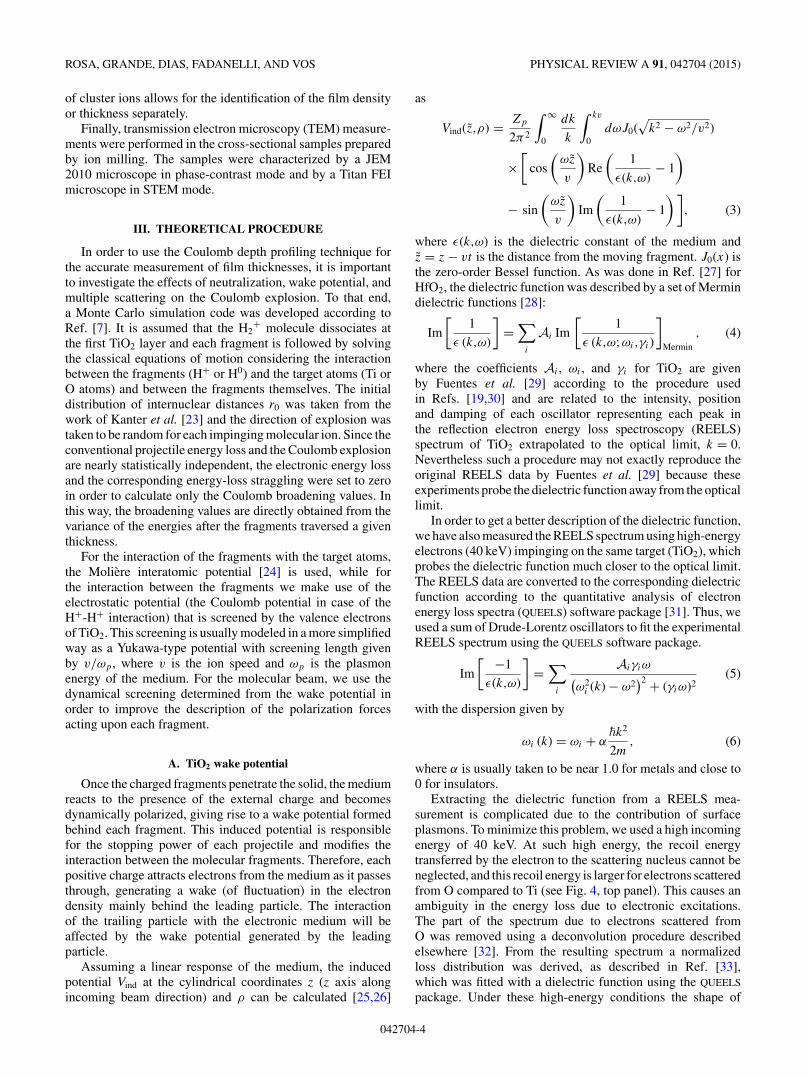
Extracting the dielectric function from a REELS mea-surement is complicated due to the contribution of surfaceplasmons. To minimize this problem, we used a high incomingenergy of 40 keV. At such high energy, the recoil energytransferred by the electron to the scattering nucleus cannot beneglected, and this recoil energy is larger for electrons scatteredfrom O compared to Ti (see Fig. 4, top panel). This causes anambiguity in the energy loss due to electronic excitations.The part of the spectrum due to electrons scattered fromO was removed using a deconvolution procedure describedelsewhere [32]. From the resulting spectrum a normalizedloss distribution was derived, as described in Ref. [33],which was fitted with a dielectric function using the QUEELS
package. Under these high-energy conditions the shape of
042704-4
NEUTRALIZATION AND WAKE EFFECTS ON THE . . . PHYSICAL REVIEW A 91, 042704 (2015)
FIG. 4. (Color online) The top panel shows the measured REELSspectrum (dots) which shows separate elastic peaks for electronsscattered from Ti an O. After deconvolution of the O contribution(solid line) the spectrum can be treated in the same way as a lowenergy REELS spectrum. The bottom panel shows the obtainednormalised loss function (dots), and a fit using a Drude-Lorentzdielectric function (solid line). The dashed line corresponds to thelimit of Im[−1/ε(k,ω)] for k = 0.
the normalized loss distribution is very close to the shapeof Im[−1/ε(k,ω)] for k = 0 as is illustrated in Fig. 4, bottompanel. The coefficients of the obtained fit are reproduced inTable II.
TABLE II. Drude-Lindhard parameters used in Eq. (5) for the fitin Fig. 4. α was taken to be 0.05.
ωi (eV) Ai (eV2) γi (eV)
6.2 1.65 2.411 16.8 314 17.9 318 25.5 727 266.9 1239.5 16.6 347.5 124 5.553.5 366 1790 102 50
For optical limit k = 0, the Drude-Lorentz and Mermindielectric functions coincide for a given set of ωi,Ai , and γi .However, since Eq. (3) involves an integration over the transfermomentum k, the values for the induced potential will differfor a Drude-Lorentz and Mermin dielectric functions, even ifthey coincide at k = 0. It should be pointed out that althoughwe used α = 0.05 as Fuentes et al. [29] to extract the dielectricfunction for TiO2 from REELS measurements, we used α = 1to calculate the wake potential from Eq. (3) in order to improvethe description of high-energy transfers.
In fact electron scattering measurements mainly probe elec-tronic excitations with low momentum transfer. Ion stopping(as wake potential) is also sensitive to excitations at highmomentum transfer [34]. Therefore, calculations of the ionstopping based on these REELS-derived dielectric functionsfail when a small value of α is used. This is mainly due tothe fact that the Bethe ridge part of the electronic excitations(binary collisions between particle and target electron, at highmomentum transfer, where this particle acts as a free particle)implies that α is 1 at high momentum transfer. If we usethe same oscillators as derived from a REELS experiment,but assuming a Mermin-type momentum dependence or anα value of 1, then we do get values for the ion stopping inreasonable agreement with experiment. Assuming a Mermin-type dispersion or α = 1 would decrease the accuracy withwhich the REELS data are described. As a matter of fact,extracting the dielectric function with the right momentumdependence that is consistent with all sum rules is currentlystill an open question [35].
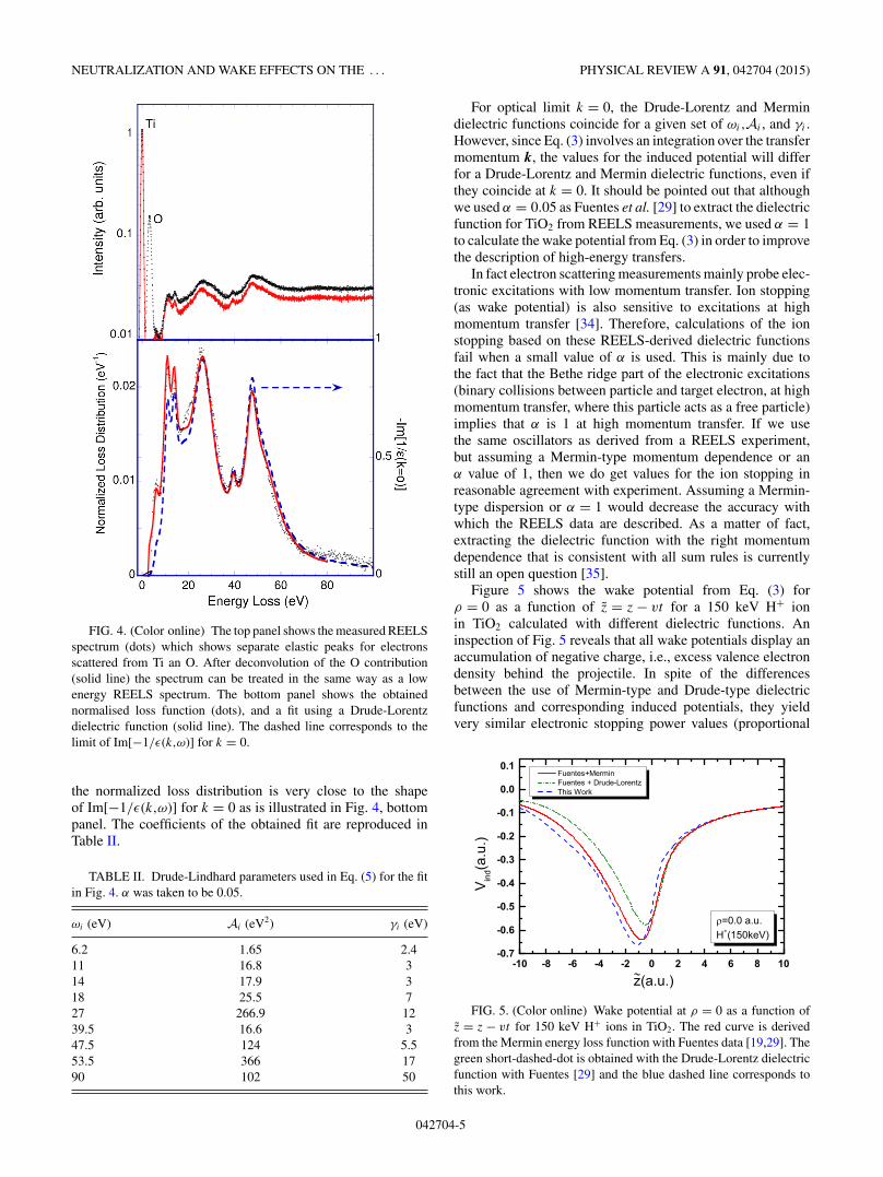
Figure 5 shows the wake potential from Eq. (3) forρ = 0 as a function of z = z − vt for a 150 keV H+ ionin TiO2 calculated with different dielectric functions. Aninspection of Fig. 5 reveals that all wake potentials display anaccumulation of negative charge, i.e., excess valence electrondensity behind the projectile. In spite of the differencesbetween the use of Mermin-type and Drude-type dielectricfunctions and corresponding induced potentials, they yieldvery similar electronic stopping power values (proportional
FIG. 5. (Color online) Wake potential at ρ = 0 as a function ofz = z − vt for 150 keV H+ ions in TiO2. The red curve is derivedfrom the Mermin energy loss function with Fuentes data [19,29]. Thegreen short-dashed-dot is obtained with the Drude-Lorentz dielectricfunction with Fuentes [29] and the blue dashed line corresponds tothis work.
042704-5
ROSA, GRANDE, DIAS, FADANELLI, AND VOS PHYSICAL REVIEW A 91, 042704 (2015)
to the first derivative of Vind at ρ = 0 and z = 0). Indeed, theelectronic stopping powers are 11.9 eV/A and 12.0 eV/Afor Mermin and Drude-Lorentz respectively using Fuentesdata, and 15.7 eV/A for Drude-Lorentz using the presentdata. Moreover, all of them produce about the same valuesfor the Coulomb broadening when one considers a H2
+ beam(150 keV/u, normal incidence) into a 10 nm thick TiO2 film,namely 530 eV and 529 eV for Mermin and Drude-Lorentzrespectively using Fuentes data, and 529 eV for Drude-Lorentzusing present data. In particular, the very similar resultsobtained for the Coulomb broadening is a direct consequenceof the similarity of the wake potentials shown in Fig. 5. Despitethe differences, the shape of the curves are nearly the same,leading to similar wake forces. In what follows, we make useof the Mermin-type dielectric function because of its improvedbuilt-in dispersion relation.
B. Neutralization
According to semiempirical formulas [36] for the meancharge state of protons traversing a solid, the neutralizationof protons is expected to be important for energies smallerthan 100 keV/u. From the experimental point of view, theneutralization of the fragments is observed by comparing thetotal yield of the MEIS spectrum for different energies. Sincethe neutral fraction of H0 is not detected, as the MEIS setupuses an electrostatic analyzer, the corresponding reduction ofthe total yield of the Ti peak (normalized by the scattering crosssection) is consistent with the increasing of H0 charge-statefraction [36,37].
In the Monte Carlo simulations we assume that theneutralization of H+ ions takes place inside the solid accordingto the electron capture and loss cross sections as discussed inRef. [6]. An estimate of the electron loss cross section for H0
particles in TiO2 as a function of energy was obtained fromexperimental results for CO2 gas targets [38]. The capture crosssection is then calculated based on the constraint that it shouldreproduce the value of the mean charge state from Ref. [36].The uncertainty in the determination of the electron loss crosssection affects only the mean distance for charge-exchangeprocesses and has a minor effect on the simulated values ofthe Coulomb broadening.
The mean charge state of each moving fragment is affectedby the presence of the nearby fragment. This vicinage effecton the charge state was observed in carbon foils [39] as afunction of the H2
+ ion energy and was used here to correct thevalues of the mean charge state from Ref. [36]. A multiplicativecorrection factor was used and amounts 0.91 at 30 keV/u and0.94 at 120 keV/u [39] and interpolated/extrapolated valueswere used for the energies studied in this work.
Another effect that has to considered is the modification ofthe atomic radius of the H0 fragment by the wake potentialgenerated by its nucleus just after the capture process. Infact, the atomic radius of an embedded H0 is larger thanthe Bohr radius a0 (0.529 A). This effect modifies the totalforce acting upon that neutral fragment due to a neighboringpositive fragment. In order to take this effect into account, wedescribe the H0 bound electron density ρ(�r) by an effectivecharge Zeff according to ρ(�r) = A exp(−2Zeffr), where A isa normalization constant. Thus, the atomic radius will simplybe a0/Zeff . The Zeff is obtained from the variational principle
TABLE III. The values of Zeff and bound state radius rbound
(=1/Zeff ) as a function of H0 kinetic energy.
Energy (keV) Zeff rbound
30 0.74 1.3550 0.79 1.2770 0.83 1.20100 0.87 1.15150 0.90 1.11200 0.93 1.08
taking into account not only the Coulomb interaction from themoving nucleus but also the wake potential generated by thesame nucleus according to Eq. (3) (see Table III). No vicinageeffect was taken into account for the determination of Zeff .
C. Multiple scattering
The effect of the nuclear collisions are taken into accountby the Monte Carlo simulations but turn out to be of minorimportance since the TiO2 films are not thick enough toproduce a sizable effect. While multiple scattering events affectthe distribution of internuclear distances at larger depths, it hasa negligible effect at smaller depths according to the presentsimulations. Therefore, at the energies studied in this work, nu-clear multiple scattering does not contribute significantly to σC .
IV. RESULTS AND DISCUSSIONS
The results for the Coulomb broadening σC for 50, 120, and150 keV/u H2
+ ions traversing the thin TiO2 film as a functionof the apparent thickness [thickness/cos(θ ), where θ is theincidence angle] are shown in Fig. 6. The Coulomb broadeningvalues and the apparent thicknesses were obtained from thebest fit of the MEIS energy spectra for three different scatteringangles (112◦, 120◦ and 128◦). The uncertainties associatedwith the Coulomb broadening were estimated based on theχ2 minimization procedure. The thicknesses are expressed interms of atoms/cm2. Moreover, it is important to stress that theexperimental results for the Coulomb broadening σC (verticalaxis) and the total amount of atoms (horizontal axis) do notdepend on the film density used in the fitting analysis. Thesolid curves displayed in Fig. 2 correspond to the simulationsdescribed in Sec. III assuming a TiO2 density of 3.4 g/cm3
to convert the depth scale from nanometers to atoms/cm2. Infact, this density is the one that provides the best fit of the 1Denergy spectra such as those shown in Fig. 2. It is important tonote that this density is 10% lower than the one of TiO2 in theamorphous phase, i.e 3.8 g/cm3 [40]). The use of 3.4 g/cm3 inthe simulations led to a thickness of 10 nm for the TiO2 film.This thickness is in good agreement with transmission electronmicroscopy (TEM) images as the ones shown in Fig. 7. Thisimage also shows the presence of voids in the TiO2 film, whichconfirms a decrease in film density. Figure 6 also shows thatthe values of σC as well as its dependence on film thicknessare larger for increasing projectile energies.
We have also performed ellipsometry measurements todetermine the thickness of the TiO2 film. Wavelengths rangingfrom 0.37–0.63 μm were used in these experiments. Theresults obtained for the film thickness was 10 ± 2 nm, while
042704-6

NEUTRALIZATION AND WAKE EFFECTS ON THE . . . PHYSICAL REVIEW A 91, 042704 (2015)
FIG. 6. (Color online) The Coulomb broadening as a function ofthe depth traversed by the fragments of H2
+ molecules in ultrathinTiO2 film obtained at energies of 50, 120, and 150 keV/u. The curvesrepresent the calculations of the Coulomb broadening assuming aCoulomb repulsive potential screened by the wake potential.
the refractive index varied from 2.58 at 0.37 μm to 2.21 at0.63 μm. These values are slightly smaller than those frombulk TiO2, which confirms the relatively lower density of theTiO2 film.
The Coulomb depth profile technique yields the same filmthickness for all energies only if wake and neutralizationeffects are properly taken into account. As expected, theseeffects decrease the energy loss straggling due to the Coulombexplosion at lower energies. The importance of each effect asa function of the energy for the H2
+ molecules is depicted inFig. 8. The Coulomb broadening σC , evaluated after 20 nm,traversed by two H+ fragments and assuming a pure Coulombrepulsion potential, is shown as a black solid curve. It increaseswith the ion energy because it depends on the product ofthe ion velocities in the laboratory and CM systems after acomplete Coulomb explosion. On the other hand, for a fixed
FIG. 7. (a) High-resolution TEM image of the TiO2 film usingphase contrast. (b) Scanning transmission electron microscope(STEM) image of the same sample. In (b) no difference could beobserved between the contributions of TiO2 and SiO2, present as anative oxide.
traversed distance, the dwell time decreases as the ion velocityincreases, thus reducing the effect of the explosion but thesimulation shows that this is less important. The screeningeffect from the Yukawa screening function (with screeninglength given by v/ωp, with ωp = 12.1 eV [41]) reduces theeffect of the Coulomb explosion mainly for low energies.The Coulomb explosion assisted by wake forces (dashed linein Fig. 8) yields σC values that are smaller at low energiesand larger at higher energies when compared with the freeCoulomb explosion (i.e., the Coulomb explosion expected forH+-H+ in vacuum). For lower energies, the wake potential hasa screening effect similar to the Yukawa potential. At energieslarger than 150 keV/u, the wake force on the trailing protontends to align this fragment behind the leading proton [13].This alignment increases the value of σC . In all cases, theneutralization, which plays an important role at energieslower than 100 keV/u, weakens the Coulomb explosion andthus decreases σC . At 150 keV/u, the simple free Coulomb
042704-7

ROSA, GRANDE, DIAS, FADANELLI, AND VOS PHYSICAL REVIEW A 91, 042704 (2015)
FIG. 8. (Color online) The Coulomb broadening (σC) as a func-tion of the H2
+ energy for ions traversing a 10 nm thick TiO2 filmalong a trajectory 60 degrees away from the surface normal. Thelines stand for calculations of σC in different scenarios: assuminga free Coulomb explosion (full black line); assuming the Coulombrepulsive potential screened by Yukawa potential (dot-dashed redline); assuming a Coulomb repulsive potential screened by wakepotential (dashed blue line). All calculations were obtained assumingtwo H+ fragments. The purple curve includes the possibility of havingH0 fragments.
explosion model yields the same Coulomb broadening as themore sophisticated calculations for the present case.
Figure 9 shows the results of Monte Carlo simulationswhere the initial orientation �in of the clusters is notrandomized for a H2
+ beam at 200 keV/u. On the left panels,σC is shown as a function of thickness for two interactingpotentials and three initial orientations, namely �in = 0◦,30◦,and 80◦ (see inset of Fig. 9). On the right panels, it is shownthe corresponding distributions of angular orientations after athickness of 200 A is traversed by the ions. For the sake ofclarity, multiple-scattering effects due to collisions with thetarget nuclei (Ti and O) have been turned off. Therefore, fora central potential as the Coulomb potential, the final angularorientations are identical to the initial orientations. On theother hand, a change in alignment is clearly observed forthe wake interaction. For initial orientations correspondingto �in = 30◦ and 80◦, a large fraction of the fragments arerotated towards smaller value of �, which will enhance σC .In fact, the wake potential has two effects: alignment andscreening. The enhancement of σC due to the alignment isclearly visible for �in = 80◦. For this case, screening is ofminor importance since trailing H+ ions are out of the wake.For �in = 0◦, no alignment takes place and the wake potentialjust screens the Coulomb interaction, yielding a smaller valueof σC . For �in = 30◦, both effects compensate each other.
V. CONCLUSIONS
In this work we exploit the Coulomb explosion of H2+
molecules in order to improve the depth profiling capabilities
FIG. 9. (Color online) On the right the Coulomb broadening σC
for three fixed initial H2+ orientations (�in = 0◦,30◦, and 80◦) at
200 keV/u. On the left the distribution of final cluster orientationsafter traversing a layer of 200 A.
of ion scattering techniques at energies lower than 200 keV/u.The correct interpretation of the energy-loss straggling σC
due to the Coulomb explosion allows the determination of thedwell time of the fragments before the backscattering collision.Consequently, the film thickness and the corresponding densitycan be obtained in a straightforward manner. At low energies,neutralization and wake effects are shown to be importantand their influence on σC can be accurately modeled byMonte Carlo simulations. The neutralization is also affectedby the presence of the wake potential and vicinage effects. Athigh energies, only the wake forces acting upon the chargedfragments are important and can enhance the value of σC
compared to a free Coulomb explosion. For the TiO2 filmstudied in this work we obtained the same thickness at differentprojectile energies indicating that the energy dependences ofthe different contributions are modeled correctly. The validityof the present results were checked by TEM and STEMmeasurements. The lower density found in our targets isexplained by the presence of voids as observed by STEM.
ACKNOWLEDGMENTS
We are indebted to the Brazilian agencies CAPES, CNPq,and PRONEX-FAPERGS for the partial support of thisresearch project. We also thank D. L. Baptista for the STEMmeasurements. M.V. acknowledges support of the AustralianResearch Council.
[1] S. M. Shubeita, R. C. Fadanelli, J. F. Dias, and P. L. Grande,Surf. Sci. 608, 292 (2013).
[2] M. J. Gaillard, J.-C. Poizat, A. Ratkowski, and J. Remillieux,Nucl. Instrum. Methods 132, 69 (1976).
042704-8

NEUTRALIZATION AND WAKE EFFECTS ON THE . . . PHYSICAL REVIEW A 91, 042704 (2015)
[3] R. Levi-Setti, K. Lam, and T. Fox, Nucl. Instrum. Methods 194,281 (1982).
[4] A. L’Hoir, C. Cohen, J. J. Ganem, I. Trimaille, I. C. Vickridge,and S. M. Shubeita, Phys. Rev. A 85, 042901 (2012).
[5] M. Gaillard, J.-C. Poizat, J. Remillieux, A. Chateau-Thierry,A. Gladieux, and W. Brandt, Nucl. Instrum. Methods 132, 547(1976).
[6] M. C. Cross, Phys. Rev. B 15, 602 (1977).[7] C. D. Denton, I. Abril, M. D. Barriga-Carrasco, R. Garcia-
Molina, G. H. Lantschner, J. C. Eckardt, and N. R. Arista,Nucl. Instrum. Methods Phys. Res., Sect. B 193, 198 (2002).
[8] P. de Vera, I. Abril, and R. Garcia-Molina, Appl. Radiat. Isot.83, Part B, 122 (2014).
[9] I. Abril, J. C. Moreno-Marın, J. M. Fernandez-Varea,C. D. Denton, S. Heredia-Avalos, and R. Garcia-Molina,Nucl. Instrum. Methods Phys. Res., Sect. B 256, 172 (2007).
[10] P. de Vera, R. Garcia-Molina, I. Abril, and A. V. Solov’yov,Phys. Rev. Lett. 110, 148104 (2013).
[11] S. Heredia-Avalos, R. Garcia-Molina, J. M. Fernandez-Varea,and I. Abril, Phys. Rev. A 72, 052902 (2005).
[12] R. Garcia-Molina, C. D. Denton, I. Abril, and N. R. Arista,Phys. Rev. A 62, 012901 (2000).
[13] C. D. Denton, I. Abril, R. Garcia-Molina, and S. Heredia-Avalos,Nucl. Instrum. Methods Phys. Res., Sect. B 256, 137 (2007).
[14] J. F. V. der Veen, Surf. Sci. Rep. 5, 199 (1985).[15] J. R. Tesmer and M. A. Nastasi, Handbook of Modern Ion Beam
Materials Analysis: Materials Research Society, 1st ed. (MRS,Pittsburgh, 1995).
[16] M. A. Sortica, P. L. Grande, G. Machado, and L. Miotti, J. Appl.Phys. 106, 114320 (2009).
[17] W. K. Chu, Backscattering Spectrometry (Academic Press,New York, 1978).
[18] J. F. Ziegler, M. D. Ziegler, and J. P. Biersack Nucl. Instrum.Methods Phys. Res., Sect. B 268, 1818 (2010).
[19] S. Limandri, R. Fadanelli, M. Behar, L. Nagamine, J. Fernandez-Varea, I. Abril, R. Garcia-Molina, C. Montanari, J. Aguiar, D.Mitnik, J. Miraglia, and N. Arista, Eur. Phys. J. D 68, 194 (2014).
[20] W. K. Chu, Phys. Rev. A 13, 2057 (1976).
[21] S. M. Shubeita, M. A. Sortica, P. L. Grande, J. F. Dias, andN. R. Arista, Phys. Rev. B 77, 115327 (2008).
[22] P. L. Grande, A. Hentz, R. P. Pezzi, I. J. R. Baumvol, and G.Schiwietz, Nucl. Instrum. Methods Phys. Res., Sect. B 256, 92(2007).
[23] E. P. Kanter, P. J. Cooney, D. S. Gemmell, K. O. Groeneveld,W. J. Pietsch, A. J. Ratkowski, Z. Vager, and B. J. Zabransky,Phys. Rev. A 20, 834 (1979).
[24] G. Moliere, Z. Naturforsch. A 2, 133 (1947).[25] I. Abril, R. Garcia-Molina, C. D. Denton, F. J. Perez-Perez, and
N. R. Arista, Phys. Rev. A 58, 357 (1998).[26] M. D. Barriga-Carrasco and C. Deutsch, Plasma Phys.
Controlled Fusion 48, 1787 (2006).[27] M. Behar, R. C. Fadanelli, I. Abril, R. Garcia-Molina, C. D.
Denton, L. C. C. M. Nagamine, and N. R. Arista, Phys. Rev. A80, 062901 (2009).
[28] N. Mermin, Phys. Rev. B 1, 2362 (1970).[29] G. G. Fuentes, E. Elizalde, F. Yubero, and J. M. Sanz,
Surf. Interface Anal. 33, 230 (2002).[30] I. Abril (personal communication).[31] F. Yubero and S. Tougaard, Surf. Interface Anal. 19, 269 (1992).[32] M. Vos and P. L. Grande [Nucl. Instrum. Methods Phys. Res.,
Sect. B (to be published)], doi: 10.1016/j.nimb.2014.11.083.[33] S. Tougaard and I. Chorkendorff, Phys. Rev. B 35, 6570 (1987).[34] U. Fano, Annu. Rev. Nucl. Sci. 13, 1 (1963).[35] C. T. Chantler and J. D. Bourke, Phys. Rev. B 90, 174306 (2014).[36] G. Schiwietz and P. L. Grande, Nucl. Instrum. Methods Phys.
Res., Sect. B 175–177, 125 (2001).[37] G. Schiwietz and P. L. Grande, Nucl. Instrum. Methods Phys.
Res., Sect. B 153, 1 (1999).[38] Y. Nakai, T. Shirai, T. Tabata, and R. Ito, At. Data Nucl. Data
Tables 37, 69 (1987).[39] B. Mazuy, A. Belkacem, M. Chevallier, M. J. Gaillard, J.-C.
Poizat, and J. Remillieux, Nucl. Instrum. Methods Phys. Res.,Sect. B 33, 105 (1988).
[40] D. Mergel, D. Buschendorf, S. Eggert, R. Grammes, and B.Samset, Thin Solid Films 371, 218 (2000).
[41] D. W. Fischer, Phys. Rev. B 5, 4219 (1972).
042704-9