Neurohistology. Neuron Components Research by Purpura – Mental retardation (MR) is from failure of...
7
Neurohistology
-
Upload
kathleen-horn -
Category
Documents
-
view
221 -
download
3
Transcript of Neurohistology. Neuron Components Research by Purpura – Mental retardation (MR) is from failure of...

Neurohistology
Neuron Components• Research by Purpura
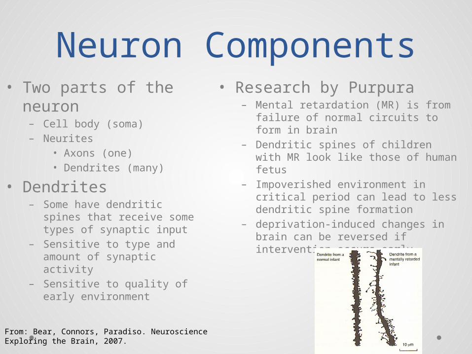
– Mental retardation (MR) is from failure of normal circuits to form in brain
– Dendritic spines of children with MR look like those of human fetus
– Impoverished environment in critical period can lead to less dendritic spine formation
– deprivation-induced changes in brain can be reversed if intervention occurs early
• Two parts of the neuron– Cell body (soma)– Neurites
• Axons (one)• Dendrites (many)
• Dendrites– Some have dendritic spines
that receive some types of synaptic input
– Sensitive to type and amount of synaptic activity
– Sensitive to quality of early environment
From: Bear, Connors, Paradiso. Neuroscience Exploring the Brain, 2007.
Classification of Neurons
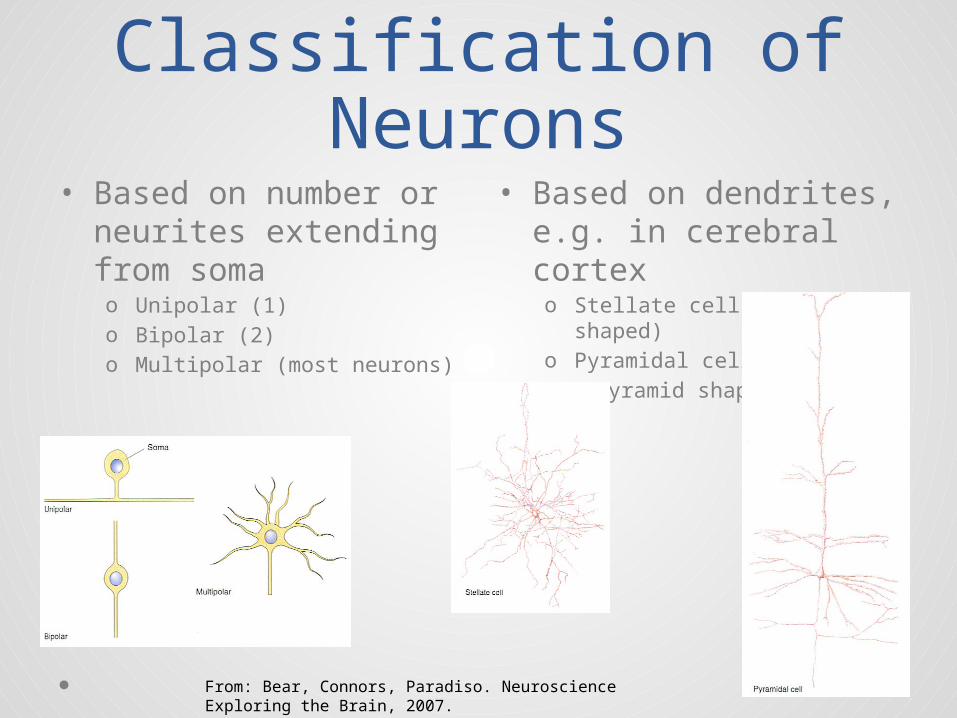
• Based on dendrites, e.g. in cerebral cortexo Stellate cells (star shaped)o Pyramidal cells (pyramid shaped)
• Based on number or neurites extending from somao Unipolar (1)o Bipolar (2)o Multipolar (most neurons)
From: Bear, Connors, Paradiso. Neuroscience Exploring the Brain, 2007.

Classification of Neurons
• Based on neurotransmitter– Cholinergic, i.e. cells that
release acetylcholine• Used in the motor system
to command voluntary movements
• Based on connections– Primary sensory– Primary motor– Interneurons (connect only
with other neurons)
• Based on axon length– Golgi type I extend from one
part of brain to another (e.g. pyramidal cells)
– Golgi type II are more local (e.g. stellate cells that never extend beyond cerebral cortex)
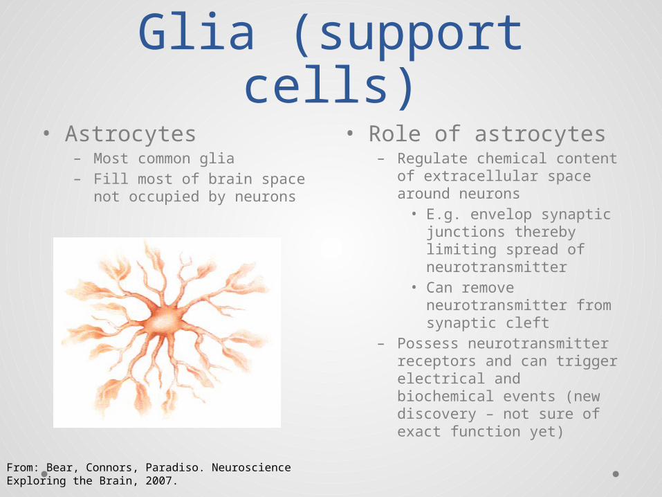
Glia (support cells)• Role of astrocytes
– Regulate chemical content of extracellular space around neurons• E.g. envelop synaptic
junctions thereby limiting spread of neurotransmitter
• Can remove neurotransmitter from synaptic cleft
– Possess neurotransmitter receptors and can trigger electrical and biochemical events (new discovery – not sure of exact function yet)
• Astrocytes– Most common glia– Fill most of brain space not
occupied by neurons
From: Bear, Connors, Paradiso. Neuroscience Exploring the Brain, 2007.

Myelinated Glia• Myelin spiral around
axon to form the myelin sheath
• Myelin sheath is interrupted periodically by nodes of Ranvier
• Oligodendrocytes (in CNS)– Can myelinate several
neurons
• Schwann cells (in PNS)– Only myelinates one neuron
From: Bear, Connors, Paradiso. Neuroscience Exploring the Brain, 2007.
Cross section of optic nerve myelinated fibers
Other Non-Neuronal Cells
• Microgliao Function as phagocytes to
remove debris from degenerating neurons and glia
• Ependymal cellso Line the fluid filled
ventricleso Play role in directing cell
migration during brain development