NEBNext Ultra II RNA Library Prep Kit for Illumina for use ...
46
1 INSTRUCTION MANUAL NEBNext ® Ultra ™ II RNA Library Prep Kit for Illumina ® for use with NEBNext Multiplex Oligos for Illumina (Unique Dual Index UMI Adaptors RNA Set 1, NEB #E7416) NEB #E7770S/L, #E7775S/L 24/96 reactions Version 1.0_7/20 Table of Contents Required Materials Not Included ........................................................................................................................................................ 2 Considerations on Choosing an RNA-seq Library Preparation Method .......................................................................................... 2 NEBNext Ultra II RNA Product Selection Guide ............................................................................................................................... 3 NEBNext Ultra II RNA Protocol Selection Guide .............................................................................................................................. 3 Overview ............................................................................................................................................................................................. 3 Section 1 Protocol for use with NEBNext Poly(A) mRNA Magnetic Isolation Module.................................................................................... 4 Section 2 Protocol for use with NEBNext rRNA Depletion Kit (Human/Mouse/Rat) ..................................................................................... 12 Section 3 Protocol for use with FFPE RNA, NEBNext rRNA Depletion Kit (Human/Mouse/Rat) ................................................................ 20 Section 4 Protocol for use with Purified mRNA or rRNA Depleted RNA ....................................................................................................... 27 Section 5 Protocol for use with rRNA Depleted FFPE RNA............................................................................................................................ 33 Section 6 Appendix A ...................................................................................................................................................................................... 39 Section 7 Troubleshooting Guide ..................................................................................................................................................................... 42 Kit Components ................................................................................................................................................................................. 44 Revision History ................................................................................................................................................................................ 46 The Library Prep Kit Includes The volumes provided are sufficient for preparation of up to 24 reactions (NEB #E7770S/#E7775S) and 96 reactions (NEB #E7770L/#E7775L). Package 1: Store at –20°C. (lilac) NEBNext First Strand Synthesis Reaction Buffer (lilac) Random Primers (lilac) NEBNext First Strand Synthesis Enzyme Mix (orange) NEBNext Second Strand Synthesis Enzyme Mix (orange) NEBNext Second Strand Synthesis Reaction Buffer (green) NEBNext Ultra II End Prep Enzyme Mix (green) NEBNext Ultra II End Prep Reaction Buffer (red) NEBNext Ultra II Ligation Master Mix
Transcript of NEBNext Ultra II RNA Library Prep Kit for Illumina for use ...

1
INSTRUCTION MANUAL NEBNext® Ultra™ II RNA Library Prep Kit for Illumina®
for use with NEBNext Multiplex Oligos for Illumina (Unique Dual Index UMI Adaptors RNA Set 1, NEB #E7416) NEB #E7770S/L, #E7775S/L 24/96 reactions Version 1.0_7/20 Table of Contents
Required Materials Not Included ........................................................................................................................................................ 2
Considerations on Choosing an RNA-seq Library Preparation Method .......................................................................................... 2
NEBNext Ultra II RNA Product Selection Guide ............................................................................................................................... 3
NEBNext Ultra II RNA Protocol Selection Guide .............................................................................................................................. 3
Overview ............................................................................................................................................................................................. 3
Section 1 Protocol for use with NEBNext Poly(A) mRNA Magnetic Isolation Module .................................................................................... 4
Section 2 Protocol for use with NEBNext rRNA Depletion Kit (Human/Mouse/Rat) ..................................................................................... 12
Section 3 Protocol for use with FFPE RNA, NEBNext rRNA Depletion Kit (Human/Mouse/Rat) ................................................................ 20
Section 4 Protocol for use with Purified mRNA or rRNA Depleted RNA ....................................................................................................... 27
Section 5 Protocol for use with rRNA Depleted FFPE RNA ............................................................................................................................ 33
Section 6 Appendix A ...................................................................................................................................................................................... 39
Section 7 Troubleshooting Guide ..................................................................................................................................................................... 42
Kit Components ................................................................................................................................................................................. 44
Revision History ................................................................................................................................................................................ 46
The Library Prep Kit Includes The volumes provided are sufficient for preparation of up to 24 reactions (NEB #E7770S/#E7775S) and 96 reactions (NEB #E7770L/#E7775L).
Package 1: Store at –20°C.
(lilac) NEBNext First Strand Synthesis Reaction Buffer
(lilac) Random Primers
(lilac) NEBNext First Strand Synthesis Enzyme Mix
(orange) NEBNext Second Strand Synthesis Enzyme Mix
(orange) NEBNext Second Strand Synthesis Reaction Buffer
(green) NEBNext Ultra II End Prep Enzyme Mix
(green) NEBNext Ultra II End Prep Reaction Buffer
(red) NEBNext Ultra II Ligation Master Mix

2
(red) NEBNext Ligation Enhancer
(blue) NEBNext Ultra II Q5® Master Mix
NEBNext Adaptor Dilution Buffer (0.1X) TE Buffer Nuclease-free Water
Package 2: Store at room temperature. Do not freeze.
Supplied only with NEBNext Ultra II RNA Library Prep with Sample Purification Beads, NEB #E7775.
NEBNext Sample Purification Beads
Required Materials Not Included • Magnetic rack/stand (NEB #S1515, Alpaqua®, cat. #A001322 or equivalent)
• Magnetic rack or plate (for example NEBNext Magnetic Separation Rack NEB #S1515, Alpaqua® 96S Super Magnet Plate #A001322, or equivalent)
• 80% Ethanol (freshly prepared)
• Thermal cycler
• Any thin wall 200 µl PCR tube
• Agilent® Bioanalyzer® or similar fragment analyzer and associated consumables
For NEB #E7770 only:
• SPRIselect® Reagent Kit (Beckman Coulter, Inc. #B23317) or AMPure® XP Beads (Beckman Coulter, Inc. #A63881)
• In NEB #E7775 beads are included [Sample Purification Beads (NEB #E7767)]
For use with NEBNext Poly(A) mRNA Magnetic Isolation Module:
• NEB #E7490 (not included)
• 96-well 0.2 ml PCR Plates and Microseal® 'B' Adhesive Sealer (BioRad MSB-1001) or 0.2 ml RNase-free tube
• 1.5 ml Microcentrifuge tube and NEB #S1506 Magnet stand or equivalent (for washing beads only)
For use with NEBNext Ribosomal Depletion Kit:
• NEB #E6310 (not included) Agencourt® RNAClean® XP Beads (Beckman Coulter, Inc. #A63987)
• In NEB #E6350, beads are included [RNA Sample Purification Beads (NEB #E6351)]
Considerations on Choosing an RNA-seq Library Preparation Method The library preparation protocol should be chosen based on the goals of the project and quality of the RNA sample. Total cellular RNA is mainly composed of rRNA and often is not of interest. rRNA can be removed from total cellular RNA by one of two common methods. The first method uses oligo d(T) beads, which bind to the poly(A) tail of eukaryotic mRNA. Alternatively, rRNA can be depleted using rRNA specific probes. NEB offers the NEBNext Poly(A) mRNA Magnetic Isolation Module (NEB #E7490) and the NEBNext rRNA Depletion Kit (Human/Mouse/Rat) (NEB #E6310) for the enrichment of non-ribosomal RNA.
In the oligo d(T) approach, only mRNA with poly(A) tails will be enriched; other cellular RNA without a poly(A) tail, such as non-coding RNA or mRNA lacking poly(A) will not bind to the beads. In addition, mRNA from some organisms (e.g., prokaryotes) or degraded RNA (e.g., FFPE RNA) does not have poly(A) tails and will not be captured by oligo d(T) beads. On the other hand, the probe based rRNA depletion kit will remove the targeted ribosomal RNA, but it will preserve other biologically relevant cellular RNA such as non-coding RNA or mRNA.
The quality of an RNA sample should also be considered when deciding on a library preparation protocol. The NEBNext Poly(A) mRNA Magnetic Isolation Module should only be used with high quality RNA samples (RIN > 7), since degradation results in a loss of poly(A) tails from mRNA molecules. For partially degraded or heavily degraded samples (e.g., RIN ≤ 7, FFPE RNA), the NEBNext rRNA Depletion Kit should be used or total RNA should be directly converted to RNA-seq libraries without any enrichment or depletion.
Please refer to the product page on NEB.com for FAQs about this product.

3
NEBNext Ultra II RNA Product Selection Guide Use the following chart to determine the required kits to make RNA libraries based on starting input material. The chart contains kit recommendations only for the protocols described in this manual. For other RNA library needs or related products, please refer to www.nebnext.com.
Figure 1. NEBNext Ultra II RNA Product Selection Guide.
NEBNext Ultra II RNA Protocol Selection Guide Use the following chart to determine the most suitable protocol in this manual. Every chapter in this manual contains a different protocol based on the starting material. More detailed information is available at the beginning of each chapter. Please read the RNA sample recommendations and input amount requirements in its entirety before starting the protocol.
Figure 2. NEBNext Ultra II RNA Protocol Selection Guide.
Overview The NEBNext Ultra II RNA Library Prep Kit for Illumina contains the enzymes and buffers required to convert a broad range of input amounts of RNA into high quality non-directional libraries for next-generation sequencing on the Illumina platform. The fast, user-friendly workflow has minimal hands-on time and is compatible with poly(A) mRNA enrichment and rRNA depletion methods.
Each kit component must pass rigorous quality control standards, and for each new lot the entire set of reagents is functionally validated together by construction and sequencing of an indexed transcriptome library on the Illumina sequencing platform.
For larger volume requirements, customized and bulk packaging is available by purchasing through the OEM/Bulks department at NEB. Please contact [email protected] for further information.
NEBNext Poly(A) mRNA Magnetic Isolation Module (NEB #E7490)
NEBNext rRNA Depletion Kit
(NEB #E6310)
Total RNA
NEBNext rRNA Depletion Kit
(NEB #E6310)
FFPE RNAPoly(A)-enrichedor rRNA-depleted
RNA
rRNA-depletedFFPE RNA*
NEBNext Ultra II RNA Library Prep Kit for Illumina (NEB #E7770)
Choose from one of the following:• NEBNext Multiplex Oligos for Illumina (Index Primers Set 1) (12 indices – NEB #E7335)• NEBNext Multiplex Oligos for Illumina (Index Primers Set 2) (12 indices – NEB #E7500)• NEBNext Multiplex Oligos for Illumina (Index Primers Set 3) (12 indices – NEB #E7710)• NEBNext Multiplex Oligos for Illumina (Index Primers Set 4) (12 indices – NEB #E7730)• NEBNext Multiplex Oligos for Illumina (96 Index Primers) (NEB #E6609)• NEBNext Multiplex Oligos for Illumina (Dual Index Primers Set 1) (NEB #E7600)
Startingmaterial
* or Total FFPE RNA when rRNA depletion is not desirable
Chapter 1 Chapter 2
Total RNA
Chapter 3
FFPE RNA
Chapter 5Chapter 4
Poly(A)-enrichedor rRNA-depleted
RNA
rRNA-depletedFFPE RNA*
RNA fragmentation(sample dependent)
First strand cDNA synthesis
Second strand cDNA synthesis
End repair/dA-tailing
Adaptor ligation
PCR enrichment of libraries
Startingmaterial
Starting chapter
NEBNext Poly(A) mRNA Magnetic Isolation Module (NEB #E7490)
Poly(A) mRNA isolation using:
NEBNext rRNA Depletion Kit
(NEB #E6310)
rRNA depletion using:
NEBNext rRNA Depletion Kit
(NEB #E6310)
rRNA depletionusing:
* or Total FFPE RNA when rRNA depletion is not desirable

4
Section 1 Protocol for use with NEBNext Poly(A) mRNA Magnetic Isolation Module (NEB #E7490) Symbols
This caution sign signifies a step in the protocol that has two paths leading to the same end point but is dependent on a user variable, like the type of RNA input.
This is a point where you can safely stop the protocol and store the samples prior to proceeding to the next step in the protocol.
Colored bullets indicate the cap color of the reagent to be added.
The protocol has been optimized using high quality Universal Human Reference Total RNA.
RNA Sample Recommendations RNA Integrity: Assess the quality of the Input RNA by running the RNA sample on an Agilent Bioanalyzer® RNA 6000 Nano/Pico Chip. For PolyA mRNA enrichment, high quality RNA with a RIN score > 7 is required.
RNA Sample Requirements: The RNA sample should be free of salts (e.g., Mg2+ and guanidinium salts), divalent cation chelating agents (e.g., EDTA and EGTA) or organics (e.g., phenol or ethanol). RNA must be free of DNA. gDNA is a common contaminant from RNA preps. It may be carried over from the interphase of organic extractions or when the silica matrix of solid phase RNA purification methods is overloaded. If the total RNA sample may contain gDNA contamination, treat the sample with DNase I to remove all traces of DNA (DNase is not provided in this kit). After treatment with DNase I the enzyme should be removed from the sample. Any residual activity of the DNase I may degrade the oligos necessary for the enrichment. DNase I can be removed from the extraction using phenol/chloroform extraction and ethanol precipitation.
Input Amount Requirements
10 ng–1 µg DNA-free total RNA quantified by Qubit® Fluorometer and quality checked by Bioanalyzer. The protocol is optimized for approximately 200 bp RNA inserts. To generate libraries with longer RNA insert sizes, refer to Appendix A (Section 6) for recommended fragmentation times and size selection conditions.
Keep all the buffers on ice, unless otherwise indicated.
1.1. Preparation of First Strand Reaction Buffer and Random Primer Mix
1.1.1. Prepare the First Strand Synthesis Reaction Buffer and Random Primer Mix (2X) in a nuclease-free microcentrifuge tube as follows:
COMPONENT VOLUME
(lilac) NEBNext First Strand Synthesis Reaction Buffer 8 µl
(lilac) NEBNext Random Primers 2 µl
Nuclease-free Water 10 µl
Total Volume 20 µl
You can prepare the first strand synthesis reaction buffer later in the protocol, but it is important that it is ready before the elution in Step 1.2.36. The beads should not be allowed to dry out.
1.1.2. Mix thoroughly by pipetting up and down ten times.
Note: Keep the mix on ice until mRNA is purified. It will be used in Step 1.2.36.
1.2. mRNA Isolation, Fragmentation and Priming Starting with Total RNA
1.2.1. Dilute the total RNA with nuclease-free water to a final volume of 50 μl in a nuclease-free 0.2 ml PCR tube and keep on ice.
1.2.2. To wash the Oligo dT Beads, add the following to a 1.5 ml nuclease-free tube. If preparing multiple libraries, beads for up to 10 samples can be added to a single 1.5 ml tube for subsequent washes (use magnet NEB #S1506 for 1.5 ml tubes). The purpose of
5
this step is to bring the beads from the storage buffer into the binding buffer. The 2X Binding Buffer does not have to be diluted for this step.
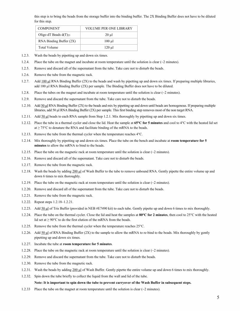
COMPONENT VOLUME PER ONE LIBRARY
Oligo dT Beads d(T)25 20 µl
RNA Binding Buffer (2X) 100 µl
Total Volume 120 µl
1.2.3. Wash the beads by pipetting up and down six times.
1.2.4. Place the tube on the magnet and incubate at room temperature until the solution is clear (~2 minutes).
1.2.5. Remove and discard all of the supernatant from the tube. Take care not to disturb the beads.
1.2.6. Remove the tube from the magnetic rack.
1.2.7. Add 100 μl RNA Binding Buffer (2X) to the beads and wash by pipetting up and down six times. If preparing multiple libraries, add 100 µl RNA Binding Buffer (2X) per sample. The Binding Buffer does not have to be diluted.
1.2.8. Place the tubes on the magnet and incubate at room temperature until the solution is clear (~2 minutes).
1.2.9. Remove and discard the supernatant from the tube. Take care not to disturb the beads.
1.2.10. Add 50 μl RNA Binding Buffer (2X) to the beads and mix by pipetting up and down until beads are homogenous. If preparing multiple libraries, add 50 μl RNA Binding Buffer (2X) per sample. This first binding step removes most of the non target RNA.
1.2.11. Add 50 μl beads to each RNA sample from Step 1.2.1. Mix thoroughly by pipetting up and down six times.
1.2.12. Place the tube in a thermal cycler and close the lid. Heat the sample at 65°C for 5 minutes and cool to 4°C with the heated lid set at ≥ 75°C to denature the RNA and facilitate binding of the mRNA to the beads.
1.2.13. Remove the tube from the thermal cycler when the temperature reaches 4°C.
1.2.14. Mix thoroughly by pipetting up and down six times. Place the tube on the bench and incubate at room temperature for 5 minutes to allow the mRNA to bind to the beads.
1.2.15. Place the tube on the magnetic rack at room temperature until the solution is clear (~2 minutes).
1.2.16. Remove and discard all of the supernatant. Take care not to disturb the beads.
1.2.17. Remove the tube from the magnetic rack.
1.2.18. Wash the beads by adding 200 μl of Wash Buffer to the tube to remove unbound RNA. Gently pipette the entire volume up and down 6 times to mix thoroughly.
1.2.19. Place the tube on the magnetic rack at room temperature until the solution is clear (~2 minutes).
1.2.20. Remove and discard all of the supernatant from the tube. Take care not to disturb the beads.
1.2.21. Remove the tube from the magnetic rack.
1.2.22. Repeat steps 1.2.18–1.2.21.
1.2.23. Add 50 μl of Tris Buffer (provided in NEB #E7490 kit) to each tube. Gently pipette up and down 6 times to mix thoroughly.
1.2.24. Place the tube on the thermal cycler. Close the lid and heat the samples at 80°C for 2 minutes, then cool to 25°C with the heated lid set at ≥ 90°C to do the first elution of the mRNA from the beads.
1.2.25. Remove the tube from the thermal cycler when the temperature reaches 25°C.
1.2.26. Add 50 μl of RNA Binding Buffer (2X) to the sample to allow the mRNA to re-bind to the beads. Mix thoroughly by gently pipetting up and down six times.
1.2.27. Incubate the tube at room temperature for 5 minutes.
1.2.28. Place the tube on the magnetic rack at room temperature until the solution is clear (~2 minutes).
1.2.29. Remove and discard the supernatant from the tube. Take care not to disturb the beads.
1.2.30. Remove the tube from the magnetic rack.
1.2.31. Wash the beads by adding 200 μl of Wash Buffer. Gently pipette the entire volume up and down 6 times to mix thoroughly.
1.2.32. Spin down the tube briefly to collect the liquid from the wall and lid of the tube.
Note: It is important to spin down the tube to prevent carryover of the Wash Buffer in subsequent steps.
1.2.33 Place the tube on the magnet at room temperature until the solution is clear (~2 minutes).

6
1.2.34. Remove and discard all of the supernatant from the tube. Take care not to disturb the beads that contains the mRNA.
Note: It is important to remove all of the supernatant to successfully fragment the mRNA in the subsequent steps. Spin down the tube. Place the tube on the magnetic rack and with a 10 µl tip, remove all of the wash buffer. (Caution: Do not disturb beads that contain the mRNA). Avoid letting the beads dry out before adding elution buffer.
1.2.35. Remove the tube from the magnetic rack.
Note: The next step provides a fragmentation incubation time resulting in an RNA insert size of ~ 200 nt. For RNA insert sizes > 200 nt, refer to Section 6 (Appendix A) for recommended fragmentation times in Step 1.2.37.
1.2.36. To elute the mRNA from the beads and fragment, add 11.5 μl of the First Strand Synthesis Reaction Buffer and Random Primer Mix (2X) prepared in Step 1.1.2, pipette up and down six times to resuspend the beads.
1.2.37 Incubate the sample in a thermal cycler with the heated lid set at 105°C as follows: 15 minutes at 94°C Hold at 4°C* *Immediately transfer the tube to ice for 1 minute as soon as it is cool enough to handle (~65°C)
1.2.38. Quickly spin down the tube in a microcentrifuge to collect the liquid from the sides of the tube and place on the magnet right away until the solution is clear (~1-2 minutes).
1.2.39. Collect the fragmented mRNA by transferring 10 μl of the supernatant to a nuclease-free 0.2 ml PCR tube.
Note 1: If the supernatant volume recovered is less than 10 μl for any reason, bring the volume up to 10 μl by adding the First Strand Synthesis Reaction Buffer and Random Primer Mix (2X) prepared in Step 1.1.2 and continue with the protocol.
Note 2: Avoid transferring any of the magnetic beads.
1.2.40. Place the tube on ice and proceed directly to First Strand cDNA Synthesis.
1.3. First Strand cDNA Synthesis
1.3.1. Assemble the first strand cDNA synthesis reaction on ice by adding the following components into fragmented and primed RNA from Step 1.2.40.
FIRST STRAND cDNA SYNTHESIS REACTION VOLUME
Fragmented and primed RNA (Step 1.2.40) 10 µl
Nuclease-free Water 8 µl
(lilac) NEBNext First Strand Synthesis Enzyme Mix 2 µl
Total Volume 20 µl
1.3.2. Mix thoroughly by pipetting up and down at least 10 times.

7
1.3.3. Incubate the sample in a preheated thermal cycler with the heated lid set at ≥ 80°C as follows:
Note: If you are following recommendations in Appendix A, for longer RNA fragments (creating inserts > 200 bases), increase the incubation at 42°C from 15 minutes to 50 minutes at Step 2.
Step 1: 10 minutes at 25°C Step 2: 15 minutes at 42°C Step 3: 15 minutes at 70°C Step 4: Hold at 4°C
1.3.4. Immediately, perform Second Strand cDNA Synthesis.
1.4. Second Strand cDNA Synthesis
1.4.1. Assemble the second strand cDNA synthesis reaction on ice by adding the following components into the first strand synthesis reaction product from Step 1.3.4.
SECOND STRAND SYNTHESIS REACTION VOLUME
First-Strand Synthesis Product (Step 1.3.4) 20 µl
(orange) NEBNext Second Strand Synthesis Reaction Buffer (10X) 8 µl
(orange) NEBNext Second Strand Synthesis Enzyme Mix 4 µl
Nuclease-free Water 48 µl
Total Volume 80 µl
1.4.2. Keeping the tube on ice, mix thoroughly by pipetting the reaction up and down at least 10 times.
1.4.3. Incubate in a thermal cycler for 1 hour at 16°C with the heated lid set at ≤ 40°C (or off).
1.5. Purification of Double-stranded cDNA using SPRIselect Beads or NEBNext Sample Purification Beads
1.5.1. Vortex SPRIselect Beads or NEBNext Sample Purification Beads to resuspend.
1.5.2. Add 144 μl (1.8X) of resuspended beads to the second strand synthesis reaction (~80 μl). Mix well on a vortex mixer or by pipetting up and down at least 10 times.
1.5.3. Incubate for 5 minutes at room temperature.
1.5.4. Briefly spin the tube in a microcentrifuge to collect any sample from the sides of the tube. Place the tube on a magnetic rack to separate beads from the supernatant. After the solution is clear, carefully remove and discard the supernatant. Be careful not to disturb the beads, which contain DNA. (Caution: do not discard beads).
1.5.5. Add 200 μl of freshly prepared 80% ethanol to the tube while in the magnetic stand. Incubate at room temperature for 30 seconds and then carefully remove and discard the supernatant.
1.5.6. Repeat Step 1.5.5 once for a total of 2 washing steps.
1.5.7. Air dry the beads for up to 5 minutes while the tube is on the magnetic rack with the lid open.
Caution: Do not over-dry the beads. This may result in lower recovery of DNA target. Elute the samples when the beads are still dark brown and glossy looking, but when all visible liquid has evaporated. When the beads turn lighter brown and start to crack, they are too dry.
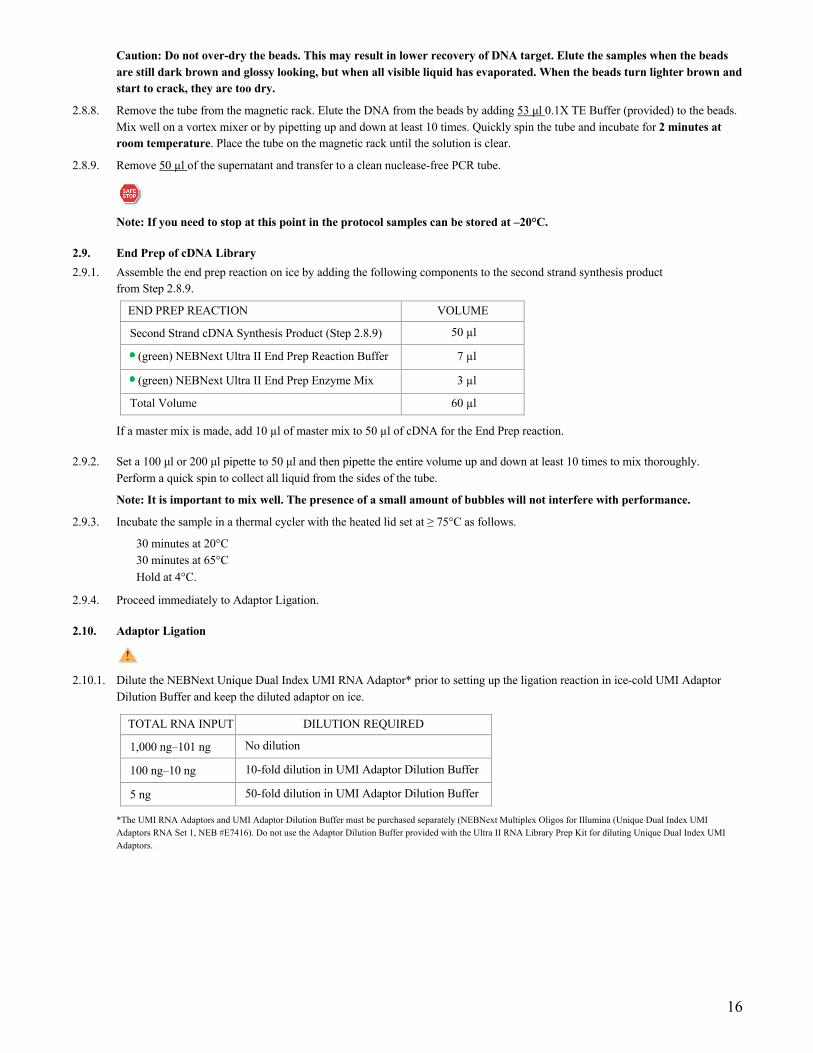
1.5.8. Remove the tube from the magnet. Elute the DNA target from the beads by adding 53 μl 0.1X TE Buffer (provided) to the beads. Mix well on a vortex mixer or by pipetting up and down ten times. Briefly spin the tube and incubate for 2 minutes at room temperature. Place the tube on the magnetic rack until the solution is clear.
1.5.9. Remove 50 µl of the supernatant and transfer to a clean nuclease-free PCR tube.
Note: If you need to stop at this point in the protocol, samples can be stored at –20°C.

8
1.6. End Prep of cDNA Library
1.6.1. Assemble the end prep reaction on ice by adding the following components to second strand synthesis product from Step 1.5.9.
END PREP REACTION VOLUME
Second Strand cDNA Synthesis Product (Step 1.5.9) 50 µl
(green) NEBNext Ultra II End Prep Reaction Buffer 7 µl
(green) NEBNext Ultra II End Prep Enzyme Mix 3 µl
Total Volume 60 µl
1.6.2. Set a 100 μl or 200 μl pipette to 50 μl and then pipette the entire volume up and down at least 10 times to mix thoroughly. Perform a quick spin to collect all liquid from the sides of the tube.
Note: It is important to mix well. The presence of a small amount of bubbles will not interfere with performance.
1.6.3. Incubate the sample in a thermal cycler with the heated lid set at ≥ 75°C as follows: 30 minutes at 20°C 30 minutes at 65°C Hold at 4°C
1.6.4. Proceed immediately to Adaptor Ligation.
1.7. Adaptor Ligation
1.7.1. Dilute the NEBNext Unique Dual Index UMI RNA Adaptor* prior to setting up the ligation reaction in ice-cold UMI Adaptor Dilution Buffer and keep the diluted adaptor on ice.
TOTAL RNA INPUT DILUTION REQUIRED
1,000 ng–250 ng No dilution
249 ng–100 ng 10-fold dilution in UMI Adaptor Dilution Buffer
99 ng–10 ng 50-fold dilution in UMI Adaptor Dilution Buffer
*The UMI RNA Adaptors and UMI Adaptor Dilution Buffer must be purchased separately (NEBNext Multiplex Oligos for Illumina (Unique Dual Index UMI Adaptors RNA Set 1, NEB #E7416). Do not use the Adaptor Dilution Buffer provided with the Ultra II RNA Library Prep Kit for diluting Unique Dual Index UMI Adaptors.
1.7.2. Assemble the ligation reaction on ice by adding the following components, in the order given, to the end prep reaction product from Step 1.6.4.
LIGATION REACTION VOLUME
End Prepped DNA (Step 1.6.4) 60 µl
Diluted Adaptor (Step 1.7.1) 5 µl
(red) NEBNext Ligation Enhancer 1 µl
(red) NEBNext Ultra II Ligation Master Mix 30 µl
Total Volume 96 µl
The Ligation Master Mix and Ligation Enhancer can be mixed ahead of time and is stable for at least 8 hours @ 4°C. We do not recommend premixing the Ligation Master Mix, Ligation Enhancer and adaptor prior to use in the Adaptor Ligation Step.
1.7.3. Set a 100 μl or 200 μl pipette to 80 μl and then pipette the entire volume up and down at least 10 times to mix thoroughly. Perform a quick spin to collect all liquid from the sides of the tube.
Caution: The NEBNext Ultra II Ligation Master Mix is very viscous. Care should be taken to ensure adequate mixing of the ligation reaction, as incomplete mixing will result in reduced ligation efficiency. The presence of a small amount of bubbles will not interfere with performance.
1.7.4. Incubate 15 minutes at 20°C (heated lid off) in a thermal cycler.

9
1.7.5 Proceed immediately to Purification of the Ligation Reaction.
1.8. Purification of the Ligation Reaction Using SPRIselect Beads or NEBNext Sample Purification Beads
Note: If you are selecting for larger insert size libraries (> 200 nt) follow the size selection recommendations in Appendix A, Section 6.
1.8.1. Add 70 μl (0.7X) resuspended SPRIselect Beads or NEBNext Sample Purification Beads and mix well on a vortex mixer or by pipetting up and down at least 10 times.
1.8.2. Incubate for 10 minutes at room temperature.
1.8.3. Quickly spin the tube in a microcentrifuge and place the tube on an appropriate magnetic rack to separate beads from the supernatant. After the solution is clear (~ 5 minutes), discard the supernatant that contain unwanted fragments (Caution: do not discard beads).
1.8.4. Add 200 μl of freshly prepared 80% ethanol to the tube while in the magnetic rack. Incubate at room temperature for 30 seconds and then carefully remove and discard the supernatant.
1.8.5. Repeat Step 1.8.4 once for a total of 2 washing steps.
1.8.6. Briefly spin the tube and put the tube back in the magnetic rack.
1.8.7. Completely remove the residual ethanol and air dry beads until the beads are dry for up to 5 minutes while the tube is on the magnetic rack with the lid open.
Caution: Do not over-dry the beads. This may result in lower recovery of DNA target. Elute the samples when the beads are still dark brown and glossy looking, but when all visible liquid has evaporated. When the beads turn lighter brown and start to crack, they are too dry.
1.8.8. Remove the tube from the magnet. Elute DNA target from the beads by adding 22 μl 0.1X TE (provided) to the beads. Mix well on a vortex mixer or by pipetting up and down. Quickly spin the tube and incubate for 2 minutes at room temperature. Put the tube in the magnetic rack until the solution is clear.
1.8.9. Without disturbing the bead pellet, transfer 20 μl of the supernatant to a clean PCR tube and proceed to PCR enrichment.
Note: If you need to stop at this point in the protocol samples can be stored at –20°C.

10
1.9. PCR Enrichment of Adaptor Ligated DNA
1.9.1. Set up the PCR reaction as described below.
COMPONENT VOLUME PER ONE LIBRARY
Adaptor Ligated DNA (Step 1.8.9) 20 µl
(blue) NEBNext Ultra II Q5 Master Mix 25 µl
NEBNext Primer Mix* 5 µl
Total Volume 50 µl
* NEBNext Primer Mix is provided in NEBNext Multiplex Oligos for Illumina (Unique Dual Index UMI Adaptors RNA Set 1, NEB #E7416).
1.9.2. Mix well by gently pipetting up and down 10 times. Quickly spin the tube in a microcentrifuge.
1.9.3. Place the tube on a thermal cycler with the heated lid set to 105°C and perform PCR amplification using the following PCR cycling conditions (refer to Table 1.9.3A and Table 1.9.3B):
Table 1.9.3A:
CYCLE STEP TEMP TIME CYCLES
Initial Denaturation 98°C 30 seconds 1
Denaturation 98°C 10 seconds 7–16*, **
Annealing/Extension 65°C 75 seconds
Final Extension 65°C 5 minutes 1
Hold 4°C ∞
* The number of PCR cycles should be adjusted based on RNA input (Table 1.9.3B).
** It is important to limit the number of PCR cycles to avoid overamplification. If overamplification occurs, a second peak ~ 1,000 bp will appear on the Bioanalyzer trace (See Figure 7.1 on page 43).
Table 1.9.3B: Recommended PCR cycles based on total RNA input amount:
TOTAL RNA INPUT RECOMMENDED PCR CYCLES
1,000 ng 7–8
100 ng 11–12
10 ng 14–16
Note: PCR cycles are recommended based on high quality Universal Human Reference Total RNA. It may require optimization based on the sample quality to prevent PCR over-amplification.
1.10. Purification of the PCR Reaction using SPRIselect Beads or NEBNext Sample Purification Beads
1.10.1. Vortex SPRIselect Beads or NEBNext Sample Purification Beads to resuspend.
1.10.2. Add 45 μl (0.9X) of resuspended beads to the PCR reaction (~ 50 μl). Mix well on a vortex mixer or by pipetting up and down at least 10 times.
1.10.3. Incubate for up to 5 minutes at room temperature.
1.10.4. Quickly spin the tube in a microcentrifuge and place the tube on an appropriate magnetic rack to separate beads from the supernatant. After the solution is clear (about 5 minutes), carefully remove and discard the supernatant. Be careful not to disturb the beads that contain DNA targets.
1.10.5. Add 200 μl of freshly prepared 80% ethanol to the tube while in the magnetic rack. Incubate at room temperature for 30 seconds and then carefully remove and discard the supernatant.
1.10.6. Repeat Step 1.10.5 once for a total of 2 washing steps.
1.10.7. Air dry the beads for up to 5 minutes while the tube is on the magnetic rack with the lid open.

11
Caution: Do not over-dry the beads. This may result in lower recovery of the DNA target. Elute the samples when the beads are still dark brown and glossy looking, but when all visible liquid has evaporated. When the beads turn lighter brown and start to crack, they are too dry.
1.10.8. Remove the tube from the magnetic rack. Elute the DNA target from the beads by adding 23 μl 0.1X TE (provided) to the beads. Mix well on a vortex mixer or by pipetting up and down ten times. Quickly spin the tube in a microcentrifuge and incubate for 2 minutes at room temperature. Place the tube in the magnetic rack until the solution is clear.
1.10.9. Transfer 20 μl of the supernatant to a clean PCR tube and store at –20°C.
1.11 Assess Library Quality on an Agilent Bioanalyzer DNA Chip
1.11.1. Use a Bioanalyzer or Tape Station to determine the size distribution and concentration of the libraries.
1.11.3. Check that the electropherogram shows a narrow distribution with a peak size approximately 300 bp.
Note: If a peak at ~ 80 bp (primers) or ~ 150 bp (adaptor-dimer) is visible in the Bioanalyzer traces, bring up the sample volume (from Step 1.10.9) to 50 μl with 0.1X TE buffer and repeat the SPRIselect Bead or NEBNext Sample Purification Bead Cleanup Step (Section 1.10).
Figure 1.11.1. Example of a representative RNA library size distribution on a Bioanalyzer.

12
Section 2 Protocol for use with NEBNext rRNA Depletion Kit (Human/Mouse/Rat) (NEB #E6310) Symbols
This caution sign signifies a step in the protocol that has two paths leading to the same end point but is dependent on a user variable, like the type of RNA input.
This is a point where you can safely stop the protocol and store the samples prior to proceeding to the next step in the protocol.
Colored bullets indicate the cap color of the reagent to be added.
This protocol has been optimized using Universal Human Reference Total RNA.
RNA Sample Requirements RNA Integrity: Assess the quality of the input RNA by running the RNA sample on an Agilent Bioanalyzer RNA 6000 Nano/Pico Chip to determine the RNA Integrity Number (RIN). RNA with different RIN values require different fragmentation times or no fragmentation at all.
For intact (RIN > 7) or partially degraded RNA samples (RIN = 2 to 7) follow the library preparation protocol in Section 2 (current Section). See Table 2.5.1. for the recommended fragmentation times, based on RIN.
For highly degraded samples (RIN = 1 to 2) (e.g., FFPE), which do not require fragmentation, follow the library preparation protocol in Section 3.
RNA Sample Requirements: The RNA sample should be free of salts (e.g., Mg2+ and guanidinium salts) or organics (e.g., phenol and ethanol). RNA must be free of DNA. gDNA is a common contaminant from RNA preps. It may be carried over from the interphase of organic extractions or when the silica matrix of solid phase RNA purification methods is overloaded. If the total RNA sample may contain gDNA contamination, treat the sample with DNase I to remove all traces of DNA (not provided in this kit). After treatment with DNase I the enzyme should be removed from the sample. Any residual activity of DNase I will degrade the single stranded DNA probes necessary for the ribosomal depletion. DNase I can be removed from the extraction using phenol/ chloroform extraction and ethanol precipitation.
Input Amount Requirements
5 ng–1 μg total RNA (DNA free) in a 12 μl total volume, quantified by Qubit Fluorometer and quality checked by Bioanalyzer.
The protocol is optimized for approximately 200 nt RNA inserts. To generate libraries with longer RNA insert sizes, refer to Appendix A (Section 6) for recommended fragmentation times and size selection conditions.
Keep all of the buffers on ice, unless otherwise indicated.
2.1. Probe Hybridization to RNA
2.1.1. Dilute the total RNA with Nuclease-free Water to a final volume of 12 μl in a PCR tube. Keep the RNA on ice.
2.1.2. Prepare RNA/Probe master mix as follows:
RNA/PROBE MASTER MIX VOLUME
NEBNext rRNA Depletion Solution 1 µl
Probe Hybridization Buffer 2 µl
Total Volume 3 µl
2.1.3. Add 3 µl of the above mix to 12 µl total RNA (from Step 2.1.1), resulting in a total volume of 15 µl.
2.1.4. Mix thoroughly by pipetting up and down at least 10 times.
2.1.5. Briefly spin down the sample in a microcentrifuge.
13
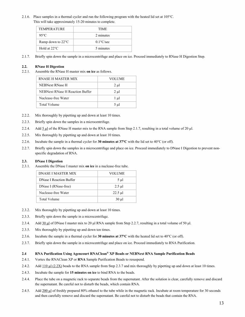
2.1.6. Place samples in a thermal cycler and run the following program with the heated lid set at 105°C. This will take approximately 15-20 minutes to complete.
TEMPERATURE TIME
95°C 2 minutes
Ramp down to 22°C 0.1°C/sec
Hold at 22°C 5 minutes
2.1.7. Briefly spin down the sample in a microcentrifuge and place on ice. Proceed immediately to RNase H Digestion Step.
2.2. RNase H Digestion 2.2.1. Assemble the RNase H master mix on ice as follows.
RNASE H MASTER MIX VOLUME
NEBNext RNase H 2 µl
NEBNext RNase H Reaction Buffer 2 µl
Nuclease-free Water 1 µl
Total Volume 5 µl 2.2.2. Mix thoroughly by pipetting up and down at least 10 times.
2.2.3. Briefly spin down the samples in a microcentrifuge.
2.2.4. Add 5 µl of the RNase H master mix to the RNA sample from Step 2.1.7, resulting in a total volume of 20 µl.
2.2.5. Mix thoroughly by pipetting up and down at least 10 times.
2.2.6. Incubate the sample in a thermal cycler for 30 minutes at 37°C with the lid set to 40°C (or off).
2.2.7. Briefly spin down the samples in a microcentrifuge and place on ice. Proceed immediately to DNase I Digestion to prevent non-specific degradation of RNA.
2.3. DNase I Digestion 2.3.1. Assemble the DNase I master mix on ice in a nuclease-free tube.
DNASE I MASTER MIX VOLUME
DNase I Reaction Buffer 5 µl
DNase I (RNase-free) 2.5 µl
Nuclease-free Water 22.5 µl
Total Volume 30 µl 2.3.2. Mix thoroughly by pipetting up and down at least 10 times.
2.3.3. Briefly spin down the sample in a microcentrifuge.
2.3.4. Add 30 μl of DNase I master mix to 20 μl RNA sample from Step 2.2.7, resulting in a total volume of 50 μl.
2.3.5. Mix thoroughly by pipetting up and down ten times.
2.3.6. Incubate the sample in a thermal cycler for 30 minutes at 37°C with the heated lid set to 40°C (or off).
2.3.7. Briefly spin down the sample in a microcentrifuge and place on ice. Proceed immediately to RNA Purification.
2.4 RNA Purification Using Agencourt RNAClean® XP Beads or NEBNext RNA Sample Purification Beads 2.4.1. Vortex the RNAClean XP or RNA Sample Purification Beads to resuspend.
2.4.2. Add 110 µl (2.2X) beads to the RNA sample from Step 2.3.7 and mix thoroughly by pipetting up and down at least 10 times.
2.4.3. Incubate the sample for 15 minutes on ice to bind RNA to the beads.
2.4.4. Place the tube on a magnetic rack to separate beads from the supernatant. After the solution is clear, carefully remove and discard the supernatant. Be careful not to disturb the beads, which contain RNA.
2.4.5. Add 200 µl of freshly prepared 80% ethanol to the tube while in the magnetic rack. Incubate at room temperature for 30 seconds and then carefully remove and discard the supernatant. Be careful not to disturb the beads that contain the RNA.

14
2.4.6. Repeat Step 2.4.5 once for a total of 2 washing steps.
2.4.7. Completely remove residual ethanol and air dry the beads for up to 5 minutes while the tube is on the magnetic rack with the lid open.
Caution: Do not over-dry the beads. This may result in lower recovery of RNA target. Elute the samples when the beads are still dark brown and glossy looking, but when all visible liquid has evaporated. When the beads turn lighter brown and start to crack, they are too dry.
2.4.8. Remove the tube from the magnet. Elute the RNA from the beads by adding 7 µl Nuclease-free Water. Mix well by pipetting up and down at least 10 times and briefly spin the tube.
2.4.9. Incubate for 2 minutes at room temperature. Place the tube in the magnet until the solution is clear (~2 minutes).
2.4.10. Remove 5 µl of the supernatant containing RNA and transfer to a nuclease-free tube.
2.4.11. Place the sample on ice and proceed to RNA Fragmentation and Priming.
2.5. RNA Fragmentation and Priming
RNA fragmentation is only required for intact or partially degraded RNA. Recommended fragmentation times can be found in Table 2.5.1.
2.5.1. Assemble the following fragmentation and priming reaction on ice:
FRAGMENTATION AND PRIMING REACTION VOLUME
Ribosomal RNA Depleted Sample (Step 2.4.11) 5 µl
(lilac) NEBNext First Strand Synthesis Reaction Buffer 4 µl
(lilac) Random Primers 1 µl
Total Volume 10 µl 2.5.2. Mix thoroughly by pipetting up and down ten times.
2.5.3. Place the sample on a thermal cycler and incubate the sample at 94°C following the recommendations in Table 2.5.1 below for libraries with inserts ~200 nt.
Table 2.5.1. Suggested fragmentation times based on RIN value of RNA input.
RNA TYPE RIN FRAG. TIME
Intact RNA > 7 15 min. at 94°C
Partially Degraded RNA 2–6 7–8 min. at 94°C
Note: Refer to Appendix A (Section 6) for fragmentation conditions if you are preparing libraries with large
inserts (> 200 bp). Conditions in Appendix A only apply for intact RNA.
2.5.4. Immediately transfer the tube to ice and proceed to First Strand cDNA Synthesis.

15
2.6. First Strand cDNA Synthesis
2.6.1. Assemble the first strand synthesis reaction on ice by adding the following components to the fragmented and primed RNA from Step 2.5.4:
FIRST STRAND SYNTHESIS REACTION VOLUME
Fragmented and primed RNA (Step 2.5.4) 10 µl
Nuclease-free Water 8 µl
(lilac) NEBNext First Strand Synthesis Enzyme Mix 2 µl
Total Volume 20 µl 2.6.2. Mix thoroughly by pipetting up and down ten times.
2.6.3. Incubate the sample in a preheated thermal cycler with the heated lid set at ≥ 80°C as follows:
Note: If you are following recommendations in Appendix A (Section 6), for libraries with longer inserts (> 200 bases), increase the incubation at 42°C from 15 minutes to 50 minutes at Step 2 below.
Step 1: 10 minutes at 25°C Step 2: 15 minutes at 42°C Step 3: 15 minutes at 70°C Step 4: Hold at 4°C
2.6.4. Proceed directly to Second Strand cDNA Synthesis.
2.7. Second Strand cDNA Synthesis
2.7.1. Assemble the second strand cDNA synthesis reaction on ice by adding the following components into the first strand synthesis product from Step 2.6.4.
SECOND STRAND SYNTHESIS REACTION VOLUME
First-Strand Synthesis Product (Step 2.6.4) 20 µl
(orange) NEBNext Second Strand Synthesis Reaction Buffer (10X) 8 µl
(orange) NEBNext Second Strand Synthesis Enzyme Mix 4 µl
Nuclease-free Water 48 µl
Total Volume 80 µl
2.7.2. Keeping the tube on ice, mix thoroughly by pipetting up and down at least 10 times.
2.7.3. Incubate in a thermal cycler for 1 hour at 16°C with the heated lid set at ≤ 40°C (or off).
2.8. Purification of Double-stranded cDNA Using SPRIselect Beads or NEBNext Sample Purification Beads
2.8.1. Vortex SPRIselect Beads or NEBNext Sample Purification Beads to resuspend.
2.8.2. Add 144 μl (1.8X) of resuspended beads to the second strand synthesis reaction (~80 μl). Mix well on a vortex mixer or by pipetting up and down at least 10 times.
2.8.3. Incubate for 5 minutes at room temperature.
2.8.4. Briefly spin the tube in a microcentrifuge to collect any sample on the sides of the tube. Place the tube on a magnet to separate beads from the supernatant. After the solution is clear, carefully remove and discard the supernatant. Be careful not to disturb the beads, which contain DNA. (Caution: do not discard beads).
2.8.5. Add 200 μl of freshly prepared 80% ethanol to the tube while in the magnetic rack. Incubate at room temperature for 30 seconds and then carefully remove and discard the supernatant.
2.8.6. Repeat Step 2.8.5 once for a total of 2 washing steps.
2.8.7. Air dry the beads for up to 5 minutes while the tube is on the magnetic rack with lid open.
16
Caution: Do not over-dry the beads. This may result in lower recovery of DNA target. Elute the samples when the beads are still dark brown and glossy looking, but when all visible liquid has evaporated. When the beads turn lighter brown and start to crack, they are too dry.
2.8.8. Remove the tube from the magnetic rack. Elute the DNA from the beads by adding 53 μl 0.1X TE Buffer (provided) to the beads. Mix well on a vortex mixer or by pipetting up and down at least 10 times. Quickly spin the tube and incubate for 2 minutes at room temperature. Place the tube on the magnetic rack until the solution is clear.
2.8.9. Remove 50 µl of the supernatant and transfer to a clean nuclease-free PCR tube.
Note: If you need to stop at this point in the protocol samples can be stored at –20°C.
2.9. End Prep of cDNA Library 2.9.1. Assemble the end prep reaction on ice by adding the following components to the second strand synthesis product
from Step 2.8.9.
END PREP REACTION VOLUME
Second Strand cDNA Synthesis Product (Step 2.8.9) 50 µl
(green) NEBNext Ultra II End Prep Reaction Buffer 7 µl
(green) NEBNext Ultra II End Prep Enzyme Mix 3 µl
Total Volume 60 µl
If a master mix is made, add 10 µl of master mix to 50 µl of cDNA for the End Prep reaction.
2.9.2. Set a 100 μl or 200 μl pipette to 50 μl and then pipette the entire volume up and down at least 10 times to mix thoroughly. Perform a quick spin to collect all liquid from the sides of the tube.
Note: It is important to mix well. The presence of a small amount of bubbles will not interfere with performance.
2.9.3. Incubate the sample in a thermal cycler with the heated lid set at ≥ 75°C as follows.
30 minutes at 20°C 30 minutes at 65°C Hold at 4°C.
2.9.4. Proceed immediately to Adaptor Ligation.
2.10. Adaptor Ligation
2.10.1. Dilute the NEBNext Unique Dual Index UMI RNA Adaptor* prior to setting up the ligation reaction in ice-cold UMI Adaptor Dilution Buffer and keep the diluted adaptor on ice.
TOTAL RNA INPUT DILUTION REQUIRED
1,000 ng–101 ng No dilution
100 ng–10 ng 10-fold dilution in UMI Adaptor Dilution Buffer
5 ng 50-fold dilution in UMI Adaptor Dilution Buffer
*The UMI RNA Adaptors and UMI Adaptor Dilution Buffer must be purchased separately (NEBNext Multiplex Oligos for Illumina (Unique Dual Index UMI Adaptors RNA Set 1, NEB #E7416). Do not use the Adaptor Dilution Buffer provided with the Ultra II RNA Library Prep Kit for diluting Unique Dual Index UMI Adaptors.

17
2.10.2. Assemble the ligation reaction on ice by adding the following components, in the order given, to the end prep reaction product from Step 2.9.4.
LIGATION REACTION VOLUME
End Prepped DNA (Step 2.9.4) 60 µl
Diluted Adaptor (Step 2.10.1) 5 µl
(red) NEBNext Ligation Enhancer 1 µl
(red) NEBNext Ultra II Ligation Master Mix 30 µl
Total Volume 96 µl Note: The Ligation Master Mix and Ligation Enhancer can be mixed ahead of time and is stable for at least 8 hours @
4°C. We do not recommend premixing the Ligation Master Mix, Ligation Enhancer and adaptor prior to use in the Adaptor Ligation Step.
2.10.3. Set a 100 μl or 200 μl pipette to 80 μl and then pipette the entire volume up and down at least 10 times to mix thoroughly. Perform a quick spin to collect all liquid from the sides of the tube.
Caution: The NEBNext Ultra II Ligation Master Mix is very viscous. Care should be taken to ensure adequate mixing of the ligation reaction, as incomplete mixing will result in reduced ligation efficiency. The presence of a small amount of bubbles will not interfere with performance.
2.10.4. Incubate 15 minutes at 20°C (heated lid off) in a thermal cycler.
2.10.7. Proceed immediately to Purification of the Ligation Reaction.
2.11. Purification of the Ligation Reaction Using SPRIselect Beads or NEBNext Sample Purification Beads
Note: If you are selecting for libraries with larger insert size (> 200 nt) follow the size selection recommendations in Appendix A, Section 6.
2.11.1. Add 70 μl (0.7X) resuspended SPRIselect Beads or NEBNext Sample Purification Beads and mix well on a vortex mixer or by pipetting up and down at least 10 times.
2.11.2. Incubate for 5 minutes at room temperature.
2.11.3. Quickly spin the tube in a microcentrifuge and place the tube on an appropriate magnetic rack to separate beads from the supernatant. After the solution is clear (~ 5 minutes), discard the supernatant that contains unwanted fragments. (Caution: do not discard beads).
2.11.4. Add 200 μl of freshly prepared 80% ethanol to the tube while in the magnetic rack. Incubate at room temperature for 30 seconds and then carefully remove and discard the supernatant.
2.11.5. Repeat Step 2.11.4 once for a total of 2 washing steps.
2.11.6. Briefly spin the tube and put the tube back in the magnetic rack.
2.11.7. Completely remove the residual ethanol and air dry beads until the beads are dry for up to 5 minutes while the tube is on the magnetic rack with the lid open.
Caution: Do not over-dry the beads. This may result in lower recovery of DNA target. Elute the samples when the beads are still dark brown and glossy looking, but when all visible liquid has evaporated. When the beads turn lighter brown and start to crack, they are too dry.
2.11.8. Remove the tube from the magnetic rack. Elute DNA target from the beads by adding 22 μl 0.1X TE (provided) to the beads. Mix well on a vortex mixer or by pipetting up and down. Quickly spin the tube and incubate for 2 minutes at room temperature. Put the tube in the magnet until the solution is clear.
2.11.9. Without disturbing the bead pellet, transfer 20 μl of the supernatant to a clean PCR tube and proceed to PCR enrichment. Note: If you need to stop at this point in the protocol samples can be stored at –20°C.

18
2.12. PCR Enrichment of Adaptor Ligated DNA
2.12.1. Set up the PCR reaction as described below.
COMPONENT VOLUME PER ONE LIBRARY
Adaptor Ligated DNA (Step 2.11.9) 20 µl
(blue) NEBNext Ultra II Q5 Master Mix 25 µl
NEBNext Primer Mix* 5 µl
Total Volume 50 µl
* NEBNext Primer Mix is provided in NEBNext Multiplex Oligos for Illumina (Unique Dual Index UMI Adaptors RNA Set 1, NEB #E7416).
.2.12.2. Mix well by gently pipetting up and down 10 times. Quickly spin the tube in a microcentrifuge.
2.12.3. Place the tube on a thermal cycler with the heated lid set to 105°C and perform PCR amplification using the following PCR cycling conditions (refer to Table 2.12.3A and Table 2.12.3B):
Table 2.12.3A:
CYCLE STEP TEMP TIME CYCLES
Initial Denaturation 98°C 30 seconds 1
Denaturation 98°C 10 seconds 6–16*, **
Annealing/Extension 65°C 75 seconds
Final Extension 65°C 5 minutes 1
Hold 4°C ∞
* The number of PCR cycles should be adjusted based on RNA input.
** It is important to limit the number of PCR cycles to avoid overamplification. If overamplification occurs, a second peak ~ 1,000 bp will appear on the Bioanalyzer trace (See Figure 7.1 on page 43).
Table 2.12.3B: Recommended PCR cycles based on total RNA input amount:
TOTAL RNA INPUT RECOMMENDED PCR CYCLES
1,000 ng 6–7
100 ng 10–11
10 ng 13–15
5 ng 15–16
Note: PCR cycles are recommended based on high quality Universal Human Reference Total RNA. It may require optimization based on the sample quality to prevent PCR over-amplification.
2.13. Purification of the PCR Reaction using SPRIselect Beads or NEBNext Sample Purification Beads 2.13.1. Vortex SPRIselect Beads or NEBNext Sample Purification Beads to resuspend.
2.13.2. Add 45 μl (0.9X) of resuspended beads to the PCR reaction (~ 50 μl). Mix well on a vortex mixer or by pipetting up and down at least 10 times.
2.13.3. Incubate for 5 minutes at room temperature.
2.13.4. Quickly spin the tube in a microcentrifuge and place the tube on an appropriate magnetic rack to separate beads from the supernatant. After the solution is clear (about 5 minutes), carefully remove and discard the supernatant. Be careful not to disturb the beads that contain DNA targets. (Caution: do not discard beads).
2.13.5. Add 200 μl of freshly prepared 80% ethanol to the tube while in the magnetic rack. Incubate at room temperature for 30 seconds and then carefully remove and discard the supernatant.
2.13.6. Repeat Step 2.13.5 once for a total of 2 washing steps.
2.13.7. Air dry the beads for up to 5 minutes while the tube is on the magnetic rack with the lid open.

19
Caution: Do not over-dry the beads. This may result in lower recovery of DNA target. Elute the samples when the beads are still dark brown and glossy looking, but when all visible liquid has evaporated. When the beads turn lighter brown and start to crack, they are too dry.
2.13.8. Remove the tube from the magnetic rack. Elute the DNA target from the beads by adding 23 μl 0.1X TE (provided) to the beads. Mix well on a vortex mixer or by pipetting up and down ten times. Quickly spin the tube in a microcentrifuge and incubate for 2 minutes at room temperature. Place the tube in the magnetic rack until the solution is clear.
2.13.9. Transfer 20 μl of the supernatant to a clean PCR tube and store at –20°C.
2.14. Assess Library Quality on an Agilent Bioanalyzer DNA Chip 2.14.1. Use a Bioanalyzer or Tape Station to determine the size distribution and concentration of the libraries.
2.14.2. Check that the electropherogram shows a narrow distribution with a peak size approximately 300 bp.
Note: If a peak at ~ 80 bp (primers) or ~ 150 bp (adaptor-dimer) is visible in the bioanalyzer traces, bring up the sample volume (from Step 2.13.9) to 50 μl with 0.1X TE buffer and repeat the SPRIselect Bead or NEBNext Sample Purification Bead Cleanup Step (Section 2.13).
Figure 2.14.1. Example of a representative RNA library size distribution on a Bioanalyzer.

20
Section 3 Protocol for use with FFPE RNA, NEBNext rRNA Depletion Kit (Human/Mouse/Rat) (NEB #E6310) Symbols
This caution sign signifies a step in the protocol that has two paths leading to the same end point but is dependent on a user variable, like the type of RNA input.
This is a point where you can safely stop the protocol and store the samples prior to proceeding to the next step in the protocol.
Colored bullets indicate the cap color of the reagent to be added.
This protocol has been optimized using Universal Human Reference Total RNA.
RNA Sample Requirements RNA Integrity: Assess the quality of the input RNA by running the RNA sample on an Agilent Bioanalyzer RNA 6000 Nano/Pico Chip to determine the RNA Integrity Number (RIN). RNA with different RIN values require different fragmentation times or no fragmentation at all.
For intact (RIN > 7) or partially degraded RNA samples (RIN = 2 to 7) follow the library preparation protocol in Section 2. See Table 2.5.1. for the recommended fragmentation times, based on RIN.
For highly degraded samples (RIN = 1 to 2) (e.g., FFPE), which do not require fragmentation, follow the library preparation protocol in Section 3 (current Section).
RNA Sample Requirements: The RNA sample should be free of salts (e.g., Mg2+ and guanidinium salts) or organics (e.g., phenol and ethanol). RNA must be free of DNA. gDNA is a common contaminant from RNA preps. It may be carried over from the interphase of organic extractions or when the silica matrix of solid phase RNA purification methods is overloaded. If the total RNA sample may contain gDNA contamination, treat the sample with DNase I to remove all traces of DNA (not provided in this kit). After treatment with DNase I the enzyme should be removed from the sample. Any residual activity of DNase I will degrade the single stranded DNA probes necessary for the ribosomal depletion. DNase I can be removed from the extraction using phenol/ chloroform extraction and ethanol precipitation.
Input Amount Requirements
10 ng–100 ng FFPE RNA (DNA free) in a 12 μl total volume, quantified by Qubit Fluorometer and quality checked by Bioanalyzer.
The protocol is optimized for approximately 200 nt RNA inserts.
Keep all of the buffers on ice, unless otherwise indicated.
3.1.1. Probe Hybridization to RNA
3.1.1. Dilute the total RNA with Nuclease-free Water to a final volume of 12 μl in a PCR tube. Keep the RNA on ice.
3.1.2. Prepare a RNA/Probe master mix as follows:
RNA/PROBE MASTER MIX VOLUME
NEBNext rRNA Depletion Solution 1 µl
Probe Hybridization Buffer 2 µl
Total Volume 3 µl 3.1.3. Add 3 µl of the above mix to 12 µl total RNA sample (from Step 3.1.1), resulting in a total volume of 15 µl.
3.1.4. Mix thoroughly by pipetting up and down ten times.
3.1.5. Briefly spin down the sample in a microcentrifuge.

21
3.1.6. Place samples in a thermal cycler and run the following program with the heated lid set at 105°C. This will take approximately 15-20 minutes to complete:
TEMPERATURE TIME
95°C 2 minutes
Ramp down to 22°C 0.1°C/sec
Hold at 22°C 5 minutes 3.1.7. Briefly spin down the samples in a microcentrifuge and place on ice. Proceed immediately to RNase H Digestion Step.
3.2. RNase H Digestion 3.2.1. Assemble the RNAse H master mix on ice as follows.
RNASE H MASTER MIX VOLUME
NEBNext RNase H 2 µl
NEBNext RNase H Reaction Buffer 2 µl
Nuclease-free Water 1 µl
Total Volume 5 µl 3.2.2. Mix thoroughly by pipetting up and down ten times.
3.2.3. Briefly spin down the samples in a microcentrifuge.
3.2.4. Add 5 µl of the RNase H master mix to the RNA sample from Step 3.1.7, resulting in a total volume of 20 µl.
3.2.5. Mix thoroughly by pipetting up and down ten times.
3.2.6. Incubate the sample in a thermal cycler for 30 minutes at 37°C with the lid set to 40°C (or on).
3.2.7. Briefly spin down the samples in a microcentrifuge and place on ice. Proceed immediately to DNase I Digestion to prevent non-specific degradation of RNA.
3.3. DNase I Digestion 3.3.1. Assemble DNase I digestion master mix on ice in a nuclease-free tube.
DNASE I MASTER MIX VOLUME
DNase I Reaction Buffer 5 µl
DNase I (RNase-free) 2.5 µl
Nuclease-free Water 22.5 µl
Total Volume 30 µl 3.3.2. Mix thoroughly by pipetting up and down ten times.
3.3.3. Briefly spin down the samples in a microcentrifuge.
3.3.4. Add 30 μl of DNase I digestion master mix to 20 μl RNA sample from Step 3.2.7, resulting in a total volume of 50 μl.
3.3.5. Mix thoroughly by pipetting up and down ten times.
3.3.6. Incubate the samples in a thermal cycler for 30 minutes at 37°C with the heated lid set to 40°C (or on).
3.3.7. Briefly spin down the samples in a microcentrifuge and place on ice. Proceed immediately to RNA purification.
3.4. RNA Purification Using Agencourt RNAClean XP Beads or NEBNext RNA Sample Purification Beads 3.4.1. Vortex the RNAClean XP or RNA Sample Purification Beads to resuspend.
3.4.2. Add 110 µl (2.2X) beads to the RNA sample from Step 3.3.7 and mix thoroughly by pipetting up and down at least 10 times.
3.4.3. Incubate the sample for 10 minutes on ice to bind RNA to the beads.
3.4.4. Place the tube on a magnetic rack to separate beads from the supernatant. After the solution is clear, carefully remove and discard the supernatant. Be careful not to disturb the beads, which contain RNA.
22
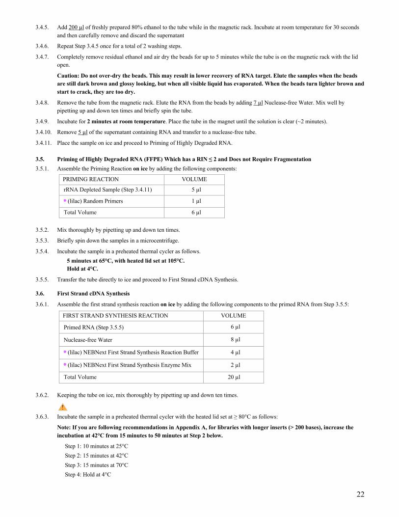
3.4.5. Add 200 µl of freshly prepared 80% ethanol to the tube while in the magnetic rack. Incubate at room temperature for 30 seconds and then carefully remove and discard the supernatant
3.4.6. Repeat Step 3.4.5 once for a total of 2 washing steps.
3.4.7. Completely remove residual ethanol and air dry the beads for up to 5 minutes while the tube is on the magnetic rack with the lid open.
Caution: Do not over-dry the beads. This may result in lower recovery of RNA target. Elute the samples when the beads are still dark brown and glossy looking, but when all visible liquid has evaporated. When the beads turn lighter brown and start to crack, they are too dry.
3.4.8. Remove the tube from the magnetic rack. Elute the RNA from the beads by adding 7 µl Nuclease-free Water. Mix well by pipetting up and down ten times and briefly spin the tube.
3.4.9. Incubate for 2 minutes at room temperature. Place the tube in the magnet until the solution is clear (~2 minutes).
3.4.10. Remove 5 µl of the supernatant containing RNA and transfer to a nuclease-free tube.
3.4.11. Place the sample on ice and proceed to Priming of Highly Degraded RNA.
3.5. Priming of Highly Degraded RNA (FFPE) Which has a RIN ≤ 2 and Does not Require Fragmentation 3.5.1. Assemble the Priming Reaction on ice by adding the following components:
PRIMING REACTION VOLUME
rRNA Depleted Sample (Step 3.4.11) 5 µl
(lilac) Random Primers 1 µl
Total Volume 6 µl 3.5.2. Mix thoroughly by pipetting up and down ten times.
3.5.3. Briefly spin down the samples in a microcentrifuge.
3.5.4. Incubate the sample in a preheated thermal cycler as follows. 5 minutes at 65°C, with heated lid set at 105°C. Hold at 4°C.
3.5.5. Transfer the tube directly to ice and proceed to First Strand cDNA Synthesis.
3.6. First Strand cDNA Synthesis
3.6.1. Assemble the first strand synthesis reaction on ice by adding the following components to the primed RNA from Step 3.5.5:
FIRST STRAND SYNTHESIS REACTION VOLUME
Primed RNA (Step 3.5.5) 6 µl
Nuclease-free Water 8 µl
(lilac) NEBNext First Strand Synthesis Reaction Buffer 4 µl
(lilac) NEBNext First Strand Synthesis Enzyme Mix 2 µl
Total Volume 20 µl
3.6.2. Keeping the tube on ice, mix thoroughly by pipetting up and down ten times.
3.6.3. Incubate the sample in a preheated thermal cycler with the heated lid set at ≥ 80°C as follows:
Note: If you are following recommendations in Appendix A, for libraries with longer inserts (> 200 bases), increase the incubation at 42°C from 15 minutes to 50 minutes at Step 2 below.
Step 1: 10 minutes at 25°C Step 2: 15 minutes at 42°C Step 3: 15 minutes at 70°C Step 4: Hold at 4°C
23
3.6.4. Proceed directly to Second Strand cDNA Synthesis Reaction.
3.7. Second Strand cDNA Synthesis
3.7.1 Assemble the second strand cDNA synthesis reaction on ice by adding the following components to the first strand reaction product from Step 3.6.4.
SECOND STRAND SYNTHESIS REACTION VOLUME
First-Strand Synthesis Product (Step 3.6.4) 20 µl
(orange) NEBNext Second Strand Synthesis Reaction Buffer (10X) 8 µl
(orange) NEBNext Second Strand Synthesis Enzyme Mix 4 µl
Nuclease-free Water 48 µl
Total Volume 80 µl 3.7.2 Keeping the tube on ice, mix thoroughly by pipetting up and down ten times.
3.7.3 Incubate in a thermal cycler for 1 hour at 16°C with the heated lid set at ≤ 40°C (or off).
3.8. Purification of Double-stranded cDNA Using SPRIselect Beads or NEBNext Sample Purification Beads
3.8.1. Vortex SPRIselect Beads or NEBNext Sample Purification Beads to resuspend.
3.8.2. Add 144 μl (1.8X) of resuspended beads to the second strand synthesis reaction (~80 μl). Mix well on a vortex mixer or by pipetting up and down at least 10 times.
3.8.3. Incubate for 5 minutes at room temperature.
3.8.4. Briefly spin the tube in a microcentrifuge to collect any sample on the sides of the tube. Place the tube on a magnet to separate beads from the supernatant. After the solution is clear, carefully remove and discard the supernatant. Be careful not to disturb the beads, which contain DNA. (Caution: do not discard beads).
3.8.5. Add 200 μl of freshly prepared 80% ethanol to the tube while in the magnetic stand. Incubate at room temperature for 30 seconds and then carefully remove and discard the supernatant.
3.8.6. Repeat Step 3.8.5 once for a total of 2 washing steps.
3.8.7. Air dry the beads for up to 5 minutes while the tube is on the magnet with lid open.
Caution: Do not over-dry the beads. This may result in lower recovery of DNA target. Elute the samples when the beads are still dark brown and glossy looking, but when all visible liquid has evaporated. When the beads turn lighter brown and start to crack they are too dry.
3.8.8. Remove the tube from the magnet. Elute the DNA from the beads by adding 53 μl 0.1X TE Buffer (provided) to the beads. Mix well on a vortex mixer or by pipetting up and down ten times. Quickly spin the tube and incubate for 2 minutes at room temperature. Place the tube on the magnetic rack until the solution is clear.
3.8.9. Remove 50 µl of the supernatant and transfer to a clean nuclease free PCR tube.
Note: If you need to stop at this point in the protocol samples can be stored at –20°C.
3.9. End Prep of cDNA Library 3.9.1. Assemble the end prep reaction on ice by adding the following components to the second strand
synthesis product from Step 3.8.9.
END PREP REACTION VOLUME
Second Strand Synthesis Product (Step 3.8.9) 50 µl
(green) NEBNext Ultra II End Prep Reaction Buffer 7 µl
(green) NEBNext Ultra II End Prep Enzyme Mix 3 µl
Total Volume 60 µl If a master mix is made, add 10 µl of master mix to 50 µl of cDNA for the End Prep reaction.

24
3.9.2. Set a 100 μl or 200 μl pipette to 50 μl and then pipette the entire volume up and down at least 10 times to mix thoroughly. Perform a quick spin to collect all liquid from the sides of the tube.
Note: It is important to mix well. The presence of a small amount of bubbles will not interfere with performance.
3.9.3. Incubate the sample in a thermal cycler with the heated lid set at ≥ 75°C as follows. 30 minutes at 20°C 30 minutes at 65°C Hold at 4°C.
3.9.4. Proceed immediately to Adaptor Ligation.
3.10. Adaptor Ligation
3.10.1. Dilute the NEBNext Unique Dual Index UMI RNA Adaptor* prior to setting up the ligation reaction in ice-cold UMI Adaptor Dilution Buffer and keep the diluted adaptor on ice.
FFPE RNA DILUTION REQUIRED
100 ng–10 ng 10-fold dilution in UMI Adaptor Dilution Buffer
*The UMI RNA Adaptors and UMI Adaptor Dilution Buffer must be purchased separately (NEBNext Multiplex Oligos for Illumina (Unique Dual Index UMI Adaptors RNA Set 1, NEB #E7416). Do not use the Adaptor Dilution Buffer provided with the Ultra II RNA Library Prep Kit for diluting Unique Dual Index UMI Adaptors.
3.10.2. Assemble the ligation reaction on ice by adding the following components, in the order given, to the end prep reaction product from Step 3.9.4.
LIGATION REACTION VOLUME
End Prepped DNA (Step 3.9.4) 60 µl
Diluted Adaptor (Step 3.10.1) 5 µl
(red) NEBNext Ligation Enhancer 1 µl
(red) NEBNext Ultra II Ligation Master Mix 30 µl
Total Volume 96 µl Note: The Ligation Master Mix and Ligation Enhancer can be mixed ahead of time and is stable for at least 8 hours @
4°C. We do not recommend premixing the Ligation Master Mix, Ligation Enhancer and adaptor prior to use in the Adaptor Ligation Step.
3.10.3. Set a 100 μl or 200 μl pipette to 80 μl and then pipette the entire volume up and down at least 10 times to mix thoroughly. Perform a quick spin to collect all liquid from the sides of the tube.
Caution: The NEBNext Ultra II Ligation Master Mix is very viscous. Care should be taken to ensure adequate mixing of the ligation reaction, as incomplete mixing will result in reduced ligation efficiency. The presence of a small amount of bubbles will not interfere with performance.
3.10.4. Incubate 15 minutes at 20°C (heated lid off) in a thermal cycler.
3.10.5. Proceed immediately to Purification of the Ligation Reaction.
3.11. Purification of the Ligation Reaction Using SPRIselect Beads or NEBNext Sample Purification Beads
Note: If you are selecting for larger size fragments (> 200 nt) follow the size selection recommendations in Appendix A, Section 6.
3.11.1. Add 70 μl (0.7X) resuspended SPRIselect Beads or NEBNext Sample Purification Beads and mix well on a vortex mixer or by pipetting up and down at least 10 times.
3.11.2. Incubate for 5 minutes at room temperature.

25
3.11.3. Quickly spin the tube in a microcentrifuge and place the tube on an appropriate magnetic rack to separate beads from the supernatant. After the solution is clear (about 5 minutes), discard the supernatant that contains unwanted fragments. (Caution: do not discard beads).
3.11.4. Add 200 μl of freshly prepared 80% ethanol to the tube while in the magnetic rack. Incubate at room temperature for 30 seconds and then carefully remove and discard the supernatant.
3.11.5. Repeat Step 3.11.4 once for a total of 2 washing steps.
3.11.6. Briefly spin the tube and put the tube back in the magnetic rack.
3.11.7. Completely remove the residual ethanol and air dry beads until the beads are dry for up to 5 minutes while the tube is on the magnetic rack with the lid open.
Caution: Do not over-dry the beads. This may result in lower recovery of DNA target. Elute the samples when the beads are still dark brown and glossy looking, but when all visible liquid has evaporated. When the beads turn lighter brown and start to crack, they are too dry.
3.11.8. Remove the tube from the magnetic rack. Elute DNA target from the beads by adding 22 μl 0.1X TE (provided) to the beads. Mix well on a vortex mixer or by pipetting up and down, incubate for 2 minutes at room temperature. Put the tube in the magnet until the solution is clear.
3.11.9. Without disturbing the bead pellet, transfer 20 μl of the supernatant to a clean PCR tube and proceed to PCR enrichment.
Note: If you need to stop at this point in the protocol samples can be stored at –20°C.
3.12. PCR Enrichment of Adaptor Ligated DNA
3.12.1. Set up the PCR reaction as described below.
COMPONENT VOLUME PER ONE LIBRARY
Adaptor Ligated DNA (Step 3.11.9) 20 µl
(blue) NEBNext Ultra II Q5 Master Mix 25 µl
NEBNext Primer Mix* 5 µl
Total Volume 50 µl
* NEBNext Primer Mix is provided in NEBNext Multiplex Oligos for Illumina (Unique Dual Index UMI Adaptors RNA Set 1, NEB #E7416).
3.12.2. Mix well by gently pipetting up and down 10 times. Quickly spin the tube in a microcentrifuge.
3.12.3. Place the tube on a thermal cycler with the heated lid set to 105°C and perform PCR amplification using the following PCR cycling conditions (refer to Table 3.12.3A and Table 3.12.3B):
Table 3.12.3A:
CYCLE STEP TEMP TIME CYCLES
Initial Denaturation 98°C 30 seconds 1
Denaturation 98°C 10 seconds 12–17*, **
Annealing/Extension 65°C 75 seconds
Final Extension 65°C 5 minutes 1
Hold 4°C ∞
* The number of PCR cycles should be adjusted based on RNA input.The recommendation of PCR cycles based on internal tests for FFPE RNA. ** It is important to limit the number of PCR cycles to avoid overamplification.
If overamplification occurs, a second peak ~ 1,000 bp will appear on the Bioanalyzer trace (See Figure 7.1 on page 43).

26
Table 3.12.3B: Recommended PCR cycles based on input amount:
FFPE RNA INPUT RECOMMENDED PCR CYCLES
100 ng 12–14
10 ng 15–17
3.13. Purification of the PCR Reaction using SPRIselect Beads or NEBNext Sample Purification Beads
3.13.1. Vortex SPRIselect Beads or NEBNext Sample Purification Beads to resuspend.
3.13.2. Add 45 μl (0.9X) of resuspended beads to the PCR reaction (~ 50 μl). Mix well on a vortex mixer or by pipetting up and down at least 10 times.
3.13.3. Incubate for up to 5 minutes at room temperature.
3.13.4. Quickly spin the tube in a microcentrifuge and place the tube on an appropriate magnetic rack to separate beads from the supernatant. After the solution is clear (about 5 minutes), carefully remove and discard the supernatant. Be careful not to disturb the beads that contain DNA targets. (Caution: do not discard beads).
3.13.5. Add 200 μl of freshly prepared 80% ethanol to the tube while in the magnetic rack. Incubate at room temperature for 30 seconds and then carefully remove and discard the supernatant.
3.13.6. Repeat Step 3.13.5 once for a total of 2 washing steps.
3.13.7. Air dry the beads for up to 5 minutes while the tube is on the magnetic rack with the lid open.
Caution: Do not over-dry the beads. This may result in lower recovery of DNA target. Elute the samples when the beads are still dark brown and glossy looking, but when all visible liquid has evaporated. When the beads turn lighter brown and start to crack, they are too dry.
3.13.8. Remove the tube from the magnetic rack. Elute the DNA target from the beads by adding 23 μl 0.1X TE (provided) to the beads. Mix well on a vortex mixer or by pipetting up and down ten times, quickly spin the tube in a microcentrifuge and incubate for 2 minutes at room temperature. Place the tube in the magnetic rack until the solution is clear.
3.13.9. Transfer 20 μl of the supernatant to a clean PCR tube and store at –20°C.
3.14. Assess Library Quality on an Agilent Bioanalyzer DNA Chip 3.14.1. Use a Bioanalyzer or Tape Station to determine the size distribution and concentration of the libraries.
3.14.2. Check that the electropherogram shows a narrow distribution with a peak size approximately 300 bp.
Note: If a peak at ~ 80 bp (primers) or ~ 150 bp (adaptor-dimer) is visible in the bioanalyzer traces, bring up the sample volume (from Step 3.13.9) to 50 μl with 1X TE Buffer and repeat the SPRIselect Bead or NEBNext Sample Purification Bead Cleanup Step (Section 3.13). Adaptor-dimer is common for low inputs of FFPE samples and if observed, a second cleanup is recommended
Figure 3.14.1. Example of a representative RNA library size distribution on a Bioanalyzer.

27
Section 4 Protocols for use with Purified mRNA or rRNA Depleted RNA Symbols
This caution sign signifies a step in the protocol that has two paths leading to the same end point but is dependent on a user variable, like the type of RNA input.
This is a point where you can safely stop the protocol and store the samples prior to proceeding to the next step in the protocol.
Colored bullets indicate the cap color of the reagent to be added.
RNA Sample Requirements This Section can be used for libraries without any enrichment or depletion of total RNA with RIN scores > 7.
RNA Integrity: RNA Integrity Number (RIN) is computed using ribosomal RNA (rRNA) amount in the sample. If rRNA is removed by any method, the RIN value should not be used to evaluate the integrity of the RNA sample. In this case, we recommend that the fragmentation time is empirically determined if the RNA sample is suspected to be low quality. The following recommendation apply to the total RNA samples only.
Assess the quality of the input RNA by running the RNA sample on an Agilent Bioanalyzer RNA 6000 Nano/Pico Chip to determine the RNA Integrity Number (RIN). RNA with different RIN values require different fragmentation times or no fragmentation at all.
For intact (RIN > 7) or partially degraded RNA samples (RIN = 2 to 7) follow the library preparation protocol in Section 4 (current Section). See Table 4.1.1 for the recommended the fragmentation times.
For highly degraded samples (RIN = 1 to 2) (e.g., FFPE), which do not require fragmentation, follow the library preparation protocol in Section 5.
RNA Sample Requirements: The RNA sample should be free of salts (e.g., Mg2+ and guanidinium salts), divalent cation chelating agents (e.g., EDTA and EGTA) or organics (e.g., phenol and ethanol). RNA must be free of DNA. gDNA is a common contaminant from RNA preps. It may be carried over from the interphase of organic extractions or when the silica matrix of solid phase RNA purification methods is overloaded. If the total RNA sample may contain gDNA contamination, treat the sample with DNase I to remove all traces of DNA (DNase is not provided in this kit). After treatment with DNase I the enzyme should be removed from the sample. DNase I can be removed from the extraction using phenol/chloroform extraction and ethanol precipitation.
Input Amount Requirements
1 ng – 100 ng purified mRNA or rRNA depleted RNA that is quantified after the purification. RNA should be DNA free in up to 5 µl of Nuclease-free Water, quantified by Qubit Fluorometer and quality checked by Bioanalyzer.
The protocol is optimized for approximately 200 nt RNA inserts. To generate libraries with longer RNA insert sizes, refer to Appendix A (Section 6) for recommended fragmentation times and size selection conditions.
This protocol has been optimized using Universal Human Reference Total RNA.

28
4.1. RNA Fragmentation and Priming
RNA fragmentation is only required for intact or partially degraded RNA. Recommended fragmentation times can be found in Table 4.1.1.
4.1.1. Assemble the fragmentation and priming reaction on ice in a nuclease-free tube by adding the following components:
FRAGMENTATION AND PRIMING MIX VOLUME
Purified mRNA or rRNA Depleted RNA 5 µl
(lilac) NEBNext First Strand Synthesis Reaction Buffer 4 µl
(lilac) Random Primers 1 µl
Total Volume 10 µl 4.1.2. Mix thoroughly by pipetting up and down ten times.
4.1.3. Place the sample in a thermal cycler and incubate the sample at 94°C following the recommendations in Table 4.1.1 below for fragment sizes ~200 nt.
Table 4.1.1 Suggested fragmentation times based on RIN value of RNA input.
RNA TYPE RIN FRAG. TIME
Intact RNA > 7 15 min. at 94°C
Partially Degraded RNA 2–6 7–8 min. at 94°C
Note: Refer to Appendix A (Section 6) for fragmentation conditions if you are preparing libraries with
large inserts (> 200 bp). Conditions in Appendix A (Section 6) only apply for intact RNA.
4.1.4. Immediately transfer the tube to ice and proceed to First Strand cDNA Synthesis.
4.2 First Strand cDNA Synthesis Reaction
4.2.1. Assemble the first strand synthesis reaction on ice by adding the following components to the fragmented and primed RNA from Step 4.1.4:
FIRST STRAND SYNTHESIS REACTION VOLUME
Fragmented and Primed RNA (Step 4.1.4) 10 µl
Nuclease-free Water 8 µl
(lilac) NEBNext First Strand Synthesis Enzyme Mix 2 µl
Total Volume 20 µl
4.2.2. Mix thoroughly by pipetting up and down ten times.
4.2.3. Incubate the sample in a preheated thermal cycler with the heated lid set at ≥ 80°C as follows:
Note: If you are following recommendations in Appendix A, (Section 6), for longer RNA fragments (creating inserts > 200 bases), increase the incubation at 42°C from 15 minutes to 50 minutes at Step 2 below.
Step 1: 10 minutes at 25°C Step 2: 15 minutes at 42°C Step 3: 15 minutes at 70°C Step 4: Hold at 4°C
4.2.4. Proceed directly to Second Strand cDNA Synthesis.

29
4.3. Second Strand cDNA Synthesis
4.3.1. Assemble the second strand cDNA synthesis reaction on ice by adding the following components to the first strand reaction product from Step 4.2.4.
SECOND STRAND SYNTHESIS REACTION VOLUME
First-Strand Synthesis Product (Step 4.2.4) 20 µl
(orange) NEBNext Second Strand Synthesis Reaction Buffer (10X) 8 µl
(orange) NEBNext Second Strand Synthesis Enzyme Mix 4 µl
Nuclease-free Water 48 µl
Total Volume 80 µl 4.3.2. Keeping the tube on ice, mix thoroughly by pipetting the reaction up and down ten times.
4.3.3. Incubate in a thermal cycler for 1 hour at 16°C with the heated lid set at ≤ 40°C (or off).
4.4. Purification of Double-stranded cDNA Using SPRIselect Beads or NEBNext Sample Purification Beads
4.4.1. Vortex SPRIselect Beads or NEBNext Sample Purification Beads to resuspend.
4.4.2. Add 144 μl (1.8X) of resuspended beads to the second strand synthesis reaction (~80 μl). Mix well on a vortex mixer or by pipetting up and down at least 10 times.
4.4.3. Incubate for 5 minutes at room temperature.
4.4.4. Briefly spin the tube in a microcentrifuge to collect any sample on the sides of the tube. Place the tube on a magnetic rack to separate beads from the supernatant. After the solution is clear, carefully remove and discard the supernatant. Be careful not to disturb the beads, which contain DNA. (Caution: do not discard beads).
4.4.5. Add 200 μl of freshly prepared 80% ethanol to the tube while in the magnetic rack. Incubate at room temperature for 30 seconds and then carefully remove and discard the supernatant.
4.4.6. Repeat Step 4.4.5 once for a total of 2 washing steps.
4.4.7. Air dry the beads for up to 5 minutes while the tube is on the magnetic rack with lid open.
Caution: Do not over-dry the beads. This may result in lower recovery of DNA target. Elute the samples when the beads are still dark brown and glossy looking, but when all visible liquid has evaporated. When the beads turn lighter brown and start to crack, they are too dry.
4.4.8. Remove the tube from the magnetic rack. Elute the DNA from the beads by adding 53 μl 0.1X TE Buffer (provided) to the beads. Mix well on a vortex mixer or by pipetting up and down several times. Quickly spin the tube and incubate for 2 minutes at room temperature. Place the tube on the magnetic rack until the solution is clear.
4.4.9. Remove 50 µl of the supernatant and transfer to a clean nuclease free PCR tube.
Note: If you need to stop at this point in the protocol samples can be stored at –20°C.
4.5. End Prep of cDNA Library 4.5.1. Assemble the end prep reaction on ice by adding the following components to second strand synthesis product from Step 4.4.9.
END PREP REACTION VOLUME
Second Strand Synthesis Product (Step 4.4.9) 50 µl
(green) NEBNext Ultra II End Prep Reaction Buffer 7 µl
(green) NEBNext Ultra II End Prep Enzyme Mix 3 µl
Total Volume 60 µl
If a master mix is made, add 10 µl of master mix to 50 µl of cDNA for the End Prep reaction.
4.5.2. Set a 100 μl or 200 μl pipette to 50 μl and then pipette the entire volume up and down at least 10 times to mix thoroughly. Perform a quick spin to collect all liquid from the sides of the tube.
Note: It is important to mix well. The presence of a small amount of bubbles will not interfere with performance.
30
4.5.3. Incubate the sample in a thermal cycler with the heated lid set at ≥ 75°C as follows: 30 minutes at 20°C 30 minutes at 65°C Hold at 4°C
4.5.4. Proceed immediately to Adaptor Ligation.
4.6. Adaptor Ligation
4.6.1. Dilute the NEBNext Unique Dual Index UMI RNA Adaptor* prior to setting up the ligation reaction in ice-cold UMI Adaptor Dilution Buffer and keep the diluted adaptor on ice.
PURIFIED RNA DILUTION REQUIRED
100 ng–11 ng No dilution
10 ng–1 ng 10-fold dilution in UMI Adaptor Dilution Buffer
*The UMI RNA Adaptors and UMI Adaptor Dilution Buffer must be purchased separately (NEBNext Multiplex Oligos for Illumina (Unique Dual Index UMI Adaptors RNA Set 1, NEB #E7416). Do not use the Adaptor Dilution Buffer provided with the Ultra II RNA Library Prep Kit for diluting Unique Dual Index UMI Adaptors.
4.6.2. Assemble the ligation reaction on ice by adding the following components, in the order given, to the end prep reaction product from Step 4.5.4:
LIGATION REACTION VOLUME
End Prepped DNA (Step 4.5.4) 60 µl
Diluted Adaptor (Step 4.6.1) 5 µl
(red) NEBNext Ligation Enhancer 1 µl
(red) NEBNext Ultra II Ligation Master Mix 30 µl
Total Volume 96 µl The Ligation Master Mix and Ligation Enhancer can be mixed ahead of time and is stable for at least 8 hours @ 4°C. We do not
recommend premixing the Ligation Master Mix, Ligation Enhancer and adaptor prior to use in the Adaptor Ligation Step.
4.6.3. Set a 100 μl or 200 μl pipette to 80 μl and then pipette the entire volume up and down at least 10 times to mix thoroughly. Perform a quick spin to collect all liquid from the sides of the tube.
Caution: The NEBNext Ultra II Ligation Master Mix is very viscous. Care should be taken to ensure adequate mixing of the ligation reaction, as incomplete mixing will result in reduced ligation efficiency. The presence of a small amount of bubbles will not interfere with performance.
4.6.4. Incubate 15 minutes at 20°C (heated lid off) in a thermal cycler.
4.6.5. Proceed immediately to Purification of the Ligation Reaction.
4.7 Purification of the Ligation Reaction Using SPRIselect Beads or NEBNext Sample Purification Beads
Note: If you are selecting for larger size fragments (> 200 nt) follow the size selection recommendations in Appendix A, Section 6.
4.7.1. Add 70 μl (0.7X) resuspended SPRIselect Beads or NEBNext Sample Purification Beads and mix well on a vortex mixer or by pipetting up and down at least 10 times.
4.7.2. Incubate for 5 minutes at room temperature.
4.7.3. Quickly spin the tube in a microcentrifuge and place the tube on an appropriate magnetic rack to separate beads from the supernatant. After the solution is clear (about 5 minutes), discard the supernatant that contain unwanted fragments (Caution: do not discard beads).

31
4.7.4. Add 200 μl of freshly prepared 80% ethanol to the tube while in the magnetic rack. Incubate at room temperature for 30 seconds and then carefully remove and discard the supernatant.
4.7.5. Repeat Step 4.7.4 once for a total of 2 washing steps.
4.7.6. Briefly spin the tube and put the tube back in the magnetic rack.
4.7.7. Completely remove the residual ethanol and air dry beads until the beads are dry for up to 5 minutes while the tube is on the magnetic rack with the lid open.
Caution: Do not over-dry the beads. This may result in lower recovery of DNA target. Elute the samples when the beads are still dark brown and glossy looking, but when all visible liquid has evaporated. When the beads turn lighter brown and start to crack, they are too dry.
4.7.8. Remove the tube from the magnet. Elute DNA target from the beads by adding 22 μl 0.1X TE (provided) to the beads. Mix well on a vortex mixer or by pipetting up and down several times and incubate for 2 minutes at room temperature. Put the tube in the magnetic rack until the solution is clear.
4.7.9. Without disturbing the bead pellet, transfer 20 μl of the supernatant to a clean PCR tube and proceed to PCR enrichment.
Note: If you need to stop at this point in the protocol samples can be stored at –20°C.
4.8. PCR Enrichment of Adaptor Ligated DNA
4.8.1. Set up the PCR reaction as described below.
COMPONENT VOLUME PER ONE LIBRARY
Adaptor Ligated DNA (Step 4.7.9) 20 µl
(blue) NEBNext Ultra II Q5 Master Mix 25 µl
NEBNext Primer Mix* 5 µl
Total Volume 50 µl
* NEBNext Primer Mix is provided in NEBNext Multiplex Oligos for Illumina (Unique Dual Index UMI Adaptors RNA Set 1, NEB #E7416).
4.8.2. Mix well by gently pipetting up and down 10 times. Quickly spin the tube in a microcentrifuge.
4.8.3. Place the tube on a thermal cycler with the heated lid set to 105°C and perform PCR amplification using the following PCR cycling conditions (refer to Table 4.8.3A and Table 4.8.3B):
Table 4.8.3A:
CYCLE STEP TEMP TIME CYCLES
Initial Denaturation 98°C 30 seconds 1
Denaturation 98°C 10 seconds 5–13*, **
Annealing/Extension 65°C 75 seconds
Final Extension 65°C 5 minutes 1
Hold 4°C ∞
* The number of PCR cycles should be adjusted based on RNA input.
** It is important to limit the number of PCR cycles to avoid overamplification. If overamplification occurs, a second peak ~ 1,000 bp will appear on the Bioanalyzer trace (See page 43).

32
Table 4.8.3B: Recommended PCR cycles based on input amount:
PURIFIED mRNA or rRNA DEPLETED RNA (QUANTIFIED AFTER PURIFICATION)
RECOMMENDED PCR CYCLES
100 ng 5–6
50 ng 6–7
10 ng 8–10
1 ng 11–13
Note: PCR cycles are recommended based on high quality Universal Human Reference Total RNA. It may require optimization based on the sample quality to prevent PCR over-amplification.
4.9. Purification of the PCR Reaction using SPRIselect Beads or NEBNext Sample Purification Beads
4.9.1. Vortex SPRIselect Beads or NEBNext Sample Purification Beads to resuspend.
4.9.2. Add 45 μl (0.9X) of resuspended beads to the PCR reaction (~ 50 μl). Mix well on a vortex mixer or by pipetting up and down at least 10 times.
4.9.3. Incubate for 5 minutes at room temperature.
4.9.4. Quickly spin the tube in a microcentrifuge and place the tube on an appropriate magnetic rack to separate beads from the supernatant. After the solution is clear (about 5 minutes), carefully remove and discard the supernatant. Be careful not to disturb the beads that contain DNA targets. (Caution: do not discard beads).
4.9.5. Add 200 μl of freshly prepared 80% ethanol to the tube while in the magnetic rack. Incubate at room temperature for 30 seconds and then carefully remove and discard the supernatant.
4.9.6. Repeat Step 4.9.5 once for a total of 2 washing steps.
4.9.7. Air dry the beads for up to 5 minutes while the tube is on the magnetic rack with the lid open.
Caution: Do not over-dry the beads. This may result in lower recovery of DNA target. Elute the samples when the beads are still dark brown and glossy looking, but when all visible liquid has evaporated. When the beads turn lighter brown and start to crack, they are too dry.
4.9.8. Remove the tube from the magnetic rack. Elute the DNA target from the beads by adding 23 μl 0.1X TE (provided) to the beads. Mix well on a vortex mixer or by pipetting up and down several times. Quickly spin the tube in a microcentrifuge and incubate for 2 minutes at room temperature. Place the tube in the magnetic rack until the solution is clear.
4.9.9. Transfer 20 μl of the supernatant to a clean PCR tube and store at –20°C.
4.10. Assess Library Quality on an Agilent Bioanalyzer DNA Chip
4.10.1. Use a Bioanalyzer or Tape Station to determine the size distribution and concentration of the libraries.
4.10.2. Check that the electropherogram shows a narrow distribution with a peak size approximately 300 bp.
Note: If a peak at ~ 80 bp (primers) or ~ 150 bp (adaptor-dimer) is visible in the Bioanalyzer traces; Bring up the sample volume (from Step 4.9.9) to 50 μl with 0.1X TE buffer and repeat the SPRIselect Bead or NEBNext Sample Purification Bead Cleanup Step (Section 4.9).
Figure 4.10.1. Example of a representative RNA library size distribution on a Bioanalyzer.

33
Section 5 Protocol for use with rRNA Depleted FFPE RNA Symbols
This caution sign signifies a step in the protocol that has two paths leading to the same end point but is dependent on a user variable, like the type of RNA input.
This is a point where you can safely stop the protocol and store the samples prior to proceeding to the next step in the protocol.
Colored bullets indicate the cap color of the reagent to be added.
The protocol has been optimized using high quality Universal Human Reference Total RNA.
RNA Sample Requirements This Section can be used for libraries without any enrichment or depletion of total RNA with RIN scores 1–2.
RNA Integrity: RNA Integrity Number (RIN) is computed using ribosomal RNA (rRNA) amount in the sample. If rRNA is removed by any method, the RIN value should not be used to evaluate the integrity of the RNA sample. The following recommendation apply to the total RNA samples only.
Assess the quality of the input RNA by running the RNA sample on an Agilent Bioanalyzer RNA 6000 Nano/Pico Chip to determine the RNA Integrity Number (RIN). RNA with different RIN values require different fragmentation times or no fragmentation at all.
For intact (RIN > 7) or partially degraded RNA samples (RIN = 2 to 7) follow the library preparation protocol in Section 4.
For highly degraded samples (RIN = 1 to 2) (e.g., FFPE), which does not require fragmentation, follow the library preparation protocol in Section 5 (current Section).
RNA Sample Requirements: The RNA sample should be free of salts (e.g., Mg2+ and guanidinium salts), divalent cation chelating agents (e.g., EDTA and EGTA) or organics (e.g., phenol and ethanol). RNA must be free of DNA. gDNA is a common contaminant from RNA preps. It may be carried over from the interphase of organic extractions or when the silica matrix of solid phase RNA purification methods is overloaded. If the total RNA sample may contain gDNA contamination, treat the sample with DNase I to remove all traces of DNA (DNase is not provided in this kit). After treatment with DNase I the enzyme should be removed from the sample. DNase I can be removed from the extraction using phenol/chloroform extraction and ethanol precipitation.
Starting Material: 1 ng – 100 ng rRNA depleted FFPE RNA that is quantified after rRNA depletion. RNA should be DNA free in up to 5 μl of Nuclease-free Water, quantified by Qubit Fluorometer and quality checked by Bioanalyzer.
5.1. Priming of Highly Degraded RNA (FFPE) Which has a RIN ≤ 2 and Does not Require Fragmentation 5.1.1. Assemble the Priming Reaction on ice by adding the following components:
PRIMING REACTION VOLUME
rRNA Depleted FFPE RNA (1 ng–100 ng) 5 µl
(lilac) Random Primers 1 µl
Total Volume 6 µl 5.1.2. Mix thoroughly by pipetting up and down several times.
5.1.3. Briefly spin down the samples in a microcentrifuge.
5.1.4. Incubate the sample in a preheated thermal cycler as follows. 5 minutes at 65°C, with heated lid set at 105°C Hold at 4°C
5.1.5. Transfer the tube directly to ice and proceed to First Strand cDNA Synthesis.

34
5.2. First Strand cDNA Synthesis
5.2.1. Assemble the first strand synthesis reaction on ice by adding the following components:
FIRST STRAND SYNTHESIS REACTION VOLUME
Primed RNA (Step 5.1.5) 6 µl
(lilac) NEBNext First Strand Synthesis Reaction Buffer 4 µl
Nuclease-free Water 8 µl
(lilac) NEBNext First Strand Synthesis Enzyme Mix 2 µl
Total Volume 20 µl
5.2.2. Keeping the tube on ice, mix thoroughly by pipetting up and down ten times.
5.2.3. Incubate the sample in a preheated thermal cycler with the heated lid set at ≥ 80°C as follows:
Step 1: 10 minutes at 25°C Step 2: 15 minutes at 42°C Step 3: 15 minutes at 70°C Step 4: Hold at 4°C
5.2.4. Proceed directly to Second Strand cDNA Synthesis.
5.3. Second Strand cDNA Synthesis
5.3.1 Assemble the second strand cDNA synthesis reaction on ice by adding the following components to the first strand reaction product from Step 5.2.4:
SECOND STRAND SYNTHESIS REACTION VOLUME
First-Strand Synthesis Product (Step 5.2.4) 20 µl
(orange) NEBNext Second Strand Synthesis Reaction Buffer (10X) 8 µl
(orange) NEBNext Second Strand Synthesis Enzyme Mix 4 µl
Nuclease-free Water 48 µl
Total Volume 80 µl 5.3.2 Keeping the tube on ice, mix thoroughly by pipetting up and down ten times.
5.3.3 Incubate in a thermal cycler for 1 hour at 16°C with the heated lid set at ≤ 40°C (or off).
5.4. Purification of Double-stranded cDNA Using SPRIselect Beads or NEBNext Sample Purification Beads
5.4.1. Vortex SPRIselect Beads or NEBNext Sample Purification Beads to resuspend.
5.4.2. Add 144 μl (1.8X) of resuspended beads to the second strand synthesis reaction (~80 μl). Mix well on a vortex mixer or by pipetting up and down at least 10 times.
5.4.3. Incubate for 5 minutes at room temperature.
5.4.4. Briefly spin the tube in a microcentrifuge to collect any sample on the sides of the tube. Place the tube on a magnetic rack to separate beads from the supernatant. After the solution is clear, carefully remove and discard the supernatant. Be careful not to disturb the beads, which contain DNA. (Caution: do not discard beads).
5.4.5. Add 200 μl of freshly prepared 80% ethanol to the tube while in the magnetic rack. Incubate at room temperature for 30 seconds and then carefully remove and discard the supernatant.
5.4.6. Repeat Step 5.4.5 once for a total of 2 washing steps.
5.4.7. Air dry the beads for up to 5 minutes while the tube is on the magnet with lid open. Caution: Do not over-dry the beads. This may result in lower recovery of DNA target. Elute the samples when the beads
are still dark brown and glossy looking, but when all visible liquid has evaporated. When the beads turn lighter brown and start to crack, they are too dry.

35
5.4.8. Remove the tube from the magnet. Elute the DNA from the beads by adding 53 μl 0.1X TE Buffer (provided) to the beads. Mix well on a vortex mixer or by pipetting up and down ten times. Quickly spin the tube and incubate for 2 minutes at room temperature. Place the tube on the magnetic rack until the solution is clear.
5.4.9. Remove 50 µl of the supernatant and transfer to a clean nuclease free PCR tube.
Note: If you need to stop at this point in the protocol samples can be stored at –20°C.
5.5. End Prep of cDNA Library 5.5.1. Assemble the end prep reaction on ice by adding the following components to the second strand synthesis product
from Step 5.4.9.
END PREP REACTION VOLUME
Second Strand Synthesis Product (Step 5.4.9) 50 µl
(green) NEBNext Ultra II End Prep Reaction Buffer 7 µl
(green) NEBNext Ultra II End Prep Enzyme Mix 3 µl
Total Volume 60 µl
If a master mix is made, add 10 µl of master mix to 50 µl of cDNA for the End Prep reaction.
5.5.2. Set a 100 μl or 200 μl pipette to 50 μl and then pipette the entire volume up and down at least 10 times to mix thoroughly. Perform a quick spin to collect all liquid from the sides of the tube.
Note: It is important to mix well. The presence of a small amount of bubbles will not interfere with performance.
5.5.3. Incubate the sample in a thermal cycler with the heated lid set at ≥ 75°C as follows.
30 minutes at 20°C 30 minutes at 65°C Hold at 4°C.
5.5.4. Proceed immediately to Adaptor Ligation.
5.6. Adaptor Ligation
5.6.1. Dilute the NEBNext Unique Dual Index UMI RNA Adaptor* prior to setting up the ligation reaction in ice-cold UMI Adaptor Dilution Buffer and keep the diluted adaptor on ice.
PURIFIED RNA DILUTION REQUIRED
100 ng–11 ng No dilution
10 ng–1 ng 10-fold dilution in UMI Adaptor Dilution Buffer
*The UMI RNA Adaptors and UMI Adaptor Dilution Buffer must be purchased separately (NEBNext Multiplex Oligos for Illumina (Unique Dual Index UMI Adaptors RNA Set 1, NEB #E7416). Do not use the Adaptor Dilution Buffer provided with the Ultra II RNA Library Prep Kit for diluting Unique Dual Index UMI Adaptors.
5.6.2. Assemble the ligation reaction on ice by adding the following components, in the order given, to the end prep reaction product from Step 5.5.4.
LIGATION REACTION VOLUME
End Prepped DNA (Step 5.5.4) 60 µl
Diluted Adaptor (Step 5.6.1) 5 µl
(red) NEBNext Ligation Enhancer 1 µl
(red) NEBNext Ultra II Ligation Master Mix 30 µl
Total Volume 96 µl
Note: The Ligation Master Mix and Ligation Enhancer can be mixed ahead of time and is stable for at least 8 hours @ 4°C. We do not recommend premixing the Ligation Master Mix, Ligation Enhancer and adaptor prior to use in the Adaptor Ligation Step.

36
5.6.3. Set a 100 μl or 200 μl pipette to 80 μl and then pipette the entire volume up and down at least 10 times to mix thoroughly. Perform a quick spin to collect all liquid from the sides of the tube.
Caution: The NEBNext Ultra II Ligation Master Mix is very viscous. Care should be taken to ensure adequate mixing of the ligation reaction, as incomplete mixing will result in reduced ligation efficiency. The presence of a small amount of bubbles will not interfere with performance.
5.6.4. Incubate 15 minutes at 20°C (heated lid off) in a thermal cycler.
5.6.5. Proceed immediately to Purification of Ligation Reaction.
5.7. Purification of Ligation Reaction Using SPRIselect Beads or NEBNext Sample Purification Beads
5.7.1. Add 70 μl (0.7X) resuspended SPRIselect Beads or NEBNext Sample Purification Beads and mix well on a vortex mixer or by pipetting up and down at least 10 times.
5.7.2. Incubate for 5 minutes at room temperature.
5.7.3. Quickly spin the tube in a microcentrifuge and place the tube on an appropriate magnetic rack to separate beads from the supernatant. After the solution is clear (about 5 minutes), discard the supernatant that contains unwanted fragments. (Caution: do not discard beads).
5.7.4. Add 200 μl of freshly prepared 80% ethanol to the tube while in the magnetic rack. Incubate at room temperature for 30 seconds and then carefully remove and discard the supernatant.
5.7.5. Repeat Step 5.7.4 once for a total of 2 washing steps.
5.7.6. Briefly spin the tube and put the tube back in the magnetic rack.
5.7.7. Completely remove the residual ethanol and air dry beads until the beads are dry for up to 5 minutes while the tube is on the magnetic rack with the lid open.
Caution: Do not over-dry the beads. This may result in lower recovery of DNA target. Elute the samples when the beads are still dark brown and glossy looking, but when all visible liquid has evaporated. When the beads turn lighter brown and start to crack, they are too dry.
5.7.8. Remove the tube from the magnetic rack. Elute DNA target from the beads by adding 22 μl 0.1X TE (provided) to the beads. Mix well on a vortex mixer or by pipetting up and down ten times. Quickly spin the tube and incubate for 2 minutes at room temperature. Put the tube on the magnet until the solution is clear.
5.7.9. Without disturbing the bead pellet, transfer 20 μl of the supernatant to a clean PCR tube and proceed to PCR enrichment.
Note: If you need to stop at this point in the protocol samples can be stored at –20°C.
5.8. PCR Enrichment of Adaptor Ligated DNA
5.8.1. Set up the PCR reaction as described below.
COMPONENT VOLUME PER ONE LIBRARY
Adaptor Ligated DNA (Step 5.7.9) 20 µl
(blue) NEBNext Ultra II Q5 Master Mix 25 µl
NEBNext Primer Mix* 5 µl
Total Volume 50 µl
* NEBNext Primer Mix is provided in NEBNext Multiplex Oligos for Illumina (Unique Dual Index UMI Adaptors RNA Set 1, NEB #E7416).
5.8.2. Mix well by gently pipetting up and down 10 times. Quickly spin the tube in a microcentrifuge.

37
5.8.3. Place the tube on a thermal cycler with the heated lid set to 105°C and perform PCR amplification using the following PCR cycling conditions (refer to Table 5.8.3A and Table 5.8.3B):
Table 5.8.3A:
CYCLE STEP TEMP TIME CYCLES
Initial Denaturation 98°C 30 seconds 1
Denaturation 98°C 10 seconds 7–15*, **
Annealing/Extension 65°C 75 seconds
Final Extension 65°C 5 minutes 1
Hold 4°C ∞
* The number of PCR cycles should be adjusted based on RNA input. The recommendation of PCR cycles based on internal tests for FFPE RNA.
** It is important to limit the number of PCR cycles to avoid overamplification. If overamplification occurs, a second peak ~ 1,000 bp will appear on the Bioanalyzer trace (See Figure 7.1 on page 43).
Table 5.8.3B: Recommended PCR cycles based on input amount:
rRNA DEPLETED FFPE RNA (QUANTIFIED AFTER rRNA PURIFICATION)
RECOMMENDED PCR CYCLES
100 ng 7–8
50 ng 8–9
10 ng 10–12
1 ng 13–15
Note: PCR cycles are recommended based on internally tested FFPE RNA. It may require optimization based on the sample quality to prevent PCR over-amplification.
5.9. Purification of the PCR Reaction using SPRIselect Beads or NEBNext Sample Purification Beads
5.9.1. Vortex SPRIselect Beads or NEBNext Sample Purification Beads to resuspend.
5.9.2. Add 45 μl (0.9X) of resuspended beads to the PCR reaction (~ 50 μl). Mix well on a vortex mixer or by pipetting up and down at least 10 times.
5.9.3. Incubate for 5 minutes at room temperature.
5.9.4. Quickly spin the tube in a microcentrifuge and place the tube on an appropriate magnetic rack to separate beads from the supernatant. After the solution is clear (about 5 minutes), carefully remove and discard the supernatant. Be careful not to disturb the beads that contain DNA targets.
5.9.5. Add 200 μl of freshly prepared 80% ethanol to the tube while in the magnetic rack. Incubate at room temperature for 30 seconds and then carefully remove and discard the supernatant.
5.9.6. Repeat Step 5.9.5 once for a total of 2 washing steps.
5.9.7. Air dry the beads for up to 5 minutes while the tube is on the magnetic rack with the lid open.
Caution: Do not over-dry the beads. This may result in lower recovery of DNA target. Elute the samples when the beads are still dark brown and glossy looking, but when all visible liquid has evaporated. When the beads turn lighter brown and start to crack, they are too dry.
5.9.8. Remove the tube from the magnetic rack. Elute the DNA target from the beads by adding 23 μl 0.1X TE (provided) to the beads. Mix well on a vortex mixer or by pipetting up and down ten times. Quickly spin the tube in a microcentrifuge and incubate for 2 minutes at room temperature. Place the tube on the magnetic rack until the solution is clear.
5.9.9. Transfer 20 μl of the supernatant to a clean PCR tube and store at –20°C.
5.10. Assess Library Quality on an Agilent Bioanalyzer DNA Chip 5.10.1. Use a Bioanalyzer or Tape Station to determine the size distribution and concentration of the libraries.

38
5.10.2. Check that the electropherogram shows a narrow distribution with a peak size approximately 300 bp. Note: If a peak at ~ 80 bp (primers) or ~ 150 bp (adaptor-dimer) is visible in the bioanalyzer traces, bring up the sample volume
(from Step 5.9.9) to 50 μl with 0.1X TE Buffer and repeat the SPRIselect Bead or NEBNext Sample Purification Bead Cleanup Step (Section 5.9). Adaptor-dimer is common for low inputs of FFPE samples and if observed, a second cleanup is recommended.
Figure 5.10.1. Example of a representative RNA library size distribution on a Bioanalyzer.

39
Section 6 Appendix A 6.1. Fragmentation
Note: These recommendations have been optimized using Universal Human Reference Total RNA. Other types of RNA may require different fragmentation times.
Figure 6.1. Modified fragmentation times for longer RNA inserts.
Red Ladder Blue 150-300 bp, mRNA fragmented for 15 minutes at 94°C Green 200-500 bp, mRNA fragmented for 10 minutes at 94°C Cyan 400-1,000 bp, mRNA fragmented for 5 minutes at 94°C
Modified fragmentation times for longer RNA inserts. Bioanalyzer traces of RNA as shown in an RNA Pico Chip. mRNA isolated from Universal Human Reference RNA and fragmented with First Strand Synthesis Reaction Buffer and Random Primer Mix (2X) at 94°C for 5, 10 or 15 minutes, and purified using 2.2X volume of Agencourt® RNAClean® XP Beads. For libraries with RNA insert sizes larger than 300 bp, fragment RNA between 5–10 minutes and remember to increase the incubation at 42°C from 15 to 50 minutes during the first strand cDNA synthesis reaction.
6.2. Size Selection of Adaptor Ligated DNA
Note: Size selection should be done after adaptor ligation.
The size selection protocol is based on a starting volume of 96 μl. Size selection conditions were optimized with SPRIselect Beads and NEBNext Sample Purification Beads; however, AMPure XP Beads can be used following the same conditions. If using Ampure XP Beads, please allow the beads to warm to room temperature for at least 30 minutes before use.
Please adjust recommended bead volumes for each target size according to Table 6.2. The protocol below is for libraries with a 300 bp insert size (450 bp final library size).
Table 6.2: Recommended size selection conditions for libraries with insert sizes larger than 300 bp.
Note: Size selection for < 100 ng total RNA input is not recommended.
LIBRARY PARAMETER
APPROXIMATE INSERT SIZE 300 bp 400 bp 450 bp
Approx. Final Library Size
450 bp 550 bp 600 bp
BEAD VOLUME TO BE ADDED (µl)
1st Bead Selection 25 20 15
2nd Bead Selection 10 10 10
Note: Any differences in insert sizes between the Agilent Bioanalyzer and that obtained from paired end sequencing can be
attributed to the higher clustering efficiency of smaller sized fragments.

40
6.2.1. Vortex SPRIselect Beads or NEBNext Sample Purification Beads to resuspend.
6.2.2. Add 25 μl of resuspended beads to the 96 μl ligation reaction. Mix well by pipetting up and down at least 10 times.
6.2.3. Incubate for 5 minutes at room temperature.
6.2.4. Place the tube on an appropriate magnetic rack to separate the beads from the supernatant. If necessary, quickly spin the sample to collect the liquid from the sides of the tube or plate wells before placing on the magnetic rack. After the solution is clear (about 5 minutes), carefully transfer the supernatant containing your DNA to a new tube (Caution: do not discard the supernatant). Discard the beads that contain the unwanted large fragments.
6.2.5. Add 10 μl resuspended beads to the supernatant, mix well by pipetting up and down at least 10 times and incubate for 5 minutes at room temperature.
6.2.6. Place the tube/plate on an appropriate magnetic rack to separate the beads from the supernatant. If necessary, quickly spin the sample to collect the liquid from the sides of the tube or plate wells before placing on the magnetic rack. After the solution is clear (about 5 minutes), carefully remove and discard the supernatant that contains unwanted DNA. Be careful not to disturb the beads that contain the desired DNA targets (Caution: do not discard beads).
6.2.7. Add 200 μl of 80% freshly prepared ethanol to the tube while in the magnetic rack. Incubate at room temperature for 30 seconds and then carefully remove and discard the supernatant.
6.2.8. Repeat Step 6.2.7 once for a total of 2 washing steps.
6.2.9. Air dry the beads for up to 5 minutes while the tube/plate is on the magnetic rack with the lid open.
Caution: Do not over-dry the beads. This may result in lower recovery of DNA target. Elute the samples when the beads are still dark brown and glossy looking, but when all visible liquid has evaporated. When the beads turn lighter brown and start to crack, they are too dry.
6.2.10. Remove the tube/plate from the magnetic rack. Elute the DNA target from the beads by adding 22 μl of 0.1 X TE (provided) to the beads. Mix well on a vortex mixer or by pipetting up and down ten times. Quickly spin the tube and incubate for 2 minutes at room temperature.
6.2.11. Place the tube on a magnetic rack. After the solution is clear (about 5 minutes), transfer 20 μl to a new PCR tube for amplification.
6.3. PCR Enrichment of Size-selected Libraries Note: Size-selected libraries require 2 additional PCR cycles due to loss during size selection steps compared to non-size-selected
libraries.
6.3.1. Set up the PCR reaction as described below.
COMPONENT VOLUME PER ONE LIBRARY
Adaptor Ligated DNA (Step 6.2.11) 20 µl
(blue) NEBNext Ultra II Q5 Master Mix 25 µl
NEBNext Primer Mix* 5 µl
Total Volume 50 µl
* NEBNext Primer Mix is provided in NEBNext Multiplex Oligos for Illumina (Unique Dual Index UMI Adaptors RNA Set 1, NEB #E7416)
6.3.2 Mix well by gently pipetting up and down 10 times. Quickly spin the tube in a microcentrifuge.

41
6.3.3. Place the tube in a thermal cycler with the heated lid set to 105°C. Perform PCR amplification using the following PCR cycling conditions:
CYCLE STEP TEMP TIME CYCLES
Initial Denaturation 98°C 30 seconds 1
Denaturation 98°C 10 seconds variable*, **
Annealing/Extension 65°C 75 seconds
Final Extension 65°C 5 minutes 1
Hold 4°C ∞
* The number of PCR cycles should be adjusted based on RNA input. Size-selected libraries require additional 2 PCR cycles and should be adjusted accordingly. For example if a non-size selected library requires 8 PCR cycles, the size-selected library should be amplified for 10 cycles (8 + 2) after the size selection.
** It is important to limit the number of PCR cycles to avoid overamplification. If overamplification occurs, a second peak ~ 1,000 bp will appear on the Bioanalyzer trace (See Figure 7.1 on page 43).
Figure 6.3: Bioanalyzer traces of size selected RNA libraries.
50 ng mRNA was fragmented with First Strand Synthesis Reaction Buffer and Random Primer Mix (2X) at 94°C for 15, 10 or 5 minutes. Libraries were size-selected as described in Table 6.2, then amplified by PCR, and run on Agilent Bioanalyzer DNA 1000 chip. Fragmentation times and corresponding size selection conditions are shown in the table below.
Table 6.3:
LIBRARY SAMPLE FRAGMENTATION TIME 1st BEAD SELECTION 2nd BEAD SELECTION
Red 10 minutes 25 µl 10 µl Blue 5 minutes 20 µl 10 µl Green 5 minutes 15 µl 10 µl
For libraries with longer inserts (> 200 bp), remember to increase the incubation at 42°C from 15 to 50 minutes during the First Strand cDNA Synthesis reaction.

42
Section 7 Troubleshooting Guide
OBSERVATIONS POSSIBLE CAUSES EFFECT SUGGESTED SOLUTIONS
Presence of Bioanalyzer peaks < 85 bp (Figure 7.1)
• Presence of Primers remaining after PCR clean up
Primers cannot cluster or be sequenced, but can bind to flowcell and reduce cluster density
• Clean up PCR reaction again with 0.9X SPRIselect Beads or NEBNext Sample Purification Beads (second clean up may result in reduction of library yield)
Presence of ~150 bp adaptor-dimer Bioanalyzer peak (Figure 7.2)
• Addition of non-diluted adaptor
• RNA input was too low • RNA was over
fragmented or lost during fragmentation
• Inefficient Ligation
Adaptor-dimer will cluster and be sequenced. If ratio is low compared to library, may not be a problem but some reads will be dimers.
• Dilute adaptor before setting up ligation reaction
• Clean up PCR reaction again with 0.9X SPRIselect Beads or NEBNext Sample Purification Beads (second clean up may result in reduction of library yield)
Presence of additional Bioanalyzer peak at higher molecular weight than the expected library size (~ 1,000 bp) (Figure 7.1)
• PCR artifact (over-amplification). Represents single-stranded library products that have self-annealed. If the PCR cycle number (or PCR input amount) is too high; in the late cycles of PCR the primers become limiting. Therefore, the adaptor sequences on either end of the fragment anneal to each other. This creates heteroduplexes with different insert sequences that run slower in the Bioanalyzer.
If ratio is low compared to library, may not be a problem for sequencing
• Reduce number of PCR cycles.
Broad library size distribution
• Under-fragmentation of the RNA
Library size will contain longer insert sizes
• Increase RNA fragmentation time
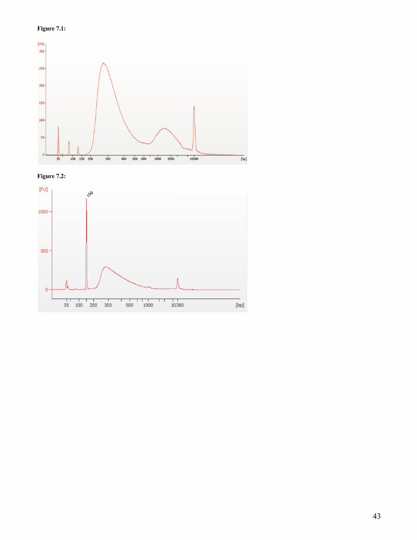
43
Figure 7.1:
Figure 7.2:

44
Kit Components Each set of reagents is functionally validated and compared to the previous lot through construction of libraries using the minimum and maximum amount of Universal Human Reference Total RNA. The previous and current lots are sequenced together on the same Illumina flow cell and compared across various sequence metrics including individual transcript abundances, 5´® 3´ transcript coverage and fraction of reads mapping to the reference
NEB #E7770S Table of Components
NEB # PRODUCT VOLUME E7374A NEBNext Ligation Enhancer 0.024 ml E7421A NEBNext First Strand Synthesis Reaction Buffer 0.192 ml E7422A Random Primers 0.048 ml E7425A NEBNext Second Strand Synthesis Enzyme Mix 0.096 ml E7427A NEBNext Second Strand Synthesis Reaction Buffer 0.192 ml E7646A NEBNext Ultra II End Prep Enzyme Mix 0.072 ml E7647A NEBNext Ultra II End Prep Reaction Buffer 0.168 ml E7648A NEBNext Ultra II Ligation Master Mix 0.720 ml E7649A NEBNext Ultra II Q5 Master Mix 0.6 ml E7761A NEBNext First Strand Synthesis Enzyme Mix 0.048 ml E7762A NEBNext Adaptor Dilution Buffer 2.4 ml E7763A (0.1X) TE Buffer 2.78 ml E7764A Nuclease-free Water 1.25 ml
NEB #E7770L Table of Components
NEB # PRODUCT VOLUME E7374AA NEBNext Ligation Enhancer 0.096 ml E7421AA NEBNext First Strand Synthesis Reaction Buffer 0.768 ml E7422AA Random Primers 0.192 ml E7425AA NEBNext Second Strand Synthesis Enzyme Mix 0.384 ml E7427AA NEBNext Second Strand Synthesis Reaction Buffer 0.768 ml E7646AA NEBNext Ultra II End Prep Enzyme Mix 0.288 ml E7647AA NEBNext Ultra II End Prep Reaction Buffer 0.672 ml E7648AA NEBNext Ultra II Ligation Master Mix 3 x 0.960
ml E7649AA NEBNext Ultra II Q5 Master Mix 2 x 1.2 ml E7761AA NEBNext First Strand Synthesis Enzyme Mix 0.192 ml E7762AA NEBNext Adaptor Dilution Buffer 9.6 ml E7763AA (0.1X) TE Buffer 11.5 ml E7764AA Nuclease-free Water 5.7 ml

45
NEB #E7775S Table of Components
NEB # PRODUCT VOLUME E7374A NEBNext Ligation Enhancer 0.024 ml E7421A NEBNext First Strand Synthesis Reaction Buffer 0.192 ml E7422A Random Primers 0.048 ml E7425A NEBNext Second Strand Synthesis Enzyme Mix 0.096 ml E7427A NEBNext Second Strand Synthesis Reaction Buffer 0.192 ml E7646A NEBNext Ultra II End Prep Enzyme Mix 0.072 ml E7647A NEBNext Ultra II End Prep Reaction Buffer 0.168 ml E7648A NEBNext Ultra II Ligation Master Mix 0.720 ml E7649A NEBNext Ultra II Q5 Master Mix 0.6 ml E7761A NEBNext First Strand Synthesis Enzyme Mix 0.048 ml E7762A NEBNext Adaptor Dilution Buffer 2.4 ml E7763A (0.1X) TE Buffer 2.78 ml E7764A Nuclease-free Water 1.25 ml E7767S NEBNext Sample Purification Beads 8.65 ml
NEB #E7775L Table of Components
NEB # PRODUCT VOLUME E7374AA NEBNext Ligation Enhancer 0.096 ml E7421AA NEBNext First Strand Synthesis Reaction Buffer 0.768 ml E7422AA Random Primers 0.192 ml E7425AA NEBNext Second Strand Synthesis Enzyme Mix 0.384 ml E7427AA NEBNext Second Strand Synthesis Reaction
Buffer 0.768 ml
E7646AA NEBNext Ultra II End Prep Enzyme Mix 0.288 ml E7647AA NEBNext Ultra II End Prep Reaction Buffer 0.672 ml E7648AA NEBNext Ultra II Ligation Master Mix 3 x 0.960 ml E7649AA NEBNext Ultra II Q5 Master Mix 2 x 1.2 ml E7761AA NEBNext First Strand Synthesis Enzyme Mix 0.192 ml E7762AA NEBNext Adaptor Dilution Buffer 9.6 ml E7763AA (0.1X) TE Buffer 11.5 ml E7764AA Nuclease-free Water 5.7 ml E7767L NEBNext Sample Purification Beads 4 x 8.65 ml

46
Revision History REVISION # DESCRIPTION DATE 1.0 N/A 7/20
This product is intended for research purposes only. This product is not intended to be used for therapeutic or diagnostic purposes in humans or animals.
This product is covered by one or more patents, trademarks and/or copyrights owned or controlled by New England Biolabs, Inc. For more information about commercial rights, please email us at [email protected]. While NEB develops and validates its products for various applications, the use of this product may require the buyer to obtain additional third party intellectual property rights for certain applications.
This product is licensed for research and commercial use from Bio-Rad Laboratories, Inc., under U.S. Pat. Nos. 6,627,424, 7,541,170, 7,670,808, 7,666,645, and corresponding patents in other countries. No rights are granted for use of the product for Digital PCR or real-time PCR applications, with the exception of quantification in Next Generation Sequencing workflows.
AGENCOURT®, AMPURE®, RNACLEAN® AND SPRISELECT® are registered trademarks of Beckman Coulter, Inc. B CORPORATION™ is a trademark of B Lab, Inc. E-GEL® is a registered trademark of Life Technologies, Inc. SIZESELECT™ is a trademark of Life Technologies, Inc. BIOANALYZER® is a registered trademark of Agilent Technologies, Inc. ILLUMINA® is a registered trademark of Illumina, Inc. ALPAQUA® is a registered trademark of Alpaqua Engineering. LLC QUBIT® is a registered trademark of Molecular Probes, Inc.
© Copyright 2020, New England Biolabs, Inc.; all rights reserved
New England Biolabs, Inc., 240 County Road, Ipswich, MA 01938-2723 Telephone: (978) 927-5054 Toll Free: (USA Orders) 1-800-632-5227 (USA Tech) 1-800-632-7799 Fax: (978) 921-1350 e-mail: [email protected]
www.neb.com
be INSPIRED drive DISCOVERY stay GENUINE
ISO 13485Registered
Medical Devices
ISO 14001Registered
EnvironmentalManagement
ISO 9001Registered
QualityManagement