NAOSITE: Nagasaki University's Academic Output...
14
This document is downloaded at: 2020-02-18T10:39:02Z Title A Case of Pulmonary Alveolar Proteinosis associated with Pulmonary Silicosis Author(s) Tashiro, Takayoshi; Goto, Jun; Goto, Ikuo; Akashi, Mitsunobu; Nasu, Masaru; Itoga, Takashi Citation Acta medica Nagasakiensia. 1983, 28(1-4), p.22-34 Issue Date 1983-10-25 URL http://hdl.handle.net/10069/15638 Right NAOSITE: Nagasaki University's Academic Output SITE http://naosite.lb.nagasaki-u.ac.jp
Transcript of NAOSITE: Nagasaki University's Academic Output...
This document is downloaded at: 2020-02-18T10:39:02Z
Title A Case of Pulmonary Alveolar Proteinosis associated with PulmonarySilicosis
Author(s) Tashiro, Takayoshi; Goto, Jun; Goto, Ikuo; Akashi, Mitsunobu; Nasu,Masaru; Itoga, Takashi
Citation Acta medica Nagasakiensia. 1983, 28(1-4), p.22-34
Issue Date 1983-10-25
URL http://hdl.handle.net/10069/15638
Right
NAOSITE: Nagasaki University's Academic Output SITE
http://naosite.lb.nagasaki-u.ac.jp
Acta Med. Nagasaki 28; 22-34
A Case of Pulmonary Alveolar Proteinosis
associated with Pulmonary Silicosis
Takayoshi TASHIRO, Jun GOTO, Ikuo GOTO,
Mitsunobu AKASHI, Masaru NASU, and
Takashi ITOGA
Department of Internal Medicine,
Medical College of Oita
Received for publication, December 5, 1982
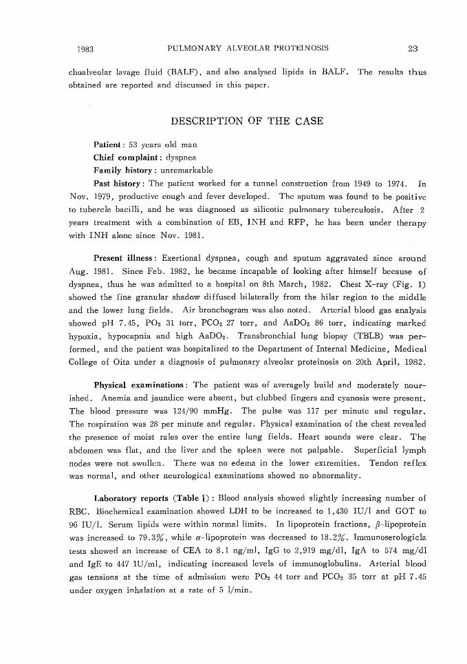
A 53 years old man complaining productive cough and dyspnea admitted to a hos-
pital on 8th March, 1982. Chest X-ray showed the fine granular shadow through out
lung fields. Arterial blood gas tensions were PO2 31 torr, PCO2 27 torr and AaDO2 86
torr at pH 7.45. Transbronchial lung biopsy was performed and the microscopic findings
of which showed eosinophilic, PAS positive granular materials filled in the intraalveolar
space and the swelling of the alveolar lining cells. The patient was diagnosed as pulmo-
nary alveolar proteinosis and was saved by bronchoalveolar lavage with the aid of extra-
corporeal circulation. Histochemical and electron microscopic observation and biochemi-
cal analysis were performed, and it was suggested that the materials accumulated in the
intraalveolar space were mostly originated from the lung surfactant secreted by the pul-
monary alveolar cells type II.
INTRODUCTION
Pulmonary alveolar proteinosis is the disease in which eosinophilic and PAS-stain-
able granular materials accumulate in the alveoli. This disease was first reported by
ROSEN, CASTLEMAN and LIEBOWI) in 1958. In Japan, OKA et al.2) reported the first
case in 1960, and a total of 79 cases have been found out until 19793).
We have experienced a case with pulmonary alveolar proteinosis complicated with
silicosis and pulmonary tubercurosis. This patient was saved by bronchoalveolar lavage
(BAL) with the aid of extracorporeal circulation. During the treatment, we have carried out histochemical and electron microscopic observations on biopsied lung and the bron-
田代 隆良,後 藤 純,後 藤 育郎,明 石 光 伸,那 須 勝,糸 賀 敬
choalveolar lavage fluid (BALF), and also analysed lipids in BALF. The results thus
obtained are reported and discussed in this paper.
DESCRIPTION OF THE C SE
Patient : 53 years old man
Chief complaint : dyspnea
Family history : unremarkable
Past history : The patient worked for a tunnel construction from 1949 to 1974. In
Nov. 1979, productive cough and fever developed. The sputum was found to be positive
to tubercle bacilli, and he was diagnosed as silicotic pulmonary tuberculosis. After 2
years treatment with a combination of EB, INH and RFP, he has been under therapy
with INH alone since Nov. 1981.
Present illness : Exertional dyspnea, cough and sputum aggravated since around
Aug. 1981. Since Feb. 1982, he became incapable of looking after himself because of
dyspnea, thus he was admitted to a hospital on 8th March, 1982. Chest X-ray (Fig. 1)
showed the fine granular shadow diffused bilaterally from the hilar region to the middle
and the lower lung fields. Air bronchogram was also noted. Arterial blood gas analysis
showed pH 7.45, P02 31 torr, PCO2 27 torr, and AaDO2 86 torr, indicating marked
hypoxia, hypocapnia and high AaDO2. Transbronchial lung biopsy (TBLB) was per-
formed, and the patient was hospitalized to the Department of Internal Medicine, Medical
College of Oita under a diagnosis of pulmonary alveolar proteinosis on 20th April, 1982.
Physical examinations : The patient was of averagely build and moderately nour-
ished. Anemia and jaundice were absent, but clubbed fingers and cyanosis were present.
The blood pressure was 124/90 mmHg. The pulse was 117 per minute and regular.
The respiration was 28 per minute and regular. Physical examination of the chest revealed
the presence of moist rales over the entire lung fields. Heart sounds were clear. The
abdomen was flat, and the liver and the spleen were not palpable. Superficial lymph
nodes were not swollen. There was no edema in the lower extremities. Tendon reflex
was normal, and other neurological examinations showed no abnormality.
Laboratory reports (Table 1) : Blood analysis showed slightly increasing number of
RBC. Biochemical examination showed LDH to be increased to 1,430 IU/1 and GOT to
96 IU/l. Serum lipids were within normal limits. In lipoprotein fractions, ~-lipoprotein
was increased to 79.3%, while a-lipoprotein was decreased to 18.2%. Immunoserologicla
tests showed an increase of CEA to 8.1 ng/ml, IgG to 2,919 mg/dl, IgA to 574 mg/dl
and IgE to 447 IU/ml, indicating increased levels of immunoglobulins. Arterial blood
gas tensions at the time of admission were P02 44 torr and PCO2 35 torr at pH 7.45
under oxygen inhalation at a rate of 5 1/min.
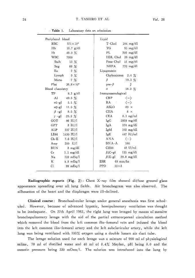
. Table 1. Laboratory data on admission
Peripheral blood Lipid RBC 571 x 104 T-Chol 204 mg/dl
Hb 16.7 g/dl TG 95 mg/dl Ht 49.0 % PL 209 mg/dl WBC 7000 HDL-Chol 26 mg/dl Stab 13 % Free-Chol 51 mg/dl Seg 69 % NEFA 270 mg/dl Eo 2 % Lipoprotein
Lymph 9 % Chylomicron 2.4 Mono 7 % /3 79.3
Plat 36.8 x 10⁴ pre-/3 J Blood chemistry a 18.2
TP 8.1 g/dl Immunoserological Al 49.5 % CRP (-)
al-gl 5.1 % RA (-) a2-gl 11.5 % ASLO 80 x
/3 -gl 8.5 % CHA 8 x r -gl 25.3 % ~ CEA 8.1 ng/ml
GOT 96 IU/1 IgG 2919 mg/dl GPT 8 IU/1 IgA 574 mg/dl ALP 237 IU/1 IgM 192 mg/dl LDH 1430IU/1 IgE 447IU/ml
Ch-E 7.6IU/1 ANA (-) Amy 200 U/1 DNA-A 160 BUN 8 mg/dl CH50 48 U/ml
Cr 1.1 mg/dl /31C-gl 135 mg/dl Na 139 mEq/1 /31E-gl 29.8 mg/dl K 4.8 mEq/1 ESR 69 mm/hr
Cl 99 mEq/1 PPD 10x8
Radiographic reports (Fig. 2) : Chest X-ray film showed diffuse ground-glass
appearance spreading over all lung fields. Air bronchogram was also observed. The
silhouettes of the heart and the diaphragm were ill-defined.
Clinical course : Bronchoalveolar lavage under general anesthesia was first sched-
uled. However, because of advanced hypoxia, hemipulmonary ventilation was thought
to be inadequate. On 27th April 1982, the right lung was lavaged by means of massive
bronchopulmonary lavage with the aid of the partial extracorporeal circulation method
which removed the blood from the left common ilio-femoral vein and infused the blood
into the left common ilio-femoral artery and the left subclavicular artery, while the left
lung was being ventilated with 100% oxygen using a double lumen air duct tube.
The lavage solution used for each lavage was a mixture of 900 ml of physiological
saline, 70 ml of distilled water and 40 ml of 8.4% Meylon, pH being 8.0 and the
osmotic pressure being 330 mOsm/l. The solution was introduced into the lung by
gravity feeding from the level about 30 cm higher than patient's midchest. Then, the chest was lightly tapped with hands and vibrated with a vibrator for about 5 min, and
the solution was drained by gravity out of the lung into a drain bottle placed at about
60 cm lower level. These procedures were repeated for 10 times. The total volume of
the lavage solution introduced was 9,840 mi, and that of the recovered solution was
10,090 ml.
As shown in Fig. 3, the X-ray picture of the chest after the lavage of the right
lung showed marked improvement. Arterial blood gas tensions were also improved such
as P02 73 torr, PCO2 40 torr and pH 7.43 under 3 1/min oxygen inhalation. The left
and right lungs were then lavaged without using extracorporeal circulation. After 3 such
lavagings, marked improvement was achieved in the chest X-ray pictures (Fig. 4). Ar-
terial blood gas tensions were also improved considerably to be pH 7.46, P02 71 torr,
PCO2 34 torr, AaD02 38 torr under air inhalation. The patient was discharged on 30th
June, 1982.
Respiratory function tests (Table
2) : The result of the test performed
before discharge showed % VC of 68.2
%, indicating restraint disorder, and DLCO was decreased to 50.4 %.
Pathohistological findings : The
septum of pulmonary alveoli was mildly
thickened, alveolar lining cells were swo-
llen, and eosinophilic granular materials
were packed in the alveoli (Fig. 5).
The large mononuclear cells alike exfol-
iated alveolar lining cells type II or al-
veolar macrophages were also observed in
the intraalveolar space (Fig. 6). In other
Table 2. Respiratory function tests
VC 2340 ml
VC 68.2
FEV 1.0 1990 ml FEV1.0% 90.5%
TV 610 ml
FRC 1980 ml
ERV 1240 ml
RV 740 ml
TLC 3080 ml
RV/TLC 24 V 75 7 1/s
V 50 2.6 1/s
V25 1I/s
DLCO 11.8 m1/min/mmHg
DLCO 50.4
biopsy specimen, hyalinized collagen fibers were spiralled, and there was marked deposi-
tion of coal powder around them (Fig. 7). Observation under a polarizing microscope
showed the presence of considerable numbers of polarizable fine granular materials.
Therefore, this was thought to be a silicotic nodule.
Histochemistry : As shown in Table 3, the granular material and the large mono-
nuclear cell contained in both biopsy specimen and the precipitate of lung lavaged solu-
tion exhibited similar histochemical reactivities. The granular material was most strongly
positive to PAS staining, and the mononuclear cell also contained PAS-positive substan-ces. These substances were not digested by a diastase. All lipid stainings tested, i. e. ,
with Oil red 0, Suddan black B and Nile blue, were strongly positive to the large mono-
nuclear cell, and partly positive to the granular material (Fig. 8). Both granular mate-

Table 3. Histochemistry
Biopsy specimen Lavage fluid
Granular material Mononuclear cell Granular material Mononuclear cell
HE + + + +
PAS -1-f- + -1+ +
diastase investigation - - - -
Oil red O + -+ + -H-
Sudan black B + ~f- + I f
Nile blue + -}{- + H LFB -F}- + -I-I- +
Alcian blue - - -!- ±
Toluidin blue - - ± -
Mucicarmin - - - -PTAH - - - -
GMS - - - -
rial and mononuclear cell were well stained with LFB staining (for phospholipids), but
the granular material was strongly positive. Staining with Alcian blue, Toluidine blue
or Mucicarmin was negative, and that with PTAH was also negative. Pneumocystis car-
inii was denied by GMS staining.
Electron microscopic observation : In the intraalveolar space, there were many my-
elin-like materials with a concentric structure and various sizes and shapes, and elec-
tron-opaque bodies with various sizes (Fig. 9). The myelin-like material was formed pri-
marily around the electron-opaque body. Lamellar bodies were also observed, some of
them being surrounded with the myelin-like materials (Fig. 10). The precipitate of la-
vaged solution also showed similar images.
Biochemical analysis of the lavage fluid : A portion of the lavage fluid recovered
from the lst lavage was centrifuged at 500 x g for 10 min. The grey precipitate with a
yellowishwhite tint thus obtained was largest in amount in the 1st lavage fluid and de-
creased in subsequent lavage fluids, thus the supernatant became clearer in later lavage
fluids (Fig. 11). The supernatant and the precipitate of the 1st lavage fluid was supplied
to biochemical analyses. Proteins were rich (73.1%) in the supernatant, while lipids were
more abundant (65.1%) in the precipitate.
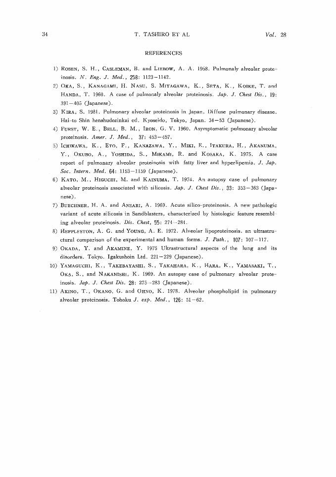
The proteins consisted of the major pro-
teins of serum such as prealbumin, albu-
min, al-antitrypsin, transferrin and IgG
(Fig. 12). Lipids in either the super-
natant or the precipitate contained phos-
pholipids abundantly (Tadle 4), in which
phosphatidylcholin was the highest cons-tituent (Table 5).
Table 4. Lipid composition of lavage fluid
Lipid composition (weight %)
Supernatant Precipitate
Phospholipid 72.2 80.8
Triacylglycerol 0.1 0.8
Free fatty acid 10.1 1.4
Total cholesterol 17.5 17.1

Table 5. Phospholipid composition of lavage fluid
Phospholipid Composition (mole %) Spot No. Lipid
Supernatant Precipitate
I Phosphatidylcholine 35.7 54.9
2 Phosphatidylethanolamine 4.5 3.6
3 Sphingomyelin 28.6 11.9
4 Phosphatidylinositol 0.9 6.4
5 Phosphatidylserine 7.5 1.8 6 Lysophosphatidylcholine 6.9 6.8
7 Unknown - - 8 Phosphatidylglycerol 2.2 3.2
9 Unknown (Glycolipid) - -
10 Lyso-bis-phosphatidic acid 10.8 7.6
11 Unknown (Glycolipid) - - 12 Unknown (Glycolipid) - -
13 Unknown (Glycolipid) - -
14 Unknown (Neutral lipid) - -
15 Unknown (Neutral lipid) - -
16 Origin (Degradated lipid) 2.9 3.7
DISCUSSION
Pulmonary alveolar proteinosis develops regardless of age, but it occurs most fre-
quently at the age between 30-50th, and the incidence in man is twice of that in female. Subjective symptoms are exertional dyspnea, cough and sputum.
The rise of the LDH level is frequently observed in biochemical blood test. In
the present case, it was also raised to 1,430 IU/1. Although cases complicated with
hyperlipemia have been reported4)5), the serum lipid level has been usually reported to
be normal. The lipoprotein fraction from the serum has been studied by a few inves-
tigators, but the rise of ji-lipoprotein and the decrease of pre-/3-lipoprotein and a-
lipoprotein were demonstrated in the present case. The serum CEA level in the present
case was as high as 8.1 ng/ml before lavage. Repetitive pulmonary lavages, however,
decreased the CEA value successively, and it was 2.1 ng/ml at the time of discharge.
The chest X-ray film frequently shows fine granular shadow which bilaterally
spread from the hilus. Such a shadow is sometimes described as frosted glass-like or
cloud-like shadow, air bronchogram is frequently observed, which is thought to be due
to dense and fine granular alveolar shadow.
Pulmonary function tests show restraint disorder and lowered DLCO. Blood gas
analysis reveals hypoxemia and increased AaDO2. The present case also showed marked
hypoxemia and the increase of AaDO2. Although these symptoms were improved by
pulmonary lavage, respiratory function tests before discharge indicated the presence of restraint disorder and lowered DLCO. Since this particular case had pulmonary silicosis

and tuberculosis as basal diseases, their influence may not be ignored. It was also
conceivable that protein-like material accumulated in the intraalveolar space had not been
completely eliminated.
Regarding the complication of pulmonary alveolar proteinosis with pulmonary sill-
cosis, 6 cases have been reported in Japan, and one case out of 27 cases reported by
ROSEN et al.1) also experienced the inhalation of silica. In 1969 BUECHNER et al .7)
described 4 cases in which acute silicosis and pulmonary alveolar proteinosis were com-
plicated. The term given by him to these dead cases was `acute silico-proteinosis' . Although experimental inhalation of silica by animals may produce the morbid image
similar to pulmonary alveolar proteinosis8), the inhalation of silica can be one of inducing
factors. However, many other cases are entirely unrealated to silica inhalation. There-
fore, it is unlikely that the inhalation of silica is a direct cause of pulmonary alveolar
proteinosis.
Pathohistological characteristic of the proteinosis is the presence of eosinophilic
granular materials which fill the intraalveolar space. The existence of the large mono-nuclear cells appeared to be exfoliated alveolar lining cells type II or alveolar macrophages
are also characteristics. The alveolar septum is mildly thickened. The proliferation of
the alveolar lining cells type If is present in some region and absent in the other.
These regions were classified by OKADA et al.9), based on electron microscopic obser-
vation, to be the area of desquamation and sloughing and the area of accumulation.
Histochemical findings in the present case mostly agreed with those reported by
other investigators')') 10) . Granular materials were strongly stained with PAS and LFB,
thus they were thought to be phospholipid-rich proteins. Large mononuclear cells were
strongly positive to lipid stainings such as Oil red 0, Suddan black B and Nile blue
stainings, indicating the high content of neutral lipids.
Electron microscopic observation showed the presence of electron-opaque bodies
with various sizes and of myelin-like structures, the former being surrounded by the
latter. Lamellar bodies were also observed, and some of the electron-opaque bodies were
surrounded by such lamellar bodies. ROSEN et al.1) and OKADA et al.9) have stated
that the material accumulated in the intraalveolar space is that formed by the decompo-
sition of alveolar lining cells type 0 . Electron microscopic observation of ours also
suggested degenerated and decomposed alveolar lining cells type If or degenerated lung
surfactants to be the origin of the accumulated material.
Biochemical analysis of the lavage fluid showed the abundancy of lipids in the
precipitate and of proteins in the supernatant. Although the phospholipid was the major
constituent of the lipid, the supernatant had different phospholipid composition from the
precipitate. In the supernatant, phosphatidylcholine and sphingomyelin were major, while
phosphatidylcho line was high and sphingomyelin was low in the precipitate. It has been
stated by AKINO et al.11) that the phospholipid in the precipitate is mostly derived from
the lung surfactant secrected from the alveolar cells type f , rather than derived from
intraalveolar free cells. According to a study using radioactive isotopes, there is no

particular facilitation of the synthesis of phosphatidylcholine, though the synthesis of
this phospholipid de novo and via the lysolecithin formation system is basically active in
the lung tissue from a patient with pulmonary alveolar proteinosis. Moreover, the se-
cretion of phosphatidylcho line into the intraalveolar space is not known to be facilitated.
Therefore, it may be thought that disturbed removal mechanism is responsible for the
accumulation of surfactant-origin lipids in the intraalveolar space.
CONCLUSION
A case of pulmonary alveolar proteinosis developed as a complication of pulmonary
silicosis was reported. The patient was saved by bronchoalveolar lavage with the aid of
extracorporeal circulation, because of advanced dyspnea. On the basis of pathohistologi-
cal, histochemical and electron microscopic observations and of biochemical analyses, it
was thought that the material accumulated in the intraalveolar space was mostly originated
from the lung surfactant secreted by the pulmonary alveolar cells type I .
ACKNOWLEDGMENT
We would like to thank Dr. T. AKINO, Sapporo Medical College, for making a
chemical analysis of the bronchoalveolar lavage fluid of the patient.
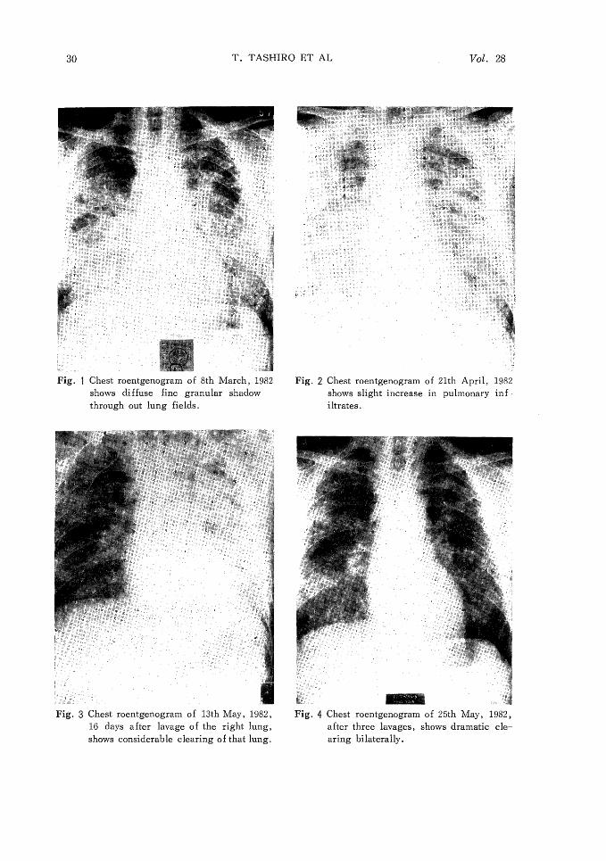
Fig. 1 Chest roentgenogram of 8th March, 1982
shows diffuse fine granular shadow
through out lung fields.
Fig. 2 Chest roentgenogram of 21th April, 1982
shows slight increase in pulmonary inf-
iltrates.
Fig. 3 Chest roentgenogram of 13th May, 1982,
16 days after lavage of the right lung,
shows considerable clearing of that lung.
Fig. 4 Chest roentgenogram of 25th May, 1982,
after three lavages, shows dramatic cle-
aring bilaterally.
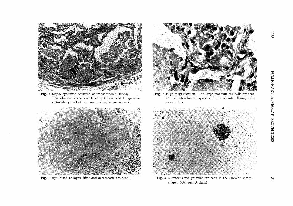
Fig. 5 Biopsy specimen obtained at transbronchial biopsy.
The alveolar space are filled with eosinophilic granular
materials typical of pulmonary alveolar proteinosis.
Fig. 6 High magnification. The large mononuclear cells are seen
in the intraalveolar space and the alveolar lining cells
are swollen.
Fig. 7 Hyalinized collagen fiber and anthracosis are seen. Fig. 8 Numerous red granules are seen in the alveolar macro-
phage, (Oil red 0 stain).

Fig. 9 Electron micrograph of bronchoalveolar lavage fluid (13,000x). The electron-
opaque bodies and the myelin-like materials surrounding them are observed.
Fig. 10 Electron micrograph of bronchoalveolar lavage fluid (10,000x). The extracellular lamellar bodies surrounded by myelin-like materials are observed.
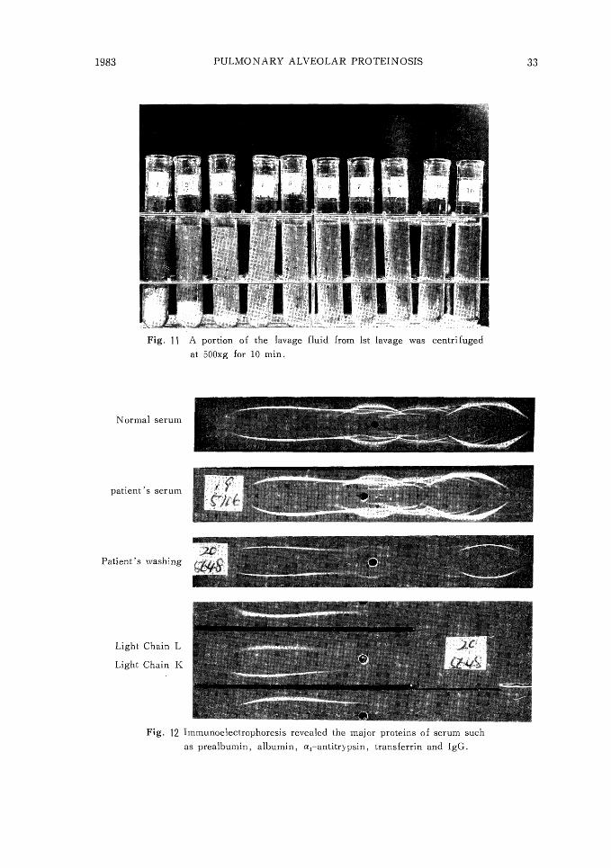
Fig. 11 A portion of the lavage fluid from Ist lavage was centrifuged
at 500xg for 10 min.
Normal serum
patient's serum
Patient's washing
Light Chain L
Light Chain K
Fig. 12 Immunoelectrophoresis revealed the major proteins of serum such
as prealbumin, albumin, al-antitrypsin, transferrin and IgG.
REFERENCES
1) ROSEN, S. H., CASLEMAN, B. and LIEBOW, A. A. 1958. Pulmonaly alveolar prote-
inosis. N. Eng. J. Med., 258: 1123-1142.
2) OKA, S., KANAGAMI, H. NASU, S. MIYAGAWA, K., SETA, K., KOIKE, T. and
HANDA, T. 1960. A case of pulmonaly alveolar proteinosis. Jap. J. Chest Dis., 19:
391-405 (Japanese).
3) KIRA, S. 1981. Pulmonary alveolar proteinosis in Japan. Diffuse pulmonary disease.
Hai-to-Shin henshudozinkai ed. Kyoseido, Tokyo, Japan. 34-53 (Japanese).
4) FURST, W. E., BELL, B. M., IRON, G. V. 1960. Asymptomatic pulmonary alveolar
proteinosis. Amer. J. Med., 37: 453-457.
5) ICHIKAWA, K., ETO, F., KANAZAWA, Y., MIKI, E., ITAKURA, H., AKANUMA,
Y., OKUBO, A., YOSHIDA, S., MIKAMI, R. and KOSAKA, K. 1975. A case
report of pulmonary alveolar proteinosis with fatty liver and hyperlipemia. J. Jap.
Soc. Intern. Med. 64: 1153-1159 (Japanese).
6) KATO, M., HIGUCHI, M. and KAINUMA, T. 1974. An autopsy case of pulmonary
alveolar proteinosis associated with silicosis. Jap. J. Chest Dis. , 33: 353-363 (Japa-
nese).
7) BUECHNER, H. A. and ANSARI, A. 1969. Acute silico-proteinosis. A new pathologic
variant of acute silicosis in Sandblasters, characterized by histologic feature resembl-
ing alveolar proteinosis. Dis. Chest, 55: 274-284.
8) HEPPLESTON, A. G. and YOUNG, A. E. 1972. Alveolar lipoproteinosis. an ultrastru-
ctural comparison of the experimental and human forms. J. Path., 107: 107-117.
9) OKADA, Y. and AKAMINE, Y. 1975 Ultrastructural aspects of the lung and its
disorders. Tokyo. Igakushoin Ltd. 221-229 (Japanese).
10) YAMAGUCHI, K . , TAKEBAYASHI, S., TAKAHARA, K . , HARA, K . , YAMASAKI, T.,
OKA, S., and NAKANISHI, K. 1969. An autopsy case of pulmonary alveolar prote-
inosis. Jap. J. Chest Dis. 28: 275-283 (Japanese).
11) AKINO, T., OKANO, G. and OHNO, K. 1978. Alveolar phospholipid in pulmonary
alveolar proteinosis. Tohoku J. exp. Med., 126: 51-62.