Nanosuspension Based Electrolyte Sensitive In Situ Gel for ...
46
University of Mississippi University of Mississippi eGrove eGrove Electronic Theses and Dissertations Graduate School 2019 Nanosuspension Based Electrolyte Sensitive In Situ Gel for Nanosuspension Based Electrolyte Sensitive In Situ Gel for Topical Ocular Delivery of Natamycin Topical Ocular Delivery of Natamycin Poorva H. Joshi University of Mississippi Follow this and additional works at: https://egrove.olemiss.edu/etd Part of the Pharmacy and Pharmaceutical Sciences Commons Recommended Citation Recommended Citation Joshi, Poorva H., "Nanosuspension Based Electrolyte Sensitive In Situ Gel for Topical Ocular Delivery of Natamycin" (2019). Electronic Theses and Dissertations. 1624. https://egrove.olemiss.edu/etd/1624 This Thesis is brought to you for free and open access by the Graduate School at eGrove. It has been accepted for inclusion in Electronic Theses and Dissertations by an authorized administrator of eGrove. For more information, please contact [email protected].
Transcript of Nanosuspension Based Electrolyte Sensitive In Situ Gel for ...
University of Mississippi University of Mississippi
eGrove eGrove
Electronic Theses and Dissertations Graduate School
2019
Nanosuspension Based Electrolyte Sensitive In Situ Gel for Nanosuspension Based Electrolyte Sensitive In Situ Gel for
Topical Ocular Delivery of Natamycin Topical Ocular Delivery of Natamycin
Poorva H. Joshi University of Mississippi
Follow this and additional works at: https://egrove.olemiss.edu/etd
Part of the Pharmacy and Pharmaceutical Sciences Commons
Recommended Citation Recommended Citation Joshi, Poorva H., "Nanosuspension Based Electrolyte Sensitive In Situ Gel for Topical Ocular Delivery of Natamycin" (2019). Electronic Theses and Dissertations. 1624. https://egrove.olemiss.edu/etd/1624
This Thesis is brought to you for free and open access by the Graduate School at eGrove. It has been accepted for inclusion in Electronic Theses and Dissertations by an authorized administrator of eGrove. For more information, please contact [email protected].
NANOSUSPENSION BASED ELECTROLYTE SENSITIVE IN SITU GEL FOR
TOPICAL OCULAR DELIVERY OF NATAMYCIN
A Thesis
Presented for the degree of
Master of Science in Pharmaceutical Science
With emphasis in Pharmaceutics and Drug Delivery
The University of Mississippi
by
POORVA H JOSHI
May 2019

Copyright © Poorva H Joshi 2019
All rights reserved

ii
ABSTRACT
Natamycin (NT) is a commercially available antifungal drug used for the treatment of fungal
keratitis, an infection which affects the clear corneal surface and its associated layers. Currently,
NT is available commercially as a 5% w/v ophthalmic suspension, to be administered
topically. The main objective of the present investigation was to develop and
evaluate the natamycin loaded nanosuspension (NT-NS), as well as it‘s corresponding in-situ
gel (NT-NS-GG), for the treatment of fungal keratitis. NT-NS was prepared using
homogenization technique and optimized based on size, PDI, zeta potential (ZP), assay and
process stability. Further, optimized NT-NS was modified into an in-situ gel with the addition of
0.2% gellan gum as a gelling agent and evaluated for rheological properties. In vitro release and
trans corneal permeation studies were performed. Differential scanning calorimetry studies
showed no interaction between the drug and other excipients being formulated into a NS. Particle
size, PDI, ZP and assay of the optimized NT-NS formulation (formulation LP1) were 586. ± 61.3,
0.447 ± 0.12, -38.97 and 107 ± 0.6%, respectively. LP1 formulation was stable for up to 4 weeks
under refrigerated and at room temperature conditions. In-situ gels showed satisfactory
rheological properties, prolonged drug release and permeation through rabbit corneas. The
results suggest that NT loaded NS could be an alternative topical ocular dosage form.
iii
DEDICATION
I dedicate this Master of Science degree to my parents and my friends back in India for their
constant support and belief in me. I am thankful to the University of Mississippi for giving me
an opportunity to study at this university.

iv
LIST OF ABBREVIATIONS AND SYMBOLS
ACN Acetonitrile
DPBS Dulbecco‘s phosphate buffer saline
DSC Differential Scanning Calorimetry
GFT Gel formation time
GG Gellan Gum
GRT Gel retention time
HPLC High Pressure Liquid Chromatography
IPBS Isotonic phosphate buffer saline
NLC Nano lipid carriers
NS Nanosuspension
NT Natamycin
NT-NS Natamycin nanosusupension
NT-NS-GG Natamycin Nanosuspension in situ gel
PDI Polydispersity index
PVP Polyvinyl pyrrolidone
RMβCD Randomly methylated β cyclodextrin
STF Simulated Tear Fluid
UV Ultraviolet detection

v
ZP Zeta potential

vi
ACKNOWLEDGEMENT
I am thankful to my research advisor Dr. Soumyajit Majumdar for giving me an
opportunity to work with him in the Department of Pharmaceutics and Drug Delivery at the
University of Mississippi. My committee members, Dr. Michael Repka and Dr. Eman Ashour for
their encouragement and feedback.
I would like to thank my postdoc Dr. Narendar Dudhipala for guiding me throughout my
Masters project, my lab members Prit Lakhani, Akash Patil, Corinne Sweeney, Rama Kashikar,
Kai-Wei Wu, Kanika Goel and Samir Senapati, for their help and advice.

vii
TABLE OF CONTENTS
ABSTRACT…………………………………………………………………………...……….…ii
DEDICATION………………………………………………………………………………........iii
LIST OF ABBREVATIONS AND SYMBOLS……………………………………….………....v
ACKNOWLEDGEMENT………………………………………………………………...……...vi
LIST OF TABLES…………………………………………………………………………...…viii
LIST OF FIGURES……………………………………………………………………...……… ix
CHAPTER I
INTRODUCTION………………………………………………………………………………...1
CHAPTER II
METHODOLOGY………………………………………………………………………..............8
CHAPTER III
RESULTS………………………………………………………………………………..............16
CHAPTER IV
CONCLUSION……………………………………………………………………………..........28
LIST OF REFERENCES…………………………………………………………………….......29
VITA………………………………………………………………………………………..…....33

viii
LIST OF TABLES
1. Composition of natamycin nanosuspension (NT –NS) with single
surfactant………………………………………………………………….……..…….......9
2. Composition of natamycin nanosuspensions (NT-
NS)………………………………………………..……………………………………...10
3. Physico-chemical characteristics - Particle size, PDI, ZP and assay of natamycin
nanosuspension (NT-NS) (mean±SD n=3)…………………………………………………….17
4. List of Formulations selected…………………………………………….…………..…..18
5. Stability studies of optimized NT-NS (LP1) formulation at 4°C and
25°C……………………………………………………………………………….……..19
6. Composition of natamycin in situ gel (NT-NS-
GG)...…………………………………………………………………....................…….20
7. Rheological evaluation of natamycin in situ gel (NT-NS-
GG)……………………………………..……………………………………...…….......21
8. In vitro drug release profiles of natamycin nanosuspensions (NT-NS), in situ gel (NT-NS
GG) and natamycin control……………………………………………..……………….24
9. Transcorneal permeation studies of natamycin nanosuspensions (NT-NS) and in situ gel
(NT-NS-GG)……………….........................................................................................….26

ix
LIST OF FIGURES
1. Viscosity of optimized NT-NS-GG with and without simulated tear fluid (STF)
(mean±SD, n=3) ……………………………………………………………………..…..21
2. DSC thermograms of pure natamycin and optimized NT-NS ……………...…...............22
3. Pre-autoclave (3A) and post-autoclave (3B) images of NT NS and Natacyn® (marketed
formulation) ……………………………………………….………...……………................23
4. In vitro release studies of optimized NT-NS (LP1), NT-NS-GG and NT control
formulations (mean±SD, n=3) …………………………………………………….….…25
5. In vitro transcorneal flux and apparent permeability coefficient of natamycin from
optimized NT-NS (LP1), NT-NS-GG (mean ± SD; n=3) …………………..……..….…26

1
CHAPTER I
INTRODUCTION
Eyes are one of the most important and complex sensory organs; they act as a gateway to
collect external images and transmit them to the brain as signals through the optic nerve [1]. This
maintains a connection between the body and the surroundings. Its visual capabilities may be
affected by bacterial, viral or fungal infections of the eye as well as age related disorders. The
complex anatomy, physiology and biochemistry of the eye, renders this organ highly impervious
to drugs/treatment [1].
The eyes can broadly be classified into two segments: the posterior segment and the anterior
segment. The anterior segment consists of the cornea, conjunctiva, aqueous humor, iris, ciliary
body and the lens. The sclera, choroid, Bruch’s membrane, retinal pigmented epithelium, neural
retina and vitreous humor make up the posterior segment of the eye [1]. The anterior segment of
the eye occupies one-third of the eye.
Anatomy and Physiology of the eye.
Anterior segment
The cornea is known to be the largest penetration barrier, inhibiting the passage of drug
into the eye, avascular in nature and comprising of 5 layers. The primary diffusional barrier is the
corneal epithelium, which controls the entry of drug into the eye [2]. The external surface of the
cornea is exposed to the environment, and is covered by a tear film; its inner surface is in contact
with the aqueous humor [1]. The efficiency of drug penetration across the cornea is governed by
2
various factors, such as the integrity of the cornea, the properties of the drug and the type
of formulation the drug is administered in [2].
The conjunctiva is a thin, semitransparent, elastic, mucous secreting tissue forming the
lining of the upper and lower eyelids [1]. A thin vascular layer known as the ciliary body
contains the ciliary muscle responsible for adjusting the shape of the lens to focus on an object
[3]. The epithelial cells of the ciliary body continuously produce an alkaline ocular fluid, at a rate
of 2.5µL/min, known as the aqueous humor which supplies nutrients to the cornea and lens
Located at the posterior region of the cornea, the iris consists of 3 layers. the endothelium,
stroma and the epithelium [1]. The central aperture is a pupil which is controlled by the iris,
regulating the amount of light entering the eye [4].
The ciliary body is located anterior to the iris and has three major functions: secreting
aqueous humor, adjusting focus and draining aqueous humor [1].
Located behind the iris and the pupil, the lens is transparent and biconcave in nature. The
anterior part is in contact with the aqueous humor while the posterior part is in contact with the
vitreous humor [1].
Posterior segment of the eye
The sclera also known as the ‗white of the eye‘ is an elastic tissue present below the
conjunctiva. It gives the eyeball its shape and protects the internal organs of the eye from
damage [5]. The sclera is made up of a network of collagen fibers responsible for the scattering
of the visible light.
Present between the peripheral sclera and the inner retinal membrane, the choroid is made
of the outer suprachoroid, which is 6-10 layers thick, the vascular layer below
the suprachoroid and the Bruch‘s membrane which is the innermost layer consisting of
3
photoreceptor cells [1]. Each eye of an individual contains approximately 3.5 million non-
dividing retinal pigmented epithelium cells (RPE). It plays a vital role in the functioning of
photoreceptors and is essential for maintaining visual function [1].
Light sensitive neural cells line the inner lining of the eyeball which makes up the
neural retina. The neural retina interacts with the external environment by transmitting sensory
information to the brain. Rods and cones are collectively known as the photoreceptor cells.
These cells capture and convert the collected photons into neural signals. The cone cells help in
distinguishing between black and white colors in dim light while the rod cells differentiate colors
in bright light [1].
A gel like fluid is present between the lens and the retina known as the vitreous humor.
The vitreous humor is 99.9% water,0.01% of collagen fibrils, hyaluronic acid and ions, and aids
in maintaining the structure of the eye.
Topical dosage forms
Topical administration is preferred for treating disorders of the anterior segment of the
eye as it offers a few major advantages [6]. It helps in localizing the drug effects by minimizing
systemic exposure. It‘s ease of administration makes it convenient for self-administration by
patients and is a painless method which in turn leads to increased patient compliance [6]. Due to
these reasons ophthalmic drugs are primarily administered topically, in the form of solutions,
suspensions, and ointments. However, ocular tissues are biologically protected from external
toxic elements by a variety of mechanisms [6]. The most prominent mechanism is tear secretion,
which continuously flushes the surface of the eye. There is also the surface epithelium which is
almost impermeable, and possesses an active efflux-transport system [6]. The major goal in
ocular therapeutics is to circumvent these structural obstacles and protective mechanisms in

4
order to elicit the desired pharmacological responses [7]. Physiological barriers to topically
administered drugs are present mainly in the precorneal and corneal regions. Precorneal factors
such as lacrimation, dilution, and conjunctival absorption cause loss of more than 90% of the
topically administered formulation [1]. Due to these formidable physiological barriers, frequent
dosing is often necessitated to achieve satisfactory therapeutic results. This results in a pulsating
dosing pattern with extreme fluctuations of drug concentration in the ocular region. These
extreme fluctuations may cause local or systemic adverse effects. For maximum bioavailability
of the topically administered drug, it is thus important that duration of contact with the cornea be
increased.
Fungal keratitis
Fungal keratitis (keratomycosis) is a fungal infection of the cornea the incidence being between 6%
-20% of all microbial keratitis cases, depending on the geographic location. It primarily affects
the corneal epithelium and stroma, although the endothelium and anterior chamber of the eye
may get involved in more severe cases. Fungal keratitis is common in tropical countries
compared to the temperate regions. Its incidence in developed countries is reported to be
increasing due to the widespread use of contact lenses. Infections due to filamentous fungi such
as Fusarium and Aspergilillus are found to be more common in tropical regions while temperate
regions show a higher incidence of yeast infections such as Candida [8].
The main outcomes desired in an ophthalmic therapy for fungal keratitis is the resolution of the
infection as rapidly as possible to give way to good visual outcome and to eliminate the need for
therapeutic keratoplasty or permanent loss of vision[8]. Fungal keratitis is a challenging
ophthalmologic condition that requires a high level of suspicion and aggressive treatment to
prevent untoward outcomes [9].

5
Natamycin (NT) has been one of the mainstays in the treatment of fungal keratitis [10]. NT
suspension is currently the only FDA approved ophthalmic antifungal formulation.
Natamycin is stable at pH 5-9. At pH levels higher or lower, they are found to undergo
saponification and/or bond cleavage which leads to instability and hence loss of antifungal
activity. NT is also susceptible to photo-oxidation and hence should be stored in the dark [10].
NT shows antifungal activity against both filamentous as well as non-filamentous fungal species,
with potent activity against the filamentous species compared to other antifungal agents. It is
found to be potent against Aspergillus and Fusarium species apart from Candida species. NT has
low retention at the ocular surface which requires it to be frequently administered. Despite these
limitations, NT has shown efficacy in treating superficial ophthalmic fungal infections mainly
due to its trans-corneal penetration capability [10].
Currently, a formulation of NT (Natacyn® Alcon Laboratories, Fort Worth, TX) is available
commercially for the treatment of fungal keratitis. It is available as an aqueous suspension
containing 5% NT (50 mg/mL); pH adjusted to 5-7.5 to prevent chemical instability of NT.
Benzalkonium chloride is added to the suspension to prevent bacterial growth [10]. Its higher
penetration across intact cornea upon topical administration and its broad spectrum of activity
makes natamycin the forerunner in the treatment of fungal keratitis. NT eye-drops have, thus,
been the primary option for superficial corneal infections. Other antifungals such as
Amphotericin B. echinocandin, and azoles are also used off label as a second-line of treatment
[10].
Nanosuspensions (NS) are sub-micron colloidal dispersions of pure particles of drug, stabilized
by surfactants. The particle size of the drug in the dispersed phase ranges from 100 to 1000 nm
[11]. Surfactants act as stabilizing agents by lowering the surface tension between the dispersed

6
drug molecules and the dispersion liquid, allowing the formulation to stabilize [12].
Nanosuspensions contain particles of 100% pure drug devoid of any carriers or vehicles
[13]. Hence a high drug loading in nanosuspensions could result in highly efficient drug
transportation into the cells leading to high therapeutic concentrations, which in turn would
maximize the pharmacological effects. However, surfactant-stabilized suspensions must
possess sufficiently high energy barriers to prevent the suspended particles from coming close
together and agglomerating.
PEGylated natamycin NLCs (nanostructured lipid carriers) with 0.3% drug load [14] and ion
sensitive in situ gels of natamycin bilosomes [15] have been previously reported. The optimized
PEGylated NLC (0.3%) had a particle size with a narrow PDI, high NT entrapment and drug
content. In the in vitro studies, the NLC formulation also showed an improved transcorneal
permeation and flux compared to the Natacyn® (5%) the marketed suspension, as well as
concentrations statistically similar to Natacyn®
in the inner ocular tissues [14].
The optimized NT bilosomes (NB) showed a 6-9 fold enhancement of transcorneal flux and
ocular penetration. The in situ gel with 0.3% gellan gum was found to be cytocompatible with
the desired viscoelastic and adhesive properties [15].
In situ forming gels are liquids at the time of instillation into the eye, but then undergoes
rapid gelation in the cul-de-sac of the eye to form viscoelastic gels in response to environmental
changes [16]. In situ gels tend to increase precorneal residence time of the drug in the cul-de-sac,
leading to sustained release, enhanced bioavailability and reduced dosing frequency. The main
reason for the popularity of in situ gels is the ease of administering accurate quantities compared
to pre-gelled formulation [17]. Electrolyte sensitive in situ gel systems undergo a gel-sol

7
transformation when in contact with mono or divalent cations in the tear fluid, particularly
Na+,
Mg2+
and Ca2+
, thus forming a gel on the ocular surface [17]. Naturally
occurring polysaccharides have been used as a matrix to obtain sustained drug
delivery [18]. Gellan gum is an anionic polysaccharide which undergoes a sol-gel transition due
to the temperature and ionic conditions present in the tear fluid, forming an ordered state
of gellan chains responsible for the gelling effect [18].
A majority of the drug instilled into the eye, is drained out via reflexive blinking or through
lacrimation. Thus, the desired formulation characteristics include prolonged pre-corneal
residence time, non-irritating characteristics and suitable rheological properties [19]. From the
perspective of patient compliance, a dosage form capable of maintaining contact with the cornea
for extended periods of time, which would reduce the frequency of dosing, is desired [20].
In the current study, we attempted to develop a NT-NS and corresponding in situ gel (NT-NS-
GG) with gellan gum as the gelling agent. Particle size, polydispersity index, assays and zeta
potential of the NT-NS were evaluated. The optimized NT-NS was evaluated for stability studies
at 25°C and 4°C for 30 days. For the NT-NS-GG, characteristics such as gelling time, gel
retention time, viscosity of the formulation with and without the addition of simulated tear fluid
were investigated. The optimized NT NS and NT-NS-GG were evaluated for in vitro release
studies and transcorneal penetration studies.

8
CHAPTER II
METHODOLOGY
Materials
NT was purchased from Cayman Chemicals (Ann Arbor, MI). Polysorbate 80, poloxamer
188 was purchased from Acros Organics (Morris, NJ). Laboratory grade lecithins (Fair Lawn,
New Jersey), Tyloxapol was purchased from Sigma-Aldrich Life Science (St Louis,
MO). Gellan gum was obtained from Alfa Aesar (Ward Hill, MA). High performance liquid
chromatography (HPLC) grade solvents and other chemicals (analytical grade) were supplied by
Fisher Scientific (Hampton, NH). Slide-A-Lyzer™ MINI Dialysis Device, 10K was purchased
from Thermo Fisher (Rockford, IL).
Animal tissues
Whole eyes of male albino New Zealand rabbits were acquired from Pel-Freez
Biologicals (AR, USA).
Preparation of natamycin nanosuspension (NT-NS) and in-situ gels (NT-NS-GG)
Selection of surfactants combination
Two types of surfactant combinations were used: polysorbate 80 with poloxamer 188 and
lecithin with poloxamer 188. Tyloxapol was used as a surface-active agent. PVP K 30 was used
as a stabilizer.
Method of Preparation of natamycin nanosuspension (NT-NS):

9
NT loaded NS were prepared using homogenization method as per the compositions in Table 1
and 2. The surfactants along-with tyloxapol and poloxamer 188 were dissolved in sufficient
volume of water with the aid of magnetic stirring. On forming a clear solution, NT was added to
the above solution and stirred at 2000 rpm on a magnetic stirrer for 5 minutes at room
temperature. This suspension was then homogenized using a T-25 digital Ultra-Turrax at 16,000
rpm for 5 minutes.
Table 1: Composition of natamycin nanosuspension (NT-NS) with single surfactant
Ingredients NT-Tylo NT-HPMC NT-Leci
Lecithin - - 0.1%
HPMC 4KM - 0.1% -
Tyloxapol 0.1% - -
Natamycin 0.3% 0.3% 0.3%

10
Table 2: Composition of natamycin nanosuspension (NT-NS)
Ingredients LP1 LP2 TB5 TwB TP1 TP2 TP3 TP4 F1
Natamycin 0.3% 0.3% 0.3% 0.3% 0.3% 0.3% 0.3% 0.3% 1%
Lecithin 0.5% - 0.1% - - - - - -
Polysorbate
80
- 0.1% - 0.1% 0.1% 0.5% 0.75% 1% 0.5%
Poloxamer
188
0.5% 0.5% 0.1% 0.2% 0.5% 0.5% 0.5% 0.75% -
Tyloxapol 0.1% 0.1% 0.1% 0.1% - 0.1% 0.1% 0.1% 0.3%
PVP K 30 - - - - - - - - 1.5%
Water Qs 10 Qs 10 Qs 10 Qs 10
Qs 10
Qs 10
Qs 10 Qs 10 Qs 10
The natamycin nanosuspension in-situ gels (NT-NS-GG) were prepared using the NT-NS
optimized formulation and with the addition of gellan gum to sufficient quantity of water and
stirred continuously at 30°C till a clear solution was formed. The gellan gum was then added to
the NT-NS with constant stirring at room temperature.
Characterization of NT-NS
Measurement of particle size, polydispersity index (PDI) and zeta potential (ZP)
The particle size and PDI of the NT-NS formulation was determined by photon
correlation spectroscopy at 25°C and with 173° backscatter detection, in disposable folded
capillary clear cells. The samples for analysis were prepared as follows: the formulation was

11
vortexed for a couple of seconds, 10 µL of the formulation was diluted to 1000 µL using Milli-
Q water filtered through a 0.2µ syringe filter before addition. This sample was used to measure
particle size and PDI.
Zeta potential (ZP) measurements were also carried out at 25°C using the laser Doppler
velocimetry function of the same instrument.
Quantification of natamycin - HPLC conditions
The natamycin content in the formulations were determined by the High-Performance
Liquid Chromatography-Ultraviolet (HPLC-UV) technique. The mobile phase consisted of a
mixture of phosphate buffer (0.2 M, pH 5.5) and ACN (acetonitrile) in the ratio of 70:30. The
flow rate was maintained at 1 mL/min. A C18 Phenomenex Luna® (5μ, 250 x 4.6 mm) column
was used. The temperature for the analyses was 25°C, 20µl of the sample was injected into the
column and the UV detection wavelength was set to 304 nm at AUFS 1.00 [14]. The HPLC
system consisted of a Waters 717 plus auto-sampler coupled with a Waters 2487 Dual λ
Absorbance UV detector, a Waters 600 controller pump, and an Agilent 3395 Integrator.
Drug content of NT-NS and NT-NS-GG
Briefly, the assay samples were prepared by diluting 100µl of the formulation to 10ml
using methanol in calibrated volumetric flasks. Methanol was used since NT is soluble in
methanol. The volumetric flask was then sonicated using a bath sonicator for 15 minutes. 1ml of
this solution was then centrifuged at 13,000 rpm for 15 minutes (AccuSpin 17 R centrifuge,
Fisher Scientific, Hampton, NH) and analyzed using the HPLC-UV method mentioned above.
Differential Scanning Calorimetry (DSC)
DSC of pure natamycin and NT-NS was obtained using TA instruments DSC 25
Discovery series, New Castle, DE) equipped with Trios manager software. The NT-NS was
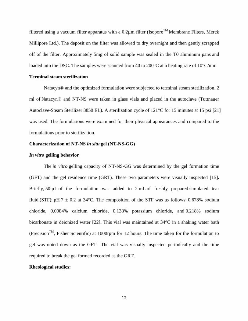
12
filtered using a vacuum filter apparatus with a 0.2µm filter (IsoporeTM
Membrane Filters, Merck
Millipore Ltd.). The deposit on the filter was allowed to dry overnight and then gently scrapped
off of the filter. Approximately 5mg of solid sample was sealed in the T0 aluminum pans and
loaded into the DSC. The samples were scanned from 40 to 200°C at a heating rate of 10°C/min
Terminal steam sterilization
Natacyn® and the optimized formulation were subjected to terminal steam sterilization. 2
ml of Natacyn® and NT-NS were taken in glass vials and placed in the autoclave (Tuttnauer
Autoclave-Steam Sterilizer 3850 EL). A sterilization cycle of 121°C for 15 minutes at 15 psi [21]
was used. The formulations were examined for their physical appearances and compared to the
formulations prior to sterilization.
Characterization of NT-NS in situ gel (NT-NS-GG)
In vitro gelling behavior
The in vitro gelling capacity of NT-NS-GG was determined by the gel formation time
(GFT) and the gel residence time (GRT). These two parameters were visually inspected [15].
Briefly, 50 µL of the formulation was added to 2 mL of freshly prepared simulated tear
fluid (STF); pH 7 ± 0.2 at 34°C. The composition of the STF was as follows: 0.678% sodium
chloride, 0.0084% calcium chloride, 0.138% potassium chloride, and 0.218% sodium
bicarbonate in deionized water [22]. This vial was maintained at 34°C in a shaking water bath
(PrecisionTM
, Fisher Scientific) at 1000rpm for 12 hours. The time taken for the formulation to
gel was noted down as the GFT. The vial was visually inspected periodically and the time
required to break the gel formed recorded as the GRT.
Rheological studies:

13
The viscosity of NT-NS-GG was measure using the Brookfield cone and plate viscometer
(Model: LVDV-II + Pro Viscometer, Middleboro, MA). It was calibrated before measuring the
viscosity of the in-situ gel. About 500 µL of the in-situ gel formulation was added to the center
of the spindle, and the gap was adjusted accordingly. The spindle used was 52. The cup was
maintained at 25°C. A CPE-52 Cone/Spindle was operated at 1, 2 and 5 rpm and the viscosity
recorded using the Rheocalc software. The in-situ gel was evaluated first by itself and then with
the addition of STF, wherein the formulation and STF were added in the ratio of 50:7.
In vitro release studies:
In vitro release studies were carried out using a multi-station hot plate magnetic stirrer (RT 10
power, IKA® WERKE, Wilmington, NC). Briefly, 20 mL of isotonic phosphate buffer saline
(IPBS) (pH 7.4), containing 2.5% randomly methylated β-cyclodextrin (RMβCD), was taken in
scintillation vials. The lower end of the 10K membrane (Slide-A-Lyzer™ MINI dialysis), was
submerged into the receiving medium (IPBS+ RMβCD). Once the setup reached a temperature
of 34 ± 0.2°C, 200µl of the formulation was placed onto the membrane. The setup was
maintained at 34 ± 0.2°C, with constant stirring for the entirety of the study. The control
consisted of a coarse suspension of NT (3 mg/mL). Aliquots were withdrawn from the vials at
predetermined time intervals and replaced with equal volume of IPBS with 2.5% RMβCD. NT-
NS-GG was added, along with STF, into the membrane in the ratio of 50:7 [17]. The amount of
drug released was quantified using the HPLC method mentioned previously. The study was
carried out for 24 hours.
Transcorneal studies:
Transcorneal studies were carried out using corneas separated from whole rabbits' eye, obtained
from Pel-Freez Biologicals® (Inc Rogers, AR, USA) shipped overnight in Hanks balanced salt

14
solution over ice. The eyes were used immediately upon arrival [22]. The cornea was excised
and washed with Dulbecco‘s phosphate buffer saline (DPBS) (pH 7.4) prior to being mounted
on vertical Valia-Chien cells (PermeGear, Inc®). The cornea was placed with the endothelial
side facing the receiver chamber while the epithelial side faced the donor compartment. 200µl
each of NT-NS and NT-NS-GG were added to the individual donor chambers. DPBS (5ml) (pH
7.4) with 2.5% w/v of RMβCD medium was filled into each of the receiving chambers
maintained at 34 ± 0.2°C for the entirety of the study. Aliquots of 800 µL were withdrawn from
the receiving chambers at predetermined time points and replenished by adding an equal volume
of fresh medium. The study was carried out for a total time period of 3 hours.
Data analysis of Transcorneal studies.
The transcorneal permeation across the rabbit cornea were calculated as follows [15]. The
cumulative amount of drug permeated M was calculated:
Where n indicates sampling point (n= 1,2,3...8 which corresponds to 15, 30, 45, 180
minutes respectively) Vr and Vs represent volume of the medium in the receiver chamber (mL)
and volume of the sample collected at the time point n (ml). Cr(n) indicates the concentration of
the drug in the receiver medium at the time point n (µg/ml). The rate of NT permeation across
the rabbit cornea was determined from the slope of cumulative amount of NT permeated vs. time
(t).
The flux was calculated as per equation 3:
Flux (J) = (dM/dt)/A (3)
Where, M represent the cumulative amount of drug transported and A designates the surface area
of the cornea utilized in the experiment (0.636 cm2)

15
The transcorneal permeability of NT is calculated from the ratio of steady-state flux (J) and the
amount of drug added to the donor chamber (Cd) as follows
Stability studies
The optimized formulations were stored at 4°C, 25°C, and at 40°C. Samples were
withdrawn and analyzed according to predetermined time points and changes in particle size,
PDI and assay % were evaluated.
Statistical analysis:
Results were presented in their mean values ± standard deviation.
.

16
CHAPTER III
RESULTS AND DISCUSSION
Quantification of natamycin
NT was quantified using standard calibration plots ranging from 0.5-50 µg/mL. The standard
curve was linear for the above range of concentration. The limit of detection and quantification
were 0.05 and 0.1 µg/mL. The standard curves in all the assays, in vitro drug release and
transcorneal permeation studies were linear with a coefficient of determination (r2) ≥ 0.99.
NT-NS formulation development and characterization
Natamycin loaded nanosuspensions were prepared by homogenization method. The
selected placebos were loaded with NT and analyzed for particle size. An almost 2-fold
increase was observed in the particle size after probe sonication. Hence, probe sonication process
was not used for further development of NT-NS.
The particle size of the formulation was measured prior to stirring at 2000 rpm with a magnetic
stirrer and post homogenization using T-25 digital Ultra-Turrax for 5 minutes at 16,000 rpm.
Table 3 summarizes the physico-chemical properties of the NT loaded nanosuspensions
formulated after Ultra-Turrax processing.

17
Table 3: Physio-chemical characteristics-Particle size, PDI, ZP and assay of natamycin
nanosuspension (NT-NS) (mean ± SD, n=3)
Formulation Particle Size (nm) PDI ZP (mV)
NT-Tylo 1237 ± 130.9 0.75 ± 0.03 -15
NT-HPMC 2585 ± 725.2 1 -10.3
NT-Leci 1046 ± 741.7 0.80 ± 0.169 -35.5
LP1 586 ± 61.3 0.44 ± 0.4 -38.9
LP2 708 ± 181.5 0.68 ± 0.0 -44.2
TB5 929 ± 84.1 0.63 ± 0.0 -45.6
TwB 3923 ± 1990 0.77 ± 0.1 -44.6
TP1 3834 ± 2157.9 1± 0 -37.3
TP2 1283 ± 200.0 0.85 ± 0.1 -33.8
TP3 2846 ± 241.1 1±0 -40.2
TP4 790 ± 59.8 0.71 ± 0.1 -30.8
F1 859 ± 77.7 0.77 ± 0.0 -39.9
An increase in the percentage of NT caused an increase in the particle size of the formulation.
Formulations containing single surfactants showed a high particle size (refer Table 3), hence it
was hypothesized that a surfactant combination would be more successful in reducing the
particle size. Formulations containing lecithin with poloxamer 188 and polysorbate 80 with
poloxamer 188 were formulated keeping the drug load (0.3%) and tyloxapol (0.1%) content
constant while varying the concentration of the surfactant. Lecithin with poloxamer 188 was
found to be a better combination compared to polysorbate 80 with poloxamer 188 as it was more

18
effective at reducing the particle size. The results suggested that a higher level of surfactants was
needed to prevent the suspended drug particles from agglomerating. Hence, the drug load of NT
was kept at 0.3% during the preparation of NT-NS [22].
Probe sonication resulted in an increase in the particle size of the formulations. The
probe sonicator uses high energy causing the reduction in particle size, however in case of
nanosuspensions, the high energy produced resulted in the suspended nanoparticles getting
charged and attracted towards each other, facilitating agglomeration.
A smaller particle size and a narrower PDI is desirable for the formulation of NS as larger
particles may cause irritation in the eye upon instillation while a narrow PDI is indicative of
possible long term stability owing to the decrease in Ostwald ripening [23].
Zeta potential indicates the surface charge of the particle which could be used to predict the
degree of repulsion present between the suspended particles [23].
From the twelve formulations prepared, four were selected on the basis of their particle size and
are listed in Table 4. The formulations selected were LP1, TB5, TP4 and LP2.
Table 4: List of formulations selected
Parameter TP4 TB5 LP2 LP1 *
Particle size
(nm)
790 ± 59.8 929 ± 84.1 708 ± 181.5 586 ± 61.3
PDI 0.71 ± 0.1 0.63 ± 0.0 0.68 ± 0.0 0.44 ± 0.4
ZP (mV) -30.8 -45.6 -44.2 -38.97
Assay (%) - - - 107 ± 0.6
*LP1 is the formulation selected for future studies.
19
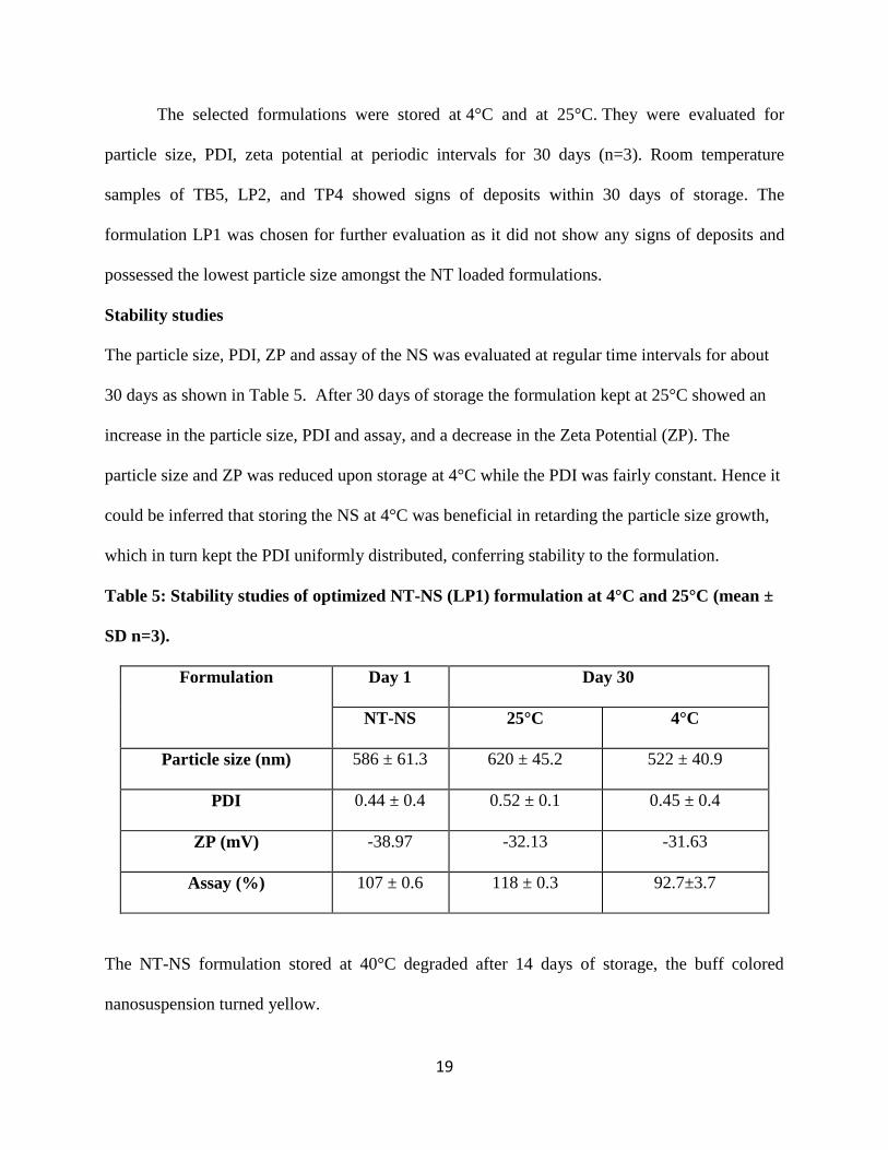
The selected formulations were stored at 4°C and at 25°C. They were evaluated for
particle size, PDI, zeta potential at periodic intervals for 30 days (n=3). Room temperature
samples of TB5, LP2, and TP4 showed signs of deposits within 30 days of storage. The
formulation LP1 was chosen for further evaluation as it did not show any signs of deposits and
possessed the lowest particle size amongst the NT loaded formulations.
Stability studies
The particle size, PDI, ZP and assay of the NS was evaluated at regular time intervals for about
30 days as shown in Table 5. After 30 days of storage the formulation kept at 25°C showed an
increase in the particle size, PDI and assay, and a decrease in the Zeta Potential (ZP). The
particle size and ZP was reduced upon storage at 4°C while the PDI was fairly constant. Hence it
could be inferred that storing the NS at 4°C was beneficial in retarding the particle size growth,
which in turn kept the PDI uniformly distributed, conferring stability to the formulation.
Table 5: Stability studies of optimized NT-NS (LP1) formulation at 4°C and 25°C (mean ±
SD n=3).
Formulation Day 1 Day 30
NT-NS 25°C 4°C
Particle size (nm) 586 ± 61.3 620 ± 45.2 522 ± 40.9
PDI 0.44 ± 0.4 0.52 ± 0.1 0.45 ± 0.4
ZP (mV) -38.97 -32.13 -31.63
Assay (%) 107 ± 0.6 118 ± 0.3 92.7±3.7
The NT-NS formulation stored at 40°C degraded after 14 days of storage, the buff colored
nanosuspension turned yellow.

20
Gellan gum is an anionic polysaccharide polymer used as an in situ gelling agent as it
undergoes a sol-gel transformation when in contact with the cations present in the tear fluid [15].
In situ gelling properties are desired to prolong the residence time in the cul-de-sac [24]. Gellan
gum has been used previously in the preparation of NT bilosomes, and was found to be
compatible with natamycin [15]. On addition of gellan gum in NT Nanosuspension (0.3%), a
thick gel was immediately formed which suggested that the addition of gellan gum at lower
concentrations would be suitable. The formulation should possess optimum viscosity for it to be
easily instilled into the eye but should contain a sufficient amount of gellan gum to form a gel
rapidly on contact with the tear fluid. Table 6 represents the components present in the in situ gel
formulation.
Table 6: Composition of natamycin in situ gel (NT-NS-GG)
Ingredients LP1 0.1% GG (%w/v) LP1 0.2% GG (%w/v) LP1 0.3% GG (%w/v)
Natamycin 0.3 0.3 0.3
Lecithin 0.5 0.5 0.5
Poloxamer 188 0.5 0.5 0.5
Tyloxapol 0.1 0.1 0.1
Gellan Gum 0.1 0.2 0.3
LP1 indicates the optimized nanosuspension, and GG stands for in-situ gel
prepared using gellan gum. Each formulation was prepared for 10ml.
Gelling capacity refers to the time taken for the formation of gel and the time it takes for the gel
to remain intact in the tear fluid [23]. Formulations LP1 0.1% GG and LP1 0.2% GG were
evaluated for gel formation time (GFT) and gel retention time (GRT). Both the formulations
gelled immediately on addition to STF at 34°C, however the gel formed by LP1 0.1% GG did

21
not maintain its integrity for 12 hours. LP1 0.2% GG was selected for further studies as it
showed a satisfactory GFT and GRT. The outcomes of GFT, GRT and the viscosity of the
selected formulation (LP1 0.2% GG) is represented in Table 7.
Table 7: Rheological evaluation of natamycin in situ gel (NT-NS-GG) (n = 3)
Formulation Gellan gum
(%)
Gel formation
time (sec)
Gel retention
time (h)
Viscosity
(cP)
Viscosity with
STF (cP)
LP1 0.1% GG 0.1% < 5 < 12 - -
LP1 0.2% GG 0.2% < 5 > 12 130.2 562.9
LP1 0.3% GG 0.3% Gelled in the
vial
- - -
The spindle was operated at 1, 2 and 5 RPM respectively.
Figure 1: Viscosity of optimized NT-NS-GG (LP1 0.2% GG) with and without simulated
tear fluid (STF).
204.6 130.2
94.82
1060
562.9
266
0
200
400
600
800
1000
1200
1 2 5
Vis
cosi
ty
(cP
)
RPM
Viscosity of LP1 with 0.2% GG
without STF with STF
22
Viscosity of the formulation is essential in determining the residence time of the drug into
the eye [23]. Increase in the RPM (revolutions per minute) caused a linear increase in the shear
rate. An increasing shear rate caused a decrease in the viscosity. Viscosity of LP1 0.2% GG was
evaluated using the Brookfield Viscometer; the spindle was operated at 1, 2 and 5 RPM
respectively. The addition of freshly prepared STF instantly increased the viscosity of the
formulation as shown in Figure 1. The rapid increase in the viscosity of the formulation
immediately on addition of freshly prepared STF is indicative of the fact that the gellan gum
incorporated in the NT NS underwent a sol to gel transformation. This further confirmed the
theory that the formulation was being activated in the presence of ions similar to those present in
the eye.
Differential scanning calorimetry (DSC)
The sample of pure drug and LP1 was run form 25°C to 200°C with a gradual increase of
10°C/min.

23
Figure 2: DSC thermogram of pure NT and optimized NT-NS (LP1)
The DSC thermogram of pure NT and of NT-NS is portrayed in Figure 2. The DSC study was
carried out to determine any change in the NT which may have occurred during the formulation
process of the NS. The DSC thermogram of pure NT showed an endothermic peak at 115°C
corresponding to the melting point of pure NT. The thermogram of NT-NS showed a pattern
similar to that of pure NT with the endothermic peak at around 107°C. It could be inferred from
the DSC thermogram peaks that the formulation process used did not cause any changes in NT.
The slight flattening of the peak in the DSC thermogram of the NS could be due to the presence
of excipients in formulation or the presence of residual water molecules.
Terminal steam sterilization
The NT-NS and Natacyn® showed immediate signs of degradation after autoclaving.
Figure 3A and 3B depicts the NT-NS and Natacyn®
prior to autoclave and post autoclave in

24
Figure 3. NT was proven to undergo degradation when autoclaved at 110oC for 20 minutes [25].
The buff colored formulations turned yellow-orange on autoclaving at 121°C for 15 minutes
indicating signs of possible degradation. Analyzing the NS formulation and Natacyn® for assay
confirmed that both samples were degraded.
NT®
and Formln refer to Natacyn
® and NT-NS respectively.
Figure 3: Pre autoclave (3A) and post autoclave (3B) images of NT nanosuspension and
Natacyn® (marketed formulation).
Since the formulation could not withstand the autoclave conditions, it would be recommended
that the NT-NS be formulated in an aseptic environment.
In-vitro release study
The study was carried out for 24 hours using 10K membrane. Table 8 shows the in vitro release
study profile of the NS, the in situ gel and the control.
Table 8: In vitro drug release profiles of natamycin nanosuspension (NT-NS), in situ gel
(NT-NS-GG) and NT control (mean ± SD, n=3)
Time (h) In vitro drug release
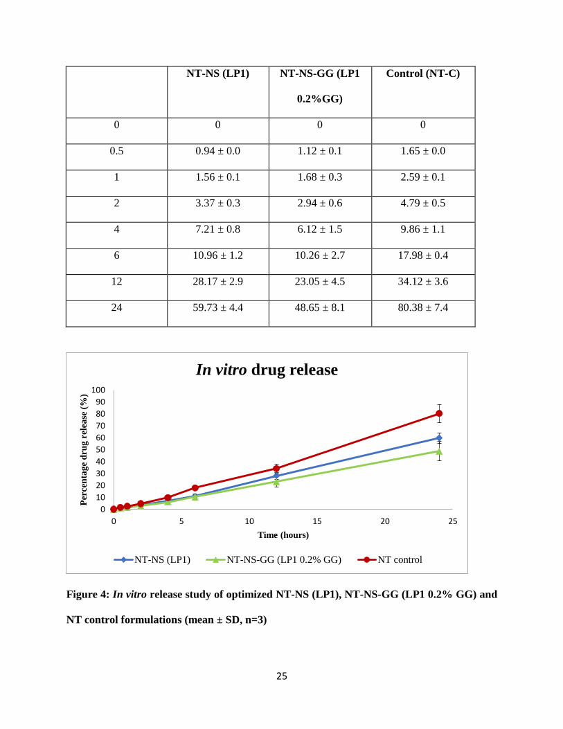
25
NT-NS (LP1) NT-NS-GG (LP1
0.2%GG)
Control (NT-C)
0 0 0 0
0.5 0.94 ± 0.0 1.12 ± 0.1 1.65 ± 0.0
1 1.56 ± 0.1 1.68 ± 0.3 2.59 ± 0.1
2 3.37 ± 0.3 2.94 ± 0.6 4.79 ± 0.5
4 7.21 ± 0.8 6.12 ± 1.5 9.86 ± 1.1
6 10.96 ± 1.2 10.26 ± 2.7 17.98 ± 0.4
12 28.17 ± 2.9 23.05 ± 4.5 34.12 ± 3.6
24 59.73 ± 4.4 48.65 ± 8.1 80.38 ± 7.4
Figure 4: In vitro release study of optimized NT-NS (LP1), NT-NS-GG (LP1 0.2% GG) and
NT control formulations (mean ± SD, n=3)
0
10
20
30
40
50
60
70
80
90
100
0 5 10 15 20 25
Per
cen
tag
e d
rug
rel
ease
(%
)
Time (hours)
In vitro drug release
NT-NS (LP1) NT-NS-GG (LP1 0.2% GG) NT control

26
The drug release profile of the optimized NT-NS and its corresponding NT-NS-GG was
compared to that of NT control formulation containing 0.3% NT similar to the drug load present
in the optimized formulation. The drug release profile for the NT-NS (LP1) and NT-NS-
GG (LP1 with 0.2% GG) is shown in Figure 4. LP1 0.2% GG showed a higher drug release
(compared to LP1) in the initial hour of the study; however its release gradually diminished as
the study progressed. Overall, the control formulation of NT showed the highest release (80.38 ±
7.49%) followed by LP1 (59.73 ± 4.45%) and lastly the LP1 0.2% GG (48.65 ± 8.1%) in 24
hours. The slow release from the NT-NS in situ gel could be due to the transformation into gel
form on addition of the STF - leading to a sustained release of drug from the gel matrix as the
study progressed. This slow release could be beneficial in providing a rapid initial release
followed by a sustained release of NT which could potentially lead to a reduced dosing
frequency.
Transcorneal studies
Table 9: Transcorneal permeation studies of natamycin nanosuspension (NT-NS) and in
situ gel (NT-NS-GG).
LP1 (NT-NS) LP1 0.2% GG (NT-NS-GG)
Flux (µg/min/cm) 0.034 ± 0.0 0.047 ± 0.0
Permeability (cm/min x 10-5
) 5.6 ± 0.1
7.8 ± 0.2

27
indicates statistically significant at level of p<0.05, compared with NT-NS.
Figure 5: In vitro transcorneal flux and apparent permeability coefficient of natamycin
from optimized NT-NS (LP1), NT-NS-GG (LP1 0.2% GG) (mean ± SD; n=3).
The transcorneal permeation profile is summarized in Table 10 and represented graphically in
figure 5. At the end of 3 hours LP1 0.2% GG showed a higher mean permeability and flux
compared to LP1. This could be due to the addition of gellan gum which may have enhanced the
penetration of NT from the formulation. This was almost similar to the pattern observed in the in
vitro release data conducted wherein LP1 0.2% GG showed a higher release rate compared to
LP1 in the first hour of the study. It could be hypothesized that LP1 0.2% GG is somewhat more
effective in delivering NT to the ocular tissues as compared to the NS.
0
0.01
0.02
0.03
0.04
0.05
0.06
0.07
0
1
2
3
4
5
6
7
8
9
NT-NS (LP1) NT-NS-GG (LP1 0.2% GG)
Flu
x(µ
g/m
in/c
m)
Per
mea
bil
ity *
10 -
5 (c
m/m
in)
Transcorneal permeation studies
Permeability Flux

28
CHAPTER IV
CONCLUSION
In this study, natamycin loaded nanosuspensions and its corresponding in situ gels were
successfully formulated using lecithin and poloxamer 188 as the surfactants. The formulation
had a better particle size with narrower PDI. The in-situ gel formulated, showed a
higher permeation and flux in the transcorneal studies when compared to the natamycin
nanosuspension, the in vitro release data showed a higher release from the nanosuspension
compared to the in situ gel over a 24 hour time period. The in situ gel formulated was activated
with the addition of simulated tear fluid, as was confirmed by the rheological studies.

28
LIST OF REFERENCES

29
[1] A. K. Mitra, Ocular transporters and receptors : their role in drug delivery. 2013.
[2] E. Sánchez-López, M. Espina, S. Doktorovova, E. B. Souto, and M. L. García, ―Lipid
nanoparticles (SLN, NLC): Overcoming the anatomical and physiological barriers of the
eye – Part I – Barriers and determining factors in ocular delivery,‖ Eur. J. Pharm.
Biopharm., vol. 110, pp. 70–75, Jan. 2017.
[3] D. Achouri, K. Alhanout, P. Piccerelle, and V. Andrieu, ―Recent advances in ocular drug
delivery,‖ Drug Dev. Ind. Pharm., vol. 39, no. 11, pp. 1599–1617, Nov. 2013.
[4] V. K. Yellepeddi and S. Palakurthi, ―Recent Advances in Topical Ocular Drug Delivery.,‖ J.
Ocul. Pharmacol. Ther., vol. 32, no. 2, pp. 67–82, Mar. 2016.
[5] M. Rawas-Qalaji and C.-A. Williams, ―Advances in Ocular Drug Delivery.,‖ Curr. Eye
Res., vol. 37, no. 5, pp. 345–356, May 2012.
[6] N. M. Davies, ―Biopharmaceutical Considerations In Topical Ocular Drug Delivery,‖ Clin.
Exp. Pharmacol. Physiol., vol. 27, no. 7, pp. 558–562, Jul. 2000.
[7] P. A. Thomas, ―Fungal infections of the cornea,‖ Eye, vol. 17, p. 852, Nov. 2003.
[8] S. S. Tuli, ―Fungal keratitis,‖ Clin. Ophthalmol. Auckl. NZ, vol. 5, pp. 275–279, 2011.
[9] B. H. Jeng, ―Challenges in the Management of Fungal KeratitisChallenges in the
Management of Fungal KeratitisResearch,‖ JAMA Ophthalmol., vol. 135, no. 6, pp. 525–
526, Jun. 2017.
[10] A. Patil, P. Lakhani, and S. Majumdar, ―Current perspectives on natamycin in ocular fungal
infections,‖ J. Drug Deliv. Sci. Technol., vol. 41, pp. 206–212, Oct. 2017.

30
[11]Y. Liu, P. Xie, D. Zhang, and Q. Zhang, ―A mini review of nanosuspensions development.,‖
J. Drug Target., vol. 20, no. 3, pp. 209–223, Mar. 2012.
[12] A. Leonardi et al., ―Influence of different surfactants on the technological properties and in
vivo ocular tolerability of lipid nanoparticles,‖ Int. J. Pharm., vol. 470, no. 1, pp. 133–140,
Aug. 2014.
[13] L. Wang, J. Du, Y. Zhou, and Y. Wang, ―Safety of nanosuspensions in drug delivery,‖
Nanomedicine Nanotechnol. Biol. Med., vol. 13, no. 2, pp. 455–469, Feb. 2017.
[14] A. Patil et al., ―Formulation Development, Optimization, and In Vitro–In Vivo
Characterization of Natamycin-Loaded PEGylated Nano-Lipid Carriers for Ocular
Applications,‖ J. Pharm. Sci., vol. 107, no. 8, pp. 2160–2171, Aug. 2018.
[15] K. Y. Janga et al., ―Ion-sensitive in situ hydrogels of natamycin bilosomes for enhanced
and prolonged ocular pharmacotherapy: in vitro permeability, cytotoxicity and in vivo
evaluation,‖ Artif. Cells Nanomedicine Biotechnol., vol. 46, no. sup1, pp. 1039–1050, Oct.
2018.
[16] Y. Wu et al., ―Research progress of in-situ gelling ophthalmic drug delivery system,‖ Asian
J. Pharm. Sci., vol. 14, no. 1, pp. 1–15, Jan. 2019.
[17] C. Le Bourlais, L. Acar, H. Zia, P. A. Sado, T. Needham, and R. Leverge, ―Ophthalmic
drug delivery systems—Recent advances,‖ Prog. Retin. Eye Res., vol. 17, no. 1, pp. 33–58,
Jan. 1998.

31
[18] Y. D. Sanzgiri, S. Maschi, V. Crescenzi, L. Callegaro, E. M. Topp, and V. J. Stella,
―Gellan-based systems for ophthalmic sustained delivery of methylprednisolone,‖ J. Controlled
Release, vol. 26, no. 3, pp. 195–201, Sep. 1993.
[19] A. K. Mitra, Ophthalmic drug delivery systems. New York: Marcel Dekker, 2005.
[20] S. Gupta, M. K. Samanta, and A. M. Raichur, ―Dual-drug delivery system based on in situ
gel-forming nanosuspension of forskolin to enhance antiglaucoma efficacy,‖ AAPS
PharmSciTech, vol. 11, no. 1, pp. 322–335, Feb. 2010.
[21] S. P. Balguri, G. R. Adelli, and S. Majumdar, ―Topical ophthalmic lipid nanoparticle
formulations (SLN, NLC) of indomethacin for delivery to the posterior segment ocular
tissues,‖ Eur. J. Pharm. Biopharm., vol. 109, pp. 224–235, Dec. 2016.
[22] A. Tatke et al., ―In Situ Gel of Triamcinolone Acetonide-Loaded Solid Lipid Nanoparticles
for Improved Topical Ocular Delivery: Tear Kinetics and Ocular Disposition Studies,‖
Nanomaterials, vol. 9, no. 1, 2018.
[23] M. K. Pathak, G. Chhabra, and K. Pathak, ―Design and development of a novel pH
triggered nanoemulsified in-situ ophthalmic gel of fluconazole: Ex-vivo transcorneal
permeation, corneal toxicity and irritation testing,‖ Drug Dev. Ind. Pharm., vol. 39, no. 5,
pp. 780–790, May 2013.
[24] I. Elsayed and S. Sayed, ―Tailored nanostructured platforms for boosting transcorneal
permeation: Box-Behnken statistical optimization, comprehensive in vitro, ex vivo and in
vivo characterization,‖ Int. J. Nanomedicine, vol. 12, no. Journal Article, pp. 7947–7962,
2017.
32
[25] ―Preliminary studies on the stability of natamycin eye drops after sterilization by
autoclaving,‖ Aust. J. Hosp. Pharm., vol. 14, no. 4, pp. 159–162, Jan. 1984.
33
VITA
Experience
Internship
June 2015--Vergo Pharma Research Laboratories Pvt. Ltd, Goa India
-Overview of the workings of a formulation development laboratory
-Standard Operating Procedures of all the departments.
-Analytical development laboratory: Exposure to working of the HPLC and interpretation
of data.
Release study of tablets using dissolution apparatus, working of IR spectrophotometer.
-Calibration of pH meters, weighing balance and analysis of water quality in the
manufacturing plant.
December 2012--Watson Pharma, Goa India
-A step by step demonstration of large scale manufacture of tablets by wet granulation
technique.
-Working of equipment such as V cone blenders, Fluidized Bed Driers, granulators,
automatic tablet compression and capsule filling machine demonstrated by the staff.
-Quality Control Department: Calibration of pH meters, working of HPLC.
-Microbiology testing laboratory
Education
Master of Science in Pharmaceutical Science with emphasis in Pharmaceutics and Drug
Delivery-May 2019

36
The University of Mississippi, Oxford MS
Research advisor: Dr. Soumyajit Majumdar
Bachelors in Pharmaceutical Sciences- July 2016
PES Rajaram and Tarabai Bandekar College of Pharmacy, Goa India
Skills
Formulation development of nanoformulations (Nanostructured Lipid Carriers,
Nanosuspensions and Solid Lipid Nanoparticles)
Development of analytical method using HPLC-UV
In vitro release study of nanosuspensions
Transcorneal permeation studies using isolated rabbit cornea
Measurement of viscosity using Brookfield cone and plate viscometer
Measurement and interpretation of particle size, PDI and zeta potential using Zetasizer
Nano ZS Zen3600 (Malvern Instruments)
Proficient with Zotero software for reference and citation management